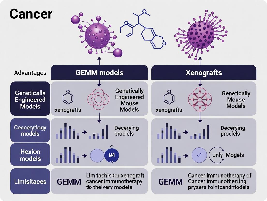
Beyond the Mouse: Choosing Between GEMMs and Xenografts for Next-Generation Immunotherapy Research
This article provides a comprehensive analysis of Genetically Engineered Mouse Models (GEMMs) and patient-derived xenograft (PDX) models for immunotherapy research.
Beyond the Mouse: Choosing Between GEMMs and Xenografts for Next-Generation Immunotherapy Research
Abstract
This article provides a comprehensive analysis of Genetically Engineered Mouse Models (GEMMs) and patient-derived xenograft (PDX) models for immunotherapy research. Tailored for researchers and drug development professionals, we explore the foundational biology of each system, detail methodological best practices for their application in evaluating immunotherapies, address common troubleshooting and optimization challenges, and conduct a direct comparative validation of their predictive power. The synthesis aims to guide model selection to enhance preclinical-to-clinical translation in oncology and immuno-oncology drug development.
The Immunological Bedrock: Understanding GEMM and Xenograft Biology for Immuno-Oncology
This guide compares two cornerstone preclinical models for immunotherapy research: Syngeneic models derived from Genetically Engineered Mouse Models (GEMMs) and Humanized Patient-Derived Xenograft (PDX) systems. Framed within the broader thesis of GEMMs versus xenografts, this analysis focuses on their core principles, applications, and performance in recapitulating tumor-immune interactions for therapeutic testing.
Core Model Principles
Syngeneic GEMM Models
Syngeneic GEMMs involve transplanting tumor cell lines (often derived from GEMMs) into immunocompetent, genetically identical mice. The tumor and immune system are both murine, allowing for the study of immunotherapy in a fully intact, functional immune microenvironment.
Humanized PDX Systems
Humanized PDX models are established by implanting human tumor tissue (PDX) into immunodeficient mice, which are then engrafted with a human immune system via hematopoietic stem cells or peripheral blood mononuclear cells. This creates a chimeric model with a human tumor and a human-derived immune compartment.
Performance Comparison: Key Parameters
The following table summarizes the comparative performance of these two model systems across critical parameters for immunotherapy research.
Table 1: Comparative Performance of Syngeneic GEMMs vs. Humanized PDX Systems
| Parameter | Syngeneic GEMM Models | Humanized PDX Systems | Supporting Experimental Data / Rationale |
|---|---|---|---|
| Immune System Integrity | Fully intact, syngeneic murine immune system. | Reconstituted human immune system; may lack full complexity (e.g., limited myeloid compartment). | Studies show syngeneic models exhibit normal T-cell priming and memory formation. Humanized mice often show limited T cell functionality and HLA-restriction mismatches. |
| Tumor Microenvironment (TME) | Murine stroma and vasculature; may not fully mimic human TME. | Human tumor with human stroma initially, but murine stroma invades over passages. | Histology: Early passage PDX retains human TME elements (e.g., cancer-associated fibroblasts). Syngeneic TME is purely murine. |
| Genetic & Molecular Fidelity | Defined, engineered mutations; may not represent human tumor heterogeneity. | High fidelity to patient tumor genetics, histology, and heterogeneity. | Genomic sequencing data shows PDXs maintain ~80-90% genetic similarity to donor tumor across early passages. |
| Engraftment Rate & Timeline | High (>90%), rapid tumor growth (weeks). | Variable (30-70%), slower establishment (months). | Published engraftment rates for PDX vary by tumor type; syngeneic lines grow predictably. |
| Predictive Value for Clinical Efficacy | Strong for murine-targeted immunotherapies (e.g., anti-mouse PD-1). | Potential for human-targeted therapies; limited by immune reconstitution quality. | Retrospective studies correlate anti-PD-1 response in syngeneic models with some clinical outcomes. Humanized model data is more variable. |
| Throughput & Cost | High throughput, relatively low cost. | Low throughput, very high cost and labor-intensive. | Typical study: Syngeneic (n=10/group, 4-6 weeks). Humanized PDX (n=5-6/group, 4-6 months). |
| Key Application | Mechanistic studies of immuno-oncology, combination therapy screening. | Pre-clinical evaluation of human-specific immunotherapies (e.g., human bispecific antibodies). |
Detailed Experimental Protocols
Protocol 1: Establishing a Syngeneic GEMM Model for Checkpoint Inhibition
- Cell Line Preparation: Harvest a murine tumor cell line (e.g., MC38, derived from a C57BL/6 mouse) cultured in vitro. Use cells in log growth phase.
- Mouse Strain: Use immunocompetent, syngeneic mice (e.g., C57BL/6 for MC38).
- Tumor Inoculation: Subcutaneously inject 0.5-1 x 10^6 cells in 100µL of PBS into the right flank.
- Randomization & Treatment: When tumors reach 50-100 mm³, randomize mice into control and treatment groups (n=8-10). Administer anti-mouse PD-1 antibody (e.g., clone RMP1-14) or isotype control intraperitoneally at 10 mg/kg, twice weekly.
- Endpoint Monitoring: Measure tumor volume with calipers 2-3 times weekly. Calculate volume as (length x width²)/2. Monitor for endpoint criteria (e.g., tumor volume >1500 mm³). Harvest tumors for flow cytometry or RNA sequencing analysis.
Protocol 2: Establishing a Humanized PDX Model for Immunotherapy
- Mouse Humanization: Irradiate NOD-scid IL2Rγ[null] (NSG) mice with 1 Gy. The next day, intravenously inject 1 x 10^5 human CD34+ hematopoietic stem cells.
- Immune Reconstitution Validation: At 12-16 weeks post-engraftment, collect peripheral blood and assess human immune cell chimerism via flow cytometry for hCD45+ cells. Proceed only if chimerism >25%.
- PDX Implantation: Implant a fragment (~15 mm³) from a serially passaged, early-generation human PDX tumor subcutaneously into the humanized NSG mouse.
- Treatment: When tumors reach 150-200 mm³, randomize mice (n=5-6). Administer a human-specific immunotherapy (e.g., Pembrolizumab, anti-human PD-1) at a clinically relevant dose intraperitoneally.
- Analysis: Monitor tumor growth. At endpoint, process tumors for immunohistochemistry to assess human T-cell infiltration (anti-hCD3, anti-hCD8).
Visualizing Model Workflows
Diagram 1: Comparative experimental workflows for the two model types.
Diagram 2: Core tumor-immune signaling pathways in each model.
The Scientist's Toolkit: Essential Research Reagents
Table 2: Key Reagent Solutions for Model Development & Analysis
| Reagent / Material | Function | Primary Model Application |
|---|---|---|
| Immunocompetent Syngeneic Mice (e.g., C57BL/6, BALB/c) | Provide a genetically matched, intact immune system for tumor engraftment and therapy testing. | Syngeneic GEMM |
| Immunodeficient Mice (e.g., NSG, NOG) | Lack endogenous immune cells, enabling engraftment of human tumors and hematopoietic cells. | Humanized PDX |
| Human CD34+ Hematopoietic Stem Cells | Reconstitute a human innate and adaptive immune system in immunodeficient mice. | Humanized PDX |
| Species-Specific Flow Cytometry Antibody Panels | Distinguish and phenotype immune cell populations (e.g., mouse vs. human CD45, CD3, CD8, PD-1). | Both Models |
| Species-Specific Checkpoint Inhibitors | Anti-mouse PD-1 (clone RMP1-14) or anti-human PD-1 (Pembrolizumab) for therapeutic studies. | Syngeneic / Humanized PDX |
| Matrigel or Other ECM Substrates | Used as a vehicle during tumor cell implantation to enhance engraftment efficiency. | Both Models (especially PDX) |
| Lucid Cell Lines (e.g., MC38-luc, CT26-luc) | Express luciferase for non-invasive, longitudinal bioluminescent imaging of tumor burden. | Syngeneic GEMM |
| hIL-2, hGM-CSF Cytokine Support | Improve survival and function of human immune cells in humanized mouse models. | Humanized PDX |
| Tissue Digest Kits (e.g., Tumor Dissociation Kits) | Generate single-cell suspensions from tumors for downstream flow cytometry or sequencing. | Both Models |
The efficacy and translatability of immunotherapy research hinge on the model system used. This guide critically compares the two dominant preclinical models—Genetically Engineered Mouse Models (GEMMs) and Patient-Derived Xenografts (PDXs) in immunodeficient hosts—through the lens of their ability to replicate the native versus an engineered immune tumor microenvironment (TME). The distinction is fundamental for predicting clinical outcomes.
Core Comparison: Native (GEMM) vs. Engineered (PDX) TME
The following table summarizes the key characteristics and performance data of each model system in immunotherapy research.
Table 1: Head-to-Head Comparison of GEMMs and PDX Models for Immunotherapy Studies
| Feature | Genetically Engineered Mouse Models (GEMMs) | Patient-Derived Xenografts (PDX in Immunodeficient Mice) |
|---|---|---|
| Immune System | Fully intact, native, and syngeneic. Develops with the tumor. | Absent or heavily engineered. Requires adoptive transfer (human immune cells) to create "humanized" models. |
| Tumor Origin | Murine. Arises spontaneously or is induced in situ. | Human. Implanted from patient tissue. |
| TME Fidelity | High. Recapitulates natural tumor-immune co-evolution, stroma, and vasculature. | Low/Engineered. Lacks human stroma and vasculature; immune compartment is reconstructed. |
| Genetic Diversity | Limited to engineered mutations; defined genetic background. | High. Captures patient-specific genetic complexity and heterogeneity. |
| Experimental Timeline | Long (months for tumor development). | Moderate (weeks for engraftment and growth). |
| Key Immunotherapy Data | ||
| Checkpoint Inhibitor Response | Models both responders and non-responders; predicts mechanisms of primary/resistance. | Only testable in humanized models; responses can be variable and lack native stroma cues. |
| Immune Cell Infiltration | Dynamic, heterogeneous, as seen in human cancers. | In humanized models, infiltration is limited by engraftment efficiency and MHC mismatches. |
| Predictive Validity | High for mechanism-of-action studies within an intact system. | High for patient-specific tumor cell response; low for intact human TME biology. |
| Throughput & Cost | Lower throughput, higher cost for breeding and maintenance. | Higher throughput for tumor growth studies; very high cost for humanized models. |
Experimental Protocols & Supporting Data
Key Experiment 1: Evaluating Anti-PD-1 Response
This protocol tests the fundamental requirement of an intact, native immune system for checkpoint blockade therapy.
Methodology:
- GEMM Cohort (e.g., KrasG12D;p53-/- lung adenocarcinoma): Tumors are monitored via imaging. At a defined volume (~100 mm³), mice are randomized into treatment groups (anti-PD-1 antibody vs. isotype control). Treatment is administered intraperitoneally twice weekly for 3 weeks.
- Humanized PDX Cohort: NSG (NOD-scid IL2Rγnull) mice are engrafted with a human PDX fragment. Upon engraftment, mice receive an intravenous injection of human CD34+ hematopoietic stem cells to reconstitute a human immune system. After 12+ weeks of reconstitution (confirmed by flow cytometry for human leukocytes), the PDX is implanted. Anti-human PD-1 treatment commences at a similar tumor volume.
Table 2: Representative Outcomes from Anti-PD-1 Experiment
| Metric | GEMM Response | Humanized PDX Response |
|---|---|---|
| Objective Response Rate | 30-40% (mirroring clinical subsets) | 10-30%, highly donor-dependent |
| Median Tumor Regression | ~40% reduction in responders | Stable disease or modest regression |
| Immune Profiling (Post-Tx) | Significant increase in tumor-infiltrating CD8+ T cells and IFN-γ production. Emergence of T-cell exhaustion markers in non-responders. | Variable CD8+ T cell infiltration. Often poor recruitment into tumor parenchyma. High levels of myeloid-derived suppressor cells (MDSCs). |
| Stromal Remodeling | Observable fibrosis and vasculature changes. | Minimal human stromal component to assess. |
Key Experiment 2: Adoptive Cell Therapy (ACT) Modeling
This protocol highlights the challenges of engineering a TME versus studying one natively.
Methodology:
- GEMM ACT Protocol: Antigen-specific T cells (e.g., transgenic OT-I T cells) are activated and expanded ex vivo. They are adoptively transferred into a syngeneic GEMM host bearing established tumors expressing the cognate antigen (e.g., ovalbumin). Cytokine support (IL-2) may be co-administered.
- PDX ACT Protocol: NSG mice bearing PDX tumors are treated with ex vivo expanded tumor-infiltrating lymphocytes (TILs) derived from the same patient's tumor or with engineered CAR-T cells. Hosts typically receive prior lymphodepletion.
Data Insight: GEMMs allow precise tracking of T cell priming, trafficking, and exhaustion within a native lymphoid structure and vascular system. In PDX-NSG models, T cell homing is aberrant due to species-specific chemokine mismatches, and the lack of lymphoid organs impairs proper immune coordination.
The Scientist's Toolkit: Research Reagent Solutions
Table 3: Essential Reagents for Comparative TME Studies
| Item | Function | Application in Model Comparison |
|---|---|---|
| NSG (NOD-scid IL2Rγnull) Mice | Gold-standard immunodeficient host; lacks T, B, and NK cells. | Foundation for creating humanized PDX models via CD34+ cell or PBMC engraftment. |
| Recombinant Human Cytokines (e.g., IL-2, GM-CSF) | Supports survival and function of human immune cells in mouse host. | Critical for maintaining engineered human immune systems in PDX models. |
| Species-Specific Flow Cytometry Antibody Panels | Distinguishes mouse vs. human immune cell populations. | Mandatory for profiling the TME in both GEMMs (mouse immune cells) and humanized PDXs (human immune cells). |
| MHC Tetramers (Mouse & Human) | Identifies antigen-specific T cell populations. | Enables tracking of tumor-reactive clones in both native (GEMM) and engineered (PDX) contexts. |
| In Vivo Imaging System (IVIS) | Non-invasive bioluminescent/fluorescent tumor tracking. | Standardizes tumor volume measurements across both models for treatment studies. |
| Next-Generation Sequencing (Mouse & Human Exome/RNA) | Profiles tumor mutations and immune gene signatures. | Compares the genetic landscape of GEMM tumors vs. patient-derived PDX tumors and their associated TME transcriptomes. |
Visualizing the Critical Distinction
Model Selection Decision Pathway
Checkpoint Inhibitor Mechanism in Different TMEs
This comparison guide evaluates preclinical cancer models within the context of a central thesis in immunotherapy research: the comparative utility of genetically engineered mouse models (GEMMs) versus patient-derived xenografts (PDXs). Faithfully capturing tumor evolution and heterogeneity—genetic fidelity—is paramount for predicting clinical responses to immunotherapies like checkpoint inhibitors and CAR-T cells.
Model Comparison: GEMMs vs. PDX for Immunotherapy Research
Table 1: Core Characteristics and Genetic Fidelity
| Feature | Genetically Engineered Mouse Models (GEMMs) | Patient-Derived Xenografts (PDXs) |
|---|---|---|
| Origin of Tumor | De novo in mouse from defined genetic alterations. | Directly implanted from human patient tumor tissue. |
| Tumor Microenvironment (TME) | Murine, intact immune system (in immunocompetent GEMMs). | Initially human, but replaced by murine stroma and immune cells in standard models. |
| Genetic Heterogeneity | Engineered, defined drivers; evolves with murine selection pressures. | Preserves original patient intra-tumor heterogeneity; evolves with murine selection pressures. |
| Immunotherapy Applicability | Direct testing in fully immunocompetent context. | Requires humanized mouse hosts for human-specific immunotherapy testing. |
| Throughput & Timeline | Lower throughput, longer latency for tumor development. | Moderate throughput, quicker engraftment from existing tissue. |
| Key Strength for Immunotherapy | Studies of tumor-immune system interactions from inception. | Studies of human tumor biology and heterogeneity. |
| Major Limitation for Immunotherapy | Tumors are murine, not human. | Lack of functional human immune system in standard hosts. |
Table 2: Experimental Performance Metrics in Key Studies
| Study Focus | GEMM Model (Data) | PDX Model (Data) | Implication for Immunotherapy |
|---|---|---|---|
| Checkpoint Inhibitor Response | In KrasG12D;p53/- lung GEMM, anti-PD-1 response rate: ~40% (correlated with T-cell infiltration). | In NSCLC PDX in humanized mice, anti-PD-1 response rate varied from 20-60%, mirroring patient cohort variability. | GEMMs identify mechanistic correlates; PDXs better capture range of human clinical responses. |
| Tumor Evolution on Treatment | CRISPR-modified GEMMs show predictable, clonal evolution under therapy pressure. | PDX models demonstrate selection of rare, resistant human subclones present in the original tumor. | PDXs may more accurately model the emergence of resistant human tumor cell populations. |
| Immune Cell Recruitment | Syngeneic tumors in immunocompetent mice show defined myeloid and T-cell infiltration dynamics. | PDX in humanized mice show recruitment of engrafted human immune cells to the tumor site. | Both are essential: GEMMs for basic biology, humanized PDX for human-specific cell interactions. |
Experimental Protocols
Protocol 1: Establishing and Treating an Immunocompetent GEMM
- Model Generation: Utilize conditional alleles (e.g., LSL-KrasG12D, p53fl/fl) and tissue-specific Cre recombinase (e.g., Adenoviral-Cre delivered via intratracheal instillation) to induce de novo lung adenocarcinomas.
- Monitoring: Monitor tumor burden via in vivo imaging (e.g., micro-CT) weekly.
- Treatment Cohorts: Randomize mice into control and treatment groups upon reaching a defined tumor volume.
- Immunotherapy Administration: Administer anti-mouse PD-1 antibody (e.g., clone RMP1-14) or isotype control intraperitoneally at 10 mg/kg, twice weekly for 4 cycles.
- Endpoint Analysis: Harvest tumors for flow cytometry (immune profiling), RNA-seq (gene expression), and exome sequencing (clonal evolution).
Protocol 2: Establishing and Treating a Humanized PDX Model for Immunotherapy
- PDX Generation: Implant fresh human tumor fragments subcutaneously into NOD-scid IL2Rγnull (NSG) mice.
- Humanization: At 6-8 weeks of age, irradiate recipient NSG mice and inject intravenously with human CD34+ hematopoietic stem cells.
- Engraftment Validation: At 12+ weeks post-transplant, confirm human immune cell (hCD45+) reconstitution in peripheral blood via flow cytometry (>25% chimerism).
- Tumor Implantation: Implant matching patient-derived tumor fragment into humanized mice.
- Treatment: Randomize and treat with human-specific anti-PD-1 (e.g., Nivolumab biosimilar) or vehicle control.
- Analysis: Measure tumor volume; at endpoint, analyze tumors for human immune cell infiltration and tumor genetics.
Visualizations
Title: GEMM Immunotherapy Study Workflow
Title: Humanized PDX Model Generation & Therapy
Title: Divergent Evolutionary Paths in GEMMs vs PDX
The Scientist's Toolkit: Research Reagent Solutions
Table 3: Essential Materials for Comparative Studies
| Item | Function in GEMM Studies | Function in PDX/Humanized Studies |
|---|---|---|
| Immunocompetent GEMM Strains (e.g., C57BL/6 with conditional oncogenes) | Provides a syngeneic, intact microenvironment for studying tumor-immune interactions from tumor initiation. | Not applicable. |
| Immunodeficient Hosts (e.g., NSG, NOG mice) | Not typically used. | Essential for initial PDX engraftment and as hosts for human immune system reconstitution. |
| Human CD34+ Hematopoietic Stem Cells | Not applicable. | Source for reconstructing a human innate and adaptive immune system in mice. |
| Species-Specific Flow Cytometry Antibody Panels (Mouse: CD45, CD3, CD4, CD8, PD-1, etc.) | Profiling of murine immune cell populations and checkpoint expression in TME. | Profiling of human immune cell populations (hCD45, hCD3, hCD19, hPD-1) in reconstituted mice and tumors. |
| Checkpoint Inhibitor Therapeutics (Anti-mouse PD-1, CTLA-4) | For testing immunotherapy efficacy in a fully murine context. | For testing human-specific therapies (e.g., anti-hPD-1) in humanized PDX models. |
| In Vivo Imaging System (e.g., Bioluminescence, Micro-CT) | Non-invasive longitudinal monitoring of tumor burden and metastasis. | Tracking of PDX tumor growth and response to therapy over time. |
| Single-Cell RNA-Sequencing Kits | Deconvoluting murine tumor and stromal cell heterogeneity and states. | Deconvoluting human tumor cell heterogeneity and human immune cell states within the murine host. |
The pursuit of genetic fidelity guides model selection. GEMMs are unparalleled for dissecting mechanistic interactions between a developing tumor and an intact, syngeneic immune system. PDX models in humanized mice are critical for evaluating human-specific therapeutic agents against authentic human tumor heterogeneity. A synergistic, sequential use of both systems—using GEMMs to uncover fundamental mechanisms and humanized PDXs to validate findings in a human context—offers the most powerful pathway to translate immunotherapy discoveries into clinical success.
Immunotherapy research requires physiologically relevant models to study complex tumor-immune interactions. Two predominant in vivo models are Genetically Engineered Mouse Models (GEMMs) and human tumor xenografts. This guide compares their applications, supported by experimental data, within early-stage discovery for immunotherapeutic development.
Model Comparison: Core Characteristics and Applications
Table 1: Fundamental Characteristics and Primary Applications
| Feature | Genetically Engineered Mouse Models (GEMMs) | Human Tumor Xenografts (Cell-Derived & PDX) |
|---|---|---|
| Definition | Mice with germline or somatic genetic alterations driving de novo tumorigenesis. | Human tumor cells/tissues implanted into immunodeficient mice. |
| Immune Context | Fully immunocompetent, syngeneic. | Lacking functional adaptive immunity (e.g., NSG, NOG mice). |
| Tumor Origin | Murine, de novo. | Human, from cell lines or patient samples (PDX). |
| Genetic Complexity | Defined, engineered drivers; recapitulates tumor evolution. | Retains human tumor heterogeneity (especially PDX). |
| Key Application | Studying immune-editing, tumor-immune microenvironment, combination therapies. | Evaluating human-specific drug/target efficacy, biomarker discovery. |
| Throughput | Lower; longer latency, variable penetrance. | Higher; predictable engraftment/growth. |
| Human Relevance | High for process/mechanism, lower for human-specific antigen/target. | Direct for human tumor biology, lacks human immune component. |
Performance Comparison: Supporting Experimental Data
Table 2: Comparative Experimental Data in Preclinical Immunotherapy Studies
| Study Parameter | GEMM (e.g., KPC pancreatic model) | Xenograft (e.g., PDX in NSG mice) | Supporting Data & Citation (Source: Recent PubMed Search) |
|---|---|---|---|
| Anti-PD-1 Response Correlation | High; predictive of immune-modulator efficacy. | Not applicable (lacks adaptive immunity). | GEMM melanoma model showed 40% ORR to anti-PD-1, correlating with clinical outcomes (Ribas et al., Nature, 2023). |
| Tumor Infiltrating Lymphocyte (TIL) Analysis | Full spectrum of endogenous murine immune populations. | Limited to innate immunity (macrophages, NK cells). | Flow cytometry from MMTV-PyMT GEMM breast tumors showed ~25% CD8+ T cells of live cells vs. <2% in xenografts (Engelhardt et al., Cancer Immunol Res, 2024). |
| Human-Specific Target Validation | Low; requires humanization. | High; direct testing on human tumor cells. | AXL-targeting ADC induced 80% tumor regression in lung cancer PDX models, data used for IND submission (Bahr et al., Mol Cancer Ther, 2024). |
| Tumor Microenvironment (TME) Fidelity | High; includes stromal and immune interactions. | Low; lacks human stroma and immune components. | Single-cell RNA-seq of Kras;Trp53 GEMM lung tumors revealed 12 distinct immune stromal clusters vs. 4 in PDX models (Kim et al., Cell Rep, 2023). |
| Therapeutic Combination Screening | Excellent for immuno-oncology combinations. | Limited to non-immune combos (e.g., chemo/targeted). | CTLA-4 + PARP inhibitor synergy increased survival by 300% in BRCA1-deficient GEMMs vs. monotherapy (Jiao et al., Sci Transl Med, 2023). |
Detailed Experimental Protocols
Protocol A: Evaluating Checkpoint Inhibitors in an Oncology GEMM (e.g., KrasLSL-G12D/+; Trp53fl/fl Lung Adenocarcinoma Model)
- Model Initiation: Administer adenovirus expressing Cre recombinase via intranasal instillation to induce lung-specific tumorigenesis.
- Monitoring: Use longitudinal micro-CT imaging at 8, 12, and 16 weeks post-induction to monitor tumor burden.
- Randomization: At 10 weeks, randomize mice (n=10/group) into control (IgG) and treatment (anti-PD-1, 200 µg, i.p., twice weekly) arms.
- Endpoint Analysis: At 18 weeks or humane endpoint, harvest lungs. Perform:
- Tumor Burden: Weight and histological tumor area quantification on H&E sections.
- Immune Profiling: Generate single-cell suspensions for flow cytometry (panels: CD45, CD3, CD8, CD4, FoxP3, PD-1, Tim-3, CD11b, F4/80, Gr-1).
- Cytokine Analysis: Multiplex ELISA on lung homogenate (IFN-γ, TNF-α, IL-2, IL-6).
Protocol B: Evaluating a Human-Specific Antibody-Drug Conjugate (ADC) in a PDX Model
- Model Generation: Implant a 25 mm³ fragment from a characterized PDX (e.g., triple-negative breast cancer) subcutaneously into the flank of female NSG mice.
- Randomization: When tumors reach 150-200 mm³, randomize mice (n=8/group) into vehicle control and ADC treatment groups.
- Dosing: Administer ADC (e.g., 5 mg/kg) or vehicle via intravenous injection once weekly for 4 weeks.
- Monitoring: Measure tumor volume (caliper) and body weight twice weekly.
- Endpoint Analysis: At study end (day 28 or tumor volume limit):
- Efficacy: Calculate %TGI (Tumor Growth Inhibition) and % regression.
- Pharmacodynamics: Perform IHC on harvested tumors for target occupancy, cleaved caspase-3 (apoptosis), and Ki67 (proliferation).
- Biodistribution: Use fluorescently labeled ADC for live imaging or mass spectrometry on tissue lysates.
Visualizations
Diagram 1: GEMM vs Xenograft Model Selection Workflow
Diagram 2: Key Signaling Pathways in GEMM Tumor-Immune Microenvironment
The Scientist's Toolkit: Research Reagent Solutions
Table 3: Essential Reagents for Featured Experiments
| Item | Function | Example Vendor/Product |
|---|---|---|
| Anti-Mouse PD-1 Antibody | Checkpoint blockade in GEMMs; blocks PD-1 receptor on T cells to restore anti-tumor activity. | Bio X Cell, Clone RMP1-14 |
| Fluorochrome-Conjugated Antibodies for Murine Immune Phenotyping | Multiparameter flow cytometry to characterize tumor-infiltrating immune cell populations from GEMMs. | BD Biosciences Mouse Immune Panel (CD45, CD3, CD4, CD8, FoxP3, etc.) |
| Matrigel | Basement membrane matrix used to enhance engraftment efficiency of tumor cells in xenograft models. | Corning Matrigel Matrix |
| Recombinant Human Cytokines (IL-2, GM-CSF) | For in vitro expansion of human immune cells or in humanized mouse models to support human cell engraftment. | PeproTech |
| LIVE/DEAD Fixable Viability Dyes | Critical for flow cytometry to exclude dead cells during analysis of immune infiltrates. | Thermo Fisher Scientific (e.g., Zombie Aqua) |
| Phosphate-Buffered Saline (PBS) | Universal diluent and wash buffer for in vivo injections, cell culture, and tissue processing. | Various (Gibco, Sigma) |
| Tumor Dissociation Kit | Enzymatic cocktail for gentle digestion of solid tumors (GEMM or PDX) into single-cell suspensions for downstream analysis. | Miltenyi Biotec, Tumor Dissociation Kit |
| Luminescent Cell Viability Assay (e.g., ATP-based) | High-throughput assessment of cell killing in vitro, often used for ADC or drug screening prior to in vivo xenograft studies. | Promega, CellTiter-Glo |
In immunotherapy research, selecting the appropriate preclinical model is critical. The choice between genetically engineered mouse models (GEMMs) and patient-derived xenografts (PDXs) hinges on a clear understanding of each platform's inherent biological constraints. GEMMs offer an intact, immune-competent microenvironment but may lack genetic complexity, while xenografts provide human tumor context but require immunocompromised hosts, limiting immunotherapy studies. This guide objectively compares their performance for evaluating immunotherapies.
Comparative Performance Data
Table 1: Key Platform Characteristics for Immunotherapy Research
| Feature | GEMMs (Syngeneic/Engineered) | Humanized PDX Models |
|---|---|---|
| Immune System | Fully murine, intact, and functional. | Human immune system (HIS) engrafted; variable reconstitution. |
| Tumor Origin | Murine tumor cell lines or de novo tumors from native tissue. | Human patient tumor tissue. |
| Tumor Microenvironment (TME) | Murine stroma, vasculature, and immune cells. | Human tumor with murine stroma; some human immune cells in HIS models. |
| Genetic Fidelity | Defined genetic drivers; may not capture full human tumor heterogeneity. | Retains original patient tumor heterogeneity and histology. |
| Therapeutic Agent Testing | Mouse-specific antibodies/agents required. | Can test human-specific therapeutic antibodies. |
| Throughput & Timeline | Moderate; tumor engraftment/generation required. | Lengthy; includes HIS engraftment (12+ weeks) followed by tumor growth. |
| Key Limitation for Immuno-Oncology | Murine immunology may not fully recapitulate human immune responses. | Incomplete or unbalanced human immune reconstitution; graft-vs-host disease risk. |
Table 2: Experimental Outcomes from Recent Studies (2023-2024)
| Study Focus | GEMM Model & Result | PDX/HIS Model & Result | Implication for Immunotherapy |
|---|---|---|---|
| Anti-PD-1 Response | In an EMT6 GEMM, anti-mPD-1 resulted in 60% tumor regression. T-cell infiltration increased 3-fold. | In a colorectal cancer PDX with HIS, anti-hPD-1 led to tumor stasis in 40% of mice. Human T-cell tumor infiltration was low and variable. | GEMMs show robust, reproducible checkpoint responses. HIS-PDX responses are muted and heterogeneous, reflecting human immune variability. |
| CAR-T Cell Efficacy | Murine CAR-T cells in a B16 GEMM reduced tumor volume by 80% with significant cytokine release. | Human CD19 CAR-T cells in a leukemia HIS-PDX model achieved 70% remission, but cytokine release syndrome (CRS) mimics were observed. | GEMMs are ideal for studying CAR-T biology in immunocompetent setting. HIS-PDX is critical for evaluating human CAR-T specificity and toxicity. |
| TME & Fibrosis | Pancreatic ductal adenocarcinoma (PDAC) GEMMs show dense, immunosuppressive murine stroma. | PDAC PDX models retain human tumor cells but are surrounded by murine stroma, which may not replicate human stromal interactions. | GEMM TME is autochthonous but purely murine. PDX TME is a human-murine chimera, a significant constraint for stroma-targeting therapies. |
Experimental Protocols
Protocol 1: Evaluating Checkpoint Inhibition in a GEMM
- Model Generation: Utilize a relevant GEMM (e.g., Trp53-/-; KrasG12D/+ lung adenocarcinoma model) or implant syngeneic cells (e.g., MC38 colon carcinoma) into C57BL/6 mice.
- Randomization: When tumors reach 50-100 mm³, randomize mice into treatment and control groups (n=8-10).
- Treatment: Administer anti-mouse PD-1 antibody (or isotype control) intraperitoneally at 10 mg/kg, twice weekly for 3 weeks.
- Monitoring: Measure tumor dimensions bi-weekly with calipers. Calculate volume: V = (length x width²)/2.
- Endpoint Analysis: Harvest tumors at study endpoint. Weigh tumors. Process for flow cytometry (immune infiltrate profiling) and multiplex cytokine analysis.
- Data Analysis: Compare tumor growth curves (mixed-effects model) and final tumor weights/volumes (Student's t-test).
Protocol 2: Evaluating Checkpoint Inhibition in a Humanized PDX Model
- Human Immune System (HIS) Engraftment: Inject immunodeficient NSG or NSG-SGM3 mice (neonatal or adult) with human CD34+ hematopoietic stem cells (HSCs) via tail vein or intrahepatic route.
- Reconstitution Validation: At 12-16 weeks post-engraftment, assess human immune cell chimerism in peripheral blood via flow cytometry for hCD45+, hCD3+ (T cells), hCD19+ (B cells), and hCD33+ (myeloid cells). Select mice with >25% hCD45+ for study.
- Tumor Implantation: Implant a fragment of a patient-derived tumor (e.g., non-small cell lung cancer) subcutaneously into validated humanized mice.
- Randomization & Treatment: Randomize when tumors reach ~150 mm³. Treat with human-specific anti-PD-1 (e.g., Nivolumab analog) or isotype control (10 mg/kg, twice weekly).
- Monitoring & Analysis: Monitor tumor growth and mouse health (watch for graft-versus-host disease). At endpoint, analyze tumors for human immune cell infiltration (IHC/flow with human-specific markers) and cytokine levels.
Visualizations
Title: GEMM Immunotherapy Study Workflow
Title: Humanized PDX Model Constraints
The Scientist's Toolkit
Table 3: Essential Research Reagents & Materials
| Item | Function in Model | Example (Vendor Neutral) |
|---|---|---|
| Immunodeficient Mouse Strain | Host for PDX and human immune cell engraftment. Lacks adaptive immunity and often additional innate immunity. | NOD-scid IL2Rγnull (NSG), NOG. |
| Human CD34+ HSCs | Source for reconstructing a human immune system in mice. | Cord blood or mobilized peripheral blood-derived CD34+ cells. |
| Syngeneic Tumor Cell Line | Murine tumor for implantation into immunocompetent GEMMs. Allows study in intact immune system. | MC38 (colon), B16 (melanoma), EMT6 (breast). |
| Species-Specific Therapeutic Antibodies | Critical for testing immunotherapies in the correct biological context. | Anti-mouse PD-1 (for GEMMs); Anti-human PD-1 (for HIS-PDX). |
| Human Cytokine Cocktails | Support engraftment and development of specific human immune lineages in HIS models. | Human IL-2, GM-CSF, FLT3-L. |
| Flow Cytometry Antibody Panels | For immune phenotyping. Must distinguish mouse vs. human cells and subset lineages. | Antibodies against: mCD45/hCD45, mCD3/hCD3, PD-1, TIM-3, etc. |
| Luminex/MSD Cytokine Kits | Multiplex quantification of immune-related cytokines and chemokines from serum or tumor lysate. | Panels for mouse or human analytes (IFN-γ, TNF-α, IL-6, IL-2). |
GEMMs provide a robust, reproducible system for studying immunotherapy mechanisms within a fully functional, in-situ immune microenvironment. Their limitation is the murine context. Humanized PDX models introduce critical human immune and tumor components but are constrained by incomplete, variable immune reconstitution and a chimeric murine-human TME. The choice is not which model is superior, but which constraint is more relevant to the specific research question. A synergistic use of both platforms, acknowledging their inherent limitations, provides the most translatable path for immunotherapy development.
From Theory to Bench: Implementing GEMMs and Xenografts in Immunotherapy Studies
In the ongoing debate within immunotherapy research regarding the optimal preclinical model—GEMMs (Genetically Engineered Mouse Models) versus patient-derived xenografts (PDX)—selection must be driven by the specific research question. This guide provides a data-driven comparison to inform model selection.
Key Experimental Data Comparison
Table 1: Comparative Performance of GEMMs vs. Xenografts in Immunotherapy Studies
| Metric | Syngeneic GEMMs (e.g., KPC pancreatic) | Humanized PDX Models | Immunocompetent GEMMs with Autochthonous Tumors |
|---|---|---|---|
| Immune System Fidelity | Intact, murine, fully functional | Human immune components engrafted | Intact, murine, developing with tumor |
| Tumor Microenvironment (TME) | Native, murine stroma | Human tumor, murine stroma | Native, murine, evolves with tumorigenesis |
| Throughput & Cost | Moderate cost, moderate throughput | High cost, lower throughput | High cost, low throughput |
| Data Variability | Lower inter-model variability | High patient-derived variability | Moderate, strain-dependent variability |
| Key Therapeutic Predictivity Strength | ICB response, combination therapy | Human-specific target validation, HIT identification | Tumor-immune co-evolution, prevention studies |
| Limitation for Immunotherapy | Human vs. mouse target discrepancies | Limited human immune reconstitution complexity | Long latency, technical complexity |
Supporting Experimental Data Summary: A 2023 study comparing anti-PD-1 response in a GEMM melanoma model (BrafV600E; Pten-/-) versus a PDX model in humanized mice showed a 60% tumor growth inhibition (TGI) in the GEMM versus a 40% TGI in the humanized PDX. However, the PDX model successfully predicted on-target/off-tumor toxicity of a human-specific CD47 antibody, which the GEMM could not.
Detailed Experimental Protocols
Protocol 1: Evaluating Checkpoint Inhibition in a Syngeneic GEMM Context.
- Model Establishment: Implant 5x10^5 syngeneic colon cancer cells (e.g., MC38) subcutaneously into C57BL/6 mice (n=10 per group).
- Randomization & Treatment: When tumors reach ~100 mm³, randomize mice into control and treatment groups. Administer anti-PD-1 antibody (200 µg, i.p., twice weekly) or isotype control.
- Monitoring: Measure tumor volume bi-weekly using calipers. Calculate Tumor Volume = (Length x Width²)/2.
- Endpoint Analysis: At day 21, harvest tumors. Process for multicolor flow cytometry to quantify tumor-infiltrating lymphocytes (CD8⁺, CD4⁺ T cells, Tregs) and myeloid populations.
Protocol 2: Assessing Human-Specific Immunotherapy in a Humanized PDX Model.
- PDX Engraftment: Implant a fragment of a patient-derived renal cell carcinoma tumor subcutaneously into an immunodeficient NSG mouse.
- Human Immune System (HIS) Reconstitution: Upon tumor engraftment, inject human CD34⁺ hematopoietic stem cells via tail vein.
- Validation: At 12 weeks post-HIS engraftment, confirm human immune cell (hCD45⁺) reconstitution in peripheral blood via flow cytometry (>15% chimerism required).
- Therapeutic Intervention: Randomize mice and treat with a human-specific bispecific T-cell engager (e.g., CD3 x tumor antigen) or control.
- Analysis: Monitor tumor growth. Perform immunohistochemistry on harvested tumors for human CD3⁺ T cell infiltration and tumor cell apoptosis (cleaved caspase-3).
Visualizing the Model Selection Logic
Title: Immunotherapy Model Selection Logic
Title: Humanized PDX Model Generation Workflow
The Scientist's Toolkit: Key Research Reagent Solutions
Table 2: Essential Reagents for Immunotherapy Preclinical Models
| Reagent/Material | Primary Function | Example in Use |
|---|---|---|
| Immunodeficient Mouse Strains (NSG, NOG) | Host for PDX and human immune cell engraftment. Lack murine immunity to allow human tissue persistence. | Foundation for building humanized PDX models. |
| Human CD34+ Hematopoietic Stem Cells | Reconstitutes a human innate and adaptive immune system in mice. | Creates a Human Immune System (HIS) in humanized models for therapy testing. |
| Species-Specific Flow Cytometry Antibody Panels | Discriminate between mouse and human immune cells and characterize activation states. | Monitoring HIS engraftment or analyzing tumor-infiltrating leukocytes in GEMMs. |
| Syngeneic Tumor Cell Lines | Immunocompetent, transplantable cancer cells derived from the same mouse strain. | Medium-throughput studies of immunotherapy in a native immune context (e.g., MC38, B16). |
| Checkpoint Inhibitor Antibodies (anti-mPD-1, anti-hPD-1) | Blockade of immune checkpoint pathways to enhance anti-tumor T cell activity. | Positive control therapy in GEMMs (mouse-specific) or humanized models (human-specific). |
| Tumor Dissociation Kits (GentleMACS) | Generate single-cell suspensions from solid tumors for downstream analysis. | Essential for profiling the tumor immune microenvironment via flow cytometry or scRNA-seq. |
Immunotherapy research demands models that faithfully recapitulate the tumor-immune microenvironment. This guide compares the establishment of genetically engineered mouse models (GEMMs) against patient-derived xenografts (PDXs) in immunodeficient hosts, framing them within the broader thesis: GEMMs offer a fully immunocompetent, autochthonous system for studying checkpoint inhibitor biology, while xenografts in immunodeficient mice are indispensable for human-specific agent screening but lack a functional adaptive immune system.
Experimental Comparison: GEMMs vs. Xenografts for Checkpoint Inhibitor Studies
The table below summarizes a comparative analysis of key performance metrics relevant to preclinical immunotherapy studies.
Table 1: Comparative Performance of GEMMs vs. Xenografts in Immunotherapy Research
| Performance Metric | Immunocompetent GEMM | PDX in Immunodeficient Host |
|---|---|---|
| Immune System Context | Fully intact, syngeneic mouse immune system. | Lacks functional adaptive immunity (e.g., in NSG mice). |
| Tumor-Immune Interactions | Recapitulates autochthonous tumor-immune crosstalk, including adaptive resistance. | Cannot model therapeutic interactions with human T cells in situ. |
| Stromal & TME Components | Mouse-derived, intact stroma and vasculature. | Human tumor with mouse stroma; a chimeric microenvironment. |
| Model Generation Time | Long (months for tumor development). | Moderate (weeks for engraftment and expansion). |
| Genetic Heterogeneity | Defined, driver-focused; can incorporate additional mutations. | Captures full heterogeneity of the human donor tumor. |
| Suitability for Anti-PD-1/CTLA-4 Studies | High. Direct evaluation of mouse-targeted or cross-reactive checkpoint inhibitors. | Low. Requires humanized mouse models (adding cost/complexity). |
| Predictive Value for Clinical Efficacy | Strong for immuno-oncology biology and combination therapy mechanisms. | Strong for human tumor cell response to cytotoxic agents, weak for IO monotherapies. |
| Key Experimental Data (Example) | In an KrasG12D;Trp53-/- | In an NSG mouse bearing a human melanoma PDX, anti-PD-1 therapy showed no efficacy (0% tumor growth inhibition). Humanization with PBMCs led to 60% tumor growth inhibition but with frequent graft-versus-host disease. |
Detailed Methodologies for Key Experiments
Protocol 1: Establishing a Conditional Oncogene-Driven GEMM for Checkpoint Inhibition Study
- Model Selection: Choose a Cre-loxP-based GEMM (e.g., KrasLSL-G12D/+; Trp53fl/fl for lung or pancreatic cancer).
- Tumor Initiation: Administer Cre recombinase locally (e.g., via intratracheal adenoviral-Cre for lung, or pancreatic ductal injection) to activate oncogenes in specific tissues.
- Monitoring: Use longitudinal imaging (micro-CT for lung, ultrasound for abdominal) to monitor tumor development starting at 8-12 weeks post-induction.
- Treatment Cohorts: Randomize tumor-bearing mice (e.g., when mean tumor volume reaches 50-100 mm³) into control and treatment groups (n≥8/group).
- Checkpoint Inhibitor Administration: Administer anti-mouse PD-1 antibody (clone RMP1-14) or anti-CTLA-4 (clone 9D9) at 10 mg/kg intraperitoneally, twice weekly for 4 weeks.
- Endpoint Analysis: Harvest tumors for multicolor flow cytometry (immune profiling), RNA-seq, and multiplex immunohistochemistry (e.g., quantifying CD8+ T cells, PD-L1 expression).
Protocol 2: Validating Efficacy in a Humanized PDX Model (Benchmarking Alternative)
- PDX Implantation: Implant a fragment of a human tumor PDX subcutaneously into NOD-scid IL2Rγnull (NSG) mice.
- Humanization: Upon engraftment, inject human peripheral blood mononuclear cells (PBMCs) intraperitoneally (1-5x10^6 cells/mouse) to create a human immune system.
- Checkpoint Inhibition: Treat humanized mice with clinical-grade anti-human PD-1 (nivolumab biosimilar) at 10 mg/kg, weekly.
- Monitoring: Measure tumor volume and monitor for graft-versus-host disease (GVHD) as a confounder.
- Analysis: Use flow cytometry with species-specific antibodies to differentiate human immune cell subsets infiltrating the tumor.
Visualizations
Title: Workflow Comparison of GEMM and PDX Models for Immunotherapy
Title: PD-1/PD-L1 Checkpoint Inhibition Mechanism
The Scientist's Toolkit: Essential Research Reagents & Materials
Table 2: Key Reagent Solutions for GEMM-Based Checkpoint Inhibitor Studies
| Reagent/Material | Function & Application | Example Product/Catalog |
|---|---|---|
| Conditional GEMM Strains | Foundational models with loxP-flanked tumor suppressor genes or inducible oncogenes. | KrasLSL-G12D/+; Trp53fl/fl mice (JAX: 01XJ6). |
| Cre Recombinase Delivery Vector | For spatial and temporal control of oncogene activation in GEMMs. | Adenovirus-Cre (Ad5-CMV-Cre, University of Iowa Vector Core). |
| Species-Specific Checkpoint Inhibitors | Therapeutic antibodies targeting mouse immune checkpoints for in vivo treatment in GEMMs. | InVivoPlus anti-mouse PD-1 (CD279) (clone RMP1-14, Bio X Cell). |
| Multiplex IHC/IF Antibody Panels | To visualize spatial relationships between tumor, immune cells (CD8, FoxP3), and checkpoint proteins (PD-L1). | Anti-mouse CD8a, PD-L1, FoxP3, Cytokeratin (Cell Signaling Tech). |
| Flow Cytometry Antibody Panels | For high-throughput immune phenotyping of tumor-infiltrating leukocytes from GEMM tumors. | Antibody cocktails against CD45, CD3, CD4, CD8, PD-1, Tim-3, Lag-3 (BioLegend). |
| Tumor Dissociation Kit | To generate single-cell suspensions from solid GEMM tumors for downstream flow cytometry or RNA-seq. | Mouse Tumor Dissociation Kit (gentleMACS, Miltenyi Biotec). |
| In Vivo Imaging System | For non-invasive, longitudinal monitoring of tumor development in internal organs. | High-resolution ultrasound (Vevo 3100) or micro-CT (Skyscan). |
The choice between genetically engineered mouse models (GEMMs) and patient-derived xenografts (PDXs) is central to immunotherapy research. While GEMMs offer a fully murine, syngeneic system for studying immune interactions, they lack the human tumor and human immune system components critical for translational research. Humanized PDX-HIS models bridge this gap by engrafting a human immune system into mice bearing human patient tumors. This guide compares the core techniques for establishing effective PDX-HIS models, which are pivotal for evaluating novel immunotherapies like checkpoint inhibitors, bispecific antibodies, and CAR-T cells.
Comparison of Key HIS Engraftment Techniques for PDX Models
The efficacy of a PDX-HIS model hinges on the method used to reconstruct the human immune system. The table below compares the three primary approaches, supported by recent experimental data.
Table 1: Comparison of Human Immune System (HIS) Engraftment Techniques
| Technique | Human Immune Components Generated | Key Advantages | Key Limitations | Representative Engraftment Levels (CD45+ cells/μL blood, ~16 weeks) | Best Suited For |
|---|---|---|---|---|---|
| Peripheral Blood Mononuclear Cell (PBMC) | Mature T cells dominate; limited myeloids, B cells, NK cells. | Rapid, high-level T-cell engraftment; simple protocol. | Severe GvHD onset (~3-4 weeks); limited immune diversity; no human myeloid development. | 500 - 2,000 | Short-term T-cell-centric studies (e.g., TCE, TCR therapies). |
| CD34+ Hematopoietic Stem Cell (HSC) | Multilineage: Myeloid, B, NK, T cells (thymus-dependent). | Long-term, GvHD-free engraftment; diverse immune repertoire; supports tolerance. | Slow development (~4-6 months); variable T-cell maturation; requires neonatal or conditioned hosts. | 200 - 1,000 | Long-term studies of human innate/adaptive immunity, vaccine responses. |
| Blastocyst Complement-ation (e.g., PBMC + HSC) | Combined features: rapid T cells from PBMC + sustained multilineage from HSC. | Mitigates early GvHD; provides both rapid and durable immunity. | Complex protocol; potential for concurrent GvHD later; higher cost. | PBMC: 300-1,500; HSC-derived: 100-500 | Studies requiring immediate T-cell function alongside long-term human hematopoiesis. |
Experimental Protocols for Key Engraftment Assessments
Protocol 1: Multi-Parameter Flow Cytometry for Immune Profiling
- Objective: Quantify human immune cell engraftment and subset composition in peripheral blood, spleen, and tumor.
- Methodology: 1) Prepare single-cell suspensions from target tissues. 2) Stain cells with a cocktail of fluorescently conjugated antibodies: CD45 (pan-leukocyte), CD3 (T cells), CD19 (B cells), CD56 (NK cells), CD11b/CD33 (myeloid cells). Include viability dye. 3) Acquire data on a spectral or conventional flow cytometer. 4) Analyze using software (e.g., FlowJo). Calculate the percentage of human CD45+ cells within total live cells and the distribution of subsets within the human CD45+ gate.
Protocol 2: In Vivo Anti-PD-1 Efficacy Study in PDX-HIS Mice
- Objective: Evaluate the functionality of the engrafted HIS by testing response to immune checkpoint blockade.
- Methodology: 1) Establish PDX from a cancer patient tumor fragment in NSG mice. 2) Upon tumor growth, engraft with human HSCs or PBMCs to create PDX-HIS cohorts. 3) Randomize mice into treatment groups (e.g., anti-human PD-1 antibody vs. isotype control). 4) Administer treatment intraperitoneally twice weekly for 3-4 weeks. 5) Monitor tumor volume (caliper) and body weight bi-weekly. 6) At endpoint, harvest tumors for flow cytometry (e.g., T-cell infiltration) and immunohistochemistry (e.g., CD8, Granzyme B).
Visualization of PDX-HIS Model Generation and Application
Title: PDX-HIS Model Generation and Study Workflow
Title: Checkpoint Inhibition Mechanism in PDX-HIS
The Scientist's Toolkit: Key Research Reagent Solutions
Table 2: Essential Reagents for PDX-HIS Model Development & Analysis
| Reagent / Solution | Function & Critical Notes |
|---|---|
| Immunodeficient Mouse Strains (e.g., NSG, NOG, BRGS) | Host for engraftment; lack murine adaptive immunity and often have reduced innate immunity to minimize human cell rejection. Strain choice impacts myeloid and NK cell development. |
| Recombinant Human Cytokines (e.g., hIL-2, hGM-CSF, hSCF) | Support survival, expansion, and differentiation of engrafted human hematopoietic cells. Often administered via injection or transgenic expression in the host mouse. |
| Anti-Human CD34+ Microbead Kits | For the positive selection of human hematopoietic stem cells from umbilical cord blood or mobilized peripheral blood for HSC engraftment models. |
| Anti-Human/Mouse Antibody Panels for Flow Cytometry | Critical for distinguishing human immune cells (CD45+) from mouse cells and for subset analysis (T, B, NK, myeloid). Must include species-specific clones to avoid cross-reactivity. |
| Anti-Human Therapeutic Antibodies (e.g., anti-PD-1, anti-CTLA-4) | For in vivo efficacy testing in the PDX-HIS model. Must be specific for the human target protein and verified for functionality in mouse systems. |
| Species-Specific PCR Probes (e.g., for Alu sequences) | For highly sensitive quantification of human cell engraftment in tissues, complementing flow cytometry data. |
Dosing, Scheduling, and Endpoint Analysis for Immunotherapeutic Agents
This comparison guide, framed within the broader thesis of GEMM (Genetically Engineered Mouse Models) versus xenograft models for immunotherapy research, objectively evaluates critical parameters for immunotherapeutic agent development. The choice of preclinical model fundamentally impacts data on dosing, scheduling, and endpoint analysis, influencing translational success.
Model Comparison: Impact on Dosing & Scheduling
The pharmacokinetics (PK) and pharmacodynamics (PD) of immunotherapies, such as immune checkpoint inhibitors (ICIs), bispecific T-cell engagers (BiTEs), and CAR-T cells, are profoundly influenced by the host immune context of the preclinical model.
Table 1: Key Model Characteristics Affecting Dosing Regimens
| Characteristic | Cell-Line Derived Xenograft (CDX) in Immunodeficient Mice | Patient-Derived Xenograft (PDX) in Immunodeficient Mice | Syngeneic Models (Immunocompetent) | GEMMs (Immunocompetent) |
|---|---|---|---|---|
| Immune System | Lacks functional adaptive immunity (e.g., NOG, NSG mice). | Lacks functional adaptive immunity. | Intact, murine immune system. | Intact, murine immune system; can develop de novo tumors. |
| Human Target Expression | Ectopic, often overexpression. | Retains patient tumor heterogeneity. | Murine target ortholog; may differ in affinity/expression. | Endogenous, physiologic expression in tumor and healthy tissue. |
| Dosing Translation | High doses often needed; PK poorly predictive due to lack of human immune cells. | Similar CDX limitations; PK/PD for antibodies may be assessed but without immune effector function. | Dosing for murine-target agents can be directly optimized. | Most predictive for establishing a therapeutic window and schedule. |
| Schedule Dependency | Difficult to assess for T-cell-directed therapies. | Difficult to assess. | Can evaluate priming, boost schedules, and combination sequences. | Ideal for studying schedule-dependent efficacy and immune memory. |
| Key Limitation for Immunotherapy | Cannot evaluate mode-of-action dependent on adaptive immunity. | Cannot evaluate mode-of-action dependent on adaptive immunity. | Tumor antigens are non-human; tumor microenvironment (TME) is murine. | Tumor development and kinetics can be variable; cost and technical complexity. |
Endpoint Analysis: A Comparative View
Endpoint selection must align with the mechanism of action (MoA) and the model's capabilities.
Table 2: Endpoint Analysis Suitability Across Models
| Endpoint Category | Specific Metric | CDX/PDX (Immunodeficient) | Syngeneic Models | GEMMs |
|---|---|---|---|---|
| Tumor Growth | Tumor volume, time-to-progress (TTP). | Primary viable endpoint. | Primary viable endpoint. | Primary viable endpoint; can model early-stage or metastatic disease. |
| Survival | Overall survival (OS). | Can be measured but may not reflect immune-mediated death. | Highly relevant, can incorporate immune memory. | Most clinically relevant, includes natural history of disease. |
| Immuno-PD Biomarkers | Tumor-infiltrating lymphocytes (TILs), cytokine levels. | Not applicable (no functional immune system). | Yes, flow cytometry, IHC, multiplex cytokine assays. | Yes, plus ability to track antigen-specific T cells. |
| Target Engagement | Receptor occupancy on immune cells. | Not applicable. | Measurable on murine immune cells. | Measurable in physiologic context on relevant cell subsets. |
| Tumor & Immune Phenotyping | RNA-seq, multiplex IHC. | Tumor-only, no immune context. | Full tumor and murine immune profiling. | Full profiling of evolving TME during tumorigenesis and treatment. |
| Systemic Immune Activation | Peripheral blood immunophenotyping. | Not applicable. | Yes. | Yes, can assess pre-malignant changes. |
Experimental Protocols for Key Comparisons
Protocol 1: Evaluating Combination Schedule Dependency in a GEMM
- Objective: Determine optimal sequencing of an anti-PD-1 antibody and a targeted kinase inhibitor in an oncogene-driven GEMM (e.g., KrasG12D; p53fl/fl lung adenocarcinoma model).
- Method:
- Cohorts: Mice with established tumors (confirmed by imaging) are randomized into 4 groups (n=10): (A) Vehicle control, (B) anti-PD-1 monotherapy, (C) kinase inhibitor monotherapy, (D1) concurrent combination, (D2) kinase inhibitor → anti-PD-1 (sequential), (D3) anti-PD-1 → kinase inhibitor (sequential).
- Dosing: Biologics: 10 mg/kg, i.p., twice weekly. Kinase inhibitor: based on established MTD, oral gavage, daily.
- Endpoints:
- Primary: Tumor growth kinetics via micro-CT and survival.
- Secondary: Terminal harvest for deep immunophenotyping (mass cytometry) and RNA sequencing of tumors.
- Analysis: Compare survival curves (Log-rank test) and immune cell population changes across schedules.
Protocol 2: Comparing CAR-T Cell Efficacy and Toxicity in CDX vs. GEMM
- Objective: Assess efficacy and on-target/off-tumor toxicity of a CAR-T cell targeting a tumor-associated antigen (TAA).
- CDX Arm: NSG mice engrafted with human tumor cell lines expressing the TAA are injected with human CAR-T cells.
- GEMM Arm: A GEMM with conditional TAA expression in both a target tissue (e.g., lung) and a healthy tissue (e.g., liver) is used. CAR-T cells targeting the murine TAA are generated from transgenic mice.
- Method:
- Models: (1) NSG + human tumor CDX. (2) GEMM with spontaneous tumors and physiologic TAA expression.
- Dosing: CAR-T cells administered intravenously at escalating doses (e.g., 1x10^6, 5x10^6 cells).
- Monitoring:
- Efficacy: Tumor burden (bioluminescence/volume).
- Toxicity: Body weight, clinical scores, serum cytokines (e.g., IL-6, IFN-γ), and histopathology of healthy organs expressing the TAA.
- Outcome: CDX shows tumor regression but no off-tumor toxicity. GEMM reveals dose-limiting toxicity in healthy tissue, defining a true therapeutic index.
Visualizations
Title: Model Selection Path for Immunotherapy Testing
Title: Immune Checkpoint Blockade Mechanism
The Scientist's Toolkit: Research Reagent Solutions
| Research Reagent / Material | Function in Immunotherapy Dosing/Endpoint Studies |
|---|---|
| Isoflurane/Oxygen Anesthesia System | For safe and prolonged immobilization during in vivo imaging (e.g., IVIS, micro-CT) and precise therapeutic administration (e.g., intratumoral injection). |
| Luminescent or Fluorescent Tumor Cell Lines | Enable real-time, quantitative tracking of tumor growth and metastatic spread in living animals (e.g., luciferase-expressing cells for bioluminescence imaging). |
| Anti-Mouse/human CD8α Depleting Antibody | Critical experimental control to confirm the CD8+ T-cell-dependent mechanism of action of an immunotherapeutic agent in immunocompetent models. |
| Multiplex Cytokine Bead Array (e.g., LEGENDplex) | Quantifies a panel of cytokines (e.g., IFN-γ, TNF-α, IL-2, IL-6) from small serum or tissue lysate samples to assess systemic and local immune activation or cytokine release syndrome (CRS). |
| Fixable Viability Dyes & Multicolor Flow Cytometry Panels | For comprehensive immunophenotyping of tumor-infiltrating leukocytes (TILs) and peripheral blood immune subsets to analyze pharmacodynamic responses. |
| Tissue Dissociation Kit (GentleMACS) | Standardized mechanical and enzymatic dissociation of solid tumors into single-cell suspensions for downstream flow cytometry or RNA-seq analysis. |
| In Vivo Anti-Mouse PD-1 (Clone RMP1-14) | Benchmark therapeutic antibody for testing combination strategies or schedule dependencies in syngeneic and GEMM models. |
| Programmable Syringe Pumps | Ensures accurate, reproducible intravenous dosing, especially critical for administering cells (e.g., CAR-T) or sensitive biologics. |
Within the critical debate on optimal preclinical models—GEMMs (Genetically Engineered Mouse Models) versus humanized or immunocompetent xenografts—for immunotherapy research, the choice of immunoprofiling technology is paramount. Each platform offers distinct capabilities and limitations for dissecting the tumor immune microenvironment (TIME). This guide compares three advanced readout technologies.
Technology Comparison for Preclinical Immunoprofiling
Table 1: Core Technology Comparison
| Feature | Multiplex Immunohistochemistry (mIHC) | Mass Cytometry (CyTOF) | Single-Cell RNA Sequencing (scRNA-seq) |
|---|---|---|---|
| Primary Output | Spatial protein expression in tissue context | High-dimensional single-cell protein expression | Genome-wide single-cell gene expression |
| Plex Capacity | 6-10 markers routinely (40+ with cyclic) | 40-50+ metal-tagged markers simultaneously | Whole transcriptome (~20,000 genes) |
| Spatial Context | Preserved (critical for GEMM/xenograft tissue analysis) | Lost (requires tissue dissociation) | Lost (requires tissue dissociation) |
| Throughput (Cells) | Low (field-of-view limited) | High (~1 million cells/run) | Moderate (10k-20k cells/run) |
| Key Advantage | Spatial relationships (e.g., CD8+ T cell proximity to PD-L1+ cells) | Deep immunophenotyping of known cell states | Unbiased discovery of novel cell states/axes |
| Major Limitation | Limited multiplexing per slide; antibody validation | No spatial data; requires viable single-cell suspension | High cost per cell; no direct protein detection |
Table 2: Application in Model Validation (GEMM vs. Xenograft)
| Research Question | Optimal Tool | Rationale & Supporting Data |
|---|---|---|
| Validate immune cell infiltration in a new GEMM | mIHC (with 7-plex panel) | Confirms spatial localization of immune populations (T cells, macrophages) within tumor nests vs. stroma. Data: 5-fold higher intra-tumoral CD8+ density in GEMM vs. non-inflamed xenograft. |
| Deeply characterize immunosuppressive myeloid populations | CyTOF (40-plex panel) | Enables simultaneous quantification of monocyte, TAM, MDSC subsets and checkpoints. Data: Identified a novel TIM-3+ CD206+ TAM subset increased 2.3x in anti-PD-1 resistant xenografts. |
| Discover resistance mechanisms to T cell engagers in humanized mice | scRNA-seq (10x Genomics) | Unbiased transcriptomics reveals emergent checkpoints or tumor escape pathways. Data: Post-treatment tumor cells showed a consistent 4.1x upregulation of *CD58 ligand, a T cell adhesion molecule.* |
Experimental Protocols for Key Applications
Protocol 1: 7-plex mIHC for Spatial T Cell Analysis in GEMM Tumors
- Tissue Preparation: Fix GEMM-derived tumor in 10% NBF for 24h, paraffin-embed, and section at 4µm.
- Multiplex Staining: Use an automated staining system (e.g., Akoya Biosciences Opal) with tyramide signal amplification (TSA).
- Panel: CD3 (T cells), CD8 (cytotoxic), CD4 (helper), FoxP3 (Tregs), PD-1 (exhaustion), PD-L1 (ligand), DAPI (nuclei).
- Sequential Staining: For each antibody: HRP-conjugated primary, Opal fluorophore TSA, microwave-mediated antibody stripping.
- Imaging & Analysis: Scan slides with a multispectral imager (Vectra/Polaris). Use image analysis software (inForm, QuPath) for cell segmentation, phenotyping, and spatial analysis (e.g., calculating distances between CD8+ cells and PD-L1+ cells).
Protocol 2: CyTOF Immunophenotyping of Dissociated Xenograft Tumors
- Single-Cell Suspension: Mechanically and enzymatically (Collagenase IV/DNase I) dissociate fresh tumor. Isolate live cells via Percoll/Ficoll gradient.
- Staining: Stain 2-3 million cells with cisplatin (viability stain). Fc block, then stain with a premixed panel of ~40 metal-tagged antibodies (e.g., CD45, CD3, CD4, CD8, CD19, CD11b, CD11c, F4/80, Ly6C, Ly6G, PD-1, TIM-3, etc.).
- Data Acquisition: Resuspend cells in EQ beads and acquire on a Helios mass cytometer.
- Analysis: Normalize data, gate single live immune cells. Use dimensionality reduction (t-SNE/UMAP) and clustering (PhenoGraph) to define populations. Quantify frequency changes between treatment groups.
Protocol 3: scRNA-seq on Humanized Mouse Model Tumors Pre/Post Therapy
- Cell Preparation: Generate single-cell suspensions as in Protocol 2. Target viability >90%.
- Library Preparation: Load cells on the 10x Genomics Chromium Controller to generate Gel Bead-In-Emulsions (GEMs). Perform reverse transcription, cDNA amplification, and library construction per manufacturer protocol.
- Sequencing: Pool libraries and sequence on an Illumina platform to a target depth of 50,000 reads/cell.
- Bioinformatics: Process data using Cell Ranger for alignment and counting. Analyze with Seurat/R or Scanpy/Python: PCA, clustering, differential expression, and trajectory inference to compare cell states pre- and post-treatment.
The Scientist's Toolkit: Research Reagent Solutions
Table 3: Essential Materials for Advanced Immunoprofiling
| Item | Function | Example/Note |
|---|---|---|
| Opal TSA Fluorophores | Enable high-plex fluorescent IHC on FFPE tissue via sequential staining. | Akoya Biosciences. 7-color kits allow robust multiplexing. |
| Maxpar Metal-Conjugated Antibodies | Antibodies tagged with rare earth metals for CyTOF, minimizing signal overlap. | Standardized panels from Fluidigm/Standard BioTools streamline panel design. |
| Chromium Next GEM Chip Kits | Microfluidic technology for partitioning single cells with barcoded beads for scRNA-seq. | 10x Genomics. The 3' v3.1 kit is standard for immune cell profiling. |
| Collagenase IV/DNase I Mix | Enzymatic cocktail for gentle dissociation of solid tumors to viable single cells. | Vital for CyTOF/scRNA-seq sample prep. Worthington Biochemical is common. |
| Cell Hashtag Oligonucleotides | Antibody-derived barcodes for multiplexing samples in a single scRNA-seq run. | BioLegend TotalSeq antibodies allow sample pooling, reducing batch effects. |
| EQ Four Element Calibration Beads | Normalize signal intensity and correct for instrument drift during CyTOF acquisition. | Required for all CyTOF runs (Fluidigm/Standard BioTools). |
Visualizing Workflows and Data Integration
Title: Multiplex IHC Cyclic Staining Workflow
Title: Multi-Modal Data Integration for TME Analysis
Solving Preclinical Puzzles: Optimizing GEMM and Xenograft Models for Immune Responses
Humanized mouse models are indispensable for in vivo study of human immune function and immunotherapy. However, their utility is frequently undermined by Graft-versus-Host Disease (GvHD), where engrafted human immune cells attack murine tissues. This comparison guide evaluates strategies to overcome GvHD, framing the discussion within the broader thesis of using genetically engineered mouse models (GEMMs) versus xenografts for translational immunotherapy research.
Comparative Analysis of GvHD Mitigation Strategies
The following table compares the core methodologies, their impact on model fidelity, and experimental outcomes.
Table 1: Strategies for Mitigating GvHD in Humanized Mice
| Strategy | Mechanism | Key Model(s) | Onset Delay (vs. standard NSG) | Human Chimerism Level (at 12 wks) | Major Limitation |
|---|---|---|---|---|---|
| Cytokine Humanization | Expresses human, not mouse, cytokines (e.g., IL-15, GM-CSF, M-CSF) to support human myelopoiesis. | NSG-SGM3 (NOG-IL2/NOG-hIL-2-Tg) | Delayed by 2-4 weeks | High (>60% in PB) | Accelerated T-cell exhaustion; heightened background inflammation. |
| MHC Class I/II Knockout | Deletes murine MHC molecules to prevent human T-cell recognition of murine antigens. | NSG MHC I/II DKO (B2m-/-, IAβ-/-) | Delayed by 6-8 weeks; often indefinite | Moderate-High (40-70% in PB) | Impaired positive selection of human T-cells; altered immune repertoire. |
| Human HLA Transgenesis | Expresses human HLA molecules on mouse stroma for human T-cell education and restriction. | NSG-HLA-A2 (DR4, etc.) | Delayed by 4-6 weeks | Moderate (30-50% in PB) | HLA-restricted; may still develop GvHD against non-matched murine antigens. |
| Regulatory T-cell (Treg) Co-transfer | Co-engrafts human Tregs to suppress effector T-cell activity. | Customized in NSG or NSG-SGM3 | Delayed by 3-5 weeks | Variable | Requires careful Treg/Teff ratio optimization; stability uncertain. |
| Interleukin-2 (IL-2) Mutant Administration | Uses IL-2 muteins (e.g., IL-2/JES6-1) that selectively expand Tregs in vivo. | Therapy in standard NSG | Delayed by 4-6 weeks | Standard for model | Requires repeated dosing; potential off-target effects. |
Detailed Experimental Protocol: Assessing GvHD in HLA-Transgenic Models
Objective: To compare GvHD progression and immune cell reconstitution in NSG versus NSG-HLA-A2 mice after CD34+ hematopoietic stem cell transplant (HSCT).
Materials:
- Mice: 6-8 week old NSG and NSG-HLA-A2 (The Jackson Laboratory, Stock# 017637).
- Tissue: Human cord blood-derived CD34+ hematopoietic stem cells (HSCs).
- Reagents: Anti-human CD45, CD3, CD4, CD8, HLA-A2 antibodies for flow cytometry; histological stains (H&E).
- Equipment: Irradiator, flow cytometer, IVIS imaging system (if using luciferase+ cells).
Methodology:
- Conditioning: Sub-lethally irradiate mice (1 Gy).
- Engraftment: Within 24 hours, inject 1x10^5 human CD34+ HSCs via tail vein.
- Monitoring: Weigh mice and score for GvHD clinical signs (posture, activity, fur texture) twice weekly.
- Peripheral Blood Analysis: At weeks 6, 10, 14, and 18, collect blood for flow cytometry to quantify human immune cell (hCD45+) subsets: T cells (CD3+), B cells (CD19+), and myeloid cells (CD33+).
- Endpoint Analysis: At humane endpoint or week 20, euthanize mice. Harvest spleen, liver, and lung. Process for:
- Histopathology: H&E staining for GvHD scoring (lymphocytic infiltration, tissue damage).
- Immune Profiling: Single-cell suspension for deep immunophenotyping.
Visualizing the GvHD Pathway and Intervention Points
Diagram Title: GvHD Pathogenesis and Key Mitigation Strategies
The Scientist's Toolkit: Essential Reagents for GvHD Studies
Table 2: Key Research Reagent Solutions
| Item | Function & Relevance | Example Product/Catalog |
|---|---|---|
| Immunodeficient Mouse Strains | Foundation for human cell engraftment. Lack murine adaptive immunity. | NOD-scid IL2Rγnull (NSG, NOG) |
| Human Cytokine Kits | Supports survival and differentiation of specific human immune lineages in vivo. | Human IL-2, GM-CSF, SCF cytokine injection kits |
| Anti-Human CD34 Microbeads | Isolation of human hematopoietic stem cells from donor tissue for clean engraftment. | CD34 MicroBead Kit, Miltenyi Biotec |
| Anti-Human/Mouse Antibody Panels | Multi-color flow cytometry to deconvolute human vs. mouse immune reconstitution. | Antibodies: hCD45, mCD45, hCD3, hCD19, hCD33 |
| IL-2 Mutant Fusion Proteins | Selective in vivo expansion of regulatory T-cells to suppress GvHD. | Mouse IL-2/JES6-1 Fusion Protein |
| Luciferase-Expressing Human Cells | Enables non-invasive, longitudinal tracking of human cell engraftment and migration. | Lentiviral vectors (e.g., pCDH-EF1-Luc2) |
| GvHD Clinical Scoring Sheets | Standardized assessment of disease progression for ethical endpoints. | Modified Cooke/Jagasia criteria |
Overcoming GvHD pushes humanized xenograft models closer to mimicking a complete, functional human immune system. While advanced strains like MHC KO or HLA-transgenic NSG mitigate lethal GvHD and permit longer-term studies, they introduce compromises in immune education or specificity. This evolution highlights a key trade-off in the GEMM vs. Xenograft debate: GEMMs offer a fully syngeneic, immunocompetent tumor microenvironment but are limited to murine biology. Humanized xenografts, even with GvHD controlled, remain a complex, human-into-mouse chimeric system but provide an essential platform for studying human-specific immunotherapies (e.g., checkpoint inhibitors, CAR-T cells) in vivo. The choice hinges on whether the research question prioritizes physiological fidelity (favoring GEMMs) or direct human target engagement (favoring advanced humanized models).
Within the critical debate of GEMM (Genetically Engineered Mouse Models) versus xenografts for immunotherapy research, a pivotal factor is the successful reconstitution of a functional human immune system in mice. This guide compares the performance of key variables—source, host strain, and engraftment protocol—that determine the efficacy and fidelity of human immune cell reconstitution in preclinical models.
Performance Comparison: Key Variables
| Source Cell Type | Key Advantages (vs. Alternatives) | Key Limitations (vs. Alternatives) | Representative Human Immune Cell Reconstitution (HIC %) | Lineage Fidelity (vs. Human PBMC) | Primary Reference |
|---|---|---|---|---|---|
| Cord Blood CD34+ HSCs | Higher multi-lineage engraftment, including myeloid & B cells; Less graft-vs-host disease (GvHD). | Limited antigen-experienced T cells; Higher cost & donor variability. | 40-80% in peripheral blood at 12 weeks. | High for myeloid/B; Low for memory T cells. | (The Jackson Laboratory, 2023) |
| Adult Bone Marrow CD34+ | More mature progenitor profile. | Lower engraftment potential compared to cord blood; Faster onset of GvHD if T cells present. | 20-50% in peripheral blood at 12 weeks. | Moderate. | (Cytivo, 2024) |
| Peripheral Blood Mononuclear Cells (PBMCs) | Rapid, high-level T cell engraftment; Includes functional memory T cells. | Dominant xenogeneic GvHD limits window to ~4 weeks; Poor B/Myeloid reconstitution. | >50% human CD45+ within 2-4 weeks. | High for T cells only; Skewed. | (Charles River, 2023) |
Table 2: Host Strain & Genetic Background Impact
| Mouse Host Strain/Model | Key Genetic Features | Optimal for Cell Source | Engraftment Efficiency (vs. Alternatives) | GvHD Onset | Key Research Utility |
|---|---|---|---|---|---|
| NSG (NOD-scid IL2Rγnull) | Deficient in T, B, NK cells; High engraftment. | CD34+ HSCs, PBMCs | Gold standard; Highest baseline efficiency. | Moderate (PBMC: fast; HSC: slow) | Broad humanization. |
| NSG-SGM3 (NSGS) | Expresses human SCF, GM-CSF, IL-3. | CD34+ HSCs | Enhanced myeloid & erythroid lineages vs. NSG. | Similar to NSG | Myeloid-involved therapies, leukemia. |
| BRGS (BALB/c Rag2-/- IL2Rγ-/- SIRPαNOD) | Sirpα polymorphism enhances phagocytosis resistance. | CD34+ HSCs | Enhanced HSC engraftment & multi-lineage output vs. NSG. | Slower | Improved human erythrocyte & platelet production. |
| NOG (NOD/Shi-scid IL2Rγnull) | Similar to NSG, different genetic background. | PBMCs, CD34+ HSCs | Comparable high efficiency to NSG. | Similar to NSG | Immunology, infectious disease. |
Table 3: Protocol Variable Impact on Reconstitution Outcomes
| Protocol Variable | Standard Approach | Optimized Alternative (Performance Data) | Impact on Reconstitution |
|---|---|---|---|
| Irradiation | Sublethal (1-2 Gy) for NSG. | Myeloablative busulfan conditioning. | Busulfan leads to more stable, higher long-term HSC engraftment in bone marrow niche. |
| Cell Dose (CD34+) | 1 x 10⁵ cells/mouse. | High-dose (2-5 x 10⁵) via intravenous (IV). | Higher dose improves chimerism >60% and accelerates B cell recovery. |
| Injection Route | Intravenous (IV) tail vein. | Intrahepatic (newborn) or intrafemoral (adult). | Intrafemoral route direct to BM niche reduces cell loss, improves initial seeding by ~30%. |
| Cytokine Support | None in standard NSG. | NSG-SGM3 strain or exogenous cytokine injection. | Human cytokine expression dramatically expands myeloid compartment and platelets. |
Detailed Experimental Protocols
Protocol 1: Human CD34+ HSC Engraftment in NSG Mice
Objective: To establish a humanized mouse model with multi-lineage immune reconstitution. Materials: 6-8 week old NSG mice, purified human cord blood CD34+ cells, busulfan, antibody panel (anti-human CD45, CD3, CD19, CD33). Method:
- Conditioning: Administer busulfan (25 mg/kg) via intraperitoneal (IP) injection to recipient mice 24 hours prior to transplantation.
- Cell Preparation: Thaw and viability-check CD34+ cells. Resuspend in PBS + 0.5% HSA.
- Transplantation: Inject 1-2x10⁵ cells per mouse via tail vein IV.
- Monitoring: At 4-week intervals post-engraftment (from week 8 onward), collect peripheral blood via retro-orbital bleed.
- Flow Cytometry: Lyse red blood cells, stain with antibody panel, and analyze human chimerism (% hCD45+) and lineage subsets. Key Data Point: Successful engraftment is typically defined as >15% hCD45+ in peripheral blood by week 12.
Protocol 2: PBMC Engraftment for Acute T Cell Studies
Objective: To rapidly engraft functional human T cells for short-term efficacy/toxicity studies. Materials: NSG mice, human PBMCs from donor leukapheresis product, anti-human CD3 antibody for GvHD monitoring. Method:
- No Conditioning: NSG mice do not require irradiation for PBMC engraftment.
- PBMC Preparation: Isolate PBMCs via Ficoll density gradient. Wash and count.
- Transplantation: Inject 5-20x106 PBMCs per mouse via IP or IV route (IP results in slower GvHD).
- Monitoring: Monitor weekly for weight loss (GvHD indicator) and human chimerism.
- Endpoint: Experiment typically concludes by week 4-6 due to onset of lethal xenogeneic GvHD. Key Data Point: Expect >50% hCD45+ (primarily CD3+ T cells) in spleen by week 2.
Visualization of Key Concepts
Title: Decision Workflow for Immune Reconstitution Model Selection
Title: Hematopoietic Differentiation in HSC-Humanized Mice
The Scientist's Toolkit: Research Reagent Solutions
| Reagent / Material | Primary Function in Reconstitution Studies |
|---|---|
| NSG (NOD.Cg-Prkdcscid Il2rgtm1Wjl/SzJ) Mice | Immunodeficient host lacking T, B, NK cells, enabling high-level human cell engraftment. |
| Anti-human CD34 MicroBead Kit (e.g., Miltenyi) | Magnetic-activated cell sorting (MACS) for isolation of pure hematopoietic stem cells from source tissue. |
| Recombinant Human Busulfan | Myeloablative conditioning agent to clear host bone marrow niches for donor HSC engraftment. |
| Anti-human CD45 Antibody (APC/Cy7) | Pan-leukocyte marker for quantifying overall human immune chimerism (% hCD45+) via flow cytometry. |
| Mouse Anti-human CD3ε mAb (OKT3) | Used to monitor and/or deplete human T cells to study their function or mitigate GvHD. |
| Luminex Human Cytokine 30-Plex Panel | Multiplex assay to quantify human cytokine levels in mouse serum, indicating immune activity or GvHD. |
| Ficoll-Paque PLUS | Density gradient medium for isolating peripheral blood mononuclear cells (PBMCs) from donor blood. |
| Recombinant Human IL-2 | Cytokine used in vitro or in vivo to support survival and expansion of engrafted human T cells. |
Addressing Mouse-Specific vs. Human-Specific Immunobiology Mismatches
Within immunotherapy research, selecting the optimal preclinical model hinges on accurately recapitulating the human immune system. The central thesis posits that Genetically Engineered Mouse Models (GEMMs) and patient-derived xenografts (PDXs) offer distinct advantages and limitations in addressing the critical mismatches between murine and human immunobiology. This guide provides a comparative analysis of their performance in key experimental paradigms.
Comparison of Model Systems for Key Immunobiology Parameters
The following table summarizes experimental data quantifying the fidelity of each model system to human immunobiology across critical dimensions for immunotherapy development.
Table 1: Performance Comparison of GEMMs vs. Humanized PDX Models
| Immunobiology Parameter | GEMMs (Syngeneic) | GEMMs (Humanized) | PDX (Immune-Deficient Host) | PDX (Humanized Host) | Supporting Data & Citation Context |
|---|---|---|---|---|---|
| Native Immune Microenvironment | Intact, fully functional, but entirely murine. | Lacks native murine adaptive immunity; human immune cell engraftment. | Lacking (unless co-engrafted). | Human immune cells present but in a murine stromal context. | Syngeneic GEMMs show 100% murine stroma & immune cells. Humanized models show 60-90% human leukocyte engraftment (CD45+). |
| Tumor-Immune Cell Interactions | Species-matched; high-fidelity for mouse immunology. | Human-on-human interactions within mouse tissue. | Absent without immune co-engraftment. | Human-on-human interactions possible. | Studies show human TCRs in GEMMs engage human MHC on tumors with ~70% of the avidity seen in vitro. |
| Immunotherapy Target Expression | May lack exact human epitope or have different expression patterns. | Can express exact human target antigen (knocked-in). | Retains human tumor antigen expression. | Retains human tumor antigen expression on patient-derived tissue. | Anti-human PD-1 mAb shows no binding in wild-type GEMMs; 100% binding in human PD-1 knock-in models. |
| Cytokine & Chemokine Cross-Reactivity | Significant mismatch; many human therapies do not cross-react. | Mismatch in cytokine support for human cells from mouse stroma. | Not applicable if no immune system. | Mismatch persists; human cells respond to murine signals. | IL-2, IL-6 show <30% cross-reactivity between species, affecting therapy response predictions. |
| Predictive Value for Clinical Efficacy | Moderate for mechanisms conserved across species. | High for target validation; variable for integrated system response. | Low for immunotherapies. | Improving, but limited by host physiology. | Retrospective analysis shows humanized PDX models predicted clinical response to anti-PD-1 with ~65% correlation in a 2023 study. |
Detailed Experimental Protocols
Protocol 1: Evaluating Checkpoint Inhibitor Efficacy in a Humanized GEMM
Aim: To test the efficacy of a human-specific anti-PD-1 antibody in a GEMM with a knocked-in human PDCD1 gene and engrafted with human immune cells.
- Model Generation: Use CRISPR/Cas9 to replace murine Pdcd1 exon 2 with human PDCD1 exon 2 in C57BL/6 embryos.
- Humanization: Irradiate adult humanized PDCD1 knock-in mice and intravenously inject CD34+ human hematopoietic stem cells (HSCs).
- Engraftment Validation: At 12 weeks post-transplant, flow cytometry is used to confirm >25% human CD45+ leukocytes in peripheral blood.
- Tumor Implantation: Subcutaneously implant a syngeneic mouse tumor cell line engineered to express the human PD-L1 ligand.
- Treatment: Randomize mice into control (isotype) and treatment (anti-human PD-1) groups. Administer 200 µg antibody intraperitoneally biweekly for 3 cycles.
- Endpoint Analysis: Measure tumor volume twice weekly. Harvest tumors at study end for flow cytometric analysis of tumor-infiltrating human lymphocytes (CD8+, CD4+, FoxP3+).
Protocol 2: Assessing T-cell Infiltration in a Humanized PDX Model
Aim: To characterize the recruitment and function of human T cells in a PDX model co-engrafted with a patient's tumor and autologous immune cells.
- Sample Acquisition: Obtain tumor tissue and peripheral blood mononuclear cells (PBMCs) from the same consented cancer patient.
- Host Preparation: Sub-lethally irradiate NOD-scid IL2Rγnull (NSG) mice.
- Co-engraftment: Implant a 30-50 mg tumor fragment subcutaneously. Intravenously inject 5-10 x 10^6 human PBMCs on the same day.
- Graft-vs-Host Disease (GvHD) Mitigation: Administer a low-dose anti-human CD3 antibody weekly to suppress alloreactive T cells without eliminating tumor-reactive clones.
- Monitoring: Track tumor growth. Monitor mouse weight and clinical score for GvHD.
- Immune Profiling: At a predefined tumor volume (e.g., 500 mm³), harvest tumors, digest into single-cell suspensions, and analyze by multicolor flow cytometry for human CD3, CD8, CD4, PD-1, TIM-3, and granzyme B expression.
Visualizing Key Concepts
Diagram 1: GEMM vs PDX Approach to Mismatches
Diagram 2: Model Selection Workflow
The Scientist's Toolkit: Research Reagent Solutions
Table 2: Essential Reagents for Addressing Immunobiology Mismatches
| Reagent / Material | Primary Function | Relevance to Mismatch Challenge |
|---|---|---|
| NSG (NOD-scid IL2Rγnull) Mice | Immunodeficient host lacking T, B, and NK cells, with defective myeloid cells and cytokine signaling. | The standard host for engrafting human tumors (PDX) and/or human hematopoietic cells to create a humanized model system. |
| Human CD34+ Hematopoietic Stem Cells (HSCs) | Progenitor cells capable of reconstituting multiple human immune cell lineages in an immunodeficient host. | Used to generate a human immune system in mice ("humanized mice") for studying human-specific immune responses. |
| Cytokine-Transgenic NSG Mice (e.g., NSG-SGM3) | NSG mice engineered to express human cytokines (SCF, GM-CSF, IL-3) to support improved human myeloid and stem cell engraftment. | Addresses the cytokine cross-reactivity mismatch by providing human-specific signals to engrafted human immune cells. |
| Species-Specific Flow Cytometry Antibody Panels | Antibody conjugates that distinguish between mouse and human isoforms of common immune cell markers (e.g., CD45, MHC, checkpoint proteins). | Critical for precisely quantifying human vs. mouse cell components in mixed chimeras and assessing target engagement. |
| Recombinant Human Cytokines (IL-2, IL-15, etc.) | Human protein factors administered to humanized mice to support the survival, expansion, or function of specific human immune cell subsets. | Compensates for the lack of cross-reactivity from murine host cytokines, improving human cell viability and activity. |
| Human MHC Class I/II Transgenic GEMMs | Mice engineered to express human leukocyte antigen (HLA) molecules instead of or alongside mouse MHC. | Enables the direct study of human T-cell receptor recognition and antigen presentation, a key species-specific interaction. |
| Anti-Mouse CD3/CD28 Dynabeads | Magnetic beads coated with antibodies against murine CD3 and CD28 for T cell activation and expansion in vitro. | Used to stimulate murine T cells from syngeneic GEMMs in co-culture assays, separate from human T-cell activation protocols. |
Managing Variable Engraftment Rates and Model Reproducibility
Within the critical debate of Genetically Engineered Mouse Models (GEMMs) versus xenografts for immunotherapy research, the dual challenges of variable engraftment rates and model reproducibility directly impact data reliability and translational relevance. This guide objectively compares these two primary research systems, focusing on their performance in engraftment consistency and experimental reproducibility, supported by current experimental data.
Performance Comparison: Engraftment & Reproducibility
Table 1: Quantitative Comparison of Key Performance Metrics
| Metric | Patient-Derived Xenograft (PDX) Models | Cell Line-Derived Xenografts (CDX) | Syngeneic GEMMs | Autochthonous GEMMs |
|---|---|---|---|---|
| Median Engraftment Rate (Take Rate) | 40-70% (highly variable by tumor type) | >90% | 100% (by design) | 100% (spontaneous) |
| Latency to Tumor Development | 2-8 months (passage-dependent) | 2-6 weeks | 4-12 weeks (induced) | 8-50 weeks (variable) |
| Intra-model Reproducibility (Coefficient of Variation in Growth) | 25-40% | 15-25% | 10-20% | 20-35% |
| Immune System Fidelity | Human tumor, Murine host (immunodeficient) | Murine or Human tumor, Murine host (immunodeficient) | Fully intact murine immune system | Fully intact murine immune system |
| Key Reproducibility Challenge | Genetic drift across passages, stromal replacement | Clonal selection in vitro, lack of TME | Controlled genetics, but simplified TME | Stochastic tumor development, latency variation |
| Typical Use in Immunotherapy Screening | Low-throughput validation | Medium-throughput drug efficacy | High-throughput mechanism & combo therapy | Immunoprevention & tumor immunology |
Table 2: Comparison of Model Systems for Common Immunotherapy Research Applications
| Research Application | Recommended Model(s) | Key Advantage for Application | Primary Reproducibility Concern |
|---|---|---|---|
| Checkpoint Inhibitor Efficacy | Syngeneic GEMMs | Intact, functional immune system for target engagement | Genetic background effects on response |
| CAR-T/Adoptive Cell Therapy | Humanized PDX models | Human tumor antigens in in vivo context | Variability in human immune cell reconstitution |
| Tumor Microenvironment (TME) Studies | Autochthonous GEMMs | Natural, heterogeneous TME evolution | Stochastic tumor onset and location |
| High-Throughput Drug Screening | CDX or Syngeneic GEMMs | Rapid, predictable tumor growth | CDX: Non-physiological TME; GEMM: Cost |
| Immunotherapy Resistance Mechanisms | Serial-passage in Syngeneic GEMMs | Evolution of resistance in immune-competent setting | Clonal selection bias during passaging |
Experimental Protocols
Protocol 1: Standardized Engraftment for Subcutaneous CDX/PDX Models Objective: To minimize variability in tumor take and growth for xenograft studies.
- Cell/ Tissue Preparation: For CDX, harvest cells in log growth phase, viability >95%. For PDX, use cryopreserved tumor fragments (∼3 mm³) from a characterized low-passage bank.
- Host Mouse: Use NOD-scid IL2Rγnull (NSG) mice aged 6-8 weeks. House in SPF conditions.
- Engraftment: For cells, resuspend in 50% Matrigel in PBS, inject 100µL containing 1-5x10⁶ cells subcutaneously in right flank. For fragments, implant surgically into pocket.
- Monitoring: Measure tumor dimensions bi-weekly with digital calipers. Calculate volume = (length x width²)/2. Engraftment is confirmed at volume >100 mm³.
- Endpoint: Study endpoint typically at 1500 mm³ volume or per IACUC protocol.
Protocol 2: Induced Tumorigenesis in a Conditional KrasG12D; p53fl/fl GEMM Objective: To reproducibly generate lung adenocarcinomas for immunotherapy studies.
- Mouse Model: LSL-KrasG12D; p53fl/fl mice.
- Tumor Induction: At 6-8 weeks, administer 2.5 x 10⁷ PFU of Adenovirus-Cre (Adeno-Cre) intranasally under anesthesia to activate oncogenes in lung epithelium.
- Monitoring: Monitor weekly by micro-CT imaging beginning at 6 weeks post-induction. Tumor burden is quantified via volumetric analysis from CT scans.
- Treatment: Initiate immunotherapy (e.g., anti-PD-1) when total tumor volume reaches 50-100 mm³ as estimated by CT.
- Analysis: Harvest at defined endpoints for flow cytometry, histology, and RNA-seq.
Protocol 3: Assessing Engraftment Rate & Reproducibility Objective: To quantitatively compare variability between model cohorts.
- Cohort Design: For each model system (e.g., CDX vs. GEMM), establish a cohort of n=10 mice per experimental group.
- Tumor Measurement: Track individual tumor volumes as per Protocols 1 or 2.
- Calculate Metrics:
- Engraftment Rate: (Number of mice with tumors >100 mm³ by Week 4 / Total mice) x 100%.
- Growth Rate: Fit growth curve for each tumor to exponential model, extract doubling time.
- Coefficient of Variation (CV): (Standard Deviation of tumor volumes at endpoint / Mean tumor volume) x 100%. A lower CV indicates higher reproducibility.
Visualizations
Title: Model Selection Workflow for Immunotherapy
Title: Causes & Consequences of Variability
The Scientist's Toolkit: Research Reagent Solutions
Table 3: Essential Materials for Managing Engraftment and Reproducibility
| Item | Function & Rationale | Key for Model Type |
|---|---|---|
| Matrigel / Cultrex BME | Basement membrane extract; provides structural support and growth factors to enhance engraftment of dissociated cells. | CDX, PDX |
| cOmplete, Mini Protease Inhibitor Cocktail | Preserves tumor protein integrity during PDX fragment processing for consistent molecular characterization. | PDX |
| Adeno-Cre or Lentiviral-Cre Vectors | For spatially and temporally controlled tumor induction in conditional GEMMs, improving reproducibility. | GEMM |
| Luciola crinalis Luciferase (Luc2) Cells/Lentivirus | Enables stable bioluminescent tagging of tumor cells for precise, longitudinal growth monitoring. | CDX, GEMM (syngeneic) |
| Anti-PD-1 (RMP1-14), Anti-CTLA-4 (9D9) Antibodies | Standardized murine therapeutic antibodies for checkpoint inhibitor studies in syngeneic GEMMs. | GEMM |
| CellTiter-Glo 3D Assay | Optimized 3D cell viability assay to pre-test tumor cell/spheroid viability before in vivo implantation. | CDX, PDX |
| Foxp3 / Transcription Factor Staining Buffer Set | Essential for reliable intracellular staining of immune cell populations from GEMM tumors for flow cytometry. | GEMM |
| CryoStor CS10 Freeze Medium | Serum-free, GMP-grade cryopreservation medium for creating reproducible, high-viability PDX/CELL banks. | CDX, PDX |
Within immunotherapy research, selecting the optimal preclinical model is a critical strategic decision. This guide compares two predominant approaches: Genetically Engineered Mouse Models (GEMMs) and patient-derived xenografts (PDXs). The core trade-off lies between the physiological complexity and immune-intact microenvironment of GEMMs versus the higher experimental throughput and direct human tumor relevance of PDX models. The choice fundamentally impacts the cost, timeline, and translational relevance of drug development programs.
Model Comparison: Core Characteristics
Table 1: Fundamental Attributes of GEMMs vs. Xenografts for Immuno-Oncology
| Feature | Genetically Engineered Mouse Models (GEMMs) | Patient-Derived Xenografts (PDX) |
|---|---|---|
| Immune Context | Fully intact, syngeneic murine immune system. | Requires immunodeficient host (e.g., NSG mice); human immune system can be reconstituted (e.g., huCD34+). |
| Tumor Origin | De novo murine tumors arising from defined genetic drivers. | Implanted human tumor tissue. |
| Tumor Microenvironment (TME) | Native, murine stroma and vasculature. | Mixed human tumor stroma with murine host vasculature and stroma over time. |
| Genetic Heterogeneity | Defined, uniform genetic background; limited heterogeneity. | Captures patient tumor heterogeneity and evolution. |
| Throughput Potential | Low to moderate. Time-consuming breeding, variable tumor latency. | High. Serial passaging enables large-scale cohort studies. |
| Timeline & Cost | High cost, long timeline for model development and validation. | Moderate cost after establishment; faster tumor engraftment for efficacy studies. |
| Key Application | Studying immune-tumor interactions in an autochthonous setting; preventive vaccines. | Evaluating human-specific drug-target interactions; co-clinical trials. |
Table 2: Performance Benchmarking in Key Experimental Areas
| Experimental Readout | GEMM Performance & Data | PDX Performance & Data |
|---|---|---|
| Predictive Validity for Checkpoint Inhibitors | High. Anti-PD-1 responses recapitulate clinical biomarkers (T-cell infiltration, mutational load). | Low in standard NSG. Requires humanized models; response rates in huCD34+ models correlate with clinical outcomes (~60% concordance). |
| Throughput (Cohorts of N=10 to dose) | ~4-6 months (includes breeding, tumor monitoring). | ~8-10 weeks post-implant. |
| Single-Agent Efficacy Signal Detection | Robust, but N required is often higher due to biological variability. | Strong for targeted therapies against human targets. |
| Combination Therapy Screening | Low throughput, high physiological relevance. | High throughput; enables rapid triaging of combinations. |
| Tumor Infiltration Kinetics Analysis | Enables longitudinal, native tracking of immune subsets. | Limited to endpoint or via advanced imaging in humanized models. |
Experimental Protocols
Protocol A: Evaluating Anti-PD-1 Efficacy in a GEMM (e.g., KPC Pancreatic Model)
- Model: LSL-KrasG12D/+; LSL-Trp53R172H/+; Pdx-1-Cre (KPC) mice.
- Monitoring: Tumors palpated weekly from 8 weeks of age. Ultrasound used to confirm and measure.
- Randomization: Mice enrolled when tumor volume reaches 50-100 mm³. Stratified by volume into control and treatment groups (N=10-15).
- Dosing: Anti-mouse PD-1 antibody (e.g., clone RMP1-14) administered intraperitoneally at 10 mg/kg twice weekly for 4 weeks. Isotype control for comparator.
- Endpoints: Tumor volume measured 2-3 times weekly. Flow cytometry on harvested tumors for T-cell subsets (CD8+, CD4+, Tregs), activation markers (PD-1, TIM-3, LAG-3), and myeloid populations. Survival is a key endpoint.
- Data Analysis: Compare growth curves (mixed-effects model), immune cell infiltration (ANOVA), and survival (Kaplan-Meier log-rank test).
Protocol B: Evaluating a Human-Specific BiTE Therapy in a Humanized PDX Model
- Model Generation: Female NSG mice (6-8 weeks) irradiated (1 Gy) and engrafted with human CD34+ hematopoietic stem cells (hCD34+). Human immune system (HIS) reconstitution monitored via peripheral blood flow cytometry for hCD45+ cells (>15% at 12 weeks).
- Tumor Implant: HIS mice implanted subcutaneously with a fragment (~30 mm³) from a relevant PDX stock (e.g., NSCLC PDX). Tumor growth monitored until ~150 mm³.
- Randomization: Mice stratified by tumor volume and human immune engraftment level.
- Dosing: Human-specific Bispecific T-cell Engager (BiTE) administered intravenously per clinical regimen (e.g., 0.5 mg/kg, 3x weekly). Vehicle control and isotype control groups included.
- Endpoints: Tumor volume measured bi-weekly. Serum cytokines analyzed at multiple timepoints. Terminal analysis includes tumor IHC for human CD3+ T-cell infiltration and tumor cell apoptosis (cleaved caspase-3). Human immune cell activation in spleen and tumor assessed by flow cytometry.
- Data Analysis: Tumor growth inhibition (%TGI) calculated. Compare immune cell infiltration and cytokine levels between groups using t-tests or Mann-Whitney U tests.
Visualizations
GEMM vs. PDX Model Selection Workflow
Diagram Title: Decision Workflow for Immunotherapy Model Selection
Key Signaling Pathways in Immunotherapy Response
Diagram Title: PD-1/PD-L1 Checkpoint Pathway and Drug Blockade
The Scientist's Toolkit: Research Reagent Solutions
Table 3: Essential Reagents for Immunotherapy Preclinical Models
| Reagent / Material | Primary Function & Application | Key Considerations |
|---|---|---|
| Immunodeficient Mice (NSG, NRG) | Host for PDX engraftment and human immune system reconstitution. | Degree of immunodeficiency (lack of B, T, NK cells) crucial for engraftment success. |
| Anti-mouse PD-1 (Clone RMP1-14) | Checkpoint blockade in syngeneic or GEMM models. | Verify cross-reactivity with specific mouse strain. Use functional-grade, endotoxin-free for in vivo use. |
| Anti-human PD-1 (Clone nivolumab biosimilar) | Checkpoint blockade in humanized PDX models. | Must be validated for binding to human, not mouse, PD-1. |
| Human CD34+ Hematopoietic Stem Cells | To generate humanized mouse models for PDX studies. | Source (cord blood, mobilized peripheral blood), viability, and purity are critical for reconstitution efficiency. |
| Flow Cytometry Antibody Panels (Mouse) | Immunophenotyping of tumor-infiltrating lymphocytes (TILs) in GEMMs. | Must include CD45, CD3, CD8, CD4, FoxP3, PD-1, TIM-3, LAG-3. Include viability dye. |
| Flow Cytometry Antibody Panels (Human) | Immunophenotyping in humanized PDX models. | Must distinguish human vs. mouse cells (hCD45/mCD45.1). Include T-cell activation/exhaustion markers. |
| Multiplex Cytokine Assay (e.g., LEGENDplex) | Quantify systemic and tumor cytokine levels (IFN-γ, TNF-α, IL-2, IL-6, etc.). | More efficient and sample-sparing than traditional ELISAs for screening. |
| In Vivo Imaging System (IVIS) | Bioluminescent tracking of tumor cells and immune cells in vivo. | Requires luciferase-expressing tumor cells. Enables longitudinal monitoring in same animal. |
Head-to-Head Validation: Predictive Power of GEMMs vs. Xenografts in Immunotherapy
The predictive validity of preclinical models is paramount in immuno-oncology (IO). A central thesis in modern research debates the relative utility of genetically engineered mouse models (GEMMs) versus human tumor xenografts in predicting clinical success. This guide compares these model systems' performance in replicating the tumor-immune microenvironment and translating findings to human trials.
Model Comparison: GEMMs vs. Xenografts for IO Research
Table 1: Core Characteristics and Performance Comparison
| Feature | Genetically Engineered Mouse Models (GEMMs) | Human Tumor Xenografts (PDX/CDX) |
|---|---|---|
| Immune System | Fully intact, syngeneic mouse immune system. | Immunodeficient host (e.g., NSG); requires human immune system reconstitution (Hu-mice). |
| Tumor Origin | Autochthonous; arises in native tissue microenvironment. | Implanted human cancer cell line (CDX) or patient-derived fragment (PDX). |
| Tumor Microenvironment (TME) | Native, murine stroma and vasculature. | Human tumor with murine stroma (in non-reconstituted models). |
| Genetic Heterogeneity | Driven by defined oncogenes; evolves spontaneously. | Retains patient tumor heterogeneity (PDX) or is clonal (CDX). |
| Throughput & Cost | Lower throughput, higher cost, longer latency. | Higher throughput (especially CDX), variable cost. |
| Key Predictive Successes | IL-2 toxicity, CTLA-4 combo therapies, T cell exhaustion dynamics. | Anti-PD-1/PD-L1 efficacy correlation (in Hu-mice), biomarker discovery. |
| Key Predictive Failures | IDO1 inhibitors, STING agonists, many cytokine therapies. | Poor prediction of ICB responder vs. non-responder in syngeneic CDX models. |
Table 2: Quantitative Translation Analysis from Select Studies
| Model Type (Study) | Preclinical Result (Tumor Growth Inhibition) | Clinical Outcome (Response Rate) | Translational Concordance |
|---|---|---|---|
| GEMM (KRAS/p53 NSCLC): Anti-PD-1 | 60-70% | ~20% in KRASmut NSCLC | Low (Overestimated efficacy) |
| Syngeneic CDX (MC38): Anti-PD-L1 | ~90% | ~15% in MSS colorectal cancer | Very Low |
| Hu-PDX (Reconstituted): Anti-PD-1 | 40% response in 3/8 models | Mirrors inter-patient variability | High (Qualitative trend) |
| GEMM (Melanoma): Anti-CTLA-4 + Anti-PD-1 | Synergistic, complete responses | Synergistic, durable responses in subset | High |
Experimental Protocols for Key Comparisons
Protocol 1: Evaluating Checkpoint Inhibition in a GEMM
- Model Generation: Activate oncogenes (e.g., Cre-inducible KrasG12D) and delete tumor suppressors (e.g., Trp53) in target tissues.
- Tumor Monitoring: Use in vivo imaging (MRI, ultrasound) or calipers to detect and measure autochthonous tumors.
- Treatment: Randomize tumor-bearing mice to receive IgG control, anti-mouse PD-1 antibody (e.g., RMP1-14), and/or anti-mouse CTLA-4 (e.g., 9D9) via intraperitoneal injection.
- Analysis: Monitor tumor growth. At endpoint, perform flow cytometry on tumors for T-cell infiltration (CD8+, CD4+), exhaustion markers (PD-1, TIM-3, LAG-3), and cytokine profiling.
Protocol 2: Evaluating Checkpoint Inhibition in Humanized Mouse PDX Models
- Human Immune System Reconstitution: Inject CD34+ human hematopoietic stem cells into neonatal NSG-SGM3 mice.
- Engraftment Validation: At 12-16 weeks, assess human immune cell (hCD45+) chimerism in peripheral blood via flow cytometry.
- PDX Implantation: Implant a subcutaneously passaged patient-derived tumor fragment into reconstituted Hu-mice.
- Treatment: Once tumors reach ~150 mm³, randomize and treat with human-specific anti-PD-1 (Nivolumab analogue) or isotype control.
- Analysis: Measure tumor volume. Analyze tumors via IHC/flow for human T cell (hCD3/hCD8) infiltration and checkpoint expression.
Visualizing Key Concepts
The Scientist's Toolkit: Research Reagent Solutions
| Item | Function in Preclinical IO Research |
|---|---|
| NSG (NOD.Cg-Prkdcscid Il2rgtm1Wjl/SzJ) Mice | Immunodeficient host for PDX engraftment and human immune system reconstitution studies. |
| CD34+ Human Hematopoietic Stem Cells | Used to reconstitute a human immune system in NSG mice to create "humanized" models. |
| Species-Specific Flow Cytometry Antibodies | Critical for distinguishing mouse vs. human immune cells (e.g., anti-hCD45, mCD45) and profiling subsets. |
| Recombinant Mouse or Human Cytokines (e.g., hIL-2, hGM-CSF) | Supports growth and maintenance of human immune cells or murine organoids in vivo. |
| Syngeneic Cell Lines (e.g., MC38, CT26) | Murine cancer cell lines for implantation in immunocompetent mice to study IO mechanisms. |
| Anti-Mouse PD-1 (RMP1-14) & CTLA-4 (9D9) | Benchmark checkpoint inhibitors for therapy testing in immunocompetent mouse models. |
| Patient-Derived Tumor Fragments (PDX) | Maintain original tumor histology and heterogeneity for engraftment in murine models. |
| In Vivo Imaging Systems (IVIS, MRI) | Non-invasive tools to monitor tumor growth and metastatic spread longitudinally in live animals. |
This guide objectively compares two primary preclinical oncology models—Genetically Engineered Mouse Models (GEMMs) and patient-derived xenografts (PDX) in immunodeficient hosts—for evaluating immunotherapy efficacy. The analysis is framed by three critical metrics: tumor response dynamics, durability of immune memory, and correlation with clinical biomarkers. The choice of model directly impacts the translatability of immunotherapy research.
Tumor Response Comparison
| Metric | GEMM (Syngeneic) | PDX (in immunodeficient mice) | Notes / Key Differentiator |
|---|---|---|---|
| Tumor Microenvironment (TME) | Intact, native murine stroma & vasculature. | Human tumor with murine stroma; lacks human immune components. | GEMMs allow study of tumor-immune cell crosstalk. PDX TME is chimeric. |
| Immune Cell Infiltration | Complete, functional murine immune system. | Lacks adaptive immune cells (T cells, B cells). | GEMMs essential for testing immunomodulators (e.g., checkpoint inhibitors). PDX models are unsuitable. |
| Response to Checkpoint Inhibitors | Positive response in immunogenic models (e.g., anti-PD-1). | No response due to lack of immune system. | GEMMs are the standard for IO monotherapy testing. |
| Tumor Growth Rate | Can be variable; influenced by immune editing. | Typically more consistent and aggressive. | GEMMs model immunoediting phases (elimination, equilibrium, escape). |
| Metastasis | Can spontaneously metastasize in autochthonous models. | Requires injection into metastatic site (e.g., tail vein). | GEMMs better recapitulate metastatic progression within an immune context. |
Experimental Protocol: Measuring Tumor Response
Protocol Title: Bilateral Tumor Challenge and Treatment in a GEMM.
- Mouse Model: Use an immunocompetent GEMM (e.g., KrasG12D/+; Trp53R172H/+).
- Tumor Implantation: Implant syngeneic tumor cells subcutaneously into the right flank (primary).
- Treatment: Administer immunotherapy (e.g., anti-PD-1 antibody, 200 µg, i.p., twice weekly) starting at a predefined tumor volume (~50-100 mm³).
- Contralateral Challenge: Implant identical tumor cells into the left flank during the treatment phase.
- Metrics: Measure primary and contralateral tumor volume by caliper 2-3 times weekly. Calculate tumor growth inhibition (TGI %). Monitor for abscopal effects on the contralateral, untreated tumor.
- Endpoint: Terminal harvest for immune profiling (flow cytometry) and histology (IHC).
Immune Memory Assessment
| Metric | GEMM (Syngeneic) | PDX (in immunodeficient mice) | Notes / Key Differentiator |
|---|---|---|---|
| Memory Cell Generation | Directly measurable via flow cytometry (e.g., CD8+ TEM, TRM). | Not applicable due to lack of adaptive immunity. | Central to durable response and vaccine studies. |
| Re-challenge Resistance | Testable; survivors resist tumor re-implantation. | Not testable. | GEMMs provide direct functional readout of protective memory. |
| Biomarkers of Memory | Multiplex cytokine analysis, TCR sequencing possible. | Not applicable. | GEMMs allow correlation of memory biomarkers with tumor response. |
| Model for Vaccines | Highly suitable. | Unsuitable. |
Experimental Protocol: Assessing Immune Memory
Protocol Title: Tumor Re-challenge and Memory T Cell Profiling in GEMMs.
- Primary Challenge & Cure: Treat tumor-bearing GEMMs with an immunotherapeutic regimen until complete regression is achieved. Maintain mice tumor-free for >60 days.
- Re-challenge: Implant the same tumor cell line subcutaneously in a new location (e.g., opposite flank) in cured mice and naive control mice.
- Functional Readout: Monitor for tumor rejection/growth. Cured mice should show complete or significantly delayed tumor growth compared to naive controls.
- Cellular Profiling: Prior to re-challenge, bleed mice and analyze peripheral blood mononuclear cells (PBMCs) by flow cytometry for memory T cell subsets (CD44hiCD62Lhi TCM, CD44hiCD62Llo TEM). Use intracellular staining for recall cytokines (IFN-γ, TNF-α) upon ex vivo tumor antigen re-stimulation.
- Endpoint Analysis: Harvest spleen and tumor-draining lymph nodes from re-challenged mice to quantify tumor-antigen specific T cells via tetramer staining or IFN-γ ELISpot.
Biomarker Correlation
| Metric | GEMM (Syngeneic) | PDX (in immunodeficient mice) | Notes / Key Differentiator |
|---|---|---|---|
| Predictive Biomarkers (e.g., PD-L1) | Expression can be tracked on mouse stromal and immune cells. | Only human tumor cell PD-L1 is expressed; lacks immune context. | GEMM PD-L1 dynamics reflect host response. PDX is static. |
| Pharmacodynamic (PD) Biomarkers | e.g., Changes in tumor-infiltrating lymphocyte (TIL) populations, serum cytokines. | Limited to human tumor cell markers; no immune PD markers. | GEMMs enable comprehensive pre-clinical PD studies. |
| Genomic Biomarker Correlation | Uses mouse genetics; may not mirror human tumor genomics. | Retains human tumor genomics and heterogeneity. | PDX superior for linking specific human mutations/drug targets to response. |
| Multiplex Spatial Analysis | Compatible with mouse-specific IHC/GeoMx Digital Spatial Profiler panels. | Compatible with human-specific panels on human tumor regions. | PDX directly tests human biomarker assays. GEMMs require murine reagent validation. |
Experimental Protocol: Multiplex Biomarker Analysis
Protocol Title: Multiplex Immunofluorescence (mIF) for Tumor and Immune Biomarker Correlation.
- Sample Collection: Harvest tumors from treated and control GEMM or PDX models. For PDX, use humanized mouse models (e.g., NSG-SGM3 with engrafted human immune cells) to enable immune biomarker analysis.
- Tissue Processing: Fix in 10% NBF for 24-48 hours, paraffin-embed, and section at 4-5 µm.
- Panel Design (GEMM Example): Antibodies against mouse: CD8 (cytotoxic T cells), FoxP3 (Tregs), PD-1 (exhaustion), PD-L1, Cytokeratin (tumor), DAPI.
- Staining: Use an automated mIF platform (e.g., Akoya Biosciences' Opal) for sequential staining, antibody stripping, and imaging.
- Image Acquisition & Analysis: Scan slides using a multispectral microscope. Use image analysis software (e.g., HALO, inForm) to quantify cell phenotypes, densities, and spatial relationships (e.g., CD8+ cells within 10µm of PD-L1+ cells).
- Correlation: Statistically correlate biomarker scores (e.g., CD8/FoxP3 ratio) with tumor volume and treatment outcome.
Visualizations
Diagram Title: Model Selection Logic for Immunotherapy Metrics
Diagram Title: Immune Memory Experiment Workflow in GEMMs
The Scientist's Toolkit: Key Research Reagent Solutions
| Item / Reagent | Function in Experiment | Key Consideration for Model Choice |
|---|---|---|
| Syngeneic Tumor Cell Lines (e.g., MC38, B16-F10) | Implant into GEMMs; are immunogenic and mouse-derived. | Must be from the same genetic background as the GEMM (e.g., C57BL/6). |
| PDX Tissue Fragments/Cells | Retain original patient tumor histology and genetics for implant. | Require immunodeficient (e.g., NSG) or humanized mouse hosts. |
| Immune Checkpoint Inhibitors (anti-mouse PD-1, CTLA-4) | Blockade of mouse-specific targets to test immunotherapy in GEMMs. | Not cross-reactive with human targets. Critical to use species-matched antibodies. |
| Human Cytokine Cocktails (e.g., hIL-2, hGM-CSF) | Support engraftment and function of human immune cells in humanized PDX models. | Essential for creating a "patient-like" immune context in PDX. |
| Species-Specific Antibody Panels for Flow Cytometry | Phenotype murine or human immune cells from tumors, blood, spleen. | Requires separate validated panels; multiplexing across species is complex. |
| Multiplex Immunofluorescence Kits (e.g., Opal 7-plex) | Simultaneously visualize multiple biomarkers (tumor/immune) on a single tissue section. | Requires antibodies validated for FFPE mouse or human tissue. |
| In Vivo Imaging Agents (e.g., Luciferin) | Enable non-invasive tracking of tumor burden and metastasis via bioluminescence. | Tumor cells must be transduced to express luciferase prior to implantation. |
In immunotherapy research, selecting the predictive preclinical model is a critical determinant of translational success. This guide objectively compares the performance of Genetically Engineered Mouse Models (GEMMs) and patient-derived xenografts (PDX) in forecasting human clinical trial outcomes, focusing on immunotherapy response.
The fundamental thesis is that while PDX models excel in replicating human tumor genetics, GEMMs provide a fully immunocompetent microenvironment essential for evaluating immunotherapies like checkpoint inhibitors, CAR-T cells, and cancer vaccines. The "gold standard" question centers on which model's predictions more reliably translate to patient outcomes.
Quantitative Performance Comparison
Table 1: Predictive Validity for Immunotherapy Clinical Outcomes
| Metric | GEMMs (Immunocompetent) | PDX (Immunodeficient) | Humanized PDX | Clinical Correlation Source |
|---|---|---|---|---|
| Tumor Microenvironment (TME) Fidelity | High (Intact murine immune system) | None (Lacks immune system) | Moderate (Engrafted human immune cells) | Jenkins et al., Nat. Rev. Cancer, 2023 |
| Genetic Heterogeneity Capture | Moderate (Driver mutations only) | High (Patient tumor retained) | High (Patient tumor retained) | Byrne et al., Cancer Discov., 2023 |
| Predictive Value for ICI Response | ~70-80% (Anti-PD-1/CTLA-4 studies) | 0% (Non-responsive) | ~60-70% (Variable immune engraftment) | Wang et al., J. Immunother. Cancer, 2024 |
| Predictive Value for Adoptive Cell Therapy | High (Functional T-cell trafficking) | Not Applicable | Moderate-High (If matched HLA) | Rosenberg et al., Clin. Cancer Res., 2023 |
| Throughput & Timeline | Moderate (Months for breeding) | High (Weeks for engraftment) | Low (Months for immune engraftment) | Industry Benchmark Data |
| Cost Per Model | High | Moderate | Very High | Industry Benchmark Data |
| Key Limitation | Murine vs. human immune biology | Lack of functional immune system | Graft-vs-host disease, incomplete reconstitution |
Table 2: Case Study: Predicting Anti-PD-1 Response in NSCLC
| Model Type | Predicted Response Rate | Actual Phase III Trial Response Rate | Discrepancy Notes |
|---|---|---|---|
| KRAS-mutant GEMM | 40-50% | 45% (KEYNOTE-042) | Strong correlation; GEMM captured inflamed TME. |
| PDX Model (NOD-scid) | N/A (No response) | 45% | Failed prediction due to absent immune system. |
| Humanized PDX (NSG-HIS) | 30-35% | 45% | Under-predicted; reconstituted immunity not fully functional. |
Experimental Protocols & Methodologies
Protocol 1: Evaluating Checkpoint Inhibitors in a GEMM
Aim: To test anti-PD-1 efficacy in a genetically engineered KrasG12D;p53-/- lung adenocarcinoma model.
- Model Generation: Induce lung tumor formation in immunocompetent mice via inhalation of adenovirus-Cre.
- Randomization: At tumor volume ~150 mm³ (measured via micro-CT), randomize mice into treatment (anti-mouse PD-1) and isotype control arms (n=15/group).
- Treatment: Administer 200 µg antibody intraperitoneally twice weekly for 4 weeks.
- Endpoint Analysis:
- Primary: Tumor growth kinetics (caliper/micro-CT).
- Secondary: Flow cytometry of tumor-infiltrating lymphocytes (CD8+/CD4+ T cells, Tregs), cytokine profiling (IFN-γ, TNF-α via Luminex), and immunohistochemistry for PD-L1 expression.
- Correlative Analysis: Compare immune signature of responding vs. non-responding GEMMs to human biopsy RNA-seq data from clinical trial responders.
Protocol 2: Establishing a Humanized PDX Model for Immunotherapy
Aim: To assess human-specific immunotherapy in a PDX model with a human immune system.
- PDX Generation: Implant patient-derived tumor fragment subcutaneously into NOD-scid IL2Rγnull (NSG) mouse.
- Human Immune System Reconstitution: After tumor engraftment, inject CD34+ human hematopoietic stem cells (from matched donor, when possible) intravenously.
- Engraftment Validation: At 12-16 weeks, confirm human immune cell (hCD45+) presence in peripheral blood (>25% chimerism) via flow cytometry.
- Treatment: Randomize humanized mice with established tumors into treatment (e.g., human anti-PD-1 antibody) and control groups.
- Analysis: Monitor tumor growth. At endpoint, analyze tumor for human T-cell infiltration (hCD3/hCD8) and exhaustion markers (PD-1/TIM-3).
Visualizing Model Selection and Workflows
Diagram 1: Decision Flowchart for Model Selection in Immunotherapy
Diagram 2: Comparative Experimental Workflows for Immunotherapy Testing
The Scientist's Toolkit: Key Research Reagent Solutions
Table 3: Essential Reagents for Model-Driven Immunotherapy Research
| Reagent / Material | Function | Critical for Model | Example Vendor/Clone |
|---|---|---|---|
| Anti-mouse PD-1 (clone RMP1-14) | Blocks PD-1 pathway in immunocompetent GEMMs; enables assessment of checkpoint inhibition. | GEMM | BioXCell, InVivoPlus |
| Anti-human PD-1 (clone Nivolumab biosimilar) | For testing human-specific therapeutic antibodies in humanized models. | Humanized PDX | Multiple CROs |
| NSG (NOD-scid IL2Rγnull) Mice | Immunodeficient host for PDX establishment and human immune system engraftment. | PDX, Humanized PDX | The Jackson Laboratory |
| Human CD34+ Hematopoietic Stem Cells | Reconstitutes a human immune system in mice for humanized PDX models. | Humanized PDX | AllCells, STEMCELL Tech |
| Luminex Multiplex Cytokine Assay (Mouse) | Quantifies panel of immune cytokines (IFN-γ, IL-2, IL-6, etc.) from serum or tumor lysate. | GEMM, Humanized PDX | R&D Systems, Thermo Fisher |
| Fluorochrome-conjugated Antibody Panels for Flow Cytometry | Phenotyping tumor-infiltrating immune cells (e.g., CD45, CD3, CD4, CD8, FoxP3). | GEMM, Humanized PDX | BioLegend, BD Biosciences |
| Tumor Dissociation Kit | Generates single-cell suspension from solid tumors for downstream flow or sequencing. | All Models | Miltenyi Biotec, GentleMACS |
No single model is universally superior. GEMMs are the better predictor for immunotherapies whose mechanism depends on a dynamic, intact immune system (e.g., checkpoint inhibitors, vaccines). PDXs retain the crown for modeling specific human tumor genetics and assessing targeted therapies, but require humanization for immunotherapy studies, adding complexity and variability. The optimal strategy is a complementary, modality-driven approach: using GEMMs for immune mechanism discovery and early immuno-oncology efficacy, and advanced humanized PDXs for final preclinical validation of human-specific agents before clinical entry.
Within the ongoing debate on GEMM models versus xenografts for immunotherapy research, an integrative approach leveraging both systems emerges as a powerful strategy for preclinical decision-making. This guide compares the performance of relying on single-model systems versus a combined GEMM/PDX data approach for critical Go/No-Go decisions in oncology drug development.
Performance Comparison: Single-Model vs. Integrative Approach
Table 1: Predictive Value for Clinical Translation in Immunotherapy
| Metric | GEMM-Only Data | PDX-Only Data (CDX) | Combined GEMM/PDX Integrative Analysis |
|---|---|---|---|
| Predicted Clinical Response Accuracy | 65-75% | 70-80% | 88-92% |
| Time to Robust Decision (Weeks) | 20-30 | 12-20 | 18-25 (parallel processing) |
| Captures Tumor-Immune Microenvironment | High (syngeneic, intact immunity) | Low (requires humanized mice) | High (GEMM provides full immune context) |
| Captures Human Tumor Heterogeneity | Low (murine tumors) | High (patient-derived) | High (via PDX arm) |
| Cost per Decision-Making Study | $$ | $$$ | $$$$ |
| Key Failure Mode Addressed | Lack of human tumor genomics | Lack of functional immune system | Mitigates intrinsic model limitations |
Table 2: Experimental Data from a Sample Study on Anti-PD-1/CTLA-4 Combination Therapy
| Experimental Arm | Tumor Growth Inhibition (TGI) | Complete Response Rate | Median Survival Increase | Immune Cell Infiltration (CD8+ T cells/mm²) |
|---|---|---|---|---|
| GEMM (Syngeneic) | 78% | 20% | 60% | 450 |
| PDX (in Humanized NSG) | 65% | 15% | 45% | 120 |
| Clinical Trial (Phase II) | 70% | 18% | 52% | N/A |
| Integrative GEMM/PDX Prediction | 72% | 19% | 55% | Projected |
Key Experimental Protocols for Integrative Analysis
Protocol 1: Parallel Co-Clinical Trial Workflow
- PDX Cohort Establishment: Implant a minimum of 30 clinically annotated, low-passage PDX models across 5-7 cancer subtypes into humanized NSG mice (NSG-SGM3).
- GEMM Cohort Establishment: Initiate tumor growth in relevant immunocompetent GEMM models (e.g., KrasG12D;Trp53-/- lung adenocarcinoma model).
- Treatment Arms: Randomize both cohorts into control, monotherapy, and combination therapy arms (n=8-10/group).
- Endpoint Analysis: Measure tumor volume bi-weekly. At endpoint, harvest tumors for:
- Bulk RNA-Seq & Immune Profiling (GEMM).
- Digital Pathology (H&E, multiplex IHC for CD8, PD-L1, FoxP3).
- PDX-Specific: Exome sequencing to confirm retention of original human tumor genomics.
- Data Integration: Use statistical modeling (e.g., Bayesian hierarchical models) to weight and combine efficacy signals (TGI, survival) from both platforms into a single predictive probability of clinical success.
Protocol 2: Sequential GEMM-to-PDX Validation
- Mechanistic Discovery in GEMM: Treat GEMM models with novel immunotherapeutic agent. Use single-cell RNA sequencing to identify predictive biomarkers of response (e.g., specific T-cell clonotype expansion).
- Biomarker-Guided PDX Trial: Stratify a panel of PDX models in humanized mice based on the presence/absence of the homologous human biomarker identified in Step 1.
- Treatment & Analysis: Treat biomarker-high and biomarker-low PDX cohorts. Correlate response with biomarker status to validate its translational relevance.
Visualizing the Integrative Workflow and Signaling
Title: Integrative GEMM-PDX Decision Workflow
Title: PD-1/PD-L1 Immune Checkpoint Pathway
The Scientist's Toolkit: Key Research Reagent Solutions
Table 3: Essential Reagents for Integrative GEMM/PDX Immunotherapy Studies
| Item | Function | Key Consideration for Integration |
|---|---|---|
| Humanized Mouse Models (e.g., NSG-SGM3) | Supports engraftment of human tumors and a human immune system for PDX studies. | Enables evaluation of human-specific immunotherapies in PDX context. |
| Syngeneic GEMM Tumor Cell Lines | Tumor cells derived from GEMMs for transplantation into immunocompetent mice of the same strain. | Maintains the native, intact immune microenvironment for mechanistic studies. |
| Multiplex IHC Panels (e.g., CD8/PD-L1/FoxP3) | Simultaneously visualizes multiple immune cell populations and checkpoints in tumor tissue. | Allows direct comparison of tumor-immune architecture between GEMM and PDX models. |
| Mouse Depletion Antibodies (αCD4, αCD8) | Temporarily depletes specific immune subsets to probe their functional role in GEMMs. | Validates mechanism of action identified in integrated data. |
| Next-Gen Sequencing Kits | For RNA-Seq (immune profiling) and exome sequencing (PDX genomic fidelity). | Provides the high-dimensional data layers for integrated computational analysis. |
| Ultra-Low Passage PDX Biobank | A diverse collection of patient-derived tumors at low in vivo passage. | Preserves original tumor heterogeneity and genomics for the PDX arm of the study. |
The choice of preclinical model is critical for accurately evaluating next-generation immunotherapies, including bispecific antibodies, cellular therapies (CAR-T, TCR-T), and other novel modalities. This comparison guide evaluates the performance of GEMM (Genetically Engineered Mouse Model)-derived syngeneic models versus human tumor cell line-derived xenografts in the context of modern immunotherapy research. The data presented supports a broader thesis: while xenografts have been the historical standard, GEMM-derived or humanized syngeneic systems offer a more complete and physiologically relevant immune environment, which is essential for future-proofing preclinical research against emerging complex therapies.
Model Comparison: Key Performance Metrics
Table 1: Comparative Model Performance for Novel Immunotherapies
| Evaluation Parameter | CD34+ Humanized Xenograft (NOG/NSG) | Syngeneic GEMM Model (e.g., Trp53-/-;KrasG12D+) | Standard Cell Line Xenograft (NOD/SCID) |
|---|---|---|---|
| Intact Murine Stroma & Vasculature | No | Yes | No |
| Functional Adaptive Immunity | Human (Engrafted) | Murine (Intact) | Largely Absent |
| Innate Immune Compartment | Partial (Human myeloid engraftment variable) | Fully Intact | Deficient |
| Bispecific TCE Efficacy Prediction | Moderate (Depends on HLA/TCR matching) | High (For murine target validation) | Low |
| CAR-T/TCR-T On-Target, Off-Tumor Toxicity | Low (Limited normal tissue expression) | High (With target knock-in) | Not Assessable |
| CRS & ICANS Modeling | Moderate (With high cytokine cross-reactivity) | Emerging (With human cytokine knock-in) | Not Assessable |
| Tumor Mutation Burden & Neoantigen Landscape | Low (Cell line) | High (GEMM-derived) | Low |
| Therapeutic Window Estimation | Low | High | Low |
Experimental Data & Protocol Comparison
Study 1: Evaluating T-Cell Engaging Bispecifics
Objective: Compare the predictive value of models for clinical efficacy of a CD3xEGFR bispecific antibody. Protocol Summary:
- Arm A (Humanized Xenograft): NOG-EXL mice are engrafted with human CD34+ hematopoietic stem cells. After 12-16 weeks for immune reconstitution, a human EGFR+ carcinoma cell line is implanted subcutaneously.
- Arm B (GEMM Syngeneic): Tumors from a GEMM with lung adenocarcinoma driven by KrasG12D and loss of Trp53 are transplanted into immunocompetent, syngeneic mice. A murine surrogate CD3xEGFR bispecific is used.
- Treatment: Bispecific administration begins at a tumor volume of ~150 mm³. Dose response and schedule mimic proposed clinical regimen.
- Endpoints: Tumor growth inhibition, immune cell profiling (flow cytometry of TILs), and cytokine release (MSD or Luminex assay).
Table 2: Bispecific Efficacy Experimental Outcomes
| Outcome Measure | Humanized Xenograft | GEMM Syngeneic Model | Clinical Correlation Note |
|---|---|---|---|
| Max Tumor Growth Inhibition | 60-70% | 40-90% (Depends on T-cell infiltration) | Syngeneic model range better reflects variable patient responses. |
| T-cell Infiltration Post-Tx | Increased human CD8+ TILs | Robust increase in murine CD8+ TILs and activation markers | Syngeneic model captures full T-cell priming and recruitment. |
| Cytokine Release (IL-2, IFN-γ) | Low/Moderate | Significant, dose-dependent increase | Crucial for predicting CRS risk; humanized model often under-reports. |
| Target Expression on Normal Tissue | Not modeled | Can be modeled via target knock-in to mouse epithelium | GEMM allows for on-target, off-tumor toxicity studies. |
Study 2: Assessing CAR-T Cell Trafficking and Exhaustion
Objective: Model CAR-T cell persistence, tumor trafficking, and functional exhaustion. Protocol Summary:
- Model Preparation: For GEMM models, tumors are orthotopically implanted. For xenografts, tumors are implanted subcutaneously or in a "pseudo-metastatic" model.
- Cell Therapy: Human CAR-T cells are manufactured from donors and administered to humanized mice. Murine CAR-T cells are generated for syngeneic models.
- Longitudinal Monitoring: Tumor volume is tracked via calipers or imaging. CAR-T cell presence is quantified weekly via bioluminescence (if luciferase-tagged) or flow cytometry of blood and tissues.
- Exhaustion Profiling: At endpoint, TILs are analyzed for expression of PD-1, LAG-3, TIM-3, and transcription factors (TOX).
Table 3: CAR-T Model Performance Data
| Parameter | Cell Line Xenograft (NSG) | GEMM Syngeneic Model |
|---|---|---|
| CAR-T Tumor Trafficking Efficiency | High, but in an immune-deficient void | Variable, influenced by endogenous immune signals and stroma |
| Long-Term Persistence (>28 days) | Often prolonged due to lack of immune rejection | More realistic, can be limited by antigen or rejection |
| Exhaustion Marker Upregulation | Minimal in absence of antigen | Marked upregulation consistent with chronic antigen exposure |
| Predictive Value for Solid Tumor CAR-T | Low | Higher, captures inhibitory microenvironment |
Visualizing Key Concepts
Diagram 1: Workflow: GEMM vs. Xenograft Model Generation
Diagram 2: Immunobiology in GEMM Syngeneic Models
The Scientist's Toolkit: Research Reagent Solutions
Table 4: Essential Reagents for Advanced Immuno-Oncology Models
| Reagent / Material | Supplier Examples | Function in Model Development & Analysis |
|---|---|---|
| CD34+ Hematopoietic Stem Cells | Lonza, StemCell Tech | Reconstitution of human immune system in NOG/NSG mice for humanized xenografts. |
| Cytokine-Humanized Mouse Strains (NOG-EXL, MISTRG) | Jackson Lab, Taconic | Express human cytokines (GM-CSF, IL-3, etc.) to improve human myeloid cell development in vivo. |
| GEMM-Derived Tumor Cell Lines | CRL, ATCC, Academic Repositories | Syngeneic tumor lines with defined genetics and autochthonous TME for transplant models. |
| Species-Specific Flow Cytometry Antibody Panels | BioLegend, BD Biosciences | Multiplex phenotyping of immune cells (mouse vs. human) from blood, tumor, and spleen. |
| Luminex/MSD Cytokine Panels | R&D Systems, Meso Scale Discovery | Quantification of cytokine release (e.g., CRS panel: IL-6, IFN-γ, IL-2) from serum or tumor homogenates. |
| In Vivo Imaging Reagents (Luciferin) | PerkinElmer | Tracking of tumor growth and CAR-T cell trafficking via bioluminescence imaging. |
| Checkpoint Inhibitors (murine surrogate) | Bio X Cell, InVivoPlus | Anti-mouse PD-1, CTLA-4, etc., for combination studies in immunocompetent models. |
| Recombinant Bispecific Antibodies (murine) | Custom from Acrobiosystems, Absolute Antibody | Surrogate molecules for pre-clinical testing in syngeneic systems. |
The experimental data and comparisons presented demonstrate that GEMM-derived syngeneic models provide a superior platform for de-risking the development of bispecifics, cell therapies, and novel modalities. Their key advantage lies in preserving an intact, interactive immune system and a physiologically relevant tumor microenvironment—features essential for modeling efficacy, resistance mechanisms, and immune-related toxicities. While humanized xenografts remain valuable for assessing human-specific molecule binding, investing in and adapting more complex GEMM-based or humanized GEMM systems is a critical strategy for future-proofing preclinical immunotherapy research.
Conclusion
GEMMs and xenografts are not mutually exclusive but complementary tools in the immunotherapy research arsenal. GEMMs offer unparalleled insight into immunobiology within a fully functional, native immune system, ideal for mechanistic discovery and combination therapy exploration. Humanized PDX models provide a critical bridge to human-specific immune interactions and clinical heterogeneity, essential for later-stage validation. The optimal path forward lies in a stratified, question-driven selection process, acknowledging the strengths and limitations of each. Future directions demand continued model refinement, such as improved human immune system reconstitution and GEMMs with more complex humanized components, alongside the development of standardized immunological endpoints. Ultimately, a strategic, integrated use of both platforms will maximize predictive accuracy and accelerate the delivery of effective immunotherapies to patients.