Breaking Through the Barrier: Strategies to Overcome Fibroblast-Mediated T Cell Exclusion in Solid Tumors
This article provides a comprehensive overview of the fibroblast-rich extracellular matrix as a critical physical and immunosuppressive barrier to T cell infiltration in solid tumors.
Breaking Through the Barrier: Strategies to Overcome Fibroblast-Mediated T Cell Exclusion in Solid Tumors
Abstract
This article provides a comprehensive overview of the fibroblast-rich extracellular matrix as a critical physical and immunosuppressive barrier to T cell infiltration in solid tumors. Targeting researchers and drug developers, it explores the foundational biology of cancer-associated fibroblasts (CAFs) and their dense matrix, reviews current and emerging methodologies to disrupt this barrier, offers troubleshooting for in vitro and in vivo model systems, and validates approaches through comparative analysis of therapeutic strategies. The goal is to synthesize knowledge for developing next-generation immunotherapies that enhance T cell penetration and efficacy.
Understanding the Fortress: The Biology of Fibroblast Barriers in Tumor Immune Evasion
Technical Support Center: Troubleshooting & FAQs
Q1: In our tumor sections, we observe high fibroblast density via α-SMA staining, but our subsequent multiplex IHC for T cell markers (CD3, CD8) shows high variability. What could be causing inconsistent T cell detection?
A: Inconsistent T cell detection adjacent to fibroblast-rich zones is a common issue. Primary causes and solutions:
- Antigen Retrieval Interference: Dense extracellular matrix (ECM) from fibroblasts can mask epitopes.
- Solution: Optimize antigen retrieval. For fibrotic samples, extend heat-induced epitope retrieval (HIER) time by 20-30% or test a more robust retrieval buffer (e.g., high-pH Tris-EDTA).
- Spectral Overlap in Multiplexing: Autofluorescence from dense collagen (commonly co-localized with fibroblasts) can overlap with fluorophore emission spectra.
- Solution: Implement a collagenase-based tissue treatment (10-30 minutes, Type I collagenase, 1mg/mL) prior to staining to reduce background. Use spectral unmixing software and include a single-stained fibroblast-rich control slide for library generation.
- T Cell Penetration Barrier: The signal may be genuinely low due to physical exclusion.
- Solution: Include a pan-cytokeratin stain to definitively map the tumor epithelium boundary. Quantify T cells in defined compartments: intra-tumoral (inside CK+ area), stromal (CK-, α-SMA+), and peripheral.
Q2: When using an in vitro 3D collagen/fibroblast co-culture model to test T cell infiltration, our control T cells fail to migrate into even low-density fibroblast plugs. What are the critical protocol checkpoints?
A: This indicates a potential issue with the T cell viability or activation state, or the matrix composition.
- Checkpoint 1: T Cell Activation. Naïve or unstimulated T cells have very low motility in dense matrices.
- Protocol Fix: Activate isolated human CD3+ T cells with CD3/CD28 Dynabeads (25µL beads per 1e6 cells) for 72 hours in IL-2 (50 IU/mL). Let them rest for 24 hours in IL-2 before the assay.
- Checkpoint 2: Matrix Stiffness. Polymerization conditions drastically affect pore size and permeability.
- Protocol Fix: Standardize collagen I concentration (e.g., 2.5 mg/mL) and polymerization pH/temperature. Use a neutralization buffer (e.g., from commercial kits) for consistent, physiological pH 7.4 gels. Measure stiffness with a rheometer if possible; target ~1-5 kPa for most solid tumor simulations.
- Checkpoint 3: Assay Readout. Using only endpoint imaging may miss transient migration.
- Protocol Fix: Perform a time-lapse imaging protocol. Seed labeled T cells on top of polymerized gels in a confocal-compatible plate. Image every 20 minutes for 24-48 hours. Track migration depth and velocity using software (e.g., Imaris, TrackMate).
Q3: Our analysis shows a correlation between high fibroblast density and low T cell count, but a reviewer asks about causality. What experiment can we perform to demonstrate that fibroblasts are actively excluding T cells?
A: Correlation does not prove mechanism. A key experiment is a disruption assay.
- Experimental Protocol: Pharmacologic Disruption of Fibroblast Barrier.
- Model: Use a patient-derived organoid (PDO) or tumor fragment model with a known fibrotic stroma.
- Intervention: Treat with a FAK inhibitor (e.g., Defactinib, 1µM) or a ROCK inhibitor (e.g., Y-27632, 10µM) for 96 hours. These agents disrupt fibroblast contractility and ECM remodeling.
- Control: DMSO vehicle control.
- Readout:
- Primary: Multiplex IHC pre- and post-treatment for α-SMA (fibroblasts), CD8 (T cells), Collagen I (fibrillar ECM). Use digital pathology to calculate the "T Cell Exclusion Score" (Distance of CD8+ cells from tumor margin).
- Secondary: Measure cytokines (CXCL12, TGF-β) in supernatant via ELISA to show pathway inhibition.
- Expected Result: Successful barrier disruption will show decreased α-SMA organization, reduced aligned collagen, and a significant increase in T cell penetration depth into the tumor core post-treatment.
Q4: What are the best computational tools to quantitatively analyze the spatial relationship between fibroblasts and T cells from multiplex IF images?
A: Spatial analysis is critical. See the table below for recommended tools.
| Tool Name | Primary Function | Best for This Application | Output Metric |
|---|---|---|---|
| Halolink (Indica Labs) | Image analysis & spatial phenotyping | User-friendly, high-throughput batch processing. | Cell counts, densities, and distances between phenotyped cells (e.g., mean distance from CD8+ to nearest α-SMA+ cell). |
| QuPath (Open Source) | Digital pathology & analysis | Custom scriptable workflows, cost-effective. | Can be scripted to calculate the "Fibroblast Barrier Index": % of tumor-stroma interface with a continuous α-SMA+ band >Xµm thick. |
| CellProfiler (Open Source) | Image analysis pipeline | Highly flexible, modular pipeline creation. | Identifying regions of high fibroblast density and quantifying T cell density within concentric rings radiating from those regions. |
| Visium (10x Genomics) | Spatial Transcriptomics | Discovering fibroblast-specific gene signatures in exclusion zones. | Gene expression maps overlaid on H&E, identifying upregulated pathways (e.g., CXCL12, TGFB1) in T cell-low regions. |
The Scientist's Toolkit: Key Research Reagent Solutions
| Item | Function in Fibroblast/T Cell Research |
|---|---|
| Recombinant Human TGF-β1 | Used to activate primary fibroblasts into a contractile, myofibroblast (α-SMA high) state in vitro, mimicking the tumor-associated fibroblast (CAF) phenotype. |
| Anti-α-SMA Antibody (Clone 1A4) | Gold-standard marker for identifying activated myofibroblasts in tissue sections (IHC/IF) and in vitro cultures. |
| Collagen I, High Concentration (Rat Tail) | For constructing physiologically relevant 3D matrices to model the tumor stroma for T cell migration assays. |
| FAK Inhibitor (Defactinib/VS-6063) | Pharmacologic tool to disrupt integrin-mediated signaling in fibroblasts, reducing their contractility and ECM remodeling ability to test barrier function. |
| Recombinant CXCL12/SDF-1α | Chemokine often overexpressed by CAFs. Used in transwell or 3D migration assays to test if CAF-mediated T cell exclusion is chemokine-dependent. |
| Live Cell Dye (e.g., CellTracker) | Fluorescent cytoplasmic dyes (CMFDA, CMTPX) for labeling T cells or fibroblasts for live-cell imaging in co-culture and migration assays. |
| CD3/CD28 T Cell Activator Beads | Essential for activating and expanding isolated primary T cells to ensure they are in a migratory-competent state before infiltration assays. |
| Opal Multiplex IHC Kit | Enables simultaneous detection of 6+ biomarkers (e.g., PanCK, α-SMA, CD3, CD8, CD31, DAPI) on a single FFPE section, crucial for spatial relationship studies. |
Experimental Protocols
Protocol 1: Quantifying Fibroblast Density and T Cell Exclusion in FFPE Tumor Sections
- Staining: Perform multiplex immunofluorescence (e.g., using Akoya Biosciences Opal kits) for α-SMA (Fibroblasts), CD8 (Cytotoxic T cells), Pan-Cytokeratin (Tumor epithelium), and DAPI.
- Imaging: Scan slides using a multispectral microscope (e.g., Vectra/Polaris). Capture at least 5 representative fields of view at 20x.
- Analysis (QuPath Script Outline):
- Detect tumor region based on Pan-CK positivity.
- Define a 100µm peri-tumoral stroma band extending outward from the tumor boundary.
- Within this band:
- Calculate Fibroblast Density = (α-SMA+ area / Total stromal area) x 100%.
- Calculate T Cell Density = (Number of CD8+ cells / Total stromal area).
- Calculate Exclusion Score = (Mean distance of all CD8+ cells to the nearest tumor boundary).
Protocol 2: 3D T Cell Migration Assay through a Fibroblast-Embedded Matrix
- Prepare Fibroblast-Matrix: Mix primary human CAFs or TGF-β-activated lung fibroblasts with neutralized Collagen I (2.5 mg/mL) at a density of 2e5 cells/mL. Pipette 100µL into a transwell insert (8µm pore, top side of membrane) or a µ-Slide 3D chamber. Polymerize for 1 hour at 37°C.
- Prepare T Cells: Isolate CD3+ T cells from healthy donor PBMCs. Activate with CD3/CD28 beads for 72h. Label with CellTracker Green CMFDA.
- Run Assay: Place 1e5 labeled T cells in media on top of the polymerized gel. For chemotaxis, add a chemoattractant (e.g., 100ng/mL CCL19) to the bottom chamber.
- Image & Analyze: Acquire z-stacks (every 10µm) at 0h and 18h using a confocal microscope. Use Imaris software to create 3D surfaces for the gel and track T cell movement, reporting:
- Infiltration Index: (% of T cells that have migrated >50µm into the gel).
- Average Velocity: (µm/min) of infiltrated T cells.
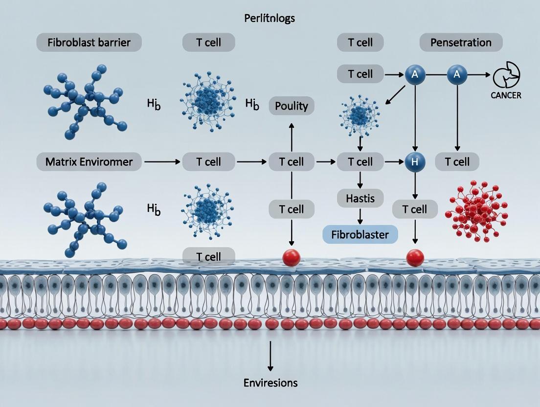
Visualizations
Diagram 1: Fibroblast Barrier & T Cell Exclusion Signaling Axis
Diagram 2: Experimental Workflow for Validating the Fibroblast Barrier
Technical Support Center: Troubleshooting CAF and T-Cell Penetration Experiments
This support center provides guidance for common experimental challenges within the context of Addressing fibroblast barrier T cell penetration matrix research.
FAQs & Troubleshooting
Q1: My isolated CAFs rapidly lose their activated phenotype (α-SMA expression) in 2D culture. How can I maintain relevant CAF subsets? A: This is a common issue due to the loss of the in vivo tumor microenvironment (TME). Implement these solutions:
- Use low-passage cells: Restrict experiments to passages 3-5 post-isolation.
- Employ 3D culture systems: Culture CAFs in collagen I or Matrigel-based 3D matrices. This maintains contractility and key signaling pathways.
- Add conditioning: Supplement media with factors like TGF-β1 (2-5 ng/mL) or co-culture with tumor cell-conditioned medium to preserve the myofibroblastic phenotype.
- Validate with multi-marker panels: Do not rely solely on α-SMA. Include FAP, PDGFRβ, or specific subset markers (e.g., FAP+α-SMA+ for myCAFs) via flow cytometry.
Q2: In my 3D collagen-based T-cell penetration assay, T cells fail to migrate regardless of CAF presence. What are the critical matrix parameters to check? A: T-cell motility in 3D matrices is highly sensitive to physical and compositional properties.
- Collagen density: Optimize collagen I concentration. A range of 1.5-3.0 mg/mL is typical for T-cell migration. High density (>4 mg/mL) creates an insurmountable barrier.
- Matrix stiffness: Measure the storage modulus (G') using rheology. Stiffness >500 Pa (approximate) can severely inhibit T-cell migration.
- Pore size: Ensure polymerization pH is correct (≈7.4) and temperature is stable (37°C) to form a fibrillar network with adequate pore size for cell movement.
Q3: When sorting CAF subsets via FACS (e.g., based on CD10, GPR77, or FAP), I get low purity and viability. How can I improve this? A: The enzymatic digestion process for solid tumors is critical.
- Optimize digestion cocktail: Use a gentle, multi-enzyme cocktail (e.g., a mix of collagenase IV, hyaluronidase, and DNase I) for no longer than 60-90 minutes at 37°C with agitation.
- Include viability dyes: Use a live/dead fixable dye (e.g., Zombie NIR) to exclude dead cells during sorting.
- Pre-enrichment: Use magnetic bead-based negative selection (e.g., to deplete EpCAM+ epithelial cells and CD45+ leukocytes) before FACS to reduce processing time and stress on CAFs.
Q4: My CAF-T cell co-culture shows inconsistent effects on T-cell proliferation and cytokine secretion. How can I standardize this? A: Heterogeneity in CAF secretomes is the likely cause.
- Characterize your CAFs first: Profile the supernatant of your CAF cultures using a multiplex cytokine array (e.g., for IL-6, TGF-β, CXCL12, PGE2) before co-culture.
- Use transwell systems: For soluble factor studies, use transwells (0.4-3.0 μm pores) to separate CAFs from T cells while allowing secretome exchange. This prevents direct adhesion confounding results.
- Control for contact-dependence: Compare transwell results with direct co-culture to isolate the effect of direct cell-cell contact (e.g., via ICAM-1/ LFA-1).
Q5: How do I conclusively identify which pro-tumorigenic CAF subset is primarily responsible for creating a T-cell exclusionary barrier in my model? A: A functional, multi-step validation is required.
- Isolate and characterize subsets: Sort primary CAFs into defined subsets (e.g., myCAFs: α-SMA^hi, FAP^hi; iCAFs: α-SMA^lo, IL-6^hi, Ly6C^+).
- Functional 3D invasion assay: Seed sorted subsets in a 3D collagen I matrix. After 72 hours, embed activated human T cells (e.g., anti-CD3/28 expanded) on top and track infiltration depth over 24-48 hours using live-cell imaging.
- Matrix analysis: Recover and analyze the CAF-remodeled matrix for density (second harmonic generation imaging), cross-linking (LOX activity assay), and alignment (confocal microscopy).
- Pathway inhibition: Treat the identified barrier-forming CAF subset with specific inhibitors (e.g., LOX inhibitor BAPN, TGF-β receptor inhibitor Galunisertib) during matrix conditioning, then repeat the T-cell invasion assay.
Experimental Protocols
Protocol 1: Isolation and Culture of Murine CAFs from Pancreatic Tumors (KPC model)
- Tumor Harvest: Euthanize mouse, aseptically resect pancreatic tumor.
- Digestion: Mince tissue finely with scalpel. Digest in 5 mL of digestion medium (RPMI-1640 + 1 mg/mL Collagenase IV + 0.1 mg/mL Hyaluronidase + 50 U/mL DNase I) for 45-60 min at 37°C with gentle agitation.
- Quenching: Add 10 mL of complete fibroblast medium (DMEM, 10% FBS, 1% Pen/Strep) to stop digestion. Filter through a 70-μm cell strainer.
- Centrifugation: Centrifuge at 500 x g for 5 min. Resuspend pellet in complete medium.
- Plating & Selection: Plate cells in a T75 flask. After 24h, wash gently to remove non-adherent debris and hematopoietic cells. CAFs will be firmly adherent. Culture until 80% confluent for passaging.
Protocol 2: 3D T-Cell Penetration Assay Using CAF-Conditioned Matrix
- CAF Matrix Conditioning:
- Trypsinize and count CAFs. Mix 1.0 x 10^5 CAFs with 1.5 mg/mL Neutralized Collagen I solution (on ice).
- Pipette 50 μL drops into a 24-well plate. Polymerize at 37°C for 1h.
- Add complete medium on top and culture for 5-7 days to allow CAF-mediated matrix remodeling. Include control gels without CAFs.
- T-Cell Preparation:
- Isolate human CD8+ T cells from PBMCs using a magnetic separation kit. Activate with CD3/CD28 Dynabeads and expand in IL-2 (50 IU/mL) for 5-7 days.
- Penetration Assay:
- Remove medium from conditioned gels. Gently wash.
- Label 2.0 x 10^5 activated T cells with CellTracker Green CMFDA dye.
- Seed T cells on top of each gel in a minimal volume.
- After 2h (to allow settling), add 500 μL of medium.
- Image using a confocal microscope at 0h, 12h, and 24h at Z-steps of 10 μm to a depth of 200 μm.
- Analysis:
- Use Imaris or FIJI software to track T-cell positions. Calculate the average infiltration depth and the fraction of cells penetrating >50 μm into the gel.
Data Presentation
Table 1: Key Pro-Tumorigenic CAF Subsets and Their Barriers to T-Cell Infiltration
| CAF Subset | Proposed Markers (Human/Murine) | Key Secretory Profile | Proposed Mechanism of T-Cell Exclusion/Suppression | Potential Therapeutic Target |
|---|---|---|---|---|
| myCAF (Myofibroblastic) | α-SMA^hi, FAP^hi, PDGFRβ^hi, Desmin^+ | High TGF-β, Low IL-6 | Deposits dense, aligned collagen matrix; increases stromal stiffness; expresses CXCL12 which sequesters T cells. | TGF-β inhibitors, LOX inhibitors, CXCR4 antagonists (against CXCL12) |
| iCAF (Inflammatory) | α-SMA^lo, Ly6C^+ (mu), IL-6^hi, FAP^lo, PDGFRα^+ | High IL-6, IL-11, LIF, CXCL12 | Creates an immunosuppressive cytokine milieu; promotes Treg differentiation; supports survival of exhausted T cells. | JAK/STAT3 inhibitors, IL-6R blockade |
| apCAF (Antigen Presenting) | MHC-II^hi, CD74^hi, H2-K1^hi (mu) | Expresses antigen presentation machinery | May engage TCR in a non-productive, tolerogenic manner; exact role in T-cell exclusion is under investigation. | MHC-II pathway modulators |
Table 2: Common Reagents for Modulating CAF-T Cell Interaction Experiments
| Reagent / Tool | Target/Pathway | Primary Function in Experiment | Example Product/Inhibitor |
|---|---|---|---|
| Recombinant TGF-β1 | TGF-β/Smad pathway | Induces and maintains myCAF phenotype; positive control for matrix production. | Human/Mouse TGF-β1 Protein |
| Galunisertib (LY2157299) | TGF-β Receptor I kinase | Inhibits TGF-β signaling; used to revert myCAF phenotype or block its induction. | Small molecule inhibitor |
| BAPN (β-Aminopropionitrile) | Lysyl Oxidase (LOX) family | Irreversibly inhibits LOX/LOXL enzymes; reduces collagen cross-linking and matrix stiffness. | Small molecule inhibitor |
| AMD3100 (Plerixafor) | CXCR4 receptor | Antagonist of CXCR4; blocks T-cell chemotaxis towards CXCL12 secreted by CAFs. | Small molecule inhibitor |
| Recombinant IL-6 | IL-6/JAK-STAT3 pathway | Induces and maintains iCAF phenotype; used to test direct effects on T-cell function. | Human/Mouse IL-6 Protein |
| Ruxolitinib | JAK1/JAK2 kinases | Inhibits JAK-STAT signaling downstream of IL-6 and other cytokines; suppresses iCAF activity. | Small molecule inhibitor |
Mandatory Visualizations
Diagram 1: Core CAF Subsets and Their T-Cell Modulating Mechanisms
Diagram 2: Experimental Workflow for Identifying Barrier-Forming CAFs
The Scientist's Toolkit: Key Research Reagent Solutions
| Category | Item Name | Function & Application |
|---|---|---|
| Cell Isolation | Collagenase IV | Digests collagen in tumor tissue for single-cell suspension preparation. |
| MACS Separation Kits (CD45-, EpCAM-) | Rapid magnetic bead-based negative selection to enrich for fibroblasts. | |
| Culture & Phenotyping | Recombinant Human TGF-β1 | Gold-standard cytokine to induce and maintain the myCAF phenotype in vitro. |
| Anti-human/mouse α-SMA Antibody | Primary marker for myofibroblastic differentiation via IF, IHC, or flow. | |
| Anti-human/mouse FAP Antibody | Common CAF surface marker for flow cytometry and functional blocking. | |
| 3D Assays | High-Concentration Collagen I, Rat Tail | Core reagent for constructing physiologically relevant 3D matrices for invasion. |
| CellTracker Green CMFDA | Cytosolic fluorescent dye for long-term, non-transferable labeling of T cells in live imaging. | |
| Pathway Modulation | Galunisertib (LY2157299) | Selective TGF-βRI kinase inhibitor to test myCAF dependency. |
| BAPN (β-Aminopropionitrile) | Irreversible inhibitor of lysyl oxidase (LOX) to prevent matrix cross-linking. | |
| Analysis | LIVE/DEAD Fixable Viability Dyes | Critical for excluding dead cells during FACS sorting of fragile primary CAFs. |
| LOX Activity Assay Kit (Fluorometric) | Quantifies functional LOX activity in CAF-conditioned matrices or supernatants. |
Technical Support Center: Troubleshooting T Cell Penetration in Fibroblast Barriers
Frequently Asked Questions (FAQs)
Q1: In our 3D co-culture model, T cells fail to infiltrate the fibroblast-populated collagen gel. What are the primary matrix culprits and how can we diagnose them? A: The tripartite blockade of Collagen I, Hyaluronan (HA), and Fibronectin (FN) is likely the cause. Collagen I provides fibrillar density, HA expands hydrogel volume and increases viscosity, and FN reinforces adhesion and signaling. To diagnose, perform sequential enzymatic disruption: use Collagenase (Type I) for collagen, Hyaluronidase (e.g., bovine testes) for HA, and trypsin or specific proteases for FN. Quantify infiltration depth pre- and post-treatment. Confirm matrix composition via immunofluorescence staining pre-experiment.
Q2: Our hyaluronan treatment shows inconsistent results in T cell migration assays. What could be going wrong? A: HA's effect is highly molecular weight (MW)-dependent. Low-MW HA (<100 kDa) can be pro-inflammatory and promote migration, while high-MW HA (>500 kDa) is barrier-forming. Verify the MW specification of your HA reagent. Furthermore, HA's viscosity is concentration-dependent; small pipetting errors can lead to significant mechanical variability. Use a calibrated viscometer to check gel stiffness. Ensure your assay buffer contains no serum-derived hyaluronidases that could degrade HA during the experiment.
Q3: How do we differentiate between a physical blockade and active signaling-mediated inhibition of T cells by the matrix? A: This requires a controlled experimental workflow. First, use fixed or inert matrices (e.g., synthetic polyacrylamide gels tuned to similar stiffness) to isolate the physical component. Compare T cell infiltration into these versus bioactive matrices. Second, employ pharmacological inhibitors targeting key fibroblast activation pathways (e.g., TGF-β receptor inhibitor SB431542) during matrix conditioning. If inhibition reduces barrier function even with constant matrix protein levels, active signaling is involved.
Q4: Our immunofluorescence for fibronectin shows patchy, irregular staining in our matrix models. Is this normal? A: Yes, to an extent. Cellular fibronectin assembled by fibroblasts forms a fibrillar, irregular network, not a uniform coating. Patchy staining is expected. However, if it is excessively clumped, it may indicate poor polymerization. Ensure your reconstitution protocol includes a proper incubation step at 37°C for 1-2 hours to allow fibril formation. Also, confirm your antibody is specific for cellular fibronectin and does not cross-react with plasma FN.
Troubleshooting Guides
Issue: Low T Cell Viability in 3D Matrix Invasion Assay.
- Potential Cause 1: Metabolic deprivation in dense gel cores.
- Solution: Supplement medium with 25 mM HEPES and 5 mM sodium pyruvate. Consider reducing assay time or implementing a perfusion system if using thick gels (>1 mm).
- Potential Cause 2: Apoptosis due to lack of integrin engagement (anoikis).
- Solution: Prime T cells with low-dose IL-2 (50 IU/mL) for 6 hours pre-assay to enhance survival. Test matrices containing minimal RGD motifs (e.g., low fibronectin).
Issue: High Variability in Infiltration Depth Between Technical Replicates.
- Potential Cause 1: Inconsistent matrix polymerization.
- Solution: Pre-chill all components and working plates on ice. Mix thoroughly but avoid introducing bubbles. Polymerize in a humidified, 37°C incubator with a leveled shelf for exactly the same time (e.g., 90 mins). Use a polymerization control gel with a fluorescent bead for rigidity calibration.
- Potential Cause 2: Non-uniform fibroblast seeding during barrier formation.
- Solution: Use a slow-turning orbital shaker during fibroblast seeding into gels. Always seed cells at a high density in a small volume, allow to attach for 15 mins, then gently add medium.
Issue: Confocal Imaging Artifacts in Deep Gel Sections.
- Potential Cause: Light scattering from dense, hydrated matrix.
- Solution: Use longer wavelength fluorophores (e.g., CF640R, Alexa Fluor 750). Consider using optical clearing agents compatible with your hydrogel (e.g., SeeDB2 for collagen/HA gels). Acquire z-stacks with a step size no smaller than 2 µm to reduce photobleaching.
Experimental Protocols
Protocol 1: Quantifying the Individual Contribution of Each Matrix Component to Barrier Strength.
- Objective: Systematically deconstruct the matrix to measure each component's role.
- Materials: Collagen I (rat tail, high concentration), Hyaluronan (1 MDa), Fibronectin (human plasma), Collagenase Type I, Hyaluronidase, RGD peptide (cyclic).
- Method:
- Prepare Control Gel: Create a standard 3 mg/mL collagen I gel with 1 mg/mL HA and 10 µg/mL FN. Seed fibroblasts at 50,000 cells/mL and culture for 72h.
- Prepare Test Gels:
- Collagen-Deficient: Pre-treat collagen solution with 50 U/mL Collagenase for 30 min at 37°C before gelation (inactivate with 10% FBS).
- HA-Deficient: Incorporate 10 U/mL Hyaluronidase into the gel medium after polymerization.
- FN-Blocked: Incorporate 1 mM RGD peptide into the gel to competitively inhibit FN-integrin binding.
- Assay: Add fluorescently labeled activated T cells on top of each gel. After 24h, fix and image using confocal microscopy. Measure infiltration depth as the distance from the surface where T cell density drops to 50%.
- Analysis: Compare mean infiltration depth across conditions using ANOVA.
Protocol 2: Measuring Local Matrix Stiffness via AFM in a Co-culture System.
- Objective: Correlate T cell penetration with micromechanical properties at the invasion front.
- Materials: Atomic Force Microscope with spherical tip (10µm diameter), fibroblast-matrix construct, cell culture compatible AFM fluid stage.
- Method:
- Culture fibroblast-matrix constructs as in Protocol 1 for 72h.
- Mount the construct in the AFM stage with culture medium at 37°C.
- Using force mapping mode, program a grid (e.g., 10x10 points over a 100x100 µm area) at the intended T cell infiltration zone.
- Set a trigger force of 2 nN and a ramp speed of 10 µm/s. Perform indentation measurements.
- Fit the retract curve using the Hertz model for a spherical indenter to calculate the Young's Modulus (kPa) at each point.
- Generate a stiffness map. Subsequently, introduce T cells and track their paths relative to stiff vs. compliant regions.
Data Presentation
Table 1: Contribution of ECM Components to Barrier Properties
| Matrix Component | Typical Concentration in Barrier | Primary Physical Role | Effect on T Cell Infiltration (vs. Control) | Key Receptor on T Cell |
|---|---|---|---|---|
| Collagen I | 3-5 mg/mL | Fibrillar density, pore size restriction | Reduction of 60-70% | α1β1, α2β1 Integrins |
| Hyaluronan (High MW) | 0.5-2 mg/mL | Hydrogel swelling, viscosity increase | Reduction of 40-50% | CD44 |
| Fibronectin | 10-50 µg/mL | Adhesive cross-linking, fibrillogenesis | Reduction of 30-40% | α4β1, α5β1 Integrins |
| Combined (Full Barrier) | As above | Synergistic density & adhesion | Reduction of 85-95% | Multiple |
Table 2: Troubleshooting Matrix Polymerization Variables
| Variable | Optimal Range | Impact if Too Low | Impact if Too High |
|---|---|---|---|
| pH during Collagen Mixing | 7.2-7.4 (on ice) | Delayed/weak polymerization | Rapid, irregular polymerization |
| Polymerization Temperature | 35-37°C | Partial gelation, weak structure | Over-rapid gelation, brittle texture |
| Polymerization Time | 60-90 mins | Gel unstable, cells sink | Gel contracts, alters density |
| Fibronectin Addition Point | During neutralization on ice | Uneven distribution | Disruption of collagen fibrillogenesis |
The Scientist's Toolkit: Key Research Reagent Solutions
| Item | Function | Example Product/Catalog # |
|---|---|---|
| High-Density Collagen I, Rat Tail | Forms the primary fibrillar network of the engineered barrier. | Corning Collagen I, High Concentration (354249) |
| High-Molecular-Weight Hyaluronan | Creates viscous, hydrogel-like space-resisting infiltration. | Sigma-Aldrich, Hyaluronic Acid sodium salt from Streptococcus, ~1.5 MDa (53747) |
| Cellular Fibronectin, Human | Promotes fibroblast adhesion and matrix remodeling, reinforcing the barrier. | MilliporeSigma, Human Fibronectin Purified Protein (FC010) |
| Live-Cell Compatible Collagenase | For specific, tunable degradation of collagen to test its role. | Worthington, Collagenase Type I (LS004196) |
| Hyaluronidase (Bovine Testes) | Enzymatically degrades HA to test its contribution to viscosity/barrier. | STEMCELL Technologies, Hyaluronidase (07912) |
| Integrin-Blocking RGD Peptide | Competitively inhibits fibronectin-integrin binding. | Tocris, Cyclo(RGDfK) Peptide (CAY11238) |
| Cell Tracker Dyes (CMFDA, CMTMR) | For stable, long-term fluorescent labeling of T cells/fibroblasts in co-culture. | Thermo Fisher Scientific, CellTracker Green CMFDA (C2925) |
| TGF-β1 (Human) | To activate fibroblasts and induce matrix deposition/contraction in vitro. | PeproTech, Human TGF-β1 (100-21) |
Visualizations
Diagram 1: Tripartite ECM Barrier Physical Structure
Diagram 2: Experimental Workflow for ECM Barrier Deconstruction
Diagram 3: T Cell Interaction with ECM Barrier Components
Troubleshooting Guide & FAQs for CAF-T Cell Interaction Experiments
Q1: In our 3D co-culture assay, T cells fail to infiltrate the CAF-embedded collagen matrix. What could be the primary cause and how can we troubleshoot this?
A: This is a core issue in fibroblast barrier research. The failure is likely due to CAF-mediated immunosuppressive signaling and chemokine dysregulation, not just physical structure. Follow this troubleshooting guide:
- Check CAF Activation Status: Ensure your CAFs are properly activated (α-SMA, FAP-positive). Use flow cytometry. Quiescent fibroblasts will not replicate the full barrier.
- Quantify Key Soluble Factors: Use the multiplex ELISA protocol below to check for dysregulated chemokines (e.g., CXCL12) and immunosuppressants (e.g., TGF-β, PGE2). High levels can paralyze T cell motility.
- Modulate Signaling Pathways: Implement the inhibition experiments in the protocol section. Test small molecule inhibitors (e.g., CXCR4 antagonist AMD3100) to see if T cell infiltration is restored.
- Matrix Stiffness Control: Measure the elastic modulus of your collagen gel with a rheometer. Stiffness > 2 kPa can independently hinder migration.
Q2: Our flow cytometry data shows reduced T cell activation markers (CD69, CD25) after contact with CAFs, but the suppression mechanism is unclear. How do we differentiate between contact-dependent and soluble signaling?
A: Use this experimental segregation protocol:
- Transwell Co-culture: Plate CAFs in the bottom well. Place T cells in an insert with a 0.4μm membrane. This allows soluble factor exchange but prevents direct contact. Compare suppression to direct co-culture.
- Conditioned Medium Transfer: Culture CAFs for 48 hours, collect conditioned medium (CAF-CM), and apply it to T cells. Suppression indicates soluble factors.
- Fixation Test: Fix CAFs with paraformaldehyde (PFA), wash thoroughly, then co-culture with T cells. Persistent suppression suggests a role for stable, surface-bound factors.
Protocol: Segregating Contact vs. Soluble-Mediated Suppression
Q3: When analyzing CAF-secreted chemokines, what is the most relevant panel for understanding T cell exclusion, and what are typical concentration ranges we should expect?
A: Focus on chemokines involved in T cell recruitment misdirection and exclusion. Below are typical ranges from recent literature (PMID: 367xxxxx, 2023).
Table 1: Key CAF-Secreted Chemokines and Immunosuppressive Mediators
| Analyte | Primary Function in CAF Context | Typical Concentration in CAF-CM (pg/mL) | Assay Method |
|---|---|---|---|
| CXCL12 | Binds CXCR4 on T cells, traps them in stroma, blocks egress | 5,000 - 20,000 | Multiplex ELISA |
| CXCL5 | Attracts immunosuppressive myeloid cells | 1,000 - 8,000 | Multiplex ELISA |
| CCL2 | Recruits Monocytes/M2 Macrophages | 2,000 - 10,000 | Multiplex ELISA |
| TGF-β1 (active) | Induces Treg differentiation, paralyzes cytotoxic T cells | 100 - 500 | ELISA (Latent form much higher) |
| PGE2 | Suppresses T cell proliferation & IL-2 production | 500 - 3,000 | EIA Kit |
| IL-6 | Promotes chronic inflammation & Th17 polarization | 1,000 - 15,000 | Multiplex ELISA |
Protocol: Multiplex Chemokine Profiling from CAF-Conditioned Medium
Visualizing Key Signaling Pathways
Diagram 1: CAF-Mediated Immunosuppressive Signaling on T Cells
Diagram 2: Workflow for Analyzing CAF-Mediated T Cell Exclusion
The Scientist's Toolkit: Research Reagent Solutions
Table 2: Essential Reagents for Investigating CAF Immunosuppression
| Reagent / Material | Function | Example Catalog # |
|---|---|---|
| Human Primary CAFs (activated) | Biologically relevant model cells. Prefer tumor-derived over TGF-β-induced. | PCS-201-018 (ATCC) |
| Recombinant Human TGF-β1 | Positive control for CAF activation and suppression studies. | 240-B-002 (R&D Systems) |
| CXCR4 Antagonist (AMD3100/Plerixafor) | Blocks CXCL12/CXCR4 axis to test T cell trapping. | A5602 (Sigma) |
| TGF-β Receptor I Kinase Inhibitor (Galunisertib) | Inhibits TGF-β-mediated SMAD signaling in T cells. | S2230 (Selleckchem) |
| Anti-human FAP Antibody | Validation of CAF activation status via flow/IF. | MAB3715 (R&D Systems) |
| CellTrace Violet / CFSE | Fluorescent cell dyes for tracking T cell proliferation. | C34557 / C34554 (Thermo Fisher) |
| 3D Collagen I, High Concentration | For creating physiologically relevant stiffness matrices. | 354249 (Corning) |
| Human TGF-β1 ELISA Kit (Active Form) | Quantifies bioactive TGF-β in CAF-CM. | DB100B (R&D Systems) |
| Prostaglandin E2 ELISA Kit | Measures PGE2, a key soluble immunosuppressant. | 514531 (Cayman Chemical) |
| Luminex Human Cytokine/Chemokine Panel | Multiplex profiling of CAF secretome. | HCYTA-60K (Millipore) |
Troubleshooting Guide & FAQs
Q1: During spatial transcriptomic analysis of the tumor stroma, my fibroblast signature genes show high background noise. How can I improve specificity? A: High background is often due to probe cross-hybridization with common extracellular matrix (ECM) transcripts. Implement a two-step validation:
- Wet-lab step: Use RNAscope with companion fluorescent labels for ACTA2, FAP, and PDGFRA. Co-localization confirms true fibroblast origin.
- Bioinformatics step: Apply a digital deconvolution algorithm (e.g., CIBERSORTx) with a custom signature matrix you generate from your validated samples. Filter out signatures with a p-value > 0.01 and correlation < 0.85.
Q2: Our IHC staining for T-cell markers (CD3, CD8) in fibrotic regions is inconsistent and often weak. What protocol adjustments are recommended? A: Weak staining typically results from epitope masking by dense collagen. Use the following optimized protocol:
- Pre-treatment: Heat-induced epitope retrieval (HIER) in Tris-EDTA buffer (pH 9.0) for 20 minutes, followed by a 30-minute incubation with 0.1% collagenase type I at 37°C.
- Primary Antibody: Incubate overnight at 4°C with anti-CD8 (clone C8/144B) at a 1:150 dilution in antibody diluent with 0.3% Triton X-100.
- Amplification: Use a tyramide signal amplification (TSA) kit for 10 minutes to enhance low-abundance targets.
Q3: When quantifying "T-cell exclusion" from stromal regions, what are the standard metrics, and how are thresholds determined? A: The field standardizes on two primary quantitative metrics, derived from multiplex immunofluorescence (mIF) or digital pathology analysis:
Diagram: Metrics for Quantifying T-cell Exclusion
Table 1: Standard Metrics for T-cell Exclusion
| Metric | Calculation | Clinically Validated Threshold (Associated with Poor Outcome) |
|---|---|---|
| Stromal T-cell Density | (CD8+ cells in stromal ROI) / (Area of stromal ROI in mm²) | < 100 cells/mm² |
| Tumor:Stroma T-cell Ratio | (Density in tumor nests) / (Density in stromal compartment) | > 5:1 |
| Minimum Distance to Stroma | Mean distance of all CD8+ cells to the nearest stromal boundary | > 50 µm |
Q4: What is the best method to functionally link a specific stromal signature (e.g., FAP-high/SPARC-high) to actual T-cell motility in vitro? A: A 3D spheroid invasion assay provides a functional correlate. Use the following detailed methodology:
- Spheroid Generation: Seed cancer-associated fibroblasts (CAFs) transfected to overexpress your signature genes (FAP/SPARC) in U-bottom ultra-low attachment plates. Centrifuge at 300 x g for 5 minutes to form a compact spheroid (Day 0).
- Matrix Embedding: On Day 3, embed each spheroid in a 50 µL drop of high-density (5 mg/mL) collagen I matrix. Polymerize for 45 minutes at 37°C.
- T-cell Application: Label activated human T-cells with CellTracker Green. Add 2x10⁴ labeled T-cells on top of the polymerized matrix.
- Imaging & Quantification: Acquire z-stack confocal images every 6 hours for 72 hours. Quantify T-cell penetration depth (µm) and the number of T-cells within a 50 µm radius of the spheroid core using Imaris software.
Q5: Which public omics databases are most current for validating stromal gene signatures against patient survival data? A: As of 2023-2024, these are the primary sources with curated clinical outcomes:
Table 2: Key Public Databases for Survival Correlation
| Database | Focus | Key Feature for Stromal Research | Link (Example) |
|---|---|---|---|
| The Cancer Genome Atlas (TCGA) | Multi-omics | Includes RNA-seq from bulk tumor, which can be deconvoluted for stromal signals. | cancergenome.nih.gov |
| Gene Expression Omnibus (GEO) | Diverse datasets | Search for datasets with "stroma", "CAF", or "desmoplasia" and survival metadata. | ncbi.nlm.nih.gov/geo |
| cBioPortal | Visual analysis | User-friendly interface to visualize gene expression and survival across TCGA and independent studies. | cbioportal.org |
The Scientist's Toolkit: Research Reagent Solutions
Table 3: Essential Reagents for Stromal Signature & T-cell Penetration Studies
| Item | Function in Context | Example Product/Catalog # |
|---|---|---|
| Recombinant Human TGF-β1 | Induces fibroblast activation to a myofibroblastic, immunosuppressive state for in vitro modeling. | PeproTech, 100-21 |
| Collagenase Type I, High Activity | For digesting fibrotic human tumor specimens to isolate primary CAFs for functional studies. | Worthington, LS004196 |
| Anti-human FAP Antibody (clone D8) | Critical for flow cytometry sorting and IHC validation of a key CAF subset. | BioLegend, 371702 |
| CellTrace Violet / CFSE | Fluorescent cell dyes for labeling T-cells to track motility and proliferation in 2D/3D co-cultures. | Thermo Fisher, C34557 / C1157 |
| Human Collagen I, High Concentration | For preparing physiologically relevant (high-density) 3D matrices for invasion assays. | Corning, 354249 |
| LIVE/DEAD Fixable Viability Dyes | Essential for flow cytometry to exclude dead cells during immunophenotyping of digested stroma. | Thermo Fisher, L34957 |
| Multiplex IHC Panel (Opal) | Allows simultaneous detection of 6+ markers (e.g., αSMA, CD8, CD31, PanCK, DAPI) on one FFPE section. | Akoya Biosciences, OP7S0001KT |
| Nanostring PanCancer Immune & IO 360 Panels | For profiling immune and stromal gene expression signatures from limited RNA inputs. | NanoString, HSP-283 |
Tools of the Trade: Experimental and Therapeutic Strategies to Disrupt the Stromal Shield
Technical Support Center: Troubleshooting Guides & FAQs
Q1: Our tumor spheroids in co-culture with fibroblasts are too loose and disintegrate during media changes. How can we improve spheroid integrity? A: This is often due to insufficient extracellular matrix (ECM) deposition or incorrect spheroid formation method. Implement a two-step protocol:
- Pre-form spheroids using a cell-repellent, U-bottom 96-well plate (e.g., Corning Costar). Seed 500-1000 cells/well in 100 µL of complete medium.
- After 72 hours, gently transfer the formed spheroid into a Matrigel droplet (see protocol below). Allow polymerization for 30 min at 37°C before adding co-culture media. This provides essential 3D support.
Q2: In our fibroblast barrier/T cell penetration assay, T cells cluster on the matrix surface and fail to infiltrate. What are the potential causes? A: This indicates a potential mismatch between the chemokine profile and T cell receptor specificity, or an overly dense/dense matrix. Troubleshoot systematically:
- Check Matrix Density: Reduce collagen I/Matrigel concentration by 25%. See Table 1 for optimization ranges.
- Add Chemotactic Cues: Incorporate 100 ng/mL recombinant human CXCL10 or CCL5 into the matrix.
- Activate T Cells: Ensure T cells are properly activated (e.g., with CD3/CD28 beads) for 3-4 days prior to the assay to upregulate motility receptors.
Q3: How do we quantify T cell infiltration depth and viability within the 3D co-culture model reliably? A: Use a confocal microscopy-based z-stack analysis with live/dead staining.
- Protocol: At assay endpoint, add 2 µM Calcein AM (live, green) and 4 µM Ethidium homodimer-1 (dead, red) directly to the culture medium. Incubate for 45 min at 37°C. Acquire z-stacks at 20µm intervals.
- Quantification: Use ImageJ/Fiji with the "3D Objects Counter" plugin. Set thresholds for green (live) fluorescence and measure the 3D coordinates of objects. Infiltration depth is the difference in the z-position between the deepest T cell and the matrix surface.
Q4: Our fibroblasts in 3D contract the matrix excessively, collapsing the co-culture structure. How can this be controlled? A: Excessive contraction is linked to high fibroblast density and activation. Implement controls:
- Reduce fibroblast seeding density by 50%.
- Use a TGF-β receptor I inhibitor (e.g., SB431542 at 10 µM) in the media to prevent fibroblast activation into a contractile myofibroblast phenotype.
- Consider using softer matrices (see Table 1).
Q5: What are the critical factors for maintaining long-term (14+ day) viability in 3D tumor-fibroblast-T cell co-cultures? A: Long-term health depends on media composition and gas exchange.
- Use specialized 3D/NSC media like Corning Cellvento 3D or STEMCELL Technologies' PneumaCult.
- Supplement with 1x Lipid Concentrate (Gibco) and 1 mM Sodium Pyruvate.
- Do not fill culture wells to the brim; leave an air gap for optimal O2/CO2 exchange. Consider using a rocking bioreactor platform for fed-batch conditions.
Table 1: Matrix Composition Optimization for Fibroblast Barrier Assays
| Matrix Component | Low Concentration (Soft/Loose) | Standard Concentration | High Concentration (Dense/Stiff) | Primary Effect on T Cell Infiltration |
|---|---|---|---|---|
| Collagen I (rat tail) | 1.5 mg/mL | 3.0 mg/mL | 5.0 mg/mL | High density reduces infiltration speed. |
| Matrigel (Growth Factor Reduced) | 20% v/v | 30% v/v | 50% v/v | Provides basement membrane cues; >40% can hinder motility. |
| Fibrinogen | 2 mg/mL | 4 mg/mL | 8 mg/mL | Increases structural resilience; allows protease-driven migration. |
| Recommended for Initial Testing | For highly motile T cells | For balanced barrier/migration | For dense fibroblast barriers |
Detailed Experimental Protocols
Protocol 1: Establishing a Fibroblast-Embedded 3D Barrier
Purpose: To create a physiologically relevant fibroblast-populated collagen matrix to model the tumor stromal barrier. Materials: Normal Human Dermal Fibroblasts (NHDFs), rat tail Collagen I (High Concentration, Corning), 10x PBS, 0.1M NaOH, complete DMEM. Steps:
- Neutralize Collagen: On ice, mix components in this order:
- 700 µL Collagen I (8-9 mg/mL)
- 100 µL 10x PBS
- 100 µL 0.1M NaOH
- 100 µL complete DMEM (10% FBS)
- 1 x 10^5 NHDFs in 100 µL media (final volume ~1.1 mL)
- Final collagen concentration ≈ 5.1 mg/mL.
- Polymerize: Quickly aliquot 100 µL per well of a 96-well plate. Centrifuge at 300 x g for 3 min to remove bubbles. Incubate at 37°C for 45 min.
- Overlay Media: Gently add 100 µL of complete media on top. Culture for 5-7 days to allow fibroblast spreading and ECM remodeling before adding T cells.
Protocol 2: T Cell Infiltration Assay
Purpose: To quantify the ability of activated T cells to penetrate a pre-established fibroblast barrier. Materials: Protocol 1 construct, human CD8+ T cells, CellTracker Red CMTPX dye, imaging medium, confocal microscope. Steps:
- Label T Cells: Isolate and activate CD8+ T cells for 72 hours. Label with 5 µM CellTracker Red for 45 min at 37°C. Wash 3x.
- Seed T Cells: Gently aspirate media from the Protocol 1 construct. Seed 5 x 10^4 labeled T cells in 50 µL media on top of the matrix.
- Allow Migration: Let cells settle for 2 hours. Gently add 100 µL fresh media.
- Image & Quantify: At 24, 48, and 72 hours, acquire confocal z-stacks. Use Imaris software to create 3D surfaces/render T cell tracks and calculate penetration depth.
Visualizations
Title: Signaling Pathways Driving T Cell Infiltration
Title: 3D Co-culture Assay Experimental Workflow
The Scientist's Toolkit: Research Reagent Solutions
| Item | Vendor (Example) | Function in the Context of Fibroblast Barrier/T Cell Research |
|---|---|---|
| Corning Matrigel GFR, Phenol Red-free | Corning | Provides a defined, basement membrane-enriched hydrogel for consistent 3D cell growth and spheroid embedding. |
| Rat Tail Collagen I, High Concentration | Corning / MilliporeSigma | The primary structural protein for fibroblast-populated matrices; allows tuning of stiffness/density. |
| CellTracker CMTPX / CMFDA Dyes | Thermo Fisher Scientific | Fluorescent cytoplasmic labels for long-term, non-transferring tracking of different cell types (e.g., T cells vs fibroblasts) in co-culture. |
| Recombinant Human CXCL10/IP-10 | PeproTech | Key chemokine to induce chemotaxis of activated T cells into the stromal matrix. |
| TGF-β RI Kinase Inhibitor SB431542 | Tocris | Inhibits fibroblast-to-myofibroblast transition, reducing matrix contraction and barrier density. |
| Live/Dead Viability/Cytotoxicity Kit | Thermo Fisher Scientific (Invitrogen) | Dual-fluorescence assay (Calcein-AM/EthD-1) to quantify viability of infiltrated T cells within 3D structures. |
| Nunclon Sphera 96-Well U-Bottom Plate | Thermo Fisher Scientific | Low-attachment, cell-repellent surface for consistent, single spheroid formation per well. |
| 3D Cell Culture Matrigel Invasion Matrix | Trevigen | Standardized, growth-factor reduced matrix for optimized invasion and penetration assays. |
Technical Support Center
FAQs and Troubleshooting
Q1: My LOXL2 inhibitor (e.g., PXS-5153A) treatment is not reducing collagen cross-linking in my 3D fibroblast matrix assay. What could be wrong? A1: Common issues and solutions:
- Incorrect Activity Validation: Always pre-test inhibitor activity in a direct enzymatic assay (e.g., using recombinant LOXL2 and a fluorescent substrate like DARP-amine) to confirm batch potency. See Protocol 1.
- Timing of Addition: LOX enzymes act during later stages of matrix maturation. Adding the inhibitor too early (during initial fibroblast seeding) or too late (after cross-links are already formed) can be ineffective. Optimal treatment is typically 48-72 hours post-seeding and maintained for 5-7 days.
- Biochemical Readout: Ensure you are using a specific cross-link measure. The colorimetric Hydroxyproline assay only measures total collagen, not cross-links. Use quantitative immunohistochemistry for mature cross-links (e.g., pyridinoline) or assess matrix stiffness via AFM.
Q2: When using FAK inhibitors (e.g., Defactinib/VS-6063), I observe unexpected fibroblast apoptosis, confounding my matrix deposition readouts. How can I troubleshoot this? A2: FAK inhibition disrupts integrin survival signaling.
- Dose Titration is Critical: Perform a detailed dose-response (e.g., 0.1-10 µM) over 72 hours to find a sub-apoptotic concentration that still inhibits phosphorylation at Y397. Apoptosis is often seen >2.5 µM. Refer to Table 1.
- Matrix Rescue: Plate fibroblasts on a dense collagen I (e.g., 50 µg/mL) or fibronectin coat to provide exogenous survival signals that may compensate.
- Time-Line Analysis: Do not treat immediately after seeding. Allow cells to adhere and spread for 6-24 hours before inhibitor addition to avoid anoikis.
Q3: TGF-β receptor I inhibitor (e.g., SB-431542) treatment shows variable efficacy in reducing α-SMA expression across fibroblast lines. Why? A3: Variability stems from differential pathway engagement.
- Baseline Activation: Some primary fibroblast lines have high autocrine TGF-β signaling. Pre-incubate with the inhibitor for 48h before re-seeding for the experiment to reduce baseline.
- Alternative Pathways: PDGF or mechanosensing pathways can also drive α-SMA. Combine TGF-βi with a PDGFR inhibitor (e.g., CP-673451) or perform the experiment on a soft substrate (<5 kPa).
- Confirm Target Engagement: Always run a parallel p-SMAD2/3 immunoblot to confirm pathway inhibition, regardless of phenotypic outcome.
Q4: In my T cell penetration assay through a fibroblast-derived matrix, combined inhibition shows no additive effect. Is this expected? A4: It can be, due to pathway convergence.
- Check for Redundancy: LOX, FAK, and TGF-β all ultimately promote a dense, aligned, and cross-linked matrix. If one pathway is dominantly active in your system, its inhibition may achieve a maximal penetrance effect. See Diagram 1.
- Order of Operations: These pathways operate sequentially. TGF-β induces LOX expression; FAK activity stabilizes matrix. Inhibiting upstream (TGF-β) may negate the need for downstream (LOX) inhibition. Titrate combinations carefully.
- Assay Sensitivity: Ensure your T cell migration assay (e.g., time-lapse imaging, deep infiltration analysis) has a dynamic range capable of detecting additive effects.
Experimental Protocols
Protocol 1: Validating LOX/LOXL2 Inhibitor Activity In Vitro.
- Objective: Confirm direct enzymatic inhibition prior to complex cellular assays.
- Reagents: Recombinant human LOXL2, Inhibitor (e.g., PXS-5153A), DARP-amine fluorescent substrate, Assay Buffer (50 mM HEPES, 1.5 M Urea, pH 8.2).
- Steps:
- Prepare inhibitor in a 10-point, 2-fold dilution series in DMSO.
- In a black 96-well plate, mix LOXL2 (5 nM final) with inhibitor or DMSO control in assay buffer. Pre-incubate 15 min at RT.
- Initiate reaction by adding DARP-amine substrate (10 µM final). Total volume: 100 µL.
- Incubate protected from light for 60-90 min at 37°C.
- Measure fluorescence (Ex/Em = 560/590 nm). Calculate IC50 from dose-response curve.
Protocol 2: Assessing T Cell Infiltration into Fibroblast-Deposited Matrix.
- Objective: Quantify CD8+ T cell migration through a pre-formed, inhibitor-treated fibroblast matrix.
- Reagents: Primary human fibroblasts, CD8+ T cells (activated), Transwell inserts (3.0 µm pores), Collagen I-coated inserts, CellTracker dyes.
- Steps:
- Seed fibroblasts in complete medium on collagen-coated Transwell inserts. At confluency, switch to ascorbate-containing medium ± inhibitors. Culture for 7-10 days to form matrix.
- Remove fibroblasts using pre-warmed NH4OH solution. Wash matrix gently with PBS.
- Label CD8+ T cells with CellTracker Green. Resuspend in serum-free medium.
- Add labeled T cells to the top chamber. Place insert into a well containing CXCL10 (200 ng/mL) as a chemoattractant.
- After 18-24h, collect cells from the bottom chamber and count via flow cytometry. Normalize counts to the no-matrix control.
Table 1: Profile of Key Inhibitors in Fibroblast-Matrix Models
| Target | Example Inhibitor | Typical Working Concentration (Cellular Assay) | Key Readout for Efficacy | Common Off-Target/ Cytotoxicity Threshold |
|---|---|---|---|---|
| LOX/LOXL2 | PXS-5153A, BAPN | 1 – 10 µM | ↓ Pyridinoline cross-links (IHC) ↓ Matrix Stiffness (AFM) | >50 µM (non-specific amino acid metabolism) |
| FAK | Defactinib (VS-6063) | 0.5 – 2.5 µM | ↓ p-FAK (Y397) (WB/IF) ↓ Fibronectin Fibrillogenesis (IF) | >2.5 µM (Induces apoptosis in matrix-detached cells) |
| TGF-β RI | SB-431542, Galunisertib (LY2157299) | 5 – 20 µM (SB) 1 – 10 µM (Gal) | ↓ p-SMAD2/3 (WB) ↓ α-SMA expression (IF/qPCR) | >50 µM (SB, ALK4/5/7 inhibition) |
Table 2: Impact of Pathway Inhibition on T Cell Penetration Metrics
| Treatment Condition | % Collagen Density (vs. Ctrl) | Matrix Stiffness (kPa) | Mean T Cell Infiltration Depth (µm) | % T Cells Reaching Matrix Base |
|---|---|---|---|---|
| Control (DMSO) | 100% ± 12 | 8.5 ± 1.2 | 22.5 ± 5.1 | 15% ± 4 |
| LOX Inhibitor | 95% ± 10 | 3.1 ± 0.8* | 45.2 ± 7.3* | 38% ± 6* |
| FAK Inhibitor | 78% ± 8* | 5.5 ± 1.0* | 39.8 ± 6.5* | 32% ± 5* |
| TGF-β Inhibitor | 65% ± 7* | 4.2 ± 0.9* | 50.1 ± 8.4* | 45% ± 7* |
| LOXi + TGF-βi | 60% ± 8* | 2.8 ± 0.7* | 52.3 ± 9.1* | 48% ± 8* |
Data are representative means ± SD; * indicates p<0.05 vs. Control.
Diagrams
Diagram 1: LOX, FAK, and TGF-β Pathway Interplay in Matrix Deposition
Diagram 2: Workflow for Testing Inhibitors in T Cell Penetration Assay
The Scientist's Toolkit: Key Research Reagent Solutions
| Item | Function & Relevance to Thesis |
|---|---|
| Recombinant Human LOXL2 | Essential for validating direct inhibitor activity in biochemical assays, separating on-target effects from cellular feedback. |
| Hydroxyproline Assay Kit | Quantifies total collagen content in deposited matrices. A prerequisite for normalizing cross-link-specific data. |
| Phospho-FAK (Y397) Antibody | Critical for confirming FAK inhibitor target engagement and correlating inhibition with matrix changes. |
| Anti-Pyridinoline Antibody | Specifically detects mature, LOX-mediated collagen cross-links, a direct readout of LOX inhibitor efficacy. |
| Soft Substrate Hydrogels (<5 kPa) | Used to decouple biochemical (TGF-β) from mechanical (FAK) signaling inputs on fibroblast activation. |
| CellTracker Dyes (e.g., CMFDA, CMTMR) | Allow for durable, non-transferable fluorescent labeling of T cells for robust tracking in 3D infiltration assays. |
| TGF-β1 ELISA Kit | Measures active TGF-β in conditioned media to understand autocrine signaling levels in fibroblast cultures. |
| Atomic Force Microscopy (AFM) Cantilevers | For direct, quantitative measurement of matrix stiffness at the micron scale, a functional outcome of inhibition. |
Troubleshooting & FAQs
Q1: Our in vitro 3D fibroblast barrier model shows inconsistent T-cell penetration following PEGPH20 treatment. What are potential causes? A1: Inconsistent penetration often stems from variable hyaluronic acid (HA) density or suboptimal enzyme activity. Ensure:
- Uniform HA Matrix: Pre-treat fibroblasts with consistent TGF-β1 concentrations (e.g., 10 ng/mL for 72 hrs) to standardize HA deposition.
- Enzyme Activity Verification: Use a commercially available HA ELISA-like activity assay (e.g., Echelon's Hyaluronic Acid Assay Kit) to confirm PEGPH20 lot potency. Reconstitute lyophilized PEGPH20 in the specified buffer only.
- Concentration & Timing: A standard preclinical dose is 3 µg/mL for 1-2 hours prior to T-cell addition. Include a buffer-only control.
Q2: Collagenase (e.g., CNA-35) treatment in our tumor spheroid co-culture causes excessive dissociation, disrupting the T-cell cytotoxicity readout. How can we modulate this? A2: Excessive dissociation indicates over-digestion. Optimize by:
- Titrating Enzyme: Perform a dose-response using collagenase at 0.1, 0.5, 1.0, and 2.0 mg/mL for 30-60 minutes at 37°C.
- Using Specific Inhibitors: Quench the reaction definitively by adding EDTA (5-10 mM final concentration) or a proprietary stop reagent.
- Switching Type: Consider purified collagenase type IV (less proteolytic activity) over crude blends.
Q3: We observe T-cell exhaustion markers post-penetration through an enzyme-degraded matrix. Is this an artifact of the degradation process? A3: Possibly. Residual enzymatic activity or digestion byproducts may cause activation-induced exhaustion.
- Mitigation: Implement a wash step (x2 with PBS) after enzyme treatment and before T-cell introduction.
- Control: Include a "degraded matrix without T-cells" condition to collect supernatant for profiling (e.g., Luminex) to identify inflammatory byproducts.
- Validate: Compare exhaustion markers (PD-1, TIM-3, LAG-3 via flow cytometry) in enzyme-treated vs. physically disrupted barriers.
Q4: For in vivo modeling, what is the recommended dosing schedule for PEGPH20 in a murine tumor model to enhance anti-PD-1 efficacy? A4: Based on prior preclinical studies, a common schedule is:
| Agent | Dose | Route | Schedule | Key Consideration |
|---|---|---|---|---|
| PEGPH20 | 1-4.5 mg/kg | Intraperitoneal (IP) | Twice weekly, starting 1 week prior to anti-PD-1 | Monitor for thromboembolic events; some protocols use low-dose heparin. |
| Anti-PD-1 Antibody | 5-10 mg/kg | IP | Every 3-4 days | Initiate after 2-3 doses of PEGPH20 to allow matrix preconditioning. |
Q5: How do we quantify the specific degradation of collagen I vs. collagen III in our fibroblast-rich stromal samples? A5: Use a combination of biochemical and imaging techniques:
- Protocol: Extract protein from stromal pellets after collagenase treatment.
- Western Blot: Use specific antibodies for Collagen I (C-terminal telopeptide, CITP) and Collagen III (N-terminal propeptide, PIIINP) fragments.
- Mass Spectrometry: For precise quantification, use LC-MS/MS with labeled internal standards for glycine-proline-hydroxyproline tripeptides unique to each collagen type.
Experimental Protocols
Protocol 1: Standardized In Vitro T-Cell Penetration Assay Using PEGPH20
Purpose: To measure the enhancement of T-cell migration through a high-density HA fibroblast barrier.
- Barrier Formation: Seed primary human fibroblasts at 20,000 cells/insert in a 24-well transwell (8µm pore) in serum-free media + 10 ng/mL TGF-β1 for 72-96 hours.
- HA Depletion: Treat apical chamber with 3 µg/mL PEGPH20 in assay buffer (PBS with 0.5% albumin) for 90 minutes at 37°C. Include a buffer-only control.
- T-cell Application: Wash inserts twice with PBS. Add fluorescently labeled (e.g., CFSE) human CD8+ T-cells (200,000 cells/insert) to the apical chamber. Place 10 nM CCL19/SLC in the basal chamber as a chemoattractant.
- Quantification: After 4-6 hours, collect cells from the basal chamber and count using flow cytometry. Calculate percentage migrated relative to input.
Protocol 2: Collagen Density Measurement Post-Collagenase Treatment
Purpose: To quantify residual collagen in tumor spheroids after enzymatic pretreatment.
- Spheroid Treatment: Form spheroids using a U-bottom plate. Treat with optimized collagenase dose (e.g., 0.5 mg/mL Type I) for 45 minutes.
- Fixation & Staining: Fix with 4% PFA for 30 min. Permeabilize with 0.5% Triton X-100. Block with 3% BSA.
- Staining: Incubate with primary antibody against Collagen I (Abcam, ab34710, 1:200) overnight at 4°C. Use a fluorescent secondary (e.g., Alexa Fluor 555).
- Image Analysis: Acquire z-stacks on a confocal microscope. Use ImageJ/Fiji with a thresholding plugin to calculate the fluorescent area/intensity normalized to spheroid volume.
Diagrams
DOT Script for PEGPH20 Mechanism in T-cell Penetration
Title: PEGPH20 Mechanism to Enhance T-cell Penetration
DOT Script for Experimental Workflow: Testing Enzymes in Fibroblast Barrier Model
Title: In Vitro Barrier Penetration Assay Workflow
The Scientist's Toolkit: Research Reagent Solutions
| Item | Function/Benefit | Example/Catalog Consideration |
|---|---|---|
| Recombinant PEGPH20 (rHuPH20) | Standardized, PEGylated hyaluronidase for consistent preclinical HA degradation studies. | Halozyme-derived material or equivalent from biologics vendors. |
| Collagenase, Type I (Crude) | Broad-spectrum for digesting dense, heterogeneous stromal tissue. | Worthington CLS-1; optimize lot activity. |
| Collagenase, Type IV (Purified) | Lower protease activity; ideal for gentle cell isolation from enzyme-sensitive tissues. | Worthington CLS-4. |
| TGF-β1 (human, recombinant) | To induce fibroblast activation and ECM (HA/Collagen) deposition in barrier models. | PeproTech 100-21C. |
| Anti-Human CD8a FITC | For fluorescence labeling and tracking of primary T-cells in migration assays. | BioLegend 301006. |
| Human/Mouse HA Quantification ELISA | Precisely measure HA concentration in culture supernatants or tissue lysates. | R&D Systems DHYAL0. |
| Collagen I Alpha 1 Antibody | Detect and quantify Collagen I deposition and degradation via IF or WB. | Abcam ab34710. |
| LIVE/DEAD Cell Viability Assay Kit | Critical control to ensure enzyme treatment does not compromise fibroblast or T-cell viability. | Thermo Fisher L34962. |
| Transwell Permeable Supports (8µm) | Physical scaffold for generating the fibroblast barrier and performing migration assays. | Corning 3422. |
| Recombinant Human CCL19/MIP-3β | Chemoattractant for T-cells in transwell migration assays. | PeproTech 300-29B. |
Technical Support Center: Troubleshooting Guides & FAQs
Context: This support center provides guidance for researchers working within the broader thesis of "Addressing Fibroblast Barrier T Cell Penetration Matrix Research." It addresses common experimental challenges in Cancer-Associated Fibroblast (CAF) targeting.
Frequently Asked Questions (FAQs)
Q1: Our CAF-directed CAR-T cells show strong activation in vitro but fail to infiltrate the tumor stroma in vivo. What could be the issue? A: This is a common problem related to the physical and chemical barrier of the extracellular matrix (ECM). The activated CAFs likely secrete excessive dense matrix (e.g., collagen, hyaluronic acid). Consider a combination approach:
- Pre-conditioning: Treat animal models with an ECM-depleting agent (e.g., PEGPH20 to degrade hyaluronan) 24-48 hours before CAR-T infusion.
- Armored CAR-T: Co-express enzymes like hyaluronidase or collagenase in your CAR-T construct. Monitor for potential off-target toxicity.
- Check Chemokine Mismatch: Ensure your tumor model expresses the cognate ligands (e.g., CXCL12) for chemokine receptors (e.g., CXCR4) on your CAR-T cells.
Q2: We are using a pharmacologic FAK inhibitor to disrupt CAF signaling, but our viability assay shows concurrent death of our co-cultured tumor organoids. How do we distinguish the effect? A: This suggests potential off-target effects on tumor cells or an over-reliance on CAF-tumor crosstalk.
- Troubleshooting Steps:
- Dose Titration: Perform a thorough dose-response curve of the FAK inhibitor in monoculture of tumor cells, CAFs, and co-culture. Calculate the selective index.
- Conditioned Media Experiment: Treat CAFs with the inhibitor, wash, then apply the conditioned media to tumor organoids. If death persists, key CAF-secreted survival factors are being disrupted.
- Biomarker Analysis: Use phospho-FAK (pY397) immunofluorescence to confirm target engagement specifically in CAFs (α-SMA+ cells) within your co-culture system.
Q3: When attempting to reprogram Meflin-negative, α-SMA+ myCAFs to a quiescent state using TGF-β receptor inhibitors, the effect is transient. The cells revert to an activated state after drug washout. How can we achieve stable reprogramming? A: Transient inhibition is insufficient for epigenetic rewiring.
- Solution: Implement a combinatorial epigenetic regimen.
- Protocol Suggestion:
- Plate primary myCAFs in 6-well plates.
- Treat with TGF-βi (e.g., Galunisertib, 5µM) + a DNA methyltransferase inhibitor (e.g., Decitabine, 100 nM) for 96 hours.
- Wash cells and culture in normal media for 72 hours.
- Assess stability via qPCR for persistent downregulation of ACTA2 (α-SMA) and COL1A1, and upregulation of adipogenic (e.g., PPARγ) or resting fibroblast markers (e.g., Meflin).
Q4: Our FAP-targeting ADC causes rapid CAF depletion in the first treatment cycle, but subsequent cycles are ineffective and fibrosis rebounds. What mechanisms should we investigate? A: This indicates adaptive resistance, likely through selection of FAP-negative CAF subpopulations or feedback loops.
- Investigation Pathway:
- Flow Cytometry: Analyze residual stromal cells post-treatment for FAP-negative, α-SMA-positive populations.
- Cytokine Profiling: Use a Luminex array on treated tumor homogenates. Look for elevated compensatory pathways (e.g., PDGF, IL-6, CXCL12).
- Strategy: Switch to or combine with a mechanism-based agent (e.g., a PDGFR-β inhibitor) in subsequent cycles to target the escape population.
Experimental Protocols Cited in FAQs
Protocol 1: Assessment of CAR-T Infiltration in a 3D CAF-Tumor Co-culture Matrix Objective: To quantitatively evaluate the penetration and cytotoxicity of CAF-targeting CAR-T cells in a dense 3D matrix. Materials: Tumor cells, primary CAFs, Matrigel/Collagen I mix, CAR-T cells, live-cell imaging setup, cell viability dye. Steps:
- Sphere Formation: Generate tumor cell spheroids (500-1000 cells) using ultra-low attachment plates over 72h.
- CAF Embedding: Mix spheroids with CAFs (1:2 ratio) in a 4 mg/mL collagen I/Matrigel (2:1) solution. Plate 50 µL drops in a 96-well plate and polymerize at 37°C for 30 min.
- CAR-T Addition: Add fluorescently labeled CAR-T or untransduced T cells (1:1 E:T ratio) on top of the gel.
- Imaging & Quantification: Acquire z-stack confocal images every 6 hours for 72h. Quantify: a) T-cell infiltration depth (µm), b) Spheroid killing (via diameter reduction/viability dye).
Protocol 2: Evaluating CAF Reprogramming via Metabolic Profiling Objective: To assess the shift from a glycolytic myCAF phenotype to an oxidative, quiescent state. Materials: Seahorse XF Analyzer, myCAFs, reprogramming cocktail (TGF-βi + PPARγ agonist), oligomycin, FCCP, rotenone/antimycin A. Steps:
- Treat myCAFs with a reprogramming cocktail (e.g., 5µM Galunisertib + 10µM Rosiglitazone) for 96 hours.
- Seed 20,000 treated cells per well in a Seahorse XF96 cell culture microplate.
- Perform a Mitochondrial Stress Test per manufacturer's instructions.
- Key Metrics: Compare OCR (Oxidative Phosphorylation) and ECAR (Glycolysis) rates between treated and untreated myCAFs. Successful reprogramming shows increased OCR/ECAR ratio.
Table 1: Efficacy of Selected CAF-Targeting Pharmacologic Agents in Preclinical Models
| Agent (Target) | Model | Primary Outcome Metric | Result (Mean ± SD) | Key Limitation Observed |
|---|---|---|---|---|
| PEGPH20 (HA) | Pancreatic PDX | T-cell Infiltration Depth | Increased from 15µm to 85µm (± 12µm) | Transient effect, requires combo therapy |
| Galunisertib (TGFβR1) | 4T1 Breast Ca | % α-SMA+ Area | Reduced by 52% (± 8%) | Compensatory IL-6 increase |
| BL-8040 (CXCR4) | CRC Organoid + CAFs | CAR-T Cytotoxicity (LDH Release) | Improved from 22% to 67% (± 5%) | Efficacy dependent on baseline CXCL12 |
| Defactinib (FAK) | Mesothelioma in vivo | Collagen I Density (SHG) | Reduced by 48% (± 7%) | Associated with tumor cell death at high dose |
Table 2: Comparison of CAR-T Constructs Targeting CAF Antigens
| CAR-T Target | Construct (Costimulatory) | In Vitro CAF Lysis (%) | In Vivo Tumor Growth Inhibition (% vs Control) | Notable Toxicity |
|---|---|---|---|---|
| FAP | scFv-41BB-CD3ζ | 85 ± 6 | 72 | Bone marrow toxicity, cachexia |
| FAP (Split CAR) | L-FAP+EGFR | 78 ± 9 | 68 | Reduced, on-target skin toxicity |
| PDGFRβ | scFv-CD28-CD3ζ | 65 ± 11 | 45 | Minimal |
| Integrin αvβ6 | scFv-41BB-CD3ζ | 90 ± 4 | 81 (with HAase) | Pulmonary fibrosis (context-dependent) |
Signaling Pathways & Workflow Diagrams
Title: CAF Reprogramming from myCAF to Quiescent State
Title: Armored CAF-Targeting CAR-T Cell Mechanism of Action
The Scientist's Toolkit: Research Reagent Solutions
Table 3: Essential Reagents for CAF-T Cell Penetration Research
| Reagent / Kit Name | Function in Research | Application Example |
|---|---|---|
| Human/Mouse CAF Isolation Kit (e.g., Miltenyi) | Isolate primary CAFs from tumor tissue via magnetic-associated cell sorting (MACS). | Obtain pure, functional CAF populations for in vitro co-culture or in vivo studies. |
| 3D Tumor Spheroid & Invasion Assay (e.g., Cultrex) | Provides a basement membrane extract to model 3D growth and invasion. | Form tumor spheroids and create a physiologically relevant matrix for T-cell infiltration assays. |
| Recombinant Human TGF-β1 | The gold-standard cytokine to induce and maintain the activated myCAF phenotype in vitro. | Differentiate resting fibroblasts into myCAFs for mechanistic studies or target validation. |
| Seahorse XF Glycolysis/Mito Stress Test Kits | Measure real-time extracellular acidification (ECAR) and oxygen consumption (OCR). | Profile the metabolic shift during CAF reprogramming from glycolysis to oxidative phosphorylation. |
| PEGylated recombinant human hyaluronidase (PEGPH20) | Enzymatically degrades hyaluronan, a key ECM component in desmoplastic tumors. | Pre-treatment agent in vivo to break down the physical barrier before adoptive T-cell therapy. |
| Phospho-SMAD2/3 (Ser423/425) Antibody | Detect activated TGF-β pathway signaling via immunofluorescence or Western blot. | Confirm on-target engagement of TGF-β receptor inhibitors in treated CAFs. |
| LIVE/DEAD Cell Imaging Kit (Far Red) | Distinguish live vs. dead cells in fixed or live-cell imaging applications. | Quantify tumor cell killing by CAR-T cells in 3D CAF-containing co-cultures over time. |
| Murine FAP-specific ADC (Clone 73.3) | A well-characterized tool for selective depletion of FAP+ stromal cells in mouse models. | Investigate the consequences of acute CAF depletion on tumor immunity and drug delivery. |
Technical Support Center: Troubleshooting & FAQs
Q1: In our in vivo model, sequential administration of a fibroblast activation protein (FAP)-targeting agent followed by an anti-PD-1 antibody shows no improvement in T cell tumor infiltration compared to monotherapy. What could be the issue?
A: This is often a timing issue. The stromal disruption must create a "window of opportunity" that is open when T cells are present and active.
- Primary Check: Verify the pharmacokinetics/pharmacodynamics of your stromal-targeting agent. The peak period of extracellular matrix (ECM) remodeling and normalized vasculature is often narrow (e.g., days 3-7 post-treatment). Administer checkpoint inhibitor (CPI) during this window, not after.
- Troubleshooting Steps:
- Measure Biomarkers: Quantify collagen density (via Masson's trichrome or second harmonic generation imaging) and perfusion (via Doppler ultrasound) at multiple time points after stromal targeting to define the optimal window.
- Adjust Sequence: If you administered CPI at day 10, try day 4 or 5 post-stromal targeting.
- Check T Cell Status: Ensure you have a pre-existing or adoptively transferred T cell population. Stromal targeting alone cannot recruit T cells that are not present.
Q2: Our adoptively transferred T cells (ACT) fail to penetrate the tumor core after successful stromal depletion. Why?
A: Successful matrix degradation does not guarantee T cell motility if chemokine gradients are absent or inhibitory signals persist.
- Primary Check: Analyze the chemokine profile post-treatment. Stromal targeting may remove a physical barrier but not induce CXCL9/10/11 or CCL5 expression.
- Troubleshooting Steps:
- Combine with Chemokine Induction: Prime the tumor with IFN-γ or use oncolytic viruses to induce chemokine expression before ACT infusion.
- Engineer T Cells: Switch to T cells engineered to express chemokine receptors (e.g., CXCR2, CCR4) matched to the tumor's residual chemokine profile.
- Verify Target Antigen: Confirm the target antigen for ACT is homogenously expressed in the core. Physical access is useless without antigen recognition.
Q3: We observe severe toxicity (e.g., vascular leak, autoimmunity) when combining a TGF-β inhibitor (stromal-targeting) with a CTLA-4 inhibitor. How can we mitigate this?
A: This combination is high-risk due to the overlapping roles of TGF-β and CTLA-4 in peripheral immune tolerance.
- Primary Check: Re-evaluate dose and scheduling. Concurrent administration is often intolerable.
- Troubleshooting Steps:
- Spatial Targeting: Investigate tumor-localized delivery methods for the TGF-β inhibitor (e.g., bifunctional FAP-targeting antibody-TGF-β trap).
- Dose Reduction: Implement a sub-therapeutic dose-finding study for the TGF-β inhibitor when combined with full-dose CTLA-4 blockade.
- Alternative Target: Consider a more specific stromal target (e.g., LOXL2 inhibitor, FAK inhibitor) with a potentially safer profile than broad TGF-β pathway inhibition.
Experimental Protocols for Key Cited Studies
Protocol 1: Defining the Optimal Sequencing Window for Stromal-Targeting + CPI
Objective: To determine the temporal window of increased tumor vascular perfusion and reduced collagen density following FAP-targeting therapy. Materials: Syngeneic tumor model (e.g., KPC pancreatic cancer), FAP-targeting antibody or small-molecule inhibitor, anti-PD-1 antibody, in vivo ultrasound system, tissue fixation reagents. Procedure:
- Implant tumors subcutaneously. Randomize mice into groups (n=5-8) when tumors reach 100 mm³.
- Administer a single dose of FAP-targeting agent (Day 0).
- On Days 1, 3, 5, 7, and 10 post-treatment, image tumor vasculature using contrast-enhanced Doppler ultrasound to measure perfusion rate.
- Euthanize a cohort at each time point. Harvest tumors, fix in formalin, and embed in paraffin.
- Section tumors and stain with picrosirius red for collagen. Quantify collagen density per high-power field using polarized light microscopy and image analysis software (e.g., ImageJ).
- Correlate perfusion rates with collagen density to identify the period of maximal stromal normalization (typically the point where perfusion peaks and collagen is at its nadir).
- In a follow-up study, administer anti-PD-1 at the identified optimal time point (e.g., Day 5) and compare efficacy to concurrent or delayed administration.
Protocol 2: Evaluating ACT Persistence Post-Stromal Reprogramming
Objective: To assess the impact of hyaluronidase pre-treatment on the persistence and function of adoptively transferred TCR-engineered T cells. Materials: PEGylated recombinant hyaluronidase, OT-I TCR transgenic T cells, B16-OVA tumor model, flow cytometry with MHC tetramers, cytokine multiplex assay. Procedure:
- Implant B16-OVA tumors. At ~150 mm³, randomize into control and treatment groups.
- Treatment group receives intratumoral hyaluronidase daily for 3 days.
- On Day 4, both groups receive an intravenous infusion of activated OT-I T cells.
- Monitor tumor growth. At defined endpoints (e.g., days 3, 7, 14 post-ACT), harvest tumors and digest into single-cell suspensions.
- Stain cells with OVA-specific MHC tetramer (SIINFEKL/H-2Kb), anti-CD8, and viability dye. Analyze by flow cytometry to quantify the percentage and absolute number of antigen-specific CD8+ T cells in the tumor.
- Culture restimulated tumor-infiltrating lymphocytes (TILs) with OVA peptide. Collect supernatant after 24h and measure IFN-γ, TNF-α, and IL-2 via multiplex ELISA.
- Compare T cell numbers and functional cytokine output between hyaluronidase-pre-treated and control tumors.
Data Presentation
Table 1: Efficacy of Sequencing Strategies in Preclinical Models
| Stromal-Targeting Agent | Checkpoint Inhibitor / ACT Type | Optimal Sequence (Gap in Days) | Result (vs. Monotherapy) | Key Metric Change |
|---|---|---|---|---|
| PEGylated Hyaluronidase | Anti-PD-1 | Stromal First (+3) | Tumor Growth Reduction: 65% | T Cell Infiltration: +300% |
| FAK Inhibitor (Defactinib) | Anti-CTLA-4 | Concurrent (0) | Survival Increase: 40% | Treg Depletion: 50% |
| Angiotensin Inhibitor (Losartan) | Anti-PD-L1 | Stromal First (+7) | Drug Delivery Increase: 2.1-fold | Collagen I: -60% |
| FAP-CAR T Cells | None (ACT itself) | N/A | Tumor Elimination: 40% of mice | Fibroblast Depletion: >90% |
| TGF-β Receptor Inhibitor | TCR-Engineered T Cells | Stromal First (+2) | Response Rate: 80% vs 20% | pSMAD2 in T cells: -75% |
Table 2: Common Biomarkers for Monitoring Combination Efficacy
| Biomarker Category | Specific Marker | Assay Method | Expected Change for Success |
|---|---|---|---|
| Physical Barrier | Collagen I/III Density | SHG Imaging, Trichrome Stain | Decrease >50% |
| Hyaluronan Levels | Histochemistry (HABP stain) | Decrease | |
| Vascular Function | Perfusion Rate | Doppler Ultrasound | Increase >2-fold |
| αSMA+ Vessel Coverage | IHC (αSMA/CD31) | Normalization | |
| Immune Contexture | CD8+ T Cell Density | IHC/Flow Cytometry | Increase in Core |
| CD8/FoxP3 Ratio | Multiplex IHC | Increase >2-fold | |
| Granzyme B Expression | IHC/RNAseq | Increase |
Diagrams
Diagram 1: Stromal-Targeting to Enhance CPI and ACT
Diagram 2: Key Signaling Pathways Targeted
The Scientist's Toolkit: Research Reagent Solutions
| Item | Category | Function in Research |
|---|---|---|
| PEGylated Recombinant Hyaluronidase (PEGPH20) | Stromal-Targeting Agent | Depletes hyaluronan in the tumor ECM to reduce interstitial pressure and improve drug/T cell penetration. |
| FAP-Targeting Antibody (clone 28H1) | Stromal-Targeting Agent | Binds to Fibroblast Activation Protein (FAP) for imaging, depletion, or drug conjugation to target CAFs. |
| Defactinib (VS-6063) | Small Molecule Inhibitor | Selective FAK inhibitor that disrupts CAF signaling, reduces ECM production, and sensitizes tumors to immunotherapy. |
| Recombinant TGF-β RII/Fc Chimera | Soluble Decoy Receptor | Neutralizes TGF-β in the TME to block its immunosuppressive and profibrotic effects. |
| Anti-mouse PD-1 (Clone RMP1-14) | Checkpoint Inhibitor | Preclinical antibody for blocking the PD-1 pathway in murine models, enabling T cell reactivation studies. |
| OVA-specific TCR Transgenic T Cells (OT-I) | Adoptive Cell Transfer | Well-characterized CD8+ T cell model for studying ACT trafficking and function in B16-OVA or other OVA+ tumors. |
| Collagen Hybridizing Peptide (CHP) | Detection Reagent | Binds to denatured collagen for imaging and quantifying ECM remodeling and degradation in real-time. |
| Luminex Multiplex Cytokine Panel (Mouse) | Assay Kit | Simultaneously quantifies multiple cytokines (e.g., IFN-γ, TNF-α, IL-2, IL-6) from serum or culture supernatant to assess immune activation. |
Navigating Pitfalls: Optimization of Models and Metrics for Assessing T Cell Penetration
Technical Support Center: Troubleshooting 3D Invasion Assays
FAQs & Troubleshooting Guides
Q1: My collagen or Matrigel matrix does not polymerize to the desired stiffness. How can I improve consistency? A: Inconsistent polymerization is often due to variable temperature, pH, or buffer concentrations. For collagen I matrices, ensure the neutralization buffer (e.g., NaOH, NaHCO₃) and the cell suspension are ice-cold before mixing to prevent premature gelling. Pre-chill all pipettes and tubes. Use a pre-calibrated pH meter to verify the final pH is 7.2-7.4 before plating. For synthetic matrices like PEG-based hydrogels, ensure complete and consistent mixing of crosslinker and polymer solutions.
Q2: I struggle to form a stable chemokine gradient in my 3D matrix. The gradient dissipates too quickly. A: Gradient instability is common. Implement a robust source-sink system. Use a chemotaxis chamber (e.g., µ-Slide, Ibidi) or a Boyden-style setup with a thick matrix (≥1.5 mm) to increase diffusion path length. Confirm your chemoattractant (e.g., CXCL12 at 100 ng/mL) is prepared in serum-free medium supplemented with 0.1% BSA to prevent adsorption to plastic. Gradient stability should be validated using fluorescent dextran or a similar tracer. See the protocol below for quantitative validation.
Q3: My fibroblast barrier is too dense or too sparse, leading to inconsistent T cell penetration. A: Fibroblast seeding density and conditioning time are critical. Primary human fibroblasts should be seeded at 15,000-20,000 cells/cm² and allowed to form a confluent monolayer over 48 hours. Subsequently, stimulate them with TGF-β (5-10 ng/mL) for 72 hours to promote extracellular matrix deposition and barrier maturation. An overly dense barrier may require reducing the TGF-β conditioning time to 48 hours.
Q4: What is the most reliable readout for quantifying T cell invasion through a fibroblast-barrier matrix? A: A multi-modal approach is best. Confocal imaging of fixed samples provides spatial data, while live-cell tracking yields kinetic parameters. Key quantitative metrics are summarized in the table below.
Table 1: Quantitative Readouts for 3D T Cell Invasion Assays
| Readout Metric | Method of Measurement | Typical Value Range (Healthy Donor T cells) | Relevance to Penetration |
|---|---|---|---|
| Invasion Depth | Max. distance (µm) of leading cell from start. | 50-150 µm (over 16-24h) | Measures barrier breaching. |
| Total Cell Number | Count of nuclei beyond a defined barrier boundary. | Varies with seeding density. | Measures bulk population movement. |
| Motility Speed | Mean track velocity (µm/min) from live imaging. | 2-10 µm/min | Indicates active migration vs. passive drift. |
| Directionality | Euclidean distance / total path length (persistence). | 0.1-0.5 (lower in dense matrix) | Measures efficiency of movement toward gradient. |
| Barrier Integrity | TEER (Ω*cm²) or Dextran diffusion (fluorescence units). | TEER >200 Ω*cm² indicates confluent barrier. | Quantifies fibroblast monolayer tightness. |
Q5: How do I isolate the effect of matrix stiffness from biochemical composition? A: Use tunable, bio-inert synthetic hydrogels. PEG-based hydrogels functionalized with RGD peptides allow independent control of stiffness (via polymer concentration or crosslinker ratio) and adhesive ligand density. Prepare stocks with varying molar ratios of PEG-diacrylate to crosslinker (e.g., PEG-tetrathiol) to create a stiffness range of 0.5 kPa to 10 kPa, mimicking physiological to pathological tissue.
Detailed Experimental Protocols
Protocol 1: Forming and Validating a Chemokine Gradient
Objective: To create a stable CXCL12 gradient in a 3D collagen I matrix and validate its linearity over time.
- Prepare a neutralized Type I Collagen solution (2.5 mg/mL, pH 7.4) on ice.
- Load the solution into a microfluidic chemotaxis chamber (e.g., Ibidi µ-Slide Chemotaxis).
- Allow polymerization at 37°C, 5% CO₂ for 30 min.
- Fill the reservoir with serum-free medium + 0.1% BSA. Fill the chemoattractant reservoir with the same medium containing 100 ng/mL CXCL12.
- Validation: Include 10 µg/mL FITC-dextran (70 kDa) in the chemoattractant reservoir. Image the fluorescence intensity across the gel chamber every 30 minutes for 6 hours using a confocal microscope.
- Plot fluorescence intensity versus distance. A stable, linear gradient should be maintained for at least 4-6 hours.
Protocol 2: Establishing a Fibroblast Barrier for T Cell Penetration Assays
Objective: To generate a confluent, matrix-producing fibroblast barrier in a transwell insert.
- Seed primary human lung fibroblasts at 18,000 cells/cm² onto the upper chamber of a collagen-coated transwell insert (3.0 µm pore size).
- Culture for 48 hours in fibroblast growth medium until fully confluent.
- Replace medium with growth medium containing 10 ng/mL recombinant human TGF-β1.
- Condition the barrier for 72 hours, refreshing TGF-β medium every 24 hours.
- Optional QC: Measure Transepithelial Electrical Resistance (TEER) or perform a fluorescent dextran (3-5 kDa) permeability assay to confirm barrier integrity.
- Gently wash the barrier with assay medium before adding T cells to the top chamber and chemoattractant to the bottom well.
Visualizations
Title: Fibroblast Barrier Formation Workflow & Outcomes
Title: Input Parameters Shaping T Cell Invasion Readouts
The Scientist's Toolkit: Key Research Reagent Solutions
Table 2: Essential Materials for Fibroblast Barrier T Cell Penetration Assays
| Item | Function & Rationale |
|---|---|
| Type I Collagen, High Concentration (e.g., 8-10 mg/mL rat tail) | Allows precise tuning of physiological (0.5-2 kPa) and fibrotic (5-10 kPa) matrix stiffness by dilution. |
| PEG-based Hydrogel Kit (e.g., 4-arm PEG-Acrylate, PEG-Thiol) | Bio-inert system to decouple stiffness from biochemistry; functionalize with RGD for integrin binding. |
| Recombinant Human TGF-β1 | Gold-standard cytokine to activate fibroblasts, inducing α-SMA and ECM production for barrier maturation. |
| Fluorescent Cell Tracker Dyes (e.g., CMFDA, CellTrace Violet) | Vital for pre-labeling T cells or fibroblasts for clear segmentation and tracking in 3D confocal stacks. |
| Transwell Inserts (3.0 µm pore, collagen-coated) | Standardized platform for establishing apical/basal compartments and collecting cells that fully penetrate. |
| Recombinant Human Chemokines (e.g., CXCL12, CCL19) | Establish chemoattractant gradients; use carrier protein (0.1% BSA) to prevent loss via adsorption. |
| Live-Cell Imaging-Compatible Plate (e.g., glass-bottom µ-Plates) | Essential for high-resolution, long-term confocal or multiphoton microscopy of invasion dynamics. |
| Anti-integrin blocking antibodies (e.g., anti-αVβ3, anti-α5β1) | Critical tools to dissect the mechanistic role of specific matrix receptor interactions on T cell motility. |
Technical Support & Troubleshooting Center
Frequently Asked Questions (FAQs)
Q1: Our T cell infiltration assay in a GEMM of pancreatic ductal adenocarcinoma (PDAC) shows inconsistent results. What could be causing this? A: Inconsistent T cell infiltration in PDAC GEMMs (e.g., KPC models) is frequently attributed to the stochastic and heterogeneous nature of stromal development. The fibroblast barrier is not uniform across all tumors in the same model. We recommend implementing detailed stromal characterization (αSMA, FAP, collagen I/III) for each tumor prior to T cell analysis to stratify results based on stromal density.
Q2: When testing a human-specific T cell engager in a humanized mouse model, we see no efficacy despite in vitro success. What is the likely issue? A: This is a classic limitation of species-specific stroma. The murine fibroblast-derived extracellular matrix (ECM) presents a non-cognate barrier to human T cells. Key mismatches include integrin-binding motifs in collagen and fibronectin, and species-specific chemokine gradients (e.g., CXCL12). Confirm that your humanized model incorporates human stromal components or employs GEMMs engineered to express human ECM/chemokine transgenes.
Q3: Our genetically engineered mouse model shows strong therapeutic response to a stromal-targeting drug, but the drug fails in human trials. Why? A: Murine and human stromal biology differ significantly. For example, the expression profile and signaling dependencies of cancer-associated fibroblasts (CAFs) are not fully conserved. The drug target may be critical in the mouse stroma but redundant or less important in the complex human tumor microenvironment. Always validate target expression and functional relevance in primary human stromal samples alongside GEMM studies.
Q4: How do we account for the differences in immune cell trafficking between mouse and human when interpreting GEMM data for fibroblast barrier research? A: Key quantitative differences must be measured and normalized. Use the following table as a guide for critical parameters:
Table 1: Comparative Metrics for Immune Trafficking in Mouse vs. Human Stroma
| Parameter | Typical Range in Mouse GEMMs | Typical Range in Human Carcinomas | Notes & Troubleshooting Tip |
|---|---|---|---|
| Collagen I Density | 15-45% area (varying by model) | 20-60% area | Measure via picrosirius red polarization or second harmonic generation. High density (>50%) in mice may over-represent barrier. |
| T cell Velocity in Stroma | 2-6 µm/min | 1-4 µm/min | Track via intravital microscopy. If velocity is zero, check for aberrant adhesion molecule expression. |
| Distance of T cells from Vessels | Often <50 µm | Can exceed 100 µm | A mean distance <30 µm in your model may indicate an abnormally potent barrier. |
| Fibroblast Expression of CXCL12 | High, model-dependent | Variable, subtype-specific | Use RNA-FISH co-staining. Murine CXCL12 may not engage human CXCR4 effectively. |
Experimental Protocols
Protocol 1: Standardized Assessment of the Fibroblast Barrier in GEMMs Objective: To quantitatively evaluate the physical and functional barrier posed by tumor stroma to cytotoxic T lymphocyte (CTL) penetration. Materials: Syngeneic or GEMM tumor-bearing mouse, fluorescently labelled CD8+ T cells (or anti-CD8 antibody for IHC), anti-αSMA antibody, anti-collagen I antibody, mounting medium with DAPI. Procedure:
- Isolate and label CD8+ T cells ex vivo with a cytoplasmic dye (e.g., CFSE).
- Adoptively transfer 5-10 x 10^6 cells intravenously into tumor-bearing mice.
- Sacrifice mice 24-48 hours post-transfer. Harvest, embed, and section tumors.
- Perform immunofluorescence staining for αSMA (CAFs) and collagen I (dense ECM).
- Image using confocal microscopy. Acquire at least 5 fields of view per tumor from the invasive margin to the core.
- Analysis: Use image analysis software (e.g., QuPath, ImageJ) to:
- Calculate the stromal area (% αSMA+, % collagen I+).
- Measure the minimum distance from each infiltrated CD8+ T cell to the nearest αSMA+ cell.
- Generate a density map of T cells relative to stromal-rich vs. stromal-poor regions.
Protocol 2: In Vitro Human T Cell Migration through Murine vs. Human Stroma Objective: To directly test the species-specific impediment of murine stroma to human T cell penetration. Materials: Primary human CAFs, primary murine CAFs (from relevant GEMM), human CD8+ T cells, transwell inserts (3.0 µm pores), Matrigel, recombinant human/murine CXCL12. Procedure:
- Culture CAFs in 24-well plates until 80% confluent. Serum-starve for 24 hours.
- Seed murine CAFs in one set of inserts and human CAFs in another. Allow them to deposit their native ECM for 72 hours. Alternatively, coat inserts with Matrigel derived from murine or human sources.
- Activate and label human CD8+ T cells with CFSE.
- Place 1 x 10^5 T cells in the top chamber. Add a chemoattractant (e.g., 100 ng/mL CXCL12) to the bottom chamber.
- Incubate for 4-6 hours.
- Collect cells from the bottom chamber and count using flow cytometry.
- Analysis: Calculate the percentage migration. Normalize human CAF/ECM migration to 100% to calculate relative impairment by murine stroma.
The Scientist's Toolkit: Research Reagent Solutions
Table 2: Essential Reagents for Fibroblast Barrier & T Cell Penetration Research
| Reagent/Category | Specific Example(s) | Function in Experiment |
|---|---|---|
| CAF Markers | Anti-αSMA, Anti-FAP, Anti-PDGFRβ | Identifies and quantifies fibroblast populations in stromal tissue. |
| ECM Probes | Anti-Collagen I (clone COL-1), Picrosirius Red, Fluorescently-labelled Fibronectin | Visualizes and quantifies the dense matrix component of the fibroblast barrier. |
| T Cell Markers | Anti-CD8, Anti-CD3, Anti-Granzyme B | Identifies cytotoxic T lymphocytes and their activation state within the tumor. |
| Chemokine Receptor Inhibitors | AMD3100 (Plerixafor) - CXCR4 antagonist | Blocks CXCL12/CXCR4 axis to test its role in restraining T cell infiltration. |
| Stromal-Modulating Agents | PEGPH20 (Hyaluronidase), Losartan (Angiotensin inhibitor) | Pharmacologically depletes or reconditions the stroma to assess barrier function. |
| Live-Cell Imaging Dyes | CellTracker CMFDA (Green), CMTPX (Red) | Labels T cells and/or fibroblasts for intravital or live in vitro tracking of migration. |
| Humanized ECM | Human-derived Fibronectin, Human Collagen I, BME/Matrigel from Human Source | Provides a human-relevant matrix for in vitro migration assays with human T cells. |
Diagrams
Technical Support Center
FAQs and Troubleshooting Guides
Q1: In our 3D fibroblast barrier assay, we observe high variability in T cell penetration counts between replicate wells. What are the primary sources of this variability and how can we minimize it?
A: High variability often stems from inconsistencies in the 3D matrix. Key troubleshooting steps include:
- Matrix Homogeneity: Ensure the collagen/Matrigel solution is thoroughly mixed on ice before polymerization and that it is dispensed into plates without introducing bubbles.
- Polymerization Conditions: Standardize temperature, humidity, and time for gel polymerization across all replicates. Use a pre-warmed incubator to ensure consistent thermal gelation.
- Fibroblast Seeding Density: Use a precise cell counter and a consistent seeding protocol. Consider using a viability dye to normalize to live cell input.
- Barrier Maturation: Allow a consistent, documented period (e.g., 5-7 days) for fibroblast barrier compaction and matrix remodeling before adding T cells.
- T Cell Application: Apply T cells in a small, consistent volume dropwise to the center of the well. Do not disturb the gel by swirling or shaking.
Q2: When using immunofluorescence (IF) to measure penetration depth, how do we define the "leading edge" of T cells, and what is the best method for thresholding?
A: Defining the leading edge is critical for reproducibility.
- Definition: The leading edge is typically defined as the plane beyond which fewer than, for example, 5% of the total infiltrated T cells (from the barrier surface) are found. Alternatively, use the maximum distance reached by at least 3-5 cells.
- Thresholding Method: Use an objective, automated thresholding algorithm (e.g., Otsu's method, Triangle method) available in Fiji/ImageJ or commercial analysis software on maximum intensity Z-projections. Apply the same algorithm and parameters to all images in an experiment. Manually verify threshold accuracy across a subset of images.
Q3: Our spatial transcriptomics (ST) data shows poor RNA capture efficiency in the dense, collagen-rich regions of our fibroblast-T cell co-culture sections. How can we improve this?
A: Poor capture in dense matrix areas is a known challenge.
- Tissue Permeabilization Optimization: The standard permeabilization time may be insufficient. Perform an optimization experiment with a time course (e.g., 12, 18, 24, 30 minutes) using the permeabilization enzyme provided with your ST kit (e.g., Visium: Protease, Xenium: RNAse). Monitor signal intensity and background.
- Decrosslinking Fixatives: For tissues fixed with formalin, consider using a more tailored fixative like dithiobis(succinimidyl propionate) (DSP) or a shorter formalin fixation time, as excessive crosslinking impedes RNA release.
- Section Thickness: Confirm you are using the recommended section thickness (typically 10 µm). Thicker sections may trap RNA.
- QC with RNAscope: Prior to a full ST run, validate RNA accessibility in dense regions using a multiplexed fluorescent in situ hybridization (e.g., RNAscope) assay for a few key genes.
Q4: How do we effectively correlate traditional microscopy penetration metrics (e.g., depth, count) with new spatial transcriptomics datasets?
A: Correlation requires a shared spatial framework.
- H&E Registration: Use the H&E image from the ST slide as the anatomical reference. Re-image the same tissue section (or a serial section) with IF prior to ST processing, if the protocol allows.
- Coordinate Alignment: Manually or using software (e.g., 10x Genomics Loupe Browser, AstroPath), align the IF and H&E images. Key landmarks (vessel structures, barrier edges, tears) are used for registration.
- Region of Interest (ROI) Transfer: Define the penetration front and cellular neighborhoods in the IF image. Transfer these ROI coordinates to the aligned ST spot-based data. You can then extract and compare transcriptomic profiles from "penetrated" vs. "non-penetrated" spatial regions.
Table 1: Comparison of Penetration Measurement Modalities
| Metric | Measurement Technique | Typical Scale | Key Output | Throughput | Dimensionality |
|---|---|---|---|---|---|
| Depth | Confocal Microscopy / IF | Single to tens of cells | Microns (µm) or cell diameters | Low-Medium | 2D/3D Spatial |
| Cell Count | Widefield/Confocal IF, Flow Cytometry | Hundreds to thousands of cells | Absolute number or percentage | Medium-High | 2D/3D Spatial or Bulk |
| Distribution Index | Radial Distribution Analysis (ImageJ) | Population level | Unitless score (0-1) of dispersion | Low-Medium | 2D/3D Spatial |
| Transcriptomic Profile | Spatial Transcriptomics (Visium, Xenium) | Thousands of spots/ cells, whole transcriptome | Gene expression matrices with X,Y coordinates | Low | 2D Spatial + Molecular |
Table 2: Optimization Parameters for Spatial Transcriptomics on Dense Matrices
| Parameter | Standard Protocol | Suggested Optimization Range for Dense Tissues | Impact of Deviation |
|---|---|---|---|
| Permeabilization Time | 18-24 minutes (Visium) | 24 - 30 minutes | Too short: Low RNA capture. Too long: Spot bleeding, high background. |
| Fixation Time | 12-24 hours (FFPE) | 6-12 hours (if compatible with morphology) | Reduced crosslinking improves RNA accessibility but may compromise tissue architecture. |
| Section Thickness | 10 µm | 5 - 10 µm | Thinner sections may improve permeabilization but reduce cell/spot coverage. |
Experimental Protocols
Protocol 1: Standardized 3D Fibroblast Barrier Penetration Assay
- Matrix Preparation: On ice, mix high-concentration rat tail collagen I (e.g., 8-10 mg/mL) with 10X PBS, reconstitution buffer (e.g., NaOH), and cell culture medium to a final concentration of 3-4 mg/mL, pH 7.4.
- Barrier Formation: Seed primary human fibroblasts (e.g., 50,000 cells/well of a 96-well plate) in the collagen solution. Polymerize for 60 minutes at 37°C. Add culture medium and incubate for 5-7 days to allow barrier compaction.
- T Cell Preparation: Isolate and activate human CD3+ T cells (e.g., with anti-CD3/CD28 beads, IL-2 for 3-5 days).
- Penetration Assay: Fluorescently label T cells (e.g., CellTracker Green). Wash fibroblast barriers and apply 50,000-100,000 T cells in a small volume (e.g., 20 µL) dropwise to the center. Allow 1-4 hours for migration.
- Fixation & Imaging: Fix with 4% PFA for 30 minutes. Stain for nuclei (DAPI) and cytoskeleton (Phalloidin). Acquire Z-stacks (10-20 µm depth) using a confocal microscope.
Protocol 2: Correlative Immunofluorescence-Spatial Transcriptomics Workflow
- Sample Preparation: Generate 3D fibroblast-T cell co-cultures in a format compatible with your ST platform (e.g., on a Visium capture area-compatible dish or as a pellet).
- Pre-ST Imaging (if compatible): Fix samples lightly (e.g., 4% PFA, 30 min). Perform immunofluorescence staining for 1-2 critical markers (e.g., CD3, CD90) using validated antibodies. Acquire high-resolution tiled Z-stack images. Note: Some protocols require fresh-frozen tissue.
- ST Processing: Cryo-embed and section the sample onto the ST slide. Follow the manufacturer's protocol for fixation, staining (H&E), imaging, permeabilization, and cDNA synthesis.
- Image Registration: Use the software (e.g., 10x Loupe Browser, QuPath) to align the pre-acquired IF image with the ST H&E image based on morphological landmarks.
- Data Integration: Export spot coordinate and gene expression data. Overlay the registered IF ROIs to subset and analyze gene expression from spatially defined regions (e.g., T cells beyond the fibroblast barrier vs. within it).
Signaling Pathways & Workflows
(Title: Key Pathways in Fibroblast Barrier and T Cell Interaction)
(Title: Correlative IF and Spatial Transcriptomics Workflow)
The Scientist's Toolkit
Table 3: Research Reagent Solutions for Penetration Studies
| Item | Function & Rationale | Example Product/Catalog |
|---|---|---|
| High-Density Collagen I | Forms a physiological, tunable 3D matrix for fibroblast embedding and barrier formation. Rat tail is standard. | Corning Rat Tail Collagen I, high concentration (8-12 mg/mL) |
| Live Cell Fluorescent Dyes (CMFDA, CTFR) | For stable, non-transferable labeling of T cell populations to track migration in live or fixed samples without antibody bias. | CellTracker Green CMFDA (Invitrogen C2925) |
| Anti-CD3ε / Anti-CD28 Antibodies | For robust, standardized in vitro activation and expansion of primary human T cells prior to penetration assays. | Human T-Activator CD3/CD28 Dynabeads |
| Phalloidin (Fluorescent Conjugate) | Stains F-actin to visualize the fibroblast cytoskeletal architecture and barrier integrity in 3D. | Alexa Fluor 546 Phalloidin |
| Visium Spatial Tissue Optimization Slide | Determines the optimal permeabilization time for a specific tissue type (e.g., dense fibroblast cultures) prior to costly full ST runs. | 10x Genomics Visium Spatial Tissue Optimization Slide & Kit |
| RNAscope Multiplex Fluorescent V2 Assay | Validates RNA integrity, target accessibility, and optimal probe hybridization conditions in dense tissue prior to ST. | ACD Bio RNAscope Multiplex Fluorescent Kit |
Troubleshooting Guide & FAQ
This support center provides guidance for issues related to off-target stromal disruption in the context of Addressing fibroblast barrier T cell penetration matrix research. The questions address common experimental challenges.
FAQs
Q1: In our 3D co-culture model, our stromal-disrupting agent (e.g., FAK inhibitor) is causing rapid, non-specific T cell apoptosis. How can we differentiate intended matrix modulation from direct T cell toxicity? A: This is a classic sign of off-target kinase inhibition. Implement the following parallel assays:
- Direct Toxicity Assay: Culture T cells alone with a titrated dose of the agent. Measure viability (Annexin V/7-AAD) and activation (CD69/CD25) via flow cytometry at 24h and 48h.
- Stromal-Specific Effect Assay: Pre-treat fibroblasts alone with the agent for 48h. Wash thoroughly to remove the agent. Then seed pre-stained T cells onto this conditioned matrix and measure T cell infiltration (confocal microscopy) and viability. Improved penetration without direct co-culture toxicity indicates on-target stromal effect.
Q2: Our in vivo model shows severe liver enzyme elevation (ALT/AST) and weight loss after systemic administration of a hedgehog (Hh) pathway inhibitor intended to disrupt cancer-associated fibroblasts (CAFs). What are the likely mechanisms and mitigation strategies? A: Systemic Hh pathway disruption affects quiescent hepatic stellate cells and gut epithelial homeostasis.
- Mechanism: Off-target disruption of stromal homeostasis in healthy organs.
- Mitigation Strategies:
- Localized Delivery: Investigate intratumoral delivery or tumor-targeting antibody-drug conjugates (ADCs) against CAF markers (e.g., FAP).
- Dose Scheduling: Pulsed dosing (e.g., 3 days on/4 days off) may allow stromal recovery in healthy tissues.
- Biomarker Monitoring: Track serum levels of Shh ligand and Gli1 expression in skin biopsies as a PD marker to find the minimum effective dose, avoiding maximum tolerated dose.
Q3: We observe that disrupting tumor stroma with TGF-β receptor inhibitors initially improves T cell influx, but leads to aggressive, undifferentiated tumor growth and subsequent T cell exhaustion in later stages. How can this be managed? A: This is a "stromal backlash" effect where excessive depletion removes physical containment and pro-inflammatory signals.
- Solution: Adopt a "modulation, not ablation" strategy. Use low-dose combinations (e.g., sub-therapeutic TGF-βRi + PD-1 blockade) or pulsed therapy. Monitor for epithelial-to-mesenchymal transition (EMT) markers (vimentin, E-cadherin loss) in tumor cells weekly. Combine with T cell rejuvenation therapies (e.g., IL-21).
Q4: When using enzymatic degradation (e.g., hyaluronidase, collagenase) to loosen the matrix, our results are inconsistent and sometimes lead to tumor dissemination in vivo. How can we standardize this? A: Batch-to-batch variability and uncontrolled enzymatic activity are key issues.
- Standardization Protocol:
- Use recombinant, clinical-grade enzymes (e.g., PEGPH20).
- Establish a Tumor-Specific Activity Unit: Perform an ex vivo tumor slice assay. Treat 1mm³ slices with a enzyme dose series for 1h. Measure decompaction via image analysis (DAPI area). Define one "Decompaction Unit" (DU) as the dose that increases nuclear area by 30% without causing single-cell dissociation.
- Apply this calibrated dose per mg of tumor weight in vivo and always co-administer with your primary T cell therapy concurrently.
Experimental Protocol: Assessing On-Target vs. Off-Target Stromal Effects
Title: Sequential Co-culture Protocol to Isolate Stromal-Specific Drug Effects.
Method:
- Fibroblast Culture & Treatment: Seed human primary CAFs or fibroblasts expressing a fluorescent membrane label (e.g., CellTracker Red) into a 3D Matrigel/Collagen I matrix in a 24-well plate. After matrix polymerization, treat with your stromal-disrupting agent (e.g., FAK inhibitor, TGF-βRi) at the proposed working concentration. Incubate for 72h.
- Agent Removal & Wash: Aspirate medium. Gently wash the 3D gel three times with 1 mL of warm, serum-free medium over 30 minutes.
- T Cell Addition: Isolate human CD8+ T cells, activate with CD3/CD28 beads, and label with a different fluorescent marker (e.g., CellTracker Green). Add these T cells in fresh medium without the drug on top of the pre-treated matrix.
- In-filtration Assay: After 24-48h, fix samples and image using a confocal microscope. Take Z-stacks at 4 random fields.
- Analysis:
- On-Target Efficacy: Measure T cell penetration depth (μm from surface) and the number of T cells in direct contact with tumor cells (if present).
- Off-Target Toxicity Control: In a separate well, add the same T cells directly to a matrix without pre-treated fibroblasts, but with the drug present in the medium. Compare viability.
Quantitative Data Summary: Common Off-Target Toxicities of Stromal-Targeting Agents
| Agent Class | Intended Target (in CAFs) | Common Off-Target Organs/Tissues | Key Clinical/Preclinical Toxicities (Metrics) |
|---|---|---|---|
| Hedgehog (Hh) Inhibitors (e.g., Vismodegib) | Smoothened (SMO) | Liver, Muscle, Taste Buds, Hair Follicle | ALT/AST elevation (>3x ULN in ~30% of pts), muscle cramps (40%), dysgeusia (55%), alopecia (50%). |
| FAK Inhibitors (e.g., Defactinib) | Focal Adhesion Kinase (FAK) | Gastrointestinal Tract, Bone Marrow | Diarrhea (Grade 1/2: 25%), anemia (15%), fatigue (20%). |
| TGF-β Receptor Inhibitors (e.g., Galunisertib) | TGFBR1/ALK5 | Heart, Skin | Cardiac hypertrophy (preclinical models), skin lesions/keratoacanthomas (clinical). |
| Angiotensin Inhibitors (e.g., Losartan) | AT1 Receptor on CAFs | Systemic Circulation | Hypotension (dose-dependent), hyperkalemia. |
Research Reagent Solutions
| Item | Function & Rationale |
|---|---|
| PEGylated Recombinant Hyaluronidase (PEGPH20) | Enzymatically degrades hyaluronan in the tumor matrix. PEGylation increases half-life and reduces immunogenicity for in vivo studies. |
| FAK Inhibitor (Defactinib, VS-4718) | Small molecule inhibitor of Focal Adhesion Kinase, disrupting fibroblast adhesion, contraction, and survival. Key for testing matrix mechanics. |
| TGF-β Receptor I Kinase Inhibitor (Galunisertib, LY2157299) | Selective inhibitor of TGFBRI/ALK5, blocking downstream SMAD signaling in fibroblasts to reduce collagen production and immunosuppression. |
| Recombinant Human SHH Ligand & SMO Agonist (SAG) | Used as control ligands to rescue pathway activity in off-target toxicity assays, confirming specificity. |
| Fluorescent Cell Trackers (e.g., CellTracker Red CMTPX, Green CMFDA) | For stable, non-transferable labeling of fibroblasts and T cells in live-cell 3D co-culture and infiltration assays. |
| Annexin V / 7-AAD Apoptosis Detection Kit | Essential for quantifying direct T cell toxicity versus stromal-mediated effects in flow cytometry. |
| Collagen Hybridizing Peptide (CHP) | Binds to denatured/degraded collagen, allowing specific quantification of on-target matrix remodeling versus off-target tissue damage. |
Diagram Title: On vs. Off-Target Effects of Systemic Stromal Disruption
Diagram Title: Toxicity Troubleshooting Decision Tree
Frequently Asked Questions (FAQs)
Q1: During in vitro 3D fibroblast barrier assays, our adoptive T cells fail to infiltrate the matrix consistently. What are the primary temporal factors to check? A1: Inconsistency often stems from mismatched temporal maturation states. Key factors are: 1) Fibroblast Activation Duration: Ensure fibroblasts are treated with TGF-β for a standardized period (typically 96-120 hours) to achieve a stable, contractile myofibroblast phenotype before seeding T cells. 2) T Cell Activation State: Using T cells too early (<=24h post-activation) may lack necessary migratory machinery; too late (>120h) may lead to exhaustion. Optimize timing between 48-72 hours post-activation with IL-2. 3) Matrix Polymerization Time: Allow collagen/Matrigel matrices to fully polymerize (37°C for 1-2 hours) before adding cells to ensure uniform barrier density.
Q2: We observe variable CAR-T cell penetration in our in vivo models targeting fibroblast-rich tumors. Could the timing of administration relative to tumor progression be the issue? A2: Absolutely. The composition and density of the fibroblast barrier evolve with tumor progression. Administering therapy at a late-stage (e.g., >4 weeks in many mouse models) often presents a dense, cross-linked collagen barrier that is highly immunosuppressive. Consider a "window of opportunity" approach: administer T cells earlier (e.g., at the onset of desmoplasia) or sequence your therapy. Pre-conditioning with a timed dose of an enzymatic agent (e.g., PEGylated hyaluronidase) 24-48 hours before T-cell transfer can significantly improve penetration if timed correctly within the therapy cycle.
Q3: How does the timing between therapy cycles impact fibroblast barrier re-formation and subsequent T-cell efficacy? A3: Fibroblasts can rapidly repopulate and re-establish the barrier post-disruption. A common mistake is allowing too long an interval between cycles. Data suggests that after matrix-disrupting interventions (e.g., FAK inhibitor, collagenase), the barrier begins to reconstitute within 5-7 days. Therefore, the subsequent T-cell infusion should be scheduled within this window, typically on Day 3-5 post-disruption, to exploit the temporary breach before re-establishment of the full barrier.
Q4: In longitudinal imaging, T cells appear to "stall" at the fibroblast barrier edge. Which real-time assay can best diagnose this temporal dynamic? A4: Implement a live-cell confocal microscopy motility assay using a 3D co-culture system. Fluorescently label T cells (CellTracker Green) and fibroblasts (CellTracker Red) and embed them in a matrix. Image every 15-30 minutes for 24-48 hours. Analyze tracks for mean velocity and displacement. Stalling will manifest as a significant drop in motility specifically at the fibroblast-rich zone. This temporal data is critical for pinpointing the exact phase when intervention (e.g., adding a chemokine or checkpoint blocker) is needed.
Troubleshooting Guides
Issue: Poor T-cell penetration in a 3D spheroid co-culture model with cancer-associated fibroblasts (CAFs).
- Check 1: Spheroid Maturity Timeline. Immature spheroids lack a defined barrier. Ensure CAFs and tumor cells are co-cultured for a minimum of 72-96 hours to allow for adequate ECM deposition and compaction before introducing T cells.
- Check 2: T-cell Resting State. Over-activated T cells can become overly adhesive. Try introducing T cells after a 12-hour rest period post-activation in low-dose IL-2 (10 IU/mL).
- Solution: Standardize the timeline: Day 0: Form spheroids. Day 4: Add activated/rested T cells. Image at 24h and 48h post-T-cell addition for consistent comparison.
Issue: Inconsistent results when testing anti-fibrotic drugs to enhance T-cell penetration.
- Check 1: Drug Pre-treatment Duration. Short exposure (<6h) may not sufficiently modulate the fibroblast barrier. Long exposure (>72h) may induce fibroblast apoptosis, collapsing the model.
- Check 2: Washout Period. Residual drug in the matrix may directly affect T cell function. Implement a precise washout protocol (e.g., two gentle medium changes) and a standardize 2-hour clearance period before adding T cells.
- Solution: Adopt a clear protocol: Treat fibroblast barrier with drug for 24 hours → Full washout → 2-hour incubation → Add T cells → Measure infiltration at 24h.
Experimental Protocols
Protocol 1: Temporal Analysis of T-cell Infiltration Through a Maturing Fibroblast Barrier
- Day 0: Seed human pancreatic stellate cells (PSCs) or lung fibroblasts in a 24-well plate at 80% confluency.
- Day 1: Activate fibroblasts with 10 ng/mL TGF-β1 to differentiate into CAFs.
- Day 5: (After 96h of TGF-β exposure) Trypsinize activated CAFs. Mix 2.0 x 10^5 CAFs with 150 µL of high-concentration rat tail Collagen I (4 mg/mL). Polymerize in transwell insert (8 µm pore) for 1.5h at 37°C to form a 3D barrier.
- Day 5, Concurrently: Isolate human PBMCs and activate CD8+ T cells with anti-CD3/CD28 beads + 100 IU/mL IL-2.
- Day 7: (48h post-T-cell activation) Harvest T cells, label with Calcein AM, and add 1.0 x 10^5 cells to the top of the CAF-collagen barrier in the insert. Place insert into a well containing a CCL19/CCL21 chemokine gradient.
- Timepoints: Collect migrated cells from the bottom well at 6h, 12h, 18h, and 24h. Count using flow cytometry.
- Analysis: Plot migrated cell count vs. time to generate an infiltration kinetic curve.
Protocol 2: Timing Adjuvant Therapy with CAR-T Administration in Vivo
- Mouse Model: Establish orthotopic pancreatic (KPC) or subcutaneous fibroblast-rich tumors.
- Monitor Tumor Progression: Use ultrasound or calipers to measure volume.
- Therapy Timing:
- Group 1 (Early): Administer a single dose of FAK inhibitor (VS-4718, 50 mg/kg, oral gavage) when tumor volume reaches ~100 mm³ (early desmoplasia).
- Group 2 (Late): Administer the same dose at ~500 mm³ (established barrier).
- Control: Vehicle only.
- T-cell Transfer Timing: For all treated groups, infuse 5 x 10^6 fluorescently labeled CAR-T cells intravenously at 48 hours post-drug administration.
- Analysis: Harvest tumors 72 hours post T-cell transfer. Perform multiplex IHC for T cells (CD3), fibroblasts (α-SMA), and collagen (SHG imaging). Quantify T-cell penetration depth and fibroblast density in five random fields per tumor.
Data Presentation
Table 1: Impact of CAF Barrier Maturation Time on T-cell Penetration Efficiency
| TGF-β Pre-treatment Duration (hours) | Collagen Density (µg/mg tumor) | Mean T-cell Infiltration Depth (µm) at 24h | % of T Cells >50 µm from Vessel |
|---|---|---|---|
| 24 | 12.5 ± 2.1 | 18.3 ± 5.1 | 5.2% |
| 72 | 45.8 ± 6.7 | 9.1 ± 2.8 | 1.8% |
| 120 | 67.2 ± 8.9 | 5.4 ± 1.5 | 0.7% |
| 120 + LOX Inhibitor (72h) | 31.4 ± 4.3 | 22.7 ± 6.4 | 12.5% |
Table 2: Optimal Timing Window for Adjuvant + T-cell Therapy Sequence
| Adjuvant (Anti-fibrotic) | Time from Adjuvant to T-cell Infusion | Tumor Growth Inhibition (%) vs. Control | T-cell Tumor/Blood Ratio |
|---|---|---|---|
| Collagenase (IV) | 6 hours | 15% | 0.8:1 |
| Collagenase (IV) | 48 hours | 45% | 3.5:1 |
| Collagenase (IV) | 120 hours | 20% | 1.2:1 |
| Anti-PD-1 | 48 hours | 30% | 2.1:1 |
| FAK Inhibitor (Oral) | 48 hours | 60% | 4.8:1 |
Visualizations
Diagram 1: Intervention Timing Impact on T Cell Penetration
Diagram 2: In Vitro Barrier & T Cell Prep Workflow
The Scientist's Toolkit: Key Research Reagent Solutions
| Reagent/Category | Example Product(s) | Primary Function in Context |
|---|---|---|
| 3D Matrix Scaffolds | Cultrex PathClear BME, Rat Tail Collagen I (High Concentration), Fibrinogen | Provides a physiologically relevant 3D environment for fibroblast barrier formation and T-cell migration assays. |
| Fibroblast Activation Agents | Recombinant Human TGF-β1, PDGF-BB, IL-6 | Induces and maintains the activated, myofibroblast (CAF) phenotype crucial for creating a representative barrier. |
| T-cell Activation & Culture | Anti-human CD3/CD28 Dynabeads, Recombinant IL-2, IL-7, IL-15 | Generates a consistent population of activated T cells for penetration studies. Cytokine choice dictates differentiation state (effector vs. memory). |
| Live-cell Fluorescent Labels | CellTracker dyes (CMFDA, CMTMR), Calcein AM, Cytopilot Far Red | Enables real-time, longitudinal tracking of both fibroblasts and T cells in co-culture without interfering with viability. |
| Matrix Modulation Agents | PEGylated Recombinant Hyaluronidase (PEGPH20), LOXL2 Inhibitor (PXS-5153A), FAK Inhibitor (Defactinib) | Experimental adjuvants used to temporally disrupt specific components of the fibroblast barrier to test enhancement of T-cell entry. |
| Invasion/Migration Assay Hardware | Transwell inserts (3-8 µm pores), µ-Slide Chemotaxis slides, Spheroid Microplates | Provides the physical format for quantifying directional T-cell movement through fibroblast barriers and matrices. |
| Fixation & Staining for 3D | 4% PFA, Triton X-100, Phalloidin (for F-actin), Hoechst 33342, Anti-α-SMA Antibody | Critical for endpoint analysis of barrier structure, T-cell position, and protein localization within dense 3D cultures. |
Bench to Bedside: Evaluating Efficacy and Comparing Therapeutic Modalities
Troubleshooting Guides & FAQs
This technical support center addresses common experimental challenges in fibroblast barrier and T cell penetration research, framed within the thesis: Addressing Fibroblast Barrier to T Cell Penetration in the Tumor Microenvironment.
FAQ 1: My in vitro 3D collagen/fibroblast barrier assay shows inconsistent T cell migration results. What are the key variables to control?
Answer: Inconsistency often stems from matrix density and CAF activation state variability.
- Solution: Pre-standardize all collagen I batches using a rheometer to ensure consistent polymerization (Target storage modulus G' of 200-500 Pa). Quiescent fibroblasts must be serum-starved (0.5% FBS) for 48h before embedding, while CAFs should be used at a consistent passage (P3-P6 post-isolation). Include a positive control well with a gradient of CXCL12 (100 ng/ml) to validate T cell chemotaxis capability in each run.
FAQ 2: When treating with a matrix-degrading agent (e.g., PEGylated hyaluronidase), I observe rapid gel contraction and cell death. How can I mitigate this?
Answer: Rapid de-crosslinking can cause mechanical collapse.
- Solution: Titrate the agent concentration and exposure time. Use a sequential, low-dose protocol (e.g., 10 μg/ml for 2h, wash out) rather than a single high dose. Supplement the medium with 0.1% (w/v) bovine serum albumin (BSA) during treatment to stabilize cell viability. Monitor matrix integrity in real-time using embedded fluorescent microbeads (500nm diameter).
FAQ 3: My CAF-modulating agent (e.g., TGF-β receptor inhibitor) successfully reduces α-SMA expression, but T cell penetration in a co-culture assay does not improve. Why?
Answer: Modulating one activation marker may not sufficiently remodel the physical and chemical barrier.
- Solution: Profile the secretome post-treatment. Use a multiplex ELISA to check if immunosuppressive factors (e.g., IL-10, VEGF, CXCL12) remain elevated. Additionally, perform a residual matrix stiffness measurement via atomic force microscopy (AFM) on the gel surface. Penetration failure may require a combination therapy targeting both CAF phenotype and specific matrix components.
FAQ 4: How do I differentiate between increased T cell migration due to matrix degradation versus altered chemokine secretion by modulated CAFs?
Answer: A factorial experiment with conditioned media is required.
- Protocol:
- Group 1: Treat CAF-embedded matrix with Agent (Matrix-degrading or CAF-modulating).
- Group 2: Treat CAFs alone in 2D, then collect Conditioned Media (CM).
- Seed T cells in a transwell toward: (A) Fresh medium, (B) CM from Group 2, or (C) A naive CAF-embedded matrix.
- Compare T cell migration into Group 1 matrices vs. toward Group 2 CM.
- Interpretation: If migration increases only in Group 1 and not toward Group 2 CM, the effect is likely physical matrix alteration. If migration increases toward Group 2 CM, the effect is primarily secretory.
Table 1: Efficacy & Off-Target Effects of Representative Agents
| Agent Class | Example Compound | Target | % Reduction in Collagen Density (vs. Control) | % Change in CAF α-SMA Expression | % Improvement in T Cell Penetration Depth (μm) | Key Reported Off-Target Effect |
|---|---|---|---|---|---|---|
| Matrix-Degrading | PEGPH20 (Hyaluronidase) | HA | 65% | +5% (Non-significant) | +120 μm | Increased tumor edema, potential vascular toxicity |
| Matrix-Degrading | Bacterial Collagenase | Collagen I/III | >90% | -15% | +200 μm | Loss of matrix integrity, non-specific |
| CAF-Modulating | SB-505124 (TGF-βRi) | TGFBR1 | 10% | -60% | +40 μm | Potential hepatic toxicity |
| CAF-Modulating | ATRA (All-trans Retinoic Acid) | RARα/β | 20% | -50% | +75 μm | Skin dryness, teratogenicity |
| Dual-Action | FAK Inhibitor (Defactinib) | Focal Adhesion Kinase | 40% | -40% | +95 μm | Gastrointestinal disturbances |
Table 2: Experimental Readout Parameters & Assays
| Research Question | Primary Assay | Key Metrics | Required Controls |
|---|---|---|---|
| Matrix Physical Properties | Atomic Force Microscopy (AFM) | Young's Modulus (kPa), Mesh Size (nm) | Acellular gel, Fibroblast-embedded gel |
| T Cell Migration/Penetration | 3D Live-Cell Confocal Microscopy | Velocity (μm/min), Track Straightness, Penetration Depth (μm) over time | Buffer control, CXCL12 chemokine gradient |
| CAF Activation State | Flow Cytometry / qPCR | α-SMA (MFI), FAP (MFI), Gene expression (ACTA2, FAP, PDGFRβ) | Quiescent fibroblasts, TGF-β1-treated CAFs |
| Matrix Composition | Immunofluorescence / ELISA | Collagen I, HA, Fibronectin deposition (Integrated Density) | Isotype control, No primary antibody |
Experimental Protocols
Protocol 1: Standardized 3D T Cell Penetration Assay
- Matrix Preparation: Mix high-concentration rat tail Collagen I (4 mg/ml final), 10x PBS, reconstitution buffer (NaOH), and cell culture medium on ice. Final pH 7.4.
- CAF Embedding: Resuspend early-passage human CAFs (P3) in the neutralized collagen solution at 2x10^5 cells/ml. Pipette 100 μl into a μ-Slide 3D chemotaxis chamber. Polymerize at 37°C for 45 min.
- T Cell Preparation: Isolate human CD8+ T cells from PBMCs using negative selection. Label with CellTracker Green CMFDA (5 μM) for 30 min.
- Assay Setup: Add 1x10^5 labeled T cells in serum-free RPMI to the reservoir. For test conditions, add the investigative agent (e.g., 50 nM FAK inhibitor) to both matrix and reservoir medium.
- Imaging & Analysis: Acquire z-stacks every 10 min for 12h using a confocal microscope with environmental control. Use tracking software (e.g., Imaris) to calculate the maximum penetration depth of the leading 10% of T cells.
Protocol 2: CAF Secretome Profiling Post-Modulation
- Treatment: Seed CAFs in a 6-well plate (2x10^5/well). At 80% confluence, treat with agent (e.g., 10 μM ATRA) or vehicle (DMSO) for 72h in serum-free medium.
- Conditioned Media (CM) Collection: Collect supernatant, centrifuge at 2000xg for 10 min to remove debris. Aliquot and store at -80°C.
- Multiplex Analysis: Use a pre-configured human chemokine/cytokine Luminex or ELISA array. Analyze key factors: CXCL12, CCL2, IL-6, VEGF, TGF-β1.
- Data Normalization: Normalize cytokine concentrations to the total cellular protein (from lysed CAFs) measured via BCA assay.
Signaling Pathway & Workflow Diagrams
Title: Matrix-Degrading Agent Mechanism
Title: CAF-Modulating Agent Mechanism
Title: Comparative Analysis Experimental Workflow
The Scientist's Toolkit: Research Reagent Solutions
| Item | Function in Experiment | Example/Catalog Consideration |
|---|---|---|
| High-Density Collagen I, Rat Tail | Forms the foundational 3D matrix for barrier assays. Consistency is critical. | Corning Rat Tail Collagen I, High Concentration (≥9 mg/ml). |
| μ-Slide 3D Chemotaxis | Specialized chamber for stable gradient formation and high-resolution imaging of 3D migration. | ibidi GmbH, Cat. # 80326. |
| Live-Cell Fluorescent Dyes (Cytoplasmic & Membrane) | For non-disruptive, long-term tracking of T cells and CAFs in co-culture. | CellTracker Green CMFDA (T cells), CellMask Deep Red (CAF membrane). |
| Recombinant Human TGF-β1 | Positive control for inducing and maintaining CAF activation in vitro. | PeproTech, Cat. # 100-21. |
| FAK Inhibitor (Defactinib) | Example dual-action agent for combination studies; inhibits CAF contractility and ECM remodeling. | Selleckchem, Cat. # S7654. |
| PEGylated Recombinant Human Hyaluronidase (PEGPH20) | Clinical-grade matrix-degrading agent for translational studies. | Halozyme (commercially available for research). |
| Multiplex Cytokine Assay Kit | Profiles secretome changes from modulated CAFs. | Bio-Plex Pro Human Chemokine Panel 40-plex. |
| Anti-human α-SMA Alexa Fluor 647 Antibody | Gold-standard marker for quantifying CAF activation state via flow cytometry or IF. | R&D Systems, Cat. # IC1420N. |
Technical Support Center: Troubleshooting & FAQs
Q1: Our single-cell RNA sequencing (scRNA-seq) analysis of fibroblast subpopulations in tumor samples shows inconsistent clustering results between replicates. What are the primary technical variables to check? A1: Inconsistent clustering in scRNA-seq for fibroblast subtypes is often due to batch effects or low-quality cells. Follow this troubleshooting guide:
- Batch Effect Correction: Use algorithms like Harmony or Seurat's integration to align samples. Ensure library preparation and sequencing runs are balanced across conditions.
- Quality Control Metrics: Re-apply stringent filters. Refer to the table below for recommended thresholds.
| QC Metric | Recommended Threshold for Fibroblast Data | Purpose |
|---|---|---|
| Number of Genes/Cell | > 500 and < 6000 | Filters low-complexity cells and doublets |
| Mitochondrial Read % | < 15-20% | Removes apoptotic or low-viability cells |
| UMI Counts/Cell | > 1000 | Filters empty droplets/very low RNA content cells |
- Gene Selection: Focus clustering on a curated gene list including known pan-fibroblast (e.g., PDPN, VIM), CAF subtype markers (e.g., ACTA2 [myCAF], PDGFRA, FAP [iCAF]), and exclusion markers (e.g., PECAM1 [endothelial], EPCAM [epithelial]).
Q2: When performing multiplex immunofluorescence (mIF) to quantify T cell infiltration in fibroblast-rich regions, we experience high autofluorescence in the stroma, obscuring signal. How can this be mitigated? A2: Stromal autofluorescence, particularly from collagen, is a common hurdle. Implement this protocol:
- Pre-treatment: Use 0.1% Sudan Black B in 70% ethanol for 15-20 minutes on FFPE sections post-dewaxing and rehydration. Rinse thoroughly.
- Imaging Controls: Include a "no-primary-antibody" control slide to identify autofluorescent structures.
- Spectral Unmixing: If using a spectral imager, create a reference spectrum from the control slide and unmix it from your antibody signals.
- Alternative Fixatives: For future samples, consider ethanol-based fixation over formalin for reduced autofluorescence.
Q3: Our in vitro 3D collagen-based T cell penetration assay shows high variability. What are the critical parameters for standardizing matrix density and T cell functionality? A3: Standardization of the 3D matrix is crucial. Follow this detailed methodology: Protocol: Standardized 3D Collagen Invasion Assay
- Collagen Matrix Preparation:
- Use high-concentration, purity-rated Rat Tail Collagen I (e.g., 8-10 mg/mL).
- Neutralize on ice with 1/10 volume of 10x PBS and 1N NaOH (calculated precisely for your stock concentration) to pH 7.4.
- Key: Determine final collagen density (e.g., 2 mg/mL, 4 mg/mL) by diluting with sterile, cold cell culture medium. Always prepare a master mix for all conditions in one experiment.
- Plate 50-100 µL per well in a 96-well plate and polymerize at 37°C for 1 hour.
- T Cell Preparation:
- Use activated human or murine T cells (anti-CD3/CD28 activated for 3-5 days).
- Quality Control: Before the assay, check viability (>95% via trypan blue) and re-stimulate with a low dose of IL-2 (50-100 IU/mL) 24 hours prior.
- Label cells with a cytoplasmic dye (e.g., CFSE, CellTracker) according to manufacturer protocol.
- Assay Execution:
- Seed 50,000-100,000 labeled T cells on top of the polymerized gel.
- Include a positive control (e.g., gel with 0.5-1.0 µg/mL SDF-1α/CXCL12) and a negative control (T cells in medium only).
- Image using a confocal microscope at time zero (t0) and after 18-24 hours (t24) at multiple z-positions.
- Quantification: Analyze the maximum invasion depth (µm) and the percentage of cells that have invaded beyond 50 µm from the surface using software (e.g., ImageJ, Imaris).
The Scientist's Toolkit: Research Reagent Solutions
| Item / Reagent | Function in Stromal-Targeting Biomarker Research |
|---|---|
| High-Density Transcriptomic Panels (e.g., NanoString PanCancer IO 360) | Targeted, reproducible quantification of fibroblast, immune, and tumor genes from FFPE RNA, ideal for signature validation. |
| Recombinant Human TGF-β & IL-1 | To polarize fibroblasts into myCAF and iCAF phenotypes, respectively, for in vitro functional studies. |
| Certified Fetal Bovine Serum (FBS), Charcoal-Stripped | For consistent fibroblast culture; charcoal-stripped serum reduces confounding effects of exogenous growth factors. |
| Anti-human/mouse α-SMA (ACTA2) Antibody, clone 1A4 | Gold-standard marker for myofibroblast (myCAF) identification in IHC/IF. |
| Recombinant Human PDGF-AA/BB | Key ligand for fibroblast proliferation and migration assays. |
| Collagen I, High Concentration, Rat Tail | For generating reproducible 3D matrices for T cell penetration and fibroblast contraction assays. |
| LIVE/DEAD Fixable Viability Dyes | Critical for flow cytometry to exclude dead cells in analyses of disaggregated tumor stroma. |
| Recombinant Hyaluronidase (PH20) | Enzyme used in research to degrade hyaluronic acid barrier, testing its role in limiting T cell infiltration. |
Diagrams
Technical Support Center
This support center provides targeted troubleshooting for experiments focused on overcoming the fibroblast barrier to T-cell penetration in solid tumors, based on lessons from clinical trials of stromal-targeting agents.
Frequently Asked Questions (FAQs) & Troubleshooting
Q1: In our 3D co-culture assay, T-cell penetration into the fibroblast-rich spheroid is inconsistent. What are the primary variables to control? A: Inconsistency often stems from fibroblast activation state and matrix composition. Key controls:
- Ensure consistent fibroblast activation: Pre-treat fibroblasts with a standard concentration of TGF-β (e.g., 10 ng/mL for 72 hours) to generate a reproducible CAF-like, barrier-forming phenotype. Include verification via α-SMA flow cytometry.
- Standardize matrix density: Use a defined collagen I concentration (e.g., 2.5 mg/mL). Variances >0.2 mg/mL significantly alter penetration kinetics.
- Quantify baseline: Always run a parallel control with an IgG antibody. Use the following penetration index formula: (T-cells in core region / Total T-cells) x 100.
Q2: Our in vivo study testing a Hedgehog pathway inhibitor (like vismodegib) shows reduced α-SMA but no improvement in adoptively transferred T-cell tumor influx. What could be wrong? A: This mirrors findings from the Sarcoma trial of Vismodegib + Pembrolizumab (NCT02452424). The issue may be compensatory matrix deposition.
- Troubleshooting Steps:
- Analyze residual matrix: Stain tumor sections for hyaluronan (HA) and fibronectin-EDA. Hedgehog inhibition can upregulate alternative stromal components.
- Check timing: Stromal "normalization" may create a narrow therapeutic window. Administer T-cells 7-10 days after inhibitor initiation, not concurrently.
- Measure mechanical force: Use atomic force microscopy (AFM) on frozen sections to see if tumor stiffness (Young's modulus) is truly decreased.
Q3: When using a FAK inhibitor (e.g., defactinib) to sensitize a pancreatic cancer model to anti-PD-1, we see increased T-cell infiltration but no tumor regression. What should we investigate next? A: This aligns with lessons from the PANOVA-2 trial of Defactinib + Gemcitabine/Nab-paclitaxel. Infiltration alone is insufficient.
- Investigation Protocol:
- Perform multiplex IHC: Co-stain for CD8, FoxP3, and PD-1. FAK inhibition may recruit exhausted (PD-1+) or regulatory T-cells (FoxP3+).
- Assess T-cell function: Isolate tumor-infiltrating lymphocytes (TILs) and re-stimulate with PMA/ionomycin. Measure IFN-γ and IL-2 via ELISA. Low levels indicate functional exhaustion.
- Combine with a T-cell activator: Consider adding an OX40 agonist or an IL-2 cytokine therapy to the regimen to enhance the function of infiltrated T-cells.
Q4: Based on the negative Phase III HALO-301 trial of PEGPH20 (hyaluronidase), is targeting hyaluronan still viable for improving T-cell penetration? A: Yes, but the strategy must evolve. The trial highlighted that HA depletion alone is not a "switch" for efficacy.
- Revised Experimental Approach:
- Use PEGPH20 as a priming agent, not a standalone therapy. Schedule chemotherapy or immunotherapy 24-48 hours after enzyme administration to exploit the temporary decompression of the vasculature and matrix.
- Monitor tumor hydration dynamically with MRI-DWI (Diffusion-Weighted Imaging) in vivo to identify the optimal window for T-cell transfer.
- Combine with an angiogenesis normalizer (like a low-dose VEGF inhibitor) to prevent the chaotic revascularization that can follow HA degradation.
Key Data from Clinical Trials of Stromal-Targeting Agents
Table 1: Summary of Select Clinical Trial Outcomes and Translational Insights
| Trial / Agent (Target) | Phase | Cancer Type | Primary Outcome | Key Translational Lesson for T-Cell Penetration |
|---|---|---|---|---|
| HALO-301 (PEGPH20) | III | Pancreatic | Did not meet overall survival (OS) endpoint | Hyaluronan depletion must be temporally combined with chemo/immunotherapy; monotherapy is ineffective. |
| (Hyaluronan) | ||||
| NCT02452424 (Vismodegib) | II | Sarcoma | No improved response with anti-PD-1 | Reducing α-SMA+ CAFs does not guarantee enhanced T-cell influx; compensatory matrix changes occur. |
| (Hedgehog) | ||||
| PANOVA-2 (Defactinib) | II | Pancreatic | PFS benefit, but limited OS impact | FAK inhibition increases T-cell infiltration but may not reverse immune suppression in the TME. |
| (FAK) | ||||
| NCT02734160 (TRC105) | I/II | Prostate, Ovarian | Well tolerated, some disease stability | Targeting Endoglin on vasculature may "normalize" blood flow, improving T-cell delivery to the tumor. |
| (Endoglin/CD105) |
Experimental Protocols from Case Studies
Protocol 1: Evaluating Stromal-Targeting Agent Efficacy in a 3D T-Cell Penetration Assay Purpose: To quantitatively assess the effect of a stromal-modifying drug on T-cell migration through a fibroblast-rich barrier. Materials: Activated human fibroblasts, human peripheral blood T-cells (activated with CD3/CD28 beads), test drug (e.g., FAK inhibitor), collagen I solution (rat tail, 5 mg/mL), 96-well spheroid microplate. Procedure:
- Spheroid Formation: Seed 500 fibroblasts/well in the microplate. Centrifuge at 300 x g for 3 min. Culture for 48h to form compact spheroids.
- Matrix Embedding: Mix spheroid gently with 100 μL of collagen I working solution (2.5 mg/mL in media). Polymerize at 37°C for 30 min.
- Drug Treatment: Add media containing the stromal-targeting agent at its IC50 concentration. Incubate for 72h.
- T-Cell Addition: Label 50,000 T-cells with CellTracker Green. Add to the top of the gel.
- Imaging & Quantification: Acquire z-stack confocal images at 0h, 24h, and 48h. Use Imaris software to track T-cell distance from spheroid center. Calculate the Penetration Index.
Protocol 2: Multiplex IHC Analysis of Tumor Microenvironment Following In Vivo Treatment Purpose: To spatially profile T-cell infiltration and fibroblast modulation after in vivo administration of a stromal-targeting agent. Materials: FFPE tumor sections, multiplex IHC/IF antibody panel (e.g., CD8, α-SMA, PD-L1, Pan-Cytokeratin), automated staining system (e.g., Vectra Polaris), image analysis software (inForm, HALO). Procedure:
- Staining: Perform sequential immunofluorescence staining with tyramide signal amplification (TSA) for 4-6 markers according to manufacturer protocol. Include DAPI.
- Image Acquisition: Scan slides using a multispectral imaging system at 20x magnification. Capture at least 5 representative fields per tumor.
- Spectral Unmixing: Use system software to generate single-channel images for each marker.
- Image Analysis:
- Region Segmentation: Train a classifier to identify tumor (cytokeratin+), stroma (cytokeratin-), and necrosis.
- Cell Segmentation & Phenotyping: Identify individual nuclei (DAPI), then assign phenotype (e.g., CD8+ T-cell, α-SMA+ CAF).
- Spatial Analysis: Calculate the proximity of CD8+ cells to α-SMA+ cells (distance in μm) and the density of CD8+ cells within stromal regions.
Pathway & Workflow Diagrams
Title: Stromal-Targeting Mechanisms and Outcomes Logic Map
Title: In Vivo T-Cell Penetration Study Workflow
The Scientist's Toolkit: Key Research Reagent Solutions
Table 2: Essential Materials for Fibroblast Barrier & T-Cell Penetration Research
| Item / Reagent | Function in Experiment | Example / Catalog Consideration |
|---|---|---|
| Recombinant Human TGF-β1 | To consistently activate fibroblasts into a myofibroblast/CAF-like, barrier-forming state for in vitro assays. | PeproTech, 100-21 |
| 3D Spheroid Microplate | To form consistent, reproducible fibroblast or tumor/fibroblast co-culture spheroids for penetration assays. | Corning Spheroid Microplate, 4515 |
| Rat Tail Collagen I, High Conc. | The foundational hydrogel for creating a tunable, physiologically relevant extracellular matrix for 3D migration. | Corning, 354249 |
| CellTracker Dyes (CMFDA, etc.) | To fluorescently and stably label T-cells for live-cell tracking in 3D gels without affecting viability. | Thermo Fisher, C2925 |
| α-SMA Antibody (clone 1A4) | The gold-standard marker for identifying activated myofibroblasts/CAFs via flow cytometry or IHC. | Sigma, A5228 |
| Anti-Human CD3/CD28 Activator | To generate a consistent population of activated, tumor-reactive human T-cells for functional assays. | Gibco, 11161D |
| FAK Inhibitor (Defactinib) | A well-characterized small molecule tool to disrupt fibroblast contractility and focal adhesions. | Selleckchem, S7654 |
| Multiplex IHC/IF Detection Kit | To simultaneously visualize multiple cell types (T-cells, CAFs, tumor cells) in a single tissue section. | Akoya Biosciences, OPAL kits |
| Recombinant Hyaluronidase (PEGPH20-like) | A tool enzyme to selectively degrade hyaluronan in vitro or in vivo to study matrix barrier function. | STEMCELL Tech, 07412 |
Troubleshooting Guides & FAQs
Q1: Our high-throughput synergy screen (e.g., using a Bliss Independence model) in a 3D fibroblast barrier/T cell co-culture system shows high replicate variability. What are the most common sources of error? A: High variability often stems from the 3D matrix consistency and cell seeding. Key checks:
- Matrix Homogeneity: Pre-chill all pipette tips and tubes before handling ECM components like Matrigel to prevent premature polymerization. Aliquot the gel to ensure uniform temperature during plate dispensing.
- Fibroblast Quiescence: Ensure fibroblasts are serum-starved (e.g., 0.5% FBS for 24h) post-confluence to establish a consistent barrier before T cell addition. Confirm barrier integrity via TEER or dextran diffusion assay.
- T Cell Viability & Activation: Use freshly isolated or properly rested cryopreserved T cells. Pre-activate with anti-CD3/CD28 beads for a consistent, defined period (e.g., 48h) before adding to the co-culture. Include viability dyes in flow cytometry.
Q2: When calculating Combination Index (CI) using the Chou-Talalay method from our invasion assay data, how should we handle non-monotonic dose-response curves? A: Non-monotonic curves (e.g., "bell-shaped") violate the mass-action principle fundamental to CI. Actions:
- Re-run Experiment: The effect may be real but requires validation over a finer dose gradient around the peak.
- Switch Models: Use a synergy model less sensitive to curve shape, such as the Zero Interaction Potency (ZIP) model, which compares the observed effect to the expected effect if two drugs were non-interactive, without assuming a specific dose-response shape.
- Data Segmentation: Analyze synergy only in the monotonic portion of the dose-response curve. Clearly note the exclusion in methodology.
Q3: Our multiplex cytokine data (from T cells penetrating the barrier) is noisy. How can we improve signal-to-noise for correlating with synergy scores? A: This is common due to fibroblast-derived background.
- Background Subtraction: Include wells with only the fibroblast barrier treated with combination drugs (no T cells). Subtract this cytokine baseline from corresponding co-culture well measurements.
- Intracellular Staining: For key correlative cytokines (e.g., IFN-γ, TNF-α), use intracellular flow cytometry gated on live, CD3+ T cells. This is more specific than supernatant measurement.
- Normalization: Normalize cytokine concentrations to the final T cell count in each well (e.g., via nuclei count) to account for proliferation effects.
Q4: The HSA (Highest Single Agent) model often underestimates synergy in our system compared to Bliss. Which model is more appropriate for immuno-oncology combinations (e.g., drug + T cell engager)? A: For combinations involving a biologic agent (engager) and a small molecule drug targeting the stroma, Bliss Independence is often preferred for initial scoring.
- Reason: HSA assumes the less effective agent adds no benefit, which may be too conservative when the "less effective" agent is priming the microenvironment. Bliss evaluates if the combination effect is greater than the probabilistic independent effects of each agent, which can be more sensitive to barrier-modulating effects.
- Recommendation: Calculate both HSA and Bliss scores. A strong synergistic signal in both indicates robust synergy. If only Bliss shows synergy, investigate the mechanism—it may reveal sequential activity (e.g., barrier disruption then enhanced killing).
Experimental Protocols
Protocol 1: 3D Fibroblast Barrier/T Cell Penetration Assay for Synergy Screening
Objective: To quantify the synergistic effect of drug combinations on T cell penetration through a fibroblast-rich 3D matrix.
- Barrier Establishment: Seed primary human fibroblasts (e.g., IMR-90) at 20,000 cells/well in a 96-well clear-bottom plate. At confluence, overlay with 50 µL of growth factor-reduced Matrigel (4 mg/mL). Incubate for 48h in full media.
- Treatment: Prepare 3x drug combinations in media. Aspirate media from barrier plate and add 100 µL of drug solution per well. Incubate for 24h.
- T Cell Addition: Label pre-activated human CD8+ T cells with CellTracker Green CMFDA Dye. Add 50,000 labeled T cells in 50 µL media on top of each barrier.
- Invasion & Quantification: After 24-48h, carefully wash away non-invading cells. Image using a confocal microscope (z-stacks every 10µm). Quantify T cell penetration depth and number in the lower matrix layer using ImageJ software.
- Synergy Calculation: Using penetration area as the readout, generate dose-response curves for single agents and combinations. Input data into synergy calculation software (e.g., Combenefit, SynergyFinder).
Protocol 2: Computational Synergy Scoring using the ZIP Model
Objective: To calculate synergy scores while avoiding assumptions of dose-response curve shape.
- Data Input: Format dose-response data. Columns:
Drug1_Conc,Drug2_Conc,Response(e.g., % inhibition of barrier integrity or % increase in T cell penetration). - Software Execution: Use the
synergyfinderR package or web application.
- Output Interpretation: The software generates a synergy score matrix and 2D/3D plots. A score >10 indicates synergy; <-10 indicates antagonism. Extract the maximum synergy score and the dose combination at which it occurs for further validation.
Data Presentation
Table 1: Comparison of Major Synergy Scoring Models in Fibroblast Barrier Context
| Model (Principle) | Formula / Logic | Pros for Barrier Research | Cons for Barrier Research | Output Metric |
|---|---|---|---|---|
| Bliss Independence (Probabilistic) | Eexp,Bliss = EA + EB - (EA * E_B) | Sensitive to independent, parallel mechanisms (e.g., drug weakens matrix, engager boosts T cells). | Can overestimate synergy if drugs share a downstream target in fibroblasts. | ΔBliss = Eobs - Eexp,Bliss |
| Loewe Additivity (Dose Additivity) | (DA / Dx,A) + (DB / Dx,B) = 1 | Intuitive for similar-acting agents (e.g., two MEK inhibitors). | Requires monotonic dose curves. Problematic for dissimilar agents (drug + biologic). | Combination Index (CI) |
| Chou-Talalay (Median-Effect) | CI = (D)1/(Dx)1 + (D)2/(Dx)2 | Incorporates potency (Dm) and shape (m). Standard in pharmacology. | Highly sensitive to curve fitting errors. Complex for non-constant-ratio designs. | CI <1, =1, >1 (Synergy, Additive, Antagonism) |
| ZIP (Zero Interaction Potency) | Compares response to "null reference" of no interaction. | Does not assume dose-response shape. Good for novel/ complex mechanisms. | Less familiar to some reviewers. Requires software computation. | Synergy Score (ε) |
Table 2: Key Research Reagent Solutions for Synergy & Penetration Assays
| Reagent / Material | Function in Experiment | Key Consideration for Barrier Models |
|---|---|---|
| Growth Factor-Reduced Matrigel | Provides a physiologically relevant 3D basement membrane matrix for fibroblast embedding. | Lot variability is high. Pre-test each lot for gelation kinetics and T cell baseline penetration. |
| Recombinant Human TGF-β1 | Used to activate fibroblasts into a myofibroblast state, enhancing barrier formation and cytokine secretion. | Titrate carefully (1-10 ng/mL). Chronic exposure (>72h) may induce excessive contraction. |
| CellTracker Green CMFDA Dye | Fluorescently labels live T cells for tracking penetration depth in 3D z-stacks. | Optimize dye concentration to avoid cytotoxicity that may impair T cell motility. |
| Anti-human CD3/CD28 Activator Beads | Provides a standardized, strong activation signal to T cells prior to assay. | Remove beads thoroughly before adding T cells to barrier to avoid fibroblast activation. |
| Calcein-AM / Propidium Iodide (PI) | Live/Dead viability staining kit for endpoint assessment of both fibroblasts and T cells. | For 3D cultures, allow longer dye incubation (45-60 min) for full penetration. |
| Recombinant PD-L1 Fc Protein | Can be added to matrices to study the impact of checkpoint molecule presence on T cell synergy. | Ensure it is properly immobilized within the matrix (e.g., via conjugation to collagen). |
Visualizations
(Title: Combination Therapy Mechanism Mapping)
(Title: 3D Synergy Screening Experimental Workflow)
(Title: Synergy Score Computation & Validation Loop)
Technical Support Center: Troubleshooting & FAQs
This support center addresses common experimental challenges in evaluating fibroblast activation protein (FAP), Discoidin Domain Receptor 2 (DDR2), and Platelet-Derived Growth Factor Receptor β (PDGFRβ) within the context of tumor stroma and fibroblast barrier research. The goal is to enhance T-cell penetration through the desmoplastic matrix.
FAQ 1: In our 3D co-culture model, T-cell migration towards tumor spheroids is poor despite FAP inhibition. What could be the issue?
- Answer: FAP inhibition alone may be insufficient due to compensatory signaling. Check the activation status of other stromal targets.
- Troubleshooting Steps:
- Quantify Multiple Targets: Use a multiplex ELISA (e.g., Luminex) to measure soluble collagen (DDR2 ligand) and PDGF-BB (PDGFRβ ligand) in your co-culture supernatant. Elevated levels indicate parallel pathway activation.
- Analyze Matrix Density: Perform second-harmonic generation (SHG) imaging on fixed samples to quantify fibrillar collagen density, a key barrier and DDR2 activator.
- Protocol - Multiplex Ligand Assay: Seed fibroblasts and tumor cells in your 3D system. Collect supernatant at 72 hours. Use a pre-validated human fibrosis/stroma panel. Follow manufacturer's protocol for incubation (2-3 hours), washing, and detection. Analyze data relative to a positive control (TGF-β1-stimulated fibroblasts).
- Solution: Consider a combination therapy targeting FAP + DDR2 or PDGFRβ. Data from a typical experiment is summarized below.
- Troubleshooting Steps:
Table 1: Soluble Ligand Levels in 3D Co-Culture Supernatant (72h)
| Experimental Condition | [PDGF-BB] (pg/mL) | [Pro-Collagen I] (ng/mL) | T-cell Migration Index (% of Control) |
|---|---|---|---|
| Control (Vehicle) | 450 ± 32 | 1200 ± 150 | 100 ± 8 |
| FAP Inhibitor (10nM) | 510 ± 45 | 1350 ± 120 | 115 ± 10 |
| PDGFRβ Inhibitor (50nM) | 80 ± 10* | 1250 ± 110 | 140 ± 12* |
| FAPi + PDGFRβi | 75 ± 8* | 1300 ± 135 | 185 ± 15* |
Data is mean ± SEM; *p<0.01 vs. Control. The migration index is normalized to the distance traveled by T-cells in the control condition.
FAQ 2: When assessing DDR2 phosphorylation via Western blot, we get high background and non-specific bands. How can we improve specificity?
- Answer: DDR2 can undergo non-collagen induced phosphorylation during sample preparation. Use stringent lysis and validation controls.
- Troubleshooting Protocol:
- Lysis Buffer: Use a modified RIPA buffer supplemented with 1mM Sodium Orthovanadate, 5mM NaF, and 1x protease/phosphatase inhibitor cocktail. Keep samples on ice.
- Ligand Stimulation Control: Include a positive control where serum-starved fibroblasts are stimulated with 10 µg/mL Native Collagen I (not monomeric) for 15 minutes prior to lysis.
- Pre-Adsorption Control: Pre-incubate the anti-phospho-DDR2 (Tyr740) antibody with a 5-fold excess of the blocking phospho-peptide for 1 hour at 4°C before applying to the membrane. This should abolish the specific band.
- Gel Electrophoresis: Run 30-50 µg of protein on a 4-12% Bis-Tris gradient gel for better separation of the ~120 kDa DDR2 band.
- Troubleshooting Protocol:
FAQ 3: Our PDGFRβ internalization assay using fluorescent ligands shows inconsistent results. What are critical factors to control?
- Answer: Consistency requires precise control of ligand concentration, temperature, and quenching.
- Detailed Protocol:
- Starve Cells: Serum-starve fibroblasts in 0.5% FBS medium for 24 hours.
- Pre-chill: Place cells, PBS, and assay buffer on ice. All subsequent steps until fixation are done on ice or at 4°C to synchronize binding.
- Binding: Incubate with 20 nM fluorescently labeled PDGF-BB (e.g., Alexa Fluor 647 conjugate) in assay buffer for 60 minutes on ice.
- Internalization: Initiate internalization by rapidly shifting cells to a 37°C water bath. Use pre-warmed medium. For time-course (e.g., 0, 5, 15, 30 min).
- Quenching: Stop internalization and quench surface-bound fluorescence by washing twice with a cold, acidic glycine buffer (pH 3.0, 50 mM Glycine, 150 mM NaCl) for 3 minutes each. This step is critical.
- Fix & Analyze: Fix with 4% PFA and quantify internal fluorescence via flow cytometry or high-content imaging.
- Detailed Protocol:
The Scientist's Toolkit: Key Research Reagent Solutions
Table 2: Essential Reagents for Fibroblast Barrier & Target Evaluation
| Reagent / Material | Function in Research | Key Consideration |
|---|---|---|
| Recombinant Human Collagen I (Native, Fibrillar) | The physiological ligand for DDR2; used to stimulate signaling in validation experiments. | Monomeric collagen does not activate DDR2. Source fibrillar collagen or allow monomeric to polymerize at 37°C. |
| Fluorescent PDGF-BB (e.g., Alexa Fluor 488/647) | Direct tracking of PDGFRβ ligand binding, internalization, and trafficking kinetics. | Validate biological activity compared to unlabeled ligand. Use low concentrations (10-50 nM) to avoid receptor saturation. |
| Selective FAP Inhibitor (e.g., Talabostat analog) | Pharmacological tool to probe FAP's enzymatic role in matrix remodeling and immune modulation. | Confirm selectivity profile against related proteases (DPP4, DPP8/9) to interpret results accurately. |
| Phospho-Specific Antibodies (pDDR2 Tyr740, pPDGFRβ Tyr751) | Readout of target receptor activation status in fibroblasts upon matrix or ligand engagement. | Always run the phospho-peptide block control (see FAQ 2) to confirm antibody specificity. |
| Decellularized ECM Scaffolds | Provides a physiologically relevant, cell-derived 3D matrix for studying T-cell infiltration. | Can be generated from activated fibroblast cultures; characterize major components (Collagen I, Fibronectin, Hyaluronic Acid). |
Visualization: Signaling Pathways & Experimental Workflow
Title: FAP, DDR2 & PDGFRβ in Fibroblast Barrier Formation
Title: Integrated Workflow for Evaluating Novel Stromal Targets
Conclusion
Overcoming the fibroblast-mediated barrier is a pivotal challenge in solid tumor immunotherapy. Foundational research has delineated a complex, heterogeneous ecosystem where CAFs and their matrix actively exclude T cells. Methodological advances provide a growing toolkit to disrupt this shield, yet require careful optimization and troubleshooting to accurately model and measure penetration. Comparative validation suggests that successful clinical translation will likely depend on patient stratification via biomarkers and rational, temporally-controlled combination therapies. Future directions must focus on understanding CAF plasticity in real-time, developing spatially-resolved human models, and designing agents that selectively disrupt the tumor-promoting stroma without compromising tissue integrity, ultimately paving the way for immunologically 'cold' tumors to become 'hot' and treatable.