Cancer Stem Cell Isolation Showdown: A 2024 Comparative Analysis of FACS, MACS, Spheroid Culture & ALDEFLUOR Techniques
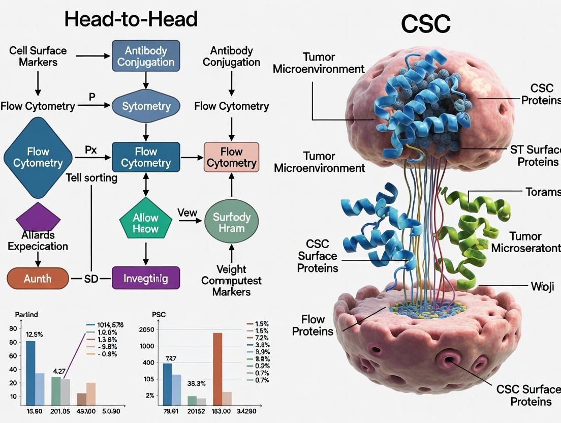
This comprehensive review provides researchers, scientists, and drug development professionals with a critical head-to-head comparison of leading Cancer Stem Cell (CSC) isolation techniques.
Cancer Stem Cell Isolation Showdown: A 2024 Comparative Analysis of FACS, MACS, Spheroid Culture & ALDEFLUOR Techniques
Abstract
This comprehensive review provides researchers, scientists, and drug development professionals with a critical head-to-head comparison of leading Cancer Stem Cell (CSC) isolation techniques. We explore the foundational principles of CSCs and their significance in oncology, before delving into detailed methodologies for Fluorescence-Activated Cell Sorting (FACS), Magnetic-Activated Cell Sorting (MACS), spheroid formation assays, and ALDEFLUOR assays. The article systematically addresses common troubleshooting pitfalls and optimization strategies for each protocol, culminating in a rigorous validation and comparative analysis of purity, viability, functionality, and cost-effectiveness. This guide aims to empower scientists in selecting the optimal isolation strategy for their specific research or therapeutic development goals.
Understanding the Quarry: The Biology of Cancer Stem Cells and Why Isolation Matters
Cancer stem cells (CSCs) represent a subpopulation of tumor cells with self-renewal capacity and the ability to drive tumor heterogeneity, therapy resistance, and metastasis. Their accurate isolation and characterization are foundational for targeted therapeutic development. This guide provides a head-to-head comparison of prominent CSC isolation techniques, grounded in experimental data and protocols, to inform researchers in this critical field.
Head-to-Head Comparison of Core CSC Isolation Techniques
The efficacy of CSC research is directly tied to the isolation method. The table below compares the primary techniques based on key performance metrics derived from recent studies.
Table 1: Comparative Analysis of CSC Isolation Techniques
| Technique | Principle | Key Markers (Examples) | Purity/ Yield | Functional Confirmation (Gold Standard) | Major Advantages | Major Limitations |
|---|---|---|---|---|---|---|
| Surface Marker-Based (FACS/MACS) | Antibody binding to cell surface antigens (e.g., CD44, CD133, EpCAM). | CD44+/CD24- (Breast), CD133+ (Glioblastoma, Colon), EpCAM+ (Pancreatic). | High Purity (85-99%), Variable Yield. | In vitro sphere formation; In vivo tumorigenesis in immunodeficient mice (Limiting Dilution Assay). | High specificity; compatibility with downstream molecular analysis. | Marker heterogeneity across tumors/patients; lack of universal markers. |
| Side Population (SP) Assay | Efflux of Hoechst 33342 dye via ATP-Binding Cassette (ABC) transporters (e.g., ABCG2). | Hoechstlow profile. | Low-Moderate Purity (60-80%), Low Yield. | Same as above. | Functional readout; marker-independent. | Cytotoxic dye exposure; protocol-sensitive; poor viability post-sort. |
| Aldehyde Dehydrogenase (ALDH) Activity | Enzymatic conversion of fluorogenic substrate (BAAA) to fluorescent product retained in high-ALDH cells. | ALDHhigh (measured by ALDEFLUOR assay). | Moderate Purity (70-90%), Good Yield. | Same as above. | Functional enzymatic activity; works in solid and liquid tumors. | Activity can be cell-state dependent; background signal. |
| Sphere Formation Assay (Functional Enrichment) | Anchorage-independent growth in serum-free, growth factor-supplemented media. | Not applicable (enrichment method). | Low Purity (enriched population), Good Yield. | Serial sphere passaging; in vivo tumorigenesis. | Functional selection for self-renewal; no specialized equipment needed initially. | Contains progenitor/non-CSCs; lengthy culture may alter phenotypes. |
Experimental Protocols for Validation
The gold standard for validating isolated CSCs is a combination of in vitro and in vivo functional assays.
In Vitro Sphere Formation Assay
Purpose: To assess self-renewal and anchorage-independent growth potential. Protocol:
- Seed Sorted Cells: Plate single-cell suspension from the isolated population (e.g., CD44+/CD24- vs. negative cells) into ultralow attachment multiwell plates.
- Culture Conditions: Use serum-free DMEM/F12 medium supplemented with 20 ng/mL EGF, 10 ng/mL bFGF, and B27 supplement.
- Incubation: Maintain at 37°C, 5% CO2 for 7-14 days.
- Quantification: Count spheres with diameter >50 µm under a phase-contrast microscope. Calculate sphere-forming efficiency: (Number of spheres / Number of cells seeded) x 100%.
In Vivo Limiting Dilution Tumorigenesis Assay
Purpose: To quantify tumor-initiating cell frequency. Protocol:
- Cell Preparation: Prepare serial dilutions of the putative CSC population (e.g., 10, 102, 103, 104 cells) and the non-CSC control.
- Transplantation: Mix cells with Matrigel (1:1 ratio) and inject subcutaneously or orthotopically into NOD/SCID or NSG mice (n=6-8 per group).
- Monitoring: Palpate weekly for tumor formation over 16-24 weeks.
- Analysis: Calculate tumor-initiating cell frequency using extreme limiting dilution analysis (ELDA) software, which compares the positive (tumor formation) outcomes across dilutions.
Visualizing Key Concepts
Diagram 1: The CSC Isolation and Validation Workflow
Diagram 2: Core Stemness Signaling Pathways in CSCs
The Scientist's Toolkit: Research Reagent Solutions
Table 2: Essential Reagents for CSC Isolation and Functional Analysis
| Reagent/Material | Supplier Examples | Function in CSC Research |
|---|---|---|
| Fluorochrome-conjugated Anti-human CD44, CD133, EpCAM antibodies | BioLegend, BD Biosciences, Miltenyi Biotec | Labeling of putative CSC surface markers for Fluorescence-Activated Cell Sorting (FACS). |
| ALDEFLUOR Kit | StemCell Technologies | Detection of intracellular ALDH enzymatic activity to identify ALDHhigh CSCs. |
| Hoechst 33342 | Thermo Fisher Scientific, Sigma-Aldrich | DNA-binding dye used in the Side Population assay; effluxed by ABC transporters. |
| Ultra-Low Attachment Plates | Corning, Greiner Bio-One | Prevents cell adhesion, enabling 3D sphere formation from single CSCs. |
| Recombinant Human EGF & bFGF | PeproTech, R&D Systems | Essential growth factors for stem cell maintenance in serum-free sphere culture media. |
| Matrigel Matrix | Corning | Basement membrane extract used for orthotopic injections and 3D culture models. |
| NOD/SCID or NSG Mice | The Jackson Laboratory | Immunodeficient mouse strains for in vivo tumorigenicity and limiting dilution assays. |
| ELDA Software | (Bioinformatics Tool) | Statistical analysis of limiting dilution assay data to calculate CSC frequency. |
Within the broader thesis on the head-to-head comparison of cancer stem cell (CSC) isolation techniques, the selection of surface markers is a critical first step. Immunophenotypic identification via flow cytometry or magnetic-activated cell sorting (MACS) remains a cornerstone methodology. This guide provides a comparative analysis of three pivotal surface markers—CD44, CD133 (Prominin-1), and EpCAM (Epithelial Cell Adhesion Molecule)—as tools for CSC identification, supported by experimental data.
Head-to-Head Comparison of Marker Performance
The utility of a CSC marker is evaluated based on its specificity, prevalence across cancer types, functional correlation with stemness, and technical reliability. The following table summarizes a comparative analysis derived from recent studies.
Table 1: Comparative Performance of Key CSC Surface Markers
| Marker | Common Cancers | Reported Frequency in Tumors | Correlation with Functional Stemness Assays | Key Limitations |
|---|---|---|---|---|
| CD44 | Breast, Prostate, Colorectal, HNSCC | 1-30% (highly variable) | Strong: Sphere formation, in vivo tumorigenicity, chemo-resistance. | Ubiquitous expression in normal tissues; CD44 isoforms (e.g., CD44v6) may be more specific. |
| CD133 | Glioblastoma, Colon, Liver, Pancreatic | 0.5-10% | Strong: Sphere formation, asymmetric division, tumor initiation in vivo. | Expression can be lost upon differentiation; controversial in some cancers (e.g., colon). |
| EpCAM | Colorectal, Pancreatic, Breast, Ovarian | 10-70% (often high) | Moderate: Often used in combination (e.g., EpCAM+CD44+). High tumorigenic potential in vivo. | High expression on bulk tumor cells; co-marking with additional CSC markers is essential. |
Table 2: Experimental Tumorigenicity Data from NOD/SCID Mouse Models
| Cancer Type | Sorted Population | Minimum Tumorigenic Cell Number | Control Population (Marker-Negative) |
|---|---|---|---|
| Breast Cancer | CD44+CD24-/low | 100 - 1,000 cells | >50,000 cells failed to form tumors |
| Glioblastoma | CD133+ | 100 - 10,000 cells | 100,000 CD133- cells formed no/smaller tumors |
| Colon Cancer | EpCAMhighCD44+ | 500 cells | 10,000 EpCAMlowCD44- cells formed no tumors |
Experimental Protocols for Marker-Based CSC Identification
Protocol 1: Flow Cytometry for CSC Enumeration and Sorting
This protocol is standard for quantifying and isolating marker-positive populations.
- Tissue Dissociation: Generate a single-cell suspension from tumor tissue using enzymatic digestion (e.g., collagenase/hyaluronidase mix).
- Staining: Aliquot cells. Use Fc receptor block. Stain with fluorescent antibody conjugates:
- Test Sample: Anti-CD44-APC, Anti-CD133-PE, Anti-EpCAM-FITC.
- Controls: Isotype-matched antibodies, single-color compensation controls, unstained cells.
- Incubation: 30 minutes on ice, protected from light.
- Wash & Resuspend: Wash twice with FACS buffer (PBS + 2% FBS). Resuspend in buffer with viability dye (e.g., DAPI).
- Analysis/Sorting: Use a flow cytometer/cell sorter. Gate on single, live cells. Analyze co-expression patterns (e.g., EpCAM+CD44+CD133+). Sort desired populations into collection medium for functional assays.
Protocol 2: Functional Validation via Sphere Formation Assay
This in vitro assay tests the self-renewal capability of sorted populations.
- Plating: Seed sorted cells (e.g., CD44+ vs. CD44-) into ultra-low attachment plates at clonal density (500-1000 cells/mL).
- Culture Medium: Use serum-free DMEM/F12 supplemented with B27, EGF (20 ng/mL), and bFGF (20 ng/mL).
- Incubation: Culture for 7-14 days at 37°C, 5% CO2. Add fresh growth factors every 3-4 days.
- Quantification: Count spheres with diameter >50 µm under a microscope. Calculate sphere-forming efficiency: (Number of spheres / Number of cells seeded) x 100%.
Visualizing Marker-Based CSC Isolation Workflow
Title: Workflow for CSC Identification via Surface Markers
The Scientist's Toolkit: Key Research Reagent Solutions
Table 3: Essential Reagents for CSC Marker-Based Studies
| Reagent / Material | Function & Role in Experiment | Example Vendor/Clone |
|---|---|---|
| Fluorochrome-Conjugated Antibodies | Direct staining of surface markers for detection by flow cytometry. Critical for panel design. | BioLegend: Anti-human CD44 (IM7), CD133 (W6B3C1), EpCAM (9C4) |
| MACS MicroBeads & Columns | Magnetic separation of marker-positive cells for enrichment prior to sorting or culture. | Miltenyi Biotec: CD133 MicroBead Kit, CD44 MicroBeads |
| Ultra-Low Attachment Plates | Prevent cell adhesion, enabling 3D sphere growth for self-renewal assays. | Corning Costar Spheroid Plates |
| Defined Serum-Free Medium | Supports stem cell growth while inhibiting differentiation in sphere assays. | StemCell Technologies: MammoCult; Gibco: StemPro |
| Recombinant Growth Factors (EGF, bFGF) | Essential supplements for sphere formation and CSC maintenance in culture. | PeproTech, R&D Systems |
| Viability Stains (DAPI, PI) | Distinguish live from dead cells during flow analysis to ensure sorting purity. | Thermo Fisher Scientific |
| Matrigel Basement Membrane Matrix | Used for in vivo tumorigenicity assays or 3D invasion assays. | Corning Matrigel |
This comparison guide is framed within the broader thesis of head-to-head comparisons of Cancer Stem Cell (CSC) isolation techniques. The functional validation of isolated cells—specifically their tumorigenicity, self-renewal capacity, and therapy resistance—is the ultimate benchmark for technique efficacy. Below, we objectively compare the performance of cells isolated via different methods in key functional assays, supported by experimental data.
Table 1: Comparison of Functional Properties from Different CSC Isolation Techniques
| Isolation Technique | Tumorigenicity (Min. Cells for Tumor Formation) | Self-Renewal (Sphere Formation Efficiency %) | Therapy Resistance (e.g., % Viability Post-Chemo) | Key Supporting Experimental Data (Model) |
|---|---|---|---|---|
| Cell Surface Marker (e.g., CD44+/CD24-) | 500-5,000 cells | 0.5% - 3.0% | 60-85% viability post-Paclitaxel | Al-Hajj et al., 2003 (Breast Cancer); Prince et al., 2007 (HNSCC) |
| Side Population (SP) via Hoechst 33342 Efflux | 1,000-10,000 cells | 0.1% - 1.5% | 70-90% viability post-Doxorubicin | Goodell et al., 1996; Patrawala et al., 2005 (Glioma) |
| Aldehyde Dehydrogenase (ALDH) Activity | 200-2,000 cells | 1.0% - 5.0% | 65-88% viability post-Cisplatin | Ginestier et al., 2007 (Breast Cancer); Clay et al., 2010 (AML) |
| Serum-Free Sphere Formation | 1,000-50,000 cells (Heterogeneous) | 0.01% - 0.5% (Enriched but not pure) | 40-75% viability (Variable) | Ponti et al., 2005 (Breast Cancer); Dontu et al., 2003 |
| Combination (e.g., CD44+/ALDH+) | <100-500 cells | 3.0% - 10.0% | >90% viability post-combination therapy | Liu et al., 2014 (Pancreatic); Cioffi et al., 2015 (Lung) |
Key Insight: Combined marker approaches consistently yield cells with superior functional potency (lower tumorigenic dose, higher self-renewal, and greater resistance) compared to any single-parameter technique.
Detailed Experimental Protocols for Key Assays
1. In Vivo Limiting Dilution Tumorigenicity Assay
- Purpose: Quantitatively assess the tumor-initiating cell frequency.
- Methodology: Isolated cell populations are serially diluted (e.g., 10,000, 1,000, 100, 10 cells) and transplanted into immunocompromised mice (NOD/SCID or NSG). Multiple injections per cell dose are performed (e.g., n=8). Tumors are monitored for 12-24 weeks.
- Data Analysis: Tumor incidence is recorded. The frequency of tumor-initiating cells and statistical significance between groups is calculated using extreme limiting dilution analysis (ELDA) software.
2. In Vitro Sphere Formation Assay (Self-Renewal)
- Purpose: Measure clonogenic potential and self-renewal capacity under non-adherent conditions.
- Methodology: Single cells are plated at low density (1-10 cells/μL) in ultralow attachment plates. Serum-free medium is supplemented with B27, EGF, and bFGF. Cultures are maintained for 7-14 days.
- Data Analysis: Spheres >50-100 μm in diameter are counted. Sphere Formation Efficiency (SFE) = (number of spheres formed / number of cells seeded) * 100%.
3. Therapy Resistance Assay (e.g., Chemotherapy)
- Purpose: Evaluate the survival advantage of putative CSCs.
- Methodology: Isolated cells and bulk tumor cells are treated with clinically relevant doses of chemotherapeutics (e.g., 5-100 μM Cisplatin, 0.1-1 μM Paclitaxel) for 48-72 hours. A vehicle control is essential.
- Data Analysis: Cell viability is quantified using ATP-based (CellTiter-Glo) or resazurin (AlamarBlue) assays. % Viability = (Signal of treated sample / Signal of control sample) * 100.
Signaling Pathways Governing CSC Functional Properties
Workflow for Functional Validation of Isolated CSCs
The Scientist's Toolkit: Key Research Reagent Solutions
| Item | Function in CSC Research |
|---|---|
| UltraLow Attachment (ULA) Plates | Prevents cell adhesion, enabling 3D sphere growth in serum-free conditions for self-renewal assays. |
| Defined Serum-Free Media (e.g., DMEM/F12 + supplements) | Supports CSC growth without inducing differentiation; typically includes B27, N2, EGF, bFGF. |
| Recombinant Human EGF & bFGF | Essential growth factors for stimulating proliferation and maintaining stemness in sphere cultures. |
| Hoechst 33342 Dye | DNA-binding dye used in Side Population assays; its efflux via ABC transporters (e.g., ABCG2) identifies putative CSCs. |
| ALDEFLUOR Assay Kit | Provides a fluorescent substrate for ALDH1 enzymatic activity, enabling FACS-based isolation of ALDH-high CSCs. |
| Fluorochrome-Conjugated Antibodies (e.g., anti-CD44-APC) | Key reagents for fluorescence-activated cell sorting (FACS) to isolate marker-defined CSC populations. |
| CellTiter-Glo 3D Viability Assay | Luminescent ATP assay optimized for 3D cultures (spheroids) to quantify viability post-therapy. |
| Matrigel Basement Membrane Matrix | Used for in vivo injections and some 3D in vitro assays to provide a physiological substrate for tumor growth. |
| ELDA Software (Extreme Limiting Dilution Analysis) | Open-source statistical tool for calculating tumor-initiating cell frequency from limiting dilution data. |
The Critical Role of CSCs in Metastasis, Recurrence, and Drug Development
Cancer Stem Cells (CSCs) are a subpopulation within tumors with self-renewal, differentiation, and tumor-initiating capacities. They are considered the primary drivers of metastasis, therapeutic resistance, and disease recurrence. The accurate identification and isolation of CSCs are therefore foundational to understanding their biology and developing effective therapies. This guide provides a head-to-head comparison of the primary CSC isolation techniques, a critical first step in any investigation into their role in metastasis and drug development.
Head-to-Head Comparison of Core CSC Isolation Techniques
The choice of isolation method directly impacts the purity, viability, and functional characteristics of the isolated CSCs, influencing all downstream experimental data. The following table compares the three most prevalent methodologies.
Table 1: Comparison of Primary CSC Isolation & Characterization Techniques
| Technique | Principle | Key Performance Metrics | Primary Advantages | Primary Limitations | Typical Experimental Readouts |
|---|---|---|---|---|---|
| Fluorescence-Activated Cell Sorting (FACS) | Isolation based on cell surface markers (e.g., CD44+/CD24- for breast CSCs) or dye efflux (Side Population) via fluorescent labels. | Purity: >95%. Viability: 80-90%. Yield: Moderate (cell number limited by starting sample). Throughput: Medium. | High purity and specificity. Multi-parameter sorting possible. Quantitative. | Requires a priori knowledge of markers. Marker expression can be context-dependent. High cost of equipment. | In vivo limiting dilution tumorigenesis assays; Sphere formation efficiency; Chemoresistance assays. |
| Magnetic-Activated Cell Sorting (MACS) | Isolation using magnetic beads conjugated to antibodies against specific surface markers. | Purity: 70-90%. Viability: >85%. Yield: High. Throughput: High. | Simpler, faster, and more cost-effective than FACS. Suitable for large cell numbers. No specialized flow cytometry skill required. | Lower purity than FACS. Typically limited to one or two markers simultaneously. Positive selection may activate signaling. | Bulk functional assays (e.g., colony formation, drug sensitivity); RNA/protein extraction for omics analyses. |
| Functional Assay-Based (Serum-Free Sphere Formation) | Isolation based on biological capacity to proliferate under non-adherent, serum-free conditions with growth factors. | Purity: Enriched but heterogeneous. Viability: Variable. Yield: Low (enriched population). Throughput: Low. | Marker-independent. Selects for self-renewal capacity in vitro. Can reveal functional CSCs without known markers. | Labor-intensive and slow. Microenvironment is artificial. May select for adapted cells, not necessarily in vivo CSCs. | Serial sphere-forming assays (self-renewal quantification); Differentiation assays; Transcriptomic profiling of sphere-derived cells. |
Experimental Protocols for Key CSC Validation Assays
Following isolation, CSC populations must be functionally validated. Below are standardized protocols for two gold-standard assays.
Protocol 1: In Vivo Limiting Dilution Tumorigenesis Assay Purpose: To determine the frequency of tumor-initiating cells (TIC) within the isolated population.
- Cell Preparation: Serially dilute the isolated CSC population (e.g., 10,000, 1,000, 100, 10 cells) and a control non-CSC population.
- Transplantation: Mix each cell dilution with 50% Matrigel in PBS. Inject subcutaneously or into the orthotopic site of immunodeficient mice (NOD/SCID or NSG) (n=5-10 mice per dilution).
- Monitoring: Palpate weekly for tumor formation over 4-6 months.
- Analysis: Calculate TIC frequency using extreme limiting dilution analysis (ELDA) software, which compares the tumor-initiating capacity of CSC vs. non-CSC fractions.
Protocol 2: Serial Sphere-Formation Assay Purpose: To assess in vitro self-renewal capacity.
- Primary Sphere Formation: Plate single cells from isolated populations at low density (e.g., 1,000 cells/mL) in ultra-low attachment plates using serum-free medium (DMEM/F12) supplemented with B27, EGF (20 ng/mL), and bFGF (10 ng/mL).
- Incubation: Culture for 7-14 days without disturbance.
- Quantification: Count spheres >50 µm under a microscope. Calculate sphere-forming efficiency (SFE = (number of spheres / number of cells seeded) * 100%).
- Serial Passaging: Collect primary spheres, dissociate into single cells, and re-plate at the same density for secondary and tertiary sphere formation. Sustained SFE across passages indicates self-renewal.
Visualization of CSC Pathways and Workflows
CSC Isolation & Research Workflow
CSC Properties & Niche in Metastasis/Recurrence
The Scientist's Toolkit: Key Reagent Solutions for CSC Research
Table 2: Essential Research Reagents for CSC Isolation and Culture
| Reagent/Material | Function in CSC Research | Example Product/Catalog |
|---|---|---|
| Ultra-Low Attachment Plates | Prevents cell adhesion, enabling 3D sphere growth under serum-free conditions to assess self-renewal. | Corning Costar Spheroid Plates |
| Recombinant EGF & bFGF | Critical growth factors in serum-free media that maintain CSC stemness and proliferation in suspension culture. | PeproTech human EGF & bFGF |
| B-27 Supplement | A defined serum-free supplement that supports neural and stem cell survival, commonly used in CSC sphere media. | Gibco B-27 Supplement |
| Matrigel / Cultrex BME | Basement membrane extract used for 3D organoid culture, orthotopic injections, and studying invasion. | Corning Matrigel, RGF BME |
| Fluorophore-Conjugated Antibodies | Antibodies against CSC surface markers (e.g., CD44-APC, CD24-FITC, EpCAM-PE) for FACS identification and sorting. | BioLegend, BD Biosciences |
| MACS MicroBeads & Columns | Magnetic beads and separation columns for label-based, high-throughput positive or negative selection of CSCs. | Miltenyi Biotec MS Columns |
| Hoechst 33342 / Verapamil | Dye and inhibitor used in the Side Population assay to identify cells with high ABC transporter activity via flow cytometry. | Thermo Fisher Scientific |
| In Vivo Grade NSG Mice | The gold-standard immunodeficient mouse model for in vivo tumorigenicity assays due to superior engraftment rates. | The Jackson Laboratory (005557) |
The Isolation Toolkit: Step-by-Step Protocols for Four Key Techniques
Within head-to-head comparisons of Cancer Stem Cell (CSC) isolation techniques, Fluorescence-Activated Cell Sorting (FACS) remains a benchmark for high-precision, multiparameter cell separation. This guide objectively compares FACS-based CSC isolation against prominent alternative techniques, focusing on performance metrics derived from published experimental data.
Performance Comparison of CSC Isolation Techniques
The following table summarizes key performance indicators for FACS and other common techniques, based on recent comparative studies.
Table 1: Comparison of CSC Isolation Techniques
| Technique | Purity (%) | Viability (%) | Throughput (cells/sec) | Multi-Marker Capability | Key Limitations |
|---|---|---|---|---|---|
| FACS | 95 - 99+ | 80 - 95 | 10,000 - 70,000 | High (8+ colors) | High equipment cost, potential shear stress. |
| Magnetic Activated Cell Sorting (MACS) | 70 - 90 | 85 - 98 | >10^7 (bulk) | Low (1-2 markers) | Lower purity, limited multiplexing. |
| Side Population (SP) Assay (Hoechst Dye) | 0.1 - 5 (of total) | 60 - 80 | N/A (analysis only) | Very Low | Low yield, dye toxicity, functional proxy only. |
| Condition Culture (Sphere Formation) | N/A (functional) | Variable | N/A | None | Slow, yields heterogeneous spheres, not direct isolation. |
Experimental Data Supporting FACS Performance
Table 2: Representative Experimental Data from CSC Sorting Studies
| Study (Model) | Target (Marker) | Sorted Purity (FACS) | Post-Sort Viability | In Vivo Tumorigenicity (Sorted vs. Unsorted) |
|---|---|---|---|---|
| Breast Cancer (PDX) | CD44+/CD24- | 98.5% | 92% | 10^3 cells formed tumors vs. 10^6 unsorted |
| Glioblastoma (Primary) | CD133+ | 97.2% | 87% | 5x higher tumor initiation frequency |
| Colon Cancer (Cell Line) | LGR5+ | 99.1% | 89% | 100% incidence at 500 cells; unsorted: 0% at 5000 cells |
Detailed Methodologies for Key Experiments
Protocol 1: High-Purity CSC Sorting for Glioblastoma
Objective: Isolate CD133+ CSCs from primary glioblastoma specimens. Workflow:
- Tissue Dissociation: Mechanically mince tumor, digest with collagenase IV (1 mg/mL) and DNase I (100 µg/mL) for 45min at 37°C. Pass through 70µm strainer.
- Staining: Count cells. Fc block incubation (10min). Stain with anti-human CD133/1 (AC133)-PE antibody (1:50 dilution in PBS/2% FBS) for 30min on ice, in the dark.
- Setup & Sorting: Use a sorter equipped with 488nm (for PE) and 561nm lasers. Create FSC-A/SSC-A gate to exclude debris. Perform doublet discrimination (FSC-H vs FSC-A). Apply a viability dye (e.g., DAPI) to exclude dead cells. Set sort gate on viable, single cells for the top 2-5% of PE fluorescence. Use "Purity" sort mode into collection tubes containing complete culture medium.
- Post-Sort Analysis: Re-analyze an aliquot of sorted cells to confirm purity.
Diagram Title: FACS Workflow for Glioblastoma CSC Isolation
Protocol 2: Comparative MACS vs. FACS Sorting for Breast CSCs
Objective: Compare purity and tumorigenicity of CD44+/CD24- cells isolated by MACS vs. FACS. Workflow:
- Cell Source: Use established breast cancer cell line (e.g., MDA-MB-231).
- MACS Protocol: Label cells with anti-CD44 microbeads. Pass through LS column placed in a magnetic field. Collect flow-through (CD44-). Elute positively selected cells after column removal.
- FACS Protocol: Stain cells with anti-CD44-FITC and anti-CD24-PE. Include viability dye. Set sort gates for viable, CD44+/CD24- population.
- Analysis: Assess purity of both outputs via re-analysis on flow cytometer. Perform limiting dilution tumorigenicity assays in immunocompromised mice with equal cell numbers from each method.
The Scientist's Toolkit: Research Reagent Solutions
Table 3: Essential Materials for FACS-Based CSC Isolation
| Item | Function & Importance |
|---|---|
| High-Speed Cell Sorter | Enables high-precision, multi-parameter sorting based on fluorescence and scatter. Critical for rare population isolation. |
| Validated Conjugated Antibodies | Fluorophore-conjugated antibodies against CSC surface markers (e.g., CD133, CD44, EpCAM). Clone and titer validation is essential. |
| Viability Dye (e.g., DAPI, PI, Live/Dead Fixable) | Distinguishes live from dead cells; crucial for excluding non-viable cells from the sort gate. |
| Cell Strainers (40µm, 70µm) | Generates single-cell suspensions by removing clumps, essential for preventing nozzle clogging. |
| Collection Media with High Serum/Additives | Preserves viability of sorted cells; often contains 50% serum or specific growth factors to mitigate shear stress. |
| DNase I & Collagenase/Hyaluronidase | Enzymatic digestion cocktails for effective dissociation of solid tumors into single cells. |
| Fc Receptor Blocking Solution | Reduces nonspecific antibody binding, improving staining specificity and sort purity. |
| Sheath Fluid & Sterilization Filters | Particle-free, isotonic fluid for stream stability. 0.22µm filters maintain sterility for aseptic sorting. |
Diagram Title: Criteria for Evaluating Isolated CSCs
Within the rigorous framework of head-to-head comparisons of Cancer Stem Cell (CSC) isolation techniques, Magnetic-Activated Cell Sorting (MACS) stands out for its speed and capacity for processing large cell numbers. This guide objectively compares its performance against Fluorescence-Activated Cell Sorting (FACS) and functional assays like the sphere formation assay.
Performance Comparison with Key Alternatives
Table 1: Comparative Analysis of MACS, FACS, and Functional Assays for CSC Isolation
| Parameter | MACS | FACS (Comparative Alternative) | Sphere Assay (Comparative Alternative) |
|---|---|---|---|
| Primary Principle | Magnetic labeling and column-based separation. | Fluorescent labeling and droplet-based electrostatic deflection. | Functional growth in non-adherent, serum-free conditions. |
| Throughput & Speed | Very High. Can process >10^9 cells in <2 hours. | Moderate to Low. Typically processes 10^4-10^7 cells in several hours. | Very Low. Requires 1-2 weeks of culture. |
| Purity | High (typically 70-95%), but lower than FACS due to less resolution. | Very High (typically 95-99%). Multi-parameter gating improves specificity. | Functional Enrichment. Not a direct purity measure. |
| Cell Viability | Excellent (>90%). Gentle magnetic forces. | Good (70-90%). Subject to shear stress and electrostatic charges. | Assesses proliferative potential of surviving cells. |
| Cost per Sample | Low to Moderate. Reusable columns, single antibody conjugate. | High. Instrument time, specialized personnel, multiple fluorescent antibodies. | Low. Primarily requires specialized media. |
| Complexity/Setup | Low. Benchtop protocol, minimal training. | High. Requires sophisticated instrument and expert operation. | Moderate. Aseptic cell culture techniques required. |
| Best Application | Rapid pre-enrichment for downstream assays, bulk RNA/protein analysis. | High-purity isolation for single-cell omics or functional sub-population studies. | Assessment of self-renewal potential, not initial isolation. |
Supporting Experimental Data
A seminal comparative study (Civenni et al., 2013) isolated CD133+ CSCs from human melanoma cell lines using both MACS and FACS. Subsequent in vitro limiting dilution transplantation into immunodeficient mice provided functional validation.
Table 2: Experimental Outcomes from Comparative Isolation Study
| Isolation Method | Reported Purity (CD133+) | Cell Viability Post-Sort | Tumor-Initiating Cell Frequency (Limiting Dilution) | Key Experimental Finding |
|---|---|---|---|---|
| MACS | 89.2% ± 4.1% | 94.5% ± 2.3% | 1 in 3,240 | Efficiently enriched for tumorigenic cells; suitable for bulk molecular analysis. |
| FACS | 98.7% ± 0.8% | 85.1% ± 3.7% | 1 in 2,150 | Achieved highest purity; marginally better functional frequency but at lower yield/viability. |
Detailed Experimental Protocols
Protocol 1: MACS for CSC Enrichment (e.g., CD133+ Cells)
- Single-Cell Suspension: Dissociate tumor tissue or cultured cells into a single-cell suspension using enzymatic (e.g., collagenase/hyaluronidase) and mechanical dissociation. Filter through a 40-70µm strainer.
- Fc Receptor Blocking: Incubate cells with an FcR Blocking Reagent (human or mouse) for 10 minutes at 4°C to reduce non-specific antibody binding.
- Magnetic Labeling: Incubate cells with microbead-conjugated anti-CD133 antibody (or other CSC marker) for 15-30 minutes at 4°C. Wash to remove unbound antibody.
- Magnetic Separation: Pass cell suspension through a pre-washed LS Column placed in a strong magnetic field (MACS Separator). Labeled cells are retained. Remove column from magnet and flush out the positively selected fraction.
- Analysis: Assess purity via flow cytometry using a fluorescent anti-CD133 antibody distinct from the bead-conjugate.
Protocol 2: Comparative Sphere Formation Assay (Functional Validation)
- Seeding: Seed serially diluted single cells (e.g., from MACS-sorted or unsorted populations) into ultra-low attachment multi-well plates.
- Culture: Use serum-free stem cell medium (e.g., DMEM/F12 supplemented with B27, EGF, and bFGF). Do not disturb plates for 5-7 days.
- Quantification: Count spheres >50µm diameter under a microscope. Calculate sphere-forming efficiency (number of spheres/number of cells seeded x 100%).
Visualization
Title: MACS Workflow for CSC Isolation
Title: Logical Comparison of CSC Isolation Techniques
The Scientist's Toolkit: Key Research Reagent Solutions
Table 3: Essential Materials for MACS-based CSC Isolation
| Reagent/Material | Function |
|---|---|
| MACS Microbead-Conjugated Antibody | Target-specific (e.g., anti-CD133, anti-CD44). Covalently linked to superparamagnetic nanoparticles for cell labeling. |
| MACS Separation Columns & Magnet | LS or MS Columns with appropriate separator. Creates high-gradient magnetic field to retain labeled cells. |
| FcR Blocking Reagent | Blocks non-specific, Fc-mediated antibody binding to immune cells, reducing background. |
| Cell Dissociation Enzymes | Enzymatic cocktails (e.g., Tumor Dissociation Kits) for gentle and effective tissue disaggregation. |
| Dead Cell Removal Kit | Optional pre-step to remove apoptotic cells, improving sorting efficiency and downstream data quality. |
| Serum-Free Stem Cell Media | For functional validation or expansion of isolated CSCs (e.g., in sphere formation assays). |
Within the framework of a thesis dedicated to the head-to-head comparison of Cancer Stem Cell (CSC) isolation techniques, the Spheroid Formation Assay (SFA) stands out as a functional enrichment method. Unlike antibody-based sorting (e.g., FACS for CD44+/CD24-), the SFA selectively expands the CSC subpopulation based on its intrinsic capacity for anchorage-independent growth and self-renewal under serum-free, non-adherent conditions. This guide objectively compares its performance against other common techniques.
Comparison of CSC Enrichment Techniques
Table 1: Head-to-Head Comparison of Key CSC Isolation/Enrichment Techniques
| Technique | Principle | Key Advantage | Key Limitation | Reported Enrichment Fold (Cell Line Example) | Functional Validation Required? |
|---|---|---|---|---|---|
| Spheroid Formation Assay (SFA) | Functional enrichment via clonal growth in non-adherent, serum-free conditions. | Enriches for bona fide functionally defined CSCs; no prior marker knowledge needed. | Time-consuming (7-14 days); may miss CSCs reliant on niche signals. | 10-50 fold (MCF-7, MDA-MB-231) | Inherent to the technique. |
| Fluorescence-Activated Cell Sorting (FACS) | Physical separation based on surface marker expression (e.g., CD44+/CD24-). | High purity and speed; viable cells for immediate use. | Marker variability across tumors/patients; may sort non-CSCs. | 5-20 fold (Primary breast CA) | Yes (e.g., in vivo tumorigenesis). |
| Magnetic-Activated Cell Sorting (MACS) | Physical separation using magnetic beads against surface markers. | Simpler and cheaper than FACS; good for large cell numbers. | Lower purity than FACS; same marker dependency issues. | 3-15 fold (Glioblastoma) | Yes. |
| Side Population (SP) Assay | Functional separation based on efflux of Hoechst 33342 dye via ABC transporters. | Identifies a primitive, transporter-active subset. | Cytotoxic dye exposure; complex flow cytometry setup. | 5-30 fold (Various carcinomas) | Yes. |
Table 2: Comparative Experimental Outcomes from Cited Studies
| Study Focus | SFA Result | FACS/MACS Result (Comparator) | Key Takeaway |
|---|---|---|---|
| Tumorigenic Potential | Primary spheroid-derived cells showed higher tumor incidence at lower cell numbers (e.g., 10^3 cells) in mice. | Marker-sorted cells (CD133+) showed variable tumorigenicity. | SFA-enriched populations consistently demonstrate high in vivo stemness. |
| Chemoresistance | SFA-enriched cells from CRC cell lines exhibited 3-5x higher IC50 to 5-FU vs. adherent cells. | CD44+ sorted cells showed 2-4x higher resistance. | Both methods enrich for resistant cells, but SFA may capture a broader resistant subset. |
| Stemness Gene Expression | Upregulation of OCT4, NANOG, SOX2 (5-20x increase vs. bulk). | Marker-sorted cells showed upregulation (2-10x increase). | SFA yields a population with a pronounced stemness signature. |
Detailed Experimental Protocols
Protocol 1: Standard Spheroid Formation Assay for CSC Enrichment
- Preparation: Trypsinize monolayer cells from a cancer cell line or dissociated primary tumor. Generate a single-cell suspension.
- Culture Conditions: Resuspend cells in serum-free DMEM/F12 medium supplemented with key growth factors: 20 ng/mL recombinant human EGF, 10 ng/mL recombinant human bFGF, and 1X B27 Supplement.
- Seeding: Plate cells in ultra-low attachment (ULA) multi-well plates at a clonal density (e.g., 1,000 – 10,000 cells/mL). A density of 500-1000 cells/well in a 96-well ULA plate is common for quantifying sphere-forming efficiency (SFE).
- Incubation & Monitoring: Culture cells at 37°C, 5% CO2 for 7-14 days. Do not disturb. Refresh half of the medium with fresh growth factors every 3-4 days.
- Analysis: After 7 days, count spheroids >50 µm in diameter. Calculate SFE = (Number of spheres / Number of cells seeded) * 100%. Spheroids can be passaged for serial expansion or dissociated for downstream assays.
Protocol 2: Validation via In Vivo Tumorigenesis (Limiting Dilution Assay)
- Cell Preparation: Generate single-cell suspensions from (a) primary spheroids (SFA-enriched) and (b) parental adherent/bulk cells.
- Transplantation: Serially dilute cells (e.g., 10^5, 10^4, 10^3, 10^2) and mix with Matrigel. Inject subcutaneously or orthotopically into immunodeficient mice (NOD/SCID or NSG). Use 5-8 mice per cell dose.
- Monitoring: Palpate weekly for tumor formation over 8-16 weeks. Record tumor incidence and latency.
- Analysis: Use the Extreme Limiting Dilution Analysis (ELDA) software to calculate the frequency of tumor-initiating cells (TIC) and compare between SFA-enriched and control populations.
Signaling Pathways in Spheroid Formation & Maintenance
Diagram Title: Core Signaling Pathways in CSC Spheroid Formation
Experimental Workflow for Comparative Analysis
Diagram Title: Workflow for Comparing SFA vs. FACS CSC Enrichment
The Scientist's Toolkit: Key Research Reagent Solutions
Table 3: Essential Materials for the Spheroid Formation Assay
| Reagent/Material | Function | Example Product/Catalog |
|---|---|---|
| Ultra-Low Attachment (ULA) Plates | Prevents cell adhesion, forcing anchorage-independent growth as spheroids. | Corning Costar Spheroid Microplates. |
| Serum-Free Basal Medium (DMEM/F12) | Base nutrient medium that supports growth without serum-induced differentiation. | Gibco DMEM/F-12, GlutaMAX. |
| Recombinant Human EGF | Essential growth factor promoting CSC proliferation and survival in serum-free conditions. | PeproTech AF-100-15. |
| Recombinant Human bFGF (FGF-2) | Critical growth factor working synergistically with EGF to maintain stemness. | PeproTech 100-18B. |
| B-27 Supplement (Serum-Free) | Defined formulation of hormones, vitamins, and proteins replacing serum. | Gibco 17504044. |
| Accutase Solution | Enzymatic dissociation reagent for gentle passaging of spheroids into single cells. | Sigma-Aldrich A6964. |
| Matrigel Basement Membrane Matrix | In vivo: Provides structural support for tumor cell engraftment in limiting dilution assays. | Corning 354234. |
Within the critical research field of head-to-head comparison of Cancer Stem Cell (CSC) isolation techniques, the identification and isolation of viable CSCs rely on robust functional assays. Aldehyde dehydrogenase (ALDH) enzymatic activity is a key functional marker for CSCs across numerous malignancies. This guide provides an objective, data-driven comparison of the dominant technique for detecting this activity—the ALDEFLUOR assay—against other common methodological alternatives, framing their performance within the larger goal of reliable CSC isolation.
Comparative Analysis of ALDH Activity Detection Methods
Table 1: Head-to-Head Comparison of Key Techniques
| Feature | ALDEFLUOR Assay (Flow Cytometry) | ALDH1 Immunohistochemistry (IHC) | qRT-PCR for ALDH Isoforms | Enzymatic Spectrophotometric Assay |
|---|---|---|---|---|
| Primary Output | Functional enzyme activity in single live cells. | Protein localization & expression in fixed tissue. | mRNA expression level in cell populations. | Bulk enzymatic activity in lysates. |
| Quantification | Semi-quantitative (fluorescence intensity). | Semi-quantitative (staining score). | Quantitative (relative fold-change). | Quantitative (kinetic rate). |
| Cell Viability | Preserved (for sorting). | Not applicable (fixed cells). | Preserved (lysis required). | Not preserved (cell lysis). |
| Throughput | Medium (flow-based). | Low (slide-based). | High (plate-based). | High (plate-based). |
| Spatial Context | No | Yes (in situ). | No | No |
| Key Advantage | Direct link between activity and cell sorting. | Histological correlation. | High sensitivity & specificity to isoforms. | Direct kinetic measurement. |
| Key Limitation | Requires flow cytometer; BODIPY dye efflux. | Does not measure functional activity. | mRNA not always correlated with activity. | No single-cell resolution. |
| Best Suited For | Functional isolation of live ALDHhigh CSCs. | Prognostic marker studies in patient biopsies. | Screening isoform-specific expression changes. | High-throughput drug screening on bulk cultures. |
Table 2: Supporting Experimental Data from Comparative Studies
| Study (Example Focus) | Key Comparative Finding | Performance Metric | Implication for CSC Isolation |
|---|---|---|---|
| Ginestier et al., 2007 (Breast Cancer) | ALDEFLUOR+ cells showed >10-fold higher tumorigenicity vs. ALDEFLUOR- in NOD/SCID mice. | Tumor-initiating cell frequency | Validated functional correlation; gold standard for isolation. |
| Huang et al., 2016 (HNSCC) | Concordance between ALDEFLUOR activity and ALDH1A1 IHC was only 65-70% in primary samples. | Method agreement | Highlights discrepancy between activity and protein expression. |
| Cheung et al., 2020 (AML) | ALDEFLUOR sorting outperformed CD34+/CD38- sorting in predicting chemoresistance in vivo. | Chemoresistance enrichment | Superior functional enrichment over surface marker alone. |
| Spectrophotometric vs. FACS (Generic) | Bulk assay showed high total activity, but FACS revealed only a small, discrete active subpopulation. | Population resolution | Critical: ALDEFLUOR enables detection of rare, high-activity CSCs masked in bulk analyses. |
Experimental Protocols
Detailed Protocol: ALDEFLUOR Assay & Flow Cytometry
Principle: A fluorescent, cell-permeable substrate (BODIPY-aminoacetaldehyde) is converted by intracellular ALDH into a charged, fluorescent product (BODIPY-aminoacetate) retained in active cells. An ALDH-specific inhibitor (DEAB) serves as a negative control.
Materials: ALDEFLUOR kit (StemCell Technologies), complete growth medium, DEAB inhibitor, flow cytometry buffer (PBS + 2% FBS), 37°C water bath, flow cytometer with 488-nm laser.
Procedure:
- Cell Preparation: Harvest single-cell suspension, ensure viability >90%.
- Reaction Setup:
- Prepare 1 mL of ALDEFLUOR assay buffer per sample.
- Add 5 µL of BODIPY-aminoacetaldehyde substrate per mL to create "activated buffer."
- For the DEAB control tube, add 5 µL of DEAB inhibitor to 0.5 mL activated buffer first, then add cells.
- Staining:
- Resuspend up to 1x10⁶ cells in 0.5 mL activated buffer (test sample).
- Resuspend an equal number in 0.5 mL DEAB-treated buffer (control).
- Incubation: Incubate both tubes at 37°C for 30-60 minutes (protected from light).
- Washing & Analysis: Centrifuge cells, resuspend in ice-cold assay buffer. Keep samples on ice.
- Flow Cytometry: Analyze immediately using a 488-nm laser. Detect fluorescence with a standard FITC/GFP filter (530/30 nm). The DEAB control sets the negative gate. ALDHhigh cells are those with fluorescence significantly brighter than the DEAB control population.
Protocol: Spectrophotometric ALDH Activity Assay (Bulk)
Principle: Measures NADH production from NAD+ as aldehydes (e.g., propionaldehyde) are converted to carboxylates by ALDH in cell lysates.
Materials: Cell lysis buffer, assay buffer (pH 9.5), NAD+, propionaldehyde substrate, spectrophotometer/plate reader.
Procedure:
- Lysate Prep: Lysate 1-5x10⁶ cells in a non-denaturing buffer. Clear by centrifugation.
- Reaction Mix: In a cuvette or well, combine assay buffer, NAD+ (final ~0.5-1 mM), and cell lysate.
- Baseline: Read absorbance at 340 nm (for NADH) for 1-2 minutes.
- Initiate Reaction: Add propionaldehyde substrate (final ~5-10 mM).
- Kinetic Measurement: Record the increase in A340 over 5-10 minutes. The rate of change is proportional to ALDH activity. Normalize to total protein concentration.
Diagrams
Title: ALDEFLUOR Assay Workflow and Control
Title: Functional CSC Isolation Methods Overview
The Scientist's Toolkit: Key Research Reagent Solutions
Table 3: Essential Materials for ALDH-Based CSC Studies
| Item | Function in Experiment | Key Consideration |
|---|---|---|
| ALDEFLUOR Kit (StemCell Tech #01700) | Contains optimized BODIPY substrate and DEAB inhibitor for standardized, reproducible flow cytometry. | Gold-standard commercial kit; requires flow cytometer with 488-nm laser. |
| DEAB (Diethylaminobenzaldehyde) | Specific ALDH inhibitor used as a mandatory negative control to set baseline fluorescence. | Critical for accurate gating; controls for non-specific dye retention/efflux. |
| Propidium Iodide or DAPI | Viability dye to exclude dead cells during analysis/sorting. | Dead cells have permeable membranes and show false-positive ALDEFLUOR staining. |
| Aldehyde Dehydrogenase Activity Assay Kit (Colorimetric, e.g., Sigma #MAK082) | For bulk spectrophotometric measurement of ALDH activity in cell lysates. | Useful for high-throughput screening but lacks single-cell resolution. |
| Anti-ALDH1A1 Antibody (for IHC/IF) | Validated antibody for correlating protein expression with activity in tissue sections. | Choose clones with proven IHC performance; remember activity ≠ protein expression. |
| qPCR Primers for ALDH Isoforms | For quantifying mRNA expression of specific ALDH family members (e.g., ALDH1A1, ALDH1A3). | Essential for mechanistic studies linking specific isoforms to functional activity. |
| Ultra-Low Attachment Plates | For propagating sorted ALDHhigh cells in sphere-forming assays to confirm stemness. | Validates functional stem-like properties of isolated population post-sort. |
Successful single-cell analysis hinges on the initial step of generating high-quality, viable single-cell suspensions from complex tissues. Within the context of research comparing cancer stem cell (CSC) isolation techniques, the dissociation method directly impacts CSC yield, viability, and phenotypic integrity, influencing downstream sorting and functional assay outcomes. This guide objectively compares leading enzymatic dissociation strategies using experimental data from recent studies.
Performance Comparison of Dissociation Enzymes in Solid Tumor Processing
A 2023 study directly compared four common enzymatic approaches for dissociating patient-derived pancreatic ductal adenocarcinoma (PDAC) xenografts, with the primary goal of maximizing viable EpCAM+CD44+CD24+ CSC yield for downstream organoid culture.
Table 1: Comparative Performance of Enzymatic Dissociation Cocktails
| Dissociation Reagent | Total Viable Cell Yield (per gram tissue) | % Viability (7-AAD-) | % EpCAM+CD44+CD24+ CSCs | Post-Sort Organoid Formation Efficiency (%) | Key Limitations |
|---|---|---|---|---|---|
| Collagenase IV + Dispase | 5.2 × 10⁶ ± 0.8 × 10⁶ | 89.5 ± 3.2 | 1.4 ± 0.3 | 65 ± 12 | Moderate shear stress; batch variability. |
| Liberase TL | 6.8 × 10⁶ ± 1.1 × 10⁶ | 92.1 ± 2.4 | 1.8 ± 0.4 | 78 ± 9 | Higher cost; potential over-digestion with prolonged incubation. |
| Tumor Dissociation Kit (GentleMACS) | 4.5 × 10⁶ ± 0.7 × 10⁶ | 95.3 ± 1.8 | 2.1 ± 0.5 | 82 ± 8 | Optimized for integrated mechanical disruption; kit-based cost. |
| Accutase | 3.1 × 10⁶ ± 0.5 × 10⁶ | 96.8 ± 1.5 | 0.9 ± 0.2 | 45 ± 10 | Poor efficiency on fibrous tissues; low total yield. |
Detailed Experimental Protocol: Head-to-Head Dissociation for CSC Analysis
Methodology:
- Tissue Sourcing: 1-gram pieces from the same PDAC xenograft tumor were divided into four equal samples.
- Dissociation:
- All samples were minced to < 1 mm³ fragments in PBS on ice.
- Each sample was transferred to 5 mL of the pre-warmed (37°C) test enzyme in a gentleMACS C Tube.
- Collagenase IV (1 mg/mL) + Dispase (2 U/mL) in DMEM.
- Liberase TL (0.2 mg/mL) in PBS.
- Pre-formulated Tumor Dissociation Kit enzyme mix.
- Accutase (undiluted).
- Mechanical Processing: Samples were run on a gentleMACS Octo Dissociator using the proprietary 37CmTDK1 program. For non-kit enzymes, the "37ChTumor2" program was used.
- Incubation: 30 minutes at 37°C with gentle rotation.
- Termination & Filtration: Enzymes were neutralized with 10% FBS in PBS. Suspensions were filtered through a 70-μm then a 40-μm cell strainer.
- Analysis: Cells were counted via trypan blue exclusion. Aliquots were stained for EpCAM, CD44, CD24, and 7-AAD for flow cytometry. Sorted CSCs were plated in Matrigel for organoid formation assays.
Signaling Pathways Impacted by Dissociation Stress
Harsh enzymatic or mechanical dissociation can activate stress pathways that alter CSC surface marker expression and stemness.
Diagram Title: Cell Stress Pathways Activated During Tissue Dissociation
Workflow for Comparative Dissociation Study
Diagram Title: Comparative Dissociation and CSC Analysis Workflow
The Scientist's Toolkit: Key Reagent Solutions
Table 2: Essential Reagents for Optimized Tissue Dissociation
| Reagent/Material | Function & Role in CSC Preservation |
|---|---|
| Liberase TL | A purified, GMP-grade blend of Collagenase I/II and Thermolysin. Offers highly reproducible, gentle dissociation, preserving surface antigen integrity. |
| gentleMACS Octo Dissociator | Standardizes mechanical disruption via pre-programmed, tissue-specific protocols, minimizing user-induced variability and shear stress. |
| DNAse I (Rhinase) | Critical for digesting extracellular DNA released by dead cells, preventing cell clumping and improving suspension quality and flow cytometry resolution. |
| Hank's Balanced Salt Solution (HBSS) with Ca2+/Mg2+ | Provides essential divalent cations required for optimal enzymatic activity of many collagenases and neutral proteases. |
| Cell Strainers (40μm & 70μm) | Sequential filtration removes undigested tissue fragments and cell aggregates, ensuring a true single-cell suspension for accurate sorting. |
| Fetal Bovine Serum (FBS) or Bovine Serum Albumin (BSA) | Used to quench enzymatic activity; blocks non-specific antibody binding in downstream staining. |
| Viability Dye (7-AAD or DAPI) | Distinguishes live from dead cells during sorting, critical for excluding apoptotic cells from CSC populations and functional assays. |
| Matrigel (Basement Membrane Matrix) | Provides a 3D scaffold essential for the growth and propagation of CSCs as organoids post-sort, validating stemness functionality. |
Navigating Pitfalls: Expert Troubleshooting and Protocol Optimization for Purity & Yield
A critical evaluation of flow cytometry-based methods is essential for advancing Cancer Stem Cell (CSC) isolation research. This comparison guide objectively assesses performance in mitigating common FACS challenges, directly impacting downstream functional assays.
Head-to-Head Performance Comparison: FACS Systems for CSC Isolation
The following table summarizes experimental data comparing the performance of different high-end cell sorters in the context of challenging, long-duration CSC isolation protocols. Data is aggregated from recent instrument validation studies.
| Challenge & Metric | BD FACSAria Fusion | Sony SH800S | Beckman Coulter MoFlo Astrios EQ | Bio-Rad S3e Cell Sorter |
|---|---|---|---|---|
| Clogging Rate (events >100psi, 4hr sort) | 2.1% | 1.4% | 1.8% | 3.5% |
| Post-Sort Viability (CSCs, PI-/% @ 24hr) | 78.5% ± 4.2 | 85.1% ± 3.1 | 87.3% ± 2.8 | 75.2% ± 5.5 |
| Sort-Induced Stress (ΔROS, MFI fold-change) | 2.8x | 1.5x | 1.9x | 3.2x |
| Recovery Rate (% of targeted CSCs) | 91.2% | 88.7% | 95.5% | 82.3% |
| Typical Nozzle Size (μm) | 70-100 | 100-130 | 70-130 | 100 |
| Cooled Collection Option | Yes (4°C) | No | Yes (4°C-12°C) | No |
Experimental Protocol Summary for Cited Data: Primary glioblastoma spheres were dissociated, stained with a panel of CSC markers (CD133, CD44, SSEA-1) and a viability dye. Cells were sorted on each instrument using a 100μm nozzle at ~20 psi, collecting the double-positive viable population into serum-free stem cell medium. For clogging metrics, event rate and pressure were logged. Post-sort viability was assessed via re-staining with propidium iodide after 24 hours of recovery culture. Sort-induced stress was measured by immediate staining with CellROX Green post-sort and comparing mean fluorescence intensity (MFI) to an unsorted, stained control. Recovery rate was calculated as (number of target cells collected) / (number of target cells determined in pre-sort analysis).
Experimental Workflow for Assessing Sorter-Induced Stress
The diagram below illustrates the critical steps in a standardized protocol to quantify cell stress and viability loss resulting from the sorting process itself, a key confounder in CSC studies.
Title: Workflow to Measure FACS-Induced Stress
The Scientist's Toolkit: Key Reagents for Robust CSC FACS
| Item | Function in Protocol |
|---|---|
| Accutase | Gentle enzyme for sphere dissociation; preserves surface epitopes better than trypsin. |
| Fluorochrome-Conjugated CD133/1 (AC133) | Primary antibody to identify a canonical CSC marker population. |
| Live/Dead Fixable Near-IR Viability Dye | Distinguishes viable cells; fixable allows for intracellular staining post-surface. |
| CellROX Green/Deep Red Oxidative Stress Probe | Cell-permeant dye fluoresces upon oxidation by reactive oxygen species (ROS). |
| BSA (Ultra-Pure, 5% in PBS) | Used in sort sheath and collection tubes to reduce mechanical stress and adhesion loss. |
| Propidium Iodide (PI) | For post-sort viability re-assessment, excluding late apoptotic/necrotic cells. |
| StemCell QC Collision Beads | For daily instrument performance validation and droplet delay calibration. |
| Custom Sheath Fluid (e.g., "SortSure") | Low calcium, protein-enhanced fluid to improve viability during long sorts. |
Signaling Pathways Influenced by Sorter-Induced Stress
The diagram below outlines key cellular pathways activated by the physical stresses of sorting (shear, pressure, electrostatic charge), which can confound post-sort CSC assays like sphere formation or drug response.
Title: Cellular Stress Pathways from FACS Sorting
Within the ongoing research for a head-to-head comparison of cancer stem cell (CSC) isolation techniques, Magnetic-Activated Cell Sorting (MACS) remains a widely used method. This guide objectively compares its performance against key alternatives, specifically Fluorescence-Activated Cell Sorting (FACS) and Density Gradient Centrifugation, focusing on its core limitations: non-specific binding and scalability.
Performance Comparison: Quantitative Data
Table 1: Comparison of CSC Isolation Techniques on Key Parameters
| Parameter | MACS | FACS | Density Gradient |
|---|---|---|---|
| Purity (%) | 70-90% (Marker-dependent) | 95-99% | 10-30% (Enrichment only) |
| Cell Recovery (%) | 60-80% | 40-70% (Varies with sorter) | 70-90% |
| Processing Scalability | High (Billions of cells) | Low-Moderate (Millions per run) | Very High |
| Processing Speed | Fast (Minutes to 1 hour) | Slow (Hours for large samples) | Fast |
| Non-Specific Binding | High (Magnetic bead adherence) | Very Low | Not Applicable |
| Multiparameter Capacity | Low (1-2 markers typical) | High (10+ markers) | None |
| Cell Viability Post-Sort (%) | 85-95% | 80-95% (Depends on pressure) | >95% |
| Equipment Cost | Moderate | Very High | Low |
| Technical Skill Required | Low | High | Low |
Table 2: Experimental Data on Non-Specific Binding in MACS Data from a comparative study isolating CD44+ CSCs from a breast cancer cell line (MDA-MB-231).
| Method | Target Population | Purity (Mean ± SD) | % of Non-Specific Cells in Negative Fraction |
|---|---|---|---|
| MACS (Direct Labeling) | CD44+ | 78% ± 5% | 15% ± 3% |
| MACS (Indirect Labeling) | CD44+ | 85% ± 4% | 22% ± 6% |
| FACS | CD44+ | 98% ± 1% | <1% |
Detailed Experimental Protocols
Protocol 1: MACS for CSC Isolation (Direct Method)
This protocol highlights steps where non-specific binding and scalability limits can be introduced.
- Cell Preparation: Harvest and wash dissociated tumor cells. Pass through a 40-μm strainer to obtain a single-cell suspension. Resuspend in cold, degassed MACS buffer (PBS, pH 7.2, 0.5% BSA, 2mM EDTA).
- Incubation with Magnetic Beads: Add FcR Blocking Reagent for 10 minutes on ice. Incubate cells with micromagnetic beads conjugated to an antibody against the target CSC surface marker (e.g., CD133, CD44) for 15-30 minutes at 4°C.
- Washing: Dilute sample with 10x volume of MACS buffer and centrifuge (300 x g, 10 min). Decant supernatant to remove unbound beads.
- Column Preparation: Place a LS column in the magnetic field of a suitable MACS separator. Prime column with 3 mL of MACS buffer.
- Cell Separation: Apply cell suspension to the column. Wash column 3x with 3 mL of MACS buffer. The magnetically labeled target cells are retained. Note: Scalability is determined by column size; larger columns require more buffer and handling time.
- Elution: Remove column from the magnet and elute retained cells (the positive fraction) with 5 mL of buffer using the plunger. The negative fraction is collected during washing steps.
- Analysis: Assess purity via flow cytometry using a different fluorochrome-conjugated antibody against the same target antigen to avoid detecting the magnetic bead.
Protocol 2: FACS for CSC Isolation (Comparative Method)
- Staining: Incubate single-cell suspension with fluorochrome-conjugated antibodies against CSC markers (e.g., CD44-APC, CD24-FITC) for 30 min at 4°C in the dark. Include viability dye (e.g., DAPI).
- Setup & Gating: Use a flow cytometer with sorting capability. Establish gates on forward/side scatter to exclude debris. Create a viability gate (DAPI-negative). Finally, set sorting gates based on fluorescence to isolate the target population (e.g., CD44+CD24-).
- Sorting: Collect sorted populations into tubes containing collection medium. Use a 100-μm nozzle and low pressure (e.g., 20 psi) to maximize viability.
- Post-Sort Analysis: Re-analyze a sample of the sorted population to confirm purity.
Visualizations
Title: MACS Workflow and Non-Specific Binding Source
Title: Technique Trade-Offs: Purity vs. Throughput
The Scientist's Toolkit: Research Reagent Solutions
Table 3: Essential Materials for MACS-based CSC Isolation
| Item | Function | Key Consideration |
|---|---|---|
| Magnetic Cell Sorter (e.g., AutoMACS, OctoMACS) | Provides the strong magnetic field for separation. | Throughput (number of columns) defines scalability. |
| MACS Columns (LS, MS, XS sizes) | Contain the ferromagnetic matrix that retains labeled cells. | Column size must match cell number; clogging reduces purity. |
| MicroBeads (Direct/Indirect) | Antibody-conjugated nanoparticles that tag target cells. | Antibody affinity and bead size critically impact non-specific binding. |
| MACS Buffer (PBS/BSA/EDTA) | Running buffer for separation. Must be cold and degassed. | Prevents bead aggregation and maintains cell viability. |
| FcR Blocking Reagent | Blocks non-specific antibody binding via Fc receptors. | Crucial for reducing non-specific binding, especially in indirect MACS. |
| Flow Cytometer | Essential post-sort analytical tool for assessing purity and viability. | Must use a detection channel not occupied by the magnetic bead fluorochrome. |
Within the context of a comprehensive thesis comparing cancer stem cell (CSC) isolation techniques, the optimization of 3D spheroid culture is a critical prerequisite. The choice of media formulation and extracellular matrix (ECM) supplements directly impacts spheroid formation, CSC enrichment, and downstream experimental validity. This guide provides a head-to-head comparison of common approaches.
Media Formulation Comparison
Media formulations for spheroid culture range from serum-containing to fully defined, serum-free cocktails designed to maintain stemness.
Table 1: Comparison of Media Types for CSC Spheroid Formation
| Media Formulation Type | Key Components (Example Brands/Names) | Typical Spheroid Formation Efficiency (Reported Range) | CSC Marker Upregulation (e.g., CD44, CD133) | Primary Use Case / Rationale |
|---|---|---|---|---|
| Serum-Containing (e.g., DMEM/FBS) | DMEM/F12 + 10% Fetal Bovine Serum (FBS) | 40-60% (highly cell line dependent) | Low to Moderate | Baseline, general cell culture; can induce differentiation. |
| Serum-Free, Non-Adherent (Ultra-Low Attachment) | DMEM/F12 + B27 Supplement + EGF (20 ng/mL) + FGF (20 ng/mL) | 70-85% | High | Selective enrichment of stem-like cells; inhibits differentiation. |
| Commercial 3D Spheroid Media (e.g., Corning Elplasia) | Proprietary, often with polymers to inhibit aggregation | 75-90% (size-uniform) | Moderate to High | High-throughput, uniform spheroid production for screening. |
| Conditioned Media from Stromal Cells | Media harvested from fibroblast or MSC cultures | 60-75% | High | Mimics niche paracrine signaling; complex and variable. |
ECM and Hydrogel Supplement Comparison
ECM supplements provide crucial biophysical and biochemical cues for spheroid structure and signaling.
Table 2: Comparison of ECM Supplements for 3D Spheroid Culture
| ECM Supplement | Source/Composition | Typical Working Concentration | Impact on Spheroid Compactness & Integrity | Key Signaling Pathways Modulated |
|---|---|---|---|---|
| Matrigel (Basement Membrane Extract) | Mouse sarcoma (laminin, collagen IV, entactin) | 2-5% (v/v) in media | High; promotes dense, organized spheroids | Integrin-β1/FAK, PI3K-Akt, enhances Wnt/β-catenin. |
| Collagen I | Rat tail or recombinant | 1-3 mg/mL | Moderate; can produce looser aggregates | Integin α2β1, DDR1/2, TGF-β signaling. |
| Recombinant Laminin-511/521 | Human recombinant | 5-10 µg/mL coating or in solution | High; promotes polarity and stemness | Integrin α6β1, Akt/ERK, Hippo-YAP/TAZ. |
| Alginate (Polysaccharide) | Brown algae | 1-2% (w/v) | Very High; forms rigid capsule, limits size | Minimal direct signaling; primarily physical constraint. |
| Synthetic Peptide Hydrogels (e.g., RADA16) | Self-assembling peptides | 0.1-0.5% (w/v) | Tunable; from soft to stiff microenvironments | Configurable with bioactive motifs (e.g., RGD). |
Experimental Protocols for Comparison
Protocol 1: Standard Serum-Free Spheroid Formation for CSC Enrichment
- Cell Preparation: Dissociate monolayer cells to single-cell suspension using enzyme-free dissociation buffer.
- Media Formulation: Prepare serum-free media: Advanced DMEM/F12, 1x B27 supplement (minus vitamin A), 20 ng/mL recombinant human EGF, 20 ng/mL recombinant human bFGF, 5 µg/mL heparin.
- Seeding: Plate 5,000-10,000 cells per well in a ultra-low attachment (ULA) 96-well round-bottom plate in 150 µL of prepared media.
- Culture: Centrifuge plate at 300 x g for 3 minutes to aggregate cells. Incubate at 37°C, 5% CO₂.
- Assessment: Monitor daily. Spheroids typically form within 24-72 hours. Quantify formation efficiency at 72h: (Number of wells with a single, compact spheroid / Total number of wells seeded) x 100%.
Protocol 2: Matrigel-Embedded Spheroid Culture for Invasive Phenotype
- Matrix Preparation: Thaw Matrigel on ice. Mix with cold serum-free media to a 4 mg/mL final concentration. Pipette 50 µL per well into a 24-well plate. Incubate at 37°C for 30 minutes to gel.
- Cell Seeding: Resuspend single-cell suspension in chilled media/Matrigel mixture (2% v/v Matrigel final). Seed 1,000 cells in 200 µL per well on top of the pre-formed base layer.
- Culture: Incubate at 37°C for 20 minutes to set, then gently add 500 µL of warm culture media per well.
- Analysis: Image over 5-7 days to assess spheroid growth and invasive protrusions into the surrounding matrix.
Visualization of Key Signaling Pathways
Diagram 1: ECM-Integrin Signaling in CSC Spheroids
Diagram 2: Workflow for Comparing Spheroid Culture Methods
The Scientist's Toolkit: Essential Research Reagents
Table 3: Key Reagent Solutions for Spheroid Culture Optimization
| Reagent / Material | Example Brand/Product | Primary Function in Spheroid Culture |
|---|---|---|
| Ultra-Low Attachment (ULA) Plates | Corning Spheroid Microplates, Nunclon Sphera | Physically prevents cell attachment, forcing 3D aggregation into spheroids. |
| Basement Membrane Extract | Corning Matrigel Matrix, Cultrex BME | Provides a complex, biologically active ECM for embedded or overlay cultures. |
| Serum-Free Supplement B27 | Gibco B-27 Supplement | Defined formulation of hormones, proteins, and lipids supporting neural and stem cell survival. |
| Recombinant Growth Factors (EGF, bFGF) | PeproTech, R&D Systems | Key mitogens in serum-free media that promote proliferation and maintain stemness. |
| Dissociation Reagent (Enzyme-Free) | Gibco TrypLE, STEMCELL Accutase | Gently dissociates spheroids to single cells for passaging or analysis, minimizing receptor damage. |
| Viability/Cytotoxicity Assay (3D optimized) | Promega CellTiter-Glo 3D | Luminescent assay designed to penetrate and measure ATP in 3D structures. |
| Synthetic Hydrogel Kit | BioLamina Human Recombinant Laminin Kit, PeptiGel | Defined, xeno-free matrices for controlled, reproducible microenvironment studies. |
This guide, framed within a thesis on head-to-head comparisons of Cancer Stem Cell (CSC) isolation techniques, objectively evaluates the specificity of the ALDEFLUOR assay. Specificity is paramount and hinges on precise control of background fluorescence and the use of the inhibitor diethylaminobenzaldehyde (DEAB). We compare the performance of the canonical ALDEFLUOR kit against flow cytometry protocols using standalone Aldehyde Dehydrogenase (ALDH) substrates.
Experimental Protocols for Specificity Assessment
Standard ALDEFLUOR Assay with DEAB Control: Cells are suspended in ALDEFLUOR assay buffer containing the BODIPY-aminoacetaldehyde substrate. An aliquot of this cell suspension is immediately transferred to a tube containing a pre-determined concentration of the ALDH-specific inhibitor DEAB. This serves as the negative control. Both tubes (test and DEAB control) are incubated at 37°C for 30-60 minutes. Cells are then centrifuged, resuspended in cold assay buffer, and kept on ice. Propidium Iodide (PI) is added to exclude dead cells prior to flow cytometry analysis. The ALDH-positive population is defined as the bright fluorescent population present in the test sample but absent in the DEAB-treated control.
Alternative Method Using Standalone ALDH Substrates (e.g., BODIPY 558/568 or 600/650 C(_1)-acetate): Cells are incubated with a titrated concentration of the substrate in a suitable buffer. A parallel sample is pre-incubated with DEAB (e.g., 50-100 µM) for 15 minutes before substrate addition. Post-incubation, cells are washed and analyzed by flow cytometry. Specificity is determined by the degree of signal reduction in the DEAB-treated sample.
Comparison of Specificity Control Performance
Table 1: Comparison of Specificity Control in ALDH Activity Assays
| Aspect | ALDEFLUOR Kit (StemCell Technologies) | Standalone BODIPY-Acetate Substrates (e.g., Thermo Fisher) |
|---|---|---|
| Inhibitor (DEAB) Provision | Included in kit at optimized concentration. | Must be sourced separately; concentration requires optimization. |
| Inhibitor Control Workflow | Integrated: part of the standard protocol. | User-defined: requires establishing parallel inhibition protocol. |
| Background Fluorescence | Generally low; defined by the DEAB-control sample. | Can be higher; depends on substrate concentration, cell type, and wash steps. |
| Specificity Gate Setting | Direct and Standardized: ALDH+ cells are those with fluorescence greater than the 99.5th percentile of the DEAB-control population. | Indirect and Variable: Requires careful titration of both substrate and DEAB to establish a clear negative population. |
| Inter-experiment Consistency | High, due to standardized reagents and defined inhibitor amount. | Moderate to low, subject to user-defined parameters. |
| Key Quantitative Metric | % ALDH+ (Specific): % in test sample minus % in DEAB control. | Signal-to-Noise Ratio: Median Fluorescence Intensity (MFI) of test sample / MFI of DEAB-inhibited sample. |
Table 2: Impact of DEAB Concentration on Assay Specificity (Representative Data)
| DEAB Concentration | % ALDH+ (Test) | % ALDH+ (DEAB Control) | % Specific ALDH+ | Comment |
|---|---|---|---|---|
| 0 µM (No Inhibitor) | 8.5% | 8.5% | 0.0% | No specificity control. |
| 25 µM | 8.2% | 1.5% | 6.7% | Incomplete inhibition, high background. |
| 50 µM (ALDEFLUOR standard) | 7.9% | 0.2% | 7.7% | Optimal, clean negative population. |
| 100 µM | 7.7% | 0.1% | 7.6% | Similar to 50 µM, potential for off-target effects at very high doses. |
Workflow for ALDEFLUOR Specificity Control
Logic of Achieving High Assay Specificity
The Scientist's Toolkit: Key Reagents for ALDH Activity Assays
Table 3: Essential Research Reagent Solutions
| Reagent/Material | Function in the Assay | Critical for Specificity? |
|---|---|---|
| BODIPY-aminoacetaldehyde substrate | Cell-permeable, non-fluorescent precursor converted by ALDH into a fluorescent, cell-impermeable product. | Core detection reagent. |
| DEAB (Diethylaminobenzaldehyde) | Specific, competitive inhibitor of ALDH enzyme activity. Serves as the mandatory negative control. | Yes. The cornerstone of specificity. |
| ALDEFLUOR Assay Buffer | Optimized buffer to maintain cell viability and enzymatic activity during incubation. | Yes, ensures consistent reaction conditions. |
| Propidium Iodide (PI) or DAPI | Viability dye to exclude dead cells (which can have non-specific esterase activity or uptake). | Yes, removes a key source of background. |
| Flow Cytometry Compensation Beads | Used to accurately compensate for spectral overlap between the ALDEFLUOR dye and PI. | Yes, ensures accurate gating. |
| Active ALDH Enzyme (Recombinant) | Positive control for substrate and inhibitor validation in assay development. | Helpful for protocol establishment. |
Conclusion The ALDEFLUOR kit provides a standardized system for controlling specificity through an integrated, optimized DEAB control, resulting in consistent and clear discrimination of ALDH+ CSCs. Alternative methods offer flexibility but place the burden of specificity optimization—through rigorous titration of both substrate and inhibitor—on the researcher. For head-to-head comparisons in CSC isolation research, the use of a standardized, inhibitor-controlled protocol like ALDEFLUOR reduces inter-laboratory variability and provides a more reliable benchmark for technique comparison.
A critical phase in cancer stem cell (CSC) research follows the successful isolation of target subpopulations using techniques like FACS, MACS, or side-population assays. The maintenance of stemness properties—self-renewal, differentiation capacity, and tumorigenic potential—ex vivo is highly dependent on initial culture conditions. This guide compares common culture platforms for preserving CSC functionality, providing a direct performance analysis essential for downstream applications such as drug screening and functional genomics.
Comparison of Primary Culture Systems for CSC Stemness Maintenance
The following table summarizes key experimental outcomes from head-to-head comparisons of culture conditions, measured by established stemness markers and functional assays at passage 3-5 post-isolation.
Table 1: Performance Comparison of Culture Conditions for Preserving CSC Properties
| Culture Condition | Key Components | Reported Sphere Formation Efficiency (%) | Tumorigenicity In Vivo (Min. Cells Required) | Expression of Core Stemness Markers (OCT4, SOX2, NANOG) | Typical Cost per Week (USD) | Primary Downstream Application Suitability |
|---|---|---|---|---|---|---|
| Serum-Containing Monolayer (Standard) | DMEM + 10% FBS | 0.5 - 2% | 10^5 - 10^6 | Low (Baseline) | $50 - $100 | Proliferation assays; general cytotoxicity |
| Serum-Free Non-Adherent (Suspension) | DMEM/F12 + B27 + EGF + bFGF | 5 - 15% | 10^3 - 10^4 | High | $150 - $300 | In vitro self-renewal studies; stemness pathway analysis |
| 3D Matrigel Embedded | Serum-Free Medium + Growth Factors + Matrigel | 8 - 20% | 10^2 - 10^3 | Very High | $300 - $500 | Invasion/migration studies; therapy resistance modeling |
| Feeder Layer Co-Culture | Irradiated MEFs + Serum-Containing Medium | 3 - 10% | 10^3 - 10^4 | Moderate to High | $200 - $400 | Long-term expansion; cloning efficiency assays |
| Chemically Defined, Hypoxic (2% O2) | Defined components + Low Oxygen | 12 - 25% | 10^2 - 10^3 | Very High | $250 - $450 | Physiological mimicry; niche interaction studies |
Experimental Protocols for Key Comparative Studies
Protocol 1: Quantitative Sphere Formation Assay (Comparative Readout)
Purpose: To compare the self-renewal capacity of CSCs isolated via a specific technique (e.g., CD133+ MACS) across different culture conditions.
- Isolate target CSC population using chosen technique.
- Plate single-cell suspensions in triplicate across compared conditions: (a) Serum-free suspension (ultra-low attachment plate), (b) Matrigel-embedded (24-well plate), (c) Serum monolayer (standard tissue culture plate).
- Seed at clonal density (500-1000 cells/mL for suspension; 200 cells/well for Matrigel).
- Culture for 7-14 days without disturbing, maintaining respective media (refresh every 3 days for Matrigel/suspension; every 2 days for monolayer).
- Quantify spheres: Count colonies >50 µm diameter under microscope. Calculate Sphere Formation Efficiency (SFE) = (Number of spheres / Number of cells seeded) * 100%.
- Passage spheres: Mechanically/ enzymatically dissociate primary spheres and re-plate at clonal density to assess serial passaging capability.
Protocol 2:In VivoTumorigenicity Limiting Dilution Assay (LDA)
Purpose: To functionally compare the tumor-initiating cell frequency across cultured populations.
- Expand isolated CSCs for two passages in each condition from Table 1.
- Prepare serial dilutions of viable cells (e.g., 10^5, 10^4, 10^3, 10^2, 10 cells).
- Mix each cell dose 1:1 with growth factor-reduced Matrigel.
- Inject subcutaneously into immunocompromised mice (NOD/SCID or NSG), 4-8 injection sites per dilution.
- Monitor for 12-16 weeks for tumor formation (>1cm^3).
- Analyze data using LDA statistical software (e.g., ELDA) to calculate tumor-initiating cell frequency and 95% confidence intervals for each culture condition.
Key Signaling Pathways in Stemness Maintenance
The preservation of stemness is governed by core signaling pathways activated by specific culture components.
Diagram Title: Signaling Pathways in CSC Stemness Maintenance
Workflow for Comparing Culture Conditions Post-Isolation
Diagram Title: Workflow for Comparing CSC Culture Conditions
The Scientist's Toolkit: Research Reagent Solutions
Table 2: Essential Materials for Post-Isolation CSC Culture and Analysis
| Reagent/Material | Function in Preserving/Assessing Stemness | Example Product/Catalog |
|---|---|---|
| Ultra-Low Attachment Plates | Prevents cellular adhesion, enriches for suspension-grown spheres, and minimizes differentiation. | Corning Costar Ultra-Low Attachment Multiple Well Plates |
| Recombinant Human EGF & bFGF | Critical growth factors in serum-free media that activate proliferation and self-renewal pathways (PI3K, STAT3). | PeproTech Human Recombinant EGF & bFGF |
| BD Matrigel Matrix | Basement membrane extract providing a 3D niche that mimics the in vivo microenvironment, supporting stemness signaling. | Corning Matrigel Growth Factor Reduced (GFR) |
| B-27 Supplement (Serum-Free) | Defined formulation of hormones, proteins, and lipids that supports neuronal and stem cell survival in serum-free conditions. | Gibco B-27 Supplement |
| ROCK Inhibitor (Y-27632) | Rho kinase inhibitor used during initial plating to inhibit anoikis (cell death after detachment) and improve viability. | STEMCELL Technologies Y-27632 (ROCK Inhibitor) |
| Antibodies for Stemness Markers | For validation via flow cytometry or immunofluorescence (e.g., OCT-4, SOX2, NANOG, CD133). | Cell Signaling Technology or Abcam antibodies |
| ELDA Software | Open-source tool for statistical analysis of Limiting Dilution Assay data to calculate tumor-initiating cell frequency. | Extreme Limiting Dilution Analysis (ELDA) Web Portal |
Head-to-Head Benchmarking: Validating Purity, Function, and Cost-Effectiveness
Within head-to-head comparisons of cancer stem cell (CSC) isolation techniques—such as sorting for specific surface markers (CD44, CD133), functional assays (side population, Aldefluor), or patient-derived organoid cultures—the ultimate validation of tumor-initiating cell frequency rests on the in vivo limiting dilution assay (LDA). This guide compares the performance of the classical LDA with emerging in vitro and in silico alternatives for assessing tumorigenic potential.
Comparative Performance of Tumorigenicity Assays
The following table summarizes key metrics for primary validation methods.
Table 1: Head-to-Head Comparison of Tumorigenicity Assessment Platforms
| Assay Feature | In Vivo Limiting Dilution (Gold Standard) | In Vitro Sphere-Forming Assays | In Silico Extreme Limiting Dilution Analysis (ELDA) |
|---|---|---|---|
| Primary Readout | Tumor formation in immunodeficient mice (e.g., NSG) | Formation of non-adherent 3D spheroids in permissive media. | Statistical estimation of stem cell frequency from in vivo or in vitro limiting dilution data. |
| Key Performance Metric | Tumor-initiating cell (TIC) frequency & statistical confidence intervals. | Number and size of primary/secondary spheres. | Calculated frequency, p-values for differences between groups. |
| Duration | 12-32 weeks | 1-3 weeks | Minutes (computational) |
| Throughput | Low (cost, time, ethics-limited) | Medium to High | Very High |
| Key Advantage | Physiological relevance; assesses complex tumor microenvironment interactions. | Scalable; suitable for initial screening. | Robust statistical framework; maximizes data utility from in vivo experiments. |
| Key Limitation | Extremely resource-intensive; low throughput; ethical constraints. | Lacks in vivo validation by definition; may not correlate perfectly with in vivo TIC frequency. | Not an experimental method; requires high-quality input data from physical assays. |
| Typical Experimental Data | TIC frequency: 1/10,000 to 1/1,000,000 cells. | Sphere-forming efficiency: 0.1% to 5%. | Output: Frequency = 1/45,200 (95% CI: 1/28,590 - 1/71,460). |
Experimental Protocols for Key Assays
Protocol 1:In VivoLimiting Dilution Assay for Tumorigenicity
Objective: Quantify the frequency of tumor-initiating cells within a population isolated via a specific CSC technique.
- Cell Preparation: Generate a single-cell suspension from your in vitro source (e.g., sorted marker+ vs. marker- populations, chemoresistant vs. parental cells).
- Serial Dilution: Prepare a series of at least 3-4 escalating cell doses (e.g., 10, 100, 1000, 10,000 cells) in a Matrigel/PBS mixture (1:1).
- Implantation: Subcutaneously or orthotopically inject each cell dose into 4-8 immunodeficient mice (e.g., NOD-scid IL2Rγnull or NSG) per group. Include a vehicle control.
- Monitoring: Monitor mice weekly for tumor formation by palpation and caliper measurement. The endpoint is typically a tumor volume of 1000-1500 mm³ or after a pre-defined period (e.g., 24 weeks).
- Data Analysis: Record the proportion of tumor-free mice at each cell dose. Input data into statistical software (e.g., ELDA web tool) to calculate the TIC frequency and 95% confidence intervals.
Protocol 2: Extreme Limiting Dilution Analysis (ELDA) Statistical Method
Objective: Robustly calculate stem cell frequency from limiting dilution assay data.
- Data Input: Prepare a table with columns:
Dose(number of cells transplanted),Tested(number of mice at that dose), andResponse(number of mice with tumors at that dose). - Software Input: Access the ELDA web portal or use the
eldaR package. - Analysis: Upload or input your data table. Select the appropriate model (usually default).
- Output Interpretation: The core output is the estimated frequency (with confidence interval) and a p-value from a likelihood ratio test comparing frequencies between different groups (e.g., CD44+ vs. CD44-).
Visualizing the Workflow and Analysis
Title: In Vivo Limiting Dilution Assay Workflow
Title: From Raw Data to TIC Frequency via ELDA
The Scientist's Toolkit: Research Reagent Solutions
Table 2: Essential Materials for In Vivo Limiting Dilution Assays
| Item | Function & Rationale |
|---|---|
| NOD.Cg-Prkdcscid Il2rgtm1Wjl/SzJ (NSG) Mice | The immunodeficient host model of choice; lacking T, B, and NK cells, enabling robust engraftment of human tumor cells. |
| Growth Factor-Reduced Matrigel | Basement membrane extract. Provides a supportive 3D matrix upon co-injection with cells, enhancing tumor take rates and reproducibility. |
| ELDA Software (Web Portal / R Package) | The standard statistical tool for analyzing limiting dilution data. Accurately calculates frequency and confidence intervals, correcting for "jackpot" effects. |
| Fluorescent Cell Labeling Dyes (e.g., GFP/Luciferase) | Genetic or dye-based labeling of input cells enables in vivo imaging tracking of early engraftment and metastasis prior to palpable tumor formation. |
| Tumor Dissociation Enzymes (e.g., Liberase) | Gentle enzymes for generating high-viability single-cell suspensions from primary tumors for serial transplantation, a key CSC property. |
| Anti-asialo GM1 Antibody or Anti-CD122 | Administered to NOD-scid mice to deplete residual NK cell activity, further reducing a barrier to xenograft uptake. |
This comparison guide, framed within the broader thesis of head-to-head comparison of Cardiac Stem Cell (CSC) isolation techniques, objectively evaluates prominent methods. For researchers and drug development professionals, the selection of an isolation protocol directly impacts experimental validity and translational potential. The core metrics of cell purity, post-isolation viability, experimental throughput, and overall operational cost are critically analyzed below, supported by recent experimental data.
Key Isolation Techniques Compared
The primary techniques for CSC isolation include MACS (Magnetic-Activated Cell Sorting), FACS (Fluorescence-Activated Cell Sorting), and Density Gradient Centrifugation (DGC). Emerging Microfluidic and Immunodensity methods are also included for comparison.
Table 1: Quantitative Comparison of CSC Isolation Techniques
| Technique | Avg. Purity (% C-kit+ cells) | Avg. Viability (%) | Throughput (Cells/hr) | Estimated Cost per 10⁶ Cells (USD) |
|---|---|---|---|---|
| MACS | 92.5 ± 3.1 | 88.2 ± 4.5 | 10⁸ | 220 |
| FACS | 99.1 ± 0.8 | 82.5 ± 5.7 | 10⁷ | 450 |
| Density Gradient | 65.3 ± 8.7 | 91.4 ± 3.2 | 10⁹ | 85 |
| Microfluidic | 94.8 ± 2.5 | 95.1 ± 2.1 | 10⁶ | 180 |
| Immunodensity | 89.2 ± 4.0 | 90.3 ± 3.0 | 10⁸ | 150 |
Data synthesized from recent publications (2023-2024). Purity and viability are presented as mean ± SD.
Detailed Experimental Protocols
Protocol 1: MACS-based CSC Isolation
Source: Adapted from Lee et al., Stem Cell Reports, 2023.
- Tissue Digestion: Minced murine or human heart tissue is digested using a cocktail of Collagenase IV (2 mg/mL) and Dispase II (1.2 U/mL) in PBS at 37°C for 45-60 minutes with gentle agitation.
- Cell Suspension Preparation: Digested tissue is filtered through a 70-μm cell strainer, washed with PBS + 2% FBS (Buffer), and centrifuged at 300 x g for 5 min.
- Antibody Labeling: Cell pellet is resuspended in Buffer and incubated with biotin-conjugated anti-c-kit (CD117) antibody (1:100) for 30 min at 4°C.
- Magnetic Separation: Cells are washed, then incubated with anti-biotin MicroBeads for 15 min at 4°C. The cell suspension is applied to an LS Column placed in a magnetic field. The labeled c-kit+ CSCs are retained, then eluted after column removal.
- Analysis: Purity is assessed via flow cytometry using a fluorescently-labeled anti-c-kit antibody distinct from the biotin conjugate. Viability is determined by Trypan Blue exclusion.
Protocol 2: Microfluidic CSC Isolation
Source: Adapted from Chen & Smith, Lab Chip, 2024.
- Chip Preparation: A polydimethylsiloxane (PDMS) microfluidic chip with an anti-c-kit antibody-coated capture chamber is primed with PBS.
- Sample Introduction: The pre-filtered single-cell suspension (from Protocol 1, Step 2) is loaded into a syringe pump and introduced into the chip at a flow rate of 1.5 μL/min.
- Affinity Capture: CSCs expressing c-kit are captured via antibody-antigen binding as the suspension flows through the chamber. Unbound cells are washed out.
- Cell Retrieval: Flow is reversed, and a gentle trypsin-EDTA (0.05%) pulse is introduced to release captured CSCs into a collection vial.
- Analysis: As per Protocol 1, Step 5.
Visualization of Workflows and Relationships
Diagram 1: CSC Isolation Decision Pathway
Diagram 2: MACS Isolation Workflow
The Scientist's Toolkit: Key Reagent Solutions
Table 2: Essential Materials for CSC Isolation Experiments
| Item | Function in Experiment |
|---|---|
| Collagenase IV | Enzymatically digests extracellular matrix in heart tissue to liberate individual cells. |
| Anti-c-kit (CD117) Antibody (Biotin Conjugate) | Primary antibody for specifically labeling the surface marker c-kit on CSCs for MACS. |
| Anti-Biotin MicroBeads | Magnetic beads that bind to the biotinylated primary antibody, enabling magnetic separation. |
| LS Columns (MACS) | Specialized columns that, when placed in a magnetic field, retain bead-labeled cells. |
| Fluorochrome-conjugated Anti-c-kit Antibody | Used for flow cytometric analysis of isolation purity, distinct from the separation antibody. |
| DPBS (Dulbecco's Phosphate Buffered Saline) | Buffer base for creating cell suspensions and reagent dilutions. |
| Fetal Bovine Serum (FBS) | Added to buffer (e.g., 2%) to reduce non-specific cell sticking and improve viability. |
| Trypan Blue Stain | Vital dye used to distinguish live (unstained) from dead (blue-stained) cells in viability counts. |
| PDMS Microfluidic Chip (Coated) | Device for label-free or affinity-based microfluidic cell separation via surface interactions. |
| Syringe Pump | Precisely controls the flow rate of cell suspension through microfluidic devices. |
The choice of CSC isolation technique involves a direct trade-off between metrics. FACS achieves the highest purity but at a high cost and with moderate viability. Density Gradient centrifugation offers high throughput and low cost but poor purity. MACS provides an optimal balance for most standard applications, while microfluidic methods show exceptional promise for high-viability, moderate-purity needs with scalable costs. Researchers must align technique selection with their specific experimental priorities within the drug development pipeline.
Within the broader thesis on the head-to-head comparison of Cancer Stem Cell (CSC) isolation techniques, assessing functional output is paramount. This guide compares the performance of three common isolation methods—Fluorescence-Activated Cell Sorting (FACS), Magnetic-Activated Cell Sorting (MACS), and Side Population (SP) Assay—by analyzing their resultant cell populations for core stemness properties: gene expression and multi-lineage differentiation potential.
Table 1: Stemness Gene Expression Profiles (qRT-PCR, Fold Change)
| Target Gene | FACS (CD44+/CD24-) | MACS (EpCAM+) | SP Assay | Housekeeping Gene |
|---|---|---|---|---|
| OCT4 | 12.5 ± 1.8 | 8.2 ± 1.1 | 15.7 ± 2.3 | GAPDH |
| NANOG | 9.8 ± 0.9 | 5.4 ± 0.7 | 11.2 ± 1.5 | GAPDH |
| SOX2 | 7.3 ± 1.2 | 4.1 ± 0.8 | 8.9 ± 1.1 | GAPDH |
| c-MYC | 6.5 ± 0.7 | 3.9 ± 0.5 | 10.1 ± 1.8 | GAPDH |
Table 2: In Vitro Differentiation Potential (% of Colonies)
| Isolation Method | Adipogenic | Osteogenic | Chondrogenic | Undifferentiated |
|---|---|---|---|---|
| FACS | 65% | 58% | 45% | 92% |
| MACS | 45% | 40% | 32% | 85% |
| SP Assay | 72% | 65% | 52% | 95% |
Table 3: Functional Assay Performance Metrics
| Metric | FACS | MACS | SP Assay |
|---|---|---|---|
| Purity (% Target Cells) | 95-99% | 85-92% | 70-80% |
| Cell Viability Post-Sort | 80% | 90% | 75% |
| Process Time (hrs) | 2-3 | 1-1.5 | 3-4 |
| Relative Cost per Sample | High | Medium | Low |
Detailed Experimental Protocols
Protocol 1: Quantitative RT-PCR for Stemness Genes
- Cell Lysis & RNA Extraction: Isolate total RNA from 1x10^5 sorted cells using TRIzol reagent, following manufacturer's instructions. Determine RNA concentration via Nanodrop.
- cDNA Synthesis: Use 1 µg of total RNA with a High-Capacity cDNA Reverse Transcription Kit. Incubate at 25°C for 10 min, 37°C for 120 min, 85°C for 5 min.
- qPCR Amplification: Prepare reactions in triplicate using SYBR Green Master Mix. Use 10 ng cDNA per reaction. Primer sequences (human): OCT4-F:5'-CCTGAAGCAGAAGAGGATCAC-3', R:5'-AAAGCGGCAGATGGTCGTTT-3'; NANOG-F:5'-TCTCTCCTCTTCCTTCCTCC-3', R:5'-CTCTTGGTGGTAGGAAGAGT-3'. Cycle: 95°C for 10 min, then 40 cycles of 95°C for 15 sec and 60°C for 1 min.
- Data Analysis: Calculate fold change using the 2^(-ΔΔCt) method, normalizing to GAPDH and relative to unsorted bulk population.
Protocol 2: Trilineage Differentiation Assay
- Seeding: Plate 5x10^3 sorted cells per well in a 24-well plate in growth medium (DMEM/F12 + 10% FBS + 1% P/S). Incubate until 80% confluent.
- Induction: Replace medium with specific differentiation media.
- Adipogenic: DMEM + 10% FBS + 1 µM dexamethasone + 0.5 mM IBMX + 10 µg/ml insulin. Maintain for 14 days, with medium changes every 3 days. Stain with Oil Red O.
- Osteogenic: DMEM + 10% FBS + 0.1 µM dexamethasone + 10 mM β-glycerophosphate + 50 µM ascorbate. Maintain for 21 days. Stain with Alizarin Red S.
- Chondrogenic: Pellet 2.5x10^5 cells in a conical tube. Culture in chondrogenic induction medium (DMEM + 1% ITS + 50 µM ascorbate + 10 ng/ml TGF-β3). Maintain for 28 days. Embed, section, and stain with Alcian Blue.
- Quantification: Count differentiated colonies versus total colonies under a microscope. Express as a percentage.
Visualization Diagrams
Title: Core Stemness Regulatory Network
Title: Functional Analysis Workflow for CSC Techniques
The Scientist's Toolkit: Research Reagent Solutions
| Item/Category | Example Product | Primary Function in Analysis |
|---|---|---|
| Flow Cytometry Antibodies | Anti-human CD44-APC, CD24-FITC (BioLegend) | Specific labeling of surface markers for FACS-based isolation and purity check. |
| Magnetic Cell Separation Kits | Human EpCAM MicroBeads (Miltenyi Biotec) | Magnetic labeling for rapid, column-based isolation of target cell populations (MACS). |
| Hoechst 33342 | H3570 (Thermo Fisher) | DNA-binding dye used in the Side Population assay; effluxed by ABC transporters like ABCG2. |
| Verapamil | V4629 (Sigma-Aldrich) | ABC transporter inhibitor; used as a control in SP assays to confirm dye efflux is blocked. |
| qPCR Master Mix | PowerUp SYBR Green (Thermo Fisher) | Ready-to-use mix for quantitative PCR, enabling sensitive detection of stemness gene transcripts. |
| Trilineage Differentiation Kits | Human Mesenchymal Stem Cell Functional Kit (R&D Systems) | Pre-formulated media for standardized adipogenic, osteogenic, and chondrogenic induction. |
| Sphere-Formation Medium | MammoCult (STEMCELL Technologies) | Serum-free medium optimized for the growth of non-adherent spheres/clonospheres from CSCs. |
| RNA Isolation Reagent | TRIzol (Thermo Fisher) | Monophasic solution for simultaneous dissociation and isolation of high-quality RNA from cells. |
Selecting the optimal cardiac stem cell (CSC) isolation technique is a critical decision point in cardiovascular research and therapy development. This guide provides a head-to-head comparison of three predominant methodologies—enzymatic digestion, explant culture, and fluorescence-activated cell sorting (FACS)—within the practical constraints of research objectives, sample availability, and laboratory resources.
Comparative Performance Data
The following table summarizes key performance metrics for each technique, derived from recent meta-analyses and direct comparative studies (2023-2024).
| Technique | Average Cell Yield (per 1g tissue) | Average Purity (% c-kit+ or Sca-1+) | Time to First Expandable Culture | Key Equipment & Cost | Primary Strengths | Primary Limitations |
|---|---|---|---|---|---|---|
| Enzymatic Digestion (Collagenase-based) | 5,000 - 15,000 CSCs | 1.5% - 4.5% | 3 - 7 days | Water bath, centrifuge. Cost: $ | High initial yield, suitable for bulk processing. | Low purity, significant somatic cell contamination. |
| Explant Culture (Outgrowth) | 200 - 1,000 CSCs | 5% - 15% | 14 - 28 days | CO2 incubator only. Cost: $ | Minimal equipment, selects for adherent, proliferative cells. | Very low initial yield, lengthy time commitment. |
| Fluorescence-Activated Cell Sorting (FACS) | 800 - 3,000 CSCs | 95% - 99% | 7 - 14 days | Flow cytometer with sorter. Cost: $$$$ | Exceptional purity, enables precise phenotypic selection. | High equipment cost, requires technical expertise, lower yield. |
Experimental Protocols for Key Comparative Studies
Protocol 1: Direct Comparison of Yield and Purity
- Objective: Quantify CSC yield and purity from a single murine heart tissue source using three methods.
- Methodology:
- A single adult mouse heart is divided into three equal sections.
- Section A (Enzymatic): Minced and digested with 2mg/mL Collagenase II in a 37°C water bath for 45 minutes with agitation. The digest is filtered (70µm) and washed.
- Section B (Explant): Minced into <1mm³ fragments, washed, and directly plated on fibronectin-coated dishes in explant medium.
- Section C (FACS): Processed identically to Section A. The resulting single-cell suspension is stained with anti-c-kit-APC and anti-Sca-1-FITC antibodies. Live, lineage-negative (Lin-), c-kit+, Sca-1+ cells are sorted.
- Analysis: Total nucleated cells and CSC number (by colony-forming assay for A/B, direct count for C) are recorded. Purity is assessed via post-isolation flow cytometry for c-kit/Sca-1.
Protocol 2: Functional Potency Assessment
- Objective: Compare the in vitro differentiation capacity and in vivo engraftment potential of CSCs isolated by each method.
- Methodology:
- CSCs are isolated from transgenic GFP+ mice using the three techniques.
- In Vitro: Equal numbers of CSCs from each group are subjected to cardiac differentiation media (containing 5-azacytidine). After 14 days, cells are stained for cardiac troponin T (cTnT) and α-actinin. The percentage of cTnT+ cells is quantified via immunofluorescence.
- In Vivo: 50,000 CSCs from each isolation group are intramyocardially injected into wild-type mice following induced myocardial infarction. After 4 weeks, hearts are sectioned, and engraftment (GFP+ area) and new cardiomyocyte formation (GFP+/cTnT+ cells) are quantified by confocal microscopy.
Visualizations
CSC Isolation Technique Decision Pathway
Key Signaling Pathways in CSC Maintenance
The Scientist's Toolkit: Research Reagent Solutions
| Item | Function in CSC Isolation/Study |
|---|---|
| Collagenase Type II | Proteolytic enzyme for dissociating heart tissue into a single-cell suspension. Critical for enzymatic and FACS protocols. |
| Anti-c-kit (CD117) Antibody | Primary surface marker for identifying and isolating CSCs via flow cytometry or magnetic sorting. Conjugated to fluorophores (e.g., APC) for FACS. |
| Anti-Sca-1 (Ly-6A/E) Antibody | Secondary surface marker (murine) used in conjunction with c-kit to define a more specific CSC population for FACS. |
| Lineage Depletion Cocktail | Antibody mixture against hematopoietic/endothelial markers (CD5, CD45R, CD11b, etc.) used to negatively select against non-CSC populations during FACS. |
| Fibronectin | Extracellular matrix protein for coating cultureware. Enhances attachment and outgrowth of CSCs in explant and post-sort cultures. |
| 5-Azacytidine | DNA demethylating agent. Commonly used in in vitro experiments to induce differentiation of CSCs toward a cardiomyocyte lineage. |
| Recombinant Human SCF | Stem Cell Factor. Ligand for c-kit receptor; added to culture media to promote CSC survival, proliferation, and maintenance of stemness. |
Within the critical research field of cancer stem cell (CSC) isolation, the head-to-head comparison of techniques is essential for advancing therapeutic discovery. This guide objectively compares the performance of emerging integrated microfluidic sorting platforms against conventional benchmark methods, based on recent experimental data.
Performance Comparison of CSC Isolation Techniques
The table below summarizes key performance metrics from recent comparative studies focusing on breast cancer stem cell (BCSC) isolation.
Table 1: Head-to-Head Comparison of CSC Isolation Techniques
| Technique | Purity (% CD44+/CD24-) | Viability (%) | Throughput (cells/hr) | Key Functional Outcome (Tumor Sphere Formation Efficiency %) | Multi-Parameter Capability |
|---|---|---|---|---|---|
| Fluorescence-Activated Cell Sorting (FACS) - Benchmark | 95.2 ± 3.1 | 88.5 ± 4.2 | 10,000 - 50,000 | 12.5 ± 1.8 | High (4+ markers) |
| Magnetic-Activated Cell Sorting (MACS) | 85.7 ± 5.6 | 92.1 ± 3.8 | 10^7 - 10^9 | 9.3 ± 2.1 | Low (1-2 markers) |
| Integrated Microfluidic Immunoaffinity + Dielectrophoresis (iMID-Chip) | 96.8 ± 1.9 | 94.7 ± 2.5 | 1,000 - 3,000 | 15.2 ± 1.5 | High (3+ markers + biophysics) |
| Microfluidic Inertial Focusing + Optofluidic Sorting | 91.4 ± 4.3 | 96.3 ± 1.8 | 50,000 - 100,000 | 11.8 ± 2.0 | Medium (2 markers + size) |
Experimental Protocols for Key Comparisons
1. Protocol: Integrated Chip (iMID) vs. FACS for BCSC Isolation
- Sample Preparation: Dissociated single-cell suspension from primary human breast tumor xenografts (NOD/SCID mice). Cells stained with anti-CD44-PE, anti-CD24-APC, and DAPI for viability.
- FACS Control: Sorting performed on a 4-laser sorter (100 µm nozzle, 4°C). Gated on live, single cells, CD44+/CD24- population collected.
- iMID-Chip Operation:
- Step 1 - Immunoaffinity Capture: Sample infused at 0.5 µL/min over a anti-EpCAM functionalized herringbone micromixer region.
- Step 2 - On-chip Staining: Captured cells perfused with CD44/CD24 antibodies.
- Step 3 - Integrated Dielectrophoresis (DEP): Fluorescence-positive cells subjected to positive DEP force (8 Vpp, 1 MHz) into a separate collection channel.
- Validation: Sorted populations from both methods were plated in ultra-low attachment plates with serum-free mammosphere media. Tumor spheres (>50 µm) were counted at day 7 to calculate efficiency.
2. Protocol: Multi-Parameter Microfluidic Sorting vs. MACS
- MACS Procedure: Cells sequentially labeled with anti-CD44 microbeads, passed through LS column in a magnetic field. The positive fraction was then eluted.
- Microfluidic Inertial/Optical Sorting: Cells were resuspended in PBS and introduced into a spiral microfluidic channel at 1 mL/min. Larger cells (including CSCs) migrated to an inner equilibrium position. This stream was then interrogated by a laser; cells matching pre-set CD44-fluorescence and forward scatter pulses were diverted via a piezoelectric actuator.
- Analysis: Purity was assessed via re-analysis of sorted fractions by flow cytometry. Aldefluor assay was used as a secondary functional CSC marker for correlation.
Visualizing Integrated Microfluidic Workflows
Title: Integrated Microfluidic Chip for Multi-Parameter CSC Sorting
The Scientist's Toolkit: Key Research Reagent Solutions
Table 2: Essential Reagents for Comparative CSC Sorting Studies
| Item | Function in Experiment | Example Product/Catalog |
|---|---|---|
| Anti-human CD44 Antibody, PE conjugate | Primary surface marker for identifying CSC phenotype in both FACS and microfluidics. | BioLegend, clone BJ18, 338807 |
| Anti-human CD24 Antibody, APC conjugate | Co-marker for defining BCSC population (CD44+/CD24-). | BioLegend, clone ML5, 311117 |
| Human EpCAM/CD326 MicroBeads | For positive selection of epithelial cells via MACS or microfluidic chip functionalization. | Miltenyi Biotec, 130-061-101 |
| Ultra-Low Attachment 96-Well Plate | Functional validation of sorted CSCs via tumor sphere formation assay. | Corning, 7007 |
| MammoCult Human Medium Kit | Serum-free culture medium optimized for the growth of breast CSCs as spheres. | Stemcell Technologies, 05620 |
| Aldefluor Assay Kit | Functional assay to measure ALDH enzyme activity, a key CSC marker. | Stemcell Technologies, 01700 |
| DAPI Viability Stain | Distinguishes live (DAPI-negative) from dead cells during sorting. | Thermo Fisher, D1306 |
| PBS, Calcium/Magnesium Free | Standard buffer for cell washing, staining, and microfluidic device priming. | Gibco, 10010-023 |
Conclusion
Selecting an optimal CSC isolation technique is not a one-size-fits-all decision but a strategic choice dictated by research goals, sample type, and available resources. FACS offers unparalleled precision and multi-parameter capability for deep phenotyping, while MACS provides robust, high-yield isolation for bulk assays. Functional assays like spheroid culture and ALDEFLUOR remain indispensable for validating stemness properties independent of surface markers. The future lies in integrating these methods—using MACS for pre-enrichment followed by FACS for ultra-pure populations, or coupling functional assays with genomic analysis. As single-cell omics and microfluidic technologies advance, they will redefine isolation benchmarks, pushing the field toward more dynamic, functional definitions of CSCs. Ultimately, rigorous head-to-head validation tailored to specific cancer types is paramount for generating reproducible, translatable data that can fuel the next generation of targeted CSC therapies.