CAR-T Cell Engineering and Manufacturing Protocols: From Foundational Concepts to Advanced Clinical Applications
This article provides a comprehensive overview of the current landscape of Chimeric Antigen Receptor (CAR)-T cell engineering and manufacturing.
CAR-T Cell Engineering and Manufacturing Protocols: From Foundational Concepts to Advanced Clinical Applications
Abstract
This article provides a comprehensive overview of the current landscape of Chimeric Antigen Receptor (CAR)-T cell engineering and manufacturing. It explores the foundational principles of CAR design, from first to next-generation constructs, and details established and emerging manufacturing protocols, including ex vivo and innovative in vivo approaches. The content delves into critical challenges such as resistance mechanisms, manufacturing bottlenecks, and high costs, while presenting optimization strategies involving process automation, cryopreservation, and decentralized models. Furthermore, it examines advanced validation techniques, including multi-omics potency assays and real-world data utilization, for ensuring product quality and efficacy. Aimed at researchers, scientists, and drug development professionals, this review synthesizes recent advances to guide the future development of more accessible, potent, and safe CAR-T cell therapies.
Deconstructing CAR Architecture and Immune Cell Engineering Fundamentals
Chimeric Antigen Receptors (CARs) are synthetic receptors that reprogram immune effector cells, most commonly T cells, to recognize and eliminate cancer cells with specified antigen specificity. The modular architecture of a CAR is fundamental to its function, consisting of three core domains: an ectodomain responsible for antigen recognition, a transmembrane domain that anchors the receptor to the cell membrane, and an endodomain that initiates intracellular signaling for T cell activation [1] [2]. This application note delineates the structure-function relationships of these domains, provides detailed protocols for their experimental evaluation, and synthesizes quantitative data to guide researchers in the rational design of novel CAR constructs.
The Structural and Functional Anatomy of CAR Domains
Ectodomain: The Antigen Recognition Module
The ectodomain is the variable region of the CAR that extends into the extracellular space and is responsible for binding the target antigen on tumor cells. Its design is critical for determining specificity, affinity, and the stability of the receptor [3] [2].
- Antigen Recognition Domain: The most common antigen-binding moiety is a single-chain variable fragment (scFv) derived from monoclonal antibodies. An scFv is a fusion protein of the variable regions of the immunoglobulin heavy (VH) and light (VL) chains, connected by a short, flexible peptide linker [1] [4]. Alternative binding domains include camelid nanobodies, natural ligands, or peptides [3].
- Linker Region: The linker that connects the VH and VL chains influences scFv stability and aggregation propensity. Common linkers include the (G4S)n repeats (e.g., (G4S)3 or (G4S)4) and the Whitlow linker (GSTSGSGKPGSGEGSTKG) [3].
- Hinge/Spacer Domain: This region connects the antigen-binding domain to the transmembrane domain, providing flexibility and steric access to the target epitope. The length and identity of the hinge (e.g., derived from CD8α, CD28, or IgG Fc regions) profoundly impact CAR signaling strength, potency, and its ability to interact with costimulatory molecules like endogenous CD28 [3].
Table 1: Common Components of the CAR Ectodomain
| Component | Common Variants | Key Function | Design Considerations |
|---|---|---|---|
| Antigen Recognition Domain | scFv, Nanobody, Ligand | Binds target antigen on tumor cell | Specificity and affinity are paramount to avoid on-target/off-tumor toxicity [1]. |
| Linker | (G4S)3, (G4S)4, Whitlow linker | Covalently links VH and VL chains in an scFv | Length and composition affect stability and can promote dimerization ("diabody" formation) with shorter linkers, potentially enhancing avidity [3]. |
| Hinge/Spacer | CD8α, CD28, IgG1-Fc | Provides flexibility and projects binding domain from membrane | Length must be optimized for target epitope accessibility; IgG-derived hinges can mediate unwanted Fc receptor binding [3]. |
Transmembrane Domain: The Membrane Anchor and Signaling Hub
The transmembrane (TM) domain is a hydrophobic alpha helix that anchors the CAR to the T cell membrane. Beyond this structural role, it influences CAR stability, dimerization, and interaction with endogenous signaling proteins [3] [2].
- Origin and Dimerization: TM domains are typically derived from native T-cell proteins such as CD3ζ, CD28, or CD8α. The choice of TM domain can drive homodimerization (e.g., CD28) or heterodimerization with endogenous proteins, which may augment or interfere with signaling [3]. The CD28 TM domain, for instance, can stabilize a heterodimer with the endogenous CD28 receptor, potentially enhancing costimulatory signaling [3].
- Stability and Expression: The TM domain contributes to the overall stability and expression level of the CAR on the T cell surface. The CD28 transmembrane domain is reported to form one of the most stable receptors [2].
Endodomain: The Intracellular Signaling Engine
The endodomain, residing in the cytoplasm, is the signaling core of the CAR and is responsible for T cell activation upon antigen engagement. The evolution of CARs is classified into generations based on the complexity of this domain [1] [2] [4].
- First Generation: Contains only the CD3ζ chain, which bears three Immunoreceptor Tyrosine-Based Activation Motifs (ITAMs). These CARs provided initial activation signal but exhibited poor persistence and efficacy in vivo due to the lack of costimulation [2] [4].
- Second Generation: Incorporates one costimulatory domain (e.g., CD28 or 4-1BB) proximal to the CD3ζ chain. This addition significantly enhances T cell proliferation, cytokine production, cytotoxicity, and in vivo persistence [1] [4]. The choice of costimulatory domain impacts the metabolic programming and persistence of CAR-T cells; CD28 domains promote potent effector functions, while 4-1BB domains favor a memory-like phenotype and longer persistence [4].
- Third Generation: Combines two or more different costimulatory domains (e.g., CD28 combined with 4-1BB or OX40) within the same endodomain, aiming to further amplify signaling and potency [1] [2].
- Fourth Generation (TRUCKs): These are second-generation CARs further engineered to inducibly express transgenic immune modifiers, such as cytokines (e.g., IL-12), upon CAR signaling. This recruits and activates innate immune cells to modify the tumor microenvironment [1] [2].
- Fifth Generation: These next-generation constructs incorporate additional signaling pathways, such as the IL-2 receptor beta chain (IL-2Rβ) with a STAT3/5 binding motif, to further promote T cell growth, memory formation, and prevent exhaustion [1].
Table 2: Evolution of CAR Endodomains and Their Functional Outcomes
| CAR Generation | Signaling Domains | Key Functional Characteristics | Clinical & Preclinical Notes |
|---|---|---|---|
| First | CD3ζ | Limited persistence, short in vivo lifespan, requires exogenous cytokines [2]. | Superseded in clinical practice due to limited efficacy. |
| Second | CD3ζ + CD28 | Potent, rapid effector response; may promote terminal differentiation and activation-induced cell death [4]. | Used in approved products (Axi-cel, Brexu-cel). |
| Second | CD3ζ + 4-1BB | Enhanced persistence, slower but sustained activation; favors a memory-like phenotype [4]. | Used in approved products (Tisa-cel, Liso-cel, Cilta-cel). |
| Third | CD3ζ + CD28 + 4-1BB (or OX40) | Augmented cytokine production and killing ability; signaling complexity requires careful optimization [1] [2]. | Not yet superior to 2nd gen in approved therapies; under investigation. |
| Fourth (TRUCK) | CD3ζ + Costim + Cytokine transgene | Enables in-situ modification of the tumor microenvironment; can recruit innate immunity [1] [2]. | Preclinical and early clinical trials for solid tumors. |
| Fifth | CD3ζ + Costim + JAK/STAT motif | Enables antigen-dependent cytokine signaling (e.g., via IL-2Rβ) to enhance persistence and prevent exhaustion [1]. | Next-generation platforms in preclinical development. |
Visualization of CAR-T Cell Signaling Pathway
The diagram below illustrates the core signaling pathway initiated upon antigen binding to a second-generation CAR, leading to T cell activation and effector functions.
CAR Signaling Pathway - Antigen binding induces CAR clustering, triggering an intracellular signaling cascade that results in T cell effector functions.
Detailed Experimental Protocol for CAR-T Cell Generation and Evaluation
This protocol outlines a standardized methodology for producing and functionally validating CAR-T cells, incorporating both manual and semi-automated processes [5] [6].
T Cell Isolation and Activation
- Objective: To isolate a pure population of human T cells and activate them for genetic modification.
- Materials:
- Leukapheresis Product: Obtained from healthy donor or patient.
- Ficoll-Paque PLUS: For density gradient centrifugation.
- Phosphate-Buffered Saline (PBS) + 1-2% Human Serum Albumin (HSA).
- T Cell Activation Beads: e.g., anti-CD3/CD28 magnetic beads.
- Cell Culture Media: X-VIVO 15 or RPMI-1640, supplemented with 5-10% Fetal Bovine Serum (FBS) or human AB serum, and L-glutamine.
- Procedure:
- Isolate Peripheral Blood Mononuclear Cells (PBMCs) from the leukapheresis product using Ficoll density gradient centrifugation.
- Wash PBMCs twice with PBS/HSA.
- Isolate T cells from PBMCs using a negative selection magnetic bead kit per manufacturer's instructions.
- Resuspend T cells in pre-warmed culture media at a concentration of 1-2 x 10^6 cells/mL.
- Add anti-CD3/CD28 beads at a bead-to-cell ratio of 1:1 to 3:1.
- Incubate cells at 37°C, 5% CO2 for 24-48 hours.
Genetic Modification with CAR Construct
- Objective: To introduce the CAR gene into activated T cells.
- Materials:
- CAR Transgene: Delivered via lentiviral/retroviral vector or as a CRISPR/Cas9 ribonucleoprotein (RNP) complex for targeted integration [5].
- Transduction Enhancer: e.g., Retronectin, Protamine Sulfate.
- Electroporation Device: e.g., Lonza 4D-Nucleofector (for non-viral methods).
- Procedure A - Viral Transduction:
- After 24 hours of activation, harvest T cells and resuspend in fresh media at 1-2 x 10^6 cells/mL.
- Pre-load non-tissue culture treated plates with retronectin (10-20 µg/mL) and the viral supernatant containing the CAR construct (Multiplicity of Infection, MOI 3-10).
- Centrifuge the plate to facilitate viral attachment (e.g., 2000 x g, 90 min, 32°C).
- Aspirate supernatant and seed activated T cells onto the viral-coated plates.
- Centrifuge again (e.g., 1000 x g, 30 min, 32°C).
- Incubate at 37°C, 5% CO2 for 24 hours before replacing with fresh media.
- Procedure B - Non-Viral Electroporation (e.g., CRISPR/Cas9):
- Pre-complex CRISPR/Cas9 RNP with a DNA donor template carrying the CAR gene.
- Wash activated T cells and resuspend in appropriate electroporation buffer.
- Mix cells with the RNP/donor complex and electroporate using a pre-optimized program (e.g., EO-115 on Lonza 4D-Nucleofector).
- Immediately transfer cells to pre-warmed media and incubate.
Cell Expansion and Formulation
- Objective: To expand the genetically modified T cells to a clinically relevant dose.
- Materials:
- Cell Culture Bioreactor: T-flasks, G-Rex bags, or automated systems like the Cocoon or CliniMACS Prodigy [6].
- Recombinant Human IL-2 or IL-7/IL-15.
- Procedure:
- After transduction/electroporation, maintain cells in culture media supplemented with cytokines (e.g., IL-2 at 50-100 IU/mL).
- Monitor cell density and split cultures as needed to maintain 0.5-1.5 x 10^6 cells/mL.
- Expand cells for 7-14 days, until the target cell number is achieved (e.g., 1-5 x 10^8 CAR+ T cells).
- Harvest cells, wash, and formulate in infusion medium (e.g., CryoStor CS10 for cryopreservation).
Quality Control and Functional Validation
- Objective: To assess the quality, phenotype, and cytotoxic function of the final CAR-T cell product.
- Materials:
- Flow Cytometer.
- Antibodies: For CAR detection (e.g., anti-linker or anti-Fab antibodies), T cell markers (CD3, CD4, CD8), activation markers (CD25, CD69), and exhaustion markers (PD-1, LAG-3, TIM-3).
- Target Tumor Cell Line: e.g., NALM6 (for CD19-CAR).
- Cytokine Detection Kit: e.g., ELISA or LEGENDplex for IFN-γ, IL-2, TNF-α.
- Procedure:
- CAR Expression & Phenotype: Stain CAR-T cells with relevant antibodies and analyze by flow cytometry. Report the percentage of CAR+ T cells and memory/effector subsets.
- In Vitro Cytotoxicity:
- Co-culture CAR-T cells with fluorescently labeled target tumor cells at various Effector:Target (E:T) ratios (e.g., 1:1 to 20:1) for 12-24 hours.
- Measure specific lysis using a real-time cell analyzer (e.g., xCelligence) or by flow cytometry using a viability dye.
- Cytokine Release:
- Co-culture CAR-T cells with target cells at a set E:T ratio (e.g., 1:1) for 24 hours.
- Collect supernatant and quantify cytokine levels via ELISA.
- Persistence & Exhaustion: Perform longitudinal assays to monitor CAR-T cell survival and the upregulation of exhaustion markers upon repeated antigen stimulation.
The Scientist's Toolkit: Essential Research Reagents
Table 3: Key Reagents for CAR-T Cell Research and Development
| Reagent/Category | Specific Examples | Primary Function in CAR-T Workflow |
|---|---|---|
| T Cell Isolation | CD3/CD28 Activation Beads, Pan T Cell Isolation Kit (human) | Isulates and activates T cells from PBMCs, initiating proliferation. |
| Gene Delivery | Lentiviral Vectors, Retroviral Vectors, CRISPR-Cas9 RNP | Introduces the CAR genetic construct into the T cell genome. |
| CAR Detection | Anti-mouse Fab-Fragment Antibodies, Labeled Antigen Recombinant Protein | Detects and quantifies surface expression of the CAR via flow cytometry. |
| Cell Culture Media | X-VIVO 15, TexMACS, RPMI-1640 | Provides nutrients and environment for T cell expansion and maintenance. |
| Cytokines | Recombinant Human IL-2, IL-7, IL-15 | Supports T cell survival, growth, and can influence differentiation during culture. |
| Flow Cytometry Antibodies | Anti-human CD3, CD4, CD8, CD45RA, CD62L, PD-1, TIM-3 | Characterizes T cell phenotype, memory subsets, and activation/exhaustion status. |
| Target Cell Lines | NALM6 (CD19+), K562 (often engineered to express target antigen) | Serves as target cells for in vitro functional assays (killing, cytokine release). |
Advanced Engineering and Future Perspectives
The field is rapidly advancing beyond the standard second-generation CAR to address challenges in solid tumors, toxicity, and manufacturing. Key innovations include:
- Logic-Gated CARs: Systems like the TME-gated inducible CAR (TME-iCAR) require multiple inputs (e.g., tumor antigen + hypoxia + small molecule inducer) for activation, enhancing tumor specificity and safety [7].
- Universal Off-the-Shelf CAR-T (UCAR-T): Derived from healthy donors, these aim to overcome patient-specific manufacturing delays. They require gene editing (e.g., CRISPR/Cas9-mediated TRAC locus disruption) to prevent Graft-versus-Host Disease (GvHD) and Host-versus-Graft Rejection (HvGR) [8].
- Automated Manufacturing: Platforms like the CliniMACS Prodigy and Cocoon enable semi-automated, closed-system manufacturing, which reduces variability, improves scalability, and shortens vein-to-vein time for patients [5] [6] [9].
In conclusion, a deep understanding of the modular blueprint of CARs—from ectodomain fine-tuning to endodomain signaling logic—is the foundation for designing the next generation of safer and more effective cellular immunotherapies. The protocols and data summarized herein provide a framework for systematic research and development in this dynamic field.
Chimeric Antigen Receptor (CAR)-T cell therapy represents a transformative breakthrough in cancer immunotherapy, harnessing the adaptive immune system to selectively eradicate cancer cells [10] [4]. This approach involves genetically engineering a patient's own T cells to express synthetic receptors that redirect their specificity toward tumor-associated antigens in a non-MHC-restricted manner [11]. The clinical success of CAR-T cell therapy, particularly for hematological malignancies, is a direct result of continuous refinements in CAR architecture [12]. These synthetic receptors have evolved from early prototypes with limited therapeutic efficacy to advanced next-generation constructs incorporating co-stimulatory domains, cytokine signaling, safety switches, and precision control mechanisms [10]. This evolution has markedly enhanced the persistence, antitumor activity, and safety profiles of CAR-T cells [4]. Understanding this developmental trajectory is essential for researchers and drug development professionals working to expand the applicability of CAR therapy to various cancer types and potentially other diseases [12].
Structural Foundations of CAR Design
The fundamental architecture of CARs consists of three core domains: an ectodomain, a transmembrane domain, and an endodomain [11]. This modular structure has remained consistent throughout the evolution of CAR designs, with refinements primarily focusing on the intracellular signaling components to enhance functional outcomes.
Core Structural Components
Ectodomain: This extracellular component contains the antigen recognition domain, typically a single-chain variable fragment (scFv) derived from monoclonal antibodies, and a hinge or spacer region that provides flexibility and access to target epitopes [11]. The scFv is formed from the variable regions of the light (VL) and heavy (VH) chains of an antibody, conferring CAR specificity toward target antigens independent of HLA presentation [11].
Transmembrane Domain: A lipophilic alpha-helical domain that anchors the CAR to the T cell membrane, facilitates stable receptor expression, and influences CAR signaling through potential interactions with endogenous membrane proteins [11]. Common sources for this domain include CD4, CD8α, CD28, or CD3ζ [11].
Endodomain: The intracellular signaling component that initiates T cell activation upon antigen recognition [11]. The primary functional unit is the CD3ζ chain from the TCR complex, which contains three immunoreceptor tyrosine-based activation motifs (ITAMs) essential for signal transduction [11] [13].
Table 1: Fundamental Structural Components of CAR Constructs
| Domain | Key Elements | Function | Common Sources |
|---|---|---|---|
| Ectodomain | Antigen recognition domain (scFv), Hinge region | Target antigen recognition, Binding specificity | Murine/humanized antibodies, Engineered binding scaffolds |
| Transmembrane Domain | Hydrophobic alpha-helix | Membrane anchoring, Receptor stability | CD4, CD8α, CD28, CD3ζ |
| Endodomain | Signaling domains (CD3ζ, co-stimulatory) | T cell activation, Cytokine production, Proliferation | CD3ζ, CD28, 4-1BB, OX40 |
The Generational Evolution of CAR Designs
First-Generation CARs
First-generation (1G) CARs, developed in 1993, consisted of a scFv fused directly to a single intracellular T cell receptor signaling domain, most often CD3ζ or, in some early studies, the Fc receptor gamma chain (FcγR) [10] [4]. These pioneering constructs were designed to utilize T cell cytotoxic effects with antibody-like specificity while bypassing MHC restriction [10].
Experimental Protocol: Evaluation of First-Generation CAR Function
- CAR Construction: Amplify VL and VH regions from hybridoma cells producing target-specific monoclonal antibodies. Assemble scFv using (Gly₄Ser)₃ linker and fuse with CD3ζ signaling domain via overlap extension PCR [10].
- Vector Assembly: Clone CAR cassette into gamma-retroviral or lentiviral transfer plasmid under control of EF-1α or CMV promoter.
- T Cell Transduction: Isolate PBMCs from leukapheresis product via Ficoll density gradient. Activate T cells with anti-CD3/CD28 beads and culture in IL-2 supplemented media. Transduce activated T cells using retroviral transduction protocols [10].
- Functional Assessment:
- Evaluate cytotoxicity via ⁵¹Cr release assay against target-positive and target-negative cell lines.
- Measure cytokine production (IFN-γ, IL-2) by ELISA after 24-hour co-culture with antigen-expressing cells.
- Assess in vivo efficacy in immunodeficient mice bearing subcutaneous tumor xenografts [10].
Despite promising in vitro results, 1G CARs demonstrated limited clinical efficacy in early trials due to the absence of co-stimulatory signals, resulting in poor in vivo persistence and failure to maintain long-term antitumor responses [10] [13]. Additionally, these early clinical trials highlighted the risk of severe side effects, such as cytokine release syndrome (CRS), necessitating adjustments to CAR design and safety mechanisms [10].
Second-Generation CARs
To address the limitations of 1G CARs, second-generation (2G) constructs incorporated one additional co-stimulatory domain alongside the CD3ζ signaling domain [10] [4]. This design innovation was based on the understanding that natural T cell activation requires two signals: (1) antigen recognition through the TCR, and (2) co-stimulation through receptors such as CD28 interacting with their ligands on antigen-presenting cells [10].
Diagram Title: Second-Generation CAR Structure
CD28 and 4-1BB (CD137) emerged as the most commonly used co-stimulatory domains in 2G CARs, with each imparting distinct functional characteristics [10]. CD28 domains promote rapid tumor elimination through enhanced IL-2 production and metabolic reprogramming, while 4-1BB domains favor longer persistence in circulation through reduced exhaustion and increased mitochondrial biogenesis [13]. The notable clinical success of CD19-redirected 2G CAR-T cell therapy in treating B-cell malignancies led to the first FDA approvals in 2017, including Kymriah (tisagenlecleucel) and Yescarta (axicabtagene ciloleucel) [10] [4].
Experimental Protocol: Comparing Co-stimulatory Domain Function
- Construct Generation: Create identical CAR constructs differing only in co-stimulatory domains (CD28 vs. 4-1BB) using isothermal assembly. Verify sequence integrity by Sanger sequencing.
- T Cell Manufacturing: Isolate naive and memory T cell subsets via magnetic bead separation. Transduce subsets separately, then combine CD4+ and CD8+ CAR-T cells at defined ratios (e.g., 1:1) [11].
- Metabolic Profiling:
- Analyze mitochondrial content via MitoTracker staining and flow cytometry.
- Measure extracellular acidification rate (ECAR) and oxygen consumption rate (OCR) using Seahorse Analyzer.
- Assess nutrient uptake through glucose and glutamine consumption assays.
- Persistence Studies:
Table 2: Comparison of Second-Generation CAR Co-stimulatory Domains
| Parameter | CD28-Based CARs | 4-1BB-Based CARs |
|---|---|---|
| Signaling Pathway | PI3K/Akt | TRAF2/NF-κB |
| Metabolic Profile | Glycolytic metabolism | Oxidative phosphorylation |
| In Vivo Persistence | Shorter (weeks to months) | Longer (months to years) |
| Cytokine Production | High IL-2, IFN-γ | Moderate IL-2, IFN-γ |
| Clinical Expansion | Robust initial expansion | Sustained lower-level expansion |
| Exhaustion Profile | Higher exhaustion markers | Reduced exhaustion |
| Representative Product | Yescarta | Kymriah |
Third-Generation CARs
Third-generation (3G) CARs incorporate multiple co-stimulatory signaling domains within the endodomain to further enhance T cell activation and persistence [13]. Common configurations include CD3ζ-CD28-OX40 or CD3ζ-CD28-41BB combinations, designed to synergistically activate multiple signaling pathways simultaneously [13].
Experimental Protocol: Assessing Synergistic Signaling
- Multisignal Validation:
- Generate 3G CARs with CD3ζ plus two co-stimulatory domains in varying orientations.
- Assess phosphorylation events via phospho-flow cytometry 15 minutes after antigen stimulation.
- Perform Western blotting for key signaling nodes (ERK, AKT, NF-κB) at 0, 5, 15, 30, and 60 minutes post-stimulation. III- Functional Multiplexing:
- Measure NF-κB and NFAT activation using dual-reporter assays (luciferase/GFP).
- Evaluate resistance to exhaustion through repeated antigen stimulation cycles (3-5 rounds, 7-day intervals).
- Compare transcriptomic profiles via RNA-seq of 2G vs. 3G CAR-T cells after antigen exposure [13].
Despite theoretical advantages, third-generation CARs have not consistently demonstrated enhanced efficacy compared to second-generation constructs in clinical settings, though they maintain favorable safety profiles and improved persistence characteristics [13].
Fourth-Generation CARs (TRUCKs)
Fourth-generation CARs, termed T cells Redirected for Universal Cytokine-Mediated Killing (TRUCKs), are based on second-generation constructs but incorporate an inducible cytokine expression cassette [13]. These designs typically utilize a nuclear factor of activated T cells (NFAT)-responsive promoter to drive transgenic cytokine expression (e.g., IL-12, IL-18) specifically upon CAR engagement with its target [13].
Diagram Title: Fourth-Generation TRUCK CAR Mechanism
Experimental Protocol: TRUCK CAR Engineering and Validation
- Inducible Cassette Design:
- Clone NFAT-responsive element (4xNFAT) upstream of cytokine gene in lentiviral vector.
- Incorporate CAR expression cassette with P2A self-cleaving peptide separated from cytokine module.
- Validate promoter specificity by stimulating with target-positive vs. target-negative cells.
- Conditional Cytokine Measurement:
- Co-culture TRUCK CAR-T cells with target cells for 24-48 hours.
- Measure cytokine secretion in supernatant via multiplex Luminex assay.
- Assess bystander immune cell recruitment in 3D tumor spheroid co-cultures.
- Tumor Microenvironment Modulation:
- Evaluate CAR-T cell infiltration and function in patient-derived xenograft (PDX) models.
- Analyze myeloid cell polarization (M1 vs. M2 macrophages) in tumor samples.
- Monitor systemic vs. localized cytokine levels to assess safety profile [13].
In preclinical models, TRUCK CARs demonstrated enhanced efficacy compared to second-generation CARs, particularly against solid tumors, while avoiding systemic toxicity through localized cytokine delivery [13].
Fifth-Generation CARs
Fifth-generation CARs represent the cutting edge of CAR design, integrating additional membrane receptor components, most commonly truncated cytokine receptors (e.g., IL-2 receptor β chain) that allow JAK-STAT signaling activation in an antigen-dependent manner [13]. These constructs aim to provide more complete T cell activation signals while maintaining precise control over therapeutic activity.
Key Advancements:
- Cytceptor Designs: Incorporation of cytokine receptor domains that activate JAK-STAT signaling upon CAR engagement, promoting T cell expansion and persistence while reducing exhaustion [13].
- Molecular Switches: Implementation of drug-dependent ON- and OFF-switches, such as lenalidomide-gated CARs, that enable precise temporal control over CAR activity for improved safety profiles [13].
- Universal CAR Platforms: Development of split, universal, and programmable CAR systems that separate targeting from signaling domains, allowing for flexible antigen targeting without re-engineering T cells [10].
Experimental Protocol: Controllable CAR Systems
- Switchable CAR Engineering:
- Fuse CAR signaling domain to drug-binding domain (e.g., FKBP12F⁷V).
- Generate separate targeting module with scFv fused to drug-binding partner.
- Titrate bridging molecule (e.g., rimiducid) to optimize ON/OFF kinetics.
- Kinetic Control Assessment:
- Measure CAR activation kinetics after drug administration in vitro.
- Evaluate reversibility by washing out drug and monitoring CAR deactivation.
- Determine therapeutic window in xenogeneic tumor models with dose-ranging studies.
- Safety Profiling:
- Assess on-target/off-tumor toxicity in human tissue cross-reactivity models.
- Test cytokine release syndrome potential in humanized mouse models.
- Evaluate maximum tolerable dose and therapeutic index in preclinical models [13].
Table 3: Progression of CAR Generations and Their Characteristics
| Generation | Intracellular Domains | Key Features | Advantages | Limitations |
|---|---|---|---|---|
| First | CD3ζ only | MHC-independent recognition | Simple design | Limited persistence, No co-stimulation |
| Second | CD3ζ + 1 co-stimulatory | One co-stimulatory signal | Enhanced persistence, Improved efficacy | Potential exhaustion, Limited solid tumor activity |
| Third | CD3ζ + 2 co-stimulatory | Multiple co-stimulatory signals | Synergistic signaling | Increased complexity, Unclear clinical benefit |
| Fourth (TRUCK) | CD3ζ + 1 co-stim + inducible cytokine | Inducible cytokine secretion | Modifies tumor microenvironment, Recruits innate immunity | Cytokine-related toxicity risk |
| Fifth | CD3ζ + 1 co-stim + cytokine receptor | JAK-STAT activation | Enhanced persistence, Reduced exhaustion, Controllable activity | Complex engineering, Immunogenicity concerns |
Emerging Technologies and Future Directions
Advanced Gene Editing Applications
Emergent genetic engineering tools, including CRISPR/Cas9, base editing, prime editing, and RNA/epigenome editing, hold significant promise for enhancing CAR-T cell function and safety [10] [4]. These technologies enable precise genomic modifications that can reduce immunogenicity, minimize graft-versus-host disease (GVHD) risk in allogeneic settings, and disrupt endogenous TCR expression to prevent mispairing [10].
Experimental Protocol: CRISPR-Mediated CAR Integration
- Target Site Selection: Identify genomic safe harbor loci (AAVS1, CCR5, TRAC) via bioinformatic analysis.
- gRNA Design and Validation: Design guide RNAs with minimal off-target potential using in silico prediction tools. Validate cleavage efficiency via T7E1 assay or next-generation sequencing.
- Multiplexed Editing: Co-electroporate Cas9 ribonucleoprotein complexes with donor template containing CAR cassette flanked by homology arms.
- Comprehensive Genotyping:
- Assess on-target integration efficiency via droplet digital PCR.
- Evaluate off-target editing through GUIDE-seq or CIRCLE-seq.
- Perform karyotypic analysis to confirm genomic stability.
- Validate TCR knockout via flow cytometry for TCRαβ and CD3 [10].
Manufacturing and Regulatory Considerations
The development of robust manufacturing processes and adherence to regulatory guidelines are critical for the successful translation of next-generation CAR-T cell therapies [14] [15]. Key considerations include maintaining quality control of starting materials, implementing comprehensive characterization assays, and ensuring lot-to-lot consistency throughout product development [14] [16].
Table 4: Essential Research Reagents for CAR-T Cell Development
| Reagent Category | Specific Examples | Research Application | Functional Role |
|---|---|---|---|
| CAR Detection | CAR Dextramer reagents, Anti-idiotype antibodies | CAR expression quantification, Binding specificity verification | Confirm CAR identity and antigen binding capability |
| Cell Isolation | Magnetic bead kits (CD3, CD4, CD8), Cytokine capture assays | T cell subset isolation, Memory cell enrichment | Define T cell population composition for manufacturing |
| Characterization | MHC Dextramer reagents, dCODE Dextramer | TCR specificity profiling, Single-cell multi-omics | Assess impact on endogenous T cell function and specificity |
| Functional Assays | Xynapse-T, Cytokine multiplex panels, Cytotoxicity assays | Potency assessment, Exhaustion profiling | Evaluate biological activity and therapeutic potential |
| Gene Editing | CRISPR/Cas9 systems, Base editors, AAV6 donor templates | Knockout of inhibitory receptors, Safe harbor integration | Enhance CAR-T cell function and persistence |
Experimental Protocol: Analytical Assay Development for Regulatory Compliance
- CAR Detection and Quantification:
- Establish flow cytometry assays using antigen-based CAR Dextramer reagents.
- Validate assay precision, accuracy, and linearity per ICH Q2(R1) guidelines.
- Determine limit of detection and quantification using serial dilutions of CAR-positive cells.
- Potency Assay Development:
- Implement multi-parameter assays measuring target cell killing, cytokine secretion, and proliferation.
- Establish release criteria based on clinical correlation data.
- Perform assay robustness testing across multiple operators and instruments.
- Comprehensive Characterization:
- Assess T cell differentiation status via CD45RA, CCR7, CD62L expression.
- Evaluate exhaustion markers (PD-1, LAG-3, TIM-3) before and after antigen stimulation.
- Perform TCR repertoire analysis via next-generation sequencing.
- Conduct viral antigen specificity testing to confirm preserved immune function [16].
The evolution of CAR designs from simple first-generation constructs to sophisticated fifth-generation systems represents a remarkable convergence of immunology, synthetic biology, and genetic engineering [10] [12]. Each generational advancement has addressed specific limitations of previous designs, culminating in CAR-T cells with enhanced persistence, superior antitumor activity, and improved safety profiles [4]. The continued refinement of CAR architectures, coupled with emerging gene editing technologies and innovative manufacturing approaches, promises to further expand the therapeutic potential of this groundbreaking modality [10]. As the field progresses toward more controllable and targeted systems, next-generation CAR-T therapies hold immense promise for overcoming current challenges in solid tumor treatment and potentially expanding into non-oncological applications, including autoimmune disorders, infectious diseases, and transplant rejection [10] [4]. For researchers and drug development professionals, understanding this evolutionary trajectory provides critical insights for designing the next wave of cellular immunotherapies that will ultimately improve patient outcomes across a spectrum of devastating diseases.
Chimeric Antigen Receptor (CAR) T-cell therapy has revolutionized the treatment of relapsed and refractory hematological malignancies. While the extracellular antigen-recognition domain dictates target specificity, the intracellular costimulatory domains are pivotal in determining the overall potency, persistence, and functional fate of CAR-T cells [1]. These domains provide the critical "second signal" required for full T-cell activation, profoundly influencing metabolic programming, differentiation, and long-term efficacy [17] [18].
All currently FDA-approved CAR-T products are second-generation constructs featuring a single costimulatory domain—either CD28 or 4-1BB—fused to the CD3ζ signaling chain [1] [19]. However, research is rapidly advancing into third-generation CARs incorporating multiple costimulatory signals (e.g., CD28/4-1BB, ICOS/4-1BB, or ICOS/OX40) to enhance complementary functionalities [20] [1]. This application note provides a structured comparison of four key costimulatory domains (CD28, 4-1BB, OX40, and ICOS), summarizes their distinct signaling pathways and functional outcomes, and presents detailed experimental protocols for evaluating their performance in CAR-T cell products.
Functional Comparison of Major Costimulatory Domains
The selection of a costimulatory domain directly impacts critical quality attributes of the final CAR-T cell product. The table below summarizes the characteristic functions and signaling pathways associated with each domain.
Table 1: Functional Characteristics of Individual Costimulatory Domains
| Costimulatory Domain | Key Signaling Pathways | Primary Functional Benefits | Associated Challenges |
|---|---|---|---|
| CD28 [18] | PI3K-AKT, GRB2-Ras, GRB2-VAV1 | Potent early activation; Enhanced cytotoxicity; Robust IL-2 production [20] [18] | Glycolytic metabolic reprogramming; Shorter persistence; Higher incidence of severe CRS/ICANS [18] |
| 4-1BB (CD137) [20] [17] | NF-κB, MAPK, PI3K-AKT | Promotes CD8+ central memory generation; Favors long-term persistence; Oxidative metabolism [20] [17] | Slower initial kinetic expansion; Potentially lower early cytotoxicity [20] |
| ICOS (CD278) [20] [17] | PI3K, NF-κB | Enhances Th1/Th17 polarization; Increases in vivo persistence; Stabilizes central memory phenotype [20] [17] | - |
| OX40 (CD134) [20] | NF-κB, PI3K-AKT | Suppresses Treg development; Sustains clonal expansion; Enhances T cell survival [20] | - |
Tandem (Third-Generation) Costimulatory Domains
To leverage the complementary advantages of individual domains, tandem (third-generation) CARs incorporating two costimulatory signals are under active investigation [20] [1]. These designs aim to create CAR-T cells with superior functionality by synergizing signaling pathways.
Table 2: Functional Benefits of Tandem Costimulatory Domains in Third-Generation CARs
| Tandem Domain Combination | Hypothesized/Observed Functional Benefits |
|---|---|
| CD28 + 4-1BB [20] [1] | Enhanced antitumor effect; improved proliferative capacity; retention of memory phenotype; reduced exhaustion [20] |
| ICOS + 4-1BB [20] [17] | Enhanced antitumor effects and increased persistence in vivo, including in solid tumor models [20] [17] |
| ICOS + OX40 [20] | Enhanced proliferative capacity after repeated challenges; long-lasting central memory phenotype; improved in vivo persistence and survival; abrogates IL-10 and Treg development [20] |
Figure 1: CAR-T Construct Architecture. Simplified structure of second and third-generation CARs, showing the placement of costimulatory domains within the intracellular module. TM: Transmembrane.
Experimental Protocol: Evaluating Novel Tandem Costimulatory Domains
The following protocol details a methodology for comparing CAR-T cells incorporating different costimulatory domains, using the evaluation of a novel ICOS.OX40ζ tandem construct as a model [20].
CAR Construct Design and Lentiviral Vector Production
Objective: To generate and package lentiviral vectors encoding CAR constructs with different costimulatory domains for T-cell transduction.
Materials:
- Plasmids: De novo synthesized CAR constructs (e.g., ROR1-targeting scFv with 4-1BBζ, CD28/4-1BBζ, ICOS/4-1BBζ, ICOS/OX40ζ) in lentiviral transfer plasmids [20].
- Packaging Cell Line: 293T cells (ATCC).
- Packaging Plasmids: VSV-G, RSV-Rev, pMDLg/pRRE.
- Transfection Reagent: Lipofectamine 2000.
- Cell Culture Media: Appropriate medium (e.g., DMEM) supplemented with Fetal Bovine Serum (FBS) and penicillin/streptomycin.
Procedure:
- Cell Seeding: Seed 293T cells in culture vessels to reach 60-80% confluency at the time of transfection.
- Transfection Complex Formation: For each CAR construct, prepare a DNA-lipid complex mixture containing:
- CAR lentiviral transfer plasmid
- VSV-G, RSV-Rev, and pMDLg/pRRE packaging plasmids
- Lipofectamine 2000 reagent in serum-free medium.
- Transfection: Incubate the DNA-lipid complex for 20 minutes, then add dropwise to the 293T cells.
- Vector Harvesting: Collect the lentivirus-containing supernatant at 48 and 72 hours post-transfection.
- Vector Concentration: Concentrate the supernatant by ultracentrifugation or using commercial concentration kits.
- Titration: Determine the viral titer (e.g., Transducing Units/mL) using a suitable method (e.g., qPCR for vector copy number or flow cytometry for functional transduction on a reporter cell line). Aliquot and store at -80°C.
CAR-T Cell Manufacturing and Transduction
Objective: To isolate, activate, and genetically modify human T-cells to express the CAR constructs.
Materials:
- Starting Material: Leukapheresis product from healthy donor (e.g., from Vitalant) [20].
- T-cell Isolation Kit: Immunomagnetic negative selection kit (e.g., against CD19, CD16, CD15, CD14, CD34, CD56, CD123, CD235a) [20].
- T-cell Activation: Dynabeads CD3/CD28.
- T-cell Medium (TCM): OpTmizer CTS medium supplemented with IL-2 (100 U/mL) and penicillin-streptomycin-glutamine.
- Lentiviral Vectors: Concentrated lentiviral preparations from section 3.1.
Procedure:
- PBMC Isolation: Isolate Peripheral Blood Mononuclear Cells (PBMCs) from the apheresis product by density gradient centrifugation.
- T-cell Isolation: Perform immunomagnetic negative selection on PBMCs to isolate untouched T-cells. Confirm purity (>95%) by flow cytometry.
- T-cell Activation: Stimulate isolated T-cells with Dynabeads CD3/CD28 at a cell-to-bead ratio of 1:3.
- Lentiviral Transduction: 24 hours post-activation, transduce activated T-cells with lentiviral vectors at a Multiplicity of Infection (MOI) of 3.0 in the presence of a transduction enhancer (e.g., protamine sulfate). Perform spinoculation if required.
- Cell Culture: Maintain cultures in TCM with IL-2. Expand cells for 10-14 days, maintaining a cell density of 0.5-1.5 x 10^6 cells/mL.
- Bead Removal: Remove CD3/CD28 beads magnetically approximately 10-14 days after activation.
- Cryopreservation: Harvest cells, cryopreserve in controlled-rate freezing medium, and store in liquid nitrogen for future assays.
In Vitro Functional Potency Assays
Objective: To comprehensively evaluate the effector function, cytokine profile, and differentiation status of generated CAR-T cells.
Materials:
- Target Cells: ROR1+ JeKo-1 (Mantle cell lymphoma) and ROR1- K562 (control) cell lines, preferably engineered to express a reporter (e.g., luciferase-ZsGreen) [20].
- Cytokine Detection: Multiplex cytokine array (e.g., Luminex) or ELISA for IFN-γ, IL-2, TNF-α, etc.
- Flow Cytometry Antibodies: Antibodies for CAR detection (e.g., F(ab')₂ anti-human IgG), T-cell phenotyping (CD4, CD8, CD45RO, CCR7, CD62L), and exhaustion markers (PD-1, TIM-3, LAG-3).
Procedure:
- Cytotoxicity Assay (Long-Term Challenge):
- Setup: Co-culture CAR-T cells with irradiated ROR1+ target cells at a specific Effector:Target (E:T) ratio.
- Stimulation: Re-stimulate weekly with fresh target cells.
- Monitoring: Monitor CAR-T cell expansion by cell counting and phenotype by flow cytometry over multiple cycles (e.g., 3-4 cycles) [20].
Cytokine Secretion Profiling:
- Stimulation: Co-culture CAR-T cells with target cells for 18-24 hours.
- Measurement: Collect supernatant and quantify secreted cytokines using a multiplex array or ELISA.
Immunophenotyping:
- Staining: Stain CAR-T cells with antibodies for memory (e.g., CD45RO, CCR7, CD62L, CD95) and exhaustion markers (PD-1, LAG-3, TIM-3).
- Analysis: Analyze by flow cytometry to identify proportions of stem cell memory (TSCM), central memory (TCM), effector memory (TEM), and exhausted subsets.
In Vivo Persistence and Efficacy Studies
Objective: To assess the anti-tumor activity and long-term persistence of CAR-T cells in an immunodeficient mouse model.
Materials:
- Animals: NSG (NOD-scid IL2Rγnull) mice.
- Tumor Model: Luciferase-expressing ROR1+ JeKo-1 cells.
Procedure:
- Tumor Engraftment: Inject mice intravenously with luciferase-expressing ROR1+ tumor cells.
- CAR-T Cell Administration: After tumor confirmation (e.g., via bioluminescent imaging), randomly group mice and treat with a single intravenous injection of CAR-T cells or untransduced T-cells (control).
- Tumor Monitoring: Monitor tumor burden weekly via bioluminescent imaging.
- Persistence Tracking: Regularly collect peripheral blood from mice and analyze by flow cytometry to quantify the presence of human CD3+ CAR+ T-cells over time.
- Survival and Endpoint Analysis: Monitor survival and at the experimental endpoint, analyze tissues (e.g., bone marrow, spleen) for tumor burden and CAR-T cell presence and phenotype.
Intracellular Signaling Pathways
The distinct functional outcomes driven by different costimulatory domains originate from their unique signaling properties. The diagram below illustrates the key pathways.
Figure 2: Core Signaling Pathways. Simplified overview of major signaling pathways initiated by CD28 versus 4-1BB/ICOS/OX40 costimulatory domains, leading to distinct functional outcomes.
The Scientist's Toolkit: Key Research Reagent Solutions
Table 3: Essential Reagents for CAR-T Costimulatory Domain Research
| Reagent/Category | Specific Examples & Functions | Experimental Application |
|---|---|---|
| CAR Construct Generation | De novo synthesized CAR genes; Lentiviral/retroviral transfer plasmids; rAAV6 for targeted integration [20] [8] | Stable expression of CAR constructs in T-cells; Targeted insertion into specific loci (e.g., TRAC) [8] |
| T-cell Isolation & Activation | Immunomagnetic beads for negative selection; Dynabeads CD3/CD28 for activation [20] [17] | Isolation of untouched T-cells; Robust initial T-cell activation prior to transduction [20] |
| Cell Culture & Expansion | OpTmizer CTS serum-free medium; Recombinant IL-2 [20] | Ex vivo T-cell expansion and maintenance; Promoting T-cell growth and viability [20] |
| Target Cell Lines | ROR1+ JeKo-1 (Mantle cell lymphoma); ROR1- K562 (Control) [20]; Lines engineered with luciferase-ZsGreen reporter [20] | In vitro cytotoxicity and challenge assays; In vivo tumor modeling and tracking via bioluminescent imaging [20] |
| Phenotyping & Detection | Flow cytometry antibodies: CD4, CD8, CD45RO, CCR7, CD62L, PD-1, TIM-3, LAG-3; F(ab')₂ anti-human IgG for CAR detection [20] [17] | Immunophenotyping for memory/exhaustion; Quantifying CAR expression and transduction efficiency [20] [17] |
| Gene Editing | CRISPR/Cas9 systems; Base Editors; ZFNs [8] | Generating universal CAR-T cells by knocking out TRAC and B2M [8] |
While Chimeric Antigen Receptor (CAR)-T cell therapy has revolutionized the treatment of hematological malignancies, its translation to solid tumors has been constrained by significant biological barriers. These include the immunosuppressive tumor microenvironment (TME), antigen heterogeneity, inefficient trafficking and infiltration, and poor persistence of CAR-T cells within the tumor mass [21] [22] [23]. In response, the field of cellular immunotherapy has expanded its arsenal to harness innate immune cells, leading to the emergence of CAR-Natural Killer (CAR-NK) cells and CAR-Macrophages (CAR-M) as promising alternative platforms [21] [24]. These effector cells offer distinct mechanistic advantages and are poised to overcome the unique challenges posed by solid tumors.
CAR-NK cells combine the targeted specificity of a CAR with the innate, MHC-unrestricted cytotoxicity of NK cells, enabling them to target tumors that evade adaptive immunity [21] [24]. They exhibit a favorable safety profile with a reduced risk of severe cytokine release syndrome (CRS), immune effector cell-associated neurotoxicity syndrome (ICANS), and graft-versus-host disease (GvHD), facilitating the development of "off-the-shelf" allogeneic therapies [24] [22]. Conversely, CAR-M therapies leverage the innate capacity of macrophages to infiltrate tumors, phagocytose target cells, and remodel the TME through pro-inflammatory cytokine secretion and antigen presentation to adaptive immune cells [21] [24]. This Application Note provides a detailed overview of the latest advances and standardized protocols for the engineering and functional assessment of these novel therapeutic platforms.
Recent Advances and Key Signaling Mechanisms
CAR-NK Cell Engineering Innovations
Substantial progress has been made between 2023 and 2025 to enhance the efficacy of CAR-NK cells. Key innovations focus on optimizing intracellular signaling and creating scalable cell sources.
- Lineage-Specific Signaling Domains: Unlike T-cells, NK cells respond robustly to signaling via adaptor proteins like DAP12, DAP10, 2B4 (CD244), and DNAM-1 (CD226). CAR constructs incorporating these domains, rather than canonical CD3ζ or CD28, demonstrate superior degranulation, IFN-γ production, and persistence [21]. For instance, an anti-mesothelin CAR-NK cell incorporating 2B4 and NKG2D domains showed significantly enhanced cytotoxicity against ovarian cancer models compared to T-cell-based CARs [21].
- Enhanced Cytotoxicity with NKG7: Overexpression of Natural Killer Granule Protein 7 (NKG7), a cytolytic effector molecule, has been shown to enhance IL-2 production, sustain surface CAR expression, and improve the cytotoxic activity of CAR-engineered cells against solid tumor cell lines [21].
- Induced Pluripotent Stem Cell (iPSC) Platforms: The generation of CAR-NK cells from iPSCs offers a homogeneous, renewable, and scalable "off-the-shelf" source [21]. These iNKs can be genetically engineered at early developmental stages to integrate synthetic enhancements such as chemokine receptors (e.g., CXCR2, CCR7) to improve tumor trafficking or cytokine support systems to enhance survival in the TME [21].
CAR-Macrophage Engineering Strategies
CAR-M engineering aims to create potent phagocytes that can simultaneously destroy tumors and reprogram the immunosuppressive TME.
- Pro-inflammatory Reprogramming: A key function of CAR-M is their ability to reshape the TME. Upon CAR activation, these cells shift from a immunosuppressive M2-like phenotype to a pro-inflammatory M1-like state, characterized by the secretion of cytokines such as IL-12, TNF-α, and IFN-γ, which can reverse TME-driven immune evasion [21] [24].
- Phagocytosis and Antigen Presentation: The primary cytotoxic mechanism of CAR-M is phagocytosis, a process distinct from the perforin/granzyme-mediated killing of T and NK cells [22]. Furthermore, CAR-M can process and present tumor-associated antigens to T cells, potentially initiating and sustaining a broader adaptive immune response [24].
- Early Clinical Translation: Early-phase clinical studies, such as the CT-0508 trial, have demonstrated the feasibility and safety of CAR-M therapies and their capacity to remodel the TME in patients [21].
The diagram below illustrates the core intracellular signaling pathways activated in CAR-NK and CAR-M cells upon antigen engagement, highlighting the key differences that drive their distinct effector functions.
Application Notes & Experimental Protocols
Protocol 1: Engineering and Expansion of CAR-NK Cells from Primary Isolates
This protocol details the generation of CAR-NK cells using non-viral mRNA electroporation of primary human NK cells, a method that ensures high transfection efficiency and minimizes safety concerns associated with viral vectors [25].
3.1.1 Materials and Reagents
- Source Material: Peripheral blood mononuclear cells (PBMCs) from healthy donors or cryopreserved PBMCs.
- NK Cell Isolation Kit: Human NK Cell Isolation Kit (e.g., Miltenyi Biotec).
- Culture Media: X-VIVO 15 or RPMI-1640, supplemented with 10% heat-inactivated human AB serum, 100 U/mL penicillin, 100 µg/mL streptomycin, and 2 mM L-glutamine.
- NK Cell Activation: Recombinant human IL-2 (500 U/mL) and IL-15 (10 ng/mL).
- Feeder Cells: Irradiated K562-derived feeder cells expressing membrane-bound IL-21 (e.g., K562-mbIL21-41BBL).
- CAR mRNA: In vitro transcribed mRNA encoding the CAR construct of interest, with optimized codons for NK cells and including lineage-specific signaling domains (e.g., 2B4, DAP12). The mRNA should be purified and capped.
- Electroporation System: Neon Transfection System (Thermo Fisher) or similar.
- Electroporation Buffer: Proprietary buffer provided with the electroporation system.
3.1.2 Step-by-Step Procedure
- NK Cell Isolation and Activation:
- Isolate NK cells from PBMCs using the negative selection NK Cell Isolation Kit according to the manufacturer's instructions.
- Count the cells and resuspend them in complete culture media at a concentration of 1-2 x 10^6 cells/mL.
- Add recombinant human IL-2 (500 U/mL) and IL-15 (10 ng/mL). Optionally, add irradiated feeder cells at a 1:2 (feeder:NK) ratio to enhance expansion.
- Culture the cells for 3-5 days at 37°C and 5% CO2 to pre-activate and initiate expansion.
- CAR mRNA Electroporation:
- On day 4 or 5 of culture, harvest NK cells and wash twice with 1X PBS.
- Resuspend the cell pellet in the provided electroporation buffer at a concentration of 1 x 10^7 cells/mL.
- Mix 1 x 10^6 cells (in 100 µL) with 2-5 µg of purified CAR mRNA.
- Transfer the cell-RNA mixture into a recommended electroporation tip.
- Electroporate using the pre-optimized program for human NK cells (e.g., Neon System: 1600V, 10ms, 3 pulses).
- Immediately transfer the electroporated cells into pre-warmed complete media containing cytokines.
- Post-Transfection Culture and Expansion:
- Culture the transfected cells for 24-48 hours to allow for robust CAR expression before functional assays.
- For large-scale expansion, continue co-culture with irradiated feeder cells and cytokines, with media changes every 2-3 days, for up to 14-21 days.
- Quality Control:
- Transfection Efficiency: Analyze CAR expression 18-24 hours post-electroporation by flow cytometry using a protein L or specific antigen staining.
- Viability: Assess cell viability using Trypan Blue exclusion or flow cytometry with a viability dye.
Protocol 2: Generation and Polarization of CAR-Macrophages
This protocol describes the differentiation of monocytes into macrophages and their subsequent engineering with lentiviral vectors to express a CAR, generating a stable and potent cellular product [21] [22].
3.2.1 Materials and Reagents
- Source Material: CD14+ monocytes isolated from human PBMCs using positive selection (e.g., CD14 MicroBeads, Miltenyi Biotec).
- Differentiation Media: RPMI-1640 or DMEM, supplemented with 10% heat-inactivated FBS, 100 U/mL penicillin, 100 µg/mL streptomycin, 2 mM L-glutamine, and 50 ng/mL recombinant human Macrophage Colony-Stimulating Factor (M-CSF).
- Polarization Cytokines: For M1 polarization: IFN-γ (20 ng/mL) and LPS (100 ng/mL). For M2 polarization: IL-4 (20 ng/mL) and IL-13 (20 ng/mL).
- Lentiviral Vector: Third-generation lentiviral particles encoding the CAR construct, with a titer > 1 x 10^8 IU/mL.
- Transduction Enhancer: Polybrene (5-8 µg/mL).
- Cell Dissociation Enzyme: Trypsin-EDTA (0.25%) or non-enzymatic cell dissociation buffer.
3.2.2 Step-by-Step Procedure
- Monocyte-Derived Macrophage Differentiation:
- Isolate CD14+ monocytes and seed them in tissue culture-treated plates at a density of 0.5-1 x 10^6 cells/cm2 in differentiation media.
- Culture the cells for 5-7 days, replenishing M-CSF-containing media every 2-3 days.
- Observe morphology daily; differentiated macrophages will appear large and adherent with an elongated or stellate morphology.
- Lentiviral Transduction:
- On day 6 of differentiation, gently detach macrophages using a cell scraper or non-enzymatic buffer. Count the cells.
- Re-seed the macrophages at a high density (1 x 10^6 cells/mL) in a small volume of fresh media containing M-CSF and polybrene.
- Add lentiviral particles at a Multiplicity of Infection (MOI) of 10-50. Gently mix and centrifuge the plate at 800 x g for 30-60 minutes (spinoculation) to enhance transduction efficiency.
- Incubate the cells at 37°C for 6-24 hours.
- After incubation, carefully remove the virus-containing supernatant and replace it with fresh differentiation media containing M-CSF.
- Culture the transduced macrophages for an additional 3-5 days to allow for CAR expression.
- Phenotypic and Functional Validation:
- CAR Expression: Detach CAR-M and analyze CAR expression by flow cytometry.
- Phenotype: Assess polarization markers by flow cytometry (e.g., CD80/CD86 for M1, CD206/CD163 for M2).
- Functional Assay: Proceed to the phagocytosis assay detailed in Section 3.3.2.
Protocol 3: In Vitro Functional Potency Assays
3.3.1 CAR-NK Cytotoxicity Assay
- Principle: This assay quantifies the specific lysis of target cells by CAR-NK cells in real-time.
- Procedure:
- Seed target tumor cells (e.g., GL-261 glioma cells) expressing the CAR antigen in a 96-well plate.
- After the target cells adhere, add CAR-NK cells or control NK cells at various Effector:Target (E:T) ratios (e.g., 1:1, 5:1, 10:1).
- Monitor tumor cell confluence in real-time using an live-cell imaging system (e.g., Incucyte) for 24-72 hours [25].
- Analysis: Calculate percentage cytotoxicity using the formula:
[1 - (Sample Confluence / Target Cell Only Confluence)] * 100. CAR-NK cells often exhibit rapid, CAR-independent killing initially, but CAR expression can sustain function under immunosuppressive conditions [25].
3.3.2 CAR-M Phagocytosis Assay
- Principle: This assay measures the ability of CAR-M to engulf and phagocytose antigen-expressing target cells.
- Procedure:
- Label target tumor cells with a fluorescent cell tracker dye (e.g., pHrodo Red, which fluoresces brightly upon phagocytosis and acidification).
- Co-culture pHrodo-labeled target cells with CAR-M or control macrophages at a defined E:T ratio (e.g., 1:5, macrophage:target) for 2-6 hours.
- Terminate the assay, wash the cells to remove non-phagocytosed targets, and analyze by flow cytometry.
- Analysis: Quantify the percentage of pHrodo-positive macrophages and the mean fluorescence intensity (MFI), which indicates the level of phagocytic activity. CAR-M should show significantly higher phagocytosis of antigen-positive targets compared to controls [21] [22].
The Scientist's Toolkit: Essential Research Reagents
The table below summarizes key reagents and their applications for researching CAR-NK and CAR-M platforms.
Table 1: Essential Research Reagents for CAR-NK and CAR-M Development
| Reagent / Tool | Function / Application | Example Use Case |
|---|---|---|
| NK Cell Isolation Kit | Negative selection of primary human NK cells from PBMCs. | Obtaining a pure NK cell population for engineering from donor apheresis products [25]. |
| Recombinant Human IL-2 & IL-15 | Critical cytokines for NK cell activation, expansion, and survival in culture. | Supplementing media during the expansion phase of CAR-NK cell manufacturing [24]. |
| K562-mbIL21 Feeder Cells | Genetically modified irradiated feeder cells to stimulate robust NK cell proliferation. | Large-scale ex vivo expansion of primary CAR-NK cells over 2-3 weeks [21]. |
| mRNA In Vitro Transcription Kit | For production of high-quality, capped CAR-encoding mRNA for transient expression. | Generating CAR mRNA for non-viral electroporation of primary NK cells [25]. |
| CD14 MicroBeads | Immunomagnetic positive selection of monocytes from PBMCs. | Isolating the starting population for macrophage differentiation [22]. |
| Recombinant Human M-CSF | Cytokine required for differentiation of monocytes into macrophages. | Standard differentiation of CD14+ monocytes into M0 macrophages over 5-7 days [22]. |
| Lentiviral CAR Constructs | Stable genetic modification of hard-to-transfect cells like primary macrophages. | Engineering CAR-M cells for persistent CAR expression and functional studies [21]. |
| pHrodo BioParticles | pH-sensitive fluorescent probes for quantitative flow-cytometry based phagocytosis assays. | Measuring the specific phagocytic capacity of CAR-M against target cancer cells [21]. |
Comparative Performance Data
The following table synthesizes quantitative data from recent preclinical studies to illustrate the functional profile of CAR-NK and CAR-M cells compared to traditional CAR-T cells.
Table 2: Comparative Preclinical Profile of CAR Immune Effector Cells in Solid Tumors
| Parameter | CAR-T Cells | CAR-NK Cells | CAR-Macrophages |
|---|---|---|---|
| Key Cytotoxic Mechanism | Perforin/Granzyme secretion, Fas/FasL [22] | Perforin/Granzyme, Death Receptors (FasL, TRAIL) [21] | Phagocytosis, Trogocytosis [21] |
| Antigen Recognition | CAR-dependent [25] | CAR-dependent & independent (via NKG2D, NKp30, etc.) [21] [25] | CAR-dependent [25] |
| Typical In Vitro Killing Efficacy | High, strictly CAR-dependent [25] | Very high, rapid kinetics; can be CAR-independent [25] | Moderate, reduces tumor confluence over time [25] |
| Cytokine Secretion Profile | IFN-γ, TNF-α, IL-2 [22] | IFN-γ, TNF-α, GM-CSF [21] | IL-12, TNF-α, IL-6 (pro-inflammatory) [21] [24] |
| Tumor Infiltration Capacity | Often limited in solid tumors [23] | Good; can be enhanced with chemokine receptor engineering [21] | Excellent; inherent tropism for TME [24] [23] |
| Risk of Severe CRS/ICANS | High [22] [1] | Low to Moderate [24] [22] | Not fully defined; early data suggests manageable [21] |
| "Off-the-Shelf" Potential | Limited (allogeneic rejection) [24] | High (MHC-unrestricted) [21] [24] | Under investigation [24] |
CAR-NK and CAR-M therapies represent a paradigm shift in cellular immunotherapy, moving beyond the limitations of CAR-T cells for solid tumors. The protocols and data outlined herein provide a foundational toolkit for researchers to engineer and evaluate these promising platforms. Future developments will likely focus on enhancing in vivo persistence, combating TME-induced suppression through combinatorial engineering (e.g., dominant-negative TGF-β receptors), and developing sophisticated logic-gated CAR systems to improve tumor specificity [21] [23]. The ongoing clinical translation of these innovative approaches, supported by robust and standardized manufacturing and analytical protocols, holds the key to unlocking their full therapeutic potential and expanding the reach of engineered cell therapy into the solid tumor domain.
{#context}
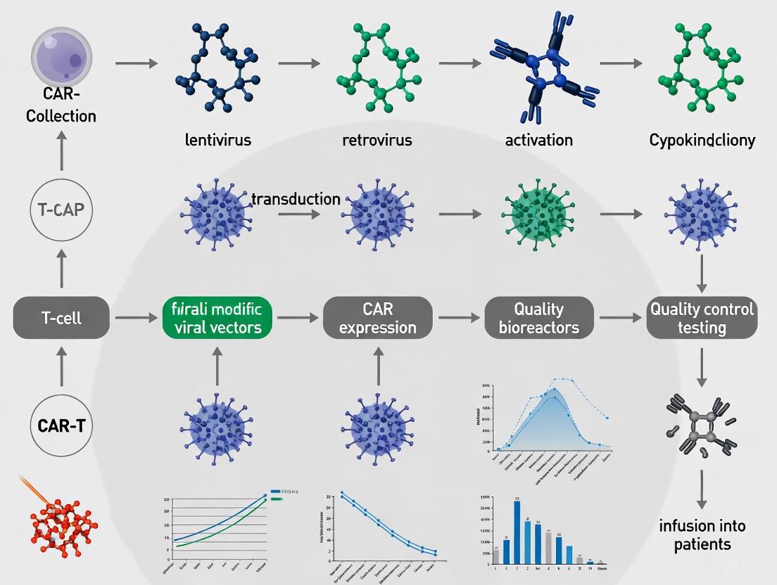
The Paradigm Shift: From Ex Vivo to In Vivo CAR-T Cell Manufacturing
Chimeric Antigen Receptor (CAR)-T cell therapy has revolutionized the treatment of relapsed/refractory hematological malignancies. However, the widespread adoption of this powerful modality is constrained by its complex, time-consuming, and costly ex vivo manufacturing process. This involves leukapheresis, T-cell activation, genetic modification, and expansion in specialized Good Manufacturing Practice (GMP) facilities before reinfusion into the patient [26] [27] [28]. This paradigm is now shifting toward in vivo CAR-T cell engineering, a transformative approach that generates therapeutic CAR-T cells directly within the patient's body. This application note details the limitations of conventional manufacturing, explores the platforms enabling this shift, and provides structured experimental data and protocols to guide research and development in this emerging field.
The Challenge of ConventionalEx VivoManufacturing
The established ex vivo manufacturing process presents significant logistical and clinical hurdles that limit patient access. A detailed breakdown of this workflow and its associated timelines is provided in Table 1.
Table 1: Key Steps and Challenges in Ex Vivo CAR-T Cell Manufacturing
| Manufacturing Step | Process Description | Key Challenges & Impact |
|---|---|---|
| Starting Material Collection | Leukapheresis to obtain patient's peripheral blood mononuclear cells (PBMCs) or T-cells [27]. | High variability in T-cell fitness and quantity from heavily pre-treated patients; can lead to manufacturing failure [27] [29]. |
| T-Cell Activation & Genetic Modification | T-cells are activated (e.g., with anti-CD3/CD28 beads) and transduced with viral (lentiviral/retroviral) or non-viral (electroporation) vectors to deliver the CAR gene [27] [28]. | Viral vectors require extensive safety testing and are costly; process is labor-intensive and requires stringent cleanroom environments [27] [28]. |
| Ex Vivo Expansion | Transduced T-cells are expanded in bioreactors or culture bags to achieve a therapeutic dose [27]. | Process typically takes 7-14 days, leading to critical treatment delays for patients with aggressive diseases [28]. |
| Final Formulation & Infusion | Cells are washed, concentrated, and cryopreserved for shipment and infusion [27] [30]. | Cryopreservation can cause quantitative and qualitative cell loss; the "vein-to-vein" time is several weeks [27] [30]. |
The complexity of this process contributes to an estimated cost exceeding $100,000 per patient in the West, creating a profound accessibility barrier [31] [28]. Furthermore, the resulting CAR-T cell products can exhibit heterogeneous composition and may contain exhausted T-cell subsets, which can compromise therapeutic efficacy and contribute to severe toxicities like Cytokine Release Syndrome (CRS) and Immune Effector Cell-Associated Neurotoxicity Syndrome (ICANS) [26] [29].
Emerging Platforms forIn VivoCAR-T Cell Engineering
In vivo CAR-T cell manufacturing aims to circumvent the limitations of ex vivo production by using injectable vector systems to genetically reprogram a patient's own T-cells directly in situ. The two primary technological approaches under development are viral vectors and non-viral transient expression systems, each with distinct characteristics summarized in Table 2.
Table 2: Comparison of In Vivo CAR-T Cell Manufacturing Platforms
| Platform Feature | Viral Vectors (Lentivirus, Gamma-retrovirus) | Non-Viral/Transient Vectors (LNP-mRNA) |
|---|---|---|
| CAR Expression | Long-term, persistent (genomic integration) [32]. | Short-term, transient (days to weeks) [32]. |
| Key Advantages | Single infusion can lead to durable CAR-T cell populations with memory potential; self-calibrates to disease burden [32]. | Tunable exposure via repeat dosing; potentially lower risk of genotoxicity and chronic on-target toxicities (e.g., B-cell aplasia) [32]. |
| Major Challenges | Risk of genotoxicity due to random integration; potential for uncontrolled expansion/persistence leading to protracted toxicity; pre-existing immunity to viral vectors [32]. | Liver tropism of LNPs can cause hepatotoxicity; innate immune activation by RNA/LNP; may lack potency against high tumor burden [32]. |
| Ideal Application | Oncology indications requiring deep, durable responses [32]. | Earlier-line oncology, minimal residual disease (MRD), autoimmune diseases, outpatient settings [32]. |
The field is in early clinical development, with over 35 companies actively pursuing in vivo CAR-T platforms. Early data demonstrate proof-of-concept and an acceptable safety profile in initial human trials [32]. This approach also holds promise for reprogramming other immune cells, such as Natural Killer (NK) cells and macrophages, directly in vivo [26].
Experimental Protocols forIn VivoCAR-T Cell Research
Protocol: EvaluatingIn VivoT-Cell Engraftment and CAR Expression using Viral Vectors
This protocol outlines the steps to assess the efficiency and safety of viral vector-based in vivo CAR-T generation in a pre-clinical mouse model.
- Objective: To quantify the generation, persistence, and tumor-killing capacity of CAR-T cells following a single intravenous infusion of a targeted lentiviral vector.
- Materials:
- Research Reagent Solutions:
- Targeted Lentiviral Vector: Engineered with a CAR construct (e.g., anti-CD19 scFv-41BB-CD3ζ) and T-cell-specific envelope/surface protein (e.g., CD8 scFv) to enhance T-cell tropism [26].
- NSG Mice: Immunodeficient mouse model engrafted with human CD34+ hematopoietic stem cells to create a humanized immune system [8].
- Flow Cytometry Panel: Antibodies against human CD3, CD4, CD8, and a protein tag or idiotype for detecting the specific CAR.
- qPCR/ddPCR Assay: Primers for detecting vector copy number (VCN) in peripheral blood and tissues [30].
- Research Reagent Solutions:
- Methodology:
- Animal Preparation: Use humanized NSG mice bearing established human CD19+ tumor xenografts.
- Vector Administration: Administer a single intravenous dose of the targeted lentiviral vector via tail vein injection. Include control groups receiving a non-targeted vector or saline.
- Longitudinal Monitoring:
- Tumor Monitoring: Measure tumor volume regularly via calipers or bioluminescent imaging to assess antitumor efficacy.
- Terminal Analysis: At study endpoint, harvest spleen, bone marrow, and tumor for deep phenotyping of CAR-T cells and assessment of VCN in tissues. Analyze for potential organ toxicity and vector biodistribution.
- Data Analysis:
- Correlate the peak and area-under-the-curve of CAR-T cells in blood with antitumor efficacy.
- Assess the relationship between VCN in the bone marrow and long-term CAR-T cell persistence.
- Monitor for signs of CRS/ICANS through serum cytokine levels (e.g., IL-6, IFN-γ) and clinical observation.
Protocol: Assessing Tunable CAR-T Cell Activity using LNP-mRNA
This protocol describes how to investigate the pharmacologically tunable activity of mRNA-based in vivo CAR-T cells.
- Objective: To demonstrate that repeated dosing of LNP-formulated CAR-encoding mRNA can control tumor growth in a calibrated manner.
- Materials:
- Research Reagent Solutions:
- LNP-mRNA Formulation: LNPs containing mRNA encoding an anti-BCMA or anti-CD19 CAR, optimized for in vivo T-cell delivery and reduced liver tropism [32].
- Syngeneic or Humanized Mouse Model: Bearing relevant antigen-positive tumors.
- ELISA/Luminex: For quantifying serum cytokine levels (e.g., IL-6, IFN-γ, IL-2).
- Research Reagent Solutions:
- Methodology:
- Tumor Implantation: Establish tumors in mice.
- Dosing Regimen: Initiate LNP-mRNA intravenous dosing once tumors are palpable. Test different regimens (e.g., single dose, multiple doses every 3-5 days).
- Pharmacodynamic Monitoring:
- CAR Expression: Analyze peripheral blood 24-48 hours after each dose for transient CAR expression on T-cells via flow cytometry.
- Tumor Measurement: Track tumor volume and survival.
- Toxicity Assessment: Measure cytokine levels 6-24 hours post-infusion to monitor for CRS.
- Immune Fitness Assessment: In a cohort of treated mice, analyze the differentiation and exhaustion status of CAR-T and endogenous T-cells (e.g., via Tim-3, LAG-3, PD-1 staining) to determine if transient expression mitigates exhaustion [32].
- Data Analysis:
- Compare tumor growth inhibition and overall survival across different dosing regimens.
- Correlate peak CAR expression levels after each dose with the magnitude of cytokine release and antitumor activity.
Visualization of Workflows and Signaling
The following diagrams illustrate the core logical relationship between ex vivo and in vivo manufacturing, as well as the critical signaling structure of a CAR molecule.
Diagram: CAR-T Manufacturing Paradigms. The diagram contrasts the multi-step, centralized ex vivo pathway (blue) with the streamlined in vivo pathway (red), highlighting key associated challenges and benefits.
Diagram: Second-Generation CAR Signaling Structure. The diagram details the modular domains of a CAR, from extracellular antigen recognition (yellow) to transmembrane anchoring (green) and intracellular signaling, which combines a costimulatory signal (red) with the primary CD3ζ activation domain (blue).
The Scientist's Toolkit: Key Reagents forIn VivoResearch
Table 3: Essential Research Reagents for In Vivo CAR-T Cell Development
| Reagent / Technology | Function in Research | Key Considerations |
|---|---|---|
| Targeted Viral Vectors | Engineered lentiviral/retroviral vectors for durable CAR gene delivery to T-cells in vivo [26] [32]. | Must be pseudotyped with T-cell-specific envelopes (e.g., CD8-scFv); requires rigorous biodistribution and genotoxicity studies [32]. |
| Lipid Nanoparticles (LNPs) | Non-viral delivery vehicles for in vivo delivery of CAR-encoding mRNA [32]. | Composition must be optimized for T-cell tropism over innate liver sequestration; mRNA sequence should include modified nucleotides to reduce immunogenicity [32]. |
| CAR-Encoding mRNA | The genetic payload for transient CAR expression; does not integrate into the genome [32]. | Sequence optimization (codon usage, UTRs) is critical for high translation efficiency and protein expression levels. |
| Flow Cytometry Panels | To detect and characterize in vivo generated CAR-T cells (phenotype, persistence, exhaustion) [29]. | Require specific antibodies against the CAR idiotype or a co-expressed tag, plus standard T-cell markers (CD3, CD4, CD8, CD45RO, CD62L) and exhaustion markers (PD-1, TIM-3, LAG-3). |
| qPCR/ddPCR Assays | To quantify vector copy number (VCN) for viral vectors, assessing transduction efficiency and biodistribution [30]. | Assays must be rigorously validated for sensitivity and specificity according to regulatory guidelines (e.g., Ph. Eur., USP) [30]. |
The trajectory of in vivo CAR-T cell therapy points toward rapid diversification. The short-term focus is on validating platforms against validated B-cell lineage targets (CD19, BCMA) in oncology and autoimmunity [32]. The subsequent wave will expand to solid tumor targets and non-oncological indications like regenerative medicine. Long-term, the field will evolve toward more sophisticated in vivo engineering, incorporating precision gene editing, logic-gated circuits, and spatiotemporal control of CAR expression [32]. Parallel advances in allogeneic, "off-the-shelf" CAR-T products from healthy donors will also continue, leveraging gene-editing tools like CRISPR/Cas9 to knock out the T-cell receptor (TCR) and HLA molecules to prevent graft-versus-host disease (GvHD) and host rejection [8].
In conclusion, the shift from ex vivo to in vivo CAR-T cell manufacturing represents a fundamental evolution in cell therapy. By eliminating complex logistics and high costs, this paradigm promises to democratize access to powerful CAR-T treatments, potentially extending their application beyond late-stage cancer to earlier lines of therapy and a broad spectrum of diseases. While challenges in targeting efficiency, safety, and potency remain, the convergence of viral engineering, nanomedicine, and immunobiology is paving the way for a more accessible and versatile future for cellular immunotherapy.
Advanced Manufacturing Workflows: Viral and Non-Viral Engineering Platforms
The selection of starting material is a critical foundational step in the manufacturing of Chimeric Antigen Receptor T (CAR-T) cell therapies. The conventional reliance on fresh peripheral blood mononuclear cells (PBMCs) presents significant logistical challenges and manufacturing constraints, including limited transportation windows and variable T-cell fitness in heavily pre-treated patients. Cryopreserved PBMCs offer a promising alternative, potentially enabling more flexible manufacturing timelines and the use of cells collected from patients at healthier stages or from healthy donors. This Application Note provides a comparative analysis of fresh versus cryopreserved PBMCs for CAR-T production, supported by quantitative data and detailed protocols to guide researchers and therapy developers in making evidence-based decisions for their manufacturing processes.
Comparative Performance Analysis
Cell Viability and Phenotypic Stability
Extensive research has demonstrated that cryopreserved PBMCs maintain sufficient viability and key T-cell subpopulations necessary for effective CAR-T manufacturing, even after long-term storage.
Table 1: Impact of Cryopreservation Duration on PBMC Viability and T-cell Composition
| Parameter | Fresh PBMCs | Cryopreserved (3-6 months) | Cryopreserved (12 months) | Cryopreserved (2 years) | Cryopreserved (3.5 years) |
|---|---|---|---|---|---|
| Viability (%) | Baseline [33] | 4.00-5.67% decrease [33] | Comparable to shorter cryopreservation [33] | Comparable to shorter cryopreservation [33] | 90.95% [33] |
| T-cell Proportion Stability | Baseline | Relatively stable [33] | Relatively stable [33] | Relatively stable [33] | N/A |
| Naïve T-cells (Tn) | Baseline | No significant change [33] | No significant change [33] | No significant change [33] | N/A |
| Central Memory T-cells (Tcm) | Baseline | No significant change [33] | No significant change [33] | No significant change [33] | N/A |
Studies indicate that while there is a statistically significant decrease in viability after cryopreservation, the actual reduction is only 4.00% to 5.67%, with viability remaining above 90% even after 3.5 years of storage [33]. Furthermore, the proportion of T-cells remains relatively stable post-cryopreservation, with preserved populations of naïve T-cells (Tn) and central memory T-cells (Tcm) that are crucial for long-lasting CAR-T efficacy [33].
CAR-T Functional Outcomes
Clinical and preclinical studies have demonstrated that CAR-T cells generated from cryopreserved PBMCs exhibit comparable functionality to those derived from fresh starting materials.
Table 2: CAR-T Functional Characteristics from Fresh vs. Cryopreserved PBMCs
| Functional Parameter | Fresh PBMC-Derived CAR-T | Cryopreserved PBMC-Derived CAR-T | Statistical Significance |
|---|---|---|---|
| Expansion Potential | Baseline | Comparable, slight reduction (not significant) [33] | P > 0.05 [33] |
| Cytotoxicity (%) | 91.02-100.00% (at E:T 4:1) [33] | 95.46-98.07% (at E:T 4:1) [33] | Not significant [33] |
| Transfection Efficiency | Baseline | Comparable [33] | Not significant [33] |
| CD3+ Purity | Baseline | Comparable [33] | Not significant [33] |
| T-cell Exhaustion Markers | Baseline | Comparable [33] | Not significant [33] |
| Cytokine Secretion (IFN-γ) | Baseline | Significant decrease in CAR-12M vs. CAR-F [33] | P < 0.05 [33] |
| 1-Year Overall Survival | 64.1% [34] | 75.4% [34] | Not significant [34] |
| 1-Year Progression-Free Survival | 44.5% [34] | 52.1% [34] | Not significant [34] |
| Complete Response Rate (3-month) | 46.2% [34] | 45.5% [34] | Not significant [34] |
A retrospective clinical study of 162 relapsed/refractory Diffuse Large B-Cell Lymphoma (DLBCL) patients receiving anti-CD19 CAR-T therapy found no significant differences in key clinical outcomes between the cryopreserved and fresh groups, including overall survival, progression-free survival, and response rates [34]. The incidence of adverse events, including cytokine release syndrome (CRS) and immune effector cell-associated neurotoxicity syndrome (ICANS), was also comparable between groups [34].
Detailed Experimental Protocols
Protocol 1: Cryopreservation of PBMCs for CAR-T Manufacturing
Principle: Preserve PBMC viability and function through controlled-rate freezing in cryoprotectant solution, maintaining T-cell fitness for future CAR-T manufacturing.
Materials:
- PBMCs isolated via density gradient centrifugation (e.g., Ficoll-Paque)
- Cryoprotectant solution (e.g., CS10 with 10% DMSO or custom formulation)
- Controlled-rate freezer
- Cryogenic storage vials or bags
- Liquid nitrogen storage system
Procedure:
- PBMC Preparation: Isolate PBMCs from leukapheresis product or whole blood using standard density gradient centrifugation. Ensure cell viability ≥95% before cryopreservation.
- Cell Counting: Determine cell concentration and calculate total cell count.
- Cryoprotectant Addition: Resuspend PBMCs in cryoprotectant solution at target concentration of 5-8×10^7 cells/mL [35]. Maintain DMSO concentration at 7.5%-10% (v/v) [35].
- Formulation: Aliquot cell suspension into cryogenic containers. Complete formulation within 120 minutes of cryoprotectant addition to minimize DMSO exposure [35].
- Controlled-Rate Freezing: Use optimized freezing curve (e.g., Thermo Profile 4) [35]. Critical parameter: cooling rate of approximately -1°C/minute to -40°C, followed by rapid cooling to -100°C before transfer to liquid nitrogen vapor phase storage.
- Storage: Maintain cells at ≤-150°C in liquid nitrogen vapor phase for long-term storage.
Quality Control:
- Post-thaw viability target: ≥90% [35]
- CD3+ T-cell proportion: Maintained relative to pre-freeze composition [33]
- Functional assessment: T-cell expansion potential and differentiation profiles
Protocol 2: Thawing and Recovery of Cryopreserved PBMCs
Principle: Maximize post-thaw recovery and functionality through optimized thawing procedures and careful removal of cryoprotectants.
Materials:
- Pre-warmed water bath (37°C)
- Thawing medium (e.g., RPMI-1640 with 20-50% human AB serum or serum-free alternatives)
- Benzonase nuclease (optional, for reducing cell clumping)
- Centrifuge
Procedure:
- Rapid Thawing: Remove cryovial from liquid nitrogen storage and immediately place in 37°C water bath with gentle agitation until only a small ice crystal remains (approximately 2 minutes).
- Dilution: Transfer cell suspension to 15mL conical tube. Slowly add 10mL pre-warmed thawing medium dropwise with gentle mixing to dilute cryoprotectant.
- Centrifugation: Centrifuge at 300-400 × g for 5-10 minutes.
- Cryoprotectant Removal: Carefully aspirate supernatant containing DMSO.
- Resuspension: Resuspend cell pellet in fresh culture medium appropriate for subsequent activation steps.
- Rest Period: Incubate cells for 4-24 hours in culture medium before activation to allow functional recovery.
Protocol 3: CAR-T Generation Using PiggyBac Transposon System
Principle: Generate CAR-T cells through non-viral gene delivery using PiggyBac transposon system, enabling stable genomic integration and CAR expression.
Materials:
- Plasmid DNA: PiggyBac transposon carrying CAR construct and PiggyBac transposase
- Electroporation system (e.g., Neon Transfection System or Gene Pulser)
- T-cell activation beads/dynabeads (anti-CD3/CD28)
- Culture medium (e.g., Optimizer, X-VIVO15, or TexMACS)
- Recombinant human IL-2
Procedure:
- T-cell Activation: Activate thawed PBMCs with anti-CD3/CD28 beads/dynabeads at 1:1 bead-to-cell ratio 24-48 hours before electroporation [33].
- Plasmid Preparation: Prepare CAR transposon and transposase plasmids at optimal ratio (typically 1:1) in electroporation buffer.
- Electroporation: Use optimized electroporation parameters for activated T-cells. For Neon Transfection System: 1600V, 10ms, 3 pulses [33].
- Post-electroporation Recovery: Immediately transfer cells to pre-warmed culture medium supplemented with IL-2 (100-300 IU/mL).
- Ex Vivo Expansion: Culture cells for 10-14 days, maintaining cell density between 0.5-2×10^6 cells/mL with regular medium replenishment.
- Harvest: Harvest CAR-T cells when target expansion and transduction efficiency are achieved.
Workflow Visualization
The Scientist's Toolkit: Essential Research Reagents
Table 3: Key Reagents for CAR-T Manufacturing from Cryopreserved PBMCs
| Reagent Category | Specific Examples | Function & Application Notes |
|---|---|---|
| Cryoprotectants | CS10, CryoSure-DEX40 [35] [34] | Cell preservation during freezing; DMSO concentration critical (7.5-10%) [35] |
| Culture Media | Optimizer, X-VIVO15, TexMACS, AIM-V [36] | Support T-cell expansion; serum-free options reduce lot-to-lot variability [36] |
| Activation Reagents | Anti-CD3/CD28 beads/dynabeads [33] | T-cell activation pre-transfection; typically 1:1 bead-to-cell ratio [33] |
| Gene Delivery Systems | PiggyBac transposon system [33], Lentiviral vectors [1] | Non-viral (PiggyBac) reduces costs vs. viral methods; each has specific protocol requirements [33] |
| Cytokines | Recombinant human IL-2 [33] [36] | Supports T-cell growth and persistence; concentration typically 100-300 IU/mL [36] |
| Electroporation Systems | Neon Transfection System [33] | Non-viral gene delivery; parameters must be optimized for cell type [33] |
Discussion and Implementation Guidelines
The accumulated evidence demonstrates that cryopreserved PBMCs represent a viable alternative to fresh starting materials for CAR-T manufacturing. The minimal impact on CAR-T functionality, coupled with the logistical advantages, positions cryopreservation as a valuable strategy for improving manufacturing flexibility.
Key Implementation Considerations:
Process Optimization is Critical: While cryopreservation itself has minimal impact, the specific manufacturing process significantly influences outcomes. Optimization of activation timing, culture conditions, and transfection parameters is essential when transitioning from fresh to cryopreserved PBMCs [33].
Logistical Advantages: Cryopreservation decouples cell collection from manufacturing, enabling more flexible production scheduling and the use of cells collected from healthy donors or patients at optimal health states [33] [37]. This approach helps mitigate issues associated with T-cell deterioration in heavily pretreated patients [35].
Alternative Starting Materials: For larger-scale applications, cryopreserved leukapheresis products offer similar advantages while preserving greater cellular diversity and potentially enhancing CAR-T potential due to higher lymphocyte proportions (66.59% in leukapheresis vs. 52.20% in PBMCs) [35].
Emerging Alternatives: Research into ambient temperature transport systems using hydrogel encapsulation for nutrient and oxygen support may provide future alternatives to cryopreservation, potentially avoiding cryoprotectant toxicity and cold chain logistics [38].
Cryopreserved PBMCs provide a practical and effective starting material for CAR-T manufacturing, with comparable performance to fresh cells across critical quality attributes including expansion potential, phenotype, and cytotoxic function. The implementation of standardized cryopreservation and thawing protocols, coupled with process optimization, enables researchers and therapy developers to leverage the significant logistical advantages of cryopreserved materials without compromising product efficacy. As the CAR-T field continues to evolve toward distributed manufacturing models and allogeneic approaches, cryopreserved starting materials will play an increasingly important role in enhancing the accessibility and scalability of these transformative therapies.
Chimeric Antigen Receptor T-cell (CAR-T) therapy has emerged as a revolutionary treatment for hematological malignancies, with viral vector-mediated transduction serving as the cornerstone of current manufacturing processes [1] [39]. Lentiviral (LV) and gamma-retroviral (γRV) vectors have become the primary delivery systems for integrating CAR genes into T lymphocytes, enabling stable, long-term expression of therapeutic constructs [40] [41]. The selection between these viral platforms and optimization of their corresponding transduction protocols directly impact critical quality attributes (CQAs) of the final cellular product, including transduction efficiency, cell viability, vector copy number (VCN), and ultimately, therapeutic efficacy and safety [40]. This application note provides detailed protocols and technical considerations for implementing LV and γRV transduction workflows within the framework of CAR-T cell manufacturing, presenting optimized parameters and analytical methods to support robust process development.
Comparative Analysis of Viral Vector Platforms
Key Characteristics of Lentiviral and Retroviral Vectors
Table 1: Comparison of Lentiviral and Gamma-Retroviral Vector Platforms
| Characteristic | Lentiviral Vectors (LV) | Gamma-Retroviral Vectors (γRV) |
|---|---|---|
| Integration Capability | Transduces dividing and non-dividing cells | Requires actively dividing cells |
| Pseudotyping Common | VSV-G (broad tropism) [40] | Gibbon Ape Leukemia Virus (GaLV) [41] |
| Payload Capacity | ~8-10 kb [39] | ~8-10 kb [39] |
| Safety Features | Self-inactivating (SIN) designs with deleted enhancer elements in LTR regions [41] [39] | Self-inactivating (SIN) designs; insulator elements [40] [41] |
| Risk Profile | Lower risk of insertional mutagenesis with SIN designs [40] [39] | Higher historical risk of insertional mutagenesis; improved safety with SIN configurations [41] |
| Approved CAR-T Products | Tisagenlecleucel (Kymriah), Lisocabtagene maraleucel (Breyanzi) [41] | Axicabtagene ciloleucel (Yescarta), Brexucabtagene autoleucel (Tecartus) [41] |
| Tropism for Primary T cells | Broad; enhanced with VSV-G pseudotyping [40] | Good for activated T cells; poor for NK cells [40] |
Quantitative Performance Parameters
Table 2: Optimized Process Parameters for Viral Transduction
| Process Parameter | Optimal Range/Conditions | Impact on Critical Quality Attributes |
|---|---|---|
| Multiplicity of Infection (MOI) | Typically 3-10 (requires empirical optimization) [40] | Higher MOI can increase transduction efficiency but may elevate VCN and cell toxicity [40] |
| Cell Activation | CD3/CD28 activation 24-72 hours pre-transduction [40] | Essential for γRV; enhances LV efficiency by upregulating viral receptors [40] |
| Transduction Duration | 8-24 hours [42] | Prolonged exposure increases efficiency but may affect viability [40] |
| Transduction Enhancers | Poloxamer 407, Protamine sulfate, Vectofusin-1 [40] | Can improve transduction efficiency 1.5-3 fold [40] |
| Cell Density | 0.5-1.5 × 10^6 cells/mL [42] | Critical for cell-vector contact; affects efficiency and viability |
| Spinoculation | 800-2000 × g for 30-120 minutes at 32°C [40] | Can increase transduction efficiency 1.5-2 fold by enhancing cell-vector contact [40] |
| Cytokine Support | IL-2 (50-300 IU/mL) or IL-7/IL-15 (10-20 ng/mL) [40] | Enhances post-transduction expansion and viability; influences memory phenotype [40] |
Detailed Transduction Protocols
Lentiviral Vector Transduction Protocol for CAR-T Cells
Principle: LV vectors enable efficient gene transfer through their ability to transduce both dividing and non-dividing cells, utilizing VSV-G pseudotyping for broad tropism and self-inactivating (SIN) designs for enhanced safety profile [40] [41].
Materials:
- Activated T-cells: Human PBMCs activated with CD3/CD28 activator for 72 hours [42]
- Lentiviral Vector: VSV-G pseudotyped, third-generation SIN vector, titer ≥ 1×10^8 IU/mL [40] [43]
- Transduction Medium: X-VIVO 15 or RPMI-1640 with 10% FBS, 2 mM L-glutamine, and IL-2 (50-100 IU/mL) [40] [42]
- Transduction Enhancer: Poloxamer 407 (0.5-1 μg/mL) or Protamine sulfate (4-8 μg/mL) [40]
- Formulation Buffer: For vector cryopreservation: 50 mM HEPES, 10% trehalose, 20 mM MgCl₂ [43]
Procedure:
- Cell Preparation: Harvest and count activated T-cells 72 hours post-activation. Adjust cell density to 1×10^6 cells/mL in transduction medium.
- Vector Thawing: Rapidly thaw LV vector aliquot in 37°C water bath and immediately place on ice. Use within 2 hours post-thaw. Avoid repeated freeze-thaw cycles.
- Transduction Mixture: In a sterile tube, combine cells, LV vector at desired MOI (typically 3-10), and transduction enhancer. Mix gently by pipetting.
- Transduction Process:
- Post-Transduction Processing: After incubation, centrifuge cells at 300 × g for 5 minutes. Remove vector-containing supernatant and resuspend cells in fresh expansion medium with IL-2.
- Expansion Culture: Culture transduced cells at 0.5-1×10^6 cells/mL with appropriate cytokine support for 7-14 days, monitoring cell growth and transduction efficiency.
Quality Control Assessment:
- Transduction Efficiency: Measure by flow cytometry for CAR expression or reporter gene expression at 72-96 hours post-transduction [40].
- Vector Copy Number (VCN): Quantify using droplet digital PCR (ddPCR) 3-5 days post-transduction. Maintain VCN < 5 copies per cell for clinical applications [40].
- Cell Viability: Assess using trypan blue exclusion or Annexin V/7-AAD staining post-transduction [40].
Retroviral Vector Transduction Protocol for CAR-T Cells
Principle: γRV vectors provide stable genomic integration but require target cell proliferation, making them suitable for ex vivo activated T-cells. GaLV pseudotyping enhances T-cell tropism, while modern SIN designs mitigate insertional mutagenesis risks [40] [41].
Materials:
- Activated T-cells: Human PBMCs activated with CD3/CD28 activator for 48-72 hours (critical for proliferation) [40]
- Retroviral Vector: GaLV pseudotyped, SIN vector, titer ≥ 1×10^8 IU/mL [41]
- Retronectin-Coated Plates: Non-tissue culture plates coated with retronectin (15-20 μg/cm²) to enhance transduction
- Transduction Medium: As above, with higher IL-2 concentration (100-300 IU/mL) to support proliferation
Procedure:
- Retronectin Coating: Coat non-tissue culture plates with retronectin (15-20 μg/cm²) for 2 hours at room temperature or overnight at 4°C. Block with 2% BSA for 30 minutes before use.
- Vector Pre-loading: Add γRV vector to retronectin-coated plates and centrifuge at 2000 × g for 2 hours at 32°C to pre-load vector onto retronectin.
- Cell Preparation: Harvest activated, proliferating T-cells and resuspend in transduction medium at 0.5-1×10^6 cells/mL.
- Transduction Process:
- Aspirate excess vector from pre-loaded plates (if necessary).
- Add cell suspension to vector-coated plates.
- Centrifuge at 400-800 × g for 30 minutes (spinoculation).
- Incubate at 37°C, 5% CO₂ for 6-24 hours.
- Repeat Transduction: For difficult-to-transduce cells, repeat transduction process 24 hours later.
- Post-Transduction Processing: Harvest cells, wash with fresh medium, and culture in expansion medium with appropriate cytokines.
Critical Considerations:
- Cell Activation State: γRV transduction efficiency directly correlates with T-cell activation and proliferation status [40].
- Vector Stability: γRV vectors are less stable than LV; use immediately after thawing.
- Safety Testing: Test for replication-competent retroviruses (RCR) in vector lots and final cellular products as regulatory requirement [41] [39].
Advanced Transduction Technologies
Novel Transduction Platform: Transduction Boosting Device (TransB)
Recent advancements in transduction technology have introduced the Transduction Boosting Device (TransB), an automated closed-system platform that utilizes hollow fibers to enhance cell-vector interactions [42]. This system demonstrates significant improvements over conventional methods:
- Enhanced Efficiency: 0.5-0.7-fold increase in transduction efficiency compared to 24-well plates [42]
- Reduced Vector Consumption: 3-fold reduction in viral vector requirements [42]
- Decreased Processing Time: Up to 1-fold decrease in processing time [42]
- Scalability: Consistent performance across different input cell numbers (0.5-2×10^6 cells) [42]
TransB Protocol:
- Prepare cell-vector mixture at desired MOI in 200 μL total volume.
- Load mixture into intracapillary (IC) space of hollow fiber.
- Continuously perfuse IL-2-supplemented medium through extracapillary (EC) space at 0.1 mL/min during transduction.
- Incubate for reduced duration (4-8 hours) compared to static transduction.
- Harvest cells by flushing IC and EC spaces with culture medium.
Quantitative CAR-T Cell Kinetics Evaluation
Accurate quantification of CAR-T cell expansion is crucial for correlating cellular kinetics with therapeutic efficacy. Traditional qPCR methods express results as transgene copies/μg genomic DNA (gDNA), but this approach can misrepresent actual cellular kinetics due to dramatic fluctuations in blood gDNA levels following lymphodepleting chemotherapy [44].
Improved qPCR Methodology:
- Unit System: Express cellular kinetics as copies/μL blood rather than copies/μg gDNA [44]
- Spike-in Calibration: Utilize standard CAR gene spiked into control blood samples for calibration [44]
- External Control: Implement dog gDNA as external control to normalize extraction efficiency variability [44]
- Validation Parameters: Demonstrates high linearity (r² = 0.999-1.000), accuracy (RE: -9.8 to 9.0%), and precision (CV: 4.4-10.3%) [44]
This volume-based unit system provides more accurate evaluation of in vivo CAR-T cell expansion and eliminates underestimation artifacts that occur with conventional gDNA-based normalization, particularly in lymphodepleted patients [44].
The Scientist's Toolkit: Essential Research Reagents
Table 3: Key Research Reagent Solutions for Viral Transduction
| Reagent/Category | Specific Examples | Function & Application Notes |
|---|---|---|
| Viral Vector Systems | VSV-G pseudotyped LV [40], GaLV pseudotyped γRV [41] | CAR gene delivery; VSV-G provides broad tropism, GaLV enhances T-cell specificity |
| Cell Activation | ImmunoCult CD3/CD28/CD2 T Cell Activator [42], Anti-CD3/CD28 beads | T-cell activation prerequisite for transduction, especially γRV; upregulates viral receptors |
| Transduction Enhancers | Poloxamer 407, Protamine sulfate, Vectofusin-1 [40] | Improve transduction efficiency by enhancing cell-vector interaction; reduce vector requirements |
| Cytokine Support | Recombinant IL-2, IL-7, IL-15 [40] | Enhance T-cell expansion, survival, and persistence post-transduction; influence memory differentiation |
| Formulation Buffers | HEPES (50 mM) with trehalose (10%) and MgCl₂ (20 mM) [43] | Maintain LV stability during cryostorage; HEPES-based buffers provide higher functional titers post-thaw |
| Analytical Tools | Flow cytometry antibodies (CD3, CD4, CD8, CAR detection) [40] | Assess transduction efficiency, immunophenotype, and CAR expression |
| Molecular Analysis | ddPCR for VCN [40], Spike-in qPCR for cellular kinetics [44] | Quantify vector integration and cellular kinetics; ddPCR provides superior precision for VCN |
Troubleshooting and Technical Notes
Low Transduction Efficiency:
- Verify cell activation status (CD25+CD69+ expression by flow cytometry)
- Confirm vector titer and functionality with permissive cell line
- Optimize MOI through dose-response testing (typically 1-20 range)
- Implement spinoculation or transduction enhancers
- For γRV: Ensure target cells are actively proliferating
Poor Cell Viability Post-Transduction:
- Reduce MOI to decrease viral load toxicity
- Shorten transduction duration (minimum 6-8 hours)
- Optimize cytokine support (consider IL-7/IL-15 as alternatives to IL-2)
- Assess vector preparation for contaminants or toxicity
High Vector Copy Number (VCN):
- Reduce MOI to decrease integration events
- Shorten transduction time to limit vector exposure
- Titrate transduction enhancers that may promote multiple integrations
Regulatory and Safety Considerations:
- Implement rigorous testing for replication-competent lentiviruses (RCL) or retroviruses (RCR) [41] [39]
- Monitor for insertional mutagenesis through integration site analysis in preclinical models
- Maintain comprehensive documentation of vector characterization and manufacturing process
Lentiviral and retroviral vector platforms continue to evolve as indispensable tools for CAR-T cell engineering, with distinct advantages and considerations for each system. The protocols detailed in this application note provide a foundation for implementing robust, efficient transduction processes that meet the critical quality attributes required for therapeutic applications. Recent advancements in transduction technologies, such as the TransB platform, and improved analytical methods, including volume-based qPCR quantification, represent significant steps toward addressing the manufacturing challenges that have limited broader clinical application of CAR-T cell therapies. As the field progresses toward more complex engineering strategies for solid tumors and allogeneic approaches, optimized viral transduction protocols will remain essential for balancing therapeutic efficacy with safety profiles.
Chimeric Antigen Receptor T-cell (CAR-T) therapy has revolutionized cancer treatment, particularly for hematologic malignancies. However, all currently approved CAR-T cell products rely on viral vectors (lentiviral or gamma retroviral vectors) for gene delivery, which present significant challenges including high costs, complex manufacturing processes, and safety concerns regarding insertional mutagenesis [45] [46]. Non-viral alternatives, primarily based on the PiggyBac (PB) transposon system combined with electroporation techniques, have emerged as promising solutions to overcome these limitations [45] [47].
The PiggyBac system offers substantial advantages over viral methods, including lower production costs, reduced immunogenicity, and a significantly larger cargo capacity (up to 100 kb) compared to viral vectors (limited to 8-10 kb) [33]. This system operates through a "cut-and-paste" mechanism where the PB transposase enzyme excises the transposon containing the CAR gene from a donor plasmid and integrates it into the host genome [45]. When combined with electroporation for delivery, this platform enables efficient, virus-free production of CAR-T cells, potentially increasing the accessibility and scalability of this revolutionary therapy [48].
Molecular Mechanism of Action
The PiggyBac transposon system functions through a precise molecular mechanism that facilitates stable genomic integration of the CAR transgene:
PiggyBac Transposition Mechanism
The PB transposase specifically recognizes inverted terminal repeats (ITRs) flanking the CAR transgene and catalyzes its integration into TTAA tetranucleotide sites within the genome [45]. This system enables high-efficiency gene transfer while minimizing genomic damage, as it operates without leaving "footprint" mutations at the excision sites [49]. The integration profile of PiggyBac is more random compared to viral vectors, which tend to integrate into transcriptionally active regions, thereby reducing the risk of oncogene activation [45].
Key Advantages Over Viral Systems
Table 1: Comparison of PiggyBac Transposon System vs. Viral Vector Systems
| Parameter | PiggyBac Transposon System | Viral Vector Systems |
|---|---|---|
| Production Cost | Significantly lower ($-$$) [47] [48] | Very high ($$$$) [46] |
| Cargo Capacity | Up to 100 kb [33] | Limited to 8-10 kb [45] |
| Integration Profile | More random, potentially safer [45] | Prefers active genes (lentiviral) or promoter regions (γRV) [45] |
| Manufacturing Complexity | Simplified, no viral packaging required [48] | Complex, requires specialized facilities [45] |
| Immunogenicity | Lower risk [33] | Higher risk of immune responses [46] |
| Regulatory Considerations | Simpler GMP compliance [48] | Stringent viral vector requirements [45] |
Electroporation Techniques for PiggyBac Delivery
Electroporation Methodology
Electroporation utilizes electrical pulses to create transient pores in cell membranes, enabling nucleic acids to enter cells. For PiggyBac delivery, both transposon DNA and transposase mRNA are typically co-electroporated into activated T cells [48]. Optimal parameters vary by instrument but generally involve square wave pulses with voltages ranging from 500V to 1500V and pulse durations of 20-30 ms [49].
Critical factors for successful electroporation include:
- Cell activation state: T cells must be pre-activated using anti-CD3/CD28 beads or antibodies for efficient nucleic acid uptake [50] [49]
- DNA:RNA ratio: Balanced ratios prevent excessive transgene copy numbers while maintaining high transfection efficiency [48]
- Post-electroporation recovery: Immediate transfer to pre-warmed, antibiotic-free media maintains cell viability [49]
Advanced Electroporation Workflow
CAR-T Manufacturing Workflow
Performance Metrics and Optimization Strategies
Quantitative Performance Data
Table 2: Performance Metrics of PiggyBac-Generated CAR-T Cells
| Metric | Typical Performance | Key Influencing Factors | Optimization Strategies |
|---|---|---|---|
| Transfection Efficiency | 60-70% CAR+ cells [48] | Cell viability, DNA quality, electroporation parameters | Use of minicircle DNA, optimized pulse protocols [48] |
| Vector Copy Number (VCN) | 1-3 copies/cell (optimized) [48] | DNA concentration, transposase activity | Titrate transposon DNA (0.3-3 μg/10⁶ cells) [48] |
| Cell Expansion | 10-100 million CAR+ cells from 10⁷ PBMCs [48] | Cytokine combination, culture duration | IL-4, IL-7, IL-21 cytokines prevent terminal differentiation [48] |
| Cryopreserved PBMC Performance | Comparable to fresh PBMCs [33] [51] | Freeze duration, thaw protocol | No significant functional loss after 2+ years cryopreservation [33] |
| Phenotypic Characteristics | Higher CAR expression vs. lentiviral [49] | Manufacturing process | PB CAR-Ts show distinct cytokine/chemokine profiles [49] |
Process Optimization Findings
Recent comparative studies demonstrate that cryopreserved peripheral blood mononuclear cells (PBMCs) can yield CAR-T products with comparable expansion potential, cell phenotype, differentiation profiles, exhaustion markers, and cytotoxicity to those derived from fresh PBMCs [33] [51]. This finding has significant implications for developing decentralized manufacturing models and "off-the-shelf" CAR-T products [47].
Key optimization strategies include:
- Cytokine supplementation: IL-4, IL-7, and IL-21 combination preserves stem-like memory phenotype and enhances persistence [48]
- Vector copy number control: Reducing transposon DNA concentration to 0.3 μg/10⁶ cells maintains VCN <3 while preserving transfection efficiency [48]
- Culture duration: Shorter ex vivo culture (14-21 days) prevents terminal differentiation and exhaustion [33]
The Scientist's Toolkit: Essential Research Reagents
Table 3: Key Research Reagent Solutions for PiggyBac CAR-T Manufacturing
| Reagent Category | Specific Examples | Function/Purpose | Considerations |
|---|---|---|---|
| Nucleic Acid Components | PCR-generated linear transposon [48] | CAR transgene delivery | Reduced size, no bacterial backbone, improved safety |
| In vitro transcribed transposase mRNA [48] | Catalyzes transposition | Transient expression enhances safety | |
| Minicircle DNA (mcDNA) [50] | Advanced DNA vector | Superior transfection efficiency and safety | |
| Cell Culture Reagents | Anti-CD3/CD28 activation beads [49] | T cell activation | Critical for electroporation efficiency |
| IL-4, IL-7, IL-21 cytokines [48] | Culture supplementation | Maintains less differentiated phenotype | |
| X-VIVO 15 serum-free medium [49] | Base culture medium | Supports T cell expansion | |
| Electroporation Systems | Celetrix CTX-1500A LE [49] | Nucleic acid delivery | Optimized for primary T cells |
| Analytical Tools | Flow cytometry with Protein L staining [49] | CAR expression detection | Alternative to target antigen-based detection |
| Digital PCR (ddPCR) [48] | Vector copy number quantification | Critical quality assessment |
Comparative Phenotypic Analysis: PiggyBac vs. Lentiviral CAR-T Cells
Transcriptomic and Functional Differences
Recent comparative analyses reveal significant differences between CAR-T cells manufactured using PiggyBac transposon systems versus lentiviral vectors [49]. PB CAR-T cells demonstrate:
- Higher CAR expression levels in a subset of cells compared to lentiviral CAR-T cells [49]
- Distinct transcriptomic signatures with greater upregulation of cytokines, chemokines, and their receptors [49]
- Unique IL-9 expression and reduced cytokine release syndrome-associated cytokines upon activation [49]
- Faster in vitro cytotoxicity against target cells while maintaining similar in vivo anti-tumor efficacy [49]
These phenotypic differences highlight the significant impact of manufacturing methodology on the final therapeutic product, emphasizing the need for careful process selection based on the desired clinical profile.
The PiggyBac transposon system combined with electroporation techniques represents a robust, cost-effective alternative to viral vector-based CAR-T manufacturing. This platform addresses several critical limitations of viral approaches, including high costs, complex production processes, and safety concerns related to insertional mutagenesis [45] [47] [46].
Future development directions include:
- In vivo CAR-T generation using targeted lipid nanoparticles to deliver PiggyBac components [50]
- Point-of-care manufacturing models utilizing cryopreserved PBMCs to increase accessibility [47] [33]
- Advanced vector engineering to enhance integration specificity and further improve safety profiles [45]
- Automated closed-system manufacturing to reduce production variability and costs [45]
As the field advances, non-viral CAR-T manufacturing platforms are poised to significantly increase the accessibility and applicability of CAR-T therapy, potentially expanding its use beyond specialized academic centers to broader clinical settings [47].
{Application Note}
Centralized vs. Decentralized Manufacturing Models: A Comparative Analysis
The commercial and clinical success of autologous Chimeric Antigen Receptor (CAR) T-cell therapies is critically dependent on the manufacturing paradigm employed. Unlike traditional biologics, autologous cell therapies are patient-specific "living drugs," introducing profound complexities in supply chain logistics, production scheduling, and product quality control [52]. The industry is thus confronted with a fundamental strategic decision: to utilize a traditional centralized manufacturing model, with its economies of scale, or to adopt a decentralized (point-of-care) manufacturing model, which promises greater agility and reduced logistics burden [53]. This application note provides a comparative analysis of these two models, underpinned by quantitative data and detailed experimental protocols, to guide researchers and drug development professionals in making informed decisions based on specific product and patient population needs.
Quantitative Model Comparison
A discrete-event simulation study modeling the UK autologous CAR-T supply chain provides robust, data-driven insights into the performance of both models across key operational metrics [52] [54]. The findings are summarized in the table below.
Table 1: Quantitative comparison of centralized and decentralized CAR-T manufacturing models based on a UK discrete-event simulation study [52] [54]
| Performance Metric | Centralized Model | Decentralized (Point-of-Care) Model | Key Findings and Context |
|---|---|---|---|
| Cost per Treatment | Lower at low demand (100-200 patients/year) | Becomes comparable at high demand (500 patients/year) | At high demand, decentralized facilities spread fixed costs over more treatments. Raw materials/consumables are a major cost driver in both models. |
| Turnaround Time (TAT) | Longer | Consistently shorter | The decentralized model eliminates cryopreservation, packaging, and long-distance transportation. In compact geographies with efficient transport, the TAT advantage may be less pronounced. |
| Critical Path Step | Sterility testing | Sterility testing | In both models, sterility testing is a major TAT driver, highlighting a universal bottleneck for process improvement. |
| Resource Utilization | High at low demand | Higher at high demand | Centralized model achieves better economies of scale at low volumes. At high demand, decentralized units can operate at high, efficient utilization. |
| System Resilience | Vulnerable to single-point failures and transport disruptions | Resilient to network-level disruptions; failure is isolated to a single node | The distributed nature of the decentralized model mitigates systemic risks. |
Decision Framework and Operational Considerations
Beyond quantitative metrics, the choice of model is governed by a set of strategic, clinical, and operational factors.
Table 2: Strategic and operational considerations for manufacturing model selection [52] [53]
| Consideration | Centralized Model | Decentralized Model |
|---|---|---|
| Ideal Use Case | Off-the-shelf (allogeneic) therapies; less aggressive diseases with longer treatment windows; large, broad patient populations. | Highly aggressive diseases; therapies requiring high, fresh cell doses; ultra-rare cancers with very small, geographically dispersed patient populations. |
| Manufacturing & Supply Chain | Established, bulk supply chains; benefits from scale-up; cold chain and logistics are critical and costly. | Eliminates complex patient material shipping; requires a network of compact, automated systems; cold chain costs are reduced but operational overhead for network management is high. |
| Regulatory & Quality Control | Single batch release site; established regulatory pathway for centralized GMP. | Requires harmonized protocols, assays, and quality programs across all sites; regulatory frameworks for multi-site POC release are still evolving. |
| Staffing & Expertise | Concentrated technical expertise at a central facility. | Requires distributed GMP-qualified technical staff at each node; recruiting and retaining specialized staff can be challenging. |
Experimental Protocol for a Semi-Automated, Decentralized Workflow
The following protocol outlines an end-to-end semi-automated method for generating non-viral, CRISPR-edited CD19-CAR T cells, suitable for a decentralized manufacturing unit [55]. This process leverages connected modular instruments controlled by automation software (e.g., CTS Cellmation) to reduce manual touchpoints, improve traceability, and enhance process consistency.
Key Research Reagent Solutions
Table 3: Essential reagents and materials for non-viral CAR-T cell manufacturing [56] [55]
| Research Reagent | Function in the Protocol |
|---|---|
| CTS Dynabeads CD3/CD28 | Provides T-cell activation signal via cross-linking CD3 and CD28 receptors. |
| ImmunoCult-XF T Cell Expansion Medium or TheraPEAK T-VIVO Medium | Serum-free, GMP-compliant culture media formulated to support T-cell viability and expansion. |
| CRISPR/Cas9 System (e.g., ribonucleoprotein complexes) | For precise genomic knock-in of the CAR transgene and knockout of the endogenous T-Cell Receptor (TCR). |
| Anti-CD19 CAR Transgene (e.g., via mRNA or plasmid) | Genetic payload encoding the chimeric antigen receptor targeting CD19. |
| Electroporation System (e.g., CTS Xenon) | Enables non-viral delivery of CRISPR components and CAR transgene into activated T cells. |
| G-Rex Bioreactor | Provides a gas-permeable membrane for high-density T-cell expansion with reduced feeding frequency. |
Detailed Step-by-Step Protocol
Day 0: T-Cell Isolation
- Starting Material: Thaw leukapheresis material from a healthy donor or patient.
- Automated Isolation: Use a connected instrument (e.g., CTS RoboSep) with immunomagnetic selection to isolate CD3+ or CD3+/CD28+ T cells.
- QC Sampling: Analyze isolated cell population for viability (expect >90%) and composition (CD3+ purity >90%) using flow cytometry [55].
Day 0-2: T-Cell Activation
- Culture Initiation: Transfer isolated T cells to a G-Rex bioreactor.
- Activation: Add CTS Dynabeads CD3/CD28 to the culture at a recommended bead-to-cell ratio.
- Culture Conditions: Maintain cells in pre-equilibrated expansion medium at 37°C, 5% CO2.
Day 2: Bead Removal and Assessment
- Automated Bead Removal: Use a connected instrument (e.g., CTS DynaCellect) to magnetically remove and count expended activation beads. The final product must contain <100 beads per 3x10^6 cells to meet FDA standards [55].
- Process Monitoring: Take a sample for flow cytometry analysis. Expect high expression of activation markers (CD69, CD25, HLA-DR) and a cell count of approximately 850 million viable cells.
Day 2: Genetic Modification via Electroporation
- Preparation: Wash and concentrate the activated T cells to the recommended density for electroporation.
- Electroporation: Use a programmed electroporator (e.g., CTS Xenon). Transfer the cell suspension to an electroporation cassette and deliver the pulse code optimized for high viability and transfection efficiency (e.g., pulse code CM-138) [56] [55]. The payload is a mixture of CRISPR/Cas9 RNP (for TCR knockout) and CAR-encoding mRNA or plasmid.
- Recovery: Immediately after electroporation, transfer cells to fresh, pre-warmed expansion medium in a new G-Rex bioreactor.
Day 2-9: Cell Expansion
- Fed-Batch Culture: Maintain cells in the G-Rex bioreactor for 7 days, feeding or diluting as necessary based on glucose consumption and cell density.
- Monitoring: Monitor cell growth, viability, and metabolism daily.
Day 9: Final Harvest and Formulation
- Harvest: Collect cells from the bioreactor.
- Formulation: Wash and formulate the final CAR-T cell product in the appropriate infusion buffer.
- Cryopreservation (if required): Cryopreserve the final product using a controlled-rate freezer.
Expected Outcomes and Quality Controls
- Cell Phenotype: The final product typically shifts to a higher proportion of CD8+ T cells and shows an increase in naïve central memory T cells (TCM), which is associated with improved persistence [55].
- Editing Efficiency: Expect >90% TCR knockout, with >20% of cells being double-positive for TCR knockout and CAR expression [55].
- Viability and Expansion: Overall viability should remain high (>90% at isolation, recovering to >80% post-electroporation). A clinically relevant fold expansion (e.g., 7-14x from day 2) should be achieved [55].
- Potency Assay: The CAR-T cells must demonstrate specific cytotoxic activity against target cells (e.g., NALM6 for CD19-CAR) in a co-culture assay, with a significantly lower EC50 than non-electroporated controls [55].
- Exhaustion Markers: Expression of exhaustion markers (LAG3, TIM3, PD1) should be low at harvest, having decreased significantly from the peak observed on day 2 post-activation [55].
Workflow Visualization
The following diagrams illustrate the logical and material flows of the two manufacturing models and the detailed steps of the semi-automated protocol.
CAR-T Manufacturing Model Flow
Semi-Automated CAR-T Production
The choice between centralized and decentralized manufacturing for CAR-T cell therapies is not a binary one but a strategic decision that must be aligned with the specific therapy profile, target patient population, and commercial goals. Centralized manufacturing remains the dominant, economically viable model for many indications, particularly as therapies move into earlier lines of treatment where timing is less critical [53]. However, decentralized manufacturing presents a compelling alternative for ultra-rare cancers, highly aggressive diseases requiring the shortest possible turnaround time, or situations where establishing a cold chain is prohibitive [52] [53]. The advent of standardized, semi-automated, and closed manufacturing systems [55] is making decentralized production more feasible, though it requires ongoing collaboration between industry, regulators, and treatment centers to harmonize standards and ensure consistent product quality across a distributed network.
Automated closed systems represent a transformative advancement in the manufacturing of cell therapies, including Chimeric Antigen Receptor T-cell (CAR-T) therapies. These systems are designed to enhance process consistency, reduce contamination risks, minimize human error, and improve scalability. Within the context of CAR-T cell engineering, the transition from manual, open-process manufacturing to automated, closed systems is critical for standardizing protocols and expanding patient access. This document details the application and protocols for two prominent platforms: the CliniMACS Prodigy from Miltenyi Biotec and the Cocoon Platform from Lonza. Both systems integrate multiple manufacturing steps into a single, functionally closed, and automated workflow, offering robust solutions for clinical and commercial-scale production of advanced cell therapies [57] [58].
The CliniMACS Prodigy and Cocoon platforms are integrated automated systems that streamline the entire cell therapy manufacturing process. While they share the common goal of standardizing production, their design philosophies and technical implementations differ.
The CliniMACS Prodigy system is a single, benchtop instrument that automates the entire process from cell separation and culture to final formulation [58]. Its magnetic selection technology is a core strength, enabling high-purity T-cell isolation. The system features an integrated culture chamber with perfusion capabilities for cell expansion [58].
The Cocoon Platform is a fully closed, automated, and flexible integrated manufacturing system. A key differentiator is its highly customizable nature, allowing researchers to select from off-the-shelf protocols or fully customize manufacturing processes with optimized programming instructions [59]. The platform uses single-use sterile cassettes and includes an integrated magnet for cell enrichment, aiming to realize a fully automated end-to-end solution [59].
Table 1: Key Technical Specifications and Performance Metrics of Automated Platforms
| Feature | CliniMACS Prodigy | Cocoon Platform |
|---|---|---|
| System Type | Integrated benchtop unit [58] | Integrated turnkey system with single-use cassettes [59] |
| Key Automation | End-to-end from selection to formulation [58] | End-to-end upstream and downstream processing [59] |
| Cell Selection Method | Magnetic separation (e.g., CD4/CD8 selection) [58] | Integrated magnetic selection [59] |
| Typical Cell Yield | ~2.5 × 10^9 CAR T cells/run from 2 x 10^8 T-cells [57] | Data not specified in search results |
| Throughput | One batch at a time | One patient batch at a time per unit [57] |
| Manufacturing Success Rate | 89% in Grade C cleanrooms [57] | Data not specified in search results |
| Scalability | Simplified tech transfer from R&D to GMP [57] | Superior scalability from pre-clinical to commercial [59] |
Application in CAR-T Cell Manufacturing Workflows
Automated systems like CliniMACS Prodigy and Cocoon are designed to execute the multi-step process of CAR-T cell manufacturing with minimal manual intervention. The workflow can be broken down into four core modules, which are integrated seamlessly within these platforms.
(CAR-T Manufacturing Workflow)
Cell Purification
The initial step involves isolating T cells from the patient's leukapheresis product.
- CliniMACS Prodigy Protocol: The system can perform a combined CD4+/CD8+ selection using GMP-compliant magnetic beads. The process is fully automated after the user connects the bag containing the leukapheresis material, buffer, and the selected magnetic bead reagents. This method routinely achieves >90% T-cell purity [58].
- Cocoon Platform Protocol: The platform utilizes its integrated magnetic selection module to enrich for target cells, addressing the high variability in patient starting material [59]. The specific reagents and protocols are part of its customizable, single-use cassette system.
Cell Activation and Transduction
Following purification, T cells are activated and genetically modified to express the chimeric antigen receptor.
- CliniMACS Prodigy Protocol: For its isolated T cells, the system uses anti-CD3/anti-CD28 co-stimulatory reagents, such as TransAct, which are added automatically to the integrated culture chamber to provide the necessary activation signals [58]. Viral vectors for transduction are similarly introduced via sterile tubing connections.
- Cocoon Platform Protocol: The platform's software-controlled fluidics system automatically handles the addition of activation reagents and viral vectors at predefined times and ratios, ensuring process consistency and minimizing operator-dependent variability [59].
Cell Expansion
Activated and transduced T cells are expanded to therapeutic doses.
- CliniMACS Prodigy Protocol: The integrated culture chamber with perfusion capabilities maintains the cells in a controlled environment, automatically feeding and monitoring the culture over a typical period of about two weeks [57] [58].
- Cocoon Platform Protocol: The platform uses its single-use bioreactor cassettes for cell expansion. Its integrated automation manages gas exchange and nutrient delivery throughout the expansion phase, which can last up to 10 days [59] [57].
Final Formulation and Harvest
The final product is concentrated, washed, and formulated into a bag for patient infusion.
- CliniMACS Prodigy Protocol: The system automatically performs concentration and wash steps, finally formulating the product into a sterile bag. The entire process from cell selection to final harvest is contained within the closed system [58].
- Cocoon Platform Protocol: The platform seamlessly integrates these downstream processing steps. The final formulated cell product is filled into an output bag, ready for cryopreservation or infusion, completing the functionally closed workflow [59].
Performance and Adoption Landscape
The adoption of automated closed systems in both academic and commercial settings is accelerating. A 2025 survey of academic institutions found that 60% of respondents used the CliniMACS Prodigy and 50% used the Lonza Cocoon for local CAR-T cell manufacturing [60]. Quantitative performance data highlights the impact of these platforms.
Table 2: Comparative Manufacturing and Economic Impact
| Performance Metric | CliniMACS Prodigy | Cocoon Platform | Manual Process (Baseline) |
|---|---|---|---|
| Annual Batches per Unit | Data not specified | ~36 batches/unit (approx. 5,000+ across 150 units) [57] | Varies widely |
| Vein-to-Vein Time Reduction | Data not specified | ~10 days (from a median of 38.3 days) [57] | Baseline |
| Labor Reduction | Significant hands-on time saved [58] | Part of overall cost and efficiency improvement | Baseline |
| Cleanroom Requirement | Can achieve 89% success rate in Grade C [57] | Reduces cleanroom stringency requirements [59] | Typically requires Grade B |
A critical driver for automation is the reduction in vein-to-vein time (V2VT). The Cocoon platform, for example, can reduce V2VT to approximately 10 days from a manual process median of 38.3 days—a reduction of over 70% [57]. This is critically important for patient outcomes; for relapsed/refractory large B-cell lymphoma patients, a 55% reduction in V2VT can increase life expectancy by more than three years [57].
The Scientist's Toolkit: Essential Research Reagents and Materials
Successful CAR-T manufacturing in automated systems relies on a suite of GMP-compliant ancillary materials.
Table 3: Key Reagent Solutions for Automated CAR-T Manufacturing
| Reagent/Material | Function | Example Use Case |
|---|---|---|
| CD4+/CD8+ Magnetic Beads | Immunomagnetic selection of target T cell populations from leukapheresis product. | CliniMACS Prodigy for high-purity T-cell isolation (>90%) [58]. |
| Cell Activation Reagents | Provides Signal 1 (anti-CD3) and Signal 2 (e.g., anti-CD28) for T-cell activation and proliferation. | TransAct or similar reagents used in Prodigy for isolated T cells [58]. |
| Viral Vector | Delivery vehicle for the genetic material encoding the CAR into the T cell (transduction). | Lentiviral or gamma-retroviral vectors used in both Prodigy and Cocoon [58]. |
| Cell Culture Media | Provides nutrients, growth factors, and cytokines necessary for T-cell survival and expansion. | X-VIVO, TexMACS, or similar media used in automated bioreactors [58]. |
| Single-Use Cassettes/Sets | Pre-assembled, sterile fluidic pathways that ensure a closed and sterile processing environment. | Cocoon Platform's disposable cassettes integrate all process steps [59]. |
Challenges and Future Directions
Despite their benefits, the widespread adoption of these platforms faces several hurdles. The high upfront capital expenditure is a significant barrier, with automation often demanding more than five times the initial equipment cost compared to manual facilities [57]. Furthermore, process rigidity in some non-modular platforms can limit adaptability for novel therapies requiring unique transduction or culture protocols [57]. Finally, regulatory uncertainty persists, as changing a manufacturing process post-IND approval typically triggers full comparability studies, adding 12-18 months to development timelines [57].
Future development is focused on enhancing flexibility and interoperability to accommodate a wider range of cell types (e.g., CAR-NK, iPSC-derived therapies) and genetic engineering techniques. Continued efforts to reduce costs and establish harmonized regulatory standards for automated systems will be crucial for global implementation [57].
Chimeric Antigen Receptor T-cell (CAR-T) therapy represents a groundbreaking advancement in the treatment of cancer and other diseases. As a living drug, the consistent quality, safety, and efficacy of CAR-T products are paramount, making robust process monitoring and control essential throughout manufacturing. The complex, multi-step production process—involving cell collection, activation, genetic modification, expansion, and formulation—creates multiple critical points where variability can impact final product quality [61] [6]. For autologous therapies, where T-cells are derived from individual patients, the inherent variability of starting material further underscores the need for stringent quality assurance measures [61]. This application note details the key parameters and methodologies for comprehensive quality assurance in CAR-T cell manufacturing, providing researchers and drug development professionals with standardized protocols to ensure product safety, identity, potency, purity, and quality (SQUIPP) [6].
The transition toward decentralized, point-of-care manufacturing models in academic institutions introduces additional challenges for maintaining consistent quality across production sites [30] [6]. Recent surveys of manufacturing practices highlight significant variability in processes across institutions, contributing to disparities in therapeutic outcomes [6] [62]. This document establishes harmonized quality control testing protocols based on current regulatory frameworks including Good Manufacturing Practices (GMP), European Pharmacopoeia standards, and guidelines from EMA and FDA [30]. By implementing these standardized monitoring and control strategies, manufacturers can improve product consistency, enhance patient safety, and facilitate broader access to these transformative therapies.
Critical Quality Attributes and Key Parameters
Critical quality attributes (CQAs) are biological, chemical, or physical properties that must be controlled within appropriate limits to ensure product quality. For CAR-T cell therapies, CQAs span from the initial apheresis material through the final drug product, with monitoring requirements evolving throughout the manufacturing process.
Table 1: Critical Quality Attributes in CAR-T Cell Manufacturing
| Manufacturing Stage | Critical Quality Attributes | Monitoring Purpose | Recommended Methods |
|---|---|---|---|
| Starting Material (Apheresis) | T-cell composition, Viability, CD4+/CD8+ ratio, Naïve/Memory subsets | Assess patient material suitability and predict manufacturing success | Flow cytometry, Cell counting, Viability assays [62] |
| In-Process Controls | Transduction efficiency, Cell expansion, Metabolic status, Vector copy number | Monitor process consistency and identify deviations early | qPCR/ddPCR, Metabolic assays, Cell counting [30] [61] |
| Drug Product Release | Identity, Purity, Potency, Viability, Sterility, Mycoplasma, Endotoxins | Ensure final product meets all safety and quality specifications | Flow cytometry, Cytotoxicity assays, Microbial tests [30] [62] |
| Post-Infusion Monitoring | CAR-T cell persistence, Functional activity, Cytokine levels | Assess in vivo performance and correlate with clinical outcomes | Flow cytometry, PCR, Cytokine assays [62] |
The identity of CAR-T products is typically confirmed through detection of the CAR transgene and expression of the CAR protein on T-cells [62]. Purity assessments focus on the percentage of CAR-positive cells and absence of undesirable cell populations. Potency, a particularly challenging attribute to measure, reflects the biological activity of the product through direct cytotoxicity assays or surrogate markers like cytokine secretion [30] [62]. Safety parameters include sterility testing, mycoplasma detection, and endotoxin testing to ensure the product is free from contaminating microorganisms and pyrogenic substances [30].
Recent surveys of European CAR-T manufacturing practices reveal significant heterogeneity in the specific analytical methods employed across different facilities, particularly for phenotypical characterization of T-cell subsets and assessment of activation/exhaustion profiles [62]. The following sections provide detailed methodologies to harmonize these critical assessments across manufacturing sites.
Analytical Methodologies for Quality Control
Mycoplasma Detection
Principle: Mycoplasma contamination represents a significant safety risk for cell therapy products. While traditional culture methods require 28 days, nucleic acid amplification techniques provide rapid alternatives with equivalent sensitivity when properly validated [30].
Protocol:
- Sample Preparation: Collect 1-5mL of cell culture supernatant or cell suspension (1×10^6 cells/mL) at harvest
- DNA Extraction: Use validated commercial DNA extraction kits (e.g., QIAamp DNA Mini Kit) according to manufacturer instructions
- PCR Setup: Utilize commercially available mycoplasma detection kits (e.g., VenorGeM Mycoplasma Detection Kit)
- Amplification: Perform PCR with positive controls (M. orale, M. hyorhinis, M. fermentans, M. pneumoniae) and internal controls
- Analysis: Verify detection limit of ≤10 CFU/mL for each mycoplasma strain [30]
Validation Requirements:
- Confirm compatibility of DNA extraction with amplification method
- Validate for both cell suspensions and culture supernatants
- Verify detection of mycoplasma strains recommended by Pharmacopoeia
- Establish specificity to prevent false positives from bacterial DNA cross-reactivity
- Perform on-site validation even for commercially validated kits [30]
Endotoxin Testing
Principle: Endotoxins from gram-negative bacteria can cause pyrogenic reactions in patients. The Limulus Amebocyte Lysate (LAL) assay or Recombinant Factor C (rFC) assay provides quantitative measurement of endotoxin levels.
Protocol:
- Sample Preparation: Dilute CAR-T cell product supernatant in endotoxin-free water
- Test Method: Use kinetic chromogenic LAL or rFC assay according to manufacturer instructions
- Controls: Include standard endotoxin curves (0.01-5.0 EU/mL) and positive product controls
- Interference Testing: Validate that sample matrix does not inhibit or enhance reaction
- Calculation: Determine endotoxin concentration against standard curve [30]
Acceptance Criteria: Endotoxin levels must be below regulatory limits (typically <5 EU/kg/hour for intravenous administration) [30].
Vector Copy Number (VCN) Quantification
Principle: VCN assessment ensures appropriate levels of genetic modification and evaluates potential risks associated with insertional mutagenesis from high vector copies.
Protocol:
- Genomic DNA Extraction: Isolate DNA from 1×10^6 CAR-T cells using validated methods
- Standard Preparation: Create standard curves with known copy numbers of CAR transgene and reference gene
- qPCR/ddPCR Setup:
- For qPCR: Use TaqMan probes targeting CAR sequence and reference gene (e.g., RNase P)
- For ddPCR: Partition samples into nanodroplets for absolute quantification
- Amplification: Run qPCR (40 cycles) or ddPCR according to optimized protocols
- Calculation:
- qPCR: Calculate VCN using ΔΔCt method with reference gene normalization
- ddPCR: Determine VCN directly from positive/negative droplet ratio [30]
Validation Requirements:
- Establish linear range (1-100 copies/cell) and limit of detection (0.1 copies/cell)
- Determine precision (CV <20%) and accuracy (80-120%)
- Validate reference gene stability across different CAR-T cell batches [30]
Potency Assessment
Principle: Potency measurements evaluate the biological activity of CAR-T cells through their ability to recognize antigen and mount appropriate effector functions.
Protocol:
- Cytotoxicity Assay:
- Label target cells (antigen-positive and antigen-negative) with fluorescent dye (e.g., Calcein-AM)
- Co-culture CAR-T cells with target cells at multiple effector-to-target ratios (e.g., 40:1, 20:1, 10:1, 5:1)
- Incubate for 4-24 hours at 37°C
- Measure fluorescence release from lysed target cells
- Calculate specific lysis: (Experimental - Spontaneous)/(Maximum - Spontaneous) × 100 [33]
Cytokine Release Assay:
Flow Cytometry-Based Activation:
- Stimulate CAR-T cells with antigen-positive cells or anti-CAR antibodies
- Stain for early activation markers (CD69, CD25) after 6-24 hours
- Analyze by flow cytometry to quantify activation percentage [62]
Acceptance Criteria: Establish product-specific ranges for cytotoxicity (typically >30% specific lysis) and cytokine secretion based on clinical correlation [33].
The following diagram illustrates the relationship between critical quality attributes and their corresponding analytical methods throughout the manufacturing workflow:
Diagram 1: CAR-T Quality Attribute Monitoring Framework. This diagram illustrates the relationship between critical quality attributes across manufacturing stages and their corresponding analytical methodologies.
Process Monitoring Technologies and Automation
Advanced monitoring technologies are increasingly important for maintaining quality throughout CAR-T manufacturing. Automated, closed-system production devices have emerged as promising solutions to facilitate consistent CAR-T cell manufacturing in academic and point-of-care settings [30] [61]. These systems integrate critical process monitoring with automated control to reduce variability.
Table 2: Automated Monitoring Solutions for CAR-T Manufacturing
| Technology Platform | Monitoring Capabilities | Key Parameters Measured | Implementation Rate |
|---|---|---|---|
| Miltenyi CliniMACS Prodigy | Integrated cell processing, transduction, expansion | Cell concentration, Viability, Transduction efficiency | 60% of surveyed institutions [6] |
| Lonza Cocoon | Automated closed-system manufacturing | Cell growth, Metabolic parameters, Media conditions | 50% of surveyed institutions [6] |
| Rocking Motion (RM) Bioreactor | Expansion process monitoring | Cell density, pH, Dissolved oxygen, Metabolites | Reported in research settings [61] |
| Real-time Cellular Analysis (RTCA) | Dynamic functional assessment | Cytotoxicity, Cell proliferation, Morphology changes | Used in research for potency assessment [33] |
The integration of automation, real-time monitoring, and digitalization represents the implementation of Pharma 4.0 principles in CAR-T cell manufacturing [61]. These systems enable continuous monitoring of critical process parameters including cell density, viability, metabolite levels (glucose, lactate), dissolved oxygen, and pH throughout the expansion phase [61]. By establishing correlations between these process parameters and critical quality attributes, manufacturers can implement quality-by-design approaches that enhance product consistency.
Digitalization complements the monitoring framework by capturing intricate process details like temperature variances during cell modifications and tracking production timelines [61]. This data serves as an invaluable analytical asset for optimizing manufacturing processes. Leveraging sophisticated algorithms, insights from this data can refine subsequent production methodologies and identify early warning signs of process deviations [61].
The following diagram illustrates how different monitoring technologies integrate throughout the CAR-T manufacturing process:
Diagram 2: Integrated Monitoring Technologies in CAR-T Manufacturing. This diagram shows how different monitoring technologies integrate throughout the manufacturing process and contribute to data-driven quality assurance.
The Scientist's Toolkit: Essential Research Reagents and Materials
Table 3: Essential Reagents for CAR-T Cell Quality Assessment
| Reagent/Category | Specific Examples | Application in Quality Assurance | Technical Notes |
|---|---|---|---|
| Mycoplasma Detection Kits | VenorGeM Mycoplasma Detection Kit | Detection of mycoplasma contamination in final product | Validate for specific matrices; includes positive controls [30] |
| Endotoxin Testing Reagents | Limulus Amebocyte Lysate (LAL), Recombinant Factor C (rFC) | Quantification of endotoxin levels for product release | Validate to prevent matrix interference [30] |
| qPCR/ddPCR Reagents | TaqMan probes for CAR sequence, Reference genes (RNase P, Albumin) | Vector copy number quantification, Transgene detection | ddPCR provides absolute quantification without standard curves [30] |
| Flow Cytometry Antibodies | Anti-CAR detection reagents, CD3, CD4, CD8, CD45RO, CCR7 | Product identity, Purity, T-cell subset characterization | Include viability dyes to exclude dead cells [33] [62] |
| Cytokine Detection Assays | IFN-γ ELISA kits, Multiplex cytokine panels | Potency assessment through cytokine release upon activation | Use antigen-positive target cells for stimulation [30] [33] |
| Cell Viability Assays | Trypan blue, Flow-based viability dyes (7-AAD, PI) | Viability assessment throughout manufacturing process | Combine with cell counting for expansion calculations [33] |
| Cell Activation Reagents | Anti-CD3/CD28 beads, Cytokines (IL-2) | T-cell activation during manufacturing process | Optimize concentration to minimize exhaustion [61] [33] |
| Target Cell Lines | Antigen-positive and antigen-negative cell lines | Potency assessment through cytotoxicity assays | Maintain consistent passage number and culture conditions [33] |
Comprehensive process monitoring and control is essential for ensuring the consistent quality, safety, and efficacy of CAR-T cell therapies. The parameters and methodologies outlined in this application note provide a framework for standardized quality assurance across manufacturing sites. As the field advances toward more decentralized production models, harmonization of these quality control practices becomes increasingly important for maintaining product consistency and expanding patient access [30] [6].
Future developments in CAR-T quality assurance will likely focus on the implementation of more sophisticated real-time monitoring technologies, advanced analytical methods for characterizing T-cell fitness and function, and the establishment of correlations between in vitro potency measurements and clinical outcomes [61] [62]. Additionally, as emerging approaches such as in vivo CAR-T cell manufacturing gain traction, novel quality control strategies will be needed to address the unique challenges presented by these platforms [63]. By adopting the standardized protocols outlined in this document, researchers and manufacturing professionals can contribute to the continued advancement and accessibility of CAR-T cell therapies while maintaining the highest standards of product quality.
Overcoming Manufacturing Hurdles and Enhancing Product Efficacy
Vein-to-vein time—the critical path from leukapheresis to infusion of the final chimeric antigen receptor (CAR) T-cell product—is a paramount determinant in the success of autologous cell therapies. prolonged manufacturing durations can compromise T-cell fitness, diminish product potency, and adversely impact patient accessibility and outcomes [29]. Within the complex manufacturing workflow, specific phases such as the starting cell population selection, T-cell activation, and ex vivo expansion are particularly susceptible to delays and represent key opportunities for process optimization [29]. This Application Note delineates detailed, evidence-based strategies and protocols designed to streamline CAR-T manufacturing, with the explicit goal of reducing vein-to-vein time without compromising product quality. The methodologies presented are framed within a broader research thesis on advancing CAR-T engineering and manufacturing protocols to achieve more robust and efficient therapeutic production.
Decoding the Manufacturing Timeline: A Quantitative Breakdown
A thorough understanding of the temporal landscape of CAR-T manufacturing is the foundation for effective intervention. The process encompasses several sequential yet interdependent stages, each contributing directly to the total vein-to-vein time. The table below synthesizes quantitative data and key parameters from established manufacturing processes for approved products, highlighting stages with high time-reduction potential [29].
Table 1: Key Process Parameters and Time Considerations in Autologous CAR-T Cell Manufacturing
| Manufacturing Stage | Key Process Parameters | Impact on Vein-to-Vein Time | Approved Product Examples |
|---|---|---|---|
| Starting Cell Population | Cell population prior to activation (Enriched T cells, PBMCs, CD4/CD8 separate) [29] | Selection and isolation can add 1-2 days; influences subsequent expansion efficiency. | Tisa-cel (Enriched T cells), Axi-cel (PBMCs), Liso-cel (CD4/CD8 separately) [29] |
| T-cell Activation | Method of activation (e.g., paramagnetic beads, polymeric nanomatrix); cytokines used [64] | Activation duration is a fixed 1-2 day step, but protocol choice impacts total expansion time. | Varies by protocol; often uses αCD3/αCD28 stimuli [64] |
| Genetic Modification | Transgene integration method (Lentivirus, Retrovirus) [29] | Transduction is a brief event, but vector choice and efficiency can affect needed expansion. | Tisa-cel (Lentivirus), Axi-cel (Retrovirus) [29] |
| Ex Vivo Expansion | Culture duration, media formulation, cytokine support (e.g., IL-7, IL-15) [64] | The most variable stage, typically requiring 7-10+ days to achieve target cell dose. | Protocol-dependent; aim for TSCM enrichment [64] |
| Formulation & Release | Final product storage (Fresh, Frozen); quality control testing [29] | Cryopreservation decouples manufacturing from infusion, adding thaw step but allowing flexible scheduling. | Most commercial products (Frozen) [29] |
Core Strategies for Time Optimization
Strategy 1: Optimizing the Starting Cell Population
The initial composition of T cells directly influences expansion kinetics and final product phenotype. Enriching for stem cell memory T cells (TSCM) and naïve T cells (TN) at the outset is correlated with superior expansion potential and persistence, potentially reducing the time required in culture to achieve a therapeutic dose [29] [64].
- Experimental Protocol: Selective T-cell Isolation
- Objective: To isolate specific T-cell subpopulations (e.g., CD4+, CD8+, or TN) to create a defined starting product.
- Materials:
- Leukapheresis product.
- Ficoll-Paque or equivalent for PBMC isolation.
- Magnetic bead-based cell sorting kits (e.g., CD4+ and CD8+ Isolation Kits, Miltenyi Biotec) [64].
- Buffer (e.g., PBS supplemented with EDTA and human serum albumin).
- Methodology:
- Isolate Peripheral Blood Mononuclear Cells (PBMCs) from the leukapheresis product via density gradient centrifugation [29] [64].
- Wash and resuspend PBMCs in appropriate buffer.
- Perform positive or negative selection using magnetic beads according to manufacturer instructions. For separate CD4+ and CD8+ manufacturing (as with liso-cel), isolate populations into separate cultures. For a mixed population, deplete non-T cells [29].
- Determine cell count and viability post-isolation.
- Key Parameters: Purity of the isolated population (typically >90%), cell viability (>95%), and total cell yield. A defined CD4:CD8 starting ratio can standardize the process and improve lot-to-lot consistency [29].
Strategy 2: Advanced Activation and Culture Protocols
Traditional manufacturing using paramagnetic beads (e.g., Dynabeads) over 6-14 days can be lengthy. Transitioning to faster, nanomatrix-based agonists (e.g., TransAct) in the presence of homeostatic cytokines like IL-7 and IL-15 can enrich for TSCM phenotypes and may allow for a shorter, more robust expansion phase [64].
- Experimental Protocol: Rapid, TSCM-Promoting Manufacturing
- Objective: To generate a TSCM-enriched CAR-T product using a rapid, cytokine-supported nanomatrix protocol compatible with automated systems like CliniMACS Prodigy [64].
- Materials:
- Methodology:
- Activation & Transduction (Day 0): Resuspend T cells in medium supplemented with IL-7 (25 U/mL) and IL-15 (50 U/mL). Stimulate with nanomatrix reagent according to manufacturer's instructions [64].
- Genetic Modification (Day 1): Transduce activated T cells with lentiviral vector at a pre-optimized MOI (e.g., MOI 5) [64].
- Nanomatrix Removal (Day 2): Remove the activation reagent by centrifugation [64].
- Ex Vivo Expansion (Days 2-14): Continue culture in IL-7/IL-15 supplemented medium. Monitor cell density, viability, and phenotype (e.g., via flow cytometry for CD62L+CD45RA+ TSCM).
- Harvest and Formulation: When target cell numbers are met (often by day 10-14), harvest cells, wash, and cryopreserve in final formulation buffer.
- Key Parameters: Fold expansion, final cell count, TSCM/TCM percentage in the final product, and transduction efficiency.
The following workflow diagram illustrates this optimized manufacturing protocol:
Figure 1: Optimized CAR-T Cell Manufacturing Workflow. This TSCM-promoting protocol uses a nanomatrix activator and homeostatic cytokines to potentially reduce culture time and enhance product quality.
The Scientist's Toolkit: Essential Reagents for Efficient Manufacturing
Table 2: Research Reagent Solutions for Streamlined CAR-T Manufacturing
| Reagent / Material | Function | Protocol Example / Impact |
|---|---|---|
| Magnetic Bead Isolation Kits | Immunomagnetic selection of specific T-cell subsets (CD4, CD8, naïve) from PBMCs. | Enables creation of a defined starting population, improving process consistency and potentially reducing expansion time [29] [64]. |
| Polymeric Nanomatrix (TransAct) | GMP-compliant reagent providing CD3/CD28 stimulation in a sterically optimal conformation. | Compatible with automated systems; promotes TSCM phenotype; allows for rapid (e.g., 2-day) activation protocol [64]. |
| Homeostatic Cytokines (IL-7, IL-15) | Cytokines added to culture media to support T-cell survival and promote stem-cell memory phenotype. | Critical for enriching TSCM populations, which exhibit superior expansion and persistence, potentially shortening culture time [64]. |
| Automated Cell Processing System (CliniMACS Prodigy) | Closed, integrated system for automated cell processing, culture, and formulation. | Reduces manual handling, improves reproducibility, standardizes protocols, and directly shortens active operator time [64]. |
| Lentiviral Vectors | Viral vector for stable genomic integration of the CAR transgene. | Commonly used in approved products (e.g., Tisa-cel); enables efficient genetic modification of T cells [29]. |
Discussion and Concluding Remarks
Reducing vein-to-vein time is a multifaceted challenge requiring a holistic view of the CAR-T manufacturing pipeline. The strategies outlined herein—strategic starting material selection, implementation of rapid activation protocols, and utilization of TSCM-promoting cytokines—are interdependent. A product beginning with a TSCM-favorable population and expanded in IL-7/IL-15 will inherently possess greater proliferative capacity, which can be leveraged to shorten culture duration while still achieving target doses [64].
Furthermore, the adoption of automated closed systems (e.g., CliniMACS Prodigy) is a critical step forward. These systems not only minimize manual hands-on time and reduce the risk of contamination but also enforce process standardization, leading to more predictable and shorter manufacturing timelines [64]. The integration of these advanced protocols and technologies directly addresses the pressing need to enhance patient accessibility by making the journey from vein to vein faster, more reliable, and more efficient.
Future research should focus on further abbreviating the expansion phase through optimized media and cytokine cocktails, developing predictive analytics to determine the optimal harvest window, and exploring the feasibility of "short-culture" products that leverage less-differentiated T cells for in vivo expansion.
Mitigating T-cell Exhaustion and Improving In Vivo Persistence
T-cell exhaustion presents a fundamental barrier to the long-term efficacy of chimeric antigen receptor (CAR)-T cell therapies. This state of T-cell dysfunction is characterized by progressive loss of effector functions, diminished cytokine production, and sustained expression of multiple inhibitory receptors [65]. In the context of CAR-T cell therapy, exhaustion undermines critical anti-tumor activity and limits in vivo persistence, resulting in suboptimal treatment responses and disease relapse [66] [67]. This application note synthesizes current mechanistic understanding and provides detailed protocols for researchers aiming to overcome these limitations through targeted engineering and manufacturing approaches. We focus specifically on strategies to modulate exhaustion pathways and enhance T-cell durability, framed within the broader objective of advancing CAR-T cell manufacturing protocols.
Mechanisms of T-cell Exhaustion in CAR-T Therapy
T-cell exhaustion arises from prolonged antigen exposure in immunosuppressive environments, a condition frequently encountered by CAR-T cells within the tumor microenvironment (TME). The molecular hallmarks of exhaustion include co-expression of inhibitory receptors such as PD-1, TIM-3, LAG-3, and TIGIT [65] [68]. These surface markers correlate with internal transcriptional reprogramming driven by regulators like TOX and members of the NR4A family, which enforce a stable dysfunctional state [65].
Beyond transcriptional changes, exhausted T-cells exhibit distinct metabolic insufficiencies and epigenetic modifications that lock in the dysfunctional phenotype. This is particularly relevant for CAR-T products, where the initial activation and expansion protocols can inadvertently promote terminal differentiation, thereby shortening therapeutic persistence [66]. The table below summarizes the key characteristics of exhausted T-cells and their impact on CAR-T function.
Table 1: Key Characteristics and Functional Impacts of T-cell Exhaustion in CAR-T Therapy
| Feature | Manifestation in Exhausted T-cells | Impact on CAR-T Function |
|---|---|---|
| Inhibitory Receptor Expression | Sustained high surface expression of PD-1, LAG-3, TIM-3, TIGIT [65] [67] | Attenuated cytolytic activity and cytokine production upon target engagement [65] |
| Transcriptional Landscape | Upregulation of transcription factors (e.g., TOX, NR4A) [65] | Epigenetic reinforcement of the exhaustion program, limiting longevity [65] |
| Metabolic Profile | Shift towards oxidative phosphorylation and lipid catabolism; impaired glycolysis [68] | Failure to meet bioenergetic demands for robust proliferation and tumor killing [66] |
| Cytokine Secretion | Impaired production of IL-2, TNF-α, and IFN-γ [65] | Reduced autocrine signaling and impaired recruitment of innate immunity [65] |
| Proliferative Capacity | Progressive loss of ability to expand following antigen re-encounter [65] [66] | Poor in vivo expansion and failure to control tumor burden [66] [67] |
The following diagram illustrates the primary signaling pathways involved in T-cell activation and the induction of exhaustion, highlighting key inhibitory checkpoints.
Experimental Protocols for Assessing Exhaustion and Persistence
Protocol: Multicolor Flow Cytometry for Exhaustion Marker Phenotyping
This protocol details the quantification of exhaustion-associated inhibitory receptors on circulating CAR-T cells, adapted from methodologies used in clinical monitoring studies [67].
1. Reagent Setup:
- Staining Buffer: Phosphate-buffered saline (PBS) supplemented with 0.2% bovine serum albumin (BSA) and 1mM EDTA.
- Lysing Solution: Ammonium chloride-based lysing solution (e.g., Cytognos) or BD FACS Lysing Solution.
- Viability Dye: 7-AAD or a comparable viability dye.
- Antibody Panel:
- CAR Detection: Biotinylated CD19 protein followed by Streptavidin-conjugated fluorochrome [67].
- Inhibitory Receptors: Anti-PD-1, Anti-LAG-3, Anti-TIM-3.
- Activation Markers: Anti-CD38, Anti-HLA-DR.
- Cytotoxicity Marker: Anti-CD107a.
- T-cell Subset Markers: Anti-CD3, Anti-CD4, Anti-CD8, Anti-CD45RO, Anti-CCR7.
2. Staining Procedure: 1. Cell Preparation: Isolate PBMCs from patient blood samples using Ficoll density gradient centrifugation. Use approximately 200,000 – 500,000 cells per staining tube. 2. Viability Staining: Resuspend cell pellet in staining buffer and add viability dye. Incubate for 10 minutes in the dark. 3. Surface Staining: Add the pre-titrated antibody cocktail. Vortex gently and incubate for 20 minutes at room temperature in the dark. 4. RBC Lysis: If using whole blood, add lysing solution, mix, and incubate for 10-15 minutes in the dark. Centrifuge and decant the supernatant. 5. Wash and Resuspend: Wash cells twice with staining buffer. Centrifuge at 500×g for 5 minutes. Resuspend the final pellet in 100-200 µL of FACS Flow solution for acquisition.
3. Data Acquisition and Analysis: Acquire data on a flow cytometer configured with appropriate lasers and filters. Analyze data using software such as FlowJo or Infinicyt. First, gate on lymphocytes, single cells, and live cells. Identify CAR-T cells via the specific CAR detection signal, then analyze the expression of inhibitory and activation markers within this population.
Protocol: Digital PCR for Quantifying CAR-T Cell Kinetics
Monitoring CAR-T expansion and persistence in peripheral blood is critical. Digital PCR (dPCR) offers high sensitivity and precision for this purpose [67].
1. Sample Processing and DNA Extraction: 1. PBMC Isolation: Collect peripheral blood at serial time points (e.g., days 5, 7, 11, 14, 28 post-infusion). Isolate PBMCs using a Vacutainer CPT tube or equivalent Ficoll method. 2. DNA Extraction: Use a commercial DNA extraction kit (e.g., QIAamp DNA Mini Kit) following the manufacturer's instructions. 3. Quality Control: Measure DNA concentration and purity using a spectrophotometer. Accept samples with a concentration ≥20 ng/µl and an A260/280 ratio >1.8.
2. Digital PCR Reaction Setup: * Primers/Probes: Design primers and a FAM-labeled TaqMan MGB probe to amplify a specific sequence within the CAR construct (e.g., the FMC63 scFv region). Use a VIC-labeled assay for a reference gene (e.g., RNase P). * Reaction Mix (10 µL final volume): * 80 ng of sample DNA * 2 µL of Absolute Q DNA dPCR Mix (5X) * 250 nM of FAM-labeled CAR probe * 900 nM of each CAR primer * 0.5 µL of RNase P reference assay (20X) * Cycling Conditions: * Hold: 96°C for 10 minutes. * 40 Cycles: 96°C for 5 seconds, 60°C for 15 seconds.
3. Data Analysis: Run samples on a dPCR system (e.g., QuantStudio Absolute Q). The software will automatically partition the sample, amplify, and provide absolute quantification of CAR transgene copies per µL of reaction or ng of DNA. Plot values over time to track expansion and persistence kinetics.
Strategic Approaches to Mitigate Exhaustion
Manufacturing and Phenotypic Engineering
A primary strategy to enhance persistence involves manufacturing CAR-T products enriched with less-differentiated T-cell phenotypes. Naive (T~N~), stem cell memory (T~SCM~), and central memory (T~CM~) T cells demonstrate superior longevity and proliferative capacity compared to effector T cells [66].
Table 2: T-cell Subsets for Improved CAR-T Persistence
| T-cell Subset | Surface Phenotype | Functional Attributes in CAR-T Therapy |
|---|---|---|
| Naive (T~N~) | CD45RA+, CCR7+, CD62L+, CD27+, CD28+ [66] | Highest proliferative potential and ability to reconstitute memory pool; associated with long-term persistence [66] |
| Stem Cell Memory (T~SCM~) | CD45RA+, CCR7+, CD95+, CD122+ [66] | Self-renewal capacity and superior longevity; can generate all other memory and effector subsets [66] |
| Central Memory (T~CM~) | CD45RO+, CCR7+, CD62L+ [66] | Strong proliferative response upon antigen re-encounter; correlates with sustained remission in clinical studies [66] [67] |
| Effector Memory (T~EM~) | CD45RO+, CCR7-, CD62L- [66] | Immediate effector function but limited proliferative capacity and persistence; contributes to initial tumor kill [66] |
The following workflow diagram outlines a manufacturing process designed to generate a less differentiated, persistence-prone CAR-T product.
Molecular and Genetic Engineering Strategies
Advanced CAR construct design and genetic manipulations directly target the molecular drivers of exhaustion.
- CAR Costimulatory Domain Selection: The choice of costimulatory domains embedded in the CAR construct profoundly impacts T-cell fate. 4-1BB (CD137) domains promote a metabolic profile favoring mitochondrial fatty acid oxidation and oxidative phosphorylation, which is associated with enhanced persistence and a memory-like phenotype. In contrast, CD28 costimulation enhances initial effector function but may predispose cells to activation-induced cell death and exhaustion [66] [12].
- Checkpoint Inhibition: Engineering CAR-T cells to resist the immunosuppressive TME is a key strategy. This can be achieved by:
- Dominant-Negative Receptors: Expressing a truncated, non-functional PD-1 receptor that competes with endogenous PD-1 for ligand binding, thus blocking the inhibitory signal [68].
- Switch Receptors: Designing receptors where the extracellular PD-1 domain is fused to a transmembrane and intracellular costimulatory domain (e.g., CD28), effectively converting an inhibitory signal into an activating one [68].
- Epigenetic and Transcriptional Reprogramming: Emerging approaches use CRISPR/Cas9 or other editors to knock out exhaustion-associated genes (PDCD1, NR4A family) or the TOX gene [68]. Alternatively, engineering inducible expression systems for transcription factors that promote memory formation (e.g., FOXO1) can help maintain a less differentiated state.
The Scientist's Toolkit: Research Reagent Solutions
Table 3: Essential Reagents for Monitoring and Engineering Persistent CAR-T Cells
| Reagent / Tool | Function / Application | Example Use Case |
|---|---|---|
| Biotinylated CD19 Protein | Flow cytometry detection of CD19-CAR expression on cell surface [67] | Monitoring circulating CAR-T cell frequency in patient blood post-infusion [67] |
| dPCR Assay for CAR Transgene | Absolute quantification of CAR transgene copies in genomic DNA [67] | Tracking in vivo CAR-T cell expansion and persistence kinetics with high sensitivity [67] |
| Cytokines (IL-7, IL-15) | Ex vivo culture supplements promoting memory T-cell differentiation [66] | Manufacturing CAR-T products with enhanced T~SCM~ and T~CM~ phenotypes for improved persistence [66] |
| CRISPR/Cas9 System | Gene editing to knock out inhibitory receptors or exhaustion drivers [68] | Generating PD-1 knockout CAR-T cells to resist TME-mediated exhaustion [68] |
| Antibody Panel: PD-1, LAG-3, TIM-3 | Immunophenotyping of T-cell exhaustion state by flow cytometry [65] [67] | Assessing the functional state of CAR-T products pre-infusion or during monitoring [67] |
| 4-1BBL Expressing Artificial APCs | Providing specific costimulation during manufacturing [66] | Expanding CAR-T cells with a 4-1BB signal to promote a persistent phenotype [66] |
Combating Antigen Escape and Resistance Mechanisms
Chimeric antigen receptor (CAR) T-cell therapy has revolutionized the treatment of hematological malignancies, achieving remarkable success against B-cell lymphomas, leukemias, and multiple myeloma [69] [1]. Despite these advances, a significant proportion of patients experience disease relapse following initial response, with antigen escape emerging as a predominant resistance mechanism [70] [71]. This phenomenon occurs when tumor cells evade immune recognition by altering the expression or accessibility of target antigens, ultimately leading to therapeutic failure [72] [73].
Antigen escape represents a critical challenge spanning both hematological malignancies and solid tumors [73]. In B-cell acute lymphoblastic leukemia (B-ALL), approximately 30-50% of patients who achieve remission with anti-CD19 CAR-T cells relapse within one year, frequently with CD19-negative disease [70]. Similarly, in multiple myeloma, resistance to B-cell maturation antigen (BCMA)-targeted CAR-T cells occurs through various antigen-dependent mechanisms [74]. The clinical significance of these escape variants necessitates innovative engineering approaches to overcome these limitations and improve long-term outcomes [72] [73].
This application note provides a comprehensive overview of antigen escape mechanisms and details experimentally validated protocols to combat this resistance. We focus on translatable strategies including engineered sensing systems, optimized signaling architectures, and combinatorial approaches designed to address the dynamic nature of tumor antigen expression.
Mechanisms of Antigen Escape
Tumor cells employ multiple sophisticated strategies to evade CAR-T cell recognition. Understanding these mechanisms is fundamental to developing effective countermeasures.
Table 1: Primary Mechanisms of Antigen Escape in CAR-T Cell Therapy
| Mechanism | Molecular Process | Clinical Example | Detection Method |
|---|---|---|---|
| Genetic Alterations | Point mutations, deletions, or alternative splicing of antigen genes | CD19 Δexon-2/5/6 variants in B-ALL; GPRC5D biallelic loss in myeloma [73] | PCR, DNA sequencing, flow cytometry |
| Impaired Antigen Processing | Disruption of chaperone proteins (CD81) or mRNA processing factors (NUDT21) | CD19-negative relapse despite CD19 mRNA presence [73] | Western blot, immunofluorescence, RNA sequencing |
| Lineage Switching | Transition from lymphoid to myeloid phenotype driven by epigenetic reprogramming | KMT2A-rearranged B-ALL to AML switch post-CD19 CAR-T [73] | Immunophenotyping, cytogenetics |
| Antigen Redistribution | Internalization of surface antigens following CAR engagement | CD19 clustering and internalization at immune synapse [73] | Live microscopy, flow cytometry |
| Trogocytosis | Bidirectional transfer of membrane proteins from tumor to CAR-T cells | CD19, BCMA transfer leading to CAR-T fratricide [73] | Flow cytometry, live imaging |
| Antigen Masking | Physical obstruction of epitopes during manufacturing | CAR-transduced leukemic cells in autologous products [73] | Single-cell RNA sequencing |
The signaling consequences of these escape mechanisms directly impact CAR-T cell function. For instance, trogocytosis not only reduces antigen density on tumor cells but also promotes CAR-T cell fratricide and exhaustion through persistent tonic signaling [73]. Similarly, antigen redistribution during immune synapse formation creates a dynamic equilibrium that ultimately diminishes surface target availability below the critical threshold required for CAR activation [73] [75].
Table 2: Signaling Pathways Implicated in Antigen Escape Mechanisms
| Escape Mechanism | Key Signaling Pathways Affected | Functional Outcome |
|---|---|---|
| Genetic Alterations | B-cell receptor signaling, survival pathways | Complete loss of target epitope |
| Impaired Antigen Processing | ER stress response, unfolded protein response | Intracellular retention of misfolded proteins |
| Lineage Switching | HOXA/MEIS1 programs, menin-KMT2A interactions | Lineage transformation with antigen loss |
| Antigen Modulation | Actin cytoskeleton remodeling, endocytic pathways | Reduced surface antigen density |
| Trogocytosis | Tonic CAR signaling, exhaustion programs | CAR-T cell fratricide and dysfunction |
Engineering Strategies to Overcome Antigen Escape
Multi-Targeting CAR Approaches
Simultaneous targeting of multiple tumor antigens prevents escape through redundant recognition systems. The following approaches have demonstrated efficacy in preclinical models:
Dual-Targeting CARs incorporate two complete CAR structures targeting distinct antigens (e.g., CD19/CD22 or BCMA/TACI) within the same T cell [73]. Clinical trials have shown reduced antigen escape rates with these constructs compared to single-target approaches.
Tandem CARs utilize a single receptor with two antigen-binding domains in tandem, requiring simultaneous engagement for optimal activation [73]. This approach increases specificity while maintaining potency against heterogeneous tumors.
Enhanced Sensing Systems
Engineering CAR-T cells with improved antigen sensitivity addresses the challenge of antigen downregulation:
Logic-Gated CAR Systems employing synthetic Notch (synNotch) receptors enable sophisticated sensing-activation circuits [73]. These systems can be programmed to activate only in the presence of multiple tumor-associated antigens, increasing specificity while reducing on-target, off-tumor toxicity.
Tumor Microenvironment-Gated CARs represent a breakthrough in spatial control of CAR-T activity. The TME-iCAR platform requires three combined inputs for full activation: (1) a small-molecule inducer (abscisic acid, ABA), (2) a tumor-associated antigen, and (3) a TME-specific signal such as hypoxia [7]. This multi-layered sensing system significantly enhances tumor selectivity.
Diagram 1: TME-gated inducible CAR circuit requiring three activation inputs.
Signaling Amplification Strategies
Overcoming the inherently high activation threshold of conventional CARs represents a promising approach to combat antigen-low escape:
Membrane-Tethered Signaling Adaptors address proximal signaling deficits in CAR architectures. Engineering a membrane-tethered version of the cytosolic signaling adaptor SLP-76 (MT-SLP-76) significantly lowers the antigen density threshold required for CAR-T cell activation [75]. This system amplifies CAR signaling through enhanced recruitment of ITK and PLCγ1, effectively restoring sensitivity to antigen-low tumor cells.
Diagram 2: Signaling amplification through membrane-tethered SLP-76 enhances sensitivity to low antigen density.
Experimental Protocols
Protocol: Evaluating MT-SLP-76 Enhanced CAR-T Cells
This protocol details the methodology for engineering and validating CAR-T cells with enhanced sensitivity to antigen-low tumor cells using membrane-tethered SLP-76 [75].
Materials
Table 3: Key Research Reagents for MT-SLP-76 Experiments
| Reagent | Function | Example Source/Catalog |
|---|---|---|
| Lentiviral Vector pLV-MT-SLP-76 | Expresses membrane-tethered SLP-76 | Addgene #(to be determined) |
| CAR Lentiviral Construct | Expresses CAR of interest (CD19, BCMA, etc.) | In-house generation |
| Human T-cell Media | T-cell expansion medium | TexMACS or X-VIVO 15 |
| RetroNectin | Enhances viral transduction | Takara Bio T100B |
| CD3/CD28 Dynabeads | T-cell activation | Gibco 11161D |
| Recombinant IL-7/IL-15 | Promotes memory differentiation | PeproTech 200-07/200-15 |
| Antigen-low Cell Lines | Target cells with defined antigen density | Nalm6 (CD19-low), MM.1S (BCMA-low) |
Procedure
Day 1: T-Cell Isolation and Activation
- Isolate peripheral blood mononuclear cells (PBMCs) from leukapheresis product by density gradient centrifugation.
- Isolate untouched human T cells using negative selection kit (e.g., Miltenyi Pan T Cell Isolation Kit).
- Activate T cells with CD3/CD28 Dynabeads at 1:1 bead-to-cell ratio in T-cell media supplemented with 10 ng/mL IL-7 and 5 ng/mL IL-15.
- Culture cells at 1×10^6 cells/mL in 24-well plates at 37°C, 5% CO₂.
Day 2: Lentiviral Transduction
- Pre-coat non-tissue culture treated 24-well plates with RetroNectin (15 μg/mL) for 2 hours at room temperature.
- Block plates with 2% bovine serum albumin (BSA) in phosphate-buffered saline (PBS) for 30 minutes.
- Load lentiviral vectors (CAR alone or CAR + MT-SLP-76) at MOI 5-10 in RetroNectin-coated plates.
- Centrifuge plates at 2000 × g for 90 minutes at 32°C.
- Wash plates and add activated T cells at 1×10^6 cells/mL in fresh cytokine-containing media.
- Centrifuge plates at 1000 × g for 30 minutes.
- Incubate transduced cells at 37°C, 5% CO₂.
Day 5: Bead Removal and Expansion
- Remove activation beads using magnetic separator.
- Continue expansion in IL-7/IL-15 containing media.
- Monitor transduction efficiency by flow cytometry for CAR expression.
Day 10-14: Functional Assays
- Evaluate CAR-T cell phenotype (memory/exhaustion markers) by flow cytometry.
- Perform co-culture assays with antigen-low target cells at various E:T ratios.
- Measure cytokine production (IL-2, IFN-γ) by ELISA after 24-hour co-culture.
- Assess cytotoxic activity using real-time cell analysis (RTCA) or flow-based killing assays.
Protocol: TME-Gated Inducible CAR-T Cell System
This protocol describes the implementation of a tumor microenvironment-gated CAR system requiring combinatorial inputs for activation [7].
Materials
- Hypoxia-activated ABA prodrug (synthesized as described [7])
- Split CAR constructs (p1 and p2 with ABI and PYL fusions)
- Hypoxia chamber or incubator (1% O₂)
- Target cells expressing tumor antigen of interest
- HPLC system for prodrug activation analysis
Procedure
Step 1: Engineering TME-iCAR-T Cells
- Clone split CAR constructs: p1 (scFv-ABI-CD3ζ) and p2 (PYL-costimulatory domain).
- Generate lentiviral vectors for both constructs.
- Transduce activated human T cells sequentially with both constructs.
- Expand transduced cells as described in Protocol 4.1.
Step 2: ABA Prodrug Activation Testing
- Treat ABA prodrug (1-10 μM) with tumor cell lysates or purified enzymes under normoxic and hypoxic conditions.
- Analyze ABA release by HPLC at various time points (0, 2, 4, 8, 24 hours).
- Confirm hypoxia-specific activation by comparing prodrug conversion rates.
Step 3: Functional Validation
- Co-culture TME-iCAR-T cells with antigen-positive target cells under three conditions:
- Normoxia (21% O₂) without prodrug
- Normoxia with prodrug
- Hypoxia (1% O₂) with prodrug
- Measure T-cell activation (CD69, CD137) by flow cytometry at 24 hours.
- Quantify cytokine secretion (IL-2, IFN-γ) by multiplex ELISA.
- Assess cytotoxic activity using real-time cell analysis over 72 hours.
The Scientist's Toolkit
Table 4: Essential Research Reagents for Combating Antigen Escape
| Category | Reagent | Specific Application | Key Function |
|---|---|---|---|
| Engineering Platforms | synNotch receptors | Logic-gated CAR circuits | Conditional CAR activation |
| MT-SLP-76 constructs | Signaling amplification | Lowers antigen threshold | |
| Hypoxia-iCAR system | TME-gated activation | Spatial control of activity | |
| Small Molecules | γ-Secretase inhibitors | BCMA shedding prevention | Enhances target density [74] |
| Menin inhibitors | Lineage switching prevention | Blocks KMT2A-driven transformation [73] | |
| ABA prodrugs | TME-iCAR activation | Inducer of CAR dimerization | |
| Analysis Tools | Phospho-flow cytometry | Signaling analysis | Proximal signaling assessment |
| Real-time cytotoxicity | Functional validation | Dynamic killing measurement | |
| scRNA-seq | Clone tracking | Antigen escape variant identification |
Antigen escape remains a formidable challenge in CAR-T cell therapy, but innovative engineering approaches provide promising paths forward. The strategies outlined herein – including multi-targeting systems, enhanced sensing mechanisms, and signaling amplification – represent the forefront of efforts to overcome this resistance mechanism. The experimental protocols provide a framework for researchers to implement and validate these advanced CAR-T cell platforms. As the field progresses, combination approaches that address both tumor-intrinsic resistance and host factors will likely yield the most durable responses, ultimately improving outcomes for patients with refractory malignancies.
Optimizing Cryopreservation Protocols for Cell Viability and Function
Cryopreservation is a critical unit operation in the chimeric antigen receptor T-cell (CAR-T) therapy manufacturing workflow, enabling logistical flexibility and ensuring product availability. In autologous cell therapy, a patient's T cells are collected via leukapheresis, genetically engineered to express CARs targeting specific tumor antigens, expanded in vitro, and then infused back into the patient [1]. The ability to cryopreserve either the starting leukapheresis material or the final CAR-T cell product itself is essential for managing complex manufacturing schedules, conducting quality control testing, and facilitating transportation between centralized manufacturing facilities and clinical treatment sites [76] [77]. The fundamental challenge of cryopreservation lies in maintaining cell viability, phenotypic characteristics, and therapeutic potency throughout the freezing, storage, and thawing processes. This protocol details optimized procedures for cryopreserving CAR-T cells and their starting materials, incorporating recent advances that preserve critical quality attributes and ultimately support successful clinical outcomes.
Comparative Performance Data: Fresh vs. Cryopreserved Cells
Extensive research has evaluated the impact of cryopreservation on key cellular attributes and clinical performance. The data below summarize findings from recent studies comparing fresh and cryopreserved cellular materials in the context of CAR-T manufacturing and therapy.
Table 1: Impact of Cryopreservation on PBMC Starting Material
| Parameter | Fresh PBMCs | Cryopreserved PBMCs | Significance | Source |
|---|---|---|---|---|
| Viability | Baseline | 4.00-5.67% decrease | Significant but minimal | [33] |
| T Cell Proportion | Stable | Relatively stable | No significant impact on CAR-T preparation | [33] |
| NK Cell Proportion | Baseline | Decreased | Presumably more sensitive to hypothermia | [33] |
| B Cell Proportion | Baseline | Decreased | Presumably more sensitive to hypothermia | [33] |
| Naïve & Central Memory T Cells | Baseline | No significant change | Crucial for long-term CAR-T persistence | [33] |
Table 2: Impact of Cryopreservation on Final CAR-T Cell Product
| Parameter | Fresh CAR-T | Cryopreserved CAR-T | Significance | Source |
|---|---|---|---|---|
| In Vitro Anti-tumor Reactivity | Higher | High, but slightly lower | Potency retained, though measurable difference | [76] |
| TIM-3 Expression | Significantly more | Less | Suggests differential exhaustion marker profile | [76] |
| Effector T Cell Proportion | Less | More | Phenotypic shift observed | [76] |
| Clinical Response Rate | Comparable | Comparable | No statistically significant difference | [76] [34] |
| In Vivo Expansion & Persistence | Comparable | Comparable | Key efficacy metrics maintained | [34] |
| Cytokine Release (e.g., IFN-γ) | Baseline | Slight decrease in some studies | Cytotoxic function not necessarily correlated | [33] |
Table 3: Clinical Outcomes from a Study of 162 DLBCL Patients
| Clinical Outcome | Fresh PBMC-Derived CAR-T (n=26) | Cryopreserved PBMC-Derived CAR-T (n=136) | P-value |
|---|---|---|---|
| 3-Month Complete Response (CR) | 46.2% | 45.5% | > 0.05 |
| Objective Response Rate (ORR) | 69.2% | 61.9% | > 0.05 |
| 1-Year Overall Survival (OS) | 64.1% | 75.4% | > 0.05 |
| 1-Year Progression-Free Survival (PFS) | 44.5% | 52.1% | > 0.05 |
| Incidence of Grade ≥3 CRS/ICANS | No significant difference | No significant difference | > 0.05 |
Detailed Experimental Protocols
Protocol 1: Cryopreservation of Peripheral Blood Mononuclear Cells (PBMCs)
This protocol is designed for the cryopreservation of leukapheresis-derived PBMCs to be used as starting material for subsequent CAR-T cell manufacturing [76] [33].
Materials:
- Leukapheresis Product: Collected per standard clinical procedures.
- Ficoll-Hypaque (Lymphocyte Separation Medium): For density gradient centrifugation.
- Cryopreservation Medium: Typically consists of a suitable base medium (e.g., Plasma-Lyte A, NaCl) supplemented with human serum albumin (HSA) and DMSO as a cryoprotectant. Commercial, GMP-grade, serum-free, DMSO-containing cryopreservation solutions like
CryoSure-DEX40are also used [34]. - Controlled-Rate Freezer (CRF)
- Cryogenic Storage Vials or Bags: Validated for liquid nitrogen storage.
- Liquid Nitrogen Storage Tank: For long-term storage in the vapor or liquid phase.
Method:
- PBMC Isolation: Isolate PBMCs from the leukapheresis product using a single-step density gradient centrifugation with Ficoll-Hypaque [76]. Centrifuge and carefully collect the mononuclear cell layer at the interface.
- Washing and Counting: Wash the isolated PBMCs twice with an appropriate buffer (e.g., PBS). Perform a viable cell count and check viability via Trypan Blue exclusion.
- Formulation: Resuspend the PBMC pellet in cold cryopreservation medium to a final concentration of 10-20 x 10^6 cells/mL. The final concentration of DMSO should be 10% [33]. Keep the cell suspension on ice or in a cold environment during formulation.
- Packaging: Aseptically aliquot the cell suspension into pre-chilled cryogenic vials or bags.
- Controlled-Rate Freezing: Place the packaged cells into a pre-programmed controlled-rate freezer. Use a standard freezing profile, for example:
- Transfer to Storage: Immediately after the freezing cycle is complete, transfer the cryovials/bags to a liquid nitrogen storage tank for long-term preservation in the vapor phase (typically ≤ -150°C).
Protocol 2: Cryopreservation of Final CAR-T Cell Infusion Product
This protocol describes the formulation and cryopreservation of the final CAR-T cell product, ready for patient infusion.
Materials:
- Expanded CAR-T Cells: Harvested from culture flasks or bioreactors (e.g., GRex flasks) [76].
- Infusion Formulation Buffer: A biocompatible solution such as Normosol or Plasma-Lyte A, adjusted to physiological pH (~7.4) and osmolarity (~290-310 mOsm/kg) [79]. It is often supplemented with human serum albumin.
- Cryoprotectant: DMSO is the most widely used. Human Serum Albumin (HSA) is often included in the formulation.
- Cryopreservation Containers: Cryobags (e.g., 5-100 mL capacity) are standard for final product. Novel rigid-walled containers (e.g.,
CellSeal CryoCase) have also been validated as alternatives [78].
Method:
- Harvest and Wash: Harvest the expanded CAR-T cells from the culture system. Wash the cells to remove culture media, cytokines, and potential debris [79].
- Final Formulation: Resuspend the CAR-T cell pellet in the pre-chilled infusion formulation buffer. The final formulation should include ~10% DMSO and may contain ~5% HSA [79]. The cell density is typically adjusted to a therapeutically relevant concentration (e.g., 1-10 x 10^6 cells/mL) based on the intended dose and volume.
- Quality Control Sampling: Aseptically remove samples for quality control testing, including viability (e.g., flow cytometry with 7-AAD), cell count, potency assays, and sterility.
- Final Packaging: Aseptically fill the final formulated product into the designated cryogenic bag or container.
- Freezing and Storage: Follow the same controlled-rate freezing profile and transfer to liquid nitrogen storage as described for PBMCs in Protocol 1. The entire process from formulation to the initiation of freezing should be performed quickly and with the cells kept cold to minimize DMSO toxicity.
Key Experiment: Evaluating Post-Thaw CAR-T Cell Potency
A critical experiment to validate any cryopreservation protocol is the assessment of the recovered CAR-T cells' anti-tumor function.
Methodology:
- Thawing: Rapidly thaw cryopreserved CAR-T cells in a 37°C water bath until only a small ice crystal remains.
- Dilution and Washing: Slowly dilute the cell suspension 1:10 with pre-warmed complete medium to reduce DMSO concentration. Centrifuge to remove the cryoprotectant-containing supernatant.
- Resting: Resuspend the cell pellet in fresh complete medium with low-dose IL-2 (e.g., 50-100 IU/mL) and incubate for 4-24 hours at 37°C to allow for recovery [76].
- Cytotoxicity Assay:
- Real-Time Cell Analysis (RTCA): Co-culture the recovered CAR-T cells with target tumor cells (e.g., SKOV-3 for mesothelin-targeting CARs) at various Effector-to-Target (E:T) ratios (e.g., 4:1, 2:1) in RTCA plates. Cytotoxicity is measured in real-time as a change in electrical impedance, providing a kinetic profile of target cell lysis [33].
- Cytokine Release Assay:
- Collect supernatant from the co-cultures after 18-24 hours.
- Use a multiplex immunoassay (e.g., Luminex) or ELISA to quantify the concentration of key cytokines such as IFN-γ, TNF-α, IL-2, IL-6, and IL-10 [33]. This measures T-cell activation and functional strength.
Visual Workflow and Pathway Diagrams
CAR-T Cell Cryopreservation and Potency Assessment Workflow
Signaling Pathways Influencing CAR-T Cell Fitness and Exhaustion
This diagram integrates concepts from the provided search results, illustrating how cryopreservation might intersect with known signaling pathways that affect CAR-T cell function and persistence, including those identified in CRISPR screening studies [80].
The Scientist's Toolkit: Essential Research Reagents and Materials
Table 4: Key Reagents and Materials for CAR-T Cell Cryopreservation
| Item | Function/Description | Example/Catalog Reference |
|---|---|---|
| Cryoprotectant | Penetrates cells to prevent lethal intracellular ice crystal formation during freezing. | Dimethyl Sulfoxide (DMSO), Pharmaceutical Grade [79] |
| Formulation Base | Provides an isotonic, physiologically compatible medium for suspending cells pre-freeze. | Plasma-Lyte A, Normosol, 0.9% NaCl with Human Serum Albumin (HSA) [79] |
| Controlled-Rate Freezer | Provides a reproducible, computer-controlled cooling rate critical for high cell viability post-thaw. | CryoMed Controlled-Rate Freezer; Planer Kryo 560-16 |
| Cryogenic Container | Sterile, sealed container designed to withstand ultra-low temperatures and allow for aseptic thawing. | Cryobags (e.g., from Cytiva); Cryovials; Novel Rigid Containers (e.g., CellSeal CryoCase) [78] |
| Lymphocyte Separation Medium | Density gradient medium for isolating mononuclear cells from leukapheresis or whole blood. | Ficoll-Paque PREMIUM; Lymphocyte Separation Medium (LSM) [76] |
| Cell Separation Kits | For post-expansion cleanup, depletion of unwanted cells (e.g., non-engineered T cells), or dead cell removal. | Buoyancy-Activated Cell Sorting (BACS) Microbubble Kits (e.g., Dead Cell Removal, T Cell Depletion) [79] |
| Cytokine Assay Kits | To quantify cytokine secretion (IFN-γ, IL-2, etc.) as a key metric of post-thaw CAR-T cell potency. | Luminex Multiplex Assays; ELISA Kits [33] |
The high cost of chimeric antigen receptor (CAR)-T cell therapies remains a significant barrier to patient access, despite their proven efficacy against hematological malignancies. These costs are driven by complex, centralized manufacturing processes, lengthy production timelines, and expensive raw materials [47] [81]. This application note details two synergistic strategies—Point-of-Care (PoC) manufacturing and upstream process intensification—that address these challenges directly. By decentralizing production and implementing intensified perfusion processes, researchers can achieve substantial reductions in vein-to-vein time and cost of goods (COG) while maintaining critical product quality attributes. The protocols herein are designed for researchers and drug development professionals aiming to optimize CAR-T manufacturing within a robust scientific framework.
Point-of-Care Manufacturing
Core Concepts and Benefits
Point-of-care manufacturing involves producing CAR-T cells in a decentralized model, typically within a hospital or clinical setting close to the patient, as opposed to a centralized, large-scale production facility [82] [81]. This paradigm shift offers several key advantages:
- Reduced Vein-to-Vein Time: Traditional centralized manufacturing can take several weeks between cell collection (leukapheresis) and patient infusion. PoC models streamline logistics, eliminating complex transport and cold chain requirements, thereby significantly shortening this critical timeline [82].
- Cost Reduction: Decentralization lowers costs associated with long-distance transportation, cryopreservation, and the large physical footprint of centralized facilities [82] [47]. One analysis suggests that PoC production can "simplify logistics" and "eliminate logistics-related costs," which constitute a major portion of the total COG [81].
- Enhanced Flexibility and Personalization: Autologous CAR-T products are inherently variable. PoC manufacturing allows clinical teams to adapt processes in real-time to accommodate patient-specific factors, such as immune health and treatment history, potentially improving manufacturing success rates [82].
Enabling Technologies and Workflow
The implementation of PoC relies on automated, closed-system platforms that integrate multiple manufacturing steps—from cell selection and transduction to expansion and harvest—into a single, streamlined workflow [82]. These systems, such as the MARS Atlas platform, are designed for a compact footprint suitable for hospital environments and support GMP-compliant operations [82]. The following diagram contrasts the traditional and PoC workflows, highlighting the reduction in complexity and timeline.
Figure 1: Workflow comparison shows PoC model eliminates complex transport and shipping steps.
Emerging clinical data underscores the potential of rapid PoC manufacturing. A recent Phase I trial demonstrated that CAR-T products manufactured in just three days and administered within five days of apheresis yielded a 52% response rate in patients who had previously failed CAR-T therapy [82]. This accelerated approach can potentially eliminate the need for separate T-cell activation and expansion stages, fundamentally streamlining the workflow [82].
Process Intensification via Perfusion
Rationale and Impact
Process intensification aims to achieve major improvements in productivity and efficiency through innovative technologies and methods [83]. In CAR-T manufacturing, intensifying the upstream expansion process is critical, as ex vivo expansion to reach a therapeutic dose represents one of the longest phases of production, typically ranging from 7–14 days [84]. Implementing perfusion culture—where fresh medium is continuously added and spent medium removed while cells are retained in the bioreactor—has been shown to drastically intensify CAR-T production.
The quantitative benefits of this approach are demonstrated in the table below, which compares process outcomes between standard fed-batch and intensified perfusion cultures.
Table 1: Quantitative benefits of perfusion process intensification for CAR-T cell expansion [84].
| Process Metric | Fed-Batch Process | Intensified Perfusion Process | Improvement |
|---|---|---|---|
| Time to First Clinical Dose | 7 days | 3 - 3.5 days | Reduced by >50% |
| Final Cell Yield (from 50M cells) | 1.0 x 10^9 cells | 4.5 x 10^9 cells | 4.5-fold increase |
| Total Doses Produced in 7 Days | 1 dose | 4.5 doses | 4.5-fold increase |
| Fold Expansion | ~20-fold | ~72-fold | 3.6-fold increase |
Protocol: Intensified CAR-T Expansion in Perfusion Bioreactors
This protocol describes the optimization of CAR-T cell expansion using an Alternating Tangential Flow (ATF) perfusion system in a stirred-tank bioreactor with xeno-free (XF)/serum-free (SF) medium [84].
Research Reagent Solutions
Table 2: Key materials and reagents for intensified CAR-T cell expansion.
| Item | Function / Application | Example / Comment |
|---|---|---|
| Ambr 250 High-Throughput Perfusion Bioreactor | Scalable, automated stirred-tank bioreactor for process optimization. | Enables scalability from 15 mL to 1 L [85]. |
| ATF (Alternating Tangential Flow) Device | Cell retention device for perfusion cultures. | Prevents filter fouling; ensures efficient cell retention [84]. |
| XF/SF Culture Medium | Chemically defined, xeno- and serum-free basal medium. | 4Cell Nutri-T GMP medium eliminates serum variability and safety risks [84]. |
| Activation Reagents | Stimulates T-cell proliferation and transduction. | Anti-CD3/CD28 beads, combined with IL-2, IL-7, and IL-15 [86]. |
| Viral or Non-Viral Vectors | Introduces CAR transgene into T cells. | Lentiviral/retroviral vectors; non-viral alternatives (e.g., CRISPR, Transposons) can reduce costs [47] [81]. |
Methodological Workflow
The following detailed workflow is based on a Design of Experiments (DOE) approach to optimize critical process parameters [84].
Figure 2: Workflow for intensified CAR-T expansion in perfusion bioreactors.
Key Steps and Parameters:
- Cell Inoculation: Isolate T cells from leukapheresis material and activate them using anti-CD3/CD28 beads in the presence of IL-2 or a cytokine combination (e.g., IL-7 and IL-15 to induce a more functional memory phenotype) [86]. Inoculate the bioreactor with 50 x 10^6 total viable cells in XF/SF medium [84].
- Perfusion Initiation: Begin with a 48-hour batch phase with no perfusion. Initiate the ATF perfusion at 48 hours post-inoculation. This early initiation is critical for supporting maximal cell growth [84].
- Perfusion Rate Control: Set the perfusion rate to 1.0 VVD (Volume Vessel Volume per Day). This high rate ensures continuous replenishment of nutrients and efficient removal of metabolic waste products, maintaining a stable environment for rapid proliferation [84].
- Process Monitoring: Monitor cell growth, viability, and metabolic profiles (e.g., glucose consumption, lactate production) daily. The transmembrane pressure of the ATF filter should be monitored to ensure no fouling occurs. Cell viability should recover and be maintained above 90% throughout the culture [84].
- Harvesting: Harvest the cells after approximately 7 days of total culture. The optimized process can achieve final cell densities of ~33.5 x 10^6 cells/mL, representing a >130-fold expansion from the initial inoculum [84].
Quality Assessment
Post-harvest, cells should be characterized for critical quality attributes (CQAs). The optimized perfusion process generates CAR-T cells that predominantly express naïve and central memory markers, exhibit low levels of exhaustion markers, and maintain potent cytotoxicity and cytokine release in vitro [84].
Point-of-Care manufacturing and Process Intensification are not mutually exclusive; they are highly complementary strategies. An intensified, short-duration perfusion process is inherently well-suited for implementation within a decentralized PoC model. Combining these approaches can yield the most significant reductions in vein-to-vein time and COG.
Conclusion: The protocols detailed in this application note provide a roadmap for researchers to address the primary cost and accessibility challenges in CAR-T therapy. By adopting PoC manufacturing, organizations can simplify logistics and lower costs. By implementing an intensified upstream perfusion process, they can dramatically increase yields, reduce expansion times, and improve process consistency. Together, these strategies represent a viable path forward to democratize access to these revolutionary cancer treatments. Future work will focus on further integrating these approaches with automation and non-viral vector technologies to achieve additional gains in efficiency and cost-reduction.
Chimeric antigen receptor (CAR) T-cell therapy has revolutionized the treatment of relapsed or refractory hematologic malignancies, offering remarkable remission rates where conventional treatments have failed [87] [88]. However, the potent immune activation that underlies its efficacy is also responsible for significant toxicities that pose substantial challenges to safe clinical implementation [89]. The most critical adverse events include cytokine release syndrome (CRS), immune effector cell-associated neurotoxicity syndrome (ICANS), and the emerging concern of therapy-associated secondary malignancies [87] [1]. For researchers and drug development professionals, understanding the pathophysiology, incidence, and management of these toxicities is paramount for developing safer, more effective CAR-T cell products and treatment protocols. This application note provides a comprehensive overview of current strategies to manage these critical toxicities within the broader context of CAR-T cell engineering and manufacturing research.
Pathophysiology and Clinical Spectrum of Major Toxicities
Cytokine Release Syndrome (CRS)
CRS is the most common toxicity observed following CAR-T cell infusion, with incidence rates reported from 57% to as high as 93% across different products, and severe (≥ grade 3) CRS occurring in 13-32% of cases [87]. It is a systemic inflammatory response triggered by the rapid activation and proliferation of CAR-T cells, leading to massive release of inflammatory cytokines including IL-6, IL-1, IFN-γ, and others [89].
The pathophysiology involves a complex cascade where CAR-T cell activation upon target recognition leads to cytokine secretion, which in turn activates secondary effector cells, particularly macrophages and other endogenous immune cells [89]. Studies in mouse models have demonstrated the critical role of recipient macrophage-derived IL-1 and IL-6 in mediating CRS pathophysiology [89]. This understanding has directly informed therapeutic approaches targeting these key cytokines.
Immune Effector Cell-Associated Neurotoxicity Syndrome (ICANS)
ICANS represents the second most common toxicity, with incidence rates between 39-69% and severe manifestations (≥ grade 3) occurring in 11-41.5% of patients [87]. The pathophysiology of ICANS is distinct from CRS, though often related, and is characterized by endothelial activation and blood-brain barrier (BBB) disruption [89] [88]. Inflammatory cytokines such as IL-6 and IL-1, along with direct CAR-T cell engagement with brain endothelial cells, lead to increased vascular permeability, enabling pro-inflammatory cytokines and immune cells to enter the cerebrospinal fluid and cause neurological damage [87] [89].
Clinical manifestations range from mild expressive aphasia, confusion, and tremor to severe cerebral edema, seizures, and coma [87] [88]. The onset of ICANS typically follows CRS, though they can occur concurrently or independently. CAR-T products incorporating a CD28 co-stimulatory domain appear to have a stronger association with ICANS, while those utilizing a 4-1BB domain tend to be more associated with CRS [88].
Secondary Malignancies
Although not covered in depth in the available search results, secondary malignancies represent an emerging concern in the field. The risk was highlighted by the U.S. Food and Drug Administration's (FDA) investigation into T-cell malignancies related to CAR-T therapy, underscoring the importance of long-term monitoring and improved safety engineering in CAR-T cell design [1]. The integration of viral vectors into the T-cell genome carries potential oncogenic risk, necessitating careful design and manufacturing controls.
Table 1: Incidence of Severe CRS and ICANS by CAR-T Product
| CAR-T Product | Target | Co-stimulatory Domain | Grade ≥3 CRS Incidence | Grade ≥3 ICANS Incidence | References |
|---|---|---|---|---|---|
| Tisagenlecleucel (tisa-cel) | CD19 | 4-1BB | 3.3% | Not specified | [88] |
| Axicabtagene ciloleucel (axi-cel) | CD19 | CD28 | 6.6-9.1% | 10% (Grades 3-4) | [87] [88] |
| Brexucabtagene autoleucel (brexu-cel) | CD19 | CD28 | 15-20% | 10% (Grade 3) | [88] |
| Idecabtagene vicleucel (ide-cel) | BCMA | 4-1BB | 13-32% (across products) | 11-41.5% (across products) | [87] |
Monitoring and Diagnostic Protocols
Grading Systems and Clinical Monitoring
Consistent and accurate grading of toxicities is essential for appropriate management and research standardization. The American Society for Transplantation and Cellular Therapy (ASTCT) consensus criteria are the established standard for grading both CRS and ICANS [88]. For CRS assessment, key parameters include fever, hypotension, and hypoxia, while ICANS evaluation incorporates the Immune Effector Cell Encephalopathy (ICE) assessment, which evaluates orientation, naming, following commands, writing, and attention [89] [88].
Practical monitoring protocols should include:
- Baseline assessment: Complete neurological examination and ICE score prior to lymphodepletion
- Frequency: At least every 12 hours for the first 7-10 days post-infusion, then daily until day 28
- High-risk patients: More frequent monitoring (every 4-8 hours) for patients with early signs of CRS or those receiving products with higher toxicity risk profiles
- Caregiver education: Training family members to recognize early neurotoxicity signs, particularly for outpatient management
Biomarker Monitoring and Predictive Tools
Several biomarkers have demonstrated utility in predicting and monitoring severe toxicities:
- Inflammatory markers: C-reactive protein (CRP), ferritin, and IL-6 levels correlate with CRS severity [89]
- Endothelial activation markers: Angiopoietin-2 (Ang-2) and von Willebrand factor (VWF) are associated with ICANS development [87] [89]
- Predictive algorithms: The Endothelial Activation and Stress Index (EASIX) and its modified version (m-EASIX) show promise as predictors of ICANS, CRS, and overall survival [88]
Research protocols should incorporate serial measurement of these biomarkers during the critical post-infusion period (days 0-14) to enable early intervention and correlate with clinical toxicity grades.
Table 2: Key Biomarkers in CAR-T Cell Toxicity Monitoring
| Biomarker | Biological Significance | Correlation with Toxicity | Typical Sampling Frequency |
|---|---|---|---|
| CRP | Acute phase reactant; rises with inflammation | Correlates with CRS severity | Daily during hospitalization |
| IL-6 | Key inflammatory cytokine in CRS pathogenesis | Direct correlation with CRS severity; levels often >1000 pg/mL in severe cases | Every 1-2 days or with CRS onset |
| Ferritin | Acute phase reactant; indicates macrophage activation | Elevated in severe CRS and ICANS | Every 2-3 days during initial phase |
| Ang-2/VWF | Endothelial activation markers | Associated with ICANS development | Baseline and days 1, 3, 7, 14 |
| IFN-γ | T-cell activation cytokine | Early marker of CAR-T cell activation | Days 1, 3, 7 post-infusion |
Proactive Management and Prevention Strategies
Prophylactic Regimens
Recent clinical experience supports the implementation of prophylactic strategies to mitigate severe toxicities. A multi-center study in Greece demonstrated successful use of:
- Levetiracetam: Administered to all CAR-T cell recipients starting on the day of infusion to prevent neurotoxicity-associated seizures [88]
- Low-dose dexamethasone: Prophylactic use in select high-risk patients significantly reduced severe toxicities without apparent impact on efficacy [88]
The rationale for prophylaxis is particularly strong for products with known higher toxicity profiles (e.g., those with CD28 co-stimulatory domains) and in patients with high pre-infusion tumor burden, which is a recognized risk factor for severe CRS [89] [88].
Pre-emptive Management Protocols
Early intervention at lower-grade toxicity represents a paradigm shift in management, with evidence suggesting this approach can prevent progression to more severe manifestations:
- CRS management: Initiation of tocilizumab at grade 1 CRS in high-risk patients or unequivocal grade 2 in standard-risk patients [88]
- ICANS management: Low-dose corticosteroids (e.g., dexamethasone 10 mg) at first signs of neurotoxicity, even with concurrent CRS [89] [88]
- Supportive care: Aggressive fluid management for CRS-associated hypotension and continuous EEG monitoring for patients with depressed consciousness [89]
Therapeutic Interventions for Established Toxicities
CRS Management Protocol
For established CRS, a stepwise approach is recommended:
- Grade 1-2: Supportive care including antipyretics and fluid management; consider tocilizumab for persistent or worsening grade 2
- Grade 3-4: Immediate administration of tocilizumab 8 mg/kg (max 800 mg) IV; repeat every 8 hours if no improvement within 8-12 hours (maximum 3 doses in 24 hours)
- Corticosteroid-refractory CRS: For patients with inadequate response to tocilizumab and corticosteroids, emerging evidence supports use of:
ICANS Management Protocol
Management of ICANS requires specialized neurological care:
- Grade 1-2: Neurological monitoring, levetiracetam prophylaxis, and initiation of dexamethasone 10 mg IV
- Grade 3-4: High-dose corticosteroids (methylprednisolone 1000 mg IV daily for 3 days, then taper based on response)
- Refractory cases: Anakinra has shown particular promise for severe ICANS due to its ability to cross the blood-brain barrier and target IL-1-mediated neuroinflammation [88]
It is crucial to note that tocilizumab should be used cautiously in isolated ICANS without concurrent CRS, as it may theoretically worsen neurotoxicity by increasing circulating IL-6 levels due to receptor blockade [89].
The Scientist's Toolkit: Research Reagent Solutions
Table 3: Essential Research Reagents for Toxicity Investigation
| Reagent/Cell Line | Function in Research | Example Application |
|---|---|---|
| Human PBMCs or T-cells | Source for CAR-T cell generation | Autologous or allogeneic CAR-T cell manufacturing |
| Viral Vectors (Lentiviral, Retroviral) | CAR gene delivery | Stable genomic integration for persistent CAR expression |
| CD3/CD28 Activator Beads | T-cell activation and expansion | Mimics in vivo T-cell activation during manufacturing |
| Cell Culture Media (X-VIVO, TexMACS) | Supports T-cell growth and differentiation | Influences final product phenotype and toxicity profile |
| Cytokine Detection Assays | Quantification of inflammatory mediators | Measuring IL-6, IL-1, IFN-γ in CRS models |
| Human Endothelial Cell Lines | Blood-brain barrier modeling | Studying ICANS pathophysiology and endothelial activation |
| Cryopreservation Media | Preservation of cellular products | Maintaining cell viability during storage and transport |
Experimental Models for Toxicity Investigation
In Vitro Models
CRS Modeling Co-culture System:
- Primary cells: Isolate monocytes and macrophages from human PBMCs
- Culture system: Establish transwell system with CAR-T cells and target tumor cells in lower chamber, macrophages in upper chamber
- Readouts: Cytokine profiling (IL-6, IL-1, IFN-γ, GM-CSF) at 24, 48, and 72 hours; macrophage polarization markers; cellular viability assays
Blood-Brain Barrier Model:
- Cell culture: Human brain microvascular endothelial cells (HBMEC) cultured on transwell inserts to form tight junctions
- Experimental setup: Apply serum from CRS/ICANS patients or activated CAR-T cell conditioned media to basal chamber
- Assessment: Transendothelial electrical resistance (TEER), permeability to dextran tracers, adhesion molecule expression (ICAM-1, VCAM-1)
In Vivo Models
Humanized Mouse CRS/ICANS Model:
- Strain selection: NSG or NRG mice for superior human cell engraftment
- Humanization: Inject human CD34+ hematopoietic stem cells or PBMCs to establish human immune system
- Tumor engraftment: Introduce human tumor cell line expressing CAR target antigen
- CAR-T cell administration: Dose with human CAR-T cells and monitor for toxicity development
- Endpoint analyses: Cytokine measurement, histopathological examination of organs, neurological assessment
Signaling Pathways in CAR-T Cell Toxicity
The following diagram illustrates the key signaling pathways involved in CRS and ICANS pathogenesis:
Toxicity Management Workflow
The following diagram outlines a comprehensive clinical management workflow for CAR-T cell toxicities:
Future Directions and Engineering Solutions
The future of toxicity management lies in proactive engineering of safer CAR-T cell products. Promising approaches include:
Safety-switch technologies: Incorporation of inducible caspase (iCaspase) systems or other suicide genes that allow selective elimination of CAR-T cells in cases of severe toxicity [90]
Logic-gated CAR systems: Boolean logic gates that require recognition of multiple antigens for full T-cell activation, enhancing tumor specificity and reducing off-target effects [90]
Tuning CAR signaling intensity: Modifying intracellular signaling domains to reduce excessive activation while maintaining antitumor efficacy [91]
Allogeneic "off-the-shelf" products: Utilizing virus-specific T-cells or gene editing to create standardized products with potentially more predictable toxicity profiles [92]
Pharmacological prevention: Pre-emptive approaches using cytokine blockade or targeted therapies administered at the time of CAR-T cell infusion [88]
As the field advances toward automated, closed-system manufacturing platforms [28] [6], opportunities emerge for more consistent production of CAR-T cells with optimized differentiation states and potentially reduced toxicity profiles. The integration of computational modeling and machine learning with manufacturing data may further enable prediction of individual patient toxicity risks based on product characteristics [91].
Analytical Methods and Real-World Performance Assessment
The remarkable clinical success of Chimeric Antigen Receptor T-cell (CAR-T) therapy in treating hematological malignancies is well-documented, with multiple products now approved for relapsed or refractory B-cell cancers and multiple myeloma [93] [1]. However, the field faces significant challenges in optimizing CAR-T cell products, improving response rates, extending the durability of remissions, and reducing toxicities such as cytokine release syndrome (CRS) and immune effector cell-associated neurotoxicity syndrome (ICANS) [94] [95]. A central obstacle in addressing these challenges has been the inadequacy of traditional, single-metric potency assays, such as interferon-gamma (IFN-γ) secretion measurements, which provide a limited snapshot of CAR-T cell functionality.
The transition to multi-omics profiling represents a paradigm shift in potency assessment. Multi-omics technologies—encompassing genomics, epigenomics, transcriptomics, proteomics, and metabolomics—enable a comprehensive, systems-level understanding of the complex and dynamic molecular phenotypes that dictate CAR-T cell efficacy and safety [96] [97]. These multidimensional datasets provide unique opportunities to dissect the mechanisms underlying CAR-T cell exhaustion, persistence, and antitumour activity, moving beyond correlation to reveal causation [94]. This application note details the experimental protocols and analytical frameworks for integrating multi-omics profiling into advanced potency assays, providing CAR-T researchers and developers with the tools to characterize products with unprecedented depth and predictive power.
Multi-Omics Technologies for Comprehensive Potency Profiling
The integration of various omics layers is critical for constructing a holistic view of CAR-T cell potency. Each technology provides a distinct yet complementary perspective on cellular state and function.
Transcriptomics and Epigenomics for Cell State Characterization
Single-cell RNA sequencing (scRNA-seq) has emerged as a powerful tool for deconvoluting the heterogeneity within CAR-T cell products and identifying transcriptional signatures correlated with critical quality attributes. Unlike bulk assays, scRNA-seq can reveal rare but therapeutically crucial subpopulations and track T cell differentiation and exhaustion states over time [96] [94]. Experimentally, CAR-T cells are captured using microfluidic devices (e.g., 10x Genomics Chromium), followed by library preparation and sequencing. Key analytical focuses include identifying gene expression patterns associated with memory phenotypes (e.g., TCF7, LEF1), effector functions (e.g., IFNG, GZMB), and exhaustion (e.g., LAG3, TOX) [94].
Complementing transcriptomics, epigenomic profiling techniques like Assay for Transposase-Accessible Chromatin using sequencing (ATAC-seq) and chromatin immunoprecipitation sequencing (ChIP-seq) illuminate the regulatory landscape that governs CAR-T cell gene expression and differentiation potential [96] [94]. For ATAC-seq, nuclei are isolated from CAR-T cells and tagmented with the Tn5 transposase, which preferentially inserts into open chromatin regions. After sequencing, bioinformatic analysis identifies differentially accessible regions and inferred transcription factor binding motifs. This approach has been instrumental in characterizing the epigenetic reprogramming associated with CAR-T cell exhaustion and in identifying key regulatory nodes that could be targeted for epigenetic engineering to enhance persistence [94].
Proteomics and Metabolomics for Functional Insight
While transcriptomics reveals potential, proteomic and metabolomic analyses provide a direct readout of functional state. Mass cytometry (CyTOF) allows for high-dimensional protein quantification at the single-cell level, enabling deep immunophenotyping and simultaneous assessment of signaling pathway activation across dozens of markers [94]. A typical panel should include markers for T cell lineage (CD4, CD8), activation (CD25, 69), exhaustion (PD-1, TIM-3), memory differentiation (CD45RO, CCR7), and intracellular signaling molecules (phospho-STATs). This enables the correlation of surface phenotypes with functional signaling capacity.
Metabolomic profiling via Liquid Chromatography-Mass Spectrometry (LC-MS) investigates the metabolic reprogramming critical for CAR-T cell function and persistence [96] [94]. CAR-T cells are quenched and metabolites extracted using a methanol/water solvent system. Extracts are then analyzed by LC-MS in both positive and negative ionization modes. Identified metabolites should be mapped to key pathways such as glycolysis, oxidative phosphorylation, and amino acid metabolism, as the balance between these pathways is known to influence T cell differentiation and longevity [94]. The data analysis requires specialized multivariate statistical methods, with sparse Partial Least Squares (SPLS) demonstrating superior performance in handling the high-dimensional, intercorrelated structure of metabolomics data compared to traditional univariate approaches [98].
Table 1: Key Multi-Omics Technologies for CAR-T Potency Assessment
| Omics Domain | Core Technology | Key Readouts for Potency | Sample Input |
|---|---|---|---|
| Transcriptomics | Single-cell RNA sequencing (scRNA-seq) | Memory/effector gene signatures, Exhaustion trajectories, CAR construct expression | 5,000-10,000 viable cells |
| Epigenomics | ATAC-seq, ChIP-seq | Chromatin accessibility, Enhancer/promoter activity, Transcription factor binding | 50,000+ nuclei for scATAC-seq |
| Proteomics | Mass Cytometry (CyTOF), Phosphoproteomics | Surface immunophenotype, Signaling pathway activation, Cytokine production | 1-3 million cells for CyTOF |
| Metabolomics | Liquid Chromatography-Mass Spectrometry (LC-MS) | Central carbon metabolism, Amino acid levels, Nucleotide synthesis | 2-4 million cells per replicate |
Integrated Experimental Workflow for Multi-Omics Potency Assessment
Implementing a robust multi-omics potency assay requires careful experimental planning and sample processing. The workflow below outlines a coordinated approach for generating complementary datasets from a single CAR-T cell product.
Sample Preparation and Multi-Omics Data Generation
The process begins with a single, well-characterized vial of the CAR-T cell product, which is thawed and allowed to recover in culture. For a comprehensive profile, the cell population is divided for parallel processing.
Stimulated vs. Unstimulated Conditions: To assess functional capacity, a portion of the cells should be stimulated. This is typically done by co-culturing CAR-T cells with antigen-positive target cells (at a 1:1 to 1:2 effector-to-target ratio) for 6-24 hours. An unstimulated control is maintained in parallel. Post-incubation, cells are processed for multi-omics analysis [94].
Integrated Sample Processing:
- scRNA-seq/scATAC-seq: A single-cell suspension is loaded onto a commercial platform (e.g., 10x Genomics). For simultaneous gene expression and chromatin accessibility, multiome kits can be used.
- Proteomics (CyTOF): Cells are stained with a metal-tagged antibody panel, fixed with paraformaldehyde, and stored in an intercalator solution until acquisition on the CyTOF instrument.
- Metabolomics (LC-MS): Cells are quickly pelleted, washed with saline, and metabolism is quenched with cold methanol. The metabolite extract is dried and reconstituted for LC-MS analysis.
This coordinated approach ensures that all omics data reflect the biological state of the CAR-T cells under consistent conditions.
Data Integration and Analytical Workflow
The analytical challenge lies in integrating these disparate data types into a unified model of CAR-T cell potency. The workflow proceeds through several stages, from data generation to biological insight.
The following diagram illustrates the logical flow and relationships between the different stages of this integrated analysis:
Diagram 1: Integrated Multi-Omics Analysis Workflow
The Scientist's Toolkit: Essential Reagents and Platforms
Implementing the protocols described requires a suite of specialized reagents, instruments, and bioinformatic tools. The following table details key solutions for establishing a multi-omics potency platform.
Table 2: Research Reagent Solutions for Multi-Omics Potency Assays
| Item/Category | Function & Application | Example Products & Platforms |
|---|---|---|
| Single-Cell Partitioning | Isolating single cells and preparing barcoded libraries for sequencing. | 10x Genomics Chromium (Single Cell 3', Single Cell Multiome ATAC + Gene Exp.) |
| Mass Cytometry Panel | High-dimensional immunophenotyping and signaling analysis. | Maxpar Metal-Conjugated Antibodies, Cell-ID Intercalator-Ir |
| Metabolomics Standards | Enabling accurate metabolite identification and quantification. | IROA Technologies Mass Spec Standards, MxP Quant 500 Kit (Biocrates) |
| Cell Stimulation | Activating CAR-T cells via antigen-specific recognition for functional assessment. | Antigen-Positive Target Cell Lines (e.g., NALM-6 for CD19), Co-culture Plates |
| Bioinformatic Suites | Processing, integrating, and visualizing multi-omics datasets. | Cell Ranger, Seurat, ArchR for single-cell analysis; MetaboAnalyst for metabolomics |
The integration of multi-omics profiling into potency assays marks a critical evolution in the development and manufacturing of CAR-T cell therapies. By moving beyond single-analyte measurements like IFN-γ, this approach provides a deep, mechanistic understanding of the product attributes that drive clinical efficacy and safety. The protocols outlined here for transcriptomic, epigenomic, proteomic, and metabolomic analysis create a powerful, predictive framework for characterizing CAR-T cell products.
Looking forward, the full convergence of multi-omics data with artificial intelligence (AI) and advanced visualization technologies is poised to deliver truly transformative insights [96]. AI-driven analysis of these complex, high-dimensional datasets will accelerate the identification of critical quality attribute signatures and optimize CAR-T construct design. Furthermore, the adoption of these advanced potency assays will be crucial as the field tackles the next frontier of CAR-T therapy: its application to solid tumors [93] [1]. The biological complexity of the solid tumor microenvironment demands a similarly sophisticated approach to product characterization. By adopting the integrated multi-omics strategies described in this application note, researchers and drug developers can significantly enhance the efficacy, safety, and ultimately, the clinical success of next-generation CAR-T cell therapies.
The therapeutic efficacy of chimeric antigen receptor (CAR)-T cell products is intrinsically linked to their cellular composition and genomic integrity. Two critical factors determining clinical success are the vector integration profile of the CAR construct and the differentiation state of the infused T-cell population. Lentiviral and retroviral vectors integrate non-randomly into the host genome, potentially disrupting gene function or altering regulation, which can lead to clonal expansion or, in rare cases, malignant transformation [99]. Simultaneously, the differentiation state of CAR-T cells—whether naïve, stem cell memory, central memory, effector memory, or terminally differentiated—profoundly impacts their in vivo expansion potential, persistence, and tumor-killing capacity [29] [100]. This application note details standardized protocols for profiling vector integration sites and assessing T-cell differentiation states to ensure consistent manufacturing and improve predictive markers for CAR-T cell therapy efficacy.
Vector Integration Site Analysis
Background and Significance
Viral vectors used in CAR-T cell manufacturing integrate their genetic payload preferentially into specific genomic regions. Lentiviral vectors favor integration into actively transcribed genes, while gammaretroviral vectors show a preference for transcription start sites and CpG islands [99]. These integration events can influence CAR-T cell function by disrupting native gene expression. In some cases, this leads to beneficial clonal expansion, such as when integration disrupts the TET2 epigenetic regulator, enhancing potency [99]. However, integration near oncogenes like LMO2 or BMI1 poses a risk of insertional mutagenesis and leukemogenesis, as observed in early gene therapy trials [99]. A documented case reported a patient developing a secondary T-cell lymphoma originating from a single infused CAR-T cell, highlighting the critical need for careful integration site monitoring [101].
Genomic Integration Site Analysis (LVIS) Protocol
This protocol outlines the steps for identifying and tracking lentiviral vector integration sites (LVIS) in CAR-T cell products and post-infusion patient samples.
- Objective: To identify the genomic locations of viral vector integration and track the clonal dynamics of CAR-T cells over time.
- Summary: The procedure involves isolating genomic DNA, amplifying vector-genome junctions using ligation-mediated polymerase chain reaction (LM-PCR), and high-throughput sequencing to map integration sites and quantify their abundance [99].
Materials and Reagents
- CAR-T cell product or patient peripheral blood mononuclear cell (PBMC) samples
- DNeasy Blood & Tissue Kit (Qiagen) or similar for genomic DNA extraction
- Restriction Enzymes (e.g., MseI, HpyCH4IV) for DNA fragmentation
- T4 DNA Ligase for linker ligation
- Vector-specific and linker-specific primers for LM-PCR amplification
- High-fidelity DNA Polymerase (e.g., Q5 Hot Start)
- Next-generation sequencing platform (e.g., Illumina MiSeq)
Experimental Workflow
Diagram 1: LVIS analysis workflow for mapping vector integration sites.
Step-by-Step Procedure
Genomic DNA Extraction
- Extract high-molecular-weight genomic DNA from ≥ 1x10^6 CAR-T cells using the DNeasy Blood & Tissue Kit, following the manufacturer's instructions. Elute DNA in nuclease-free water and quantify using a fluorometer. Ensure DNA integrity by agarose gel electrophoresis.
DNA Fragmentation and Linker Ligation
- Digest 1–2 µg of genomic DNA with a frequent-cutter restriction enzyme (e.g., MseI) in a 50 µL reaction for 2 hours.
- Purify the digested DNA using a PCR purification kit.
- Ligate a biotinylated double-stranded linker to the digested ends using T4 DNA Ligase overnight at 16°C.
Capture of Vector-Genome Junctions
- Dilute the ligation product and perform a primary PCR using a primer specific to the viral long terminal repeat (LTR) and a primer specific to the ligated linker. Use 15-20 cycles of amplification.
- Purify the PCR product and incubate with streptavidin-coated magnetic beads to capture the biotinylated fragments.
- Wash the beads to remove non-specific amplification.
Nested PCR and Library Preparation
- Perform a nested PCR using internal primers for the LTR and linker. Use a unique barcode sequence in the linker primer to multiplex samples.
- Run the final PCR product on an agarose gel and excise the smear corresponding to 200–1000 bp. Purify the DNA.
Sequencing and Bioinformatic Analysis
- Quantify the library and sequence on an Illumina platform to generate at least 100,000 reads per sample.
- Process raw sequencing data through a bioinformatics pipeline:
- Trimming: Remove adapter and primer sequences.
- Alignment: Map reads to the human reference genome (hg38).
- Identification: Determine precise integration sites (genomic coordinates).
- Quantification: Count reads for each unique integration site to estimate clonal abundance.
Data Interpretation and Key Parameters
Table 1: Key Analytical Outputs from Vector Integration Site Analysis
| Parameter | Description | Clinical/Biological Significance |
|---|---|---|
| Total Unique Clones | Number of distinct integration sites identified. | Indicates diversity of the engineered T-cell population. |
| Clonal Abundance | Frequency of reads for each integration site; identifies dominant clones. | Dominant clones may indicate selective growth advantage or pre-malignant expansion [99]. |
| Common Integration Site (CIS) | Genomic regions repeatedly targeted by the vector across samples. | Identifies genomic "hotspots" for integration. |
| Gene Annotations | List of genes disrupted by or located near integration sites. | Disruption of genes like TET2 can enhance potency; disruption of oncogenes like LMO2 poses safety risks [99]. |
| Clonal Tracking | Monitoring specific clones over time in post-infusion patient samples. | Correlates specific clones with long-term persistence and clinical response [99]. |
T-cell Differentiation State Profiling
Background and Significance
The differentiation state of CAR-T cells is a critical quality attribute (CQA) of the final product. Less differentiated naïve (TN) and stem cell memory (TSCM) cells are associated with superior in vivo expansion and long-term persistence, leading to durable clinical responses in patients with B-cell malignancies [29]. In contrast, products enriched for effector (TE) and exhausted (TEXH) cells show reduced persistence and diminished tumor control [29]. Manufacturing conditions, such as the choice of bioreactor and oxygen levels, significantly influence the final product's differentiation state. For example, the CliniMACS Prodigy system, which can experience transient hypoxia, generates products with significantly higher proportions of naïve/central memory-like cells (46%) compared to static bag cultures (16%) [100].
Multicolor Flow Cytometry Protocol for T-cell Phenotyping
This protocol provides a method for comprehensive immunophenotyping of CAR-T cell products to determine the distribution of T-cell differentiation subsets.
- Objective: To quantify the percentages of naïve, stem cell memory, central memory, effector memory, and terminally differentiated effector T cells in a CAR-T cell product.
- Summary: CAR-T cells are stained with a panel of fluorescently conjugated antibodies against surface markers that define T-cell differentiation states and analyzed by flow cytometry.
Materials and Reagents
- CAR-T cell product (washed and resuspended in FACS buffer)
- FACS Buffer: PBS + 2% FBS
- Viability dye (e.g., LIVE/DEAD Fixable Violet Dead Cell Stain)
- Fluorochrome-conjugated antibodies: Anti-CD45RA, Anti-CCR7, Anti-CD62L, Anti-CD95, Anti-CD3, Anti-CD4, Anti-CD8
- CAR detection reagent (optional, for gating on CAR+ cells)
- Flow cytometer equipped with multiple lasers (e.g., 5-laser instrument)
Experimental Workflow
Diagram 2: Flow cytometry workflow for T-cell differentiation profiling.
Step-by-Step Procedure
Sample Preparation
- Harvest approximately 5x10^5 CAR-T cells, wash once with FACS buffer, and resuspend in 100 µL of FACS buffer.
Viability Staining
- Add 1 µL of LIVE/DEAD Fixable Violet Dead Cell Stain to the cell suspension. Incubate for 20 minutes at 4°C in the dark. Wash with 2 mL of FACS buffer to remove excess dye.
Surface Antigen Staining
- Resuspend the cell pellet in 100 µL of FACS buffer containing a pre-titrated cocktail of antibodies. A typical panel includes:
- CD3-BUV395, CD4-BB700, CD8-BV650, CD45RA-APC, CCR7-PE-Cy7, CD62L-BV711, CD95-PE.
- Vortex gently and incubate for 30 minutes at 4°C in the dark.
- Wash cells twice with 2 mL of FACS buffer.
- Resuspend the cell pellet in 100 µL of FACS buffer containing a pre-titrated cocktail of antibodies. A typical panel includes:
Fixation
- (Optional) If the sample cannot be acquired immediately, fix cells in 1% paraformaldehyde in PBS.
Data Acquisition
- Resuspend cells in FACS buffer and acquire data on a flow cytometer. Collect at least 50,000 events in the lymphocyte gate.
Data Analysis
- Using flow cytometry analysis software (e.g., FlowJo):
- Gate on single cells (FSC-A vs. FSC-H).
- Gate on viable cells (LIVE/DEAD negative).
- Gate on CD3+ T cells, then separate into CD4+ and CD8+ populations.
- On CD4+ or CD8+ populations, plot CD45RA vs. CCR7 to identify differentiation subsets.
- Using flow cytometry analysis software (e.g., FlowJo):
Data Interpretation and Key Parameters
Table 2: Defining Human T-cell Differentiation Subsets by Surface Markers
| T-cell Subset | CD45RA | CCR7 | CD62L | CD95 | Functional Significance |
|---|---|---|---|---|---|
| Naïve (TN) | + | + | + | - | Greatest proliferative potential, key for long-term persistence [29]. |
| Stem Cell Memory (TSCM) | + | + | + | + | Self-renewing capacity, strong recall potential. |
| Central Memory (TCM) | - | + | + | + | Strong proliferative capacity upon re-stimulation. |
| Effector Memory (TEM) | - | - | -/+ | + | Immediate effector function, limited persistence. |
| Terminally Differentiated Effector (TE) | + | - | - | + | Short-lived, highly cytotoxic. |
The Scientist's Toolkit: Research Reagent Solutions
Table 3: Essential Reagents and Tools for Genomic and Phenotypic Profiling
| Reagent / Tool | Function / Application | Example Products / Kits |
|---|---|---|
| LVIS Analysis Kit | Reagents for ligation-mediated PCR (LM-PCR) to amplify vector-genome junctions. | Nextera DNA Library Prep Kit; custom LM-PCR protocols [99]. |
| Integration Site Analysis Software | Bioinformatics pipeline for mapping sequencing reads and annotating genes. | LVISanalyzer, INSPIIRED. |
| Multicolor Flow Cytometry Panels | Pre-configured antibody panels for immunophenotyping T-cell differentiation. | Panels based on Table 2; commercial T-cell phenotyping panels (BD Biosciences, BioLegend). |
| Epigenetic Editors (CRISPRoff/on) | For targeted, heritable gene silencing (CRISPRoff) or activation (CRISPRon) without DNA breaks. | All-RNA platform with CRISPRoff-V2.3 mRNA for durable silencing [102]. |
| CRISPR-Cas9 Knockout System | For permanent gene disruption via non-homologous end joining. | Cas9 ribonucleoprotein (RNP) complexes electroporated into T cells [103]. |
| Base Editors | For introducing precise point mutations without creating double-strand DNA breaks. | Cytidine or Adenine base editor mRNA or RNP complexes [103]. |
TCR Repertoire Analysis and Its Correlation with Clinical Outcomes
T-cell receptor (TCR) repertoire analysis has emerged as a powerful tool for understanding adaptive immune responses and their impact on clinical outcomes in cancer immunotherapy, particularly in the context of CAR-T cell engineering and manufacturing. The TCR repertoire represents the vast diversity of T-cell clones within an individual, each characterized by a unique TCR capable of recognizing specific antigens [104]. Advances in high-throughput sequencing technologies have enabled deep profiling of this diversity, revealing the repertoire's significant potential as a biomarker for predicting treatment response and patient stratification [104] [105]. Within CAR-T cell therapy manufacturing, characterizing the starting T-cell population's TCR repertoire provides critical insights into the intrinsic qualities of the final product, influencing persistence, expansion, and overall therapeutic efficacy [106] [107].
The significance of TCR repertoire analysis extends beyond mere diversity metrics. Distinct TCR features in both tumors and peripheral blood can differentiate cancer patients from healthy individuals, help stage disease, and provide prognostic insights [104]. Furthermore, the dynamic monitoring of repertoire changes during treatment offers a window into the evolving immune response, enabling more personalized and effective immunotherapeutic strategies [104] [108].
Analytical Approaches for TCR Repertoire Profiling
Methodological Landscape
Choosing an appropriate TCR sequencing method is fundamental to obtaining accurate, reproducible data that can reliably inform clinical and manufacturing decisions. The selection process involves critical considerations regarding starting material, enrichment strategy, and resolution.
Starting Material: The choice between genomic DNA (gDNA) and RNA as starting material presents distinct advantages and challenges. gDNA offers greater stability and a single template per cell, facilitating better clonotype quantification [109] [110]. However, RNA-based approaches are generally more sensitive due to higher transcript copy numbers per cell and more accurately reflect the expressed, functional TCR repertoire [109] [110]. Contrary to prior concerns, recent single-cell analyses have demonstrated that TCR RNA expression levels do not significantly bias clonotype quantification, as variation in expression is not clonotype-dependent [110].
Enrichment and Sequencing Strategies: The evolution from low-resolution techniques like spectratyping to next-generation sequencing (NGS) has revolutionized the field [104] [105]. Current dominant enrichment strategies include multiplex PCR, 5'-Rapid Amplification of cDNA Ends (5'-RACE), and bait-based capture.
Multiplex PCR, while straightforward and cost-effective, is susceptible to amplification biases due to varying primer efficiencies, potentially distorting the true clonal representation [104] [110]. The 5'-RACE method reduces primer bias by using a universal adapter but suffers from inefficient template switching, resulting in adapter addition to only 20-60% of RNA molecules [104] [110]. To overcome these limitations, novel methods like SEQTR (SEQuencing T cell receptor) have been developed, which combine in vitro transcription with a single primer pair PCR. This approach demonstrates higher sensitivity and accuracy, providing a more reliable representation of repertoire diversity [104] [110].
Furthermore, the choice between bulk and single-cell sequencing is crucial. Bulk sequencing provides a high-level overview of clonotype frequencies but cannot natively determine which alpha and beta chains pair together to form a complete TCR [109] [105]. Single-cell RNA sequencing (scRNA-seq) with integrated TCR sequencing (scTCR-seq) overcomes this by simultaneously capturing both TCR chains and linking them to the cell's full transcriptomic profile, enabling the association of TCR specificity with T-cell functional states [104] [105] [111].
Table 1: Comparison of TCR Sequencing Methodologies
| Method | Starting Material | Key Principle | Advantages | Limitations |
|---|---|---|---|---|
| Multiplex PCR | gDNA or RNA | Multiplex V/J gene primers | Cost-effective; widely used | Primer efficiency bias; distorted clonal representation [104] [110] |
| 5'-RACE | RNA | Template-switching with universal adapter | Reduces primer bias | Inefficient template switching (20-60% efficiency) [104] [110] |
| SEQTR | RNA | IVT + single primer pair PCR | High sensitivity/accuracy; quantitative | - [104] [110] |
| Bait-based Capture | gDNA or RNA | Hybridization with RNA baits | Fewer PCR cycles; reduced bias | - [105] |
| Single-cell (scTCR-seq) | Single cells (RNA) | Paired-chain isolation per cell | Reveals α/β pairing; links specificity to phenotype | Higher cost; lower cell throughput [105] [111] |
Key Analytical Metrics and Computational Tools
The massive datasets generated by TCR sequencing require distillation into interpretable metrics that describe repertoire complexity, clonality, and dynamics.
Diversity and Clonality: The TCR repertoire's composition is typically described using ecological diversity measures. Richness refers to the number of unique clonotypes in a sample, while evenness describes the homogeneity of their distribution [104] [108]. Clonality (1-evenness) indicates the extent to which the repertoire is dominated by a few expanded clones, often associated with a focused antigen-specific response [104]. The Diversity Evenness 50 (DE50) score is one clinically applied metric, where higher values correspond to less clonality and higher TCR diversity [108]. The Shannon Diversity Index is another widely used metric that considers both richness and evenness [108].
Computational Prediction of TCR Specificity: A major frontier in the field is the computational prediction of TCR specificity. Tools can be categorized into three groups:
- Clustering algorithms (e.g., GLIPH, TCRDist, ClusTCR) group TCRs by sequence similarity to infer shared antigen specificity [104].
- Machine learning-based predictors (e.g., NetTCR, MixTCRpred) directly predict TCR binding to peptide-MHC complexes [104].
- Structural prediction tools (e.g., TCRDock, TCRpcDist) model the 3D structure of the TCR-peptide-MHC interaction [104] [111].
These tools are ushering in an era of "TCR biology 3.0," integrating multi-layered insights to deepen our understanding of T-cell immunity in cancer and therapy [104]. For instance, TRTpred, an antigen-agnostic in silico predictor, leverages the distinct transcriptomic profile of tumor-reactive T cells to identify them from single-cell data [111]. When integrated with an avidity predictor and a TCR clustering algorithm (as in MixTRTpred), it enables the selection of clinically relevant TCRs for personalized T-cell therapy [111].
Correlation with Clinical Outcomes in Immunotherapy
Predictive and Prognostic Value
Extensive research has established clear correlations between specific TCR repertoire features and clinical outcomes across various immunotherapies, offering powerful tools for patient stratification and treatment selection.
Response to Immune Checkpoint Inhibitors (ICIs): TCR repertoire analysis of peripheral blood has demonstrated significant predictive value for anti-PD-1 therapy. In a study of gastrointestinal cancer patients, a high DE50 score (indicating high diversity and low clonality) in baseline peripheral blood mononuclear cells (PBMCs) was significantly associated with better clinical response and longer progression-free survival [108]. A multivariable Cox regression confirmed that a high DE50 and low platelet-lymphocyte ratio were independent predictors of better outcomes [108].
The spatial context of the TCR repertoire is also crucial. A high-clonality, focused TCR repertoire within the tumor is often associated with an active, tumor-targeted T-cell response and correlates with improved survival [104]. Furthermore, studies have shown that tumor-reactive T cells and high-avidity TCRs are preferentially enriched within the tumor core (islets) compared to the surrounding stroma, highlighting the importance of the sampling site for accurate prognostic assessment [111].
Dynamic Monitoring of Treatment Response: Beyond baseline predictions, monitoring repertoire dynamics during therapy provides real-time insights into treatment efficacy. In patients responding to ICIs, an initial increase in peripheral TCR clonality is often observed, reflecting the expansion of antigen-specific clones [104]. This dynamic shift underscores the immune system's active engagement with the tumor upon the release of inhibitory signals.
Table 2: TCR Repertoire Features and Their Clinical Correlations
| TCR Repertoire Feature | Biological Interpretation | Associated Clinical Outcome |
|---|---|---|
| High Intratumoral Clonality | Focused, antigen-specific T-cell response | Often associated with improved survival [104] |
| High Peripheral Blood Diversity (e.g., High DE50) | Robust systemic immune competence & diverse TCR repertoire | Predicts better response to anti-PD-1 therapy and longer PFS [104] [108] |
| Increase in Clonality Post-Treatment | Expansion of antigen-specific T-cell clones | Correlates with response to immunotherapy [104] |
| High TCR Richness in CAR-T Product | Polyclonal starting T-cell population | May correlate with improved efficacy and persistence [106] [107] |
| Expansion of γδ T-cells in CAR-T Product | Innate-like, non-exhausted cytotoxicity | Associated with favorable clinical responses [107] |
Application in CAR-T Cell Therapy
In CAR-T cell therapy, the characteristics of the T cells used for manufacturing are critical determinants of the final product's quality and potency. The TCR repertoire of the apheresis starting material and the resulting CAR-T infusion product serves as a window into the T-cell population's health and functional capacity.
A polyclonal, diverse TCR repertoire in the infusion product is generally indicative of a less differentiated T-cell population, which is associated with superior in vivo expansion, persistence, and sustained antitumor responses [106] [107]. Conversely, a restricted TCR repertoire may reflect a pre-existing state of T-cell exhaustion or senescence, which can compromise the longevity and efficacy of the CAR-T product [106]. Single-cell analyses have revealed that infusion products associated with poor clinical responses can exhibit moderately reduced TCR clonotypic diversity alongside molecular signatures of T-cell exhaustion [107].
Notably, the presence of non-conventional T-cell subsets, such as γδ T-cells, within the CAR-T product is also linked to positive outcomes. γδ CAR T-cells demonstrate resistance to exhaustion and have been observed to expand in patients achieving durable complete responses, suggesting their potential role in enhancing long-term tumor control [107].
Detailed Experimental Protocols
Protocol 1: Bulk TCRβ Repertoire Sequencing from PBMCs using a Multiplex PCR-Based Approach
This protocol describes a standardized method for sequencing the TCRβ chain from patient blood samples, suitable for biomarker studies like DE50 score calculation [108].
1. Sample Collection and PBMC Isolation:
- Collect peripheral blood in EDTA or heparin tubes.
- Isolate PBMCs within 24 hours using density gradient centrifugation with Ficoll-Paque Plus.
- Wash cells with PBS and count viable cells using trypan blue exclusion.
- Pellet 5-10 million PBMCs for RNA extraction or preserve in RNA stabilization reagent.
2. RNA Extraction and Quality Control:
- Extract total RNA using a magnetic bead-based kit (e.g., MagMAX mirVana Total RNA Isolation Kit).
- Quantify RNA using a fluorescence-based assay (e.g., Qubit RNA HS Assay Kit).
- Assess RNA integrity using an automated electrophoresis system (e.g., Agilent 2100 Bioanalyzer with RNA 6000 Nano Kit). RNA Integrity Number (RIN) >7.0 is recommended.
3. cDNA Synthesis and TCRβ Amplification:
- Reverse transcribe 50 ng of total RNA using a master mix (e.g., SuperScript IV VILO Master Mix).
- Amplify the TCRβ CDR3 region using 25 ng of cDNA and a targeted multiplex PCR assay (e.g., Oncomine TCR Beta-LR Assay). The assay uses a large pool of V gene and J gene primers.
- Cycling Conditions:
- Initial Denaturation: 95°C for 2 min.
- 35 cycles of: Denature at 95°C for 15 sec, Anneal/Extend at 60°C for 4 min.
- Final Extension: 72°C for 10 min.
4. Library Purification and Quantification:
- Purify amplified libraries using magnetic beads (e.g., Agencourt AMPure XP).
- Elute in 50 µL of Low TE buffer.
- Quantify the final library concentration using a sensitive fluorescence assay (e.g., Ion Library Quantitation Kit).
- Dilute libraries to 25 pM for template preparation.
5. Sequencing and Data Analysis:
- Pool libraries and sequence on a high-throughput platform (e.g., Ion Torrent S5 XL system with an Ion 530 chip).
- Process raw data through a dedicated analysis pipeline (e.g., within Ion Reporter Software) which includes:
- Adapter and primer trimming.
- Quality filtering (Q20 or higher).
- Alignment to V/D/J reference sequences.
- Clonotype calling and report generation for repertoire features (DE50, Shannon diversity, etc.).
Protocol 2: Identification of Tumor-Reactive TCRs using Single-Cell RNA-seq/TCR-seq
This protocol leverages the TRTpred algorithm to identify tumor-reactive TCRs from tumor-infiltrating lymphocytes (TILs) for personalized TCR therapy development [111].
1. Single-Cell Suspension Preparation from Tumor Tissue:
- Mechanically dissociate fresh tumor tissue using a gentleMACS Dissociator.
- Digest the tissue with a cocktail of collagenase IV (1 mg/mL) and DNase I (0.1 mg/mL) at 37°C for 30-60 minutes.
- Pass the digest through a 70-µm cell strainer and wash with PBS + 0.04% BSA.
- Enrich for live lymphocytes using a density gradient (e.g., Ficoll) or a dead cell removal kit.
2. Single-Cell Partitioning and Library Preparation:
- Load the single-cell suspension onto a microfluidic platform (e.g., 10x Genomics Chromium Controller) to partition thousands of single cells into droplets.
- Perform reverse transcription within the droplets to barcode cDNA from each individual cell.
- Generate sequencing libraries according to the manufacturer's instructions for 5' single-cell RNA-seq, which captures full-length transcriptomes and paired TCR sequences.
3. Sequencing and Primary Data Processing:
- Sequence libraries on an Illumina sequencer (NovaSeq 6000) to a sufficient depth (e.g., ≥50,000 reads/cell).
- Align sequencing reads to the reference genome (GRCh38) and call TCR contigs using the platform's cellranger (10x Genomics) or a similar pipeline.
4. In Silico Prediction of Tumor-Reactive TCRs with TRTpred:
- Export the single-cell expression matrix and filtered TCR contig annotations.
- Input the CD8+ T-cell data into the TRTpred framework, which uses a signature scoring model (edgeR-QFL) based on genes like CXCL13, LAG3, TOX, and PDCD1.
- TRTpred will assign a tumor reactivity score to each clonotype.
- Filter the list of tumor-reactive TCRs to select those with high predicted structural avidity using a complementary tool.
- Apply a TCR clustering algorithm (e.g., TCRpcDist) to group TCRs with similar physicochemical properties and select the top-ranked TCR from each cluster to ensure diversity of antigen recognition.
5. Functional Validation (Critical Step):
- Clone the selected TCR sequences into lentiviral or retroviral vectors.
- Transduce healthy donor T cells and expand them in culture.
- Co-culture TCR-engineered T cells with autologous tumor cells or antigen-presenting cells pulsed with tumor lysate.
- Measure tumor-specific reactivity by IFN-γ ELISpot, cytokine flow cytometry, or cytotoxicity assays.
Table 3: Key Research Reagent Solutions for TCR Repertoire Analysis
| Reagent/Kit | Function | Application Context |
|---|---|---|
| Ficoll-Paque PLUS | Density gradient medium for isolation of PBMCs or tumor-infiltrating lymphocytes from whole blood or tissue digest. | Sample preparation for bulk TCRseq from blood [108] or single-cell analysis from tumors [111]. |
| MagMAX mirVana Total RNA Isolation Kit | Magnetic-bead based purification of high-quality total RNA from cells. | RNA extraction for bulk TCR sequencing protocols [108]. |
| Oncomine TCR Beta-LR Assay | Targeted multiplex PCR panel for amplification of rearranged TCRβ CDR3 regions from RNA. | High-throughput bulk TCRβ repertoire profiling for biomarker studies [108]. |
| SuperScript IV VILO Master Mix | Reverse transcription enzyme mix for synthesis of first-strand cDNA from RNA templates. | cDNA synthesis for RNA-based TCR sequencing assays [108] [110]. |
| 10x Genomics Single Cell 5' Kit | Microfluidic system and reagents for partitioning single cells, barcoding cDNA, and preparing libraries for 5' RNA-seq and TCR sequencing. | Profiling paired α/β TCR chains and linking them to the transcriptional phenotype of single cells [105] [111]. |
| TRTpred Algorithm | Computational signature scoring model that uses single-cell gene expression data to identify tumor-reactive TCRs. | In silico discovery of clinically relevant TCRs for personalized T-cell therapy from single-cell data [111]. |
Workflow and Pathway Diagrams
TCR Repertoire Analysis Experimental Pathways
Clinical Decision-Making Pathway for TCR Biomarkers
Chimeric Antigen Receptor (CAR)-T cell therapy has emerged as a transformative treatment for hematologic malignancies. The functional assessment of CAR-T cell products through rigorous cytotoxicity and cytokine release assays is critical for evaluating their therapeutic potential, safety, and mechanism of action prior to clinical administration. These assays form the cornerstone of potency testing, which aims to measure the biological activity of cellular products and ideally correlate with clinical outcomes [107]. This application note provides detailed protocols and standards for conducting these essential functional assessments within the framework of CAR-T cell engineering and manufacturing research.
The MoA of CAR-T cells is a multifaceted process initiated by specific recognition and binding of CARs to target cell antigens, leading to T-cell activation, proliferation, and destruction of target cells through directed cytotoxicity [107]. Beyond immediate cytotoxic functions, CAR-T cell viability, in vivo expansion, and persistence are critical for sustained therapeutic effect [107]. This document outlines standardized approaches to quantify these key functional parameters, addressing the substantial variability in assessment methods currently noted across the field [112].
Standardized Assay Workflows for CAR-T Cell Profiling
Current potency assays for CAR-T cell products evaluate three primary aspects: (1) immediate effector function, (2) viability and expansion capacity, and (3) persistence potential [107]. The table below summarizes the core assays employed for comprehensive CAR-T cell product profiling.
Table 1: Core Functional Assays for CAR-T Cell Characterization
| Functional Category | Assay Type | Measured Parameters | Significance |
|---|---|---|---|
| Immediate Effector Function | Cytokine Release | IFN-γ, TNF-α, IL-2 secretion | Quantifies T-cell activation and functional potency [107] |
| Cytotoxicity / Degranulation | Target cell lysis, CD107a (LAMP1) surface expression | Measures target cell killing capacity and cytolytic activity [107] | |
| Viability & Expansion | Proliferation & Viability | Cell counting, dye exclusion (e.g., Trypan Blue), metabolic assays | Determines expansion potential and product fitness [56] |
| Persistence Potential | Phenotypic Characterization | Memory/naive markers (e.g., CD45RA, CD62L), exhaustion markers (e.g., PD-1, LAG-3) | Predicts in vivo persistence and durability of response [107] [56] |
The following diagram illustrates the integrated workflow for assessing CAR-T cell function from manufacturing to final product characterization, incorporating key molecular profiling levels that inform assay development.
Figure 1: Integrated Workflow for CAR-T Cell Functional Profiling. VCN: Vector Copy Number; TCR: T-cell Receptor.
Detailed Experimental Protocols
In Vitro Cytotoxicity Assay
This protocol details a flow cytometry-based method to quantify the antigen-specific cytotoxic activity of CAR-T cells against target cells, which can be adapted for co-culture periods ranging from 4 to 24 hours [113] [114].
3.1.1 Key Reagents and Materials
Table 2: Reagents for Cytotoxicity Assay
| Item | Specification | Function |
|---|---|---|
| Target Cells | CD19(+) NALM6 (ATCC) or other antigen-expressing cell line [113] | Provides antigen-positive target for CAR-T cell recognition and killing |
| Effector Cells | Anti-CD19 CAR-T cells [113] or other CAR-T product | Mediates antigen-specific cytotoxicity |
| Control Cells | Untransduced (UTD) T cells [56] | Controls for non-specific killing |
| Staining Antibody | Anti-CD107a (LAMP1) [113] | Marks degranulation of cytotoxic vesicles |
| Viability Dye | 7-AAD [113] | Distinguishes live/dead cells |
| Culture Medium | Serum-free media (e.g., LGM-3) [113] | Supports cell viability during co-culture |
3.1.2 Step-by-Step Procedure
Target Cell Preparation: Harvest and wash CD19(+) target cells (e.g., NALM6). Resuspend in serum-free medium. It is critical to include control target cells that do not express the target antigen (e.g., CD19 knockout lines) to assess off-target cytotoxicity [113] [114].
Effector Cell Preparation: Harvest and wash CAR-T cells and untransduced control T cells. Count and resuspend in the same serum-free medium to the desired concentration.
Co-culture Setup: Plate effector and target cells in a U-bottom 96-well plate at a specified Effector:Target (E:T) ratio. A common starting ratio is 1:1 [113]. Include wells for target cells alone (to determine spontaneous death) and effector cells alone (as a control).
- Positive Control: Treat target cells with a lysis buffer to determine maximum cell death.
- Negative Control: Co-culture target cells with UTD T cells.
Incubation and Staining: Incubate the co-culture plate for the desired duration (e.g., 4-6 hours) at 37°C, 5% CO₂. After incubation, add an anti-CD107a antibody to the wells to detect degranulation [113].
Flow Cytometry Analysis: After staining, wash the cells and acquire data using a flow cytometer. Use 7-AAD to gate on the live target cell population.
Data Analysis: Calculate specific cytotoxicity using the following formula:
- % Specific Lysis = [1 - (% Viable Target Cells in Test Well / % Viable Target Cells in Target Alone Well)] × 100
Cytokine Release Assay
This protocol measures T-cell activation by quantifying the secretion of key cytokines (e.g., IFN-γ, TNF-α) upon antigen engagement. Standardization of this assay is critical, as variability in platforms and reporting has been a significant challenge in the field [112].
3.2.1 Key Reagents and Materials
Table 3: Reagents for Cytokine Release Assay
| Item | Specification | Function |
|---|---|---|
| Stimulator Cells | CD19(+) Raji cells [115] or other antigen-positive cells | Provides antigenic stimulus for CAR-T cell activation |
| Detection Platform | Luminex (Fluorescence) or MSD (Electrochemiluminescence) [112] | Multiplexed quantification of multiple cytokines |
| Capture/Detection Antibodies | Pre-coated plates or bead sets for IFN-γ, TNF-α, IL-2 [107] | Binds and detects specific cytokines from supernatant |
| Culture Medium | Serum-free media | Supports cell viability during stimulation |
3.2.2 Step-by-Step Procedure
Co-culture Setup: Seed CAR-T cells (e.g., 5 × 10⁴ cells) with stimulator cells (e.g., Raji cells) at an effector-to-target ratio of 1:1 in a 96-well plate [115]. Include control wells with CAR-T cells alone and stimulator cells alone to account for background cytokine production.
Supernatant Collection: Centrifuge the plate after a 24-hour incubation at 37°C, 5% CO₂. Carefully collect the cell-free culture supernatant and transfer it to a new plate. Store at -80°C if not assayed immediately.
Cytokine Measurement: Use a standardized, validated platform for cytokine detection.
- Luminex/MSD: Follow manufacturer instructions for the multiplex assay. These platforms allow simultaneous measurement of multiple cytokines (e.g., IFN-γ, TNF-α) from a small sample volume (~25μL) [112].
- ELISA: While sensitive, ELISA measures one cytokine at a time, requires higher sample volume (~50-100μL), and is more time-intensive [112].
Data Analysis: Interpolate cytokine concentrations from a standard curve generated with recombinant cytokines. Report values in pg/mL. The secretion of IFN-γ and TNF-α are strongly indicative of CAR-T cell activation and effector function [115] [107].
Quantitative Data and Standards
Representative Experimental Data
The following table consolidates representative quantitative data from published protocols and studies for reference in assay validation and benchmarking.
Table 4: Representative Data from CAR-T Cell Functional Assays
| Assay Parameter | Protocol / Construct Details | Representative Result | Citation |
|---|---|---|---|
| Cytotoxicity (Specific Lysis) | Anti-CD19 CAR-T vs. NALM6 at 1:1 E:T ratio | 27.68% ± 6.87% | [113] |
| IFN-γ Secretion | Anti-CD19 CAR-T + Raji cells (1:1) with IL-12 EV enhancement | Significantly increased vs. control EVs and rhIL-12 | [115] |
| TNF-α Secretion | Anti-CD19 CAR-T + Raji cells (1:1) with IL-12 EV enhancement | Significantly increased vs. control EVs and rhIL-12 | [115] |
| CD107a Degranulation | Anti-CD19 CAR-T stimulated with antigen | 34.82% ± 2.08% | [113] |
| CAR-T Cell Expansion | Serum-free media, day 12 of culture | 148.4 ± 29 fold | [113] |
| T Cell Viability (Day 6) | Protocol B (TheraPEAK T-VIVO + supplements) | 94.2% ± 3.7% | [56] |
Cytokine Assay Standardization
Substantial variability exists in cytokine measurement platforms across clinical trials. The table below compares common platforms, highlighting the need for standardization to enable cross-trial comparisons [112].
Table 5: Comparison of Cytokine Measurement Platforms
| Parameter | ELISA | Meso Scale Discovery (MSD) | Luminex | Olink |
|---|---|---|---|---|
| Method | Antibody-based immunofluorescence | Antibody-based chemiluminescence | Bead-based multiplexing | Combined antibody and PCR |
| Sample Volume | ~50-100 μL | ~25 μL | ~25 μL | 1 μL |
| Multiplexing Capacity | Single-plex | 4-48 plex | 4-48 plex | >3000 plex |
| Dynamic Range | 1-2 logs | 3-4+ logs | 3-4+ logs | 5+ logs |
| Output | Absolute concentration | Absolute concentration | Absolute concentration | Relative value |
| Key Advantage | Sensitive, high throughput | Multiplexing, wide linear range | Multiplexing, wide linear range | High-plex, low volume |
The Scientist's Toolkit: Essential Research Reagents
The following table details key reagents and their critical functions for establishing robust cytotoxicity and cytokine release assays.
Table 6: Essential Research Reagents for Functional Assays
| Reagent / Solution | Function / Application | Example & Specification |
|---|---|---|
| T Cell Medium | Supports ex vivo T-cell expansion and viability | TheraPEAK T-VIVO [56] or ImmunoCult-XF [56]; should be serum-free and GMP-compliant |
| T Cell Activator | Provides signal for T-cell activation pre-transduction | Anti-CD3/CD28 antibodies; immobilized on nanomatrix prevents CD3 internalization vs. soluble mix [56] |
| Target Cell Line | Provides consistent antigen-positive target for co-culture | CD19(+) NALM6 (for leukemia models) [113] or Raji cells (for lymphoma models) [115] |
| Control Cell Line | Assesses antigen-specificity and off-target toxicity | Isogenic antigen-negative line (e.g., CD19 knockout NALM6) [113] |
| Cytokine Detection Kit | Quantifies secreted cytokines in supernatant | Multiplex panels (e.g., Luminex, MSD) for IFN-γ, TNF-α, IL-2 [107] [112] |
| Flow Cytometry Antibodies | Measures degranulation, activation, and phenotype | Anti-CD107a, anti-IFN-γ, anti-CD25 (activation), anti-LAG-3 (exhaustion) [113] [56] |
| Universal CAR Detection Reagent | Detects surface CAR expression post-transfection | F(ab′)2 fragment goat anti-mouse IgG antibody binding to scFv [56] |
The standardized protocols and data presented herein provide a framework for the rigorous functional assessment of CAR-T cell products through cytotoxicity and cytokine release assays. As the field advances with next-generation CAR constructs and manufacturing processes, these assays will remain indispensable for evaluating product potency, safety, and mechanism of action. Adopting standardized approaches, particularly for cytokine profiling, is crucial for improving cross-construct comparisons and correlating in vitro data with clinical outcomes, ultimately guiding the development of safer and more effective CAR-T cell therapies.
Real-World Data for Optimizing Therapy Delivery and Patient Outcomes
Chimeric antigen receptor (CAR) T-cell therapies have demonstrated strong curative potential, becoming a critical component in treating B-cell malignancies. As of 2025, seven CAR-T cell therapies have received FDA approval for various hematologic malignancies, including B-cell acute lymphoblastic leukemia, large B-cell lymphoma, and multiple myeloma [116]. The successful expansion of these therapies to broader indications is highly dependent on optimizing product safety, efficacy, and patient accessibility. Real-world data (RWD) has emerged as a pivotal resource for addressing these challenges, providing insights beyond controlled clinical trials that reflect actual clinical practice and patient experiences. This application note details how systematically collected RWD can enhance CAR-T cell therapy delivery, improve patient outcomes, and guide the evolution of manufacturing protocols within the broader context of CAR-T cell engineering research.
Real-World Data Applications: From Patient to Payer
Real-world data collected at patient, provider, and network levels offers multidimensional insights for optimizing CAR-T therapy. The table below summarizes key data points and their applications for enhancing therapy delivery and outcomes.
Table 1: Framework for Real-World Data Collection and Application in CAR-T Therapy
| Data Level | Key Data Points | Primary Applications | Impact on Therapy |
|---|---|---|---|
| Patient Level | Remission rates, durability of response, treatment outcomes, adverse event profiles [116] | Refine patient selection criteria, predict long-term efficacy, manage toxicities [116] | Improved clinical outcomes and personalized treatment approaches |
| Provider Level | Network performance, differences between high- and low-performing sites, value optimization metrics [116] | Support contracting and network design, identify and disseminate best practices [116] | Standardized care protocols and enhanced site performance |
| Network Level | Variations in outcomes across treatment centers, referral-to-treatment delays, identification of "treatment deserts" [116] | Guide network expansion, optimize site-of-care pathways, enable real-time intervention [116] | Expanded patient access and reduced geographic and socioeconomic disparities |
Health plans can leverage this data to refine utilization management criteria, reduce administrative delays, and develop predictive models to anticipate and manage therapy costs [116]. The overarching goal of a CAR-T real-world data program is to create a streamlined experience for patients, providers, and payers, ensuring that "the right member receives the right drug at the right time and price" [116].
Protocol: Implementing a Real-World Data Collection Program
For researchers and institutions aiming to establish a structured RWD program, the following step-by-step protocol provides a methodological framework.
Protocol for Real-World Data Program Implementation
Objective: To systematically collect and analyze real-world data on CAR-T cell therapy to improve treatment access, site performance, therapy efficacy, and safety.
Starting Materials:
- Data from electronic health records (EHR) of patients receiving CAR-T therapy
- Pharmacy and claims data related to CAR-T treatments
- Patient-reported outcome measures (PROMs)
- Center-specific operational and capacity data
Methodology:
- Define Program Goals: Clearly articulate the primary objectives, such as improving equitable access, understanding durability of response, or optimizing toxicity management [116].
- Select Priority Endpoints and Collection Intervals: Determine key metrics (e.g., 3-month response rates, incidence of severe CRS, time from referral to infusion) and standardized timepoints for data collection [116].
- Choose Collection Medium: Select appropriate data infrastructure, which may include registries, customized databases, or integrations with existing EHR systems [116].
- Develop Clear Messaging: Create communication plans for all stakeholders (patients, providers, payers) to ensure consistent understanding and participation [116].
- Implement and Scale the Program: Begin data collection, focusing on continuous quality improvement and gradual expansion of the program's scope and reach [116]. As emphasized by experts, "The most important thing you can do is start. We can’t let perfect be be the enemy of the good. The first thing you can do is start collecting so that we can start learning" [116].
Integrating RWD with Advanced CAR-T Engineering and Manufacturing
Real-world findings directly inform the development of next-generation CAR-T cells and manufacturing processes. A key clinical challenge is antigen-downregulation, a common mechanism of tumor resistance. Real-world evidence of this escape mechanism has spurred the engineering of novel CAR-T platforms, such as membrane-tethered SLP-76 (MT-SLP-76), which lowers the antigen activation threshold and overcomes resistance in antigen-low tumor models [75].
Furthermore, RWD on product phenotype and persistence has underscored the critical importance of manufacturing protocols. Ex vivo culture conditions significantly impact the final product's characteristics. The diagram below illustrates a generalized workflow for CAR-T cell manufacturing, highlighting key process parameters.
Diagram 1: CAR-T manuf. workflow.
The choice of starting cell population is a major parameter. While some processes use mixed CD4+/CD8+ T cells from peripheral blood mononuclear cells (PBMCs), others use CD4+ and CD8+ cells that are isolated and cultured separately to allow for a precisely defined CD4:CD8 ratio in the final product [29]. The activation method also influences T cell characteristics; for instance, activation with soluble anti-CD2/anti-CD3/anti-CD28 antibodies causes CD3 internalization, while activation with anti-CD3/anti-CD28 antibodies immobilized on a nanomatrix preserves CD3 surface expression [56].
These process variations directly impact critical quality attributes of the final product. The table below compares two specific lab-scale T cell expansion protocols, demonstrating how media and activator choices influence expansion and phenotype.
Table 2: Comparison of Lab-Scale T Cell Expansion Protocols [56]
| Parameter | Protocol A (ImmunoCult-XF + Soluble αCD2/αCD3/αCD28) | Protocol B (TheraPEAK T-VIVO + Immobilized αCD3/αCD28) |
|---|---|---|
| 8-Day Fold Expansion | 78.7x ± 37.1 | 158.3x ± 75.3 |
| Viability on Day 6 | 81.8% ± 7.0 | 94.2% ± 3.7 |
| CD3+ Cells on Day 3 | 23.3% ± 3.9 (due to internalization) | 84.0% ± 5.7 |
| CD4+ T Cells on Day 7 | 54.6% ± 6.8 | 37.7% ± 6.0 |
| CD8+ T Cells on Day 7 | 37.0% ± 5.4 | 53.7% ± 6.2 |
The Scientist's Toolkit: Key Research Reagent Solutions
The following reagents and systems are essential for conducting research in CAR-T cell engineering and manufacturing.
Table 3: Essential Research Reagents for CAR-T Cell Development
| Research Reagent / System | Function in CAR-T Cell Research |
|---|---|
| Membrane-Tethered SLP-76 (MT-SLP-76) | Signaling adaptor engineered to lower the antigen activation threshold of CARs, overcoming antigen-low resistance [75]. |
| TME-gated Inducible CAR Systems | CIP-based CAR platforms requiring multiple tumor-specific inputs (antigen + TME signal) for activation, enhancing tumor specificity and safety for solid tumors [7]. |
| ImmunoCult-XF T Cell Expansion Medium | A serum-free medium formulation used for the activation and expansion of human T cells in research protocols [56]. |
| TheraPEAK T-VIVO Medium | A GMP-compliant, serum-free medium designed for the culture of T cells and other immune cells, supporting high viability and expansion [56]. |
| Immobilized αCD3/αCD28 Activators | Antibodies bound to a polymeric nanomatrix used for T cell activation without causing CD3 receptor internalization [56]. |
| Hypoxia-Activated Prodrugs | Small molecules used as inducers in TME-gated CAR systems; activated specifically in the hypoxic tumor microenvironment to trigger CAR-T cell activity [7]. |
The integration of advanced engineering solutions like MT-SLP-76 can be visualized as enhancing the native CAR signaling pathway. The following diagram illustrates the proposed mechanism by which MT-SLP-76 amplifies the signal to overcome antigen-low resistance.
Diagram 2: MT-SLP-76 mechanism.
Comparative Analysis of Commercial CAR-T Products and Clinical Efficacy
Chimeric Antigen Receptor T-cell (CAR-T) therapy represents a paradigm shift in the treatment of refractory cancers and, more recently, autoimmune diseases. This innovative immunotherapy involves genetically engineering a patient's T-cells to express synthetic receptors that target specific tumor-associated antigens [1]. As of 2025, eleven CAR-T products have achieved commercialization globally, with seven approved by the U.S. Food and Drug Administration (FDA) [117]. The clinical success of these products in hematological malignancies has been remarkable, yet significant variability exists in their efficacy, safety profiles, and manufacturing processes. This comparative analysis examines the current landscape of commercial CAR-T therapies, their clinical performance across indications, manufacturing challenges, and emerging innovations that aim to expand their therapeutic application.
Current Commercial CAR-T Landscape
The CAR-T therapy market has experienced rapid expansion since the first FDA approval in 2017, with products now targeting various B-cell malignancies and multiple myeloma. These therapies primarily utilize autologous approaches, where patients serve as their own cell donors [117].
Table 1: FDA-Approved CAR-T Cell Therapies (as of 2025)
| Product Name | Target Antigen | Manufacturer | Approved Indications | CAR Generation | Costimulatory Domain |
|---|---|---|---|---|---|
| Kymriah (tisagenlecleucel) | CD19 | Novartis | R/R B-cell ALL, R/R DLBCL | Second | 4-1BB |
| Yescarta (axicabtagene ciloleucel) | CD19 | Kite/Gilead | R/R LBCL, FL, SLL | Second | CD28 |
| Tecartus (brexucabtagene autoleucel) | CD19 | Kite/Gilead | R/R MCL, R/R ALL | Second | CD28 |
| Breyanzi (lisocabtagene maraleucel) | CD19 | Bristol Myers Squibb | R/R LBCL, FL | Second | 4-1BB |
| Abecma (idecabtagene vicleucel) | BCMA | Bristol Myers Squibb/bluebird bio | R/R Multiple Myeloma | Second | 4-1BB |
| Carvykti (ciltacabtagene autoleucel) | BCMA | Janssen/Legend Biotech | R/R Multiple Myeloma | Second | 4-1BB |
| Aucatzyl | CD19 | Autolus Therapeutics | R/R B-cell malignancies | Second | Not specified |
Recent clinical data presented at major hematology conferences demonstrates continued innovation in this space. Updated results from the fully enrolled Phase 2 iMMagine-1 study of anito-cel (anitocabtagene autoleucel) for relapsed/refractory multiple myeloma show promising efficacy with a differentiated safety profile, including no delayed neurotoxicities observed to date [118]. Next-generation approaches include bicistronic autologous CAR-T therapies like KITE-363 and KITE-753, which target both CD19 and CD20 antigens and incorporate two costimulatory domains (CD28 and 4-1BB) to potentially lower the risk of antigen escape and improve safety [118].
Beyond hematological malignancies, CAR-T therapy is expanding into autoimmune diseases. Novartis is investigating rapcabtagene autoleucel (YTB323), a novel one-time investigational CAR-T cell therapy for severe refractory systemic lupus erythematosus, with biomarker data suggesting reset of the B-cell compartment [119].
Clinical Efficacy Comparison
Hematological Malignancies
CAR-T therapies have demonstrated remarkable efficacy in treating refractory hematological malignancies, with response rates substantially superior to conventional salvage therapies.
Table 2: Clinical Efficacy of Commercial CAR-T Products in Key Indications
| Product | Indication | Trial Name/Phase | Overall Response Rate (ORR) | Complete Response (CR) Rate | Duration of Response |
|---|---|---|---|---|---|
| Yescarta | 2L R/R LBCL | ZUMA-7 (Phase 3) | 83% | 65% | Median overall survival not reached at 47.2 months |
| Yescarta | 2L R/R LBCL (ASCT-ineligible) | ALYCANTE | Consistent with ZUMA-7 | Consistent with ZUMA-7 | 2-year follow-up |
| Breyanzi | 3L+ R/R LBCL | TRANSCEND NHL 001 | 73% | 53% | Median DOR: 23.1 months |
| Carvykti | R/R Multiple Myeloma | CARTITUDE-1 | 98% | 83% | 5-year OS rate: 88% |
| Anito-cel | R/R Multiple Myeloma | iMMagine-1 (Phase 2) | High ORR reported | High CR rate reported | Ongoing follow-up |
Recent data presentations have highlighted the curative potential of CAR-T therapies in earlier lines of treatment. A joint analysis of 4-year follow-up data from ZUMA-7 (evaluating Yescarta as second-line therapy in transplant-eligible patients with relapsed/refractory large B-cell lymphoma) alongside 2-year follow-up data from ALYCANTE (in transplant-ineligible patients) demonstrated consistent efficacy, safety, and health-related quality of life patterns, effectively broadening eligibility for this potentially curative, one-time treatment [118].
Regional Variations in Safety and Efficacy
A recent systematic review and meta-analysis comparing regional differences in CAR-T safety and efficacy revealed noteworthy geographic variations in patient outcomes. The analysis found that management of adverse events, particularly cytokine release syndrome (CRS) and immune effector cell-associated neurotoxicity syndrome (ICANS), differed significantly across regions, potentially influencing both safety profiles and treatment efficacy [120]. These findings highlight the importance of standardizing toxicity management protocols to optimize patient outcomes globally.
CAR-T Engineering and Signaling Pathways
CAR Structure Generations
CAR-T cells are engineered synthetic receptors consisting of an extracellular antigen-recognition domain (typically a single-chain variable fragment, scFv), a hinge region, a transmembrane domain, and intracellular signaling domains [1]. The evolution of CAR designs has progressed through multiple generations:
All currently approved commercial CAR-T products utilize second-generation CAR constructs, incorporating either CD28 or 4-1BB costimulatory domains alongside the CD3ζ activation domain [1]. The choice of costimulatory domain impacts T-cell persistence and differentiation, with 4-1BB domains associated with enhanced persistence and CD28 domains with more potent initial effector function.
CAR-T Cell Activation Signaling Pathway
Upon antigen recognition, CAR-T cells undergo activation through a coordinated signaling cascade that drives their cytotoxic activity and proliferation.
This signaling cascade results in the destruction of target cells through perforin and granzyme release, inflammatory cytokine production, and T-cell proliferation, enabling a potent antitumor response [1].
Manufacturing Processes and Protocols
Standardized CAR-T Manufacturing Workflow
The manufacturing process for autologous CAR-T therapies involves multiple critical steps that must be carefully controlled to ensure product quality and consistency.
Manufacturing Challenges and Standardization
Current CAR-T manufacturing faces significant challenges related to variability in production processes across institutions. A recent survey of 40 academic institutions engaged in CAR-T manufacturing identified cost constraints (70%), regulatory complexities (70%), and facility requirements (57%) as major barriers [6]. Additionally, 73% of institutions reported variability in product quality, highlighting the need for standardized manufacturing protocols.
The survey also revealed that 60% of institutions use the Miltenyi CliniMACS Prodigy automated system, while 50% utilize the Lonza Cocoon platform, indicating a trend toward automation to improve process consistency [6]. Centralized manufacturing models dominate commercial production, but local decentralized approaches are being explored to address logistical challenges and reduce vein-to-vein times (the period from cell collection to reinfusion), which critically impacts treatment timelines for severely ill patients [6].
Emerging Innovations and Future Directions
Next-Generation CAR-T Platforms
Several innovative approaches are advancing the CAR-T field:
In Vivo CAR-T Manufacturing: Direct in vivo engineering of T-cells represents a paradigm shift that could circumvent the complexity and costs associated with ex vivo manufacturing [26]. This approach employs injectable viral- or nanocarrier-based delivery platforms to produce therapeutic CAR-T populations directly within the patient, potentially reducing manufacturing timelines from weeks to days. Early studies suggest this method may also reduce the incidence and severity of systemic toxicities like cytokine release syndrome and neurotoxicity [26].
Allogeneic Off-the-Shelf Products: Allogeneic CAR-T therapies derived from healthy donors aim to overcome limitations of autologous approaches, including extended manufacturing times and product variability [1]. These "off-the-shelf" products could improve accessibility and reduce costs, though graft-versus-host disease remains a challenge.
Dual-Targeting CAR-Ts: Next-generation bicistronic autologous CAR T-cell therapies, such as KITE-363 and KITE-753, target both CD19 and CD20 antigens to lower the chance of antigen escape [118]. These therapies incorporate two costimulatory domains (CD28 and 4-1BB) to potentially improve safety and efficacy.
Automated Manufacturing Systems: The increasing adoption of automated closed-system platforms like the Miltenyi CliniMACS Prodigy and Lonza Cocoon aims to standardize manufacturing processes and reduce variability [6].
Expansion into New Therapeutic Areas
CAR-T therapy is rapidly expanding beyond hematological malignancies. Clinical trials are now investigating CAR-T applications in autoimmune diseases including systemic lupus erythematosus, myasthenia gravis, multiple sclerosis, and rheumatological diseases [119] [121]. Early results from studies of rapcabtagene autoleucel in severe refractory SLE show promising biomarker data suggesting reset of the B-cell compartment [119].
The Scientist's Toolkit: Essential Research Reagents
Table 3: Key Research Reagents for CAR-T Development and Analysis
| Reagent/Category | Specific Examples | Function in CAR-T Research |
|---|---|---|
| Cell Separation | CD3/CD28 Dynabeads, Miltenyi MACS beads | T-cell activation and expansion |
| Gene Delivery | Lentiviral vectors, Retroviral vectors, Transposon systems | Stable integration of CAR constructs |
| Cell Culture Media | TexMACS Medium, X-VIVO 15, AIM-V | Serum-free T-cell expansion |
| Cytokines | IL-2, IL-7, IL-15 | T-cell growth, survival, and memory formation |
| Flow Cytometry Antibodies | Anti-CD3, CD4, CD8, CD45RA, CD62L, CD69 | Immunophenotyping and activation status |
| CAR Detection Reagents | Protein L, Antigen-specific tetramers | Transduction efficiency and CAR expression |
| Automated Platforms | Miltenyi CliniMACS Prodigy, Lonza Cocoon | Standardized manufacturing processes |
| Quality Control Assays | Mycoplasma tests, Sterility tests, Potency assays | Product safety and efficacy assessment |
The comparative analysis of commercial CAR-T products reveals a rapidly evolving landscape with impressive clinical efficacy in hematological malignancies, particularly in refractory settings. While all currently approved products utilize second-generation CAR constructs, significant differences exist in their costimulatory domains, manufacturing processes, and clinical performance across indications. The field continues to face challenges related to manufacturing variability, toxicity management, and accessibility, which are being addressed through technological innovations including in vivo CAR-T manufacturing, allogeneic approaches, and automated production systems. As CAR-T therapy expands into earlier lines of treatment and new disease areas including autoimmune disorders, standardized manufacturing protocols and rigorous comparative effectiveness research will be essential to optimize product consistency and patient outcomes.
Conclusion
The field of CAR-T cell engineering and manufacturing is undergoing a transformative evolution, driven by innovations in CAR design, streamlined production protocols, and sophisticated analytical methods. The shift from complex, costly ex vivo manufacturing to streamlined in vivo generation and decentralized point-of-care models promises to significantly enhance patient access and reduce costs. Success in this next era will depend on the continued integration of advanced automation, real-time monitoring, and multi-omics data to precisely control product quality and potency. Future efforts must focus on overcoming the unique challenges of solid tumors, further mitigating toxicities, and establishing standardized, scalable manufacturing frameworks. By leveraging these advances, the next generation of CAR-based therapies holds immense potential to expand beyond hematological malignancies into autoimmune diseases and solid tumors, ultimately fulfilling the promise of personalized, curative cellular immunotherapies.
References
- 1. CAR-T cell therapy for cancer: current challenges ... - Nature [https://www.nature.com/articles/s41392-025-02269-w]
- 2. Engineering CAR-T cells | Biomarker Research | Full Text [https://biomarkerres.biomedcentral.com/articles/10.1186/s40364-017-0102-y]
- 3. Non-signaling but all important: how the linker, hinge, and ... [https://pmc.ncbi.nlm.nih.gov/articles/PMC12598016/]
- 4. Tracing the development of CAR-T cell design [https://www.frontiersin.org/journals/immunology/articles/10.3389/fimmu.2025.1615212/full]
- 5. Enhancing CAR T-cell therapy manufacturing efficiency ... [https://pubmed.ncbi.nlm.nih.gov/40469526/]
- 6. Surveying local CAR T-cell manufacturing processes to ... [https://translational-medicine.biomedcentral.com/articles/10.1186/s12967-025-06400-x]
- 7. Engineering TME-gated inducible CAR-T cell therapy for ... [https://www.sciencedirect.com/science/article/pii/S1525001625003168]
- 8. Recent advances in universal chimeric antigen receptor T cell ... [https://jhoonline.biomedcentral.com/articles/10.1186/s13045-025-01737-8]
- 9. how rapid CAR-T manufacturing can shape the cell therapy ... [https://www.insights.bio/cell-and-gene-therapy-insights/journal/article/3513/full-speed-ahead-how-rapid-cart-manufacturing-can-shape-the-cell-therapy-landscape]
- 10. Tracing the development of CAR-T cell design - PubMed Central [https://pmc.ncbi.nlm.nih.gov/articles/PMC12326545/]
- 11. Current updates on generations, approvals, and clinical trials ... [https://pmc.ncbi.nlm.nih.gov/articles/PMC9746433/]
- 12. From concept to cure: The evolution of CAR-T cell therapy [https://www.sciencedirect.com/science/article/pii/S1525001625001790]
- 13. From the first to the fifth generation of CAR-T cells [https://www.proteogenix.science/scientific-corner/car-t/car-t-generations/]
- 14. Considerations for the Development of CAR T Products ... [https://www.asgct.org/advocacy/policy-statements/considerations-for-the-development-of-car-t-products-guidance]
- 15. Guide to Cell Therapy GMP Manufacturing for Autologous ... [https://www.thermofisher.com/blog/behindthebench/guide-to-cell-therapy-gmp-manufacturing-for-autologous-car-t-and-beyond/]
- 16. Navigating FDA Guidelines for CAR T Cell Therapy [https://www.immudex.com/resources/blog/navigating-fda-guidelines-for-car-t-cell-therapy/]
- 17. Optimizing Manufacturing Protocols of Chimeric Antigen ... [https://pmc.ncbi.nlm.nih.gov/articles/PMC6940894/]
- 18. Advances in CAR optimization strategies based on CD28 [https://www.frontiersin.org/journals/immunology/articles/10.3389/fimmu.2025.1548772/full]
- 19. CAR T-Cells Therapies: Opportunities and Challenges [https://www.cap.org/member-resources/articles/car-t-cells-therapies-opportunities-and-challenges]
- 20. ICOS and OX40 tandem co-stimulation enhances CAR T-cell ... [https://pmc.ncbi.nlm.nih.gov/articles/PMC10502212/]
- 21. Engineering Innate Immunity: Recent Advances and Future ... [https://pmc.ncbi.nlm.nih.gov/articles/PMC12293738/]
- 22. CAR race to cancer immunotherapy: from CAR T, CAR NK to ... [https://jeccr.biomedcentral.com/articles/10.1186/s13046-022-02327-z]
- 23. Emerging Advances in Solid Tumor Immunotherapy [http://www.yakhak.org/journal/view.html?uid=3414&&vmd=Full]
- 24. Beyond CAR-T Cells: exploring CAR-NK, CAR-M, and ... [https://www.frontiersin.org/journals/immunology/articles/10.3389/fimmu.2025.1675807/full]
- 25. CAR T cells, CAR NK cells, and CAR macrophages exhibit ... [https://pmc.ncbi.nlm.nih.gov/articles/PMC11866521/]
- 26. From ex vivo to in vivo chimeric antigen T cells manufacturing [https://pubmed.ncbi.nlm.nih.gov/39755643/]
- 27. Scalable Manufacturing of CAR T Cells for Cancer ... [https://pmc.ncbi.nlm.nih.gov/articles/PMC8462122/]
- 28. Manufacturing CAR T Cell Therapies: Challenges, Insights ... [https://www.thermofisher.com/blog/behindthebench/manufacturing-car-t-cell-therapies-challenges-insights-and-solutions/]
- 29. CAR-T cell manufacturing: Major process parameters and next ... [https://pmc.ncbi.nlm.nih.gov/articles/PMC10791545/]
- 30. Harmonisation of quality control tests for academic ... [https://www.nature.com/articles/s41409-025-02637-8]
- 31. How Can Cellular Therapies Become More Affordable ... [https://www.cancernetwork.com/view/how-can-cellular-therapies-become-more-affordable-across-the-world-]
- 32. Could In Vivo CAR-T Cell Therapy Replace Ex Vivo? [https://theconferenceforum.org/editorial/could-in-vivo-car-t-cell-therapy-replace-ex-vivo]
- 33. Comparative analysis and process optimization for ... [https://www.nature.com/articles/s41598-025-89686-7]
- 34. Evaluating the Impact of Cryopreservation of PBMCs on ... [https://www.sciencedirect.com/science/article/pii/S0006497123101157]
- 35. a multi-platform comparative study - PMC - PubMed Central [https://pmc.ncbi.nlm.nih.gov/articles/PMC12354897/]
- 36. Impact of Manufacturing Procedures on CAR T Cell ... [https://www.frontiersin.org/journals/immunology/articles/10.3389/fimmu.2022.876339/full]
- 37. Emerging trends in clinical allogeneic CAR cell therapy [https://www.sciencedirect.com/science/article/pii/S2666634025001047]
- 38. From cold chain to ambient: Benefits, risks, and evidence ... [https://pmc.ncbi.nlm.nih.gov/articles/PMC12593556/]
- 39. Engineering strategies to safely drive CAR T-cells into the ... [https://www.frontiersin.org/journals/immunology/articles/10.3389/fimmu.2024.1411393/full]
- 40. Optimizing viral transduction in immune cell therapy ... [https://pmc.ncbi.nlm.nih.gov/articles/PMC12046913/]
- 41. The design of retroviral vectors used in the CAR‐T ... [https://pmc.ncbi.nlm.nih.gov/articles/PMC11758153/]
- 42. a novel transduction device for efficient and scalable gene ... [https://translational-medicine.biomedcentral.com/articles/10.1186/s12967-025-06836-1]
- 43. Optimizing lentiviral vector formulation conditions for ... [https://www.sciencedirect.com/science/article/pii/S1465324924006005]
- 44. Quantitative PCR methodology with a volume-based unit ... [https://www.nature.com/articles/s41598-020-74927-8]
- 45. Current Non-Viral-Based Strategies to Manufacture CAR-T ... [https://www.mdpi.com/1422-0067/25/24/13685]
- 46. The paths toward non-viral CAR-T cell manufacturing [https://www.sciencedirect.com/science/article/abs/pii/S002432052400273X]
- 47. Review Cost-effective strategies for CAR-T cell therapy ... [https://www.sciencedirect.com/science/article/pii/S2950329925000499]
- 48. Enzymatically produced piggyBac transposon vectors for ... [https://pmc.ncbi.nlm.nih.gov/articles/PMC8482285/]
- 49. Differences in the phenotypes and transcriptomic ... [https://www.frontiersin.org/journals/immunology/articles/10.3389/fimmu.2023.1068625/full]
- 50. T cell-specific non-viral DNA delivery and in vivo CAR-T ... [https://pmc.ncbi.nlm.nih.gov/articles/PMC12258353/]
- 51. Comparative analysis and process optimization for ... [https://pubmed.ncbi.nlm.nih.gov/39934258/]
- 52. Comparison between centralized and decentralized supply ... [https://www.sciencedirect.com/science/article/abs/pii/S1465324920308495]
- 53. The Cell and Gene Therapy Dilemma Centralized Vs ... [https://www.cellandgene.com/doc/the-cell-and-gene-therapy-dilemma-centralized-vs-decentralized-manufacturing-0001]
- 54. a UK case study based on discrete event simulation [https://pubmed.ncbi.nlm.nih.gov/33674239/]
- 55. Enhancing CAR T‐cell therapy manufacturing efficiency ... [https://pmc.ncbi.nlm.nih.gov/articles/PMC12129705/]
- 56. Comparison of two lab-scale protocols for enhanced ... [https://www.nature.com/articles/s41598-023-45197-x]
- 57. Exploring The Market For Closed-Loop Cell Therapy ... [https://www.cellandgene.com/doc/exploring-the-market-for-closed-loop-cell-therapy-production-0001]
- 58. Advances and challenges in manufacturing engineered T cell ... [https://pmc.ncbi.nlm.nih.gov/articles/PMC9815724/]
- 59. Cocoon® Platform | Cell Therapy Manufacturing [https://www.lonza.com/specialized-modalities/cell-and-gene/cocoon-platform]
- 60. Surveying local CAR T-cell manufacturing processes to ... [https://pmc.ncbi.nlm.nih.gov/articles/PMC12057077/]
- 61. An Analysis of Monitoring Solutions for CAR T Cell ... [https://pmc.ncbi.nlm.nih.gov/articles/PMC12073936/]
- 62. European survey on CAR T-Cell analytical methods from ... [https://www.frontiersin.org/journals/immunology/articles/10.3389/fimmu.2025.1567582/full]
- 63. From ex vivo to in vivo chimeric antigen T cells manufacturing [https://translational-medicine.biomedcentral.com/articles/10.1186/s12967-024-06052-3]
- 64. Next-Generation Manufacturing Protocols Enriching TSCM ... [https://www.frontiersin.org/journals/immunology/articles/10.3389/fimmu.2020.01217/full]
- 65. Frontiers | T-cell exhaustion in COVID-19: what do we know? [https://www.frontiersin.org/journals/immunology/articles/10.3389/fimmu.2025.1678149/full]
- 66. CAR-T Cell Performance: How to Improve Their Persistence? [https://pmc.ncbi.nlm.nih.gov/articles/PMC9097681/]
- 67. Monitoring of kinetics and exhaustion markers of circulating ... [https://pmc.ncbi.nlm.nih.gov/articles/PMC10140355/]
- 68. Frontiers | Next-generation T cell immunotherapy: overcoming exhaustion, senescence, and suppression [https://www.frontiersin.org/journals/immunology/articles/10.3389/fimmu.2025.1662145/full]
- 69. Mechanisms of Resistance to CAR T-Cells and How ... [https://www.mdpi.com/2409-9279/8/5/108]
- 70. Mechanisms of resistance to CAR T cell therapy - PMC [https://pmc.ncbi.nlm.nih.gov/articles/PMC8214555/]
- 71. Mechanisms of antigen-dependent resistance to chimeric ... [https://cancerci.biomedcentral.com/articles/10.1186/s12935-025-03697-y]
- 72. Strategies to overcome tumour relapse caused by antigen ... [https://pubmed.ncbi.nlm.nih.gov/40289115/]
- 73. Innovative gene engineering strategies to address tumor ... [https://pmc.ncbi.nlm.nih.gov/articles/PMC12587523/]
- 74. Mechanisms of resistance to CAR-T cell therapy in multiple ... [https://ehoonline.biomedcentral.com/articles/10.1186/s40164-025-00643-6]
- 75. Engineering T cells with a membrane-tethered version of ... [https://www.nature.com/articles/s43018-025-01056-4]
- 76. Impact of cryopreservation on CAR T production and clinical ... [https://pmc.ncbi.nlm.nih.gov/articles/PMC9582437/]
- 77. Advancing CAR-T Cell Therapy: Insights into Patient ... [https://pubmed.ncbi.nlm.nih.gov/40745920/]
- 78. Cryopreserving CAR-T cells in a novel rigid container ... [https://www.insights.bio/cell-and-gene-therapy-insights/journal/article/3273/Cryopreserving-CAR-Tcells-in-a-novel-rigid-container-maintains-their-phenotype-and-function-compared-to-conventional-cryobags-and-cryovials]
- 79. Formula for Success: Preparing CAR T Cells for Infusion [https://www.akadeum.com/blog/formulation-of-car-t-cells/?srsltid=AfmBOoomWiWiEikZa-L1c3vta-4vy49RGt1x8GYyT1IQB2-oeqFD5YpB]
- 80. Systematic discovery of CRISPR-boosted CAR T cell ... [https://www.nature.com/articles/s41586-025-09507-9]
- 81. Cutting CAR T Costs via Decentralization, Vector- ... [https://www.genengnews.com/topics/bioprocessing/decentralization-vector-alternatives-and-ai-can-cut-car-t-costs/]
- 82. The future of Point-of-Care (PoC) manufacturing [https://appliedcells.com/future-of-point-of-care/]
- 83. Biomanufacturing evolution from conventional to intensified ... [https://pmc.ncbi.nlm.nih.gov/articles/PMC7531520/]
- 84. Optimising and adapting perfusion feeds in serum-free ... [https://pmc.ncbi.nlm.nih.gov/articles/PMC12171161/]
- 85. Optimizing Intensified Production of CAR-T Cells [https://www.bioprocessintl.com/sponsored-content/optimizing-perfusion-parameters-for-intensified-production-of-car-t-cells-in-ambr-250-high-throughput-bioreactor]
- 86. CAR-T Cell Manufacturing for Hematological and Solid ... [https://pubmed.ncbi.nlm.nih.gov/40013750/]
- 87. The pathogenesis, diagnosis, prevention, and treatment of ... [https://pmc.ncbi.nlm.nih.gov/articles/PMC9616161/]
- 88. Management strategies for CAR-T cell therapy-related ... [https://www.frontiersin.org/journals/medicine/articles/10.3389/fmed.2025.1553966/full]
- 89. Cytokine release syndrome and associated neurotoxicity in ... [https://www.nature.com/articles/s41577-021-00547-6]
- 90. Challenges and innovations in CAR-T cell therapy [https://pmc.ncbi.nlm.nih.gov/articles/PMC11196618/]
- 91. Current advances in experimental and computational ... [https://www.frontiersin.org/journals/molecular-medicine/articles/10.3389/fmmed.2024.1310002/full]
- 92. Allogeneic off-the-shelf CAR T-cell therapy for relapsed or ... [https://pubmed.ncbi.nlm.nih.gov/39908482/]
- 93. Challenges and limitations of chimeric antigen receptor T-cell ... [https://pmc.ncbi.nlm.nih.gov/articles/PMC12557894/]
- 94. Advancing CAR T cell therapy through the use of ... [https://pmc.ncbi.nlm.nih.gov/articles/PMC11734589/]
- 95. Clinical evidence of immunogenicity of CAR-T cell ... [https://www.frontiersin.org/journals/immunology/articles/10.3389/fimmu.2025.1512494/full]
- 96. From Multi‐Omics to Visualization and Beyond: Bridging Micro and Macro Insights in CAR‐T Cell Therapy - PMC [https://pmc.ncbi.nlm.nih.gov/articles/PMC12120725/]
- 97. Advancing CAR T cell therapy through the use of multidimensional omics data | Nature Reviews Clinical Oncology [https://www.nature.com/articles/s41571-023-00729-2]
- 98. Quantitative Comparison of Statistical Methods for ... [https://pmc.ncbi.nlm.nih.gov/articles/PMC9227835/]
- 99. Leveraging Vector-Based Gene Disruptions to Enhance CAR ... [https://pmc.ncbi.nlm.nih.gov/articles/PMC11815729/]
- 100. CAR-T cell expansion platforms yield distinct T ... [https://www.sciencedirect.com/science/article/pii/S1465324924000914]
- 101. Mount Sinai Team Successfully Treats T-Cell Lymphoma ... [https://www.aabb.org/news-resources/news/article/2025/08/27/mount-sinai-team-successfully-treats-t-cell-lymphoma-secondary-to-car-t-cell-therapy]
- 102. Integrated epigenetic and genetic programming of primary ... [https://www.nature.com/articles/s41587-025-02856-w]
- 103. CRISPR Tools for T cells: Targeting the Genome, Epigenome ... [https://pmc.ncbi.nlm.nih.gov/articles/PMC12397615/]
- 104. Harnessing TCR repertoires: predictive insights and ... [https://pmc.ncbi.nlm.nih.gov/articles/PMC12615767/]
- 105. Single Cell T Cell Receptor Sequencing: Techniques and ... [https://www.frontiersin.org/journals/immunology/articles/10.3389/fimmu.2018.01638/full]
- 106. Impact of T cell characteristics on CAR-T cell therapy in ... [https://www.nature.com/articles/s41408-024-01193-6]
- 107. New insights on potency assays from recent advances and ... [https://www.frontiersin.org/journals/immunology/articles/10.3389/fimmu.2025.1597888/full]
- 108. Clinical significance of peripheral T-cell receptor repertoire ... [https://pmc.ncbi.nlm.nih.gov/articles/PMC10838612/]
- 109. Overview of methodologies for T-cell receptor repertoire analysis [https://bmcbiotechnol.biomedcentral.com/articles/10.1186/s12896-017-0379-9]
- 110. TCR sequencing and cloning methods for repertoire ... [https://pmc.ncbi.nlm.nih.gov/articles/PMC10163020/]
- 111. Identification of clinically relevant T cell receptors for ... [https://www.nature.com/articles/s41587-024-02232-0]
- 112. Need for standardization of cytokine profiling in CAR T cell ... [https://pmc.ncbi.nlm.nih.gov/articles/PMC11403224/]
- 113. Engineering safe anti-CD19-CD28ζ CAR T cells with CD8a ... [https://pmc.ncbi.nlm.nih.gov/articles/PMC12098533/]
- 114. and off-target cytotoxicity assessment of CAR T cell ... [https://www.sciencedirect.com/science/article/pii/S2666027X25000295]
- 115. Improving CAR-T cell function through a targeted cytokine ... [https://pmc.ncbi.nlm.nih.gov/articles/PMC12379361/]
- 116. Real-World Data Key to Optimizing CAR T-Cell Therapy ... [https://www.ajmc.com/view/real-world-data-key-to-optimizing-car-t-cell-therapy-delivery-outcomes]
- 117. CAR-T Cell Therapy Market Research 2024-2025: Size, ... [https://crisprmedicinenews.com/press-release-service/card/car-t-cell-therapy-market-research-2024-2025-size-forecasts-trials-and-trends-approved-car-t-t/]
- 118. Gilead and Kite Showcase Continued Progress in ... [https://www.kitepharma.com/news/press-releases/2025/11/gilead-and-kite-showcase-continued-progress-in-transforming-blood-cancer-care-with-new-cell-therapy-data-at-ash-2025]
- 119. Novartis showcases significant immunology advancements ... [https://www.novartis.com/news/media-releases/novartis-showcases-significant-immunology-advancements-acr-congress-new-data-complex-autoimmune-diseases]
- 120. Comparison of regional differences in safety and efficacy ... [https://www.sciencedirect.com/science/article/abs/pii/S1040842825003579]
- 121. CAR T-cell Therapy Market Forecasts Indicate Strong ... [https://finance.yahoo.com/news/car-t-cell-therapy-market-213100911.html]