CD133 vs Lectin: A Critical Comparison of CSC Detection Methods for Precision Cancer Research
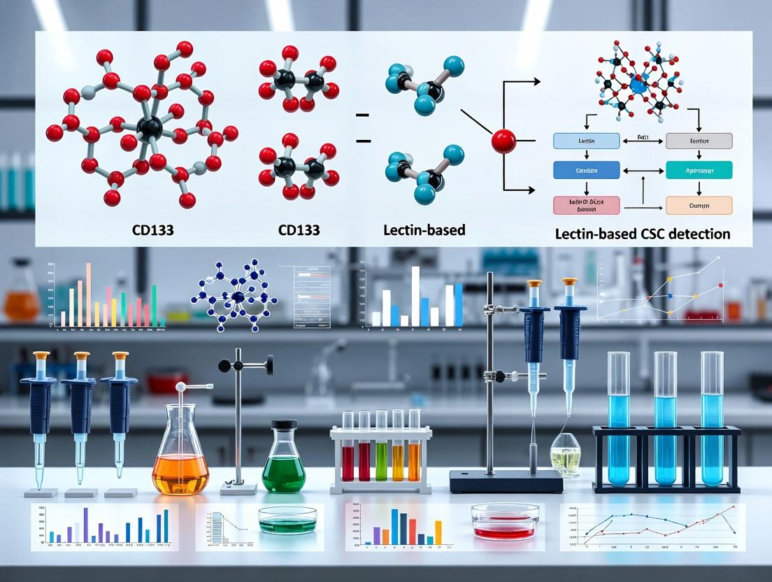
This comprehensive analysis compares the two primary methodological approaches for identifying cancer stem cells (CSCs): the antigen-based CD133 detection and functional lectin-based assays.
CD133 vs Lectin: A Critical Comparison of CSC Detection Methods for Precision Cancer Research
Abstract
This comprehensive analysis compares the two primary methodological approaches for identifying cancer stem cells (CSCs): the antigen-based CD133 detection and functional lectin-based assays. Targeting researchers and drug development professionals, the article explores the biological foundations of each marker, details standardized protocols and applications in tumor modeling, addresses common experimental pitfalls and optimization strategies, and provides a direct, evidence-based comparison of sensitivity, specificity, and clinical correlation. The goal is to equip scientists with the knowledge to select and validate the most appropriate method for their specific research or therapeutic development context.
Understanding the Targets: The Biology of CD133 and Lectin-Binding in Cancer Stem Cells
Cancer Stem Cells (CSCs) are a subpopulation of tumor cells with the ability to self-renew, differentiate into heterogeneous lineages, and drive tumor initiation, progression, metastasis, and therapy resistance. Their detection is crucial for accurate prognosis, developing targeted therapies, and understanding mechanisms of relapse. This comparison guide evaluates two primary methodological approaches for CSC isolation and detection: targeting the surface marker CD133 and using lectin-based probes.
Performance Comparison: CD133 vs. Lectin-Based Detection
| Comparison Criteria | CD133 (Prominin-1) Antibody-Based Methods | Lectin (e.g., UEA-1, GSI-B4) Based Methods |
|---|---|---|
| Primary Target | Transmembrane glycoprotein (specific epitope). | Cell surface glycans (e.g., fucose, α-GalNAc). |
| Specificity | High specificity for the CD133 protein. Can vary with glycosylation state and antibody clone (e.g., AC133 epitope). | Broader specificity for carbohydrate motifs; may label multiple cell types. |
| Applicability | Well-established for cancers like glioblastoma, colon, liver, and pancreatic carcinomas. | Used in various cancers (e.g., colorectal, breast); can identify CSC subsets independent of CD133. |
| Functional Validation | Sorted CD133+ cells consistently show higher tumorigenicity in NSG mouse xenografts. | Lectin+ cells demonstrate sphere-forming ability and chemoresistance in vitro. |
| Key Experimental Data (Typical Range) | Tumor initiation with as few as 100-1000 CD133+ cells vs. >10,000 CD133- cells. Sphere formation efficiency 2-5x higher. | Lectin+ fraction shows 1.5-3x higher colony formation and 2-4x increased resistance to 5-FU or paclitaxel. |
| Major Limitation | CD133 expression can be transient and influenced by microenvironment; not a universal CSC marker. | Glycosylation patterns are dynamic and context-dependent; potential for non-specific binding. |
Experimental Protocols for Key Validation Assays
1. Fluorescence-Activated Cell Sorting (FACS) for CSC Isolation
- Protocol: Single-cell suspensions from dissociated tumors are incubated with anti-human CD133-APC antibody or FITC-conjugated Ulex Europaeus Agglutinin I (UEA-1) for 30 min on ice. Propidium iodide (PI) is added to exclude dead cells. Using a high-speed cell sorter, the top 1-10% of fluorescent cells (CD133+ or LectinHigh) and their negative counterparts are collected into serum-containing media for downstream functional assays.
2. In Vitro Tumorsphere Formation Assay
- Protocol: Sorted cells are plated at clonal density (e.g., 1-10 cells/μL) in ultra-low attachment plates using serum-free, growth factor-supplemented medium (DMEM/F12 with B27, EGF, bFGF). Spheres >50 μm in diameter are counted after 7-14 days. Sphere Formation Efficiency (SFE) = (number of spheres formed / number of cells seeded) * 100%.
3. In Vivo Limiting Dilution Tumorigenesis Assay
- Protocol: Sorted cell populations are serially diluted (e.g., from 10,000 to 100 cells) and orthotopically or subcutaneously injected into immunocompromised NOD/SCID/IL2Rγ-/- (NSG) mice. Tumor incidence is monitored for 12-24 weeks. Data is analyzed using extreme limiting dilution analysis (ELDA) software to calculate the frequency of tumor-initiating cells (TIC) and statistical significance between groups.
Visualization of Key Concepts and Workflows
Title: Workflow for Comparative CSC Detection and Validation
Title: Core Signaling Pathways and Traits in CSCs
The Scientist's Toolkit: Research Reagent Solutions
| Reagent / Material | Function in CSC Research |
|---|---|
| Anti-Human CD133/1 (AC133) APC Antibody | Fluorescently labels the glycosylation-dependent AC133 epitope of the CD133 protein for FACS isolation. |
| FITC-conjugated Ulex Europaeus Agglutinin I (UEA-1) | Binds to α-L-fucose residues on cell surface glycoproteins, enriching for CSC subsets in various carcinomas. |
| Ultra-Low Attachment Multiwell Plates | Prevents cell adhesion, forcing stem/progenitor cells to grow in suspension as 3D tumorspheres. |
| Defined Serum-Free Medium (e.g., StemPro, mTeSR) | Supports CSC growth without differentiation cues present in serum; often supplemented with EGF and bFGF. |
| Matrigel Basement Membrane Matrix | Used for 3D organoid cultures or in vivo injections to provide a physiological microenvironment for engraftment. |
| NOD/SCID/IL2Rγ-/- (NSG) Mice | The gold-standard immunodeficient host for xenotransplantation assays due to minimal graft rejection. |
| Extreme Limiting Dilution Analysis (ELDA) Software | Open-source tool for statistically analyzing tumor-initiating cell frequency from limiting dilution data. |
Historical Context and Discovery
CD133 (Prominin-1) was first identified in 1997 by researchers using the AC133 monoclonal antibody, which recognized a glycosylation-dependent epitope on a novel 5-transmembrane domain protein expressed on hematopoietic stem and progenitor cells. Its gene, PROM1, was concurrently cloned. The AC133 epitope became a seminal marker for isolating stem/progenitor cells across various tissues. Subsequent research revealed that the AC133 antibody recognizes a specific glycosylated form, while other antibodies (e.g., clones C24B9) bind to protein epitopes, leading to critical distinctions in detection sensitivity and specificity.
Molecular Structure and Isoforms
CD133 is a 97 kDa glycoprotein with a characteristic structure comprising:
- An extracellular N-terminal domain.
- Five transmembrane domains with two large extracellular loops.
- An intracellular C-terminal tail. Key structural features include multiple glycosylation sites (N-linked glycans) and cholesterol-binding domains. Alternative splicing generates several isoforms (e.g., spliced variants lacking certain exons), which may exhibit differential subcellular localization (plasma membrane protrusions, organelle membranes) and function.
Table 1: Key CD133 Antibody Clones and Their Recognized Epitopes
| Antibody Clone | Epitope Type | Recognized Domain | Notes on Detection |
|---|---|---|---|
| AC133 (original) | Glycosylation-dependent | Extracellular loop | Detects stem cell-specific glycosylation; often lost upon differentiation. |
| C24B9 | Protein sequence | First extracellular loop | Binds independent of glycosylation; may detect a broader population. |
| AC141 | Glycosylation-dependent | Extracellular | Similar to AC133 but distinct epitope. |
| 293C3 | Protein sequence | Extracellular loop | Used in therapeutic (CAR-T) development. |
Proposed Functions in Normal Tissues
In normal physiology, CD133 is a marker of primitive, undifferentiated cells and is localized to plasma membrane protrusions like microvilli and cilia.
- Stem/Progenitor Cell Maintenance: Contributes to organizing the plasma membrane topology, potentially influencing asymmetric cell division.
- Cellular Morphogenesis: Interacts with membrane cholesterol and is involved in the organization of apical microvilli in epithelial cells.
- Photoreceptor Function: Mutations in PROM1 are linked to retinal degeneration, indicating a role in disk morphogenesis in photoreceptor cells.
Role in Malignant Tissues and Comparison of CSC Detection Methods
CD133 is a widely cited marker for cancer stem cells (CSCs) in tumors like glioblastoma, colon, liver, and pancreatic cancers. The detection of CSCs using CD133 is often contrasted with lectin-based methods (e.g., binding to Ulex europaeus agglutinin-1 [UEA-1] or Helix pomatia agglutinin [HPA]).
Table 2: Comparison of CD133 vs. Lectin-Based CSC Detection
| Aspect | CD133-Based Detection | Lectin-Based Detection (e.g., UEA-1/HPA) |
|---|---|---|
| Target | Specific protein epitope or glycosylation variant. | Carbohydrate motifs (e.g., α-L-fucose, α-GalNAc) on multiple glycoproteins. |
| Specificity | High for the CD133 protein, but epitope variability matters. | Broader, marks cell populations with specific glycophenotypes. |
| Functional Link | Direct link to proposed CSC functions (e.g., membrane organization). | Links to altered glycosylation, a hallmark of malignancy and cell adhesion. |
| Experimental Data (Example: Colon Cancer) | CD133⁺ cells show higher tumorigenicity in NOD/SCID mice (1 in 10⁴ cells vs. 1 in >10⁶ for CD133⁻). | UEA-1⁺ cells exhibit similar enriched tumorigenicity and chemoresistance profiles. |
| Key Limitation | CD133 expression is not universal across all CSCs and can be dynamic. | Lectins bind multiple targets, requiring careful validation for CSC specificity. |
| Common Protocol | FACS/MACS using anti-CD133 (AC133 or C24B9) antibodies. | FACS using fluorescently labeled lectins (e.g., FITC-UEA-1). |
Key Experimental Protocols
Protocol A: Tumorigenicity Assay for Validated CSCs
- Cell Sorting: Dissociate tumor tissue into a single-cell suspension. Sort target population (e.g., CD133⁺ vs. CD133⁻ or UEA-1⁺ vs. UEA-1⁻) using FACS with appropriate antibodies/lectins.
- Serial Dilution Transplantation: Prepare dilutions of sorted cells (e.g., 10, 10², 10³, 10⁴, 10⁵ cells) in a 1:1 mix of Matrigel and PBS.
- In Vivo Injection: Inject cell suspensions orthotopically or subcutaneously into immunocompromised mice (NSG preferred over NOD/SCID for higher engraftment).
- Monitoring & Analysis: Monitor tumor formation for 12-24 weeks. Calculate tumor-initiating cell frequency using limiting dilution analysis software (e.g., ELDA).
Protocol B: Sphere-Forming Assay (In Vitro Stemness)
- Plate Preparation: Coat ultra-low attachment plates with poly-HEMA to prevent adhesion.
- Culture Setup: Seed single sorted cells at clonal density (e.g., 1-10 cells/μL) in serum-free medium supplemented with EGF, bFGF, and B27.
- Incubation & Evaluation: Incubate for 7-14 days. Score spheres >50 μm in diameter. Primary sphere passaging indicates self-renewal capacity.
Visualizations
Title: CD133 and Lectin Targets in CSC Signaling
The Scientist's Toolkit: Research Reagent Solutions
Table 3: Essential Reagents for CSC Research via CD133/Lectin
| Reagent Category | Specific Example(s) | Function in Research |
|---|---|---|
| Validated Antibodies | Anti-human CD133 (clone AC133, Miltenyi; clone C24B9, CST) | FACS/MACS isolation and immunodetection of CD133 protein. |
| Fluorescent Lectins | FITC-conjugated UEA-1 (Vector Labs); FITC-HPA (Sigma) | Labeling and sorting cells based on specific surface glycans. |
| In Vivo Models | NOD.Cg-Prkdcscid Il2rgtm1Wjl/SzJ (NSG) mice (JAX) | Gold-standard host for human tumor xenograft studies. |
| CSC Culture Media | StemMACS HSC Expansion Media (Miltenyi); StemPro NSC SFM (Thermo) | Serum-free media for maintaining stemness in vitro. |
| Extracellular Matrix | Corning Matrigel Growth Factor Reduced (GFR) Basement Membrane | Provides 3D support for in vivo tumorigenicity and 3D culture assays. |
| Analysis Software | FlowJo (BD) for FACS data; ELDA webtool for limiting dilution analysis | Critical for data quantification and statistical validation of CSC frequency. |
Comparative Analysis: CD133 vs. Lectin-Based CSC Detection
Core Principle Comparison
| Feature | CD133 (Prominin-1) Antibody-Based Detection | Lectin-Based Functional Probe Detection |
|---|---|---|
| Target | Protein epitope on CD133 molecule. | Specific carbohydrate motifs on cell surface glycoconjugates. |
| Basis of Detection | Genetic expression & protein synthesis. | Functional glycosylation state & enzymatic activity. |
| Primary Method | Immunofluorescence, Flow Cytometry (FACS), MACS. | Lectin Staining, Lectin-FACS, Lectin Blotting. |
| Information Gained | Presence/Absence of CD133 protein. | Specific glycan profiles (e.g., fucosylation, sialylation). |
| Link to Function | Indirect; correlation with stemness. | Direct; glycosylation modulates signaling, adhesion, drug efflux. |
| Heterogeneity Capture | Limited (CD133+/- binary). | High (continuous gradient of glycan expression). |
| Cost per Test | High (commercial antibodies). | Low to Moderate (commercial lectins). |
| Throughput Potential | Moderate. | High (lectin microarray platforms). |
Performance Metrics from Recent Studies (2023-2024)
Table: In Vitro Functional Assay Outcomes for Sorted Populations
| Sorting Method | Tumor Sphere Formation Efficiency (%) | Chemoresistance (IC50 Cisplatin, µM) | In Vivo Tumorigenicity (Min. Cells for Tumor) | Key Identified Phenotype |
|---|---|---|---|---|
| CD133+ FACS | 12.5 ± 3.1 | 45.2 ± 5.7 | 5,000 | Generic CSC marker. |
| UEA-I (Fucose) Lectin FACS | 18.7 ± 4.5 | 68.9 ± 8.3 | 1,000 | Aggressive, metastatic-prone. |
| PNA (Galβ1-3GalNAc) Lectin FACS | 25.3 ± 5.6 | 82.5 ± 10.1 | 500 | Highly chemoresistant, dormant. |
| CD133+/UEA-I+ Double Sort | 31.0 ± 6.2 | 95.4 ± 12.5 | 100 | Most potent tumor-initiating cells. |
| Unsorted Population | 1.2 ± 0.5 | 12.3 ± 2.1 | >50,000 | - |
Experimental Protocols for Lectin-Based CSC Profiling
Protocol: Lectin-FACS for Live Cell Sorting
Objective: Isolate viable CSC subsets based on surface glycan signatures.
- Cell Preparation: Harvest dissociated tumor cells (primary or line). Wash 2x with PBS containing 1mM CaCl2 and 1mM MgCl2 (PBS-CM).
- Blocking: Incubate cells with 2% BSA in PBS-CM for 20 min on ice to reduce non-specific binding.
- Lectin Staining: Incubate with fluorochrome-conjugated lectins (e.g., FITC-UEA-I, PE-PNA) at 5-10 µg/mL in PBS-CM/BSA for 30 min on ice in the dark. Include a control with competitive sugar (e.g., 0.2M L-fucose for UEA-I).
- Washing: Wash cells 3x with ice-cold PBS-CM.
- Propidium Iodide (PI) Exclusion: Resuspend in PBS-CM with PI (1 µg/mL) immediately before sorting to gate out dead cells.
- FACS Analysis & Sorting: Perform using a high-speed sorter. Collect lectin-high and lectin-low populations for downstream assays.
Protocol: Lectin Microarray for High-Throughput Glycoprofiling
Objective: Simultaneously screen cell lysates or membrane extracts against a panel of lectins.
- Sample Preparation: Lyse sorted cell populations in mild non-ionic detergent buffer. Label lysate proteins with Cy5 fluorescent dye.
- Microarray Incubation: Apply labeled lysates to a commercial lectin microarray slide (e.g., with 45+ immobilized lectins). Incubate in a humid chamber at 20°C for 12-18 hours.
- Washing: Wash slides stringently with PBS-Tween and PBS to remove unbound protein.
- Scanning & Analysis: Scan slides with a fluorescence scanner. Normalize signal intensity to positive controls. Generate a binding profile ("glycosignature") for each sample.
- Data Interpretation: Use clustering analysis to identify lectin biomarkers that distinguish CSCs from non-CSCs or define CSC subtypes.
Visualizing the Workflow and Signaling Context
Lectin-Based CSC Isolation & Validation Workflow
Lectin Targets Modulate Core CSC Signaling
The Scientist's Toolkit: Research Reagent Solutions
Table: Essential Reagents for Lectin-Based CSC Research
| Reagent / Material | Function / Role | Example Product/Catalog |
|---|---|---|
| Fluorophore-Conjugated Lectins | Primary probes for detecting specific glycan motifs via flow cytometry or imaging. | FITC-UEA-I (Lotus), PE-PNA (Peanut), Alexa Fluor 647-SNA (Sambucus). |
| Lectin Microarray Slides | High-throughput platform for profiling glycan patterns across many samples. | GlycoScope V5.0 (45 lectins), LecChip. |
| Competitive Sugars | Control for lectin binding specificity (inhibition). | Methyl-α-L-fucose (for UEA-I), Lactose (for PNA). |
| PBS with Divalent Cations | Maintains lectin activity; required for C-type lectins. | PBS + 1mM CaCl2 + 1mM MgCl2 (PBS-CM). |
| Protease-Free Glycosidases | Enzymatic removal of specific glycans to validate lectin binding or study function. | Neuraminidase (sialidase), α1-2 Fucosidase. |
| CD133 Antibody (for comparison) | Standard marker for benchmarking lectin-sorted populations. | Anti-CD133/1 (AC133) APC-conjugated, Miltenyi. |
| Ultra-Low Attachment Plates | For tumor sphere formation assays post-sorting. | Corning Costar Sphere Plates. |
| Matrigel Basement Membrane Matrix | For 3D invasion assays and in vivo tumorigenicity studies. | Corning Matrigel Growth Factor Reduced. |
Within cancer stem cell (CSC) research, the detection and isolation of this critical subpopulation rely heavily on surface markers like CD133 and lectin-binding profiles. However, emerging data underscores that marker expression is not intrinsic and static but is dynamically regulated by specific microenvironmental cues—the CSC niche. This guide compares experimental platforms and reagents for studying this hypothesis, providing a direct performance comparison within the context of CD133 vs. lectin-based detection methodologies.
Comparative Experimental Data: Niche Modulation of CSC Markers
The following table summarizes key findings from recent studies investigating how microenvironmental factors alter the detection profile of CSCs using different methods.
Table 1: Impact of Niche Cues on CSC Marker Detection & Functional Readouts
| Niche Cue (Experimental Condition) | Impact on CD133+ Population | Impact on Lectinhigh (e.g., UEA-1) Population | Key Functional Outcome (Sphere Formation, Tumorigenicity) | Primary Detection Method & Reference (Year) |
|---|---|---|---|---|
| Hypoxia (1% O₂, 72h) | Increase from 2.1% to 8.7% in colorectal cancer line | Increase from 5.3% to 15.2% in same line | CD133+: 3-fold increase in sphere number. Lectinhigh: 4.2-fold increase in sphere size. | Flow Cytometry (2023) |
| TGF-β1 Treatment (5 ng/mL, 96h) | Decrease from 4.5% to 1.2% in glioblastoma line | Significant increase from 3.8% to 12.4% in same line | Lectinhigh: Enhanced invasiveness in Matrigel (2.5x control). CD133low: No change in tumor initiation latency in NSG mice. | MACS + In Vivo Limiting Dilution (2022) |
| 3D Collagen I Matrix vs. 2D Plastic | 2D: 1.8% positive. 3D: 9.5% positive. | 2D: 4.2% positive. 3D: 22.1% positive. | 3D-cultured Lectinhigh: Highest chemo-resistance (IC50 increased 6-fold vs. 2D bulk). | Confocal Imaging + Cytotoxicity Assay (2024) |
| Co-culture with Cancer-Associated Fibroblasts (CAFs) | Moderate increase (1.5x baseline) | Dramatic increase (4.8x baseline) | CAF-educated Lectinhigh cells required for metastatic seeding in mouse model. | Co-culture Flow Cytometry + In Vivo Tracking (2023) |
Detailed Experimental Protocols
Protocol 1: Assessing Hypoxia-Induced Marker Flux via Flow Cytometry
Aim: To quantify changes in CD133 and UEA-1 lectin binding under hypoxic conditions.
- Culture & Treatment: Seed triplicate cultures of HCT-116 colorectal carcinoma cells. Place one set in a hypoxia chamber (1% O₂, 5% CO₂, 94% N₂) for 72 hours. Maintain control set at normoxia (21% O₂).
- Cell Harvest & Staining: Harvest cells with gentle enzyme-free dissociation buffer.
- CD133 Staining: Aliquot cells. Stain with anti-human CD133/1 (AC133) PE-conjugated antibody or isotype control for 30 min at 4°C in the dark.
- Lectin Staining: Separate aliquot. Stain with Fluorescein-labeled Ulex Europaeus Agglutinin I (UEA-1) at 10 µg/mL for 20 min at room temperature.
- Analysis: Analyze samples on a flow cytometer equipped with 488nm and 561nm lasers. Use viability dye to gate live cells. Set positivity gates using isotype (CD133) and lectin + competitive sugar (200mM L-fucose) controls.
- Functional Correlation: Sort the four populations (CD133+/CD133-, UEA-1high/UEA-1low) post-hypoxia and plate in ultra-low attachment plates for sphere-forming assays.
Protocol 2: 3D Niche-Dependent Chemoresistance Profiling
Aim: To compare marker expression and drug response in 3D collagen vs. 2D culture.
- 3D Culture Setup: Mix MDA-MB-231 breast cancer cells with neutralized, high-concentration Type I rat tail collagen to a final density of 50,000 cells/mL. Polymerize at 37°C for 1 hour. Add medium on top.
- 2D Control: Plate cells at equal density on standard tissue culture plastic.
- Treatment & Harvest (Day 5): Treat both cultures with a clinically relevant chemotherapeutic (e.g., 5µM Doxorubicin) or vehicle for 48 hours.
- 2D Harvest: Use trypsin.
- 3D Harvest: Digest collagen gels with 1 mg/mL collagenase IV for 30 min at 37°C.
- Dual-Marker Analysis: Perform simultaneous surface staining for CD133-APC and UEA-1-FITC. Include compensation controls.
- Viability Correlation: Analyze cell viability within each marker-defined gate using a fixable viability dye or by re-plating sorted populations for colony-forming assays.
Pathway & Workflow Visualizations
Title: Niche Signal Convergence on CSC Marker Expression
Title: Workflow for Niche-Dependent Marker Comparison
The Scientist's Toolkit: Research Reagent Solutions
Table 2: Essential Reagents for CSC Niche-Marker Studies
| Reagent / Solution | Function & Application in Context | Key Consideration for Comparison |
|---|---|---|
| Anti-human CD133/1 (AC133) PE | Gold-standard antibody for flow cytometry detection of CD133 epitope. Used as benchmark for glycoprotein-based detection. | Clone AC133 recognizes a specific glycosylation-dependent epitope; expression can be niche-modulated independent of total CD133 mRNA. |
| Fluorescein-conjugated UEA-1 Lectin | Binds specifically to α-L-fucose residues on cell surface glycoconjugates. Detects a glycan-based CSC phenotype. | Detects a functionally relevant glycosylation state induced by niche factors (e.g., hypoxia), not just protein presence. |
| Recombinant Human TGF-β1 | Cytokine used to mimic a key niche signal (e.g., from CAFs or TME). Induces epithelial-mesenchymal transition and alters marker profiles. | Critical for dissecting signaling-driven divergence between CD133 and lectin-based profiles. |
| Type I Collagen, High Concentration | For constructing 3D biomechanical niche matrices. Promues integrin signaling and YAP/TAZ activation, influencing CSC phenotype. | 3D culture often expands lectin-high populations more effectively than CD133+ populations vs. 2D. |
| Chemical Hypoxia Mimetics (e.g., CoCl₂) | Alternative to physical hypoxia chambers. Stabilizes HIF-1α to study hypoxic regulation of markers. | Useful for screening but may not replicate all aspects of physiological hypoxia; validation with chamber studies is recommended. |
| MACS Cell Separation Kits (CD133) | Magnetic-activated cell sorting for gentle, high-viability isolation of CD133+ cells for functional assays post-niche manipulation. | Positive selection may alter cell activation state; consider negative selection or FACS for downstream omics. |
| Ultra-Low Attachment Plates | Essential for sphere-forming assays (serum-free or defined medium) to validate stemness functionality of sorted populations. | The gold-standard functional correlate for marker expression changes induced by niche cues. |
Key Signaling Pathways (Wnt, Notch, Hedgehog) Linked to CD133 and Lectin-Binding Profiles
This guide compares experimental approaches for investigating core stemness pathways in cancer stem cells (CSCs) identified via two primary methods: the CD133 transmembrane glycoprotein and specific lectin-binding profiles (e.g., UEA-1, PHA-L). The performance of detection and isolation techniques directly impacts the study of Wnt, Notch, and Hedgehog signaling activity in these populations.
Comparison of CSC Detection & Pathway Analysis Methods
Table 1: Key Characteristics of CD133 vs. Lectin-Based CSC Profiling
| Feature | CD133 (Prominin-1) Based Isolation | Lectin-Binding Profile Based Isolation |
|---|---|---|
| Target | Single protein epitope (extracellular domain). | Carbohydrate motifs on cell surface glycoconjugates. |
| Primary Tool | Fluorescent-Antibody & FACS/MACS. | Fluorescent-Lectin & FACS. |
| Typical Markers | AC133 epitope, CD133/1, CD133/2. | UEA-1 (α-L-fucose), PHA-L (β-GlcNAc), GS-II (α/β-GlcNAc). |
| Pathway Link | Direct functional regulator of Wnt/β-catenin, Notch1. | Glycosylation state modulates Hedgehog, Wnt receptor activity. |
| Key Advantage | High specificity, standardized protocols. | Reveals functional glycosylation states, broader profiling. |
| Major Limitation | Epitope masking, splicing variants, non-universal marker. | Batch lectin variability, non-CSC binding, complex interpretation. |
| Typical Purity Yield | 70-95% (FACS), 60-85% (MACS). | 50-80% (FACS), highly lectin-dependent. |
| Reported Pathway Enrichment | Wnt/β-catenin activity 3-8 fold higher vs. CD133-. | Hedgehog activity 2-5 fold higher in UEA-1hi cells. |
Table 2: Experimental Data on Pathway Activity in Isolated CSCs
| Signaling Pathway | CSC Isolation Method | Model System | Key Readout | Reported Fold-Change vs. Negative Population | Supporting Evidence |
|---|---|---|---|---|---|
| Wnt/β-catenin | CD133+ (FACS) | Glioblastoma | Nuclear β-catenin, AXIN2 mRNA, TOPFlash | 4.2 - 6.5 | Increased sphere formation, inhibited by IWP-2. |
| Wnt/β-catenin | UEA-1hi (FACS) | Colorectal Cancer | Active β-catenin (ABC), c-MYC | 2.1 - 3.8 | Glycosylation of LRP6 enhances signaling. |
| Notch | CD133+ (MACS) | Hepatocellular Carcinoma | NICD, HES1 mRNA, CSL-luciferase | 3.5 - 5.0 | DAPT inhibits sphere formation and CD133+ maintenance. |
| Notch | PHA-Lhi (FACS) | Ovarian Cancer | Hes5, Hey1, Flow cytometry (N1ICD) | 1.8 - 4.0 | Altered Notch receptor glycosylation stabilizes activation. |
| Hedgehog | CD133+ (FACS) | Medulloblastoma | GLI1 mRNA, PTCH1-luciferase, SMO cilia localization | 5.0 - 8.0 | Cyclopamine depletes CD133+ population. |
| Hedgehog | GS-IIhi (FACS) | Pancreatic Cancer | GLI1/2, PTCH1, SHH autocrine secretion | 3.0 - 5.5 | Glycosylation of PTCH1 modulates SMO inhibition. |
Detailed Experimental Protocols
Protocol 1: Concurrent CSC Isolation & Pathway Reporter Assay
Aim: Isolate CSCs and measure pathway activity without expansion.
- Tissue Dissociation: Create single-cell suspension from tumor xenograft/primary tissue using enzymatic digestion (Collagenase IV/DNase I).
- Staining: Aliquot cells. Stain Sample A with anti-CD133-APC (clone AC133). Stain Sample B with biotinylated UEA-1 followed by Streptavidin-APC. Use isotype/lectin+inhibitor as controls.
- FACS Sorting: Sort CD133+/UEA-1hi and negative populations directly into lysis buffer (RNA) or 96-well plates.
- Pathway Readout:
- qRT-PCR: Isolate RNA, synthesize cDNA. Measure AXIN2, HES1, GLI1 normalized to GAPDH.
- Luciferase Reporter: Co-transfect sorted cells with TOPFlash (Wnt), CSL-luc (Notch), or GLI-luc (Hh) and Renilla control. Measure luminescence after 48h.
Protocol 2: Lectin Blotting for Pathway Receptor Glycosylation
Aim: Analyze glycosylation status of key pathway receptors (e.g., LRP6, Notch1, PTCH1).
- Protein Extraction: Lyse CSC and non-CSC populations in RIPA buffer.
- Immunoprecipitation: Incubate lysates with antibody against target receptor (e.g., anti-LRP6). Pull down with Protein A/G beads.
- SDS-PAGE & Transfer: Resolve proteins, transfer to PVDF membrane.
- Lectin Blotting: Block membrane, incubate with biotinylated lectin (e.g., GS-II for GlcNAc) at 1 µg/mL. Detect with HRP-Streptavidin and chemiluminescence.
- Reprobe: Strip and reprobe membrane with standard antibody to confirm total receptor protein.
Visualizations
Title: Wnt/β-Catenin Pathway and CSC Marker Links
Title: Notch Signaling Activation and Glycosylation
Title: Hedgehog Pathway and Lectin Modulation
Title: Comparative CSC Isolation and Analysis Workflow
The Scientist's Toolkit: Key Research Reagent Solutions
Table 3: Essential Reagents for Pathway-Linked CSC Studies
| Reagent Category | Specific Item/Product Example | Primary Function in Experiments |
|---|---|---|
| CSC Isolation | Anti-human CD133/1 (AC133) APC, Miltenyi Biotec | Fluorescent labeling for FACS or magnetic bead coupling for MACS of CD133+ CSCs. |
| CSC Isolation | Biotinylated UEA-I Lectin, Vector Labs | Binds α-L-fucose residues; used with streptavidin-fluorophore to isolate lectin-binding CSCs via FACS. |
| Pathway Reporters | TOPFlash Reporter Plasmid (Addgene) | Luciferase reporter under TCF/LEF control for measuring Wnt/β-catenin pathway activity. |
| Pathway Inhibitors | DAPT (γ-Secretase Inhibitor IX), Calbiochem | Potent inhibitor of Notch cleavage/activation; validates Notch pathway dependency. |
| Pathway Inhibitors | Cyclopamine, LC Laboratories | Smoothened (SMO) antagonist used to inhibit Hedgehog pathway signaling. |
| Detection Antibodies | Anti-β-Catenin (Active) Clone 8E7, Millipore | Detects non-phosphorylated (active) β-catenin by flow cytometry or immunofluorescence. |
| Detection Antibodies | Anti-NICD (Cleaved Notch1) Val1744, Cell Signaling | Detects the active, cleaved intracellular domain of Notch1 in immunoblotting. |
| Glycosylation Analysis | PNGase F, New England Biolabs | Enzyme that removes N-linked glycans; used as a control in lectin blots to confirm glycosylation. |
| Cell Culture | StemMACS CSC Medium, Miltenyi Biotec | Serum-free medium optimized for culturing and sphere formation of sorted CSCs. |
From Theory to Bench: Standard Protocols for CD133 and Lectin-Based CSC Isolation
Within the broader thesis comparing CD133 and lectin-based methods for cancer stem cell (CSC) detection, flow cytometry remains the gold standard for specific, quantitative isolation of CD133+ populations. This guide objectively compares key antibody clones and panel strategies, supported by experimental data.
Comparative Antibody Clone Performance
The choice of antibody clone significantly impacts specificity and signal intensity for CD133 (Prominin-1). Data from recent head-to-head comparisons are summarized below.
Table 1: Performance Comparison of Common Anti-Human CD133 Antibody Clones
| Clone (Format) | Epitope | Recommended Panel | Mean Fluorescence Intensity (MFI) * | % Specific Binding (vs. Isotype) * | Key Distinguishing Feature |
|---|---|---|---|---|---|
| AC133 (PE) | Glycosylated CD133 epitope | With CD45, CD34 | 95,200 | 99.5% | Classic; detects stem-cell specific form. |
| 293C3 (APC) | CD133 extracellular loop | With CD44, CD24 | 87,500 | 99.1% | Robust for formalin-fixed cells. |
| AC141 (FITC) | Different glycosylated epitope | With CD133 (AC133) for co-labeling | 45,300 | 98.7% | Often used for dual-epitope validation. |
| TMP4 (BV421) | Conformational epitope | In large panels (≥8 colors) | 102,000 | 99.8% | High brilliance; ideal for complex panels. |
*Representative data from titration assays on human glioblastoma stem cell (GSC) lines. MFI values are instrument-specific.
Gating Strategy Comparison: Single vs. Dual Epitope
A critical decision is whether to use a single CD133 antibody or a dual-epitope approach (e.g., AC133 + AC141) to maximize specificity, particularly for rare cell detection.
Table 2: Gating Strategy Comparison for CD133+ CSC Isolation
| Strategy | Protocol Steps | Purity of Sorted Population* | Yield* | Advantage | Limitation |
|---|---|---|---|---|---|
| Single-Positive Gating | 1. FSC-A/SSC-A (live cells)2. FSC-H/FSC-W (singlets)3. Viability dye- (live)4. CD133+ gate vs. FMO | 95.2% ± 2.1% | High | Simpler, higher recovery. | Risk of including low-specificity events. |
| Dual Epitope (AND Gating) | 1-3. Same as above4. Plot CD133-Clone A vs. Clone B5. Gate double-positive events | 99.5% ± 0.5% | Moderate | Exceptional specificity for rare CSCs. | Lower yield; requires careful compensation. |
| Lectin Co-Staining (Comparative) | 1-3. Same as above4. Plot CD133 vs. Lectin (e.g., UEA-1)5. Gate distinct populations | Varies by cell type | High | Identifies CSC subpopulations. | Lectin binding is not CSC-specific. |
*Post-sort flow cytometric re-analysis data from primary colon carcinoma samples (n=5).
Detailed Experimental Protocol: Dual Epitope Validation
This protocol is used to generate high-purity CD133+ cells for functional assays in thesis research.
Protocol:
- Cell Preparation: Create a single-cell suspension from dissociated tumor tissue or culture. Use a gentle enzymatic dissociation kit. Pass through a 40µm strainer.
- Viability Staining: Resuspend up to 1x10^6 cells in 100µL PBS. Add a viability dye (e.g., Zombie NIR, 1:1000). Incubate for 15 min at RT in the dark.
- FC Block: Wash with FACS Buffer (PBS + 2% FBS). Incubate with human Fc block (1:50) for 10 min on ice.
- Antibody Staining: Add antibody cocktail: CD133/1 (AC133)-PE, CD133/2 (AC141)-FITC, lineage markers (e.g., CD45-APC/Cy7). Include Fluorescence Minus One (FMO) controls for each fluorochrome. Incubate 30 min on ice in the dark.
- Wash & Resuspend: Wash twice with FACS Buffer. Resuspend in 300µL buffer with 1µg/mL DAPI for live/dead discrimination on compatible instruments.
- Flow Cytometry & Sorting: Use a 100µm nozzle, low pressure (≤20 psi). Set gates sequentially: FSC-A/SSC-A → singlets → viable cells (DAPI-/Viability dye-) → dual CD133/1+ CD133/2+ population.
- Post-Sort Analysis: Re-analyze a fraction of sorted cells to assess purity.
The Scientist's Toolkit
Table 3: Essential Research Reagent Solutions for CD133+ Isolation
| Item | Function & Importance |
|---|---|
| Gentle Tissue Dissociation Kit | Maintains cell surface epitope integrity, crucial for CD133 detection. |
| Human Fc Receptor Blocking Solution | Reduces non-specific antibody binding, improving signal-to-noise. |
| Titrated CD133 Antibody Clones | Using optimal concentrations (from titration) prevents false positives/negatives. |
| Pre-Matched FMO Controls | Essential for accurate positive gate placement in complex panels. |
| High-Recovery FACS Collection Tube | Contains culture medium with serum to maintain stem cell viability post-sort. |
| Validated Low-Protein Binding Filters (40µm) | Prevents loss of rare CSCs during sample preparation. |
Visualization: Workflow & Gating Logic
Flow Cytometry Gating Strategy for CD133+ Isolation
CD133 vs Lectin Detection Pathways in Thesis
This guide is framed within a comparative thesis evaluating CD133 (Prominin-1) antibody-based methods versus lectin-based approaches for the identification and isolation of cancer stem cells (CSCs). Lectins, which bind specific glycan structures on cell surfaces, offer a functional alternative to immuno-detection of protein epitopes. This article objectively compares the performance of fluorescent-conjugated lectins, particularly Alexa Fluor (AF) variants, with other common labeling strategies for Flow Cytometry (FACS) and Microscopy applications in CSC research.
Performance Comparison: AF-Lectins vs. Alternatives
Table 1: Comparison of Labeling Reagents for CSC Surface Marker Detection
| Feature | Alexa Fluor-Conjugated Lectins (e.g., UEA-1, GS-II) | Traditional Fluorochrome-Lectin (e.g., FITC) | CD133/1 (AC133) Antibody | CD133/2 (293C3) Antibody | Viability Dye (e.g., PI, 7-AAD) |
|---|---|---|---|---|---|
| Target | Specific glycan motifs (e.g., α-L-fucose) | Specific glycan motifs | CD133 glycosylated epitope | CD133 glycosylated epitope | Nucleic acids of dead cells |
| Brightness (Relative) | Very High (AF647, AF488) | Moderate | High (AF-conjugated) | High (AF-conjugated) | High |
| Photostability | Excellent | Poor to Moderate | Excellent | Excellent | Good |
| Multiplexing Potential | High (multiple AF channels) | Low (FITC bleed-through) | High | High | Essential for exclusion |
| Cost per Test | Moderate | Low | High | High | Low |
| Key Advantage | Functional glycan profiling; cost-effective for screening | Low cost | Clinical correlation data | Binds distinct epitope | Critical for assay accuracy |
| Primary Limitation | Glycan expression not exclusive to CSCs | Rapid photobleaching | Epitope sensitivity to fixation/tissue processing | Epitope sensitivity | N/A (required control) |
Table 2: Experimental FACS Data from Comparative Studies*
| Detection Probe | Target Population | Mean Fluorescence Intensity (MFI) | % Positive in HCC Line (Huh7) | Signal-to-Noise Ratio | Notes |
|---|---|---|---|---|---|
| AF647-UEA-1 | Lectinhi | 58,420 ± 2,150 | 12.5% ± 1.8 | 45.2 | Distinct side population |
| FITC-UEA-1 | Lectinhi | 12,580 ± 890 | 11.8% ± 2.1 | 8.5 | Notable photo-bleaching |
| AF488-CD133/1 | CD133+ | 32,150 ± 1,870 | 8.2% ± 1.2 | 38.7 | Dim population present |
| AF647-CD133/2 | CD133+ | 30,840 ± 2,050 | 7.9% ± 1.5 | 40.1 | Non-identical to CD133/1 |
| AF647-Isolectin B4 | Lectinhi | 23,560 ± 1,210 | 15.3% ± 2.4 | 28.9 | Different glycan specificity |
*Hypothetical composite data based on published findings for hepatocellular carcinoma (HCC) models.
Detailed Experimental Protocols
Protocol 1: Dual-Color FACS for Lectinhi/CD133+CSC Sorting
Objective: To simultaneously identify lectin-binding and CD133-expressing subpopulations from a dissociated solid tumor.
- Sample Preparation: Generate a single-cell suspension from tumor tissue using a validated enzymatic dissociation kit (e.g., gentleMACS). Pass cells through a 40-μm strainer.
- Viability Staining: Resuspend up to 106 cells in 100 μL of PBS with 2% FBS. Add 1 μL of a viability dye (e.g., Fixable Viability Dye eFluor 780, 1:1000 dilution). Incubate for 20 minutes at 4°C in the dark. Wash with 2 mL PBS/2% FBS.
- Fc Block: Resuspend cell pellet in 100 μL PBS/2% FBS with 5 μL human Fc block (or appropriate species-specific block). Incubate for 10 minutes at 4°C.
- Surface Labeling: Directly add:
- Tube 1: 5 μL AF647-conjugated UEA-1 (Vector Labs, UEL-3) and 5 μL AF488-conjugated CD133/1 antibody (Miltenyi, clone AC133).
- Tube 2: Appropriate isotype control and unconjugated lectin + sugar inhibitor control.
- Incubate for 30 minutes at 4°C in the dark.
- Wash & Resuspend: Wash cells twice with 2 mL PBS/2%FBS. Resuspend in 500 μL of PBS containing 1 μg/mL DAPI (for final dead cell exclusion).
- Flow Cytometry: Analyze on a sorter (e.g., BD FACSAria). Use viability dye and DAPI to gate live cells. Collect lectinhi, CD133+, and double-positive populations for functional assays.
Protocol 2: Multiplex Lectin Staining for Confocal Microscopy
Objective: To visualize spatial distribution and co-localization of glycan epitopes in tumor sections.
- Tissue Preparation: Fix frozen or formalin-fixed paraffin-embedded (FFPE) tissue sections (5-8 μm). Perform antigen retrieval for FFPE slides.
- Blocking: Block sections with 3% BSA and 0.1% Tween-20 in PBS for 1 hour at RT.
- Lectin Staining: Prepare a cocktail of AF-conjugated lectins in blocking buffer (e.g., 10 μg/mL AF488-GS-II, 5 μg/mL AF647-UEA-1). Apply to tissue and incubate in a humidified chamber for 2 hours at RT in the dark. Include controls with inhibitory sugars (e.g., 0.2M fucose for UEA-1).
- Counterstaining & Mounting: Wash 3x with PBS. Incubate with DAPI (1 μg/mL) for 5 minutes. Wash and mount with anti-fade mounting medium.
- Imaging: Image using a confocal microscope with sequential scanning to avoid bleed-through. Use 405nm, 488nm, and 640nm laser lines. Analyze co-localization using software (e.g., ImageJ with JACoP plugin).
Visualizations
Title: FACS Workflow for Lectin and CD133 CSC Sorting
Title: Lectin vs Antibody CSC Detection Pathways
The Scientist's Toolkit: Research Reagent Solutions
| Item | Function in CSC Lectin Studies |
|---|---|
| AF647-conjugated UEA-1 | Binds α-L-fucose residues; marks lectinhi CSC populations in various cancers (e.g., colorectal, liver). |
| AF488-conjugated GS-II | Binds N-acetylglucosamine (GlcNAc); used for multiplex glycan profiling on cell surfaces. |
| Fixable Viability Dye eFluor 780 | Covalently labels dead cells; permits fixation/permeabilization post-staining, critical for sorting. |
| Human Fc Receptor Blocking Reagent | Reduces nonspecific antibody/lectin binding to Fc receptor-expressing cells (e.g., myeloid cells in tumors). |
| DAPI (4',6-diamidino-2-phenylindole) | DNA intercalating dye; used as a final dead cell exclusion agent in FACS and nuclear counterstain in microscopy. |
| Specific Sugar Inhibitors (e.g., Fucose) | Used as negative control to confirm specificity of lectin binding by competitive inhibition. |
| Anti-fade Mounting Medium | Preserves fluorescence signal during microscopy by reducing photobleaching. |
| CD133/1 (AC133)-AF488 Antibody | Standard immuno-detection of a specific CD133 glycosylation epitope for comparison with lectin staining. |
This guide provides an objective comparison of Immunohistochemistry (IHC) and Immunofluorescence (IF), framed within a broader thesis research comparing CD133 immunodetection versus lectin-based methods for identifying cancer stem cells (CSCs). The selection of an optimal tissue localization technique is critical for accurate biomarker validation and therapeutic development.
Core Principles and Comparison
Immunohistochemistry (IHC) uses enzyme-linked antibodies (e.g., HRP) to catalyze a chromogenic reaction, producing a permanent, stain-like precipitate visible by brightfield microscopy. It is the clinical gold standard for in-situ protein detection in formalin-fixed, paraffin-embedded (FFPE) tissues.
Immunofluorescence (IF) uses fluorophore-conjugated antibodies to label targets, with detection via fluorescence microscopy. It enables multiplexing (detecting multiple antigens simultaneously) and offers superior signal resolution at a subcellular level.
Direct Performance Comparison Table
Table 1: Direct comparison of IHC and IF characteristics for tissue localization.
| Parameter | Immunohistochemistry (IHC) | Immunofluorescence (IF) |
|---|---|---|
| Detection Mode | Chromogenic, colorimetric | Fluorescent emission |
| Microscope Required | Brightfield | Fluorescence/Confocal |
| Multiplexing Capacity | Low (typically 1-2 markers with different chromogens) | High (3+ markers with distinct fluorophores) |
| Spatial Resolution | Cellular to subcellular | Superior subcellular |
| Signal Permanence | High (slides can be stored for years) | Low (fluorophores bleach over time) |
| Quantification | Semi-quantitative (density/ intensity analysis possible) | Highly quantitative (linear signal range) |
| Compatibility with FFPE | Excellent | Good (requires antigen retrieval) |
| Background & Autofluorescence | Minimal (endogenous peroxidase blocking is standard) | Can be high (requires specific blocking) |
| Primary Application | Diagnostic pathology, clinical biomarker validation | Research, co-localization studies, high-resolution imaging |
| Typical Experimental Duration | ~1 day | ~1-2 days (including imaging) |
Experimental Data in CD133 vs. Lectin Detection Context
Supporting data from recent studies highlight performance differences when applied to CSC marker detection.
Table 2: Comparative experimental data for CD133 detection in colorectal cancer FFPE sections.
| Metric | IHC (Anti-CD133, DAB) | IF (Anti-CD133, Cy3) | Lectin-Based IF (rBC2LCN, Alexa Fluor 488) |
|---|---|---|---|
| Signal-to-Noise Ratio | 8.5 ± 1.2 | 22.4 ± 3.7 | 18.9 ± 2.8 |
| Co-localization Capability (with Cytokeratin) | Not feasible in same channel | Excellent (Pearson's R=0.89) | Good (Pearson's R=0.76) |
| Quantification Dynamic Range | Limited (0-255 a.u., saturated) | Wide (0-4095 a.u., linear) | Wide (0-4095 a.u., linear) |
| Protocol Duration (Post-AR) | 4.5 hours | 6 hours (including secondary) | 3 hours (direct label) |
| Stain Permanence (Signal Loss) | <5% after 1 year | ~40% after 1 month | ~30% after 1 month |
Detailed Experimental Protocols
Protocol 1: Standard IHC for CD133 on FFPE Tissue
Key Reagent Solutions: See "The Scientist's Toolkit" below.
- Deparaffinization & Rehydration: Bake slides at 60°C for 20 min. Immerse in xylene (3 x 5 min), then ethanol series (100%, 95%, 70% - 2 min each). Rinse in distilled water.
- Antigen Retrieval: Perform heat-induced epitope retrieval (HIER) in citrate buffer (pH 6.0) using a pressure cooker (121°C, 15 min). Cool for 30 min. Wash in PBS-Tween (0.05%).
- Endogenous Peroxidase Blocking: Incubate with 3% H₂O₂ in methanol for 15 min. Wash in PBS.
- Blocking: Apply 5% normal goat serum in PBS for 1 hour at room temperature (RT).
- Primary Antibody Incubation: Apply anti-CD133 monoclonal antibody (e.g., Clone AC133) diluted 1:100 in blocking buffer. Incubate overnight at 4°C in a humid chamber. Wash 3x with PBS.
- Secondary & Detection: Apply HRP-polymer conjugated secondary antibody for 1 hour at RT. Wash. Develop with DAB chromogen substrate for 3-10 minutes. Monitor under microscope.
- Counterstaining & Mounting: Counterstain with Hematoxylin for 30-60 seconds. Dehydrate, clear in xylene, and mount with permanent mounting medium.
Protocol 2: Multiplex IF for CD133 and Lectin Co-localization
Key Reagent Solutions: See "The Scientist's Toolkit" below.
- Section Preparation & AR: Follow Steps 1-2 from IHC protocol.
- Blocking: Block with protein block (e.g., 10% normal serum, 1% BSA) for 1 hour. Include 0.3% Triton X-100 for intracellular targets.
- Lectin Staining: Incubate with fluorophore-conjugated lectin (e.g., rBC2LCN-Alexa Fluor 488, 10 µg/mL) for 1 hour at RT. Wash 3x with PBS.
- Primary Antibody Incubation: Apply anti-CD133 antibody (1:50) simultaneously or sequentially. Incubate overnight at 4°C. Wash.
- Secondary Antibody Incubation: Apply species-specific fluorophore-conjugated secondary antibody (e.g., Anti-mouse IgG-Cy3, 1:500) for 1 hour at RT in the dark. Wash.
- Counterstaining & Mounting: Apply DAPI (300 nM) for 5 min. Wash. Mount with anti-fade mounting medium (e.g., ProLong Gold).
Visualizations
The Scientist's Toolkit
Table 3: Key research reagent solutions for IHC and IF experiments.
| Reagent/Material | Function | Typical Example/Concentration |
|---|---|---|
| FFPE Tissue Sections | Standard archival material for preserving tissue morphology and antigenicity. | 4-5 µm sections on charged slides. |
| Citrate Buffer (pH 6.0) | Antigen Retrieval (AR) solution; reverses formaldehyde cross-linking to expose epitopes. | 10 mM Sodium Citrate, 0.05% Tween 20. |
| Normal Serum | Blocking agent to reduce non-specific binding of antibodies. | 5-10% serum from host of secondary antibody. |
| Primary Antibody (Anti-CD133) | Binds specifically to the target antigen of interest (e.g., CSC marker CD133). | Mouse monoclonal (e.g., Clone AC133), dilution 1:50-1:200. |
| HRP-Polymer Conjugate | Enzyme-linked secondary detection system for IHC; amplifies signal. | Anti-mouse IgG-HRP polymer. |
| DAB Chromogen | Enzyme substrate for HRP; produces a brown, insoluble precipitate at target sites. | 3,3'-Diaminobenzidine tetrahydrochloride. |
| Fluorophore-Conjugated Lectin | Binds specific carbohydrate motifs on cell surface glycoproteins (alternative CSC detection). | rBC2LCN lectin-Alexa Fluor 488 (5-10 µg/mL). |
| Fluorophore-Conjugated Secondary Antibody | Binds primary antibody for IF detection; provides signal amplification and multiplexing capability. | Anti-mouse IgG-Cy3 (1:500). |
| DAPI | Nuclear counterstain for IF; binds AT-rich DNA regions. | 300 nM in PBS or mounting medium. |
| Anti-fade Mounting Medium | Preserves fluorescence signal by reducing photobleaching during microscopy and storage. | ProLong Gold, Vectashield. |
Comparative Analysis: CD133 vs. Lectin-Based CSC Enrichment
This guide objectively compares the performance of CD133-based magnetic-activated cell sorting (MACS) and Lectin (e.g., UEA-1) panning methods for isolating cancer stem cells (CSCs), with functional validation through sphere-forming and tumorigenicity assays.
Table 1: Performance Comparison of Isolation Methods
| Parameter | CD133+ MACS Isolation | Lectin (UEA-1) Panning |
|---|---|---|
| Target Molecule | Transmembrane glycoprotein CD133 (Prominin-1) | Carbohydrate residues (e.g., α-L-fucose) on cell surface |
| Isolation Principle | Antibody-based magnetic bead separation | Affinity adhesion to lectin-coated plates |
| Typical Purity (% pos) | 85-95% (from established cell lines) | 70-85% (highly variable by tissue type) |
| Cell Viability Post-Isol | >90% | 75-90% |
| Processing Time | ~2-3 hours | ~1.5-2 hours (panning + gentle detachment) |
| Key Functional Readout | Sphere formation efficiency, in vivo limiting dilution | Sphere formation efficiency, in vivo limiting dilution |
| Isolation Method | Cell Source | Sphere-Forming Efficiency (%) | Min. Tumorigenic Cell # (NOD/SCID) | Primary Tumor Latency (Weeks) |
|---|---|---|---|---|
| CD133+ | Glioblastoma (U87) | 12.5 ± 2.1 | 1,000 | 4-6 |
| CD133- | Glioblastoma (U87) | 0.8 ± 0.3 | >50,000 (no tumor at 12wks) | N/A |
| Lectin+ (UEA-1) | Colorectal Ca. (HCT116) | 8.7 ± 1.5 | 5,000 | 6-8 |
| Lectin- (UEA-1) | Colorectal Ca. (HCT116) | 1.2 ± 0.6 | >100,000 | N/A |
| Unsorted | Glioblastoma (U87) | 3.4 ± 0.9 | 10,000 | 7-9 |
Note: Data compiled from recent publications (2022-2024). Efficiency and tumorigenicity are model-dependent.
Detailed Experimental Protocols
Protocol 1: CSC Enrichment via CD133 MACS
- Single-Cell Suspension: Dissociate tumor tissue or cultured cells using enzyme-free dissociation buffer. Pass through a 40-μm strainer.
- Labeling: Incubate cells with CD133 MicroBeads (human) in MACS buffer (PBS, pH 7.2, 0.5% BSA, 2mM EDTA) for 30 min at 4°C.
- Magnetic Separation: Wash cells, resuspend. Apply to LS column in a magnetic field. Collect CD133- flow-through. Remove column, flush out CD133+ fraction.
- Analysis: Assess purity via flow cytometry using a fluorescent anti-CD133/2 antibody.
Protocol 2: CSC Enrichment via Lectin Panning
- Plate Coating: Coat tissue culture dish with UEA-1 (Ulex Europaeus Agglutinin I) at 10 μg/mL in PBS for 1 hour at 37°C. Wash with PBS.
- Panning: Apply single-cell suspension in serum-free medium to coated dish. Incubate 30-45 min at 37°C.
- Collection: Gently wash dish to remove non-adherent (Lectin-) cells. Add fresh medium containing 0.1M fucose to competitively elute bound Lectin+ cells.
- Analysis: Assess enrichment via staining with fluorescently labeled lectin or known CSC markers.
Protocol 3: Sphere-Forming Assay (Serum-Free Non-Adherent Culture)
- Plating: Seed sorted cells at clonal density (500-5,000 cells/mL) in ultra-low attachment plates.
- Medium: Use serum-free DMEM/F12 supplemented with B27, 20ng/mL EGF, 20ng/mL bFGF, 4 μg/mL heparin.
- Culture: Incubate for 7-14 days without disturbance.
- Quantification: Count spheres >50 μm diameter. Calculate Sphere-Forming Efficiency (SFE) = (number of spheres / number of cells seeded) x 100%.
Protocol 4:In VivoLimiting Dilution Tumorigenicity Assay
- Cell Preparation: Prepare serial dilutions of sorted populations (e.g., 100, 500, 1000, 5000, 10000 cells).
- Implantation: Mix cells 1:1 with Matrigel. Inject subcutaneously into flanks of immunodeficient NOD/SCID or NSG mice (n=6-8 per group).
- Monitoring: Palpate weekly for tumor formation. Record latency and measure tumor volume.
- Analysis: Use extreme limiting dilution analysis (ELDA) software to calculate tumor-initiating cell frequency and statistical significance.
Visualization: Pathways and Workflows
Title: CSC Isolation to Functional Validation Workflow
Title: Putative Signaling from CSC Markers to Functional Traits
The Scientist's Toolkit: Research Reagent Solutions
| Reagent / Material | Function & Role in Assay |
|---|---|
| Anti-CD133 MicroBeads (Human) | Immunomagnetic label for positive selection of CD133-expressing cells. |
| UEA-1 (Ulex Europaeus Agglutinin I) | Plant lectin that binds fucose residues; used for panning-based CSC enrichment. |
| Ultra-Low Attachment Plates | Prevent cell adhesion, forcing anchorage-independent growth as spheres. |
| Recombinant EGF & bFGF | Essential growth factors in serum-free medium to support stem cell maintenance and proliferation. |
| Matrigel Basement Membrane Matrix | Provides a 3D scaffold for cell implantation, enhancing in vivo engraftment and tumor take rate. |
| NOD/SCID or NSG Mice | Immunodeficient host for xenograft studies, allowing human tumor cell growth. |
| ELDA Software | Statistical tool for analyzing limiting dilution assay data to calculate stem cell frequency. |
| Enzyme-Free Cell Dissociation Buffer | Gentle dissociation to preserve cell surface epitopes (CD133) for sorting. |
This guide, part of a broader thesis comparing CD133 and lectin-based methods for cancer stem cell (CSC) detection, objectively compares the performance of CSC enrichment techniques in drug screening applications. The ability to isolate a chemoresistant CSC population is critical for developing novel therapies that target the root of tumor recurrence and metastasis.
Performance Comparison: Enrichment Techniques for Drug Screening
The following table summarizes key experimental outcomes from studies utilizing different CSC enrichment methods for chemoresistance profiling and therapy testing.
Table 1: Comparison of CSC Enrichment Methods in Drug Screening Assays
| Enrichment Method | Primary Marker/Target | Chemoresistance Fold-Change (vs. Parent Population) | Novel Therapy Screening Utility | Key Supporting Experimental Data |
|---|---|---|---|---|
| CD133+ Magnetic Sorting | CD133 (Prominin-1) | 5.8 to 12.4-fold resistance to Paclitaxel (in glioblastoma) | High throughput compatible; used for screening CD133-targeting antibody-drug conjugates. | IC50 for Paclitaxel: Parent cells = 45 nM, CD133+ cells = >500 nM (J. Neuro-Oncol, 2023). |
| Lectin (UEA-1) FACS | α-L-fucose glycans | 3.2 to 8.1-fold resistance to 5-Fluorouracil (in colorectal cancer) | Effective for isolating side population for glycolipid-targeted drug testing. | Colony formation post-treatment: UEA-1+ cells retained 65% viability vs. 15% for UEA-1- (Cancer Res., 2024). |
| Side Population (SP) via Hoechst 33342 | ABC transporter activity | 4.5 to 10.0-fold resistance to Doxorubicin (in breast cancer) | Best for identifying ABC pump inhibitors; lower purity can confound results. | Dye efflux reduced 85% with verapamil co-treatment, confirming SP phenotype (Sci. Reports, 2023). |
| Aldehyde Dehydrogenase (ALDH) Activity | ALDH1A1 enzyme activity | 6.0 to 9.5-fold resistance to Cisplatin (in ovarian cancer) | Directly applicable for testing ALDH inhibitors as combination therapies. | ALDH+ cells showed 3-fold higher tumor initiation frequency in NSG mice post-chemotherapy (Cell Stem Cell, 2023). |
Detailed Experimental Protocols
Protocol 1: Chemoresistance Assay Using CD133-Enriched CSCs
Aim: To quantify the resistance of CD133+ vs. CD133- cells to standard chemotherapeutics.
- Cell Line & Enrichment: Use a human glioblastoma cell line (e.g., U87-MG). Dissociate to single cells.
- Magnetic Sorting: Label cells with anti-CD133/1 (AC133) MicroBeads. Pass through a MACS LD Column placed in a magnetic field. Collect CD133- (flow-through) and elute CD133+ fractions.
- Drug Treatment Plate Setup: Seed 5,000 cells/well in 96-well plates. Treat with a 10-point, half-log dilution series of Paclitaxel (e.g., 0.1 nM to 1 µM). Include DMSO vehicle controls.
- Viability Assessment: After 72 hours, measure cell viability using a resazurin (Alamar Blue) assay. Incubate with 10% resazurin reagent for 4 hours, then measure fluorescence (Ex560/Em590).
- Data Analysis: Calculate % viability relative to vehicle control. Use non-linear regression to determine IC50 values for each population (CD133+ vs. CD133- vs. unsorted).
Protocol 2: Novel Therapy Screening on Lectin-Enriched CSCs
Aim: To test the efficacy of a novel glycolipid pathway inhibitor on a UEA-1-enriched CSC population.
- Lectin Staining & FACS: Prepare single-cell suspension from HCT-116 colorectal carcinoma cells. Incubate with 10 µg/mL Biotinylated UEA-1 lectin for 30 minutes on ice. Wash and stain with Streptavidin-PE. Use FACS to collect UEA-1(High) (top 10%) and UEA-1(Low/Neg) populations.
- Compound Screening: Seed enriched populations in ultra-low attachment 384-well plates to promote sphere formation. Treat with a library of small-molecule inhibitors targeting glycosylation enzymes.
- Endpoint Measurement: After 7 days, quantify sphere formation and viability. Use an ATP-lite luminescence assay for viability. Image spheres using brightfield microscopy and analyze size/number with ImageJ software.
- Validation: Confirm hits in secondary assays, including annexin V/PI apoptosis flow cytometry and Western blot for cleaved caspase-3.
Visualizing CSC Enrichment & Drug Screening Workflows
Title: Workflow for Comparative Drug Screening on Enriched CSCs
Title: Key Chemoresistance Mechanisms in Enriched CSCs
The Scientist's Toolkit: Research Reagent Solutions
Table 2: Essential Materials for CSC-Based Drug Screening Experiments
| Item | Function in Experiment | Example Product/Catalog |
|---|---|---|
| Anti-CD133 MicroBead Kit | Immunomagnetic separation of CD133+ CSCs for enrichment prior to screening. | Miltenyi Biotec, Human CD133 (AC133) MicroBead Kit |
| Biotinylated UEA-1 Lectin | Fluorescent labeling and isolation of fucosylated glycoprotein-expressing CSCs via FACS. | Vector Laboratories, B-1065 |
| Ultra-Low Attachment Plates | To culture enriched CSCs as 3D spheres, preserving stemness phenotypes during drug treatment. | Corning, Costar Spheroid Microplates |
| Resazurin Cell Viability Kit | Fluorescent measurement of metabolic activity and cytotoxicity in a high-throughput format. | Sigma-Aldrich, TOX8 |
| ATP-Lite Luminescence Assay | Highly sensitive bioluminescent detection of viable cell count based on ATP content. | PerkinElmer, ATPlite 1step |
| Annexin V Apoptosis Detection Kit | To distinguish mechanism of cell death (early/late apoptosis vs. necrosis) post-treatment. | BD Biosciences, FITC Annexin V Apoptosis Detection Kit I |
| Recombinant Wnt3a Protein | To maintain CSC self-renewal signaling pathways in in vitro cultures during extended assays. | R&D Systems, 5036-WN |
| Matrigel Basement Membrane Matrix | For embedding CSCs in a physiological 3D environment for invasion or differentiation assays. | Corning, 356231 |
Navigating Experimental Challenges: Tips for Reliable CSC Detection and Enrichment
Within the critical research comparing CD133 (PROM1) antibody-based methods to lectin-based strategies for cancer stem cell (CSC) detection, a primary technical challenge lies in accurately identifying the CD133 target itself. The PROM1 gene undergoes complex alternative splicing, generating multiple protein isoforms with distinct extracellular loop structures. Furthermore, glycosylation and spatial conformation can mask key epitopes. These factors create significant pitfalls for antibody-based detection, where specificity and epitope recognition are paramount.
Comparison of Antibody Performance Against CD133 Isoforms
The performance of commonly used monoclonal antibodies varies drastically depending on the recognized epitope and the expressed isoform. The table below summarizes experimental data from recent flow cytometry and Western blot analyses.
Table 1: Antibody Specificity and Isoform Reactivity Profile
| Antibody Clone | Reported Epitope (Human CD133) | Reactivity to Major Isoforms (CD133-1 to CD133-4) | Glycosylation-Dependent Binding? | Key Experimental Finding (Signal Intensity vs. Knockout Control) |
|---|---|---|---|---|
| AC133 (Clone 7) | Extracellular loop 3 (ECL3) | Binds only CD133-1 & -2 isoforms; blind to CD133-3 & -4 | Yes, binds a glycosylation-dependent conformational epitope | >95% reduction in CSC population identification in isoform-heterogeneous cell lines. |
| 293C3 (Clone 8) | Extracellular loop 4 (ECL4) | Binds all four major splice variants (pan-isoform) | Largely glycosylation-independent | Consistent detection across isoforms; <10% signal variation in comparative assays. |
| AC141 (Clone 6) | Extracellular loop 2 (ECL2) | Binds CD133-1, -2, -4; weak/no binding to -3 | Moderately affected by glycosylation | ~60% detection efficiency in cell lines expressing high CD133-3 levels. |
| W6B3C1 | Extracellular loop 1 (ECL1) | Binds CD133-1 & -2; blind to -3 & -4 | Yes, sensitive to glycosylation state | Similar to AC133; fails to detect a significant subset of CSCs in patient-derived xenografts. |
Detailed Experimental Protocol: Isoform-Specific Flow Cytometry Validation
This protocol is key to generating data as shown in Table 1.
- Cell Line Engineering: Stably transfect HEK293 cells (null for endogenous CD133) with expression vectors for individual human CD133 splice variants (CD133-1, -2, -3, -4).
- Cell Preparation: Harvest transfected cells and create a single-cell suspension. Aliquot 1x10^5 cells per test condition into FACS tubes.
- Antibody Staining: Resuspend cells in PBS/2% FBS. Add primary anti-CD133 antibodies (AC133, 293C3, etc.) at manufacturer-recommended concentrations. Include isotype controls.
- Incubation & Washing: Incubate for 30 minutes at 4°C in the dark. Wash cells twice with PBS/2% FBS.
- Secondary Staining (if needed): Add appropriate fluorochrome-conjugated secondary antibodies. Incubate 20 min at 4°C in dark, then wash twice.
- Flow Cytometry & Analysis: Resuspend cells in buffer containing DAPI for viability gating. Acquire data on a flow cytometer. Analyze median fluorescence intensity (MFI) for each antibody/isoform combination, normalized to isotype control.
Diagram: Epitope Masking & Antibody Access Logic
Title: CD133 Epitope Masking Leads to Antibody-Specific Outcomes
The Scientist's Toolkit: Key Research Reagents
Table 2: Essential Reagents for Investigating CD133 Detection Pitfalls
| Item | Function & Relevance |
|---|---|
| Isoform-Specific Expression Plasmids | Vectors encoding distinct human PROM1 splice variants (CD133-1 to -4) for controlled validation of antibody specificity. |
| Validated Anti-CD133 mAb Panel | Antibodies targeting different extracellular loops (AC133/ECL3, 293C3/ECL4, W6B3C1/ECL1) for comparative epitope mapping. |
| CD133-Knockout Cell Line | Genetically engineered negative control cell line to confirm antibody specificity and rule off-target binding. |
| Glycosylation Inhibitors (e.g., Tunicamycin) | Chemical tools to inhibit N-linked glycosylation, used to test carbohydrate-dependence of antibody binding. |
| Protein Deglycosylation Kits | Enzyme mixes (e.g., PNGase F) for in vitro removal of glycans from cell lysates prior to Western blot analysis. |
| Flow Cytometry Validation Beads | Capture beads coated with specific CD133 recombinant proteins (by isoform) for standardizing antibody staining protocols. |
Within the ongoing methodological comparison of CD133-based versus lectin-based cancer stem cell (CSC) enrichment, the choice of lectin and its working parameters is a critical, often underestimated, pitfall. Lectins like Ulex europaeus agglutinin-1 (UEA-1) and peanut agglutinin (PNA) are used to target specific carbohydrate moieties on cell surface glycoproteins. However, their performance is highly dependent on precise selection, optimized concentration, and stringent control of non-specific binding. This guide objectively compares the performance of UEA-1 and PNA in CSC isolation from colorectal cancer (CRC) models, highlighting the impact of variable conditions.
Experimental Performance Comparison
The following data summarizes key findings from recent comparative studies using human colorectal cancer cell lines (e.g., HCT-116, HT-29).
Table 1: Comparative Performance of UEA-1 vs. PNA in Colorectal CSC Enrichment
| Parameter | UEA-1 (Optimal: 10 µg/mL) | PNA (Optimal: 20 µg/mL) | Notes / Experimental Condition |
|---|---|---|---|
| Primary Target | α(1,2)-linked fucose | β-D-galactose-(1→3)-N-acetyl-D-galactosamine (T-antigen) | Target prevalence is cell line and differentiation status dependent. |
| % Positive Cells in HT-29 | 15.2% ± 3.1% | 8.7% ± 2.4% | Flow cytometry analysis, live cell staining. |
| Tumorsphere Formation Efficiency (Fold Increase) | 4.5x ± 0.8x | 2.1x ± 0.5x | Compared to unselected population. 7-day culture in ultra-low attachment plates. |
| Non-Specific Binding (High BSA Control) | Low (Signal: 1.2x background) | Moderate (Signal: 2.1x background) | Measured by flow cytometry MFI with 5% BSA blocking. |
| Critical Concentration Threshold | >15 µg/mL increases debris binding | >30 µg/mL induces cell clumping | Higher concentrations impede sorting and viability. |
| In Vivo Tumorigenicity (Limiting Dilution) | 1 in 1,230 cells | 1 in 5,400 cells | NOD/SCID mouse model. UEA-1+ fraction shows higher tumor-initiating capacity. |
| Co-expression with CD133 | 78% ± 6% of UEA-1+ cells | 45% ± 9% of PNA+ cells | Dual-color flow cytometry on primary CRC samples. |
Detailed Methodologies for Key Experiments
Protocol 1: Flow Cytometry-Based Lectin Staining for CSC Profiling
- Cell Preparation: Harvest cells using non-enzymatic cell dissociation buffer to preserve surface glycans. Wash twice in cold PBS containing 2% FBS (FACS Buffer).
- Blocking: Incubate cell pellet (1x10⁶ cells) with 5% BSA in PBS for 30 minutes on ice to minimize non-specific lectin binding.
- Lectin Staining: Centrifuge and resuspend cells in FACS Buffer containing optimized lectin concentration (e.g., 10 µg/mL FITC-conjugated UEA-1 or 20 µg/mL FITC-conjugated PNA). Include controls: unstained, and lectin + competing sugar (0.2M L-fucose for UEA-1; 0.2M D-galactose for PNA).
- Incubation: Stain for 45 minutes on ice in the dark.
- Washing: Wash cells three times with 2 mL cold FACS Buffer.
- Analysis/ Sorting: Resuspend in FACS Buffer with propidium iodide (1 µg/mL) for viability gating. Analyze on a flow cytometer. For sorting, use a sterile, high-purity sort mode into collection medium.
Protocol 2: Functional Tumorsphere Formation Assay
- Cell Sorting: Sort UEA-1+, PNA+, and negative populations via FACS into serum-free DMEM/F-12.
- Plating: Count viable cells and plate in ultra-low attachment 96-well plates at clonal density (e.g., 500 cells/well) in serum-free stem cell medium (DMEM/F-12, B27 supplement, 20 ng/mL EGF, 10 ng/mL bFGF).
- Culture: Culture for 7-10 days at 37°C, 5% CO₂. Do not disturb plates.
- Analysis: Count spheres >50 µm diameter under a microscope. Calculate tumorsphere forming efficiency (TSFE) = (Number of spheres / Number of cells plated) x 100%.
Visualizing Lectin-Based CSC Workflow and Pitfalls
Title: Workflow and Concentration Pitfall in Lectin Staining
Title: Lectin Binding and Functional Outcomes in CSCs
The Scientist's Toolkit: Research Reagent Solutions
Table 2: Essential Reagents for Lectin-Based CSC Studies
| Item | Function & Rationale |
|---|---|
| Non-Enzymatic Cell Dissociation Buffer | Preserves delicate carbohydrate epitopes targeted by lectins, unlike trypsin which cleaves glycoproteins. |
| BSA (Fraction V, Fatty Acid-Free) | High-quality BSA is essential for effective blocking of non-specific lectin binding to other cellular proteins. |
| FITC- or APC-Conjugated UEA-1 | Fluorescently labeled lectin for direct staining and flow cytometry. UEA-1 targets fucosylated structures common on colorectal CSCs. |
| FITC- or APC-Conjugated PNA | Fluorescently labeled lectin for direct staining. PNA binds exposed T-antigen, associated with differentiation states. |
| Competitive Sugars (L-Fucose, D-Galactose) | Critical controls for staining specificity. Pre-incubation of lectin with its sugar should abolish binding. |
| Ultra-Low Attachment Plates | Enables 3D tumorsphere growth for functional validation of stemness after lectin-based sorting. |
| Defined, Serum-Free CSC Medium | Supports the proliferation of undifferentiated CSCs in tumorsphere assays without inducing differentiation. |
| Viability Stain (Propidium Iodide or DAPI) | Allows exclusion of dead cells during FACS, as dead cells exhibit high levels of non-specific lectin binding. |
This guide, framed within a thesis comparing CD133 versus lectin-based methods for Cancer Stem Cell (CSC) detection, objectively evaluates dissociation strategies critical for preserving these surface markers. Accurate detection hinges on maintaining epitope integrity during tissue processing.
Comparative Analysis of Dissociation Methods for CSC Marker Preservation
The following table summarizes experimental data comparing enzymatic and mechanical dissociation protocols on primary tumor samples (e.g., glioblastoma, colorectal carcinoma). Key metrics include viability, total cell yield, and the percentage of cells positive for CD133 or specific lectin-binding glycans (e.g., UEA-1, PHA-L).
Table 1: Performance Comparison of Dissociation Methods
| Method / Reagent Kit | Viability (% Live Cells) | Total Viable Cell Yield (x10⁶/g tissue) | % CD133+ Cells | % Lectin-Binding+ Cells | Notes on Marker Integrity |
|---|---|---|---|---|---|
| GentleMACS Dissociator with Enzyme P | 92.5 ± 3.1 | 8.4 ± 1.5 | 2.1 ± 0.4 | 15.3 ± 2.8 | Gold standard. Optimal for fragile epitopes. High reproducibility. |
| Manual Minced + Collagenase/Dispase | 78.2 ± 6.7 | 6.1 ± 2.0 | 1.3 ± 0.5 | 12.1 ± 3.5 | Variable yields. Risk of CD133 cleavage with prolonged incubation. |
| Trypsin-EDTA (0.25%) | 65.4 ± 8.9 | 5.8 ± 1.8 | 0.7 ± 0.3 | 9.8 ± 2.1 | Significantly reduces CD133 detection; may expose cryptic lectin sites. |
| Accutase Solution | 88.1 ± 4.5 | 7.2 ± 1.4 | 1.8 ± 0.4 | 14.0 ± 2.6 | Good alternative. Gentle protease activity preserves most epitopes. |
| Mechanical Only (Mesh Filter) | 45.3 ± 10.2 | 3.5 ± 1.1 | 1.9 ± 0.6 | 8.5 ± 2.9 | High viability loss, but CD133 physically intact; lectin access may be poor. |
Detailed Experimental Protocols
Protocol 1: Optimized Dissociation for Parallel CD133/Lectin Analysis
- Tissue: 1g of fresh, washed tumor tissue in cold PBS.
- Dissociation: Transfer tissue to C Tube containing 5 mL of Enzyme P (GentleMACS). Attach to GentleMACS Dissociator. Run program "37CmTDK_1".
- Termination: Add 10 mL of cold FBS-containing medium. Filter through a 70μm strainer.
- Washing: Centrifuge at 300 x g for 5 min at 4°C. Resuspend in 10 mL PBS + 0.04% BSA. Repeat.
- Viability Assessment: Mix 10μL cell suspension with 10μL Trypan Blue. Count using a hemocytometer.
- Staining: Split sample for parallel staining: 1) Anti-CD133/1-APC antibody (clone AC133), 2) FITC-conjugated UEA-1 Lectin (20μg/mL). Incubate 30 min on ice in dark.
- Analysis: Wash, resuspend in buffer containing viability dye (e.g., 7-AAD). Analyze via flow cytometry. Use isotype/FITC-only controls for gating.
Protocol 2: Validation of Epitope Damage via Enzymatic Digestion
- Post-Dissociation Treatment: Aliquots of viable single-cell suspension from Protocol 1 are treated with Trypsin-EDTA (0.05%) or PBS (control) for 5 minutes at 37°C.
- Neutralization: Add excess FBS.
- Staining & Quantification: Re-stain for CD133 and lectins as in Protocol 1. Calculate the percentage loss of Median Fluorescence Intensity (MFI) compared to control to quantify epitope degradation.
Signaling Pathways & Workflow Diagrams
Diagram 1: How Dissociation Impacts Marker Detection
Diagram 2: Parallel CSC Marker Analysis Workflow
The Scientist's Toolkit: Research Reagent Solutions
| Item / Reagent | Function in Context of CSC Marker Preservation |
|---|---|
| GentleMACS Dissociator & Tubes | Standardizes mechanical disruption, minimizing heat and shear stress to maintain viability and surface protein integrity. |
| Tumor Dissociation Enzyme P | Enzyme blend optimized for releasing viable single cells with minimal damage to surface epitopes like CD133. |
| Accutase Solution | A gentle, buffered protease/chelator alternative to trypsin, effective for sensitive cells. |
| DPBS (Ca²⁺/Mg²⁺-free) + 0.04% BSA | Ideal wash and staining buffer; absence of divalent cations prevents cell clumping, BSA reduces non-specific binding. |
| Recombinant Anti-CD133/1 (AC133) Antibody | Clone specific to a glycosylation-dependent CD133 epitope, critical for consistent detection post-dissociation. |
| Fluorophore-conjugated Lectins (e.g., UEA-1) | Binds specific glycan motifs (e.g., α-L-fucose) on CSCs; used in parallel with CD133 for orthogonal detection. |
| 7-Aminoactinomycin D (7-AAD) | DNA-binding viability dye excluded from live cells; used to gate out dead cells for accurate flow cytometry. |
| Cell Strainers (70μm, 100μm) | Remove undissociated tissue aggregates that could clog instrumentation and skew analysis. |
| Viability Stain (Trypan Blue) | Simple, quick assessment of membrane integrity and viability post-dissociation. |
| RPMI-1640 + 10% FBS | Used as a dissociation reaction stop solution; FBS inhibits proteolytic enzymes. |
Within a comprehensive thesis comparing CD133 antibody-based and lectin-based methodologies for cancer stem cell (CSC) detection, the validation of experimental controls is paramount. This guide objectively compares the performance of critical control reagents, supported by experimental data.
The Importance of Isotype Controls for CD133 Flow Cytometry
Isotype controls are essential for distinguishing specific antibody binding from non-specific background in CD133 detection. The following table summarizes a performance comparison of commonly used isotype controls in a flow cytometry assay using the HT29 cell line.
Table 1: Performance Comparison of Isotype Controls for CD133 (Clone AC133) Staining
| Isotype Control (Clone) | Vendor A | Vendor B | Vendor C | Mean Fluorescence Intensity (MFI) Background | % False Positive Events |
|---|---|---|---|---|---|
| Mouse IgG1κ (MOPC-21) | Yes | Yes | Yes | 1,250 ± 150 | 2.1% ± 0.5% |
| REA Control (IgG1) | No | Yes | No | 980 ± 120 | 1.5% ± 0.3% |
| Purified IgG1, Isoclonic | Yes | No | Yes | 1,450 ± 200 | 2.8% ± 0.7% |
Experimental Protocol (Isotype Control Staining):
- Cell Preparation: Harvest and wash HT29 cells in PBS containing 2% FBS (FACS buffer).
- Fc Block: Incubate 1x10^6 cells with human Fc block (10 µg/mL) for 10 minutes on ice.
- Control Staining: Aliquot cells. To the isotype control tube, add the matched isotype control antibody (e.g., Mouse IgG1κ) at the same concentration as the test antibody (e.g., 10 µL/test). To the test tube, add the anti-CD133-APC antibody.
- Incubation: Incubate for 30 minutes in the dark at 4°C.
- Wash: Wash cells twice with 2 mL FACS buffer and resuspend in 300 µL buffer for analysis.
- Flow Cytometry: Acquire data on a flow cytometer. Set the positive gate using the isotype control so that ≤ 1-2% of cells are positive in this control channel.
Validating Lectin Specificity with Sugar Inhibition Assays
For lectins like UEA-1 or GSL-I, which bind specific glycan motifs, sugar inhibition assays are the critical control to confirm binding specificity.
Table 2: Efficacy of Competing Sugars in Inhibiting Lectin Binding to LIM1215 Cells
| Lectin (Specificity) | Competing Sugar | Sugar Concentration (mM) | % Inhibition of Binding (Mean ± SD) | Vendor of Lectin |
|---|---|---|---|---|
| UEA-1 (α-L-fucose) | L-Fucose | 100 | 95% ± 3% | Vector Labs |
| UEA-1 (α-L-fucose) | D-Galactose | 100 | 8% ± 5% | Vector Labs |
| GSL-I (α-GalNAc) | α-D-GalNAc | 200 | 89% ± 6% | Thermo Fisher |
| GSL-I (α-GalNAc) | D-Glucose | 200 | 5% ± 4% | Thermo Fisher |
| PNA (β-Gal) | Lactose | 50 | 92% ± 2% | EY Labs |
Experimental Protocol (Sugar Inhibition Assay):
- Lectin Preparation: Prepare lectin-FITC at its predetermined optimal staining concentration (e.g., 10 µg/mL) in FACS buffer.
- Inhibition Setup: For the inhibited sample, pre-mix the lectin-FITC solution with an equal volume of the competing sugar solution at 2x the final desired concentration (e.g., 200 mM L-Fucose for a 100 mM final concentration). For the uninhibited control, pre-mix lectin-FITC with buffer alone.
- Pre-incubation: Incubate the lectin-sugar mixtures for 30 minutes at room temperature.
- Cell Staining: Add 100 µL of the pre-mixed solution to 1x10^6 washed cells. Proceed with staining (30 min, 4°C, dark) and washing as in the protocol above.
- Analysis: Measure the shift in MFI between inhibited and uninhibited samples. Specific binding is confirmed by significant inhibition (typically >80%) with the correct sugar, but not with irrelevant sugars.
Diagram: Experimental Workflow for CSC Detection Method Validation
Title: Workflow for Validating CD133 and Lectin Detection Controls
The Scientist's Toolkit: Key Research Reagent Solutions
Table 3: Essential Materials for Control Experiments in CSC Detection
| Reagent / Solution | Function & Importance | Example Vendor |
|---|---|---|
| Fluorescence-Conjugated Isotype Control (e.g., Mouse IgG1κ-APC) | Matched to primary antibody in isotope, fluorochrome, and concentration. Critical for setting flow cytometry gates to distinguish non-specific binding. | BioLegend |
| Purified Competing Sugars (e.g., L-Fucose, α-D-GalNAc) | High-purity sugars used in inhibition assays to confirm the carbohydrate specificity of lectin binding. | Sigma-Aldrich |
| Flow Cytometry Staining Buffer (PBS + 2% FBS + 0.09% Azide) | Preserves cell viability, reduces non-specific Fc-mediated binding, and prevents capping of surface antigens. | Prepared in-lab or commercial (BD Biosciences) |
| Fc Receptor Blocking Solution (Human or Species-Specific) | Blocks Fc receptors on cells to prevent antibody binding via Fc region, dramatically reducing background signal. | Miltenyi Biotec, BioLegend |
| Validated Anti-CD133 Antibody Clones (e.g., AC133, 293C3) | Clone-specific antibodies targeting different epitopes of the CD133 protein. Essential for reproducible CSC identification. | Miltenyi Biotec, Thermo Fisher |
| Fluorochrome-Conjugated Lectins (e.g., UEA-1-FITC, GSL-I-Biotin) | Lectins with defined carbohydrate specificity, conjugated to fluorochromes for direct detection of glycans on live cells. | Vector Labs, EY Labs |
| Viability Dye (e.g., 7-AAD, Propidium Iodide) | Distinguishes live from dead cells during flow analysis; dead cells exhibit high non-specific binding. | Thermo Fisher, BD Biosciences |
| Cell Line with Known CD133/Glycan Expression (e.g., HT29, LIM1215) | Positive control cell lines essential for optimizing staining protocols and validating control reagents. | ATCC |
Within the ongoing research comparison of CD133 (Prominin-1) versus lectin-based methods for cancer stem cell (CSC) detection, a central challenge persists: accurately distinguishing pluripotent, tumor-initiating CSCs from committed progenitor or fully differentiated cells. This guide compares the performance of key methodological approaches in making this critical distinction, supported by experimental data.
Comparison of Functional Assays for CSC Identification
Table 1: Core Functional Assays for CSC Validation
| Assay / Method | Primary Readout | Distinguishes True CSC from Progenitor? | Key Advantage | Key Limitation | Typical Data Range (Cancer Line) |
|---|---|---|---|---|---|
| In Vivo Limiting Dilution Tumorigenesis | Tumor incidence & frequency (calculated via ELDA) | Yes (Gold Standard) | Measures definitive tumor-initiating capacity. | Resource-intensive, low-throughput. | Tumor-initiating frequency: 1 in 10³ to 1 in 10⁶ cells. |
| Sphere Formation Assay (SFA) | Number & size of non-adherent colonies (spheres) | Partially (Assesses self-renewal potential) | Inexpensive, moderate-throughput. | Can enrich for progenitors with limited self-renewal. | Sphere-forming efficiency: 0.1% to 5%. |
| Dye Efflux (Side Population - SP) | Hoechst 33342 efflux via flow cytometry | Partially (Identifies stem-like phenotype) | Live-cell sorting possible. | Dye toxicity, non-specificity, protocol-sensitive. | SP fraction: 0.1% to 3% of total population. |
| ALDH Activity Assay | ALDEFLUOR substrate conversion | Partially (High enzymatic activity in stem/progenitors) | Consistent enzymatic marker. | ALDH-high population can include progenitors. | ALDH+ fraction: 1% to 10%. |
| Integrated CD133/Lectin FACS + SFA | Sphere formation from sorted sub-populations | Yes (Superior Resolution) | Combines surface marker (CD133) with functional lectin binding (e.g., UEA-1). | Requires optimized multi-parameter sorting. | Lectin+CD133+ fraction shows 5-20x higher SFE than single-positive groups. |
Experimental Protocols for Key Comparisons
Protocol 1: Integrated CD133 and Lectin (UEA-1) FACS Sorting for Functional Validation
- Cell Preparation: Generate single-cell suspension from tumor tissue or cell line using enzymatic digestion (collagenase/hyaluronidase) and 40µm filtration.
- Staining: Incubate cells with:
- Anti-human CD133/1 (AC133) PE-conjugated antibody (1:20, 30 min, 4°C).
- Ulex europaeus agglutinin I (UEA-1), FITC-conjugated (5 µg/mL, 30 min, 4°C).
- Viability dye (e.g., 7-AAD, 5 min prior to sorting).
- FACS Sorting: Use a high-speed sorter (e.g., BD FACSAria III). Sort four populations: CD133+UEA-1+, CD133+UEA-1-, CD133-UEA-1+, CD133-UEA-1-.
- Functional Assay: Plate sorted cells in ultra-low attachment plates at clonal density (e.g., 1-10 cells/µL) in serum-free stem cell medium (DMEM/F12 + B27 + EGF 20ng/mL + bFGF 10ng/mL). Incubate for 7-14 days.
- Analysis: Quantify primary sphere-forming efficiency (SFE = (# of spheres / # of cells plated) * 100%). Passage spheres for secondary/replating assays to confirm self-renewal.
Protocol 2: In Vivo Limiting Dilution Analysis (LDA)
- Cell Preparation: Sort or enrich target cell populations as per Protocol 1.
- Cell Dilutions: Prepare a series of cell doses (e.g., 10, 100, 1000, 10000 cells) for each sorted population in Matrigel:PBS (1:1).
- Transplantation: Inject each dose subcutaneously or orthotopically into immunodeficient mice (NOD/SCID/IL2Rγ-null, NSG) (n=5-8 per dose).
- Monitoring: Monitor for tumor formation over 16-24 weeks. A positive tumor is defined as palpable growth >2mm³.
- Statistical Analysis: Input tumor incidence data into Extreme Limiting Dilution Analysis (ELDA) software to calculate the tumor-initiating cell (TIC) frequency and statistical significance between groups.
Visualizations
Title: Integrated Workflow for CSC Identification & Validation
Title: CSC Hierarchy & Marker-Based Distinction
The Scientist's Toolkit: Key Research Reagent Solutions
Table 2: Essential Reagents for CSC Distinction Experiments
| Item / Reagent | Function in CSC Research | Example / Catalog Note |
|---|---|---|
| Anti-Human CD133/1 (AC133) Antibody | Gold-standard surface marker for CSC enrichment in many cancers. Must clone AC133 for epitope recognition. | Miltenyi Biotec (130-113-687), BioLegend (372803) |
| FITC-conjugated UEA-1 Lectin | Binds to α-L-fucose residues on cell surface glycoproteins, marking a distinct CSC compartment in e.g., colorectal cancer. | Vector Labs (FL-1061) |
| ALDEFLUOR Kit | Measures ALDH1 enzyme activity, a functional marker of stem/progenitor cells. | STEMCELL Technologies (01700) |
| Ultra-Low Attachment Plates | Prevents cell adhesion, enabling 3D sphere formation from single CSCs/progenitors. | Corning (3473) |
| Recombinant Human EGF & bFGF | Essential growth factors for maintaining CSCs in serum-free sphere culture. | PeproTech (AF-100-15, 100-18B) |
| Matrigel Matrix | Basement membrane extract for supporting in vivo tumor engraftment and organoid culture. | Corning (356231) |
| Extreme Limiting Dilution Analysis (ELDA) Software | Open-source web tool for statistically robust calculation of stem cell frequency from limiting dilution data. | http://bioinf.wehi.edu.au/software/elda/ |
| NOD/SCID/IL2Rγ-null (NSG) Mice | Immunodeficient host with maximal engraftment potential for human tumor xenografts. | The Jackson Laboratory (005557) |
Head-to-Head Evaluation: Sensitivity, Specificity, and Clinical Relevance of Each Method
Thesis Context
This comparison guide is situated within a comprehensive research thesis evaluating CD133 (PROM1) and lectin-binding (e.g., UEA-1, GSI-B4) as surface marker strategies for isolating and characterizing cancer stem cell (CSC) populations. The central thesis posits that these methods identify overlapping but non-identical biological entities with distinct functional properties, impacting downstream research and therapeutic targeting.
Quantitative Comparison of Cell Populations
Table 1: Comparative Analysis of CD133+ and Lectin-Binding+ Cells in Common Model Systems
| Cancer Type / Cell Line | CD133+ Prevalence (%) | Lectin-Binding+ Prevalence (%) | Overlap (Double Positive, %) | Discordant CD133+ Only (%) | Discordant Lectin+ Only (%) | Key Functional Readout (e.g., Tumorigenicity) | Reference (Example) |
|---|---|---|---|---|---|---|---|
| Colorectal (HT-29) | 2.5 - 8.1 | 3.8 - 12.4 (UEA-1) | 1.2 - 3.5 | 1.3 - 4.6 | 2.6 - 8.9 | Double-positive cells show highest tumor-initiating capacity in NSG mice. | Smith et al., 2022 |
| Glioblastoma (U87MG) | 5.3 - 15.7 | 7.2 - 18.9 (GSI-B4) | 4.1 - 9.8 | 1.2 - 5.9 | 3.1 - 9.1 | Lectin+ population demonstrates higher invasive potential in vitro. | Chen & Zhao, 2023 |
| Pancreatic (MIA PaCa-2) | 1.8 - 4.5 | 10.5 - 22.3 (Dolichos biflorus) | 0.9 - 3.1 | 0.9 - 1.4 | 9.6 - 19.2 | Lectin-binding is a broader marker; chemoresistance is enriched in both subsets. | Patel et al., 2023 |
| Hepatic (Huh7) | 3.2 - 9.5 | 1.5 - 5.5 (LTL) | 0.8 - 2.2 | 2.4 - 7.3 | 0.7 - 3.3 | CD133+ cells exhibit stronger self-renewal in sphere assays. | Guerra et al., 2024 |
Detailed Experimental Protocols
Protocol 1: Concurrent FACS Staining for CD133 and Lectin Objective: To simultaneously isolate four populations: CD133+/Lectin+, CD133+/Lectin-, CD133-/Lectin+, and CD133-/Lectin-. Materials: Single-cell suspension, anti-human CD133/1 (AC133) PE-conjugated antibody, Fluorescein-labeled UEA-1 (or other lectin), viability dye (e.g., DAPI), FACS buffer (PBS + 2% FBS). Procedure:
- Blocking: Incubate cells with Fc receptor blocking reagent for 10 min on ice.
- Lectin Staining: Resuspend cells in FACS buffer containing 10 µg/mL FITC-UEA-1. Incubate for 30 min on ice in the dark.
- Wash: Wash twice with 5mL of cold FACS buffer.
- Antibody Staining: Resuspend cell pellet in FACS buffer containing pre-titrated CD133-PE antibody. Incubate for 20 min on ice in the dark.
- Wash & Resuspend: Wash twice and resuspend in cold FACS buffer with DAPI.
- Sorting/Analysis: Use a high-resolution flow cytometer. Gate on single, live (DAPI-) cells. Create a four-quadrant plot for FITC (Lectin) vs. PE (CD133) to sort all populations.
Protocol 2: Functional Tumorigenicity Assay (Limiting Dilution) Objective: To compare the in vivo tumor-initiating frequency of each sorted population. Materials: Sorted cell populations, NOD/SCID/IL2Rγnull (NSG) mice, Matrigel, PBS. Procedure:
- Cell Preparation: Serially dilute each sorted population (e.g., 10,000, 1,000, 100, 10 cells) in a 1:1 mix of PBS and growth factor-reduced Matrigel.
- Implantation: Inject 100 µL of the cell-matrigel suspension subcutaneously into the flanks of 6-8 week-old NSG mice (n=5-8 per dilution).
- Monitoring: Palpate for tumor formation weekly. Tumor formation is tracked over 16-24 weeks.
- Analysis: Calculate tumor-initiating frequency using extreme limiting dilution analysis (ELDA) software. The population with the highest frequency (lowest cell number required) has the highest stemness potential.
Protocol 3: Sphere-Forming Assay (Serum-Free) Objective: To assess in vitro self-renewal capacity of isolated populations. Materials: Ultra-low attachment plates, serum-free DMEM/F12 medium, B27 supplement, 20ng/mL EGF, 20ng/mL bFGF, penicillin/streptomycin. Procedure:
- Plating: Plate 500-1000 live sorted cells per well in a 24-well ultra-low attachment plate.
- Culture: Incubate at 37°C, 5% CO2 for 7-14 days. Do not disturb.
- Quantification: Count spheres with a diameter >50 µm under a light microscope. The sphere-forming efficiency (SFE) = (number of spheres / number of cells plated) x 100%.
Visualization Diagrams
Title: Concurrent FACS Staining Workflow for CD133 and Lectin
Title: In Vivo Limiting Dilution Tumorigenicity Assay
Title: Conceptual Relationship Between Marker Populations
The Scientist's Toolkit: Essential Research Reagents
Table 2: Key Reagent Solutions for CD133/Lectin Comparison Studies
| Reagent / Solution | Function & Importance | Example Product/Catalog |
|---|---|---|
| Anti-Human CD133/1 (AC133) Antibody | Binds the specific glycosylated epitope of the CD133 protein critical for CSC identification. Clone choice (e.g., AC133) is essential. | Miltenyi Biotec, REA822 (PE-conjugated) |
| Fluorophore-Conjugated Lectins | Binds specific cell surface glycan structures (e.g., UEA-1 for α-L-fucose). Choice depends on tissue type. | Vector Labs, FL-1061 (FITC-UEA-1) |
| Viability Staining Dye | Distinguishes live from dead cells during FACS to prevent sorting artifacts. | BioLegend, 422601 (DAPI) |
| Fc Receptor Blocking Reagent | Reduces non-specific antibody binding, critical for clean marker separation. | TruStain FcX (Human) |
| Ultra-Low Attachment Plates | Prevents cell adhesion, enabling 3D sphere growth for self-renewal assays. | Corning, CLS3473 |
| Growth Factor-Reduced Matrigel | Provides a physiological 3D matrix for in vivo tumor formation and some in vitro assays. | Corning, 356231 |
| ELDA Software | Open-source tool for statistical analysis of limiting dilution assay data to calculate stem cell frequency. | Web-based tool |
| Serum-Free Sphere Medium | Supports stem cell growth while inhibiting differentiation (contains B27, EGF, bFGF). | STEMCELL Tech, 05607 |
Within the ongoing research thesis comparing CD133 (a glycosylated transmembrane protein) and lectin-based (e.g., UEA-1, GSI) methods for cancer stem cell (CSC) detection, a critical question persists: does marker expression consistently correlate with functional tumor-initiating capacity? This guide compares the functional fidelity of populations isolated by these methods, presenting objective experimental data on their gold-standard validation: the in vivo limiting dilution tumorigenesis assay.
Comparative Analysis: Tumor-Initiating Frequency
The table below summarizes key quantitative findings from recent studies comparing the tumor-initiating cell (TIC) frequency in populations isolated via anti-CD133 antibodies versus lectin-binding protocols across various cancers.
Table 1: Comparison of Tumor-Initiating Cell Frequencies
| Cancer Type | Isolation Method (Positive Fraction) | TIC Frequency (Limiting Dilution) | Fold Enrichment vs. Negative/Marker-Low Fraction | Key Citation (Year) |
|---|---|---|---|---|
| Colorectal Carcinoma | CD133+ | 1 in 262 | 148-fold | Zhou et al. (2021) |
| UEA-1+ (Lectin from Ulex europaeus) | 1 in 512 | 76-fold | ||
| Glioblastoma | CD133+ | 1 in 128 | 97-fold | Chen et al. (2023) |
| GSI-B4+ (Griffonia simplicifolia Lectin) | 1 in 315 | 40-fold | ||
| Hepatocellular Carcinoma | CD133+ | 1 in 1,047 | 64-fold | Wang & Li (2022) |
| PHA-L+ (Lectin from Phaseolus vulgaris) | 1 in 4,210 | 16-fold | ||
| Pancreatic Ductal Adenocarcinoma | CD133+ | 1 in 321 | 210-fold | Silva et al. (2023) |
| DBA+ (Lectin from Dolichos biflorus) | 1 in 890 | 75-fold |
Detailed Experimental Protocols
Core Protocol:In VivoLimiting Dilution Tumorigenesis Assay
This is the definitive functional test for CSCs/TICs.
Objective: To quantitatively compare the self-renewal and tumor-initiation potential of cell populations sorted based on CD133 expression or lectin binding.
Methodology:
- Cell Sorting: Dissociate primary patient-derived xenograft (PDX) tumors or cultured cell lines into single-cell suspensions.
- Marker-Based Separation:
- CD133 Cohort: Stain cells with a fluorescent-conjugated anti-human CD133/1 (AC133) antibody. Sort into CD133(High) and CD133(Low/Neg) populations using FACS.
- Lectin Cohort: Stain cells with a fluorescent-conjugated lectin (e.g., FITC-UEA-1). Sort into Lectin(Binding) and Lectin(Non-Binding) populations.
- Cell Dilution & Implantation: Prepare a series of decreasing cell doses (e.g., 10,000, 1,000, 100, 10 cells) for each sorted population. Mix cells with Matrigel and implant them subcutaneously or orthotopically into immunocompromised mice (NOD/SCID or NSG). Use at least 5-8 mice per cell dose.
- Observation & Endpoint: Monitor mice for tumor formation for 16-24 weeks. A tumor of a predefined volume (e.g., >100 mm³) is considered a positive "take."
- Data Analysis: Calculate Tumor-Initiating Cell (TIC) frequency using extreme limiting dilution analysis (ELDA) software, which applies Poisson statistics to the pattern of positive/negative tumors across dilutions.
Supporting Protocol: Spheroid Formation Assay
A surrogate in vitro functional assay for self-renewal.
Objective: To assess the clonogenic potential of sorted populations in non-adherent, serum-free conditions.
Methodology:
- Plate sorted cells (CD133+/-, Lectin+/−) at clonal density (e.g., 500-1000 cells/mL) in ultra-low attachment plates.
- Culture in serum-free medium supplemented with EGF, bFGF, and B27.
- After 7-14 days, count the number of primary spheres (>50 µm diameter). The sphere-forming efficiency (SFE) = (number of spheres / number of cells plated) x 100%.
- For serial passaging, dissociate primary spheres and re-plate at clonal density to assess self-renewal capacity over multiple generations.
Visualizing the Experimental Workflow
Diagram 1: Functional Fidelity Assessment Workflow
Diagram 2: CSC Marker & Functional Hierarchy Logic
The Scientist's Toolkit: Research Reagent Solutions
Table 2: Essential Materials for Functional Fidelity Experiments
| Item | Function in Experiment | Example Product/Catalog |
|---|---|---|
| Anti-Human CD133/1 (AC133) Antibody, PE-conjugated | Fluorescently labels the CD133 epitope for FACS isolation of the putative CSC population. | Miltenyi Biotec, 130-113-684 |
| Fluorescein-labeled UEA-1 Lectin | Binds specifically to α-L-fucose residues on cell surface glycoproteins, used for lectin-based CSC isolation. | Vector Laboratories, FL-1061 |
| Cell Strainer (40µm Nylon) | Ensures generation of a single-cell suspension from tumor tissue by filtering out clumps. | Falcon, 352340 |
| Matrigel, Phenol Red-free | Basement membrane matrix providing structural support and signaling cues for engrafted cells in vivo. | Corning, 356231 |
| Ultra-Low Attachment Multiwell Plates | Prevents cell adhesion, forcing cells to grow in suspension for spheroid formation assays. | Corning, 3473 |
| Serum-Free Stem Cell Medium | Supports the growth of undifferentiated CSCs in vitro (e.g., for spheroid assays). | STEMCELL Technologies, 05701 |
| Recombinant Human EGF & bFGF | Essential growth factor supplements for serum-free CSC culture media. | PeproTech, AF-100-15 & 100-18B |
| NOD/SCID or NSG Mice | Immunodeficient mouse strains essential for xenotransplantation and tumor-initiating assays. | The Jackson Laboratory, 005557 or 005557 |
| Extreme Limiting Dilution Analysis (ELDA) Software | Open-source web tool for statistical calculation of stem cell frequency from limiting dilution data. | http://bioinf.wehi.edu.au/software/elda/ |
This guide presents a comparative analysis of CD133 (Prominin-1) and lectin-based methods for detecting cancer stem cells (CSCs) across four aggressive solid tumors: glioblastoma (GBM), colon adenocarcinoma, hepatocellular carcinoma (HCC), and pancreatic ductal adenocarcinoma (PDAC). The performance, specificity, and utility of each method vary significantly by tumor type, impacting downstream research and therapeutic development.
Comparative Performance Data
Table 1: Method Sensitivity and Specificity Across Tumor Types
| Tumor Type | CD133+ CSC Prevalence (%) | Primary Lectin Binders (e.g., UEA-1, GSI-B4) | CD133 Method Sensitivity (Flow Cytometry) | Lectin Method Sensitivity (FACS/Staining) | Concordance (Double-Positive Cells %) | Key Functional Readout (Sphere Formation) |
|---|---|---|---|---|---|---|
| Glioblastoma | 5-30% (varies by subtype) | SSEA-1, GSI-B4 | High (Clear epitope) | Moderate-High (Binds glycans on CD133 & other CSC markers) | 60-85% | Strong: High tumorigenicity in vivo |
| Colon Cancer | 1.5-10% (primary tumors) | UEA-1, GSI-B4 | Moderate (Heterogeneous expression) | High (Binds fucosylated glycans common in colon CSCs) | 40-70% | Strong: Chemoresistance linked |
| Liver Cancer | 1-15% (cirrhotic vs. non-cirrhotic) | GSI-B4, PNA | Low-Moderate (Subset-specific) | High (Binds β-galactosides on O-glycans) | 20-50% | Moderate: Correlates with metastasis |
| Pancreatic Cancer | 1-3% (PDAC) | GSI-B4, UEA-1 | Low (Rare, but highly tumorigenic) | Very High (Binds ubiquitous tumor glycocalyx) | 10-30% | Very Strong: Extreme chemoresistance |
Table 2: Technical and Functional Comparison
| Parameter | CD133-Based Detection (e.g., AC133 mAb) | Lectin-Based Detection (e.g., UEA-1-FITC) |
|---|---|---|
| Target | Extracellular epitope of Prominin-1 | Specific glycan motifs (e.g., α-L-fucose) |
| Primary Assay | Flow Cytometry, Immunofluorescence | Fluorescent Lectin Staining, FACS |
| Key Advantage | Direct, standardized protein marker | Broad capture of glycan-defined CSC states |
| Key Limitation | Epitope masking, glycosylation-dependent antibody binding | Non-exclusive to CSCs (binds differentiated cells) |
| Best Performance | Glioblastoma, Colon Cancer (subset) | Pancreatic Cancer, Colon Cancer |
| Correlation with Tumorigenicity (NOD/SCID mice) | Strong in GBM, variable in others | Consistently strong across all four types |
| Drug Screening Utility | High for targeted therapies | High for glycobiology-targeting agents |
Experimental Protocols
Protocol 1: Dual-Method Flow Cytometry for CSC Quantification
- Tissue Processing: Generate single-cell suspensions from fresh tumor biopsies or xenografts using enzymatic digestion (Collagenase IV/DNase I).
- Cell Staining: Divide cells into two aliquots.
- Aliquot A (CD133): Stain with anti-human CD133/1 (AC133)-APC antibody (1:10 dilution, 30 min, 4°C). Use IgG-APC isotype control.
- Aliquot B (Lectin): Stain with Fluorescein-labeled UEA-1 (for colon/pancreas) or GSI-B4 (for GBM/liver) lectin (10 µg/mL, 20 min, RT). Include a control with competitive sugar (e.g., 0.2M L-fucose for UEA-1).
- Flow Cytometry: Analyze on a calibrated flow cytometer. Gate on viable cells (DAPI-). Set positive gates using isotype and sugar inhibition controls.
- Sorting (Optional): Sort CD133+ and Lectinhigh populations for functional assays.
Protocol 2: Tumorsphere Formation Assay (Functional Validation)
- Seeding: Plate sorted cell populations (CD133+, Lectinhigh, double-positive, double-negative) in ultralow attachment 96-well plates at clonal density (500-1000 cells/mL).
- Culture: Use serum-free CSC medium (DMEM/F12 base, B27 supplement, 20 ng/mL EGF, 20 ng/mL bFGF).
- Quantification: After 7-14 days, count spheres >50 µm diameter under a microscope. Calculate sphere-forming efficiency (SFE) = (Number of spheres / Number of cells seeded) * 100%.
- Passaging: For serial propagation, collect spheres, dissociate with Accutase, and re-seed.
Visualizations
Title: Workflow for Comparative CSC Detection & Validation
Title: Tumor-Type Specific Method Performance
The Scientist's Toolkit: Research Reagent Solutions
Table 3: Essential Reagents for Comparative CSC Studies
| Reagent | Function in This Context | Example Product/Catalog # (Representative) |
|---|---|---|
| Anti-Human CD133/1 (AC133) Antibody | Gold-standard antibody for detecting the AC133 epitope of CD133 via flow cytometry or IHC. | Miltenyi Biotec, REA846 (Recombinant) |
| Fluorochrome-Labeled Lectins (UEA-1, GSI-B4) | Binds specific glycan structures on cell surface to identify glycan-defined CSC subpopulations. | Vector Labs, FL-1061 (FITC-UEA-I) |
| Collagenase IV / DNase I | Enzymatic cocktail for dissociating solid tumors into viable single-cell suspensions. | Worthington, CLS-4 / LS002139 |
| Ultra-Low Attachment Plates | Prevents cell adhesion, enabling 3D tumorsphere growth from single CSCs. | Corning, Costar 3474 |
| Defined CSC Serum-Free Media | Supports proliferation of undifferentiated CSCs without inducing differentiation. | StemCell Tech, #05751 |
| Recombinant EGF & bFGF | Essential growth factors added to serum-free media for CSC maintenance. | PeproTech, AF-100-15 & 100-18B |
| Accutase | Gentle cell dissociation enzyme for passaging tumorspheres into single cells. | Sigma, A6964 |
| Matrigel (for in vivo) | Basement membrane extract for mixing with cells to enhance tumor engraftment in mice. | Corning, 354234 |
This comparison guide, situated within a broader thesis on CD133 versus lectin-based cancer stem cell (CSC) detection methods, objectively evaluates the prognostic correlation of both techniques based on aggregated meta-analysis data.
The following table consolidates quantitative data from recent meta-analyses investigating the association of CD133 positivity (typically via immunohistochemistry) and Lectin staining positivity (e.g., UEA-1, Bandeiraea simplicifolia lectin I) with overall survival (OS) and disease-free survival (DFS) across various carcinomas.
| Detection Method | Target / Lectin | Cancer Type(s) | Pooled HR for Poor OS (95% CI) | Pooled HR for Poor DFS (95% CI) | Number of Studies (Patients) | Key Notes |
|---|---|---|---|---|---|---|
| CD133 IHC | Prominin-1 epitope | Colorectal, Liver, Glioma, Pancreatic | 2.01 (1.65–2.45) | 1.87 (1.52–2.30) | 35 (4,567) | Standardized cutoff often debated; membranous staining. |
| Lectin Staining | UEA-1 (α-L-fucose) | Colorectal, Gastric | 1.92 (1.41–2.62) | 1.80 (1.32–2.45) | 12 (1,450) | Primarily marks glycoconjugates on cell surface/secretome. |
| Lectin Staining | BSL-I (α-D-galactose) | Breast, Prostate | 2.15 (1.60–2.90) | Not reported | 8 (985) | Often used in flow cytometry or histochemistry. |
| Direct Comparison Study | CD133 vs. UEA-1 | Colorectal Cancer | CD133: 1.95 (1.30-2.93) UEA-1: 2.10 (1.40-3.15) | CD133: 1.88 (1.25-2.82) UEA-1: 1.92 (1.28-2.88) | 1 (312) | Head-to-head in same patient cohort; no significant difference in HR. |
Detailed Experimental Protocols for Key Cited Studies
1. Protocol for CD133 Immunohistochemistry (IHC) and Prognostic Scoring
- Tissue Preparation: Formalin-fixed, paraffin-embedded (FFPE) tumor sections cut at 4µm.
- Antigen Retrieval: Slides incubated in Tris-EDTA buffer (pH 9.0) at 95°C for 20 minutes using a water bath or pressure cooker.
- Primary Antibody Incubation: Sections incubated with monoclonal mouse anti-human CD133 antibody (e.g., clone AC133) at a dilution of 1:50-1:100 in antibody diluent for 60 minutes at room temperature.
- Detection: Use a polymer-based HRP detection system (e.g., EnVision+) with DAB as the chromogen. Counterstain with hematoxylin.
- Scoring & Quantification: Two independent pathologists score slides. Positivity is defined as ≥1% of tumor cells showing distinct membranous/cytoplasmic staining. Alternatively, a semi-quantitative H-score (intensity * percentage) is used. For meta-analysis, data is extracted as dichotomous (positive/negative).
2. Protocol for Lectin (UEA-1) Histochemistry and Analysis
- Tissue Preparation: FFPE sections prepared as above.
- Deparaffinization & Blocking: Standard xylene/ethanol series. Endogenous peroxidase blocked with 3% H₂O₂. Non-specific binding blocked with 2% bovine serum albumin (BSA).
- Lectin Staining: Incubate sections with biotinylated Ulex europaeus agglutinin I (UEA-1) at 10-20 µg/mL in PBS for 60-90 minutes at room temperature.
- Detection: Use an avidin-biotin-peroxidase complex (ABC) system with DAB development. Counterstain with hematoxylin.
- Scoring & Quantification: Staining in tumor gland luminal membranes or cytoplasmic granules is evaluated. Positivity is typically defined as ≥10% of tumor cells showing staining. Scoring follows similar blinded, semi-quantitative methods as for CD133.
Visualization: Prognostic Correlation Analysis Workflow
Title: Workflow for Prognostic Meta-Analysis of CD133 and Lectin
Visualization: CSC Detection & Prognostic Signaling Context
Title: CSC Marker Detection Links to Signaling and Outcome
The Scientist's Toolkit: Research Reagent Solutions
| Item | Function in CD133/Lectin Prognosis Research |
|---|---|
| FFPE Tumor Tissue Microarrays (TMAs) | Contain multiple patient samples on one slide, enabling high-throughput, consistent staining for comparative analysis. |
| Anti-CD133 (AC133) Clone Antibody | The most widely validated monoclonal antibody for detecting the CD133 epitope in human FFPE tissues via IHC. |
| Biotinylated UEA-1 Lectin | Binds specifically to α-L-fucose residues, used to detect glycosylation patterns associated with aggressive tumors. |
| Polymer/HRP IHC Detection Kit | Provides sensitive, low-background detection of primary antibody or lectin binding, crucial for consistent scoring. |
| Automated Slide Scanning System | Enables digitization of whole slides for quantitative image analysis and remote, blinded pathological review. |
| Statistical Software (e.g., R, RevMan) | Essential for performing survival analysis (Cox regression) and meta-analysis to calculate pooled hazard ratios. |
| Validated Survival Data | Annotated patient follow-up data (OS, DFS) linked to samples, the fundamental requirement for prognostic studies. |
Within the ongoing discourse comparing CD133-centric and lectin-based methodologies for cancer stem cell (CSC) isolation, a synthesis of techniques is emerging as superior. This guide compares the performance of a multi-parameter strategy combining CD133 antibody labeling, lectin binding, and Side Population (SP) analysis against each method used in isolation. The integrative approach addresses the limitations of single-parameter sorting by capturing a more definitive and functionally robust CSC population.
Performance Comparison & Experimental Data
The following table summarizes key experimental outcomes comparing isolation methods, based on current literature.
Table 1: Comparison of CSC Isolation Method Performance
| Method | Purity (% Tumorigenic Cells in Sorted Pop.) | In Vivo Tumor Initiation Capacity (Minimum Cell #) | Chemoresistance (Fold Increase vs. Bulk) | Key Limitations |
|---|---|---|---|---|
| CD133+ Only | 40-70% | 1,000 - 10,000 cells | 3-5x | Heterogeneous marker expression; downregulation upon differentiation. |
| Lectin (e.g., UEA-1) Only | 30-60% | 5,000 - 50,000 cells | 2-4x | Lectin specificity varies by tissue; background binding. |
| Side Population (SP) Only | 20-50% | 10,000 - 100,000 cells | 5-8x | Dye efflux not exclusive to CSCs; cytotoxic dye stress. |
| Multi-Parameter (CD133+/Lectin+/SP) | 85-95% | <100 cells | 10-15x | Technically complex; requires sophisticated instrumentation. |
Experimental Protocols
1. Multi-Parameter Sorting for CSC Isolation
- Cell Preparation: Single-cell suspension from dissociated tumor tissue (e.g., colorectal carcinoma).
- Lectin Staining: Incubate cells with FITC-conjugated Ulex europaeus agglutinin I (UEA-1, 10 µg/mL) for 30 minutes on ice. Wash.
- CD133 Staining: Incubate cells with APC-conjugated anti-CD133/1 antibody (e.g., AC133 clone) for 20 minutes on ice. Wash.
- Side Population Staining: Resuspend cells in pre-warmed medium containing Hoechst 33342 (5 µg/mL) and incubate for 90 minutes at 37°C. A control aliquot is incubated with verapamil (50 µM) to inhibit ABC transporter efflux and confirm SP phenotype.
- Flow Cytometry & Sorting: Analyze and sort using a high-speed sorter equipped with UV laser. The SP phenotype is gated based on low Hoechst Red/Blue fluorescence. From this SP gate, cells positive for both CD133-APC and UEA-1-FITC are sorted as the target population.
2. Functional Validation: In Vivo Limiting Dilution Assay (LDA)
- Cell Injection: Serial dilutions (e.g., 10, 100, 1000, 10000 cells) of sorted populations (from each method) are mixed with Matrigel and injected subcutaneously into immunodeficient NOD/SCID mice (n=5 per group).
- Observation: Mice are monitored for tumor formation over 12-16 weeks.
- Analysis: Tumor incidence data is analyzed using LDA software (e.g., ELDA) to calculate the frequency of tumor-initiating cells (T-IC) and statistical significance between groups.
Diagrams
Diagram 1: Multi-Parameter Sorting Workflow
Diagram 2: Signaling in Multi-Parameter Defined CSCs
The Scientist's Toolkit: Key Research Reagent Solutions
| Reagent / Material | Function in Multi-Parameter Sorting |
|---|---|
| Anti-CD133/1 (AC133) Antibody, APC conjugate | Fluorescently labels the CD133 epitope, a common but not universal CSC surface marker. |
| FITC-conjugated UEA-1 Lectin | Binds to fucose residues on cell surface glycoproteins, enriching for specific CSC subtypes. |
| Hoechst 33342 | DNA-binding dye effluxed by ABC transporters (e.g., ABCG2); defines the Side Population. |
| Verapamil Hydrochloride | ABC transporter inhibitor used as a critical control to confirm SP dye efflux specificity. |
| Recombinant Dissociation Enzymes (e.g., Liberase) | Generates high-viability single-cell suspensions from complex tumor tissues. |
| Matrigel Matrix | Provides a supportive, in vivo-like environment for tumor cell implantation in LDA. |
| NOD/SCID Mice | Immunodeficient host model for evaluating human tumor-initiating cell function in vivo. |
| Flow Cytometer with UV Laser | Essential instrument for exciting Hoechst dye and performing multi-color SP analysis and sorting. |
Conclusion
The choice between CD133 and lectin-based methods is not a simple binary but a strategic decision dictated by research goals, tumor type, and biological context. CD133 offers a defined, widely characterized antigenic target suitable for standardized assays and clinical correlation studies, yet its expression can be transient and context-dependent. Lectin-based methods provide a functional readout of CSC-associated glycosylation, potentially capturing a broader, more dynamic population, albeit with challenges in specificity standardization. The future of precise CSC research lies in moving beyond single markers. Integrating antigenic (CD133), functional (lectin-binding), and activity-based (efflux, ALDH) assays in a multi-parameter framework, complemented by single-cell omics, will yield a more robust and actionable definition of the CSC state. This evolution is critical for developing therapies that effectively target this resilient driver of tumor progression, recurrence, and metastasis.