Decoding Macrophage Polarization: IL-4, IFN-γ Signaling and M1/M2 Dynamics in the Tumor Microenvironment
This review provides a comprehensive analysis for researchers, scientists, and drug development professionals on the molecular mechanisms driving macrophage M1 and M2 polarization, with a focus on the opposing signals...
Decoding Macrophage Polarization: IL-4, IFN-γ Signaling and M1/M2 Dynamics in the Tumor Microenvironment
Abstract
This review provides a comprehensive analysis for researchers, scientists, and drug development professionals on the molecular mechanisms driving macrophage M1 and M2 polarization, with a focus on the opposing signals of IFN-γ and IL-4/IL-13. It explores foundational biology, advanced in vitro and in vivo methodologies for studying polarization, common experimental challenges and optimization strategies, and current approaches for validating polarization states. Special emphasis is placed on the complex role of polarized macrophages within the immunosuppressive tumor microenvironment, highlighting their impact on tumor progression, metastasis, and therapy resistance, and discussing emerging therapeutic strategies targeting these pathways.
The Biology of M1 and M2 Macrophages: Key Signals, Pathways, and Roles in Immunity
This technical guide frames macrophage polarization within the broader thesis on Macrophage M1 M2 polarization signals IL-4 IFN-gamma tumor microenvironment research. Macrophage plasticity, characterized by a spectrum of activation states from classically activated (M1) to alternatively activated (M2), is a central determinant in immunity, cancer, and tissue homeostasis. Polarization is dictated by specific cytokine signals, primarily Interferon-gamma (IFN-γ) and Interleukin-4 (IL-4), within complex tissue microenvironments like the tumor stroma.
Core Polarizing Signals and Downstream Pathways
Table 1: Core Macrophage Polarizing Signals and Primary Outcomes
| Polarization State | Primary Inducing Signal | Key Transcription Factor | Prototypical Markers | Primary Functions |
|---|---|---|---|---|
| Classical M1 | IFN-γ (with LPS priming) | STAT1, NF-κB | iNOS, CD80, CD86, IL-12, TNF-α | Pro-inflammatory, Microbial killing, Anti-tumor (cytotoxic), Th1 response promotion. |
| Alternative M2 | IL-4 / IL-13 | STAT6, PPARγ | Arg1, CD206, CD163, Ym1, FIZZ1 | Anti-inflammatory, Tissue repair, Angiogenesis, Immunosuppression, Pro-tumor. |
Signaling Pathway Visualization
Title: M1 Activation via IFN-γ Signaling
Title: M2 Activation via IL-4/STAT6 Signaling
Experimental Protocols for Polarization and Analysis
1In VitroPolarization of Human Monocyte-Derived Macrophages (hMDMs)
Detailed Protocol:
- Monocyte Isolation: Isolate CD14+ monocytes from human PBMCs using positive selection with anti-CD14 magnetic beads.
- Macrophage Differentiation: Culture monocytes for 6-7 days in RPMI-1640 medium supplemented with 10% FBS, 1% Pen/Strep, and 50 ng/mL Human M-CSF.
- Polarization Stimulation:
- M1 Polarization: Stimulate mature macrophages for 24-48 hours with 20 ng/mL IFN-γ plus a priming agent (e.g., 10 ng/mL LPS).
- M2 Polarization: Stimulate mature macrophages for 48 hours with 20 ng/mL IL-4.
- Validation: Harvest cells for RNA/protein or fix for immunostaining to confirm phenotype using markers from Table 1.
Table 2: Key Phenotypic Markers for Validation
| Method | M1-Specific Target | M2-Specific Target | Quantification Method |
|---|---|---|---|
| qRT-PCR | iNOS (NOS2), TNF-α, IL-12p35 | Arg1, CD206 (MRC1), FIZZ1 (RETNLB) | Fold change vs. unstimulated. |
| Flow Cytometry | Surface: CD80, CD86, HLA-DR | Surface: CD206, CD163 | Median Fluorescence Intensity. |
| Immunofluorescence | iNOS protein | Arg1, CD206 protein | Co-localization analysis. |
| Functional Assay | Nitrite (Griess assay) | Urea (Arg1 activity assay) | Concentration (μM). |
Co-culture Model for Tumor-Associated Macrophage (TAM) Study
Protocol: To mimic the tumor microenvironment (TME), co-culture polarized macrophages with cancer cell lines.
- Macrophage Preparation: Generate M0, M1, or M2 macrophages as in 2.1.
- Co-culture Setup: Use a transwell system (0.4 μm pores). Seed cancer cells (e.g., MDA-MB-231 for breast cancer) in the lower chamber. Place macrophages in the upper insert.
- Conditioned Medium (CM) Collection: After 48-72 hours, collect CM from co-cultures. Analyze CM via cytokine array or ELISA for factors like IL-10, TGF-β, CCL18, VEGF.
- Cancer Cell Phenotype Analysis: Assess cancer cell proliferation (MTT assay), invasion (Matrigel transwell), or apoptosis (Annexin V staining) in response to CM or direct contact.
The Tumor Microenvironment (TME): A Spectrum in Flux
The TME contains a complex mixture of signals that can drive or suppress polarization.
Title: Macrophage Polarization Dynamics in the TME
Table 3: TME-Derived Signals Influencing Macrophage Fate
| Source in TME | Pro-M1 Signal | Pro-M2 Signal | Impact on Tumor |
|---|---|---|---|
| Immune Cells | IFN-γ (Th1, NK cells) | IL-4/IL-13 (Th2, Eosinophils), IL-10 (Tregs, Bregs) | Shapes anti/pro-tumor immunity. |
| Tumor Cells | Calreticulin, HMGB1 | M-CSF, CCL2, TGF-β, VEGF | Drives TAM recruitment & education. |
| Stromal Cells | – | IL-10, TGF-β (Cancer-Associated Fibroblasts) | Promotes immunosuppression. |
| Hypoxia | HIF-1α (can promote both) | HIF-2α (promotes M2) | Drives angiogenic, invasive programs. |
The Scientist's Toolkit: Essential Research Reagents
Table 4: Key Research Reagent Solutions for Macrophage Polarization Studies
| Reagent Category | Specific Example(s) | Function in Experiment |
|---|---|---|
| Polarization Cytokines (Human) | Recombinant Human IFN-γ, IL-4, M-CSF, GM-CSF | To differentiate and polarize macrophages toward specific phenotypes. |
| Polarization Cytokines (Mouse) | Recombinant Mouse IFN-γ, IL-4, IL-13 | For polarizing bone marrow-derived macrophages (BMDMs). |
| Flow Cytometry Antibodies | Anti-human CD80-FITC, CD206-APC, CD163-PE | Surface marker profiling to validate M1/M2 phenotypes. |
| Inhibitors & Agonists | STAT6 inhibitor (AS1517499), JAK inhibitor (Ruxolitinib), PPARγ agonist (Rosiglitazone) | To dissect signaling pathway necessity and manipulate polarization. |
| ELISA Kits | Human/Mouse IL-12p70, TNF-α, IL-10, TGF-β DuoSet | Quantification of secretory profiles from polarized macrophages. |
| Activity Assay Kits | Griess Reagent Kit (Nitrite), Arginase Activity Assay Kit | Functional readout of iNOS and Arg1 activity, key for M1/M2. |
| siRNA/shRNA | STAT1 siRNA, STAT6 siRNA | Gene knockdown to confirm transcription factor role in polarization. |
| Cell Culture Inserts | Transwell Permeable Supports (0.4 µm, 8.0 µm pores) | For setting up macrophage-cancer cell co-culture experiments. |
Within the complex landscape of the tumor microenvironment (TME), macrophage polarization into classically activated (M1) or alternatively activated (M2) phenotypes is a critical determinant of anti-tumor immunity versus tumor progression. While signals like IL-4/IL-13 drive the M2 program, interferon-gamma (IFN-γ) is unequivocally established as the master regulator of M1 polarization. This process is executed via the canonical Janus kinase (JAK)-signal transducer and activator of transcription 1 (STAT1) signaling pathway, leading to the transcriptional upregulation of a suite of pro-inflammatory, anti-microbial, and anti-tumor genes. This whitepaper details the molecular mechanisms, quantitative outcomes, and essential experimental methodologies for studying this pivotal axis.
Core Molecular Mechanism: The JAK-STAT1 Signaling Cascade
IFN-γ, a homodimeric cytokine, binds to its cognate cell surface receptor, a tetramer composed of two IFNGR1 and two IFNGR2 subunits. This binding event brings the receptor-associated JAK1 and JAK2 kinases into close proximity, enabling their trans-phosphorylation and activation. The activated JAKs then phosphorylate specific tyrosine residues on the intracellular tails of IFNGR1. These phospho-tyrosines serve as docking sites for the Src homology 2 (SH2) domain of latent, cytosolic STAT1 monomers. Upon recruitment, STAT1 is phosphorylated by JAKs at a critical tyrosine residue (Y701). Phosphorylated STAT1 (p-STAT1) homodimerizes, translocates to the nucleus, and binds to gamma-activated sequence (GAS) elements in the promoters of IFN-γ-responsive genes.
Key Target Genes & Functional Outcomes of M1 Polarization:
- Inducible Nitric Oxide Synthase (iNOS/NOS2): Produces nitric oxide (NO), a potent microbial and tumoricidal agent.
- IL-12p40/p70: Promotes T helper 1 (Th1) cell differentiation, reinforcing the pro-inflammatory loop.
- Chemokines (CXCL9, CXCL10, CXCL11): Recruit more Th1 and cytotoxic T cells via the CXCR3 receptor.
- Major Histocompatibility Complex Class II (MHC-II): Enhances antigen presentation to CD4+ T cells.
- IRF1 & CIITA: Master transcription factors that further amplify the inflammatory and antigen-presenting gene programs.
Diagram Title: Canonical IFN-γ JAK-STAT1 Signaling Pathway to M1 Genes
Table 1: Quantitative Parameters of IFN-γ Signaling and M1 Output
| Parameter | Typical Experimental Range/Value | Measurement Method | Biological Significance |
|---|---|---|---|
| IFN-γ Stimulation | 10 - 100 ng/mL; 15 min - 24 hr | Dose/Time Course | Standard in vitro polarization. |
| STAT1 Phosphorylation (pY701) Peak | 15 - 30 minutes post-stimulation | Western Blot, Phosflow | Indicates pathway activation. |
| Nuclear Translocation | Detectable by 30 min, peaks ~1-2 hr | Immunofluorescence, Subcellular Fractionation | Required for transcriptional activity. |
| iNOS mRNA Upregulation | >100-fold induction (vs. unstimulated) | qRT-PCR | Hallmark M1 effector. |
| Nitric Oxide (NO) Production | Micromolar (µM) concentrations in supernatant | Griess Assay | Functional readout of iNOS activity. |
| Surface MHC-II Upregulation | 5- to 20-fold increase in MFI | Flow Cytometry | Enhanced antigen presentation capacity. |
| STAT1 Knockout/Inhibition | >80% reduction in iNOS & MHC-II | Genetic/Pharmacologic (e.g., Fludarabine) | Confirms pathway necessity. |
| Synergy with LPS/TNF-α | Often synergistic (e.g., >additive NO) | Co-stimulation Assays | Models in vivo TME complexity. |
Detailed Experimental Protocols
Protocol 1: Assessment of STAT1 Activation via Western Blotting Objective: To detect phosphorylation and total levels of STAT1 in IFN-γ-treated macrophages.
- Cell Stimulation: Seed primary bone marrow-derived macrophages (BMDMs) or cell line (e.g., RAW 264.7) in 6-well plates. Serum-starve (0.5-1% FBS) for 2-4 hours. Stimulate with recombinant murine/human IFN-γ (e.g., 20 ng/mL) for 15, 30, 60 minutes. Include an unstimulated control.
- Cell Lysis: Aspirate medium. Lyse cells on ice with RIPA buffer supplemented with phosphatase and protease inhibitors.
- Protein Quantification & Electrophoresis: Determine concentration via BCA assay. Load equal amounts (20-40 µg) onto an SDS-PAGE gel (8-10%) and run.
- Membrane Transfer & Blocking: Transfer to PVDF membrane. Block with 5% BSA in TBST (for phospho-proteins) for 1 hour.
- Antibody Probing: Incubate overnight at 4°C with primary antibodies:
- Anti-phospho-STAT1 (Tyr701)
- Anti-total STAT1
- Anti-β-actin (loading control).
- Detection: Incubate with appropriate HRP-conjugated secondary antibodies. Develop using enhanced chemiluminescence (ECL) substrate and image.
Protocol 2: Functional Readout of M1 Polarization via Griess Assay Objective: To quantify nitric oxide production as a measure of iNOS activity.
- Stimulation: Polarize macrophages towards M1 with IFN-γ (20-50 ng/mL) for 24-48 hours. Include a negative control (media only) and a positive control (IFN-γ + LPS, e.g., 100 ng/mL).
- Sample Collection: Collect cell culture supernatant. Centrifuge briefly to remove any cells/debris.
- Reaction: In a 96-well plate, mix 50 µL of supernatant with 50 µL of Griess Reagent (1:1 mix of Sulfanilamide Solution and NED Solution).
- Incubation & Measurement: Incubate at room temperature for 10 minutes, protected from light. Measure absorbance at 540 nm using a plate reader.
- Quantification: Generate a standard curve using serial dilutions of sodium nitrite (NaNO₂) in culture medium. Calculate the nitrite concentration in unknown samples from the standard curve.
The Scientist's Toolkit: Key Research Reagent Solutions
Table 2: Essential Reagents for Investigating IFN-γ/JAK-STAT1/M1 Polarization
| Reagent / Material | Category | Primary Function / Application | Example Product Codes |
|---|---|---|---|
| Recombinant IFN-γ (murine/human) | Cytokine | Primary inducer of M1 polarization via JAK-STAT1. | PeproTech 315-05, 300-02 |
| Anti-phospho-STAT1 (Tyr701) | Antibody | Detects activated STAT1 by Western Blot, Flow, IF. | Cell Signaling #9167 |
| JAK Inhibitors (e.g., Ruxolitinib) | Small Molecule Inhibitor | Pan-JAK inhibitor to block upstream signaling. | Selleckchem S1378 |
| STAT1 Inhibitor (Fludarabine) | Small Molecule Inhibitor | Selectively inhibits STAT1 phosphorylation. | Sigma-Aldrich F2782 |
| Griess Reagent Kit | Assay Kit | Quantifies nitrite, a stable metabolite of NO. | Thermo Fisher Scientific G7921 |
| L-NMMA (NG-Monomethyl-L-arginine) | Pharmacologic Inhibitor | Competitive iNOS inhibitor; negative control for NO assays. | Cayman Chemical 80210 |
| LPS (Lipopolysaccharide) | TLR4 Agonist | Used in synergy with IFN-γ for maximal M1 activation. | InvivoGen tlrl-eblps |
| Flow Antibodies: CD86, MHC-II (I-A/I-E) | Conjugated Antibodies | Surface markers for M1 polarization validation by flow cytometry. | BioLegend 105008, 107608 |
| STAT1-specific siRNA/shRNA | Genetic Tool | For knockdown studies to confirm gene function. | Horizon Dharmacon |
Diagram Title: Core Experimental Workflow for M1 Polarization Analysis
Interleukin-4 (IL-4) and Interleukin-13 (IL-13) are canonical cytokines directing macrophage polarization towards an alternatively activated (M2) phenotype, a critical process in tissue repair, fibrosis, and tumor immunomodulation. This technical guide delineates the core molecular mechanisms, focusing on the canonical JAK-STAT6 pathway and the complementary PPAR-γ-mediated metabolic reprogramming. Within the tumor microenvironment (TME), M2 macrophages promote immunosuppression, angiogenesis, and metastasis, contrasting sharply with the pro-inflammatory, anti-tumor functions of IFN-γ-driven M1 macrophages. Understanding these pathways provides actionable targets for therapeutic intervention in cancer and fibrotic diseases.
Macrophages are plastic immune cells whose functional phenotype is dictated by local signals. The classic M1/M2 dichotomy, though an oversimplification, provides a framework. IFN-γ and LPS drive M1 polarization, fostering inflammation and anti-pathogen activity. Conversely, IL-4 and IL-13 are the primary inducers of M2 polarization, promoting resolution of inflammation, tissue remodeling, and pro-tumorigenic functions. In solid tumors, tumor-associated macrophages (TAMs) often exhibit an M2-like phenotype, facilitating immune evasion and supporting tumor progression.
Core Signaling Pathways: STAT6 and PPAR-γ
The Canonical JAK-STAT6 Pathway
IL-4 and IL-13 signal through type I and type II receptors, both culminating in the activation of Signal Transducer and Activator of Transcription 6 (STAT6).
Detailed Mechanism:
- Receptor Engagement: IL-4 binds to the Type I receptor (IL-4Rα + common γ-chain) or the Type II receptor (IL-4Rα + IL-13Rα1). IL-13 primarily signals through the Type II receptor.
- JAK Activation: Receptor dimerization activates associated Janus kinases (JAK1, JAK2, JAK3, TYK2), which phosphorylate tyrosine residues on the receptor cytoplasmic tails.
- STAT6 Recruitment & Phosphorylation: Cytosolic STAT6 monomers are recruited via their SH2 domains to phospho-tyrosines, where they are phosphorylated by JAKs.
- Dimerization & Nuclear Translocation: Phosphorylated STAT6 forms homodimers, translocates to the nucleus, and binds to specific DNA response elements.
- Transcriptional Regulation: STAT6 drives expression of hallmark M2 genes, including Arg1, Fizz1, Ym1, Mrc1 (CD206), and the key transcription factor Pparg.
The PPAR-γ Amplification Loop
Peroxisome proliferator-activated receptor gamma (PPAR-γ) is a master regulator of lipid metabolism and a critical amplifier of M2 polarization.
Detailed Mechanism:
- Induction by STAT6: Initial STAT6 activation transcriptionally upregulates PPAR-γ expression.
- Ligand Activation: PPAR-γ is activated by endogenous ligands (e.g., fatty acids, 15-HETE) abundantly present in the TME.
- Transcriptional Complex Formation: Activated PPAR-γ forms a heterodimer with Retinoid X Receptor (RXR) and recruits co-activators.
- Gene Expression Modulation: The PPAR-γ/RXR complex binds to PPAR Response Elements (PPREs), enhancing the expression of a suite of M2-related genes involved in fatty acid oxidation (FAO) and immune modulation. PPAR-γ also cooperates with STAT6 to synergistically activate certain promoters.
Table 1: Core Quantitative Data on IL-4/IL-13 Signaling Components
| Component | Function | Key Interactors | Expression Change (M2 vs. Naive) | Reference (Example) |
|---|---|---|---|---|
| IL-4Rα (CD124) | Receptor subunit for IL-4/IL-13 | IL-13Rα1, γc, JAK1 | ~5-10 fold increase | Murray et al., 2014 |
| STAT6 | Signal transduction & transcription | JAKs, p-Tyr, CBP/p300 | Phosphorylation peak at 15-30 min | Szanto et al., 2010 |
| PPAR-γ | Metabolic transcription factor | RXR, fatty acids, SREBP1 | ~8-12 fold increase | Odegaard et al., 2007 |
| Arg1 | M2 marker, inhibits NO production | L-arginine substrate | ~50-100 fold increase | Munder et al., 1998 |
| MRC1 (CD206) | M2 marker, endocytic receptor | Mannose residues | ~20-50 fold increase | Stein et al., 1992 |
Experimental Protocols for Key Assays
Inducing and Verifying M2 PolarizationIn Vitro
Protocol: Bone Marrow-Derived Macrophage (BMDM) M2 Polarization
- Isolation & Differentiation: Flush bone marrow from murine femurs/tibias. Culture cells for 7 days in complete DMEM + 20% L929-conditioned medium (source of M-CSF) to generate BMDMs.
- M2 Polarization: Stimulate BMDMs (Day 7) with recombinant murine IL-4 (20 ng/mL) and/or IL-13 (20 ng/mL) for 24-48 hours.
- Verification by qPCR: Harvest RNA, synthesize cDNA. Perform qPCR for M2 markers (Arg1, Ym1, Mrc1, Pparg). Normalize to Actb or Gapdh. Expected upregulation: 20- to 100-fold for Arg1.
- Verification by Flow Cytometry: Stain cells for surface M2 markers (e.g., anti-mouse CD206-APC, CD301-PE). Analyze using flow cytometry. Compare mean fluorescence intensity (MFI) to unstimulated controls.
Assessing STAT6 Activation
Protocol: Western Blot for STAT6 Phosphorylation
- Stimulation & Lysis: Stimulate macrophages with IL-4 (20 ng/mL) for 0, 15, 30, 60 minutes. Lyse cells in RIPA buffer with phosphatase/protease inhibitors.
- Electrophoresis & Transfer: Separate 30-50 µg total protein by SDS-PAGE (8-10% gel). Transfer to PVDF membrane.
- Immunoblotting: Block membrane, then incubate with primary antibodies: anti-phospho-STAT6 (Tyr641) (1:1000) and anti-STAT6 (1:2000). Use HRP-conjugated secondary antibodies.
- Detection: Develop using ECL reagent. p-STAT6 should be visible within 15 min, peaking at 30 min.
Evaluating PPAR-γ Dependency
Protocol: Pharmacological Inhibition/Genetic Knockdown
- Inhibition: Pre-treat BMDMs with a PPAR-γ antagonist (e.g., GW9662, 10 µM) for 1 hour prior to and during IL-4 stimulation.
- Knockdown: Transfect BMDMs with Pparg siRNA or use macrophages from myeloid-specific Pparg knockout mice.
- Readout: Assess the expression of M2 markers (via qPCR or protein assay) compared to IL-4-stimulated controls. Expected: Significant attenuation of Arg1, CD206 induction, confirming PPAR-γ's role.
Pathway Visualizations
Diagram Title: IL-4/IL-13 Signal Transduction to M2 Gene Expression
Diagram Title: In Vitro M2 Macrophage Polarization Workflow
The Scientist's Toolkit: Essential Research Reagents
Table 2: Key Research Reagent Solutions
| Reagent/Category | Example Product/Specifics | Primary Function in Research |
|---|---|---|
| Recombinant Cytokines | Mouse/rHu IL-4, IL-13 (carrier-free) | Induction of M2 polarization in vitro and in vivo. |
| JAK/STAT Inhibitors | AS1517499 (STAT6 inhibitor), Ruxolitinib (JAK1/2 inhibitor) | Pharmacological validation of pathway necessity. |
| PPAR-γ Modulators | GW9662 (antagonist), Rosiglitazone (agonist) | Probing PPAR-γ's role in M2 polarization and function. |
| Antibodies (Flow Cytometry) | Anti-mouse CD206 (MMR), CD301, F4/80; Anti-human CD163, CD204 | Identification and sorting of M2-polarized macrophages. |
| Antibodies (Western Blot/IF) | Anti-p-STAT6 (Tyr641), total STAT6, PPAR-γ, Arg1, β-Actin | Detection of pathway activation and marker expression. |
| siRNA/shRNA | Stat6, Pparg, Il4ra targeted sequences | Genetic knockdown for loss-of-function studies. |
| Myeloid-Specific KO Mice | LysM-Cre;Stat6fl/fl or Ppargfl/fl | In vivo validation using cell-type-specific gene deletion. |
| M2 Gene Expression Panels | qPCR arrays for M1/M2 markers (e.g., Qiagen, Bio-Rad) | High-throughput profiling of polarization status. |
| Metabolic Assay Kits | Seahorse XF Palmitate Oxidation Stress Test Kit | Measurement of fatty acid oxidation, a key M2 metabolic shift. |
| ChIP-Grade Antibodies | Anti-STAT6, Anti-PPAR-γ | Chromatin immunoprecipitation to map transcription factor binding. |
This technical guide delineates the canonical surface markers and functional outputs defining macrophage polarization states, specifically the pro-inflammatory M1 and anti-inflammatory/pro-reparative M2 phenotypes. Framed within the broader thesis of macrophage plasticity in the tumor microenvironment (TME), this document details how master regulators like IFN-γ and IL-4 orchestrate distinct signaling cascades leading to the production of hallmark cytokines (e.g., TNF-α) and enzymes (e.g., Arginase-1). Understanding these precise molecular signatures is critical for developing therapeutics that can modulate macrophage function in cancer, fibrosis, and chronic inflammatory diseases.
Polarizing Signals and Core Pathways
M1 Polarization: IFN-γ and TLR Signaling
M1 polarization is classically induced by IFN-γ, often in synergy with microbial products like LPS (a TLR4 agonist). The IFN-γ receptor (IFNGR) engages the JAK-STAT1 pathway, while TLR4 activates NF-κB and MAPK pathways.
Diagram: M1 Polarization Signaling Pathway
M2 Polarization: IL-4/IL-13 Signaling
M2 polarization is primarily driven by the cytokines IL-4 and IL-13. They signal through type I and II IL-4 receptors, engaging the JAK-STAT6 pathway as the central axis.
Diagram: M2 Polarization Signaling Pathway
The following table consolidates key defining markers and functional outputs for human and murine macrophages. Note: Some markers differ between species.
Table 1: Canonical M1 and M2 Macrophage Markers and Outputs
| Polarization State | Key Inducing Signal | Core Surface Markers (Mouse/Human) | Signature Functional Outputs | Primary Physiological Role |
|---|---|---|---|---|
| Classical M1 | IFN-γ ± LPS | Mouse: CD80, CD86, MHC-II (High)Human: CD64, CD80, CD86, HLA-DR (High) | High: TNF-α, IL-6, IL-12, IL-23, iNOS (NO)Low: Arginase-1, IL-10 | Host defense against pathogens, anti-tumor immunity, tissue destruction. |
| Alternative M2 | IL-4 / IL-13 | Mouse: CD206 (MMR), CD301, Dectin-1Human: CD163, CD206, CD209, MSR1 | High: Arginase-1, IL-10, TGF-β, CCL17, CCL22Low: IL-12, iNOS | Immunosuppression, tissue repair & remodeling, pro-tumorigenic, anti-parasitic. |
| M2a (Wound Healing) | IL-4 / IL-13 | CD206, CD209, IL-4Rα | Arginase-1, CCL17, CCL18, CCL22 | Extracellular matrix synthesis, angiogenesis. |
| M2b (Regulatory) | Immune Complexes + TLR/LPS | CD86, MHC-II (High) | High IL-10, moderate TNF-α/IL-6 | Immune regulation, Th2 activation. |
| M2c (Deactivation) | IL-10, TGF-β, Glucocorticoids | CD163, MerTK | TGF-β, IL-10, CCL16 | Matrix deposition, tissue remodeling, suppression of inflammation. |
Table 2: Quantitative Output Ranges for Key Functional Readouts*
| Analytic | Typical M1 Output (in vitro) | Typical M2 (IL-4-induced) Output (in vitro) | Common Measurement Method |
|---|---|---|---|
| TNF-α | 500 - 5000 pg/mL (post-LPS) | 10 - 100 pg/mL | ELISA, Cytometric Bead Array (CBA) |
| IL-12p70 | 50 - 500 pg/mL | < 10 pg/mL | ELISA, CBA |
| Nitric Oxide (NO) | 20 - 50 µM (via Griess assay) | < 5 µM | Griess Reaction |
| Arginase-1 Activity | Low to undetectable | 50 - 200 mU/mg protein | Urea-based colorimetric assay |
| IL-10 | Variable, often low | 100 - 1000 pg/mL (M2b, M2c high) | ELISA, CBA |
*Ranges are approximate and highly dependent on cell source (BMDM, PBMC-derived), species, stimulus dose, and duration.
Detailed Experimental Protocols
Protocol:In VitroPolarization of Human Monocyte-Derived Macrophages (hMDMs)
Objective: Generate and validate M1 and M2 polarized macrophages from human primary monocytes.
Materials: See "Scientist's Toolkit" below.
Procedure:
- Monocyte Isolation: Isolate PBMCs from buffy coats or leukapheresis samples using density gradient centrifugation (Ficoll-Paque). Isolate CD14⁺ monocytes via positive selection (magnetic beads) or plastic adherence.
- Macrophage Differentiation: Culture monocytes for 5-7 days in complete RPMI-1640 medium supplemented with 50 ng/mL recombinant human M-CSF. Replenish M-CSF every 2-3 days.
- Polarization (Day 5-7):
- M1 Polarization: Stimulate hMDMs for 24-48 hours with 20 ng/mL recombinant human IFN-γ plus 10-100 ng/mL ultrapure LPS.
- M2 Polarization: Stimulate hMDMs for 48-72 hours with 20-50 ng/mL recombinant human IL-4 (or IL-13).
- Validation:
- Surface Markers: Detach cells (gentle scraping), stain for flow cytometry using antibodies against M1: CD80, CD86, HLA-DR; M2: CD206, CD163.
- Functional Outputs: Collect supernatant for cytokine analysis (ELISA for TNF-α (M1) or CCL18 (M2)). Harvest cell lysates for Arginase-1 activity assay or Western blot.
Protocol: Arginase Activity Assay (Colorimetric)
Objective: Quantify Arginase-1 enzymatic activity as a key M2 functional readout.
Reagents: Arginase lysis buffer (10 mM Tris-HCl, 1 µM Pepstatin A, 1 µM Leupeptin, 0.4% Triton X-100, pH 7.4), 10 mM MnCl₂, 0.5 M L-arginine (pH 9.7), Acid stop solution (H₂SO₄:H₃PO₄:H₂O = 1:3:7), 9% α-isonitrosopropiophenone (ISPF) in ethanol.
Procedure:
- Lysate Preparation: Lyse 0.5-1x10⁶ polarized macrophages in 100 µL arginase lysis buffer. Centrifuge at 13,000g for 10 min at 4°C. Retain supernatant.
- Arginase Activation: Mix 50 µL lysate with 50 µL of 10 mM MnCl₂. Heat-activate at 55-60°C for 10 minutes.
- Enzymatic Reaction: Add 50 µL of pre-warmed 0.5 M L-arginine (pH 9.7) to the activated lysate. Incubate at 37°C for 60-120 minutes.
- Reaction Termination: Add 400 µL of acid stop solution.
- Urea Detection: Add 25 µL of 9% ISPF, mix thoroughly. Heat at 100°C for 45 minutes. Protect from light.
- Measurement: Cool samples to room temperature. Measure absorbance at 540 nm using a plate reader. Calculate urea concentration using a standard curve (0-50 µg urea). Activity is expressed as mU per mg of total protein (1 unit = 1 µmol urea produced per minute).
Protocol: Intracellular Staining for iNOS and Arginase-1
Objective: Detect hallmark M1 (iNOS) and M2 (Arg-1) enzymes via flow cytometry.
Materials: Fixation/Permeabilization buffer kit (e.g., Foxp3/Transcription Factor Staining Buffer Set), anti-iNOS and anti-Arg-1 antibodies (conjugated to compatible fluorochromes).
Procedure:
- Stimulation & Harvest: Polarize macrophages as in 4.1. Harvest cells by gentle scraping.
- Surface Stain (Optional): Perform surface marker staining (e.g., CD11b) in FACS buffer (PBS + 2% FBS) for 20 min on ice. Wash.
- Fixation and Permeabilization: Fix and permeabilize cells using the commercial buffer set according to manufacturer's instructions (typically 30-60 min incubation).
- Intracellular Staining: Wash with 1x Permeabilization buffer. Resuspend cell pellet in 50-100 µL of permeabilization buffer containing titrated amounts of anti-iNOS and anti-Arg-1 antibodies. Incubate for 30-60 min at room temperature (protected from light).
- Acquisition: Wash cells twice with permeabilization buffer, resuspend in FACS buffer, and acquire data on a flow cytometer. Analyze using FMO (fluorescence minus one) controls for gating.
The Scientist's Toolkit: Essential Research Reagents
Table 3: Key Reagents for Macrophage Polarization and Analysis
| Reagent Category | Specific Item | Function & Rationale |
|---|---|---|
| Polarizing Cytokines | Recombinant Human/Mouse IFN-γ | The master regulator for classical M1 activation. |
| Recombinant Human/Mouse IL-4 | The primary driver for alternative M2 (M2a) activation. | |
| Recombinant Human/Mouse M-CSF (CSF-1) | Required for differentiation of monocytes into baseline (M0) macrophages. | |
| TLR Ligands | Ultrapure LPS (from E. coli K12) | A clean, protein-free TLR4 agonist used synergistically with IFN-γ for robust M1 polarization. |
| Flow Cytometry Antibodies | Anti-human: CD80, CD86, HLA-DR (M1); CD163, CD206 (M2) | Surface marker validation for polarized phenotypes. |
| Anti-mouse: CD11b, F4/80 (pan-mac); CD80, CD86 (M1); CD206, CD301 (M2) | Standard panels for murine macrophage identification and polarization status. | |
| Anti-iNOS (NOS2) & Anti-Arginase-1 (ARG1) | Intracellular staining for definitive functional marker detection. | |
| Detection Assays | TNF-α and IL-10 ELISA Kits | Quantification of hallmark cytokine secretion. |
| Griess Reagent Kit | Measures nitrite, a stable breakdown product of NO, indicating iNOS activity (M1). | |
| Arginase Activity Assay Kit (Colorimetric) | Measures urea production to quantify Arg-1 enzymatic activity (M2). | |
| Cell Culture & Isolation | Ficoll-Paque Premium | Density gradient medium for isolation of PBMCs from whole blood. |
| CD14⁺ MicroBeads (Human) | Magnetic bead-based positive selection for human monocytes. | |
| Cell Recovery Solution (not EDTA/Trypsin) | For gentle, non-enzymatic detachment of adherent macrophages to preserve surface markers. |
Integration in the Tumor Microenvironment (TME)
The TME is a complex milieu where both M1 and M2 signals coexist. Tumor-derived factors (e.g., CSF-1, IL-10, TGF-β) and hypoxia predominantly drive M2-like Tumor-Associated Macrophage (TAM) polarization, promoting angiogenesis, matrix remodeling, and immunosuppression via Arg-1 activity, which depletes L-arginine required for T-cell function. Conversely, immunotherapies like checkpoint inhibitors and CAR-T cells aim to repolarize TAMs towards an M1-like, tumoricidal state characterized by TNF-α and iNOS production. The dynamic balance between these states, dictated by canonical signals and outputs, is a major therapeutic target.
Diagram: Macrophage Polarization in the Tumor Microenvironment
This whitepaper explores the tumor microenvironment (TME) as a dynamic instructor, dictating the phenotype and functional plasticity of tumor-associated macrophages (TAMs). Within the broader thesis on macrophage M1/M2 polarization driven by canonical signals like IFN-γ and IL-4, the TME represents a complex, pathological classroom. It subverts classical polarization paradigms, promoting a spectrum of immunosuppressive, pro-tumorigenic, and metabolically adapted macrophage states that are critical therapeutic targets in oncology.
Core Signaling Pathways in TAM Education
The TME co-opts and overrides classical M1/M2 signals through a network of soluble factors, cell-cell contacts, and metabolic constraints.
Diagram 1: Core TAM Polarization & Education Pathways
Table 1: Key Soluble Mediators in the TME and Their Impact on Macrophages
| Factor/Signal | Primary Source in TME | Effect on Macrophage Phenotype & Function | Key Downstream Pathways |
|---|---|---|---|
| M-CSF (CSF-1) | Tumor cells, Stroma | Promotes monocyte recruitment, survival, and differentiation towards pro-tumorigenic TAMs. | PI3K/AKT, ERK1/2 |
| CCL2 | Tumor cells, CAFs, TAMs | Chemoattracts CCR2+ monocytes from bone marrow to tumor. | JAK/STAT, MAPK |
| IL-10 | Tregs, TAMs, Tumor cells | Induces deactivated, immunosuppressive (M2c-like) phenotype; inhibits pro-inflammatory cytokine production. | STAT3 |
| TGF-β | Tumor cells, CAFs, Tregs | Drives M2c-like polarization; promotes extracellular matrix remodeling and metastasis. | SMAD2/3 |
| Hypoxia / HIF-1α | Metabolic dysregulation | Induces angiogenic factors (VEGF), enhances glycolytic metabolism, promotes immunosuppression. | HIF-1α target genes |
| Lactate | Tumor glycolysis (Warburg effect) | Stabilizes HIF-1α, induces VEGF & Arg1, promotes IL-23/IL-10 expression. | NF-κB, STAT3 |
Quantitative Data on TAM Prevalence and Impact
Table 2: Correlation of TAM Density with Clinical Outcomes in Select Cancers (Meta-Analysis Summary)
| Cancer Type | High TAM Infiltration Correlation | Typical Density Range (% of Stroma) | Key Phenotype Markers |
|---|---|---|---|
| Breast Cancer | Poor prognosis, reduced relapse-free survival | 10-50% | CD163+, CD206+, TREM2+ |
| Non-Small Cell Lung Cancer | Controversial; often linked to advanced stage & metastasis | 15-40% | CD68+, CD163+, MARCO+ |
| Pancreatic Ductal Adenocarcinoma | Strongly associated with disease progression & resistance | 30-60% | CD206+, ARG1+, FAPα+ |
| Glioblastoma | Promotes tumor growth & immunosuppression | 20-40% | CD163+, TMEM119+, MRC1+ |
| Colorectal Cancer | Context-dependent; can correlate with improved survival in MSI-high | 5-30% | CD68+, iNOS+ (favorable), CD163+ (unfavorable) |
Detailed Experimental Protocols
Protocol 1: In Vitro Generation and Polarization of Human TAM-like Macrophages
- Objective: To model TAM education using TME-mimetic conditions.
- Methodology:
- Monocyte Isolation: Isolate CD14+ monocytes from human PBMCs using magnetic-activated cell sorting (MACS).
- Macrophage Differentiation: Culture monocytes for 6 days in RPMI-1640 + 10% FBS + 100 ng/ml recombinant human M-CSF. Replace media and cytokines on day 3.
- TME-conditioned Polarization: On day 6, replace media with TME-mimetic cocktail:
- Group A (M2/TAM-like): 50 ng/ml IL-4 + 20 ng/ml IL-10 + 25 ng/ml M-CSF.
- Group B (Hypoxia-mimetic): 100 µM CoCl₂ (HIF-1α stabilizer) + 20 mM Sodium Lactate.
- Control Groups: Include M1 (100 ng/ml LPS + 20 ng/ml IFN-γ) and M0 (media only).
- Incubation: Culture for an additional 48 hours.
- Analysis: Harvest cells for flow cytometry (CD163, CD206, HLA-DR), RNA extraction (qPCR for ARG1, IL10, VEGFA), and cytokine profiling (ELISA for CCL17, CCL22, IL-1β).
Protocol 2: Ex Vivo TAM Isolation and Functional Analysis from Murine Tumors
- Objective: To isolate and characterize TAMs from syngeneic mouse tumor models.
- Methodology:
- Tumor Digestion: Harvest tumors (e.g., MC38 colon carcinoma, LLC lung carcinoma) at ~1.5 cm³. Mechanically mince and enzymatically digest with a cocktail of 1 mg/ml Collagenase IV, 0.5 mg/ml Hyaluronidase, and 0.1 mg/ml DNase I in HBSS for 45 min at 37°C with agitation.
- Single-Cell Suspension: Filter through a 70 µm cell strainer and lyse red blood cells.
- TAM Enrichment: Isolate TAMs via magnetic depletion of non-target cells (e.g., CD3ε+, CD19+, Ly6G+) followed by positive selection for CD11b+ or F4/80+ cells using MACS.
- Flow Cytometry Phenotyping: Stain for: Lineage (Lin-): CD3, CD19, Ly6G; TAMs: CD11b, F4/80, MHC-II; M2/TAM Markers: CD206, CD301, ARG1 (intracellular).
- Functional Assay – Phagocytosis: Incubate 1x10⁵ isolated TAMs with pHrodo Green-labeled E. coli BioParticles for 2 hours. Quantify phagocytic capacity by flow cytometry (pHrodo Green fluorescence increase in phagolysosomes).
The Scientist's Toolkit: Research Reagent Solutions
| Reagent/Material | Vendor Examples | Function in TAM Research |
|---|---|---|
| Recombinant Human/Mouse Cytokines (M-CSF, IL-4, IL-10, IFN-γ) | PeproTech, R&D Systems | In vitro polarization of primary macrophages into M1, M2, or TAM-like phenotypes. |
| Fluorescent Conjugated Antibodies (anti-human: CD14, CD163, CD206, HLA-DR; anti-mouse: F4/80, CD11b, CD206, MHC-II) | BioLegend, BD Biosciences | Phenotypic identification and sorting of macrophage subsets via flow cytometry. |
| MACS Cell Separation Kits (Human CD14+; Mouse CD11b+) | Miltenyi Biotec | Rapid, high-viability isolation of monocytes or TAMs from PBMCs or tumor digests. |
| Collagenase IV, Hyaluronidase, DNase I | Worthington, Sigma-Aldrich | Enzymatic digestion of solid tumors to obtain single-cell suspensions for TAM analysis. |
| HIF-1α Stabilizers (CoCl₂, DMOG) | Sigma-Aldrich, Cayman Chemical | Mimicking the hypoxic conditions of the TME in in vitro cell culture experiments. |
| Metabolic Modulators (2-DG, Oligomycin, UK-5099) | Cayman Chemical, Sigma-Aldrich | Investigating the metabolic reprogramming (glycolysis, OXPHOS) of TAMs. |
| Small Molecule Inhibitors (CSF-1R: BLZ945; PI3Kγ: IPI-549) | Selleckchem, MedChemExpress | Pharmacological targeting of key TAM education pathways for functional studies. |
| Tumor Dissociation Kits (gentleMACS) | Miltenyi Biotec | Standardized, automated protocol for reproducible tumor tissue dissociation. |
Diagram 2: Experimental Workflow for TAM Analysis
The TME is a master educator, exploiting plasticity to mold macrophages into allies for tumor progression. Moving beyond the binary M1/M2 framework is essential. Current research focuses on targeting TAM recruitment (CCL2/CCR2, CSF-1/CSF-1R), re-educating TAMs towards anti-tumor states (CD40 agonists, PI3Kγ inhibitors), and disrupting their metabolic adaptations. Combination strategies integrating TAM-targeting agents with checkpoint blockade, chemotherapy, or radiotherapy represent the most promising frontier in overcoming this pillar of tumor immunity.
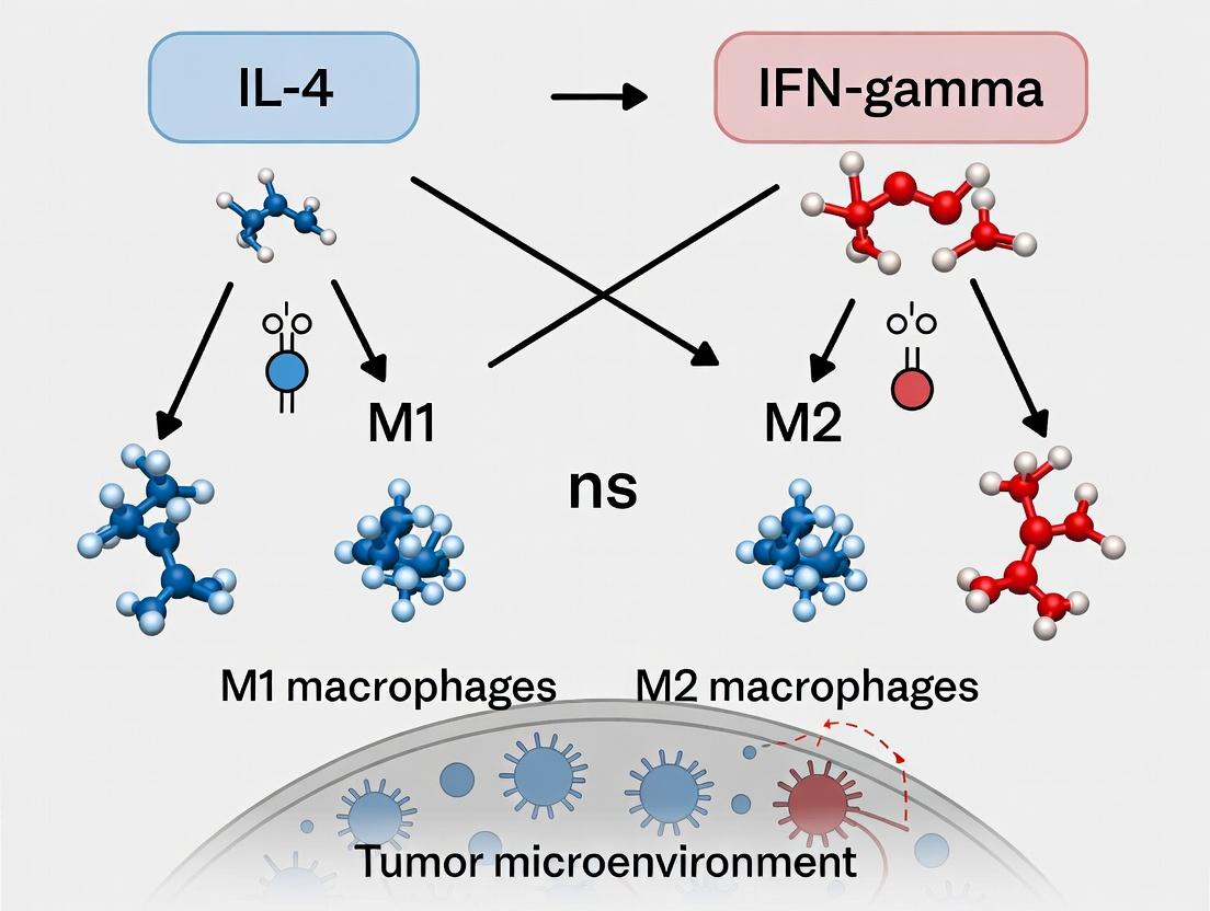
Within the tumor microenvironment (TME), macrophage polarization is a critical determinant of cancer progression. The classical M1 phenotype, driven by signals like IFN-γ, exerts anti-tumor activity, while the alternative M2 phenotype, driven by IL-4/IL-13, promotes tumor growth, angiogenesis, and immunosuppression. This whitepaper details the signaling mechanisms, experimental methodologies, and research tools central to this field.
Key Polarization Signals and Pathways
M1-Polarizing Signaling (Anti-Tumor)
IFN-γ, a key cytokine produced by T cells and NK cells, binds to its receptor (IFNγR), activating the JAK-STAT1 pathway. This induces transcription of pro-inflammatory genes (e.g., iNOS, TNF-α, IL-12) that facilitate tumor cell killing, antigen presentation, and Th1 immune response.
M2-Polarizing Signaling (Pro-Tumor)
IL-4 and IL-13 bind to their respective receptors, primarily activating the JAK-STAT6 pathway. This leads to transcription of genes (e.g., Arg1, Ym1, Fizz1) that promote tissue repair, angiogenesis, matrix remodeling, and immune suppression, facilitating tumor progression.
Diagram Title: M1/M2 Macrophage Polarization Signaling Pathways
Quantitative Comparison of M1 vs. M2 Macrophage Phenotypes
Table 1: Core Functional and Molecular Markers of Polarized Macrophages
| Parameter | M1 (Anti-Tumor) | M2 (Pro-Tumor) |
|---|---|---|
| Primary Inducers | IFN-γ, LPS, GM-CSF | IL-4, IL-13, IL-10, M-CSF |
| Key Surface Markers | CD80, CD86, MHC-II (High) | CD163, CD206, CD209 |
| Signature Enzymes | iNOS (High), Arginase-1 (Low) | Arginase-1 (High), iNOS (Low) |
| Cytokine Secretion | TNF-α, IL-12, IL-1β, IL-6 | IL-10, TGF-β, CCL17, CCL22 |
| Metabolic Profile | Glycolysis, TCA cycle disruption | Oxidative Phosphorylation, FAO |
| Tumor Outcome | Cytotoxicity, Antigen Presentation, Th1 Polarization | Angiogenesis, Matrix Remodeling, T-cell Suppression |
Table 2: Impact of TAM Phenotype on Clinical Cancer Outcomes (Representative Data)
| Cancer Type | High M2/M1 Ratio Correlation | Key Associated Metrics |
|---|---|---|
| Breast Cancer | Poor Prognosis, Reduced Overall Survival | Correlates with increased metastasis (>40% increase risk) |
| Lung Adenocarcinoma | Resistance to PD-1/PD-L1 inhibitors | M2 signature linked to non-response in ~60% of cases |
| Colorectal Cancer | Advanced Stage, Liver Metastasis | High CD163+ TAMs correlate with 2.5x higher recurrence |
| Glioblastoma | Tumor Progression, Immunosuppression | M2 cytokines IL-10 & TGF-β >5-fold increase in serum |
Experimental Protocols for Macrophage Polarization & Analysis
Protocol 4.1: In Vitro Polarization of Human Monocyte-Derived Macrophages
Objective: Generate M1 and M2 polarized macrophages from primary human monocytes.
- Monocyte Isolation: Isolate CD14+ monocytes from PBMCs using positive selection magnetic beads. Seed at 1x10^6 cells/mL in RPMI-1640 + 10% FBS.
- Macrophage Differentiation: Add 50 ng/mL recombinant human M-CSF. Culture for 6 days with medium change on day 3.
- Polarization (Day 6):
- M1: Stimulate with 20 ng/mL IFN-γ + 100 ng/mL LPS for 24-48h.
- M2: Stimulate with 20 ng/mL IL-4 + 20 ng/mL IL-13 for 48h.
- Validation: Harvest cells. Confirm phenotype via flow cytometry (CD80/86 for M1; CD206/163 for M2) and qPCR (TNF-α/IL-12 for M1; Arg1/CCL18 for M2).
Protocol 4.2: Immunofluorescence Staining of TAMs in Tumor Sections
Objective: Identify and localize M1/M2 macrophages in frozen tumor tissue.
- Tissue Preparation: Cut 5-10 μm cryosections. Fix in 4% PFA for 15 min at RT. Permeabilize with 0.1% Triton X-100 for 10 min.
- Blocking: Incubate with 5% normal goat serum + 1% BSA in PBS for 1h at RT.
- Primary Antibody Staining: Incubate overnight at 4°C with anti-mouse/human antibodies (e.g., anti-F4/80 + anti-iNOS for M1; anti-F4/80 + anti-CD206 for M2). Use species/isotype-matched IgG controls.
- Secondary Staining & Imaging: Apply fluorophore-conjugated secondary antibodies (e.g., Alexa Fluor 488, 594) for 1h at RT in the dark. Mount with DAPI-containing medium. Image using a confocal microscope.
Protocol 4.3: Functional Co-Culture Assay for Tumor Cell Phagocytosis
Objective: Quantify the phagocytic capacity of polarized macrophages toward tumor cells.
- Macrophage Preparation: Generate M1/M2 macrophages as in Protocol 4.1 in a 96-well plate.
- Target Preparation: Label tumor cells (e.g., B16-F10 melanoma, MC38 colon carcinoma) with pHrodo Red SE, a pH-sensitive dye that fluoresces upon phagocytosis.
- Co-Culture: Add labeled tumor cells to macrophages at a 5:1 (tumor:macrophage) ratio. Centrifuge briefly (300xg, 1 min) to initiate contact.
- Quantification: Incubate for 2-4h at 37°C. Measure fluorescence (Ex/Em ~560/585 nm) at 60-min intervals using a plate reader. M1 macrophages typically show 2-3 fold higher phagocytic signal.
Diagram Title: In Vitro Macrophage Polarization & Analysis Workflow
The Scientist's Toolkit: Key Research Reagent Solutions
Table 3: Essential Reagents and Tools for Macrophage Polarization Research
| Category / Reagent | Example Product (Supplier) | Key Function / Application |
|---|---|---|
| Polarization Cytokines | Recombinant Human/Mouse IFN-γ (PeproTech), IL-4 (BioLegend) | Induce specific M1 or M2 polarization in vitro. |
| Monocyte Isolation Kits | Human CD14+ MicroBeads (Miltenyi), Mouse Ly-6C+ Selection Kit (STEMCELL) | Positive selection of primary monocytes from blood or bone marrow. |
| Phenotyping Antibodies | Anti-human CD80-FITC, CD206-APC (BioLegend); Anti-mouse F4/80-PE, iNOS-APC (eBioscience) | Surface and intracellular staining for M1/M2 markers via flow cytometry. |
| Functional Assay Kits | pHrodo Red BioParticles (Thermo Fisher), Arginase Activity Assay Kit (Sigma) | Quantify phagocytosis (pHrodo) or enzymatic activity (Arginase) as functional readouts. |
| Gene Expression Analysis | RT-qPCR Primers for Human TNF-α, Arg1 (Qiagen); NanoString Myeloid Innate Immunity Panel | Quantify polarization-specific gene signatures at mRNA level. |
| In Vivo Depletion/Modulation | Clodronate Liposomes (Liposoma), Anti-CSF-1R Antibody (Bio X Cell) | Deplete or modulate TAMs in mouse tumor models for functional studies. |
Techniques to Induce, Analyze, and Target Macrophage Polarization In Vitro and In Vivo
Within the broader context of macrophage polarization signals in tumor microenvironment research, in vitro polarization protocols serve as the foundational tool for dissecting M1 and M2 phenotypes. The standardized use of IFN-γ/LPS for M1 and IL-4/IL-13 for M2 polarization provides a controlled system to model in vivo immune responses, study metabolic reprogramming, and screen therapeutic candidates targeting macrophage function.
Defining Polarization Stimuli: Key Cytokines & Reagents
The specificity of macrophage polarization is driven by distinct cytokine-receptor interactions and downstream signaling cascades. The following table summarizes the core stimuli and their molecular targets.
Table 1: Core Polarizing Stimuli and Molecular Initiation Points
| Phenotype | Primary Stimulus | Alternative/ Potentiating Stimulus | Key Receptor(s) | Initial Signaling JAK-STAT Pathway | Common Final Effectors/Markers |
|---|---|---|---|---|---|
| M1 | IFN-γ (20-100 ng/mL) | LPS (10-100 ng/mL) | IFNGR1/2 | JAK1/2, STAT1 | iNOS, TNF-α, IL-12, CD86, MHC-II |
| M2 | IL-4 (20-50 ng/mL) | IL-13 (20-50 ng/mL) | IL-4Rα/γc (Type I), IL-4Rα/IL-13Rα1 (Type II) | JAK1/3/STAT6 (IL-4), JAK1/2/TYK2/STAT6 (IL-13) | ARG1, CD206, CD209, CCL17, CCL22 |
Detailed Experimental Protocols
Protocol 1: Standard M1 Polarization of Human Monocyte-Derived Macrophages (hMDMs)
Objective: Generate classically activated M1 macrophages. 1. Monocyte Isolation & Differentiation: * Isolate CD14+ monocytes from PBMCs using magnetic-activated cell sorting (MACS). * Culture monocytes in RPMI-1640 supplemented with 10% FBS, 1% Pen/Strep, and 50 ng/mL recombinant human M-CSF for 6-7 days to differentiate into naive M0 macrophages. * Replace media with fresh M-CSF-containing media every 2-3 days. 2. M1 Polarization: * On day 6-7, aspirate media and add fresh complete media (without M-CSF) containing 50 ng/mL recombinant human IFN-γ. * After 24 hours, add a potentiating dose of ultrapure LPS at 10 ng/mL. * Incubate for an additional 24-48 hours. 3. Validation: * Harvest cells for flow cytometry analysis of surface markers (CD80, CD86, HLA-DR). * Collect supernatant for ELISA measurement of TNF-α and IL-12p70. * Perform qPCR or Western blot for iNOS (note: human iNOS is less inducible than murine).
Protocol 2: Standard M2 Polarization of Murine Bone Marrow-Derived Macrophages (BMDMs)
Objective: Generate alternatively activated M2 macrophages. 1. BMDM Differentiation: * Flush bone marrow cells from femurs and tibias of C57BL/6 mice. * Culture cells in DMEM supplemented with 10% FBS, 1% Pen/Strep, and 20 ng/mL recombinant murine M-CSF for 7 days to generate M0 BMDMs. * Replace media with fresh M-CSF-containing media on day 3. 2. M2 Polarization: * On day 7, aspirate media and wash cells with PBS. * Add fresh complete media (without M-CSF) containing 40 ng/mL recombinant murine IL-4. IL-13 can be used at the same concentration as an alternative or in combination. * Incubate for 48 hours. 3. Validation: * Harvest cells for flow cytometry analysis of surface markers (CD206, CD209). * Perform qPCR for canonical M2 genes (Arg1, Ym1/Chi3l3, Fizz1/Retnla). * Measure arginase activity via urea production assay.
Signaling Pathway Diagrams
Title: M1 Polarization via IFN-γ/LPS Signaling
Title: M2 Polarization via IL-4/IL-13-STAT6 Signaling
Title: In Vitro Macrophage Polarization Workflow
The Scientist's Toolkit: Research Reagent Solutions
Table 2: Essential Materials for Polarization Experiments
| Reagent / Material | Function / Purpose | Example / Note |
|---|---|---|
| Recombinant Cytokines | Induce specific signaling cascades for polarization. Must be species-matched. | Human/mouse IFN-γ, IL-4, IL-13, M-CSF. Use carrier protein-free, endotoxin-tested variants. |
| Ultrapure LPS | Potentiates M1 polarization via TLR4. Purity is critical to avoid unintended TLR2 activation. | E. coli O111:B4 or K12 derivatives, with Triton X-114 purification. |
| Cell Culture Media | Supports macrophage survival, differentiation, and activation. | RPMI-1640 or DMEM, supplemented with 10% certified FBS (low endotoxin). |
| Differentiation Factor | Drives monocyte/progenitor differentiation into naive M0 macrophages. | M-CSF (CSF-1) is standard. GM-CSF can be used for distinct macrophage subsets. |
| FACS Antibodies | Validation of surface phenotype markers. | Anti-human: CD80, CD86, HLA-DR (M1); CD206, CD209 (M2). Anti-mouse: CD86, MHC-II (M1); CD206, F4/80 (M2). |
| ELISA Kits | Quantitative measurement of secreted cytokines/chemokines. | TNF-α, IL-12p70 (M1); CCL17, CCL22 (M2). |
| qPCR Primers/Assays | Molecular validation of polarization at the gene expression level. | Primer sets for iNOS, TNF-α (M1); Arg1, Ym1, Fizz1 (M2 mouse); CCL18, CD200R (M2 human). |
| Arginase Activity Assay | Functional validation of M2 polarization via urea measurement. | Colorimetric assay quantifying conversion of L-arginine to urea and ornithine. |
Within macrophage polarization research—specifically the study of M1 (pro-inflammatory, IFN-γ-driven) and M2 (anti-inflammatory, IL-4-driven) phenotypes in the tumor microenvironment—the choice of cellular model is critical. Primary cells, such as Bone Marrow-Derived Macrophages (BMDMs) and human monocyte-derived macrophages, offer physiological relevance. In contrast, the THP-1 monocytic leukemia cell line provides reproducibility and scalability. This guide provides a technical comparison, protocols, and tools for researchers navigating these models.
Model Comparison: Characteristics & Applications
The table below summarizes the core quantitative and qualitative attributes of each model system.
Table 1: Comparative Analysis of Macrophage Models
| Feature | Bone Marrow-Derived Macrophages (BMDMs) | Human Primary Monocytes/Macrophages | THP-1 Cell Line |
|---|---|---|---|
| Origin | Mouse bone marrow (C57BL/6, BALB/c common) | Human peripheral blood (CD14+ selection) | Human acute monocytic leukemia |
| Differentiation Agent | M-CSF (typically 10-20 ng/mL, 5-7 days) | GM-CSF (M1-like) or M-CSF (M2-like), 5-7 days | Phorbol 12-myristate 13-acetate (PMA), 10-100 nM, 24-72h |
| Proliferation Capacity | Terminally differentiated, non-proliferative | Terminally differentiated, non-proliferative | Proliferative in suspension; PMA induces differentiation & arrest |
| Genetic Stability | Genetically stable, but donor/strain variability | High inter-donor variability | Genetically uniform but may drift; not genetically stable long-term |
| Polarization Response (Typical Markers) | M1 (IFN-γ + LPS): High iNOS, IL-6, TNF-α. M2 (IL-4/13): High Arg1, Ym1, Fizz1. | M1 (IFN-γ + LPS): High CD80, IL-12, TNF-α. M2 (IL-4): High CD206, CCL18, Arg1. | M1-like (PMA+IFN-γ/LPS): Moderate IL-1β, TNF-α. M2-like (PMA+IL-4): Moderate CD206, CCL22. |
| Key Advantages | In vivo relevance, robust polarization, suitable for transgenic models | Human-specific responses, clinical translatability | High yield, easy genetic manipulation (e.g., CRISPR), low cost |
| Major Limitations | Murine origin, time-consuming isolation, strain differences | Ethical/logistical hurdles, high cost, donor variability | Altered metabolism, muted polarization response vs. primary cells |
| Ideal For | In vivo mechanistic studies, knockout/transgenic validation | Human-specific pathway analysis, biomarker discovery | High-throughput screening, preliminary mechanistic studies |
Detailed Experimental Protocols
Generation and Polarization of BMDMs
Isolation & Differentiation:
- Euthanize mouse (e.g., C57BL/6) following institutional guidelines.
- Aseptically dissect femur and tibia. Flush marrow with cold, sterile PBS using a 25G needle.
- Pass cell suspension through a 70 µm cell strainer. Centrifuge at 300 x g for 5 min.
- Resuspend pellet in complete medium (RPMI 1640, 10% FBS, 1% Pen/Strep) supplemented with 20 ng/mL recombinant murine M-CSF.
- Plate cells in non-tissue culture-treated dishes (to prevent adhesion of progenitors). Culture at 37°C, 5% CO2.
- On day 3, add fresh medium with M-CSF (20 ng/mL). On day 5-7, harvest mature, adherent BMDMs by gentle scraping.
Polarization:
- M1 Polarization: Treat mature BMDMs with 20 ng/mL murine IFN-γ for 24h, followed by 100 ng/mL LPS for an additional 24h.
- M2 Polarization: Treat mature BMDMs with 20 ng/mL murine IL-4 for 48h.
Isolation and Differentiation of Human Primary Macrophages
Isolation (PBMCs & Monocytes):
- Collect human peripheral blood (buffy coat or whole blood) with anticoagulant.
- Dilute blood 1:1 with PBS. Layer over Ficoll-Paque PLUS density gradient medium.
- Centrifuge at 400 x g for 30 min at room temperature (brake off).
- Collect the peripheral blood mononuclear cell (PBMC) layer.
- Isolate CD14+ monocytes using positive selection magnetic beads per manufacturer's protocol.
Differentiation & Polarization:
- Culture CD14+ monocytes in RPMI 1640, 10% human AB serum, 1% Pen/Strep.
- For M1-like macrophages: Add 50 ng/mL human GM-CSF for 6 days.
- For M2-like macrophages: Add 50 ng/mL human M-CSF for 6 days.
- Polarize with 20 ng/mL IFN-γ + 100 ng/mL LPS (M1) or 20 ng/mL IL-4 (M2) for 48h after differentiation.
Differentiation and Polarization of THP-1 Cells
Maintenance: Culture THP-1 cells in suspension in RPMI 1640, 10% FBS, 1% Pen/Strep, and 0.05 mM β-mercaptoethanol.
Differentiation:
- Plate THP-1 cells at 2-5x10^5 cells/mL in tissue-culture treated plates.
- Add 100 nM Phorbol 12-myristate 13-acetate (PMA).
- Incubate for 48-72h. Remove PMA-containing medium, wash cells gently, and rest in fresh complete medium for 24h.
Polarization:
- M1-like: Treat differentiated THP-1 macrophages with 100 ng/mL LPS + 20 ng/mL IFN-γ for 24-48h.
- M2-like: Treat with 20 ng/mL IL-4 for 24-48h.
Signaling Pathways in Macrophage Polarization
Diagram Title: Core Signaling Pathways in Macrophage M1/M2 Polarization
Experimental Workflow for Polarization Studies
Diagram Title: Experimental Workflow for Macrophage Polarization Studies
The Scientist's Toolkit: Essential Research Reagents
Table 2: Key Reagent Solutions for Macrophage Polarization Research
| Category | Reagent/Material | Function & Application | Example Vendor(s) |
|---|---|---|---|
| Cytokines & Polarizing Agents | Recombinant Murine/Human M-CSF (CSF-1) | Drives monocyte-to-macrophage differentiation for BMDMs and human M2-like macrophages. | PeproTech, BioLegend, R&D Systems |
| Recombinant Murine/Human GM-CSF | Used for generating human M1-like macrophages. | PeproTech, BioLegend | |
| Recombinant IFN-γ (Mouse/Human) | Key cytokine for classical M1 macrophage activation. | PeproTech, BioLegend | |
| Recombinant IL-4 (Mouse/Human) | Key cytokine for alternative M2 macrophage activation. | PeproTech, BioLegend | |
| Ultrapure LPS (E. coli) | TLR4 agonist used in combination with IFN-γ for robust M1 polarization. | InvivoGen, Sigma-Aldrich | |
| Phorbol 12-Myristate 13-Acetate (PMA) | Differentiates THP-1 monocytes into adherent macrophage-like cells. | Sigma-Aldrich, Tocris | |
| Isolation & Culture | Ficoll-Paque PLUS | Density gradient medium for isolating PBMCs from human blood. | Cytiva |
| CD14 MicroBeads (Human) | Magnetic bead-based positive selection for human monocytes. | Miltenyi Biotec | |
| Cell Strainers (70 µm) | Removal of cell aggregates and tissue debris during BMDM isolation. | Falcon, pluriSelect | |
| Non-Tissue Culture Treated Dishes | Prevents strong adhesion of progenitor cells during BMDM differentiation. | Falcon, Corning | |
| Analysis - Flow Cytometry | Anti-mouse CD80, CD86, CD206 (MRC1) Antibodies | Surface marker analysis for M1/M2 polarization status. | BioLegend, BD Biosciences |
| Anti-human CD80, CD163, CD206 Antibodies | Human macrophage phenotyping. | BioLegend, BD Biosciences | |
| Analysis - Molecular | iNOS (NOS2), Arg1, TNF-α, IL-10 TaqMan Assays | Quantitative PCR for polarization marker gene expression. | Thermo Fisher, Bio-Rad |
| ELISA Kits for IL-6, IL-10, IL-12p70, TNF-α, TGF-β | Quantification of secreted cytokines in supernatant. | R&D Systems, BioLegend | |
| Specialized Assays | pHrodo Red BioParticles (E. coli) | Fluorescent particles for quantitative phagocytosis assays. | Thermo Fisher |
| Arginase Activity Assay Kit | Colorimetric measurement of arginase activity, an M2 functional marker. | Sigma-Aldrich, Abcam |
Introduction This technical guide addresses the critical need for robust flow cytometric assays to delineate macrophage polarization states within the context of M1/M2 polarization signals, driven by cytokines such as IFN-γ and IL-4, and their complex interplay in the tumor microenvironment (TME). Accurate phenotyping is fundamental for research in immunology, oncology, and therapeutic development.
Core Signaling Pathways in Macrophage Polarization Macrophage polarization is governed by distinct signaling cascades initiated by key cytokines.
Critical Surface Markers for Phenotyping The selection of surface markers is paramount for distinguishing polarization states via flow cytometry.
Table 1: Key Surface Markers for Human Macrophage Polarization
| Polarization State | Primary Surface Markers | Key Inducers | Function/Interpretation |
|---|---|---|---|
| M1 (Classical) | CD80, CD86, HLA-DR (hi), CD64, CCR7 | IFN-γ, LPS, GM-CSF | Antigen presentation, co-stimulation, pro-inflammatory response. |
| M2 (Alternative) | CD206 (MMR), CD163, CD200R, CD209 (DC-SIGN), IL-4R (CD124) | IL-4, IL-13, IL-10, Glucocorticoids | Scavenger receptors, immunoregulation, tissue repair, anti-inflammatory. |
| Pan-Macrophage | CD11b, CD14, CD68, F4/80 (mouse) | - | General macrophage identification. |
Table 2: Example Flow Cytometry Panels (3-Color to 8-Color)
| Panel Complexity | Fluorochrome Conjugations (Example) | Gating Strategy | Application |
|---|---|---|---|
| 3-Color Basic | CD14-BV421, CD206-FITC, CD80-PE | CD14+ -> CD206 vs CD80 | Preliminary M2 vs M1 distinction. |
| 6-Color Standard | CD11b-BV510, CD14-BV421, HLA-DR-FITC, CD86-PE, CD163-PerCP-Cy5.5, CD206-APC | CD11b+CD14+ -> HLA-DRhiCD86+ (M1) vs CD163+CD206+ (M2) | Detailed human M1/M2 profiling in tissue digests. |
| 8-Color Comprehensive | Live/Dead-NIR, CD45-BV510, CD11b-BV421, CD14-APC-Cy7, CD80-FITC, CD86-PE, CD163-PerCP-Cy5.5, CD206-APC | Live CD45+CD11b+CD14+ -> M1: CD80+CD86+; M2: CD163+CD206+ | High-parameter analysis in complex samples like TME. |
Detailed Experimental Protocol: M1/M2 Polarization and Staining Protocol 1: In Vitro Polarization & Surface Marker Staining for Flow Cytometry
- Monocyte Isolation: Isolate human CD14+ monocytes from PBMCs using magnetic bead-based positive selection.
- Differentiation: Culture monocytes for 6-7 days in RPMI-1640 with 10% FBS, 1% Pen/Strep, and 50 ng/mL M-CSF to generate monocyte-derived macrophages (MDMs).
- Polarization: Stimulate MDMs for 24-48 hours.
- M1: 20 ng/mL IFN-γ + 100 ng/mL LPS.
- M2: 20 ng/mL IL-4.
- M0: Media only (unpolarized control).
- Harvesting: Gently scrape cells in cold PBS + 2% FBS. Centrifuge at 300 x g for 5 min.
- Staining:
- Resuspend cell pellet in 100 µL of staining buffer (PBS + 2% FBS).
- Add Fc receptor blocking reagent (e.g., human IgG) for 10 min on ice.
- Add pre-titrated antibody cocktail directly. Vortex gently. Incubate for 30 min in the dark at 4°C.
- Wash twice with 2 mL staining buffer. Centrifuge at 300 x g for 5 min.
- (Optional) Fix cells in 1-2% PFA for 10 min at 4°C if not acquiring immediately.
- Acquisition & Analysis: Acquire on a flow cytometer calibrated with compensation beads. Analyze using sequential gating to identify live, single, macrophage populations before evaluating marker expression.
Experimental Workflow for TME Analysis
The Scientist's Toolkit: Key Research Reagent Solutions
Table 3: Essential Materials and Reagents
| Item | Function & Rationale | Example/Format |
|---|---|---|
| M-CSF (Human/Mouse) | Differentiates monocytes/bone marrow precursors into macrophages (M0 state). Required baseline for polarization. | Recombinant protein, carrier-free. |
| Polarizing Cytokines (IFN-γ, IL-4, LPS) | Induce specific signaling pathways leading to M1 or M2 phenotypic commitment. | High-purity, endotoxin-free recombinant cytokines. |
| Fluorochrome-Conjugated Antibodies | Detection of specific surface markers. Panel design requires careful fluorochrome brightness/marker abundance matching. | Pre-titrated clones in formats like BV421, PE, APC, FITC. |
| Fc Receptor Blocking Reagent | Reduces non-specific antibody binding, critical for high signal-to-noise ratio. | Human or mouse Fc block, purified IgG, or commercial blocking buffers. |
| Viability Dye (e.g., Live/Dead Fixable NIR) | Distinguishes live cells from dead cells, excluding debris and false-positive staining. | Fixable amine-reactive dyes. |
| Magnetic Cell Separation Kits | Rapid isolation of pure monocyte/macrophage populations from complex mixtures (PBMCs, tumors). | CD14+ or CD11b+ magnetic bead kits. |
| Cell Dissociation Enzyme (Tumor) | Generates single-cell suspensions from solid tumor tissue for TME analysis. | Gentle, enzyme-based cocktails (Collagenase/DNase). |
| Compensation Beads | Essential for setting accurate spectral compensation on flow cytometers. | Anti-mouse/rat/hamster Igκ beads. |
Advanced Considerations & Challenges
- Fluidics & High-Dimensionality: Spectral flow cytometry allows for larger panels (>15 colors), enabling simultaneous detection of M1/M2 markers, checkpoint molecules (PD-L1), and activation markers.
- Intracellular Correlation: Surface marker analysis can be combined with intracellular staining for cytokines (TNF-α, IL-10) or enzymes (iNOS, ARG1) to confirm functional polarization.
- TME Heterogeneity: Macrophages in the TME often exhibit a spectrum of activation. High-parameter analysis and tools like t-SNE or UMAP are recommended to reveal continuous phenotypes beyond binary M1/M2 classification.
Within the critical study of macrophage polarization in the tumor microenvironment, functional assays are indispensable for defining the M1 (pro-inflammatory, often IFN-γ induced) and M2 (anti-inflammatory, often IL-4 induced) phenotypes. This technical guide details core methodologies for assessing three key functional pillars: phagocytic capacity, cytokine secretion profiles, and metabolic reprogramming. These assays provide quantitative, phenotypic validation of polarization states beyond mere surface marker expression, offering insights into macrophage function in cancer immunology and therapeutic development.
Phagocytosis Assays
Phagocytosis is a cardinal macrophage function, often modulated by polarization signals. M1 macrophages typically exhibit enhanced phagocytic activity against pathogens, while TAMs (Tumor-Associated Macrophages) may show dysfunctional phagocytosis, particularly of cancer cells.
Key Methodologies:
A. Flow Cytometry-based Phagocytosis of fluorescent beads or particles:
- Protocol: Differentiate human monocyte-derived macrophages (hMDMs) with M-CSF. Polarize with IFN-γ (20 ng/mL) + LPS (100 ng/mL) for M1 or IL-4 (20 ng/mL) for M2 for 24-48h. Incubate cells with pHrodo Green-conjugated E. coli BioParticles or fluorescent latex beads (1-2 μm diameter) for 1-2 hours at 37°C (with a 4°C control for background binding). Quench extracellular fluorescence with trypan blue. Analyze by flow cytometry. Mean Fluorescence Intensity (MFI) indicates phagocytic capacity.
- Data Interpretation: M1-polarized macrophages typically show higher MFI for opsonic targets.
B. Microscopy-based Phagocytosis Assay:
- Protocol: Plate macrophages on glass coverslips. After polarization, add pH-sensitive (e.g., pHrodo) labeled particles. Phagocytosed particles fluoresce brightly in acidic phagolysosomes. Fix, stain nuclei/DAPI, and image. Quantify particles per cell using image analysis software (e.g., ImageJ).
Table 1: Representative Quantitative Data from Phagocytosis Assays (hMDMs)
| Polarization Signal | Particle Type | Assay Method | Reported Phagocytic Index (vs. M0) | Key Reference |
|---|---|---|---|---|
| IFN-γ + LPS (M1) | pHrodo E. coli Bioparticles | Flow Cytometry (MFI) | 2.1 - 3.5 fold increase | Vogel et al., 2022 |
| IL-4 (M2) | pHrodo E. coli Bioparticles | Flow Cytometry (MFI) | 0.8 - 1.2 fold change | Vogel et al., 2022 |
| IL-4 (M2) | IgG-opsonized beads | Microscopy (count/cell) | 1.5 - 2.0 fold increase | Jetten et al., 2021 |
| TCM (Tumor Cond. Media) | Apoptotic tumor cells | Flow Cytometry (% positive) | 0.5 - 0.7 fold decrease | Blando et al., 2023 |
Cytokine Secretion Profiling
Secreted cytokines define macrophage communication within the TME. M1 macrophages secrete pro-inflammatory cytokines (e.g., TNF-α, IL-12, IL-6), while M2 macrophages secrete immunoregulatory factors (e.g., IL-10, TGF-β, CCL17, CCL22).
Key Methodologies:
A. Multiplex Bead-Based Immunoassay (Luminex):
- Protocol: Culture polarized macrophages in serum-free media for 6-24h. Collect supernatant, centrifuge to remove debris. Use a pre-mixed magnetic bead-based multiplex panel (e.g., Human ProcartaPlex) per manufacturer's instructions. Acquire data on a Luminex analyzer. Generate standard curves for absolute quantification (pg/mL).
- Advantage: Simultaneously quantifies 20+ analytes from small sample volumes.
B. Enzyme-Linked Immunosorbent Assay (ELISA):
- Protocol: The gold standard for specific, high-sensitivity quantification. Coat high-binding 96-well plate with capture antibody. Block, add standards and samples, incubate. Detect with biotinylated detection antibody, streptavidin-HRP, and TMB substrate. Stop with acid and read absorbance at 450nm.
Table 2: Characteristic Cytokine Secretion Profiles of Polarized Macrophages (Secreted pg/mL/24h/10^6 cells)
| Analyte | M0 (Unpolarized) | M1 (IFN-γ + LPS) | M2 (IL-4) | Primary Source |
|---|---|---|---|---|
| TNF-α | 50 - 200 | 5,000 - 20,000 | < 100 | Murray et al., 2023 |
| IL-6 | 100 - 500 | 10,000 - 30,000 | 200 - 1,000 | Murray et al., 2023 |
| IL-12p70 | < 20 | 500 - 2,000 | < 20 | Orecchioni et al., 2022 |
| IL-10 | 50 - 300 | 500 - 2,000 | 1,000 - 5,000 | Orecchioni et al., 2022 |
| CCL17 (TARC) | < 50 | < 100 | 800 - 3,000 | Orecchioni et al., 2022 |
| CCL22 (MDC) | < 100 | < 200 | 2,000 - 8,000 | Orecchioni et al., 2022 |
Metabolic Profiling
Macrophage polarization is underpinned by metabolic reprogramming. M1 polarization is associated with a shift to glycolysis, while M2 polarization relies on oxidative phosphorylation (OXPHOS) and fatty acid oxidation (FAO).
Key Methodologies:
A. Extracellular Flux Analysis (Seahorse):
- Protocol: Plate polarized macrophages in a Seahorse microplate. Run the Cell Mito Stress Test: Sequential injections of Oligomycin (ATP synthase inhibitor), FCCP (uncoupler), and Rotenone/Antimycin A (Complex I/III inhibitors). Measure Oxygen Consumption Rate (OCR, pmol/min) for OXPHOS and Extracellular Acidification Rate (ECAR, mpH/min) for glycolysis.
- Key Outputs: Basal OCR/ECAR, ATP-linked respiration, maximal respiration, glycolytic capacity.
B. Metabolite Measurement (e.g., Lactate, Glucose):
- Protocol: Use colorimetric/fluorometric assay kits to quantify metabolites in culture supernatant. For lactate, a common endpoint is the conversion of lactate to pyruvate, generating a colorimetric product proportional to concentration.
Table 3: Metabolic Parameters of Polarized Macrophages (Seahorse Data)
| Metabolic Parameter | M0 Macrophage | M1 Macrophage | M2 Macrophage | Assay |
|---|---|---|---|---|
| Basal OCR (pmol/min) | 60 - 80 | 20 - 40 | 80 - 120 | Mito Stress Test |
| Glycolytic Capacity (ECAR mpH/min) | 30 - 50 | 80 - 150 | 20 - 40 | Glycolysis Stress Test |
| ATP-linked Respiration | Moderate | Low | High | Mito Stress Test |
| Lactate Production (nmol/10^6 cells/h) | 20 - 40 | 80 - 160 | 10 - 30 | Colorimetric Assay |
Experimental Protocols in Detail
Protocol 1: Comprehensive Polarization and Functional Profiling Workflow
- Isolation & Differentiation: Isolate CD14+ monocytes from human PBMCs using magnetic beads. Culture for 6-7 days in RPMI-1640 + 10% FBS + 50 ng/mL human M-CSF.
- Polarization: On day 7, stimulate cells for 24-48h with:
- M1: 20 ng/mL IFN-γ + 100 ng/mL LPS.
- M2: 20 ng/mL IL-4.
- Phagocytosis Assay (Flow): Harvest cells, seed in a U-bottom plate (2x10^5/well). Add pHrodo Green E. coli Bioparticles (10 μg/well). Incubate 90 min at 37°C. Wash with cold PBS containing 0.1% sodium azide. Analyze immediately on a flow cytometer (FITC channel).
- Cytokine Secretion: After polarization, replace media with serum-free X-Vivo 15. Collect supernatant after 18h. Clarify by centrifugation (500xg, 5 min). Store at -80°C. Analyze via 12-plex Luminex assay.
- Metabolic Profiling: On day 6, seed polarized macrophages in a Seahorse XF96 cell culture microplate (8x10^4/well). On day 7, equilibrate in Seahorse XF DMEM (pH 7.4) at 37°C in a non-CO2 incubator for 1h. Run the Mito Stress Test program.
Signaling Pathways in Macrophage Polarization
Diagram Title: Core Signaling Pathways Driving M1 and M2 Macrophage Polarization
Experimental Workflow for Integrated Functional Profiling
Diagram Title: Integrated Workflow for Macrophage Functional Assays
The Scientist's Toolkit: Research Reagent Solutions
Table 4: Essential Reagents and Tools for Macrophage Functional Assays
| Reagent/Tool | Vendor Examples | Function in Assays |
|---|---|---|
| Recombinant Human Cytokines (IFN-γ, IL-4, M-CSF, LPS) | PeproTech, R&D Systems | Induce and control macrophage polarization states. |
| pHrodo Green/Red E. coli or S. aureus Bioparticles | Thermo Fisher Scientific | pH-sensitive phagocytosis probes; fluoresce brightly upon phagolysosomal acidification. |
| Luminex Multiplex Assay Kits (e.g., ProcartaPlex) | Thermo Fisher Scientific, Bio-Rad | Simultaneously quantify panels of secreted cytokines/chemokines from small sample volumes. |
| ELISA Kits (TNF-α, IL-10, IL-6, etc.) | BioLegend, R&D Systems, Abcam | High-sensitivity, specific quantification of individual analytes. |
| Seahorse XF Glycolysis & Mito Stress Test Kits | Agilent Technologies | Measure real-time extracellular acidification (ECAR) and oxygen consumption (OCR) for metabolic phenotyping. |
| XF96 Cell Culture Microplates | Agilent Technologies | Specialized plates for live-cell metabolic analysis in the Seahorse analyzer. |
| Cell Staining Buffer (with Azide) | BioLegend, Tonbo Biosciences | Preserves cell surface staining and quenches reactions for flow cytometry. |
| Magnetic CD14+ MicroBeads (Human) | Miltenyi Biotec | Isolation of primary human monocytes from PBMCs for macrophage differentiation. |
| Image Analysis Software (e.g., ImageJ/Fiji) | Open Source | Quantifies particle uptake and cell morphology from microscopy images. |
| Flow Cytometry Analysis Software (e.g., FlowJo) | BD Biosciences, Treestar | Analyzes phagocytosis (MFI, % positive) and co-staining from flow cytometry data. |
Tumor-associated macrophages (TAMs) are a dominant immune cell population in the tumor microenvironment (TME), exhibiting high plasticity. Within the classic M1-M2 polarization paradigm, TAMs predominantly display an M2-like, pro-tumorigenic phenotype, driven by signals such as IL-4, IL-13, and IL-10. Conversely, IFN-γ and LPS can promote an M1-like, anti-tumor phenotype. In vivo modeling in mice is essential to understand the dynamic recruitment, differentiation, and function of TAMs, and to test therapeutic strategies aimed at reprogramming them.
Key In Vivo Models for TAM Tracing
Syngeneic Mouse Models
Implantation of murine cancer cell lines into immunocompetent mice. Commonly used models include:
- LLC (Lewis Lung Carcinoma) in C57BL/6 mice: High TAM infiltration.
- 4T1 (Breast Carcinoma) in BALB/c mice: Models metastatic disease.
Genetically Engineered Mouse Models (GEMMs)
Spontaneous tumor development driven by genetic alterations (e.g., Kras and p53 mutations), preserving a native TME and immune cell development.
Patient-Derived Xenografts (PDXs) in Humanized Mice
NSG or NSG-SGM3 mice engrafted with human hematopoietic stem cells allow study of human TAMs in a human tumor context, though with limitations in full system reconstitution.
Table 1: Comparison of Key Mouse Models for TAM Research
| Model Type | Example | Key Advantages | Key Limitations | Typical %TAMs of TME* |
|---|---|---|---|---|
| Syngeneic | LLC (C57BL/6) | Intact immune system; rapid, reproducible; cost-effective. | May not fully recapitulate human tumor genetics. | 20-50% |
| GEMM | KP model (KrasLSL-G12D/+; p53fl/fl) | Native tumorigenesis & microenvironment; immune cell education. | Variable latency; higher cost; complex breeding. | 30-60% |
| Orthotopic | E0771 (mammary fat pad) | Organ-specific microenvironment influences TAM phenotype. | Technically challenging. | 25-55% |
| Humanized PDX | NSG-SGM3 mice + HSCs + PDX | Contains human TAM precursors & tumor. | Incomplete human immunity; "mouse" cytokine milieu. | 15-40% (human CD68+ cells) |
*Data summarized from recent studies (2022-2024). Percentages are approximate and tumor-type dependent.
Methodologies for Tracing TAMs In Vivo
Genetic Lineage Tracing
- Principle: Use of Cre-lox systems under macrophage-specific promoters (e.g., Cx3cr1, Lyz2, Cd64) to permanently label macrophages and their progeny.
- Protocol (Example - Cx3cr1CreER; R26tdTomato):
- Mouse Strain: Cx3cr1CreER/+; Rosa26LSL-tdTomato/+.
- Tamoxifen Induction: Inject tamoxifen intraperitoneally (75 mg/kg in corn oil) for 3-5 consecutive days to activate Cre recombinase.
- Tumor Implantation: Perform 7 days post-final tamoxifen injection.
- Analysis: Harvest tumors at endpoint. tdTomato+ cells are macrophages derived from labeled precursors. Flow cytometry and immunofluorescence confirm identity (F4/80+ CD11b+).
Intravital Microscopy (IVM)
- Principle: Real-time visualization of TAM dynamics in living mice.
- Protocol:
- Window Chamber Implantation: For dorsal skinfold or cranial windows.
- Cell Labeling: Use transgenic reporters (e.g., Cx3cr1GFP/+) or intravenous injection of fluorescently conjugated anti-F4/80 antibodies.
- Image Acquisition: Anesthetize mouse and secure under two-photon microscope. Track cell motility, interactions with tumor cells, and blood vessels over hours.
- Key Metrics: Velocity, displacement, meandering index of TAMs.
Parabiosis for Origin Tracing
- Principle: Surgically join circulatory systems of two mice (one labeled, one unlabeled) to distinguish resident macrophage proliferation from monocyte recruitment.
- Protocol:
- Surgery: Join CD45.1 (host) and CD45.2 (parabiont) mice laterally.
- Tumor Implantation: Implant tumor in CD45.1 host after 2 weeks of circulatory anastomosis.
- Analysis: Analyze TAMs (CD11b+ F4/80hi) for CD45.1 vs. CD45.2 chimerism by flow cytometry. High % host = proliferation; high % partner = monocyte recruitment.
Single-Cell RNA Sequencing (scRNA-seq) Analysis
- Protocol for TAM Isolation & Sequencing:
- Tumor Dissociation: Use a gentle mechanical and enzymatic (Collagenase IV/DNase I) digestion protocol (30-45 min at 37°C).
- Immune Cell Enrichment: Optional: Percoll gradient or CD11b+ magnetic bead isolation.
- Cell Viability & Sorting: Stain with viability dye (DAPI) and macrophage markers (F4/80, CD11b). Sort live CD45+CD11b+F4/80+ cells.
- Library Preparation: Use 10x Genomics Chromium platform (v3.1) per manufacturer's protocol.
- Bioinformatics: Clustering (Seurat, Scanpy), trajectory inference (Monocle3), and gene signature scoring for M1 (e.g., Nos2, Il12b) vs. M2 (e.g., Arg1, Mrс1).
Signaling Pathways in TAM Polarization
Experimental Workflow for Comprehensive TAM Tracing
The Scientist's Toolkit: Essential Research Reagents & Models
Table 2: Key Research Reagent Solutions for In Vivo TAM Tracing
| Category | Item/Reagent | Function & Application | Example Vendor/Model |
|---|---|---|---|
| Mouse Models | Cx3cr1CreER-R26tdT | Genetic fate-mapping of monocyte-derived cells. | Jackson Labs (Stock #: 025524) |
| C57BL/6-Tg(Csf1r-EGFP) | Labels macrophages and microglia with GFP. | MGI: MGI:3840442 | |
| Cd64Cre mice | Targets myeloid cells, including most tissue macrophages. | Taconic (Model 12813) | |
| Fluorescent Reporters | Anti-mouse F4/80 (BV785, APC) | Pan-macrophage surface marker for flow/IF. | BioLegend (Clone BM8) |
| Anti-mouse CD206 (APC) | M2-like macrophage marker. | BioLegend (Clone C068C2) | |
| Zombie NIR Fixable Viability Kit | Distinguishes live/dead cells in cytometry. | BioLegend (423106) | |
| Cytokines & Inhibitors | Recombinant murine IL-4 | Induces M2 polarization in vitro/vivo. | PeproTech (214-14) |
| Recombinant murine IFN-γ | Induces M1 polarization. | PeproTech (315-05) | |
| CSF-1R inhibitor (PLX3397) | Depletes macrophages in vivo. | MedChemExpress (HY-16749) | |
| Enzymes & Kits | Tumor Dissociation Kit, mouse | Gentle enzymatic mix for TME single-cell prep. | Miltenyi Biotec (130-096-730) |
| Mouse Macrophage Isolation Kit (CD11b) | Magnetic bead-based enrichment. | STEMCELL Tech (19861) | |
| Chromium Next GEM Chip K (10x) | Single-cell RNA-seq library generation. | 10x Genomics (1000286) | |
| Imaging | CellTracker CM-DiI | Long-term cytoplasmic fluorescent cell label. | Thermo Fisher (C7000) |
| Matrigel Matrix | For orthotopic/s.c. co-injection with tumor cells. | Corning (356231) | |
| Dorsal Skinfold Window Chamber | For longitudinal intravital microscopy. | APJ Trading Co. |
Within the broader thesis on macrophage M1/M2 polarization signals in the tumor microenvironment (TME), this whitepaper details current drug development strategies targeting these critical pathways. The balance between pro-inflammatory, anti-tumor M1 macrophages (often induced by IFN-γ) and pro-tumorigenic, tissue-repair M2 macrophages (polarized by IL-4/IL-13) is a key determinant of cancer progression and therapeutic response. This guide provides a technical overview of experimental approaches and translational strategies for modulating these pathways.
Core Signaling Pathways & Therapeutic Targets
Polarization is governed by complex intracellular signaling cascades. Key pathways present actionable targets for pharmacological intervention.
M1-Polarizing (IFN-γ/STAT1) Signaling
IFN-γ/STAT1 Pathway Inducing M1 Polarization
M2-Polarizing (IL-4/STAT6) Signaling
IL-4/STAT6 Pathway Driving M2 Polarization
Quantitative Landscape of Polarization Markers
Table 1: Key M1 and M2 Macrophage Markers and Their Expression Levels
| Polarization State | Key Surface Marker | Expression Fold-Change (vs. M0) | Soluble Mediator | Secreted Level (Typical Range) |
|---|---|---|---|---|
| M1 (Classical) | CD80/CD86 | 5-15x | TNF-α | 500-2000 pg/mL |
| HLA-DR | 3-8x | IL-12p70 | 50-300 pg/mL | |
| CCR7 | 10-20x | IL-6 | 1000-5000 pg/mL | |
| M2 (Alternative) | CD206 (MRC1) | 20-50x | IL-10 | 1000-8000 pg/mL |
| CD163 | 10-30x | TGF-β | 200-1000 pg/mL | |
| ARG1 (Intracellular) | 50-100x | CCL17 | 500-2500 pg/mL |
Table 2: Drug Development Strategies Targeting Polarization
| Target Class | Example Target | Strategy | Therapeutic Aim | Development Phase (Examples) |
|---|---|---|---|---|
| Cytokine/Ligand | IL-4/IL-13 | Neutralizing mAbs (e.g., Dupilumab) | Inhibit M2 polarization | Preclinical/Repurposing |
| IFN-γ | Recombinant protein, Agonist mAbs | Promote M1 polarization | Phase I/II | |
| Receptors | IL-4Rα | Antagonistic mAbs, Soluble decoys | Block M2 signaling | Phase II (Cancer) |
| CSF-1R | Tyrosine kinase inhibitors | Deplete TAMs, Shift balance | Phase III (e.g., Pexidartinib) | |
| Intracellular Kinases | JAK1/2 | Small molecule inhibitors (e.g., Ruxolitinib) | Modulate STAT signaling | Approved/Repurposing |
| PI3Kγ | Inhibitors (e.g., Eganelisib) | Repolarize M2 to M1 | Phase II | |
| Transcription Factors | STAT3/STAT6 | SH2 domain inhibitors, Decoy oligos | Inhibit pro-tumorigenic transcription | Preclinical/Lead Optimization |
| Epigenetic Regulators | HDACs, BET proteins | HDACi, BET inhibitors | Reprogram gene expression | Phase I/II (Combination) |
Experimental Protocols for Polarization Studies
Protocol: In Vitro Human Macrophage Polarization & Phenotypic Validation
Purpose: Generate and validate M1 and M2 polarized macrophages from primary human monocytes. Key Steps:
- Monocyte Isolation: Isolate CD14+ monocytes from PBMCs using magnetic-activated cell sorting (MACS) or adherence. Culture in RPMI-1640 + 10% FBS + 50 ng/mL M-CSF for 6 days to generate M0 macrophages.
- Polarization:
- M1: Stimulate M0 macrophages with 20 ng/mL IFN-γ + 100 ng/mL LPS for 24-48 hours.
- M2: Stimulate M0 macrophages with 20 ng/mL IL-4 + 20 ng/mL IL-13 for 48-72 hours.
- Validation by Flow Cytometry: Harvest cells. Stain with fluorescently conjugated antibodies against surface markers: M1 Panel: CD80-FITC, CD86-PE, HLA-DR-PerCP; M2 Panel: CD206-APC, CD163-PE. Analyze on flow cytometer. M1 cells show high CD80/86/HLA-DR; M2 cells show high CD206/CD163.
- Validation by qRT-PCR: Extract RNA, synthesize cDNA. Measure expression of M1 genes (iNOS, IL-12b, CXCL10) and M2 genes (ARG1, CCL17, FIZZ1). Calculate fold-change versus M0 using the 2^(-ΔΔCt) method.
- Functional Assay - Nitrite Quantification (M1): Collect supernatant from M1-polarized cells. Use Griess reagent to measure nitrite concentration (indicative of iNOS activity). M1 supernatants typically yield >20 μM nitrite.
- Functional Assay - Arginase Activity (M2): Lyse M2-polarized cells. Measure urea production from L-arginine substrate. Activity expressed as mU per million cells. M2 lysates typically show >50 mU/10^6 cells.
Protocol: Assessing Drug-Induced Repolarization in a 3D Tumor Co-culture Model
Purpose: Evaluate candidate drug's ability to repolarize M2 macrophages in a simulated TME. Workflow:
Drug Testing in 3D Tumor-Macrophage Co-culture
Detailed Steps:
- Generate M2 macrophages as per Protocol 4.1, label with a cell tracker dye (e.g., CellTrace Violet).
- Form tumor cell spheroids (e.g., from A549 or MDA-MB-231 cells) in ultra-low attachment 96-well plates (500 cells/spheroid) over 72 hours.
- Add labeled M2 macrophages to each spheroid-containing well at a 5:1 (macrophage:tumor cell) ratio. Centrifuge gently to facilitate contact.
- After 24h of co-culture, add the candidate drug at varying concentrations (e.g., 0.1, 1, 10 µM). Include vehicle control and a positive control (e.g., recombinant IFN-γ).
- Culture for 72-96 hours.
- Terminate assay: dissociate spheroids enzymatically. Use flow cytometry to separate tracker dye-positive macrophages from tumor cells.
- Analyze Macrophages: Re-analyze surface markers (CD206, CD80) to measure phenotypic shift. Perform qPCR on sorted cells for M1/M2 gene signatures.
- Analyze Tumor Cells: Perform an ATP-based viability assay on sorted tumor cells. Alternatively, pre-stain tumor cells and use live-cell imaging to monitor spheroid growth and macrophage infiltration over time.
The Scientist's Toolkit: Key Research Reagent Solutions
Table 3: Essential Reagents for Macrophage Polarization Research
| Reagent Category | Specific Product/Example | Function in Research |
|---|---|---|
| Differentiation & Polarization Cytokines | Human/Mouse M-CSF (CSF-1) | Differentiates monocytes into naive M0 macrophages. |
| Human/Mouse GM-CSF | Alternative for generating inflammatory monocyte-derived cells. | |
| Recombinant IFN-γ | Primary cytokine for inducing M1 polarization. | |
| Recombinant IL-4 & IL-13 | Primary cytokines for inducing M2 polarization. | |
| Neutralizing/Antagonistic Antibodies | Anti-human IL-4Rα (Dupliumab clone) | Blocks IL-4/IL-13 signaling, used to inhibit M2 polarization. |
| Anti-mouse CSF-1R (AFS98) | Depletes or inhibits tumor-associated macrophages in vivo. | |
| Small Molecule Inhibitors | Ruxolitinib (JAK1/2 inhibitor) | Inhibits JAK-STAT signaling downstream of multiple cytokines. |
| SR-18292 (PGC-1α inhibitor) | Modulates metabolic reprogramming associated with polarization. | |
| IPI-549 (Eganelisib, PI3Kγ inhibitor) | Promotes repolarization from M2 to M1 phenotype. | |
| Detection Antibodies | Anti-CD86 (M1) & Anti-CD206 (M2) | Key surface markers for flow cytometry validation. |
| Phospho-STAT1 (Tyr701) & Phospho-STAT6 (Tyr641) | Readout of pathway activation via flow cytometry or Western blot. | |
| Functional Assay Kits | Griess Reagent Kit | Colorimetric quantitation of nitrite (iNOS activity) for M1 validation. |
| Arginase Activity Assay Kit | Colorimetric measurement of arginase activity for M2 validation. | |
| In Vivo Models | Ccr2 knockout mice | Impairs monocyte recruitment, studying TAM origin. |
| IL-4/IL-13 reporter mice | Visualizes Th2 cytokine activity in TME. | |
| Humanized PDX models | Tests polarization-modifying drugs in humanized TME context. |
Resolving Common Challenges in Macrophage Polarization Research and Assay Design
Within the critical research field of Macrophage M1/M2 polarization, driven by signals like IL-4 and IFN-γ in contexts such as the tumor microenvironment, the validity of all data hinges on culture purity. Contaminating cells, particularly fibroblasts and residual monocytes, can secrete confounding cytokines and drastically alter polarization outcomes, leading to irreproducible and misleading results. This guide details the technical strategies for establishing and maintaining pure macrophage cultures, a foundational requirement for rigorous polarization research.
Primary macrophage cultures are most commonly contaminated by non-adherent progenitor monocytes (during initial plating) and fast-growing fibroblasts. Identification is key:
- Fibroblasts: Exhibit elongated, spindle-shaped morphology, grow rapidly in clusters, and are highly trypsin-resistant.
- Resident/Non-Adherent Monocytes: Appear smaller and round, may be weakly adherent or in suspension.
- Microscopy: Regular phase-contrast observation (daily) is essential. Trained eyes can distinguish the larger, spread, irregular macrophage morphology from contaminants.
- Flow Cytometry: The definitive identification method. Use lineage-specific surface markers.
Table 1: Key Markers for Identifying Cells in Murine Bone Marrow-Derived Macrophage (BMDM) Cultures
| Cell Type | Positive Markers | Negative Markers | Purpose |
|---|---|---|---|
| Macrophages | F4/80 (high), CD11b (high) | Ly6C, Ly6G | Identifies mature macrophages. |
| Monocytes | CD11b (int), Ly6C (high) | F4/80 (low/neg) | Identifies progenitor monocytes. |
| Fibroblasts | CD90.2 (Thy1.2), CD140a (PDGFRα) | F4/80, CD11b | Identifies mesenchymal contaminants. |
| Neutrophils | CD11b (high), Ly6G (high) | F4/80 | Identifies granulocyte contaminants. |
Core Protocols for Ensuring Purity
Pre-Isolation & Plating Strategies
- Source Tissue: Bone marrow (murine) or PBMCs (human) are preferred over peritoneal lavage or tissue explants, which have higher inherent contamination risk.
- Density Gradient Centrifugation: For human PBMCs, use Ficoll-Paque to remove granulocytes, red blood cells, and dead cells.
- Selective Adherence: The most common purification step. Monocytes adhere to tissue-culture plastic; lymphocytes do not.
- Protocol: Plate isolated mononuclear cells in complete media (RPMI-1640 + 10% FBS + 1% Pen/Strep) without growth factors (M-CSF/GM-CSF). Incubate for 2-4 hours. Remove non-adherent cells (primarily lymphocytes) via vigorous washing with warm PBS. The remaining adherent population is monocyte-enriched.
Post-Isolation & Culture Maintenance
- Growth Factor-Dependent Expansion: Add Macrophage Colony-Stimulating Factor (M-CSF) for murine BMDMs (typically 20-100 ng/mL) or GM-CSF/G-CSF for human macrophages only after the initial adherence wash. This selectively promotes macrophage progenitor differentiation.
- Fibroblast Elimination via Trypsin Sensitivity: A critical step after 5-7 days of differentiation.
- Protocol: Aspirate media. Add 0.25% trypsin-EDTA (enough to cover monolayer). Incubate at 37°C for 3-5 minutes only. Gently tap flask. Under a microscope, observe fibroblasts detaching while macrophages remain adherent. Immediately neutralize trypsin with full-serum media, aspirate the cell suspension (containing fibroblasts), and discard. Wash the adherent macrophages twice with warm PBS before adding fresh complete media with M-CSF.
- Medium Refreshment: Replace 50-70% of the culture medium every 2-3 days to remove any floating cells and replenish growth factors.
Quality Control & Validation
- Flow Cytometry Validation: Before any polarization experiment, validate purity. Harvest a representative well/dish (using cell scrapers, not trypsin, to avoid detachment bias). Stain for F4/80/CD11b (macrophages) and CD90.2 (fibroblasts). A pure culture should be >95% positive for macrophage markers and negative for fibroblast markers.
- Functional Validation: Perform a pilot polarization. Stimulate with LPS (100 ng/mL) + IFN-γ (20 ng/mL) for M1, or IL-4 (20 ng/mL) for M2, for 18-24 hours. Assess via qPCR for marker genes (e.g., iNOS for M1, Arg1 for M2). High, expected response indicates a functionally pure population.
The Scientist's Toolkit: Research Reagent Solutions
Table 2: Essential Reagents for Pure Macrophage Culture
| Reagent | Function & Rationale |
|---|---|
| Recombinant M-CSF (murine/human) | Drives differentiation of monocyte progenitors into macrophages; selective pressure for desired cell type. |
| Ficoll-Paque Premium | Density gradient medium for isolation of mononuclear cells from bone marrow or blood with high viability. |
| Cell Culture-Tested FBS | Provides essential growth factors and nutrients; batch testing is crucial for consistent differentiation efficiency. |
| Recombinant IFN-γ & IL-4 | Gold-standard cytokines for inducing M1 and M2 polarization, respectively; used for functional validation of cultures. |
| Fluorochrome-conjugated Antibodies (anti-F4/80, CD11b, CD90.2) | Essential for flow cytometric validation of culture purity and phenotype. |
| Trypsin-EDTA (0.25%) | Selective detachment reagent for the removal of contaminating fibroblasts, which are more sensitive than mature macrophages. |
Signaling Context: Purity's Impact on Polarization Studies
Pure cultures are non-negotiable for studying polarization signaling. Contaminating fibroblasts can secrete TGF-β, which promotes an M2-like state. Residual monocytes may respond differently to polarizing cues. The canonical pathways must be studied in isolation.
Impact of Culture Purity on Polarization Signaling
Macrophage Culture Purification Workflow
Within the tumor microenvironment (TME), the functional polarization of tumor-associated macrophages (TAMs) between pro-inflammatory (M1-like) and pro-tumorigenic (M2-like) states is a critical determinant of cancer progression and therapeutic response. The canonical in vitro model for studying this plasticity involves polarizing primary human or murine macrophages with interferon-gamma (IFN-γ) plus lipopolysaccharide (LPS) to induce an M1 state, or with interleukin-4 (IL-4) to induce an M2 state. However, researchers frequently encounter inconsistent and non-reproducible polarization outcomes, often traced to the precise concentration and timing of these polarizing stimuli. This technical guide addresses these troubleshooting points within the broader thesis of understanding macrophage signaling networks in the TME for improved drug development.
Core Signaling Pathways in Canonical Polarization
The molecular pathways dictating M1/M2 polarization are complex and sensitive to signal dose and duration.
M1-Polarizing Pathway (IFN-γ + LPS): IFN-γ binding to its receptor (IFNGR) activates JAK1/2, which phosphorylates STAT1. STAT1 homodimerizes, translocates to the nucleus, and drives the transcription of pro-inflammatory genes (e.g., iNOS). Concurrent LPS activation of TLR4 engages the MyD88/NF-κB and TRIF/IRF3 pathways, synergistically amplifying the inflammatory response.
M2-Polarizing Pathway (IL-4/IL-13): IL-4 binding to the Type I (IL-4Rα + common γ-chain) or Type II (IL-4Rα + IL-13Rα1) receptor activates JAK1/3 or JAK1/2/Tyk2, respectively. This leads to the phosphorylation and dimerization of STAT6. The STAT6 dimer translocates to the nucleus to induce genes characteristic of the M2 phenotype (e.g., Arg1, Mrc1).
Quantitative Data on Stimuli Concentration & Timing
Published protocols vary significantly, leading to variability. The following tables summarize optimal and suboptimal ranges based on recent literature, primarily using bone marrow-derived macrophages (BMDMs) or primary human monocyte-derived macrophages (MDMs).
Table 1: Concentration Ranges for Common Polarizing Stimuli
| Stimulus | Target Receptor | Typical Working Concentration (Mouse BMDMs) | Typical Working Concentration (Human MDMs) | Notes & Troubleshooting |
|---|---|---|---|---|
| IFN-γ | IFNGR | 20-100 ng/mL | 10-50 ng/mL | >100 ng/mL can induce apoptosis. Low concentrations (<10 ng/mL) may yield weak M1 markers. |
| LPS | TLR4 | 10-100 ng/mL | 1-100 ng/mL | High batch-to-batch variability. Use ultrapure LPS from consistent source. |
| IL-4 | IL-4R | 10-40 ng/mL | 20-50 ng/mL | Crucial for STAT6 phosphorylation. <10 ng/mL often leads to incomplete M2 polarization. |
| IL-13 | IL-4Rα/IL-13Rα1 | 10-50 ng/mL | 10-50 ng/mL | Can be used interchangeably with IL-4, but may have subtly different kinetic profiles. |
Table 2: Impact of Stimulation Timing on Phenotype Markers
| Polarization | Minimum Duration for mRNA Peak | Minimum Duration for Protein Peak | Common Over-Stimulation Artifact | Recommended Standard Protocol (for validation) |
|---|---|---|---|---|
| M1 (IFN-γ + LPS) | 6-12 hours (e.g., TNF-α, IL-6) | 18-48 hours (e.g., iNOS, CD80) | Sustained high NO production leads to cytotoxicity. | 24-hour stimulation, followed by analysis. |
| M2 (IL-4/IL-13) | 4-8 hours (e.g., Arg1, Mrc1) | 24-72 hours (e.g., CD206, CD209) | Prolonged >72h may lead to reduced marker expression. | 48-hour stimulation, followed by analysis. |
Detailed Experimental Protocols for Troubleshooting
Protocol 4.1: Titration and Time-Course Experiment for Establishing Optimal Conditions
Objective: To determine the optimal concentration and duration of IFN-γ/LPS or IL-4 required for consistent, maximal polarization in your specific cell system.
Materials: See "The Scientist's Toolkit" below.
Method:
- Cell Preparation: Differentiate BMDMs or MDMs as per standard protocol. Plate cells at consistent density in 12-well or 24-well plates. Ensure cells are fully rested (e.g., 24 hours in minimal medium post-differentiation).
- Stimuli Preparation: Prepare serial dilutions of stock cytokines/LPS in complete medium. Typical range: IFN-γ (1, 10, 50, 100 ng/mL); IL-4 (1, 10, 20, 40 ng/mL); LPS (0.1, 1, 10, 100 ng/mL).
- Application: Replace medium on plated macrophages with stimulation media containing the titrated stimuli. For each concentration, set up multiple plates/replicates for different time points (e.g., 6h, 12h, 24h, 48h, 72h).
- Harvest: At each time point, harvest cells and supernatant.
- Supernatant: Analyze cytokine secretion (e.g., TNF-α, IL-12p40 for M1; CCL17, CCL22 for M2) via ELISA.
- Cells: Lyse for RNA extraction (qPCR analysis of marker genes: Nos2, Tnf, Il12b for M1; Arg1, Mrc1, Retnla for M2) or for protein analysis (Western blot for p-STAT1, p-STAT6, iNOS, ARG1).
- Flow Cytometry: For key time points (e.g., 24h and 48h), analyze surface markers (CD80/86 for M1, CD206/CD209 for M2) via flow cytometry.
Data Interpretation: Plot marker expression (mRNA, protein, surface) against concentration and time. The optimal condition is the lowest concentration and shortest duration that yields maximal, stable expression of key polarization markers without inducing cytotoxicity (as measured by LDH release or viability dyes).
Protocol 4.2: Validating Pathway Activation via Phospho-STAT Analysis
Objective: To confirm that inconsistent polarization results from inadequate signal initiation, not downstream blocks.
Method:
- Short-Term Stimulation: Serum-starve macrophages for 2-4 hours. Stimulate with your standard protocol concentrations of IFN-γ (15-30 min) or IL-4 (15-60 min).
- Cell Lysis: Lyse cells directly in Laemmli buffer or RIPA buffer with phosphatase/protease inhibitors.
- Western Blot: Perform immunoblotting for:
- Phospho-proteins: p-STAT1 (Tyr701), p-STAT6 (Tyr641).
- Total proteins: Total STAT1, STAT6 (loading control).
- Troubleshooting: Weak or absent p-STAT bands indicate problems with stimulus activity, receptor integrity, or JAK/STAT pathway health. Re-prepare stimuli or verify cell differentiation.
The Scientist's Toolkit: Research Reagent Solutions
| Item | Function & Importance in Polarization Studies | Example Product/Catalog # (for reference) |
|---|---|---|
| Ultrapure, TLR-grade LPS | Minimizes confounding activation of other TLRs; essential for reproducible M1 induction. | InvivoGen (tlrl-3pelps), Sigma (L4516) |
| Recombinant Cytokines (Carrier-free) | Avoids albumin or other carriers that can alter bio-availability and kinetics. | BioLegend, PeproTech, R&D Systems carrier-free grades. |
| Phospho-STAT Specific Antibodies | Critical for validating early pathway activation (p-STAT1, p-STAT6). | Cell Signaling Technology (CST #9167, #9361) |
| Flow Cytometry Antibodies (Mouse) | Surface phenotyping: M1 (CD80, CD86, MHC II), M2 (CD206, CD209). | BioLegend, BD Biosciences clones. |
| ELISA Kits for Secreted Markers | Quantifying functional output: M1 (TNF-α, IL-12), M2 (CCL17, CCL22). | DuoSet ELISA (R&D Systems) |
| Metabolic Assay Kits | Functional profiling: M1 (NO/Griess assay), M2 (Urea assay for Arg1 activity). | Thermo Fisher Scientific, Cayman Chemical |
| Cell Viability/Cytotoxicity Assay | Ruling out that polarization effects are due to cell death (e.g., LDH assay). | Promega CytoTox 96 Non-Radiative |
Achieving consistent macrophage polarization in vitro is foundational to research in the tumor microenvironment and immunotherapy development. Inconsistencies are most frequently rooted in the nuanced variables of stimulus concentration and timing. By systematically employing titration and time-course experiments, validating early signaling events, and utilizing a multi-modal analysis toolkit, researchers can identify and correct these variables. A rigorously optimized and standardized protocol is not merely a technical detail but a prerequisite for generating reliable, translatable data on macrophage biology and its therapeutic modulation.
Macrophage polarization, classically defined by the M1 (pro-inflammatory, anti-tumor) and M2 (anti-inflammatory, pro-tumor) dichotomy, is a cornerstone of immunology and tumor microenvironment (TME) research. Canonical signals are IFN-γ (+LPS) for M1 and IL-4/IL-13 for M2 polarization. However, high-dimensional single-cell analyses of the TME consistently reveal a continuum of states with mixed M1-M2 signatures, challenging the binary model. These hybrid phenotypes exhibit unique functional properties and are increasingly recognized as critical mediators of disease progression, therapy response, and immune evasion. This whitepaper provides a technical guide to defining, interrogating, and understanding these hybrid states within the framework of macrophage biology and therapeutic targeting.
Core Signaling Pathways and Their Crosstalk
The hybrid phenotype often arises from the simultaneous or sequential integration of conflicting polarization signals. The following diagrams map the core pathways and their points of intersection.
Title: Canonical M1 Polarization via IFN-γ/STAT1
Title: Canonical M2 Polarization via IL-4/STAT6
Title: Signaling Crosstalk Forming Hybrid Phenotype
Quantitative Profiling of Hybrid Phenotypes
Defining hybrid states requires multi-parametric quantification. The table below summarizes key markers and their interpretation across the spectrum.
Table 1: Quantitative Markers for Macrophage Phenotype Characterization
| Marker Category | Prototypical M1 Marker | Prototypical M2 Marker | Hybrid/Mixed Indicator | Measurement Method (Typical) | Notes on Interpretation |
|---|---|---|---|---|---|
| Surface Protein | CD80, CD86 (High) | CD206, CD163 (High) | Co-expression of CD80 & CD206 | Flow Cytometry (MFI) | MFI ratios (M1/M2) are more informative than binary gates. |
| Metabolic Enzyme | iNOS (NOS2) | Arginase-1 (ARG1) | Concurrent iNOS & ARG1 activity | Western Blot, Activity Assay | Creates competition for L-Arg substrate. Activity assays preferred. |
| Cytokine Secretion | IL-12p70, TNF-α (High) | IL-10, TGF-β (High) | IL-12 + IL-10 co-secretion | ELISA / Cytometric Bead Array | Temporal sequence matters (e.g., early IL-12, late IL-10). |
| Chemokine Secretion | CXCL9, CXCL10 | CCL17, CCL22 | CXCL10 & CCL22 | Multiplex Immunoassay | Defines distinct T cell recruitment potentials. |
| Transcription Factor | p-STAT1, IRF5 | p-STAT6, IRF4 | Nuclear p-STAT1 & p-STAT6 | Imaging Flow Cytometry | Direct measure of signaling integration. |
| Functional Assay | Phagocytosis (High) | Efferocytosis (High) | Moderate to High in Both | In vitro engulfment assays | Functional plasticity retained. |
Experimental Protocols for Generating & Analyzing Hybrid Macrophages
Protocol:In VitroGeneration of Hybrid Phenotypes
Objective: To generate human monocyte-derived macrophages with a stable hybrid M1-M2 phenotype for functional study. Reagents: See "The Scientist's Toolkit" below. Procedure:
- Monocyte Isolation: Isolate CD14+ monocytes from PBMCs using positive selection magnetic beads. Seed at 1x10^6 cells/mL in RPMI-1640 + 10% FBS (no antibiotics) in tissue culture plates.
- M0 Differentiation: Add 50 ng/mL recombinant human M-CSF. Culture for 6 days, with fresh media + M-CSF added on day 3.
- Hybrid Polarization (Sequential Stimulation):
- Group 1 (M1→M2): On day 6, stimulate with 20 ng/mL IFN-γ + 100 ng/mL LPS for 24h. Wash cells 2x with PBS. Then add 20 ng/mL IL-4 for an additional 48h.
- Group 2 (M2→M1): On day 6, stimulate with 20 ng/mL IL-4 for 48h. Wash cells 2x with PBS. Then add 20 ng/mL IFN-γ + 100 ng/mL LPS for 24h.
- Group 3 (Simultaneous): On day 6, stimulate with 20 ng/mL IFN-γ + 100 ng/mL LPS + 20 ng/mL IL-4 concurrently for 48h.
- Controls: Include M0 (media only), classic M1 (IFN-γ+LPS, 48h), and classic M2 (IL-4, 48h).
- Harvest & Analysis: Harvest cells using gentle cell scraping. Proceed to RNA/protein extraction for qPCR/Western, or stain for flow cytometry (see Table 1 markers).
Protocol: Single-Cell RNA-Seq (scRNA-seq) Data Analysis Workflow for Hybrid State Identification
Objective: To identify and characterize hybrid macrophage clusters from scRNA-seq data of tumor-infiltrating immune cells. Input: Raw FASTQ files or a pre-filtered count matrix (e.g., from 10x Genomics). Software: Cell Ranger, Seurat (R), or Scanpy (Python). Procedure:
- Pre-processing & QC: Align reads and generate feature-barcode matrices. Filter cells with low nFeature_RNA (<200) or high mitochondrial percentage (>20%).
- Integration & Clustering: Normalize data, identify high-variance features. Integrate samples using Harmony or CCA. Perform PCA, UMAP/t-SNE reduction, and graph-based clustering (e.g., FindClusters in Seurat).
- Annotation & Hybrid Scoring:
- Anonymize clusters using canonical markers: CD68, CD163 (pan-macrophage).
- Calculate module scores for curated M1 (NOS2, IL1B, TNF) and M2 (CD206, ARG1, MRC1) gene lists.
- Identify clusters that score positively for both M1 and M2 modules above a defined threshold (e.g., 75th percentile of all myeloid cells).
- Differential Analysis & Validation: Perform differential expression (DE) analysis between the hybrid cluster and pure M1/M2 clusters. Validate top DE genes by orthogonal methods (e.g., RNAscope on tissue sections).
Title: scRNA-seq Analysis Pipeline for Hybrid Macrophages
The Scientist's Toolkit: Key Research Reagent Solutions
Table 2: Essential Reagents for Hybrid Phenotype Research
| Item | Function / Application | Example Catalog # (Vendor) | Critical Notes |
|---|---|---|---|
| Recombinant Human IFN-γ | Primary M1-polarizing cytokine. Used at 20-100 ng/mL. | 285-IF (R&D Systems) | Verify species activity; use carrier-free, endotoxin-free grade. |
| Recombinant Human IL-4 | Primary M2-polarizing cytokine. Used at 20-50 ng/mL. | 204-IL (R&D Systems) | Critical for alternative activation. |
| Ultra-pure LPS | TLR4 agonist; synergizes with IFN-γ for strong M1 polarization. | tlrl-3pelps (Invivogen) | Use at low concentrations (10-100 ng/mL) to avoid excessive toxicity. |
| M-CSF (CSF-1) | Differentiates monocytes into M0 macrophages. | 216-MC (R&D Systems) | Required for in vitro model foundation. |
| Phosflow Antibodies (p-STAT1, p-STAT6) | For intracellular flow cytometry to measure pathway activation. | 612597 (p-STAT1, BD) | Requires methanol or formaldehyde-based fixation/permeabilization. |
| Fluorochrome-conjugated anti-CD80 & anti-CD206 | Surface marker co-staining for hybrid identification by flow. | 305221 (CD80, BioLegend) | Always include FMO controls for gating. |
| Arginase Activity Assay Kit | Quantifies ARG1 enzymatic activity, a key M2 function. | MAK112 (Sigma-Aldrich) | Compare activity relative to iNOS activity (Griess assay). |
| Mouse Cytokine Array Panel | Multiplex profiling of secreted M1/M2 factors from in vivo TME. | ARY006 (R&D Systems) | Ideal for conditioned media or serum/plasma analysis. |
| IL-4/IL-13 Neutralizing Antibodies | To block M2 skewing in vivo or in co-culture experiments. | MAB404 (IL-4, R&D) | Essential for functional validation of pathway contribution. |
| Seurat R Toolkit | Primary software package for scRNA-seq data analysis. | satijalab.org/seurat | Requires proficiency in R programming. |
Implications in the Tumor Microenvironment and Therapeutic Outlook
In the TME, hybrid phenotypes are not artifacts but dominant states shaped by conflicting signals (e.g., IFN-γ from T cells vs. IL-4 from eosinophils or tumor cells). They may exhibit unique pro-tumor functions, such as suppressing cytotoxic T cell activity while promoting angiogenesis. Therapeutically, targeting hybrid macrophages requires strategies distinct from shifting "M2 to M1." Approaches include:
- Disrupting signaling nodes that integrate mixed inputs (e.g., specific STAT heterodimers).
- Metabolic targeting of their unique bioenergetic profile.
- Using hybrid surface signatures (e.g., CD80+CD206+) for specific drug delivery. Moving beyond the binary paradigm to a spectrum-based model is essential for developing effective macrophage-centric cancer immunotherapies.
Optimizing Multiplex Cytokine Analysis and qPCR Panels for Polarization Markers
Understanding the dynamic balance between classically activated (M1) and alternatively activated (M2) macrophages is pivotal in tumor immunology. The polarization is driven by signals within the tumor microenvironment (TME), primarily Interferon-gamma (IFN-γ) and Interleukin-4 (IL-4). Precise measurement of these cytokines and their downstream genetic signatures is essential for dissecting TME complexity, evaluating immunotherapy efficacy, and identifying novel drug targets. This guide details optimized methodologies for multiplex cytokine quantification and qPCR panel design to accurately capture these polarization states.
Chapter 1: Multiplex Cytokine Analysis for Polarization Signals
Multiplex bead-based immunoassays (e.g., Luminex, LEGENDplex) are preferred for simultaneously quantifying key polarization cytokines from biological samples like tumor homogenates or cell culture supernatants.
Key Targets for Macrophage Polarization Studies:
- M1-Polarizing/Associated: IFN-γ, TNF-α, IL-1β, IL-6, IL-12p70
- M2-Polarizing/Associated: IL-4, IL-10, IL-13, CCL17, CCL22
- Regulatory/Contextual: TGF-β, IL-18, M-CSF, GM-CSF
Table 1: Example Multiplex Panel for Polarization Analysis
| Cytokine | Primary Role | Typical Detection Range (pg/mL) | Sample Volume Required (µL) | Key Consideration |
|---|---|---|---|---|
| IFN-γ | M1 inducer/signature | 1–10,000 | 25–50 | Acid-labile; avoid freeze-thaw |
| IL-4 | M2 inducer/signature | 0.5–5,000 | 25–50 | Often low in TME; high sensitivity kit needed |
| TNF-α | M1 signature | 1–10,000 | 25–50 | Can be membrane-bound; consider lysate prep |
| IL-10 | M2 signature/immunoregulator | 1–10,000 | 25–50 | Secreted by both M2 and regulatory cells |
| IL-6 | Pro-inflammatory, M1/TME | 1–10,000 | 25–50 | High abundance may require sample dilution |
| TGF-β | Immunosuppressive, M2/TME | 10–50,000 | 50–100 | Requires acid activation for latent form detection |
Optimized Experimental Protocol: LEGENDplex Bead-Based Assay
Materials:
- LEGENDplex Human/Mouse Macrophage/Microglia Panel (or custom panel).
- Pre-washed 96-well filter plate.
- Wash Buffer.
- Standard cocktail, reconstituted.
- Detection Antibody Cocktail.
- SA-PE (Streptavidin-Phycoerythrin).
- Flow cytometer capable of detecting 488 nm excitation.
Procedure:
- Sample Preparation: Clarify cell supernatants or tissue homogenates by centrifugation (10,000×g, 10 min, 4°C). Aliquot to avoid repeated freeze-thaw.
- Standard Dilution: Perform a serial dilution (1:4) of the standard in assay buffer to create a 7-point standard curve.
- Plate Setup: Add 25 µL of standard, control, or sample to appropriate wells in the filter plate.
- Bead Incubation: Add 25 µL of the mixed capture bead cocktail to each well. Seal and incub for 2 hours on a shaker (protected from light).
- Detection: Without washing, add 25 µL of Detection Antibody Cocktail. Incubate for 1 hour on a shaker.
- Signal Amplification: Add 25 µL of SA-PE. Incubate for 30 minutes on a shaker.
- Washing & Resuspension: Apply vacuum to filter plate. Wash twice with 200 µL Wash Buffer. Add 150 µL of Wash Buffer to resuspend beads.
- Acquisition: Analyze immediately on a flow cytometer. Acquire a minimum of 300 events per bead region.
- Data Analysis: Use the vendor's software (e.g., LEGENDplex QAnalyser) to interpolate concentrations from the standard curve.
Diagram 1: Multiplex Bead Assay Workflow
Chapter 2: Designing qPCR Panels for Polarization Markers
qPCR remains the gold standard for quantifying mRNA expression of polarization markers, offering high sensitivity and broad dynamic range.
Core Gene Panels for Macrophage Polarization:
- M1 Signature Genes: NOS2 (iNOS), IRF5, CD80, CD86, IL1B, IL6, TNF, CXCL9, CXCL10.
- M2 Signature Genes: ARG1, MRC1 (CD206), CD163, IRF4, IL10, TGFB, CCL17, CCL22, VEGF.
- Housekeeping Genes: HPRT, GAPDH, ACTB, RPLP0 (must be validated for stability under experimental conditions).
Table 2: Optimized qPCR Panel for Mouse Macrophage Polarization
| Gene Symbol | Full Name | Function | M1/M2 | Assay Type (Probe/SYBR) | Expected FC (M1 vs. M2) |
|---|---|---|---|---|---|
| Nos2 | Nitric Oxide Synthase 2 | Nitric oxide production | M1 | Probe | >100-fold increase in M1 |
| Arg1 | Arginase 1 | Arginine metabolism | M2 | Probe | >50-fold increase in M2 |
| Cd86 | CD86 Antigen | Co-stimulatory molecule | M1 | SYBR | 5–20 fold increase in M1 |
| Mrc1 | Mannose Receptor C-type 1 | Phagocytosis, endocytosis | M2 | SYBR | 10–100 fold increase in M2 |
| Il1b | Interleukin-1 Beta | Pro-inflammatory cytokine | M1 | Probe | >50-fold increase in M1 |
| Retnla | Resistin-like alpha | Immunoregulation, tissue repair | M2 | SYBR | >100-fold increase in M2 |
| Hprt | Hypoxanthine Phosphoribosyltransferase 1 | Housekeeping | N/A | Both | Stable (ΔCt < 1) |
Detailed qPCR Protocol: SYBR Green-Based Detection
Materials:
- High-quality total RNA (RIN > 8).
- cDNA reverse transcription kit (e.g., High-Capacity cDNA Reverse Transcription Kit).
- SYBR Green qPCR Master Mix (e.g., PowerUp SYBR Green).
- Sequence-specific forward and reverse primers (validated for efficiency: 90–110%).
- Nuclease-free water.
- Optical 96- or 384-well reaction plate.
- Real-Time PCR System.
Procedure:
- RNA Isolation & QC: Isolate RNA using a column-based kit with DNase I treatment. Quantify using a spectrophotometer (e.g., NanoDrop) and assess integrity (e.g., TapeStation).
- cDNA Synthesis: For each 20 µL reaction, mix 1 µg total RNA, 1x Reverse Transcription Buffer, 4 µL dNTP Mix (100 mM), 1x Random Primers, 50 U Reverse Transcriptase, and RNase Inhibitor. Incubate: 25°C for 10 min, 37°C for 120 min, 85°C for 5 min. Dilute cDNA 1:5 in nuclease-free water.
- qPCR Reaction Setup: Prepare a master mix for each primer pair: 5 µL SYBR Green Master Mix, 0.5 µL each forward/reverse primer (10 µM), and 3 µL nuclease-free water per reaction. Aliquot 9 µL of master mix per well. Add 1 µL of diluted cDNA sample. Include no-template controls (NTC) for each primer pair.
- Run qPCR: Use the following cycling conditions on a real-time PCR instrument: UDG activation at 50°C for 2 min; Polymerase activation at 95°C for 2 min; 40 cycles of: Denature at 95°C for 15 sec, Anneal/Extend at 60°C for 1 min; followed by a melt curve stage.
- Data Analysis: Calculate ΔCt values relative to housekeeping genes. Use the comparative ΔΔCt method to determine fold-change (2^(-ΔΔCt)) between experimental groups (e.g., M1 vs. M2 polarized cells).
Diagram 2: Core M1/M2 Polarization Signaling Pathways
The Scientist's Toolkit: Research Reagent Solutions
Table 3: Essential Materials for Polarization Studies
| Reagent Category | Specific Example/Product | Function in Polarization Research |
|---|---|---|
| Polarization Inducers | Recombinant Mouse IFN-γ, LPS, Recombinant Mouse IL-4 | To stimulate and drive naive macrophages (e.g., BMDMs) toward M1 or M2 phenotypes in vitro. |
| Cell Isolation Kits | CD11b+ MicroBeads (Miltenyi), F4/80 Antibody | For positive selection of monocytes/macrophages from spleen, bone marrow, or tumor tissue. |
| Multiplex Assay Kits | LEGENDplex Macrophage/Microglia Panel, Bio-Plex Pro Cytokine Assays | Simultaneous quantification of key polarization-associated cytokines from limited sample volumes. |
| qPCR Master Mix | TaqMan Fast Advanced Master Mix, PowerUp SYBR Green | For robust, sensitive detection of mRNA expression levels of polarization markers. |
| Validated qPCR Assays | TaqMan Gene Expression Assays (e.g., Mm00440502_m1 for Nos2) | Pre-optimized, highly specific primer-probe sets for accurate gene quantification. |
| RNA Stabilization | RNAlater Stabilization Solution | Preserves RNA integrity in tissue samples prior to homogenization and extraction. |
| High-Sensitivity RNA Kit | RNeasy Plus Micro Kit (Qiagen) | Ideal for extracting high-quality total RNA from low-cell-number samples like TAMs. |
| Flow Cytometry Antibodies | Anti-mouse F4/80-APC, CD206-PE, CD86-FITC | For surface marker-based identification and sorting of macrophage subsets by flow cytometry. |
Within the broader research on macrophage M1/M2 polarization signals (e.g., IL-4, IFN-γ) in the tumor microenvironment (TME), a core challenge is creating physiologically relevant in vitro models. These models are crucial for dissecting cellular crosstalk, testing therapeutics, and understanding polarization dynamics. This technical guide explores the challenges and methodologies in developing advanced co-culture systems and 3D models to better recapitulate the complex TME.
Core Challenges in TME Mimicry
The in vitro replication of the TME must contend with its multifaceted nature, which includes diverse cell types, biochemical gradients, physical forces, and spatial architecture. Key specific challenges include:
- Maintaining Functional Polarization States: Macrophages in vitro often default to or drift from desired M1 (IFN-γ/LPS-driven) or M2 (IL-4/IL-13-driven) states without sustained, context-specific signals from other TME components (e.g., cancer cells, T cells, ECM).
- Recreating 3D Spatial Heterogeneity: Simple 2D monolayers fail to replicate the oxygen, nutrient, and signaling gradients found in tumors, which critically influence macrophage localization and phenotype.
- Incorporating Dynamic ECM Cues: The biochemical and mechanical properties of the tumor stroma (e.g., collagen density, stiffness) provide essential polarization cues absent in plastic dishes.
- Sustaining Multi-Cellular Crosstalk: Long-term co-culture viability and the logistical complexity of assembling multiple cell types (tumor cells, fibroblasts, endothelial cells, immune cells) in a controllable ratio are non-trivial.
Quantitative Comparison of Common TME Model Systems
The table below summarizes the capabilities and limitations of different model systems in capturing key TME features relevant to macrophage polarization research.
Table 1: Comparison of In Vitro Model Systems for TME and Macrophage Studies
| Model Feature | 2D Mono-culture | 2D Co-culture | 3D Spheroids/Organoids | 3D Bioprinted/Biomatrix Models |
|---|---|---|---|---|
| Spatial Architecture | None | Low | High (cell-cell) | High (customizable) |
| ECM Interaction | None/Passive | Low | Moderate (often secreted) | High (defined) |
| Gradient Formation | None | Limited | High (e.g., hypoxia) | Programmable |
| Macrophage Infiltration | N/A | Surface-level | Limited (passive) | Designed/Active |
| Phenotype Stability | Low (drifts) | Moderate | High (contextual) | High (contextual) |
| Throughput & Scalability | Very High | High | Moderate | Low-Moderate |
| Cost & Technical Complexity | Low | Low-Moderate | Moderate | High |
| Best For | Signaling basics, high-throughput drug screens | Initial crosstalk studies | Tumor-immune interactions, gradient studies | Mechanistic studies, vascularization, complex crosstalk |
Detailed Experimental Protocols
Protocol 1: Establishing a 3D Tumor Spheroid-Macrophage Co-culture
This protocol creates a multicellular spheroid to study macrophage infiltration and polarization.
Spheroid Formation (Day 0):
- Seed a suspension of tumor cells (e.g., MDA-MB-231) at 5,000 cells/well in a 96-well ultra-low attachment (ULA) plate in complete medium.
- Centrifuge the plate at 300 x g for 3 minutes to aggregate cells.
- Culture for 72 hours to form compact spheroids.
Macrophage Incorporation & Polarization (Day 3):
- Differentiate THP-1 monocytes into M0 macrophages with 100 nM PMA for 48 hours in standard plates.
- Gently wash spheroids with PBS. Using a wide-bore tip, add pre-polarized macrophages (2,000 cells/spheroid) in suspension to the spheroid-containing well.
- To induce polarization, supplement medium with:
- M1-like: 20 ng/mL IFN-γ + 100 ng/mL LPS.
- M2-like: 20 ng/mL IL-4.
- Co-culture for an additional 48-96 hours.
Analysis (Day 5-7):
- Imaging: Fix with 4% PFA, stain for markers (e.g., CD86 for M1, CD206 for M2 via immunofluorescence), and image with confocal microscopy.
- Flow Cytometry: Dissociate spheroids with collagenase IV (1 mg/mL, 37°C, 30 min), stain for surface markers, and analyze.
Protocol 2: Investigating Paracrine Signaling in a Transwell Co-culture System
This protocol separates cell types to study cytokine-mediated polarization signals.
Setup (Day 0):
- Seed cancer-associated fibroblasts (CAFs) or tumor cells in the lower chamber of a 24-well plate.
- Differentiate THP-1 cells into M0 macrophages in the upper chamber of a 0.4 µm pore transwell insert.
- Culture in shared medium for 48 hours to allow soluble factor exchange.
Stimulation & Sample Collection (Day 3):
- Add relevant inhibitors or neutralizing antibodies (e.g., anti-IL-10, anti-CSF-1) to the shared medium.
- After 24-48 hours, collect conditioned medium from both chambers.
- Harvest macrophages from the upper insert for RNA/protein analysis.
Analysis:
- Macrophage Phenotype: Perform qPCR for NOS2 (M1), ARG1 (M2), CD163, IL10.
- Cytokine Profile: Analyze conditioned medium using multiplex ELISA (e.g., for IL-10, TGF-β, TNF-α, IL-12).
Signaling Pathways in Macrophage Polarization within the TME
Title: Core Signaling in M1 and M2 Macrophage Polarization
Experimental Workflow for 3D TME Model Development
Title: Sequential Workflow for Building a 3D TME Model
The Scientist's Toolkit: Key Research Reagent Solutions
Table 2: Essential Materials for Advanced TME and Macrophage Co-culture Studies
| Reagent / Material | Primary Function | Example Product/Brand | Key Consideration for TME Models |
|---|---|---|---|
| Ultra-Low Attachment (ULA) Plates | Promotes 3D spheroid formation via forced aggregation. | Corning Spheroid Microplates, Nunclon Sphera | Well geometry (U vs V-bottom) controls spheroid size and uniformity. |
| Basement Membrane Matrix | Provides in vivo-like 3D scaffold for cell growth and invasion. | Corning Matrigel, Cultrex BME | Lot-to-lot variability; temperature-sensitive; contains endogenous growth factors. |
| Defined Synthetic Hydrogels | Tunable 3D microenvironment with controlled stiffness and biochemistry. | PEG-based kits (e.g., Cellendes), AlgiMatrix | Allows incorporation of adhesive peptides (RGD) and MMP-cleavable sites. |
| Transwell/Permeable Supports | Enables study of paracrine signaling without direct cell contact. | Corning Transwell, Falcon Cell Culture Inserts | Pore size (0.4µm-5.0µm) determines whether cells can migrate through. |
| Polarization Cytokines | Directs macrophage differentiation into M1 or M2 phenotypes. | Recombinant human IFN-γ, IL-4, IL-13 (PeproTech, R&D Systems) | Requires validation of dose and timing for specific cell lines (e.g., THP-1 vs. primary). |
| Neutralizing Antibodies | Blocks specific cytokine/receptor interactions to dissect signaling. | Anti-human IL-10, anti-CSF-1 (BioLegend, BioXCell) | Essential for identifying key mediators in co-culture conditioned media. |
| Live-Cell Imaging Dyes | Tracks multiple cell types and viability in co-culture over time. | CellTracker dyes (Thermo Fisher), Cytol.ight kits (Sartorius) | Photostability and cytotoxicity must be tested for long-term (5+ day) assays. |
| Dissociation Enzymes for 3D | Recovers cells from 3D matrices/spheroids for downstream analysis. | Collagenase IV, Dispase, Accutase | Must be optimized to preserve cell surface epitopes for flow cytometry. |
The classification of macrophages into two opposing states—classically activated (M1) by signals like IFN-γ and alternatively activated (M2) by signals like IL-4—has provided a valuable heuristic framework. However, within complex environments like the tumor microenvironment (TME), this binary model represents a dangerous oversimplification. In reality, macrophages exist along a multidimensional continuum of functional states, shaped by dynamic and simultaneous signals. This whitepaper details the pitfalls of dichotomous interpretation and provides methodologies for robust, nuanced data generation and analysis in macrophage biology.
Quantitative Data: Beyond Binary Metrics
The following tables summarize key quantitative data that challenge a simple M1/M2 dichotomy, highlighting the spectrum of macrophage responses.
Table 1: Cytokine & Signal-Induced Marker Expression Spectra Data derived from *in vitro human monocyte-derived macrophage models exposed to polarizing cues.*
| Stimulus | Canonical Designation | High-Expression Markers | Suppressed/ Variable Markers | Notes on Mixed Signals |
|---|---|---|---|---|
| IFN-γ + LPS | M1-like | CD80, CD86, HLA-DR, IL-12, TNF-α, CXCL10 | MRC1 (CD206), IL-10 | Co-stimulation with TLR ligands amplifies response. |
| IL-4 / IL-13 | M2-like | MRC1 (CD206), CD209, ARG1, CCL17, CCL22 | IL-12, IL-1β | ARG1 induction is species-sensitive (high in murine, low in human). |
| IL-10 | Regulatory | CD163, IL-10, SIRPα | IL-12, TNF-α, HLA-DR | Often overlaps with M2-like but distinct transcriptional profile. |
| IFN-γ + IL-4 | Hybrid/ Mixed | Moderate: HLA-DR, CD86, MRC1 | Low: IL-12, ARG1 | Exhibits functional antagonism; produces unique chemokine profile. |
| TGF-β | Immunosuppressive | VEGF, MMP9, PDL1 | CD80, IL-1β | Promotes pro-fibrotic and tissue-remodeling phenotypes. |
Table 2: Tumor Microenvironment (TME) Metrics Illustrating Heterogeneity Summary of single-cell RNA sequencing (scRNA-seq) analyses from human carcinomas.
| Tumor Type | Reported Macrophage Subsets (Beyond M1/M2) | Key Defining Markers/Programs | Association with Clinical Outcome |
|---|---|---|---|
| Non-Small Cell Lung Cancer | SPP1+ TAMs, FOLR2+ TAMs, IFN-active TAMs, C1Q+ TAMs | SPP1, FOLR2, APOE, C1Q, ISG15 | SPP1+ TAMs correlate with matrix remodeling and poor prognosis. |
| Triple-Negative Breast Cancer | Angiogenic TAMs, Immunosuppressive TAMs, Lipid-Associated TAMs | VEGFA, MRC1, CCL18, APOC1, TREM2 | Lipid-associated TAMs linked to metabolic suppression of T cells. |
| Colorectal Cancer | CCR2+ Inflammatory Monocytes, RES-like TAMs, MACRO+ TAMs | CCR2, FCN1, CD163, C1QC, MACRO (SCAMP5) | RES-like TAMs associated with better response to checkpoint therapy. |
Experimental Protocols for Nuanced Analysis
Protocol 1: In Vitro Generation of a Mixed-Signal Macrophage Population Aim: To model TME-like complexity by stimulating macrophages with combined M1 and M2 signals.
- Isolate CD14+ monocytes from human PBMCs using magnetic-activated cell sorting (MACS).
- Differentiate monocytes into macrophages (MDMs) over 6 days in RPMI-1640 + 10% FBS + 50 ng/mL M-CSF.
- On day 6, replace medium and stimulate cells for 48 hours under one of five conditions:
- M0: Media only.
- M1: 20 ng/mL IFN-γ + 100 ng/mL LPS.
- M2: 20 ng/mL IL-4.
- Mixed Signal 1: 20 ng/mL IFN-γ + 20 ng/mL IL-4.
- Mixed Signal 2: Sequential stimulation: 20 ng/mL IL-4 for 24h, then add 20 ng/mL IFN-γ for 24h.
- Harvest cells for multi-parametric flow cytometry (see Protocol 2) and supernatant for multiplex cytokine analysis.
Protocol 2: High-Dimensional Flow Cytometry Panel Design Aim: To simultaneously quantify surface, intracellular, and phosphorylated markers to capture state heterogeneity.
- Surface Staining: Harvest stimulated cells, block Fc receptors, and stain with a viability dye and surface antibody cocktail (30 min, 4°C). Recommended Panel: CD14, CD16, CD80, CD86, HLA-DR, CD206 (MRC1), CD163, PD-L1.
- Fixation & Permeabilization: Fix cells using BD Cytofix buffer (20 min, RT). For phospho-proteins, use pre-warmed BD Phosflow Lyse/Fix buffer (10 min, 37°C). Permeabilize with ice-cold BD Phosflow Perm Buffer III (30 min, -20°C).
- Intracellular Staining: Stain for transcription factors (e.g., STAT1 pY701, STAT6 pY641) and cytokines (e.g., TNF-α, IL-10) for 60 min at RT.
- Acquisition & Analysis: Acquire on a 3-laser (or more) flow cytometer capable of detecting 12+ colors. Analyze using dimensionality reduction tools (t-SNE, UMAP) and clustering algorithms (PhenoGraph, FlowSOM) to visualize continua and clusters.
Signaling Pathway & Experimental Workflow Visualizations
Diagram Title: M1/M2 Signaling Crosstalk and Outcome Continuum
Diagram Title: Workflow for Analyzing Macrophage State Complexity
The Scientist's Toolkit: Research Reagent Solutions
Table 3: Essential Reagents for Macrophage Polarization & Analysis
| Reagent Category | Specific Item/Product Example | Function & Rationale |
|---|---|---|
| Differentiation Cytokines | Recombinant Human/Murine M-CSF (CSF-1) | Drives monocyte-to-macrophage differentiation, producing a baseline "M0" population. Essential for consistent starting material. |
| Polarizing Cytokines | Recombinant IFN-γ, IL-4, IL-13, IL-10, TGF-β | Used singly or in combination to induce distinct or hybrid activation states. High-purity, carrier-free versions are recommended. |
| TLR Ligands | Ultrapure LPS (from E. coli K12), Pam3CSK4 | To induce strong pro-inflammatory (M1-like) signaling via TLR4 or TLR1/2, respectively. Mimics pathogen-associated signals. |
| Flow Cytometry Antibodies | Anti-human: CD14, CD16, CD80, CD86, HLA-DR, CD206, CD163, PD-L1, p-STAT1, p-STAT6. Viability Dye. | Enables high-dimensional surface and intracellular phenotyping. Phospho-specific antibodies are critical for signaling node analysis. |
| Functional Assay Kits | Nitric Oxide (NO) Detection, Arginase Activity Assay, Phagocytosis Assay Kits (pHrodo beads) | Measures functional outputs (M1: NO; M2a: Arginase; universal: phagocytosis) to correlate phenotype with biology. |
| Single-Cell RNA-seq Platforms | 10x Genomics Chromium, BD Rhapsody | For unbiased, genome-wide profiling of macrophage heterogeneity within complex co-cultures or ex vivo TME samples. |
| Bioinformatics Tools | Seurat, Scanpy, CellPhoneDB | Software for scRNA-seq analysis, clustering, trajectory inference, and ligand-receptor interaction prediction from expression data. |
Validating Macrophage Phenotype: Comparative Analysis of Markers, Models, and Clinical Correlates
Within the macrophage M1/M2 polarization paradigm—central to immunology, oncology, and therapeutic development—surface markers serve as essential operational definitions. The signals driving polarization, primarily IFN-γ toward M1 and IL-4/IL-13 toward M2, create a complex spectrum of phenotypes, especially within the tumor microenvironment (TME). This whitepaper provides a critical, technical evaluation of four canonical markers: the M1-associated CD80 and CD86, and the M2-associated CD206 and CD163. Their validity as "gold standards" is assessed in the context of modern, high-resolution single-cell analyses which reveal significant heterogeneity beyond binary classification.
Marker Biology and Signaling Context
CD80 (B7-1) & CD86 (B7-2) These are transmembrane glycoproteins acting as co-stimulatory ligands for T-cell CD28 and inhibitory CTLA-4. Their expression on antigen-presenting cells, including classically activated (M1) macrophages, is induced by IFN-γ and TLR agonists (e.g., LPS). They are not merely markers but functional drivers of adaptive immune activation.
CD206 (Mannose Receptor, C-type 1) A type I transmembrane pattern recognition receptor involved in endocytosis and phagocytosis of glycoproteins. Its expression is robustly upregulated by IL-4 and IL-13 via the STAT6 signaling pathway, making it a hallmark of alternatively activated (M2) macrophages, particularly those involved in tissue remodeling and immune regulation.
CD163 (Hemoglobin Scavenger Receptor) A hemoglobin-haptoglobin complex scavenger receptor, exclusively expressed on cells of the monocyte-macrophage lineage. Its expression is highly sensitive to anti-inflammatory stimuli like IL-10 and glucocorticoids. It marks a subset of M2 macrophages with strong anti-inflammatory and tissue-protective functions, often prevalent in tumor-associated macrophages (TAMs).
Table 1: Core Characteristics of Evaluated Macrophage Markers
| Marker | Primary Polarization Signal | Key Inducing Cytokines | Main Function | Common Detection Methods | Critical Caveats |
|---|---|---|---|---|---|
| CD80 | M1 | IFN-γ, LPS (via NF-κB) | T-cell co-stimulation (CD28/CTLA-4 ligand) | Flow cytometry, IHC, IF | Lower basal expression than CD86; transient induction. |
| CD86 | M1 | IFN-γ, LPS, GM-CSF | T-cell co-stimulation (CD28/CTLA-4 ligand) | Flow cytometry, IHC, IF | Constitutively expressed; modulation is key. |
| CD206 | M2a | IL-4, IL-13 (via STAT6) | Endocytosis of glycoproteins, immune modulation | Flow cytometry, IHC, IF, mRNA analysis | Also expressed on some dendritic cells and endothelial cells. |
| CD163 | M2c (Regulatory) | IL-10, Glucocorticoids | Hemoglobin-haptoglobin scavenging, anti-inflammatory | Soluble form (sCD163) in serum, IHC, Flow cytometry | Shedding via ADAM17; TAMs often CD163+. |
Table 2: Representative Expression Levels in Polarized Human Monocyte-Derived Macrophages (hMDMs) Data compiled from recent studies (2020-2023); MFI = Mean Fluorescence Intensity by flow cytometry.
| Polarization State | CD80 (MFI ± SEM) | CD86 (MFI ± SEM) | CD206 (MFI ± SEM) | CD163 (MFI ± SEM) |
|---|---|---|---|---|
| M0 (Unstimulated) | 520 ± 45 | 2150 ± 210 | 850 ± 120 | 3200 ± 450 |
| M1 (IFN-γ + LPS) | 12,500 ± 980 | 9,800 ± 760 | 1,100 ± 200 | 1,050 ± 180 |
| M2a (IL-4 + IL-13) | 800 ± 110 | 2,900 ± 340 | 45,000 ± 3,200 | 4,100 ± 520 |
| M2c (IL-10) | 600 ± 90 | 1,800 ± 230 | 2,800 ± 410 | 28,500 ± 2,900 |
Critical Evaluation in the Tumor Microenvironment
In the TME, macrophage phenotypes exist on a continuum, and marker co-expression is common. Recent single-cell RNA sequencing (scRNA-seq) studies of human tumors (e.g., breast, lung, melanoma) consistently show:
- CD163 is a dominant marker of TAMs across most cancer types, often co-expressed with CD206, but also with variable levels of CD86.
- CD80 expression on TAMs is frequently low or absent, suggesting a predominantly immunosuppressive phenotype.
- "M1-like" TAMs identified in scRNA-seq often express both pro-inflammatory genes (IL1B, TNF) and canonical "M2" markers like CD163, challenging the binary model.
- The polarization signals IFN-γ and IL-4 are often spatially segregated within the TME, leading to distinct niches of marker expression.
Detailed Experimental Protocols
Protocol 1: In Vitro Polarization and Flow Cytometric Analysis of Human Monocyte-Derived Macrophages (hMDMs)
- Monocyte Isolation: Isolate CD14+ monocytes from human PBMCs using magnetic-activated cell sorting (MACS) or adherence selection.
- Differentiation: Culture monocytes for 6 days in RPMI-1640 medium supplemented with 10% FBS, 1% Pen/Strep, and 50 ng/mL recombinant human M-CSF.
- Polarization (24-48h):
- M0: Fresh medium only.
- M1: Add 20 ng/mL IFN-γ + 100 ng/mL LPS.
- M2a: Add 20 ng/mL IL-4 + 20 ng/mL IL-13.
- M2c: Add 20 ng/mL IL-10.
- Harvesting & Staining: Gently scrape cells. Block Fc receptors with human IgG. Stain with fluorochrome-conjugated antibodies against CD80, CD86, CD206, CD163, and a viability dye. Include isotype controls.
- Acquisition & Analysis: Acquire data on a flow cytometer (≥3-laser). Gate on single, live, CD68+ macrophages. Report Median or Mean Fluorescence Intensity (MFI) for each marker, normalized to isotype control or M0 condition.
Protocol 2: Immunofluorescence Co-localization Analysis in Tumor Tissue Sections
- Tissue Preparation: Fix fresh-frozen or formalin-fixed, paraffin-embedded (FFPE) tumor sections.
- Antigen Retrieval: For FFPE, perform heat-induced epitope retrieval in citrate buffer (pH 6.0).
- Multiplex Staining: Use sequential staining with tyramide signal amplification (TSA) or directly conjugated antibody panels. A suggested panel: CD68 (pan-macrophage), CD163 (M2), CD86 (M1), and DAPI.
- Image Acquisition: Use a confocal or multispectral microscope. Acquire high-resolution Z-stacks.
- Image Analysis: Use software (e.g., QuPath, HALO) for cell segmentation and intensity quantification. Calculate metrics like %CD68+ cells expressing each marker or the Pearson's correlation coefficient for marker co-localization.
Signaling Pathway & Experimental Workflow Diagrams
Title: Signaling Pathways for M1 and M2a Macrophage Polarization
Title: In Vitro Macrophage Polarization & Flow Cytometry Workflow
The Scientist's Toolkit: Essential Research Reagents
Table 3: Key Reagent Solutions for Macrophage Polarization & Marker Analysis
| Reagent Category | Specific Example | Function & Purpose | Key Considerations |
|---|---|---|---|
| Cytokines for Polarization | Recombinant Human IFN-γ, IL-4, IL-13, IL-10, M-CSF | To induce specific macrophage polarization states in vitro. | Use carrier-protein-free (CPF) versions for flow cytometry; verify species specificity. |
| Polarization Inducers | Ultrapure Lipopolysaccharide (LPS from E. coli K12) | TLR4 agonist to synergize with IFN-γ for robust M1 polarization. | Use ultrapure grade to avoid confounding TLR2 activation. |
| Fluorochrome-Conjugated Antibodies | Anti-human CD80 (clone 2D10), CD86 (clone IT2.2), CD206 (clone 15-2), CD163 (clone GHI/61), CD68 (clone Y1/82A) | For detection and quantification of surface markers via flow cytometry or IF. | Titrate antibodies; check compatibility with laser/filter sets. Use clones validated for specific applications (IHC vs. flow). |
| Cell Isolation Kits | Human CD14 MicroBeads (MACS) | Rapid, positive selection of monocytes from PBMCs. | Yields high purity (>95%) for consistent differentiation. |
| Cell Culture Media Supplements | Human Serum or FBS, Penicillin-Streptomycin, L-Glutamine | Supports monocyte survival, differentiation, and polarization. | Use consistent serum batch; consider using defined, serum-free media for translational work. |
| Blocking Reagents | Human TruStain FcX (Fc Receptor Blocking Solution) | Blocks non-specific antibody binding via Fc receptors, reducing background. | Essential for all flow cytometry/IF staining of myeloid cells. |
| Multiplex Imaging Kits | Opal (Akoya) or TSA (PerkinElmer) Multiplex Kits | Enables simultaneous detection of 4+ markers on a single tissue section. | Requires spectral unmixing; optimal antibody validation is critical. |
| scRNA-seq Library Kits | 10x Genomics Chromium Single Cell 5' or 3' Kits | For high-throughput profiling of macrophage heterogeneity and marker gene expression. | Allows discovery of new markers beyond the canonical set. |
CD80, CD86, CD206, and CD163 remain invaluable consensus markers for defining macrophage polarization states in vitro and providing spatial context in situ. However, their expression is not exclusive or binary, particularly within the complex cytokine milieu of the TME. A critical evaluation mandates their use as part of a broader panel, incorporating functional assays and spatial transcriptomics. The field is moving towards a spectrum-based, context-dependent classification system, where these markers serve as anchoring points rather than absolute definitions. Future research and drug development, particularly targeting TAMs for cancer immunotherapy, must account for this complexity to design effective therapeutic strategies.
The study of macrophage polarization, specifically the classical (M1) and alternative (M2) activation states, is central to understanding immune regulation, cancer progression, and therapeutic intervention. The M1 phenotype, typically induced by interferon-gamma (IFN-γ) and lipopolysaccharide, is pro-inflammatory and anti-tumorigenic. Conversely, the M2 phenotype, induced by interleukin-4 (IL-4) and IL-13, promotes tissue repair, angiogenesis, and often pro-tumorigenic functions within the tumor microenvironment (TME). A unimodal omics approach provides an incomplete picture of this complex process. True mechanistic understanding requires multiparameter validation through the integrated analysis of transcriptomics (RNA-level), proteomics (protein-level), and metabolomics (metabolite-level) data. This technical guide outlines the rationale, methodologies, and analytical frameworks for such an integrative approach within macrophage polarization research.
Core Methodologies for Each Omics Layer
Transcriptomic Profiling of M1/M2 Macrophages
Primary Goal: To quantify genome-wide changes in gene expression in response to polarizing signals (IFN-γ, IL-4). Standard Protocol: Bulk RNA-Sequencing
- Cell Source & Polarization: Human monocytic cell line (e.g., THP-1) differentiated with PMA (100 nM, 24h) into macrophages, followed by polarization with IFN-γ (20 ng/mL, 24h) for M1 or IL-4 (20 ng/mL, 24h) for M2. Primary human monocyte-derived macrophages (MDMs) are the gold standard.
- RNA Extraction: Use TRIzol or column-based kits (e.g., RNeasy) with DNase I treatment. Assess integrity (RIN > 8.0 via Bioanalyzer).
- Library Preparation: Utilize stranded mRNA-seq library prep kits (e.g., Illumina TruSeq). Poly-A selection enriches for mRNA.
- Sequencing: Run on platforms like Illumina NovaSeq for >30 million paired-end reads per sample.
- Bioinformatic Analysis:
- Alignment: Map reads to a reference genome (e.g., GRCh38) using STAR or HISAT2.
- Quantification: Generate gene counts using featureCounts or HTSeq.
- Differential Expression: Use DESeq2 or edgeR in R. Key M1 markers: IL1B, TNF, NOS2, CXCL9/10. Key M2 markers: ARG1, MRC1 (CD206), CCL17/18, CD209.
Proteomic Profiling of M1/M2 Macrophages
Primary Goal: To identify and quantify proteins and post-translational modifications, providing a direct functional readout. Standard Protocol: Liquid Chromatography-Tandem Mass Spectrometry (LC-MS/MS)
- Cell Lysis: Lyse polarized macrophages in RIPA buffer with protease/phosphatase inhibitors.
- Protein Digestion: Reduce (DTT), alkylate (IAA), and digest proteins with trypsin/Lys-C overnight.
- Peptide Cleanup/Desalting: Using C18 solid-phase extraction tips or columns.
- LC-MS/MS Analysis:
- Chromatography: Separate peptides on a reverse-phase C18 nano-column with a long gradient (60-120 min).
- Mass Spectrometry: Use a high-resolution Q-Exactive HF or timsTOF Pro mass spectrometer operating in Data-Dependent Acquisition (DDA) or Data-Independent Acquisition (DIA/SWATH) mode for deeper quantitation.
- Data Analysis: Search data (via MaxQuant, Spectronaut, DIA-NN) against a human protein database. Quantify label-free or using TMT/iTRAQ multiplexing. Validate polarization via proteins like iNOS (M1) and Arginase-1 (M2).
Metabolomic Profiling of M1/M2 Macrophages
Primary Goal: To characterize the dynamic small-molecule metabolites that define the functional state and metabolic reprogramming. Standard Protocol: Dual-platform Metabolomics
- Metabolite Extraction: Quench cells in cold 80% methanol. Use a biphasic (methanol/chloroform/water) or single-phase (methanol/acetonitrile) extraction.
- Analysis Platforms:
- Liquid Chromatography-MS (LC-MS): For polar and non-polar metabolites (e.g., TCA cycle intermediates, amino acids, lipids). Use HILIC and RP columns coupled to a high-resolution MS.
- Gas Chromatography-MS (GC-MS): For volatile compounds and derivatives of central carbon metabolites (e.g., sugars, organic acids).
- Data Processing: Use software like XCMS, MS-DIAL, or vendor-specific tools for peak picking, alignment, and annotation against databases (HMDB, METLIN).
Integrative Analysis & Multiparameter Validation Workflow
The power of multi-omics lies in integration, not parallel reporting.
- Pathway-Centric Integration: Tools like Ingenuity Pathway Analysis (IPA) or MetaboAnalyst accept gene, protein, and metabolite lists to identify commonly enriched pathways (e.g., "Glycolysis," "Arginine and Proline Metabolism").
- Multi-Omic Clustering: Joint dimensionality reduction (Multi-Omics Factor Analysis - MOFA) identifies latent factors driving variation across all data types, revealing coordinated molecular programs.
- Correlation Networks: Construct pairwise correlation networks (e.g., mRNA-protein-metabolite) to identify key regulatory nodes. This validates that transcriptional changes manifest at the functional protein/metabolite level.
Table 1: Key Quantitative Signatures in M1 vs. M2 Macrophages Across Omics Layers
| Omics Layer | M1 Signature (Induced by IFN-γ) | M2 Signature (Induced by IL-4) | Typical Fold-Change (M1 vs. M2) | Validation Technique |
|---|---|---|---|---|
| Transcriptomics | TNF, IL6, NOS2, CXCL9 | ARG1, MRC1, CCL18, PPARG | NOS2: >100x ↑ ; ARG1: >50x ↓ | qRT-PCR (TaqMan assays) |
| Proteomics | iNOS, IRF5, STAT1 phosphorylated | Arginase-1, IRF4, STAT6 phosphorylated | iNOS: Often not detected in M2 | Western Blot, Flow Cytometry |
| Metabolomics | Succinate accumulation, Itaconate (from Irg1), ↓ Arginine | Polyamines (spermidine), ↑ Ornithine, Urea | Itaconate: M1-specific | Targeted LC-MS/MS (MRM) |
| Functional Assay | Nitrite (NO) production in supernatant | Urea production in supernatant | Nitrite: M1 >10-fold higher | Griess Assay; Urea Assay Kit |
The Scientist's Toolkit: Key Research Reagent Solutions
Table 2: Essential Reagents and Kits for Macrophage Multi-Omics Studies
| Item | Function & Application | Example Product/Catalog |
|---|---|---|
| Polarization Cytokines | Induce specific macrophage phenotypes. | Recombinant Human IFN-γ (PeproTech, #300-02); IL-4 (PeproTech, #200-04) |
| RNA Isolation Kit | High-quality, genomic DNA-free RNA for sequencing. | Qiagen RNeasy Mini Kit (#74104) with DNase I step |
| Stranded mRNA-seq Kit | Library preparation for transcriptome sequencing. | Illumina Stranded mRNA Prep (#20040534) |
| Proteomic Lysis Buffer | Complete protein extraction with PTM preservation. | RIPA Buffer (Thermo, #89900) + Halt Protease Inhibitor Cocktail |
| TMTpro 16plex | Multiplexed quantitative proteomics for up to 16 samples. | Thermo Scientific TMTpro 16plex Label Reagent Set (#A44520) |
| HILIC Column | Separation of polar metabolites for LC-MS metabolomics. | Waters XBridge BEH Amide Column (2.1 x 150 mm, 2.5 μm) |
| Metabolite Standard Mix | Retention time alignment and peak identification in metabolomics. | Millipore Sigma MSK-CA-MIX1 (for GC-MS) |
| Multi-Omics Analysis Software | Integrated statistical analysis of transcript, protein, and metabolite data. | QIAGEN OmicSoft (for pathway integration) |
M1/M2 Signal Transduction to Functional Output
Multi-Omics Integration Workflow Diagram
Application in Tumor Microenvironment (TME) Research
Within the TME, tumor-associated macrophages (TAMs) often display a spectrum of M2-like properties, driven by factors like IL-4, IL-10, and TGF-β from cancer and stromal cells. Multiparameter validation is critical here:
- Transcriptomics of sorted TAMs may show an M2-skewed gene signature.
- Proteomics validates the actual expression of immunosuppressive proteins (e.g., Arginase-1, PD-L1, VEGF).
- Metabolomics reveals the functional consequence: depletion of L-arginine (inhibiting T-cell function) and accumulation of immunosuppressive metabolites like lactate. Integrated analysis can identify the master regulatory nodes (e.g., a specific transcription factor or metabolic enzyme) that drive the pro-tumor phenotype, presenting novel targets for therapy (e.g., disrupting a key metabolic pathway to re-polarize TAMs toward an M1 state).
Integrating transcriptomics, proteomics, and metabolomics provides a powerful, validated framework for deconstructing the complex biology of macrophage polarization. This multiparameter approach moves beyond correlation to establish causative relationships between gene expression, protein function, and metabolic activity. In the context of the TME, such a comprehensive analysis is indispensable for identifying robust, therapeutically actionable targets to modulate macrophage function for cancer immunotherapy.
Macrophage polarization into classically activated (M1) and alternatively activated (M2) phenotypes is a cornerstone of immunology, central to both physiological homeostasis and disease pathogenesis. Research in this field is fundamentally framed within the broader thesis of understanding how signals like IFN-γ and IL-4 orchestrate polarization, and how the resultant phenotypes influence complex environments like the tumor microenvironment (TME). This process is highly conserved, yet critical species-specific differences between mice and humans can confound translation. This whitepaper provides an in-depth technical comparison of mouse and human macrophage polarization, detailing signaling pathways, experimental protocols, and key reagents, to guide preclinical research and therapeutic development.
Core Polarization Signals and Receptors
Table 1: Key Polarizing Cytokines: Receptor and Signaling Initiators
| Cytokine/Signal | Primary Receptor (Mouse) | Primary Receptor (Human) | Core Downstream Initiator | Key Polarization Outcome |
|---|---|---|---|---|
| IFN-γ | IFNGR1/IFNGR2 | IFNGR1/IFNGR2 | JAK1/2, STAT1 phosphorylation | M1 Polarization |
| IL-4 | IL-4Rα / Common γ-chain or IL-13Rα1* | IL-4Rα / Common γ-chain or IL-13Rα1* | JAK1/3/4, STAT6 phosphorylation | M2a Polarization |
| IL-13 | IL-13Rα1/IL-4Rα | IL-13Rα1/IL-4Rα | JAK1/2, STAT6 phosphorylation | M2a Polarization |
| LPS | TLR4/MD-2/CD14 | TLR4/MD-2/CD14 | MyD88/TRIF, NF-κB/IRF3 activation | M1 (with IFN-γ priming) |
| IL-10 | IL-10Rα/IL-10Rβ | IL-10Rα/IL-10Rβ | JAK1, STAT3 phosphorylation | M2c Polarization |
| TGF-β | TGF-βRII/TGF-βRI | TGF-βRII/TGF-βRI | SMAD2/3 phosphorylation | M2d-like/Regulatory |
Note: The IL-4 receptor complex can vary; Type I receptor uses the common γ-chain, Type II uses IL-13Rα1.
Comparative Signaling Pathways
M1-Polarizing IFN-γ/LPS Pathway
Diagram Title: Core M1 Polarization Signaling in Mouse vs Human
M2-Polarizing IL-4/IL-13 Pathway
Diagram Title: Core M2a Polarization via IL-4/IL-13
Critical Species Differences
Table 2: Major Functional & Phenotypic Differences in Polarized Macrophages
| Aspect | Mouse Macrophages | Human Macrophages | Translational Implication |
|---|---|---|---|
| Primary M1 Marker | iNOS (NOS2) → High NO production | IDO; iNOS expression is low/transient → minimal NO | Mouse anti-tumor models may overstate RNS role. |
| Key M2a Markers | Arg1, Ym1 (Chil3), Fizz1 (Relmα) | No direct orthologs for Ym1/Fizz1. CHI3L1 (YKL-40), CCL18 | Mouse-specific markers cannot be directly translated. |
| Metabolic Shift (M1) | Strong glycolytic upregulation, SDH inhibition (succinate), TCA break. | Glycolytic upregulation, but TCA cycle may remain more active. | Metabolic drug targets may have differing efficacy. |
| Metabolic Shift (M2) | Oxidative Phosphorylation (OXPHOS), FAO upregulation. | OXPHOS and FAO upregulation. | More conserved; targeting metabolism may be translatable. |
| Cytokine Response | High IL-12p70, IL-23, IL-1β from M1. | Lower IL-12p70, higher IL-23, IL-1β; higher regulatory capacity. | Human responses are more complex and regulated. |
| Source Variability | Bone marrow-derived (BMDM) or peritoneal. More homogeneous. | Monocyte-derived (MDM) or tissue-specific. High donor variability. | Human data has higher inherent variability. |
Experimental Protocols
Protocol: Generation and Polarization of Mouse Bone Marrow-Derived Macrophages (BMDMs)
Objective: Differentiate and polarize primary mouse macrophages in vitro. Reagents: See "Scientist's Toolkit" below. Procedure:
- Euthanize & Isolate: Euthanize C57BL/6 mouse (6-12 weeks). Aseptically remove femur and tibia.
- Flush Bone Marrow: Using a 25G needle and 10ml cold PBS, flush marrow into sterile dish.
- Cell Suspension: Pass through 70μm cell strainer. Centrifuge at 300 x g for 5min.
- Red Cell Lysis: Resuspend in 2ml ACK lysis buffer for 2min at RT. Neutralize with 10ml PBS. Centrifuge.
- Differentiation Culture: Resuspend cells in complete BMDM medium (RPMI-1640, 10% FBS, 1% P/S, 20ng/ml M-CSF). Plate at 1x10^6 cells/ml in non-tissue culture treated petri dishes (to facilitate detachment later). Incubate at 37°C, 5% CO2 for 7 days.
- Feed Cells: Add 2ml fresh M-CSF medium on day 4.
- Harvest: On day 7, wash with PBS and detach using cold PBS + 2mM EDTA for 20-30min on ice. Gently scrape.
- Polarization: Seed harvested BMDMs in TC-treated plates. After 24h, replace medium with polarization medium:
- M0: M-CSF medium only.
- M1: 20ng/ml IFN-γ + 100ng/ml LPS.
- M2a: 20ng/ml IL-4.
- M2c: 20ng/ml IL-10.
- Analysis: Incubate for 24-48h before RNA/protein harvest or functional assays.
Protocol: Generation and Polarization of Human Monocyte-Derived Macrophages (MDMs)
Objective: Differentiate and polarize primary human macrophages in vitro. Reagents: See "Scientist's Toolkit". Procedure:
- PBMC Isolation: Isulate peripheral blood from healthy donors (Li-Heparin tube). Dilute 1:1 with PBS. Layer over Ficoll-Paque PLUS. Centrifuge at 400 x g for 30min at RT, no brake.
- Monocyte Isolation: Collect PBMC layer. Wash 2x with PBS+2%FBS. Isolate CD14+ monocytes using magnetic bead-based negative or positive selection kit per manufacturer's instructions.
- Differentiation Culture: Resuspend monocytes in complete MDM medium (RPMI-1640, 10% Human AB Serum or FBS, 1% P/S, 50ng/ml GM-CSF for M1-bias or 50ng/ml M-CSF for M2-bias). Plate at 0.5-1x10^6 cells/ml. Incubate at 37°C, 5% CO2 for 6-7 days.
- Feed Cells: Add 1ml fresh cytokine-containing medium on day 3 or 4.
- Polarization: On day 6/7, replace medium with polarization medium:
- M0: No additional cytokines.
- M1: 20ng/ml IFN-γ + 20ng/ml LPS (or 100ng/ml for high stimulation).
- M2a: 20ng/ml IL-4 (or IL-13).
- M2c: 20ng/ml IL-10.
- Analysis: Incubate for 24-48h before analysis.
The Scientist's Toolkit: Key Research Reagent Solutions
Table 3: Essential Reagents for Macrophage Polarization Research
| Item | Function & Specificity | Example Product/Catalog # (Research-Use Only) |
|---|---|---|
| Recombinant Mouse M-CSF | Differentiates bone marrow progenitors into BMDMs. | PeproTech, 315-02; BioLegend, 576406 |
| Recombinant Human GM-CSF | Differentiates monocytes into pro-inflammatory (M1-biased) MDMs. | PeproTech, 300-03; BioLegend, 572902 |
| Recombinant Human M-CSF | Differentiates monocytes into anti-inflammatory (M2-biased) MDMs. | PeproTech, 300-25; BioLegend, 574806 |
| Recombinant IFN-γ (Mouse & Human) | Primary signal for classical M1 polarization. Activates STAT1. | PeproTech (mouse: 315-05; human: 300-02) |
| Recombinant IL-4 (Mouse & Human) | Primary signal for alternative M2a polarization. Activates STAT6. | PeproTech (mouse: 214-14; human: 200-04) |
| Ultra-Pure LPS (E. coli) | TLR4 agonist; synergizes with IFN-γ for full M1 activation. | InvivoGen, tlrl-3pelps (from E. coli K12) |
| CD14+ Isolation Kit (Human) | Magnetic bead-based isolation of primary monocytes from PBMCs. | Miltenyi Biotec, 130-050-201; STEMCELL, 18058 |
| Anti-mouse F4/80 Antibody | Flow cytometry marker for mature mouse macrophages. | BioLegend, clone BM8, 123116 |
| Anti-human CD68 Antibody | Flow cytometry/IHC marker for human macrophages. | BioLegend, clone Y1/82A, 333810 |
| Anti-mouse iNOS (NOS2) Antibody | Key M1 marker for western blot/IHC in mouse models. | Cell Signaling Technology, 13120S |
| Anti-mouse/human Arg1 Antibody | M2 marker (stronger in mouse). | Cell Signaling Technology, 93668S (D4E3M) |
| Anti-mouse CD206 (MRC1) Antibody | Pan-species M2 marker for flow cytometry. | BioLegend, clone C068C2, 141706 |
| STAT1 (pY701) Phospho-Specific Antibody | Readout for IFN-γ pathway activation. | Cell Signaling Technology, 7649S |
| STAT6 (pY641) Phospho-Specific Antibody | Readout for IL-4/IL-13 pathway activation. | Cell Signaling Technology, 93618S |
Application in Tumor Microenvironment Research
Diagram Title: Macrophage Roles in the Tumor Microenvironment
Table 4: TAM Targeting Strategies & Species Considerations
| Strategy | Mechanism | Mouse Model Evidence | Human Translation Challenge |
|---|---|---|---|
| Block Recruitment | Anti-CCL2/CSF-1R antibodies | Reduces TAMs, slows tumor growth in mice. | Limited efficacy in trials; compensatory mechanisms. |
| Deplete TAMs | CSF-1R inhibitors, Clodronate liposomes | Effective depletion, often combined with therapy. | Depletion may not fully reprogram; systemic toxicity. |
| Repolarize to M1 | Agonists: CD40 mAb, TLR agonists, STING agonists. | Strong synergy with checkpoint inhibitors in mice. | Cytokine storm risk; human TAMs may be less plastic. |
| Inhibit M2 Functions | Arg1 inhibitors, PI3Kγ inhibitors. | Blocks immunosuppression, enhances T cell function. | Human TAMs rely less on Arg1; target redundancy. |
The fundamental framework of macrophage polarization is conserved between mice and humans, with IFN-γ/STAT1 and IL-4/STAT6 as central opposing axes. However, critical differences in marker expression (iNOS vs. IDO, species-specific lectins), metabolic nuances, and cytokine output necessitate cautious translation. Successful drug development targeting TAMs requires validation in human systems—using primary MDMs, patient-derived co-cultures, and careful biomarker selection—to ensure that promising mouse data yields effective human therapies. The experimental protocols and toolkit provided herein form a basis for rigorous comparative studies within this critical field.
This whitepaper serves as an in-depth technical guide for validating cellular phenotypes within the complex architecture of solid tumors. Framed within a broader thesis on Macrophage M1/M2 polarization signals (IL-4, IFN-γ) in the tumor microenvironment (TME), the focus is on Immunohistochemistry (IHC). IHC is the gold standard for visualizing protein expression and cell localization in situ, making it indispensable for confirming phenotypes proposed by genomic or transcriptomic data. For researchers investigating the spatial balance of pro-inflammatory (M1, IFN-γ-driven) and pro-tumorigenic (M2, IL-4-driven) macrophages, IHC provides the critical spatial context that bulk assays lack.
The Central Paradigm: Polarization Signals in the TME
Macrophage polarization is a plastic process directed by cytokines in the TME. The functional dichotomy is governed by key signals:
- M1 Polarization: Primarily driven by IFN-γ (from T cells, NK cells) and LPS. Activates STAT1 signaling, leading to expression of pro-inflammatory mediators (iNOS, TNF-α, IL-12).
- M2 Polarization: Induced by IL-4 and IL-13 (from Th2 cells, eosinophils, tumor cells). Activates STAT6 signaling, leading to expression of anti-inflammatory and pro-fibrotic markers (ARG1, CD206, CD163, Ym1).
In tumors, these populations coexist and interact spatially, influencing prognosis and therapy response. Validating their presence and distribution is crucial.
Core Signaling Pathways
M1 Polarization via IFN-γ/STAT1 Pathway
M2 Polarization via IL-4/STAT6 Pathway
IHC Experimental Workflow for Phenotype Validation
Detailed IHC Protocol for Macrophage Markers
Objective: To detect M1 (iNOS) and M2 (CD163) macrophages in formalin-fixed, paraffin-embedded (FFPE) tumor sections.
Materials: See "Research Reagent Solutions" table below.
Protocol:
- Sectioning: Cut 4-5 µm sections from FFPE blocks using a microtome. Float on a warm water bath (42-45°C) and mount on charged slides. Dry overnight at 37°C.
- Deparaffinization & Rehydration:
- Xylene: 2 changes, 5 minutes each.
- 100% Ethanol: 2 changes, 3 minutes each.
- 95% Ethanol: 2 minutes.
- 70% Ethanol: 2 minutes.
- Deionized (DI) water: 2 minutes.
- Antigen Retrieval (Heat-Induced, pH 6.0):
- Fill a decloaking chamber or pressure cooker with citrate buffer (pH 6.0).
- Submerge slides, heat to ~95-100°C (or according to retrieval system protocol) for 20 minutes.
- Cool at room temperature (RT) for 30 minutes in the buffer.
- Rinse in DI water, then transfer to wash buffer (e.g., 1X PBS, 0.025% Triton X-100).
- Peroxidase Blocking: Incubate slides with 3% H₂O₂ in methanol for 10 minutes at RT to quench endogenous peroxidase activity. Rinse with wash buffer (3 x 2 mins).
- Protein Blocking: Apply 2.5% normal horse serum (or appropriate serum matching the secondary antibody host) for 20 minutes at RT to reduce non-specific binding.
- Primary Antibody Incubation:
- Tap off blocking serum.
- Apply optimally titrated primary antibody in antibody diluent.
- Anti-iNOS (M1): Suggested dilution 1:200.
- Anti-CD163 (M2): Suggested dilution 1:400.
- Incubate overnight at 4°C in a humidified chamber.
- Detection (Avidin-Biotin Complex Method):
- Rinse slides with wash buffer (3 x 5 mins).
- Apply biotinylated secondary antibody (e.g., anti-rabbit IgG) for 30 minutes at RT.
- Rinse slides with wash buffer (3 x 5 mins).
- Apply VECTASTAIN ABC Reagent (prepared 30 minutes prior) for 30 minutes at RT.
- Chromogen Development:
- Rinse slides with wash buffer (2 x 5 mins), then in DI water.
- Apply DAB substrate working solution. Monitor development under a microscope (typically 30 seconds to 3 minutes).
- Stop reaction by immersing slides in DI water.
- Counterstaining & Mounting:
- Counterstain with Hematoxylin for 30-60 seconds. Rinse in tap water.
- Dehydrate quickly through graded alcohols (70%, 95%, 100%) and clear in xylene.
- Mount with a permanent mounting medium (e.g., Cytoseal) and a coverslip.
Quantification and Spatial Analysis
Quantitative analysis moves validation from qualitative to objective.
Table 1: Common IHC Quantification Methods
| Method | Description | Application in Polarization Research | Advantages | Limitations |
|---|---|---|---|---|
| Manual Scoring (H-Score) | Semi-quantitative score incorporating intensity (0-3+) and % positive cells. | Scoring iNOS+ vs. CD163+ cells in defined regions (e.g., tumor core vs. invasive margin). | Simple, low cost, good for heterogeneous staining. | Subjective, time-consuming, prone to inter-observer variability. |
| Digital Image Analysis | Software-based (e.g., QuPath, HALO, ImageJ) segmentation and quantification of staining area and intensity. | Quantifying M1/M2 cell density, co-localization with other markers, or spatial distribution maps. | High-throughput, objective, enables complex spatial analysis. | Requires optimization, sensitive to staining artifacts and tissue quality. |
| Automated Multiplex Analysis | Using multispectral imaging to separate and quantify multiple markers (e.g., CD68/CD163/iNOS) on a single slide. | Precisely defining double-positive or transition-state populations within the TME. | Maximizes data from precious samples, reveals cellular co-expression. | Expensive equipment, complex data analysis pipeline. |
Table 2: Example Quantitative Data from IHC Analysis in Tumor Sections
| Study Focus | M1 Marker (eNOS) | M2 Marker (CD163) | Key Spatial Finding | Correlation with Outcome |
|---|---|---|---|---|
| Pancreatic Ductal Adenocarcinoma | 12.4 ± 3.1 cells/mm² (Tumor Core) | 45.7 ± 8.9 cells/mm² (Tumor Core) | M2 density 3.7x higher than M1 in the core; M1 enriched in stroma. | High M2/M1 ratio correlated with reduced overall survival (HR = 2.1, p<0.01). |
| Triple-Negative Breast Cancer | 8.2% positive area (Invasive Margin) | 22.5% positive area (Invasive Margin) | Co-localization of M2 macrophages with cancer-associated fibroblasts (CAFs) at the margin. | M2-CAF proximity score predictive of lymph node metastasis (p=0.003). |
| Colorectal Carcinoma | High (MSI-H subtype) | Low (MSI-H subtype) | Inverse correlation between iNOS+ cells and CD163+ cells across molecular subtypes. | High iNOS (M1) signature associated with improved response to immunotherapy. |
The Scientist's Toolkit: Research Reagent Solutions
Table 3: Essential Reagents for Macrophage Phenotype IHC
| Item | Function in IHC | Example/Note |
|---|---|---|
| FFPE Tissue Sections | The analyte. Provides spatial architecture. | Use sections 4-5 µm thick on charged slides. |
| Citrate Buffer (pH 6.0) | Antigen retrieval solution. Reverses formaldehyde-induced cross-linking to expose epitopes. | Alternative: Tris-EDTA buffer (pH 9.0) for some antigens. |
| Primary Antibodies | Specifically bind to target proteins (phenotype markers). | Mouse anti-iNOS (Clone 6, Abcam). Rabbit anti-CD163 (Clone EPR14643-78, Abcam). Validate for FFPE. |
| Biotinylated Secondary Antibodies | Bind to primary antibody, conjugated to biotin for signal amplification. | Horse anti-mouse IgG or Goat anti-rabbit IgG. Match the host species of the primary antibody. |
| VECTASTAIN ABC Kit | Pre-formed Avidin-Biotinylated enzyme Complex. Binds to biotin on secondary antibody, amplifying signal. | Contains avidin and biotinylated HRP. Prepare 30 mins before use. |
| DAB (3,3'-Diaminobenzidine) Substrate Kit | Chromogen. HRP catalyzes its oxidation to produce a brown, insoluble precipitate at the antigen site. | A common, stable precipitate. Handle with care as a potential carcinogen. |
| Hematoxylin | Nuclear counterstain. Stains nuclei blue, providing histological context. | Differentiates tissue morphology (e.g., tumor vs. stroma). |
| Automated Slide Stainer | (Optional but recommended) Standardizes staining protocol, improving reproducibility and throughput. | Platforms from Leica, Roche, Agilent, etc. |
| Whole Slide Scanner | Digitizes stained slides at high resolution for quantitative analysis and archiving. | Enables digital pathology workflows (e.g., using Aperio, Hamamatsu, or 3DHistech scanners). |
| Image Analysis Software | Quantifies staining intensity, area, and cell counts from digital slides. | Open Source: QuPath, ImageJ. Commercial: HALO (Indica Labs), Visiopharm, Aperio ImageScope. |
This whitepaper is situated within the broader research thesis on macrophage M1/M2 polarization, driven by signals such as IL-4 and IFN-γ, and its dysregulation within the tumor microenvironment (TME). Tumor-associated macrophages (TAMs) are pivotal players in cancer progression, frequently exhibiting an M2-like, pro-tumorigenic phenotype. A critical challenge in immunology and oncology drug development is the accurate modeling of these in vivo TAM states using controlled in vitro systems. This document serves as a technical guide for researchers aiming to design, execute, and interpret experiments that effectively bridge this gap, ensuring that in vitro polarized macrophages meaningfully correlate with their in vivo counterparts.
Key Signaling Pathways in Macrophage Polarization
Understanding the core signaling pathways is essential for designing relevant in vitro models.
Canonical M1 (Classical) Activation via IFN-γ
IFN-γ, often from T helper 1 (Th1) cells or NK cells, binds to its receptor (IFNGR), activating the JAK-STAT1 signaling cascade. This leads to the transcription of genes for pro-inflammatory cytokines (TNF-α, IL-6, IL-12), iNOS, and MHC class II molecules.
Canonical M2 (Alternative) Activation via IL-4/IL-13
IL-4 and IL-13, typically from Th2 cells, bind to their respective receptors, primarily engaging the JAK-STAT6 pathway. This drives the expression of genes like Arg1, Ym1, Fizz1, and Mrc1 (CD206), promoting tissue repair, immunoregulation, and pro-tumor functions.
TAM-Specific Modulation in the TME
In vivo, TAMs are exposed to a complex milieu beyond IL-4/IFN-γ, including CSF-1, TGF-β, hypoxia, lactate, and apoptotic cell debris. This results in a unique and often hybrid polarization state that is challenging to recapitulate.
Visualizing Core Signaling Pathways
Title: Core M1/M2 Signaling Pathways and TAM Integration
Experimental Protocol: Generating and Validating In Vitro Polarized Macrophages
Protocol: Human Monocyte-Derived Macrophage (MDM) Polarization
Objective: To generate M1 and M2 polarized macrophages from primary human monocytes for comparison with TAMs.
Detailed Methodology:
- Monocyte Isolation: Isolate CD14+ monocytes from human PBMCs using positive selection (anti-CD14 magnetic beads) or plastic adherence. Culture in RPMI-1640 + 10% FBS + 1% Pen/Strep + 50 ng/mL recombinant human M-CSF for 6 days to differentiate into M0 macrophages.
- Polarization (Day 6-8):
- M1: Treat M0 macrophages with 100 ng/mL IFN-γ + 100 ng/mL LPS (or 20 ng/mL TNF-α) for 24-48 hours.
- M2: Treat M0 macrophages with 20-50 ng/mL IL-4 + 20 ng/mL IL-13 for 48 hours.
- M0 Control: Maintain in M-CSF medium only.
- Validation (Day 8):
- Flow Cytometry: Surface staining for M1 markers (e.g., CD80, CD86, HLA-DR) and M2 markers (e.g., CD206, CD163, CD200R).
- qPCR: Analyze gene expression of TNF, IL12B, NOS2 (M1) and ARG1, MRC1, CCL18 (M2). Use GAPDH or HPRT1 as housekeeping.
- Functional Assays:
- M1: Measure nitric oxide production (Griess assay) or supernatant levels of IL-12p70/IL-6 (ELISA).
- M2: Measure arginase activity (cell lysate) or supernatant levels of CCL17/CCL22 (ELISA).
Protocol: Co-culture Systems to Mimic TME
Objective: To introduce tumor-derived signals for generating more TAM-like phenotypes in vitro.
Detailed Methodology:
- Conditioned Medium (CM) Model: Culture a relevant human cancer cell line (e.g., MDA-MB-231 for breast cancer) to 70-80% confluence. Replace medium with serum-free macrophage medium for 48h. Collect, filter (0.22µm), and use at 50-75% v/v to treat M0 or pre-polarized macrophages for 48-72h.
- Transwell Co-culture: Plate cancer cells in the lower chamber and differentiated macrophages in a transwell insert (0.4µm or 3.0µm pores for contact/non-contact). Co-culture for 3-5 days before analyzing macrophages.
- Analysis: Profile macrophages using high-dimensional flow cytometry (30+ markers) or RNA-seq and compare to FACS-sorted TAMs from mouse models or patient samples.
Workflow for Correlating In Vitro with In Vivo States
Title: In Vitro-In Vivo Correlation Workflow
Table 1: Key Surface and Gene Expression Markers for Human Macrophage States
| Marker | M1 (IFN-γ/LPS) | M2 (IL-4/IL-13) | In Vivo TAMs (Typical) | Detection Method |
|---|---|---|---|---|
| CD80 | High ↑↑ | Low | Low/Intermediate | Flow Cytometry |
| CD86 | High ↑↑ | Moderate | Variable | Flow Cytometry |
| HLA-DR | High ↑↑ | Moderate | Variable | Flow Cytometry |
| CD206 (MRC1) | Low | High ↑↑ | High ↑↑ | Flow Cytometry, IHC |
| CD163 | Low | High ↑↑ | High ↑↑ | Flow Cytometry, IHC |
| NOS2 (iNOS) | High ↑↑ (Gene/Protein) | Low | Low (Mouse: High) | qPCR, WB, IHC |
| ARG1 | Low | High ↑↑ (Gene) | High ↑↑ (Activity) | qPCR, Activity Assay |
| TNF | High ↑↑ | Low | Low/Moderate | qPCR, ELISA |
| IL-10 | Low | Moderate/High | Often High ↑↑ | qPCR, ELISA |
| CCL18 | Low | High ↑↑ | Often High ↑↑ | qPCR, ELISA |
The Scientist's Toolkit: Essential Research Reagents
Table 2: Key Research Reagent Solutions for TAM Modeling
| Reagent / Material | Function / Purpose | Example Product/Catalog |
|---|---|---|
| Recombinant Human/Mouse Cytokines | To polarize macrophages in vitro (M-CSF for differentiation, IFN-γ/LPS for M1, IL-4/IL-13 for M2). | PeproTech, R&D Systems |
| M-CSF (CSF-1) | Critical for the survival, proliferation, and differentiation of monocytes into macrophages. | |
| Magnetic Cell Separation Kits | Isolation of pure CD14+ monocytes (human) or CD11b+ cells from tissues for in vitro work or ex vivo TAM analysis. | Miltenyi Biotec, STEMCELL Tech |
| Collagenase/DNase Mix | Enzymatic digestion of solid tumors for the isolation of viable TAMs for downstream analysis. | Sigma-Aldrich, Worthington |
| Flow Cytometry Antibody Panels | High-dimensional phenotyping of surface markers (CD45, CD11b, F4/80, CD206, CD86, etc.) to define states. | BioLegend, BD Biosciences |
| Phospho-STAT1/STAT6 Antibodies | To validate activation of key polarization signaling pathways via intracellular flow or western blot. | Cell Signaling Tech |
| Arginase Activity Assay Kit | Functional validation of M2-like/TAM activity by measuring urea production. | Sigma-Aldrich, Abcam |
| Griess Reagent Kit | Functional validation of M1 polarization by measuring Nitrite (NO) production. | Thermo Fisher, Promega |
| Multiplex Cytokine ELISA/LEGENDplex | Quantify a broad panel of secreted chemokines/cytokines from conditioned media. | BioLegend, R&D Systems |
| Transwell Inserts (3.0 µm, 0.4 µm) | For setting up co-culture systems with tumor cells to mimic paracrine signaling. | Corning, Falcon |
Effectively correlating in vitro states with in vivo TAMs requires moving beyond simple cytokine-driven polarization. Researchers must integrate complex TME signals—through advanced co-culture systems, patient-derived conditioned media, or 3D organoids—and employ multi-omic validation (transcriptomic, proteomic, metabolic) against carefully isolated in vivo TAMs. This iterative process of model generation, comparative profiling, and computational integration, as outlined in this guide, is essential for developing preclinical models that accurately predict therapeutic responses, ultimately accelerating the development of effective TAM-targeting therapies.
Macrophage polarization is a central process in the immune landscape of cancer, dictated by signals within the TME. The classical M1 phenotype, driven by IFN-γ and LPS, exhibits pro-inflammatory, anti-tumor activity. The alternative M2 phenotype, induced by IL-4 and IL-13, promotes tissue repair, angiogenesis, and tumor progression. The dynamic balance, or ratio, of M1 to M2 tumor-associated macrophages (TAMs) is a critical determinant of patient prognosis and response to immunotherapies, positioning it as a promising quantitative biomarker.
Key Signaling Pathways Governing Polarization
The core pathways defining M1/M2 states are driven by specific cytokine signals.
Diagram Title: Core M1 and M2 Macrophage Polarization Pathways
Correlative Data: M1/M2 Ratios and Clinical Outcomes
Quantitative assessments from immunohistochemistry (IHC), flow cytometry, and gene expression profiling consistently link M1/M2 metrics to prognosis.
Table 1: Correlation of M1/M2 Ratios with Patient Prognosis Across Cancers
| Cancer Type | High M1/M2 Ratio Association | Key Markers Measured | Hazard Ratio (HR) for Overall Survival (High vs. Low Ratio) | Reference (Example) |
|---|---|---|---|---|
| Non-Small Cell Lung Cancer (NSCLC) | Favorable Prognosis | CD86+ (M1) / CD163+ (M2) by IHC | HR: 0.45 (95% CI: 0.28-0.72) | Zhang et al., 2020 |
| Triple-Negative Breast Cancer (TNBC) | Favorable Prognosis | iNOS+ (M1) / CD204+ (M2) by IHC | HR: 0.51 (95% CI: 0.32-0.81) | Medrek et al., 2012 |
| Hepatocellular Carcinoma (HCC) | Favorable Prognosis | HLA-DR+ (M1) / CD206+ (M2) by Flow | HR: 0.38 (95% CI: 0.21-0.69) | Yeung et al., 2015 |
| Colorectal Cancer (CRC) | Unfavorable Prognosis* | CD80+ (M1) / CD163+ (M2) by IHC | HR: 2.1 (95% CI: 1.3-3.4) | Zhou et al., 2017 |
| Glioblastoma Multiforme (GBM) | Favorable Prognosis | Gene Signature (IRF5/MAF) | HR: 0.62 (95% CI: 0.42-0.91) | Müller et al., 2017 |
*Note: Some CRC studies show conflicting data, underscoring TME complexity.
Table 2: M1/M2 Ratios as Predictors of Therapy Response
| Therapy Modality | Cancer Type | High Pre-Treatment M1/M2 Predicts | Experimental Readout |
|---|---|---|---|
| Anti-PD-1/PD-L1 Immunotherapy | Melanoma | Improved Response & PFS | Flow cytometry (CD68+CD80+/CD68+CD163+) in biopsies |
| Chemotherapy (e.g., Gemcitabine) | Pancreatic Adenocarcinoma | Improved Response | IHC for CD11c+ (M1) vs. CD206+ (M2) in TME |
| Radiotherapy | Soft Tissue Sarcoma | Improved Local Control | Nanostring Gene Expression (M1 vs. M2 signature) |
| CAR-T Cell Therapy | B-cell Lymphoma | Reduced Cytokine Release Syndrome & Improved Efficacy | Plasma cytokine (IL-6, IFN-γ) and IHC correlation |
Experimental Protocols for Determining M1/M2 Ratios
Protocol 4.1: Multiplex Immunofluorescence (mIF) for Spatial Profiling
This protocol allows in-situ quantification and spatial relationship analysis of M1/M2 macrophages within the TME.
Workflow:
Diagram Title: Multiplex Immunofluorescence Workflow for M1/M2 Analysis
Detailed Steps:
- Staining Cycles: After deparaffinization, perform a primary antibody incubation (e.g., mouse anti-CD80, Opal 570). Apply corresponding Opal polymer HRP secondary and Opal fluorophore. Heat-treated antigen retrieval (microwave in pH 9 buffer) is performed to strip antibodies before the next cycle.
- Panel Design: A typical panel: DAPI (nuclei), CD68 (pan-macrophage, Opal 520), CD80 or HLA-DR (M1, Opal 570), CD163 or CD206 (M2, Opal 620), Pan-cytokeratin (tumor, Opal 690).
- Analysis: Use image analysis software (e.g., Akoya's inForm, Indica Labs' HALO) for spectral unmixing, cell segmentation, and phenotyping. Calculate ratios from cell counts within defined tumor regions.
Protocol 4.2: Flow Cytometric Analysis of Dissociated Tissue
This protocol provides high-throughput, quantitative cell surface profiling of fresh or viably frozen tumor samples.
Detailed Steps:
- Tumor Dissociation: Mechanically mince and enzymatically digest tumor tissue (e.g., using collagenase IV/DNase I cocktail) for 30-60 mins at 37°C to create a single-cell suspension.
- Staining: Fc receptor block. Stain with viability dye (e.g., Zombie NIR). Surface antibody cocktail: CD45 (leukocytes), CD11b, F4/80 or CD64 (macrophages), CD86 or MHC-II (M1), CD206 or CD301 (M2). Include isotype controls.
- Gating Strategy: Live cells -> single cells -> CD45+ -> CD11b+F4/80+ -> quantify %CD86+ (M1) and %CD206+ (M2). Ratio can be calculated as (%M1 / %M2) or mean fluorescence intensity (MFI) ratio.
- Optional: Intracellular staining for iNOS (M1) or Arg1 (M2) post-permeabilization.
The Scientist's Toolkit: Key Research Reagent Solutions
Table 3: Essential Reagents for M1/M2 Macrophage Research
| Reagent Category | Specific Example(s) | Function in Research |
|---|---|---|
| Polarizing Cytokines | Recombinant mouse/human IFN-γ, IL-4, IL-13, LPS | To induce and study canonical M1 or M2 polarization in vitro from primary cells or cell lines (e.g., bone marrow-derived macrophages). |
| Cell Surface Marker Antibodies (Flow/IHC) | Anti-CD68, CD11b, F4/80; Anti-CD80, CD86, MHC-II (M1); Anti-CD163, CD206, CD301 (M2) | Identification, quantification, and spatial localization of macrophage subsets in tissue or cell suspensions. |
| Intracellular/Functional Assay Kits | iNOS Activity Assay, Arginase Activity Assay, NO Detection Kits (e.g., Griess Reagent) | Functional validation of polarized phenotypes. M1 macrophages produce high iNOS/NO; M2 express high arginase-1. |
| Gene Expression Analysis | qPCR Primer Assays for: IRF5, TNF-α, IL-12 (M1); IRF4, Arg1, Ym1, Fizz1 (M2); Nanostring PanCancer Immune Profiling Panel | Quantitative mRNA-level assessment of polarization signatures from sorted cells or bulk tissue. |
| Signaling Pathway Modulators | STAT1 inhibitor (Fludarabine), STAT6 inhibitor (AS1517499), JAK inhibitor (Ruxolitinib), PPAR-γ agonist (Rosiglitazone) | To manipulate polarization pathways and study functional consequences on phenotype and tumor cell interactions. |
| Cell Lines & Co-culture Systems | Human THP-1 (PMA-differentiated), Mouse RAW 264.7; Primary tumor cell lines | Standardized in vitro models for polarization studies and investigating macrophage-tumor cell crosstalk in transwell or direct co-culture. |
Challenges and Future Directions
While promising, translating M1/M2 ratios into validated clinical biomarkers faces hurdles: Spatial Heterogeneity (invasive front vs. tumor core), Plasticity (states are not terminal), and Simplification (the M1-M2 spectrum is a continuum with hybrid states). Future work requires high-dimensional single-cell technologies (scRNA-seq, CyTOF) to define better subsets, dynamic imaging to track plasticity, and standardized, multiplexed assays for clinical trial integration. The ultimate goal is to use these refined metrics to stratify patients for macrophage-targeted therapies (e.g., CSF-1R inhibitors, CD40 agonists) and combination immunotherapies.
Conclusion
Macrophage polarization represents a critical, plastic axis of immune regulation, centrally governed by the IFN-γ and IL-4/IL-13 signaling pathways, with profound implications in cancer. A robust understanding of the foundational biology, coupled with optimized and validated methodological approaches, is essential for accurate research. The field is moving beyond a simple M1/M2 dichotomy towards a spectrum-based understanding, particularly within the complex tumor microenvironment. Future directions include developing more sophisticated humanized models, defining context-specific functional subsets, and advancing therapeutic strategies that repolarize TAMs or block pro-tumor signaling. Successfully targeting these pathways holds significant promise for next-generation immunotherapies, making the continued refinement of polarization research a high priority for translational oncology and drug development.