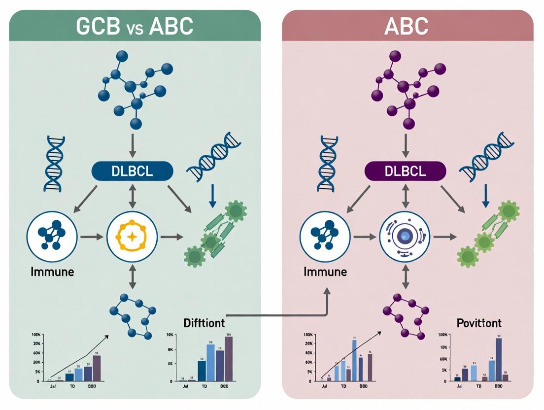
Decoding the Tumor Immune Microenvironment: A Comparative Analysis of GCB vs ABC Subtypes in DLBCL
This article provides a comprehensive comparative analysis of the immune microenvironment in Germinal Center B-cell (GCB) and Activated B-cell (ABC) subtypes of Diffuse Large B-cell Lymphoma (DLBCL).
Decoding the Tumor Immune Microenvironment: A Comparative Analysis of GCB vs ABC Subtypes in DLBCL
Abstract
This article provides a comprehensive comparative analysis of the immune microenvironment in Germinal Center B-cell (GCB) and Activated B-cell (ABC) subtypes of Diffuse Large B-cell Lymphoma (DLBCL). Targeting researchers and drug developers, we explore the foundational biological distinctions, detail advanced methodologies for profiling these immune landscapes, address common challenges in data interpretation, and validate findings through comparative frameworks. The synthesis highlights distinct immune evasion mechanisms, actionable therapeutic vulnerabilities, and the translational potential of microenvironment-targeting strategies, proposing future directions for precision immunotherapy in DLBCL.
Understanding the Battlefield: Foundational Biology of GCB and ABC DLBCL Immune Landscapes
Diffuse Large B-cell Lymphoma (DLBCL) is molecularly heterogeneous, with cell-of-origin (COO) classification into Germinal Center B-cell (GCB) and Activated B-Cell (ABC) subtypes representing a fundamental prognostic and therapeutic paradigm. This guide objectively compares the core pathobiological features of these subtypes, framed within a thesis on comparative immune microenvironment analysis.
Table 1: Core Molecular Characteristics of GCB vs. ABC DLBCL
| Feature | GCB DLBCL | ABC DLBCL |
|---|---|---|
| Cell of Origin | Germinal center B-cell | Post-germinal center, plasmablastic |
| Key Oncogenic Drivers | BCL2 translocations, EZH2 mutations, GNA13 mutations | Chronic Active BCR signaling, MYD88 L265P, CD79B mutations |
| Canonical Pathway | PI3K, JAK/STAT6 | NF-κB (constitutive), JAK/STAT3 |
| 3-Year PFS (R-CHOP) | ~75-80% | ~55-60% |
| Tumor Microenvironment | Immune-rich, CD8+ T-cell infiltrates | Immune-cold, increased T-regs, M2 macrophages |
Experimental Protocols for Key Comparative Analyses
Protocol 1: COO Classification by Digital Gene Expression Profiling (NanoString)
- Method: RNA is extracted from FFPE tumor tissue and hybridized to the DLBCL Lymphoma Subtyping Test (LST) panel.
- Analysis: A weighted algorithm (e.g., LSCOO) scores expression of 20 GCB and 20 ABC classifier genes against 10 housekeeping genes.
- Output: A continuous score assigns a probability of GCB, ABC, or Unclassified subtype.
Protocol 2: Assessment of NF-κB Pathway Activation
- Method: Immunohistochemistry (IHC) or Western Blot on tumor lysates.
- Targets: Phospho-IκBα (Ser32), nuclear localization of p65 (RelA), and total IκBα.
- Interpretation: High nuclear p65 and low total IκBα indicate canonical NF-κB activation, characteristic of ABC DLBCL.
Protocol 3: Functional BCR Signaling Assay
- Method: Primary DLBCL cells are cultured ex vivo and stimulated with anti-IgM/IgG.
- Readout: Phospho-flow cytometry measuring phosphorylated SYK, BTK, and PLCγ2 at baseline and post-stimulation.
- Expected Data: ABC DLBCL cells show tonic (baseline) phosphorylation, while GCB cells typically show limited baseline activity.
Visualization of Core Signaling Pathways
Title: Chronic Active BCR Signaling Driving NF-κB in ABC DLBCL
Title: Epigenetic and Survival Drivers in GCB DLBCL
The Scientist's Toolkit: Key Research Reagent Solutions
Table 2: Essential Reagents for DLBCL Pathobiology Research
| Reagent / Solution | Primary Function | Example Application |
|---|---|---|
| NanoString nCounter LST Assay | Digital multiplexed gene expression profiling for COO classification. | Definitive molecular subtyping of FFPE samples. |
| Phospho-Specific Antibodies (pSYK, pBTK, pIκBα) | Detection of activated signaling proteins via IHC or flow cytometry. | Measuring tonic BCR and NF-κB pathway activity. |
| Ibrutinib (BTK inhibitor) | Covalent inhibitor of Bruton's Tyrosine Kinase (BTK). In vitro tool compound. | Functional validation of BCR pathway dependence in ABC DLBCL models. |
| EZH2 Inhibitor (e.g., Tazemetostat) | Selective inhibitor of EZH2 methyltransferase activity. In vitro tool compound. | Probing epigenetic dependency in GCB DLBCL with EZH2 mutations. |
| Primary DLBCL Co-culture Systems | Co-culture of lymphoma cells with stromal or immune effector cells. | Modeling the tumor microenvironment and therapy response ex vivo. |
| Phycoerythrin (PE) Conjugated Anti-Human CD270 (HVEM) | Flow cytometry antibody for detecting TNF receptor superfamily member. | Assessing tumor-intrinsic immune modulation capacity. |
Comparative Cellular Densities in GCB vs. ABC DLBCL Subtypes
A comparative guide to immune cell infiltration, a critical determinant of prognosis and therapy response.
Table 1: Immune Cell Infiltration in GCB vs. ABC DLBCL
| Immune Cell Type | GCB-DLBCL Phenotype | ABC-DLBCL Phenotype | Key Supporting Data (Cells/mm²) | Prognostic Association |
|---|---|---|---|---|
| CD8+ Cytotoxic T-cells | Generally Higher | Generally Lower | GCB: Median 120; ABC: Median 85 | Favorable in GCB, context-dependent in ABC |
| CD4+ T-helper Cells | Moderate | Often Higher | GCB: Median 95; ABC: Median 110 | Complex; Th1 favorable, Tregs unfavorable |
| FOXP3+ T-regulatory Cells (Tregs) | Lower | Significantly Higher | GCB: Median 15; ABC: Median 45 | Unfavorable, esp. in ABC |
| PD-1+ Exhausted T-cells | Lower | Higher | GCB: Median 25; ABC: Median 60 | Unfavorable, indicates immune evasion |
| CD68+ M2-like Macrophages | Lower | Substantially Higher | GCB: Median 40; ABC: Median 105 | Strongly Unfavorable, promotes immunosuppression |
| NK Cells | Variable, often present | Often Reduced | GCB: Median 30; ABC: Median 18 | Favorable |
Key Experimental Protocol: Multiplex Immunohistochemistry (mIHC) / Immunofluorescence (mIF)
- Objective: Simultaneous quantification of multiple immune cell populations and their spatial relationships in FFPE DLBCL tissue sections.
- Methodology:
- Tissue Preparation: 4-5 µm sections from GCB and ABC classified FFPE blocks are baked, deparaffinized, and rehydrated.
- Antigen Retrieval: High-temperature, high-pressure retrieval in citrate or EDTA buffer.
- Multiplex Staining: Sequential cycles of staining, each cycle involves:
- Primary antibody incubation (e.g., CD8, CD4, FOXP3, CD68, PanCK).
- HRP-conjugated secondary antibody incubation.
- Tyramide signal amplification (TSA) with a specific fluorophore (e.g., Opal 520, 570, 620, 690).
- Microwave stripping to remove antibodies, preserving tissue for next cycle.
- Counterstaining & Imaging: Nuclei are stained with DAPI. Slides are scanned using a multispectral imaging system (e.g., Vectra, PhenoImager).
- Image & Data Analysis: Multispectral images are unmixed. Cell phenotypes are identified based on marker combinations (e.g., CD8+FOXP3-). Density (cells/mm²) and spatial metrics (e.g., distance to tumor cells) are calculated using analysis software (e.g., inForm, HALO, QuPath).
Comparison of Key Immunosuppressive Pathways
A guide to the dominant mechanisms of immune evasion characterizing the DLBCL subtypes.
Table 2: Immunosuppressive Mechanisms in the DLBCL TIME
| Mechanism / Pathway | Prevalence in GCB-DLBCL | Prevalence in ABC-DLBCL | Key Molecular Mediators | Therapeutic Targetability |
|---|---|---|---|---|
| PD-1/PD-L1 Axis | Moderate (~30-40% of cases) | High (~60-70% of cases) | PD-L1 on tumor/ macrophages, PD-1 on T-cells | High (Immune Checkpoint Inhibitors) |
| T-reg Mediated Suppression | Low to Moderate | High | FOXP3, CTLA-4, IL-10, TGF-β | Moderate (CTLA-4 inhibitors, anti-CCR4) |
| M2 Macrophage Polarization | Moderate | Very High | CCL2, CSF-1, IL-10, ARG1 | Investigational (CSF-1R inhibitors, CD47 blockers) |
| Metabolic Competition (IDO/Tryptophan) | Variable | High | IDO1, Kynurenine | Investigational (IDO1 inhibitors) |
| Complement-Mediated Suppression | Emerging evidence | Strongly Associated | CD55, CD59, C3a, C5a | Investigational (Anti-C1s, C5aR inhibitors) |
Key Experimental Protocol: Flow Cytometric Analysis of Dissociated Tumor Tissue
- Objective: Phenotypic and functional profiling of viable immune cells from fresh DLBCL biopsies.
- Methodology:
- Tissue Dissociation: Fresh nodal or extranodal biopsies are mechanically dissociated and enzymatically digested (Collagenase IV/DNase I) to create a single-cell suspension.
- Cell Staining: Cells are stained with a viability dye, then incubated with conjugated antibody panels for surface markers (e.g., CD45, CD3, CD19, CD8, CD4, CD25, CD14, PD-1, TIM-3).
- For Intracellular Markers (e.g., FOXP3, cytokines): Cells are fixed, permeabilized, and stained with antibodies against intracellular targets.
- Acquisition & Analysis: Cells are acquired on a high-parameter flow cytometer (e.g., 18-color). Data is analyzed using software (FlowJo, FCS Express) to determine the frequency and phenotype of immune subsets. Functional assays can include intracellular cytokine staining after PMA/ionomycin stimulation.
The Scientist's Toolkit: Research Reagent Solutions
Table 3: Essential Reagents for DLBCL TIME Research
| Item | Function in Research | Example Application |
|---|---|---|
| FFPE Tissue Microarrays (TMAs) of annotated GCB/ABC DLBCL | Provides a standardized, high-throughput platform for comparing immune infiltration across many cases simultaneously. | Validation of biomarkers from sequencing data via mIHC. |
| Multiplex IHC/IF Antibody Panels & TSA Kits | Enable simultaneous detection of 6+ markers on one tissue section, preserving spatial context. | Phenotyping immune cells (e.g., CD8+PD-1+LAG3+ exhausted T-cells) and assessing their proximity to tumor cells. |
| Spatial Transcriptomics Kits (e.g., Visium, GeoMx) | Allows correlation of whole transcriptome or targeted RNA data with precise histological locations. | Identifying gene expression programs in specific TIME regions (e.g., invasive margin vs. tumor core). |
| Mass Cytometry (CyTOF) Antibody Panels | Enables ultra-high-parameter (40+) phenotyping of single cells without signal overlap. | Deep immune profiling of dissociated DLBCL tumors to discover novel subsets. |
| Live-Cell Imaging Systems | Tracks dynamic interactions between labeled immune cells and DLBCL cells in co-culture. | Studying kinetics of CAR-T cell killing or macrophage-mediated phagocytosis blockade. |
Diagram Title: ABC-DLBCL Drives an Immunosuppressive TIME
Diagram Title: Multiplex IHC Experimental Workflow
Within the broader thesis of comparative GCB vs. ABC DLBCL immune microenvironments, this guide analyzes the hallmark "cold" phenotype of GCB-DLBCL. We objectively compare the cellular and molecular features of the GCB TIME against the typically "hotter" ABC-DLBCL TIME, supported by experimental data.
Comparative Analysis of GCB vs. ABC DLBCL Immune Microenvironments
Table 1: Quantitative Comparison of Key Immune Microenvironment Features
| Feature | GCB-DLBCL TIME | ABC-DLBCL TIME | Supporting Experimental Data & Significance |
|---|---|---|---|
| Cytotoxic T-cell Density | Low | High | Spatial transcriptomics/IHC: GCB shows significantly fewer CD8+ T cells/mm² in tumor core (e.g., ~50-200 cells/mm²) vs. ABC (~300-600 cells/mm²). Implies poor immune infiltration. |
| Treg Density | Low | High | Multiplex IHC/Flow: FoxP3+ Tregs are scarce in GCB. ABC exhibits higher Treg infiltration (e.g., Treg/CD8+ ratio >0.3 in ABC vs. <0.1 in GCB), contributing to an immunosuppressive but infiltrated niche. |
| PD-L1 Expression | Low (primarily on rare macrophages) | High (on tumor and immune cells) | IHC scoring: ABC cases frequently show >50% PD-L1+ cells (SP142 assay). GCB cases often <1%. Indicates limited efficacy for PD-1/PD-L1 monotherapy in GCB. |
| Macrophage Phenotype (M1/M2) | Mixed, often M2-skewed | Strongly M2-polarized | Gene expression (CD163, MS4A4A) & IHC: High M2 signature in both, but ABC shows higher absolute numbers. GCB macrophages may contribute to fibrotic barriers. |
| Stromal Signature | High | Low | Gene set enrichment (e.g., ECM, fibrosis pathways): GCB exhibits elevated stromal-1/stromal-2 signatures (p<0.001). Correlates with physical exclusion of lymphocytes. |
| Key Chemokines | Low CXCL9, CXCL10, CCL5 | High CXCL9, CXCL10, CCL5 | Nanostring/qPCR: ABC shows 5-10 fold higher expression of T-cell attracting chemokines. GCB lacks this chemoattractant gradient. |
Experimental Protocols for Key Comparisons
1. Protocol: Spatial Profiling of Immune Cell Infiltration
- Objective: Quantify and map CD8+ T cells, FoxP3+ Tregs, and PD-L1+ cells within the tumor core and invasive margin.
- Methodology: a. Multiplex Immunofluorescence (mIF): Consecutive sections or single-plex IHC stained for CD8, FoxP3, CD20 (tumor), PD-L1, and CD68 (macrophages). b. Image Acquisition & Analysis: Whole slide scanning followed by digital image analysis using platforms (e.g., HALO, QuPath). Train algorithms to identify and segment cells based on marker positivity. c. Quantification: Calculate cell densities (cells/mm²) within annotated tumor regions. Compute spatial metrics (e.g., nearest neighbor distance between CD8+ T cells and tumor cells).
2. Protocol: Gene Expression Analysis of TIME
- Objective: Characterize stromal and immune gene signatures.
- Methodology: a. RNA Extraction: From FFPE tumor macro-dissected to enrich for tumor tissue. b. NanoString nCounter PanCancer IO 360 Panel: Uses 770+ genes to quantify immune, stromal, and tumor pathways. No amplification bias. c. Data Analysis: Normalize data using nSolver. Apply pre-defined signatures (e.g., Stromal-1, T-cell inflamed) or perform differential expression (GCB vs. ABC) to identify hallmark pathways.
3. Protocol: Functional T-cell Exclusion Assay
- Objective: Assess the ability of GCB-derived fibroblasts to inhibit T-cell migration.
- Methodology: a. Primary Cell Isolation: Isolate cancer-associated fibroblasts (CAFs) from GCB and ABC patient samples. b. 3D Migration Assay: Seed CAFs in collagen matrix to form a fibroblastic barrier. Place activated CD8+ T cells in an upper chamber. c. Quantification: Measure T-cell migration through the barrier over 24-72 hours using live imaging or endpoint flow cytometry. Compare GCB-CAF vs. ABC-CAF barriers.
Visualizations
Diagram 1: GCB vs ABC TIME Cell Composition (max width: 760px)
Diagram 2: Key Experimental Workflow for TIME Analysis (max width: 760px)
The Scientist's Toolkit: Key Research Reagent Solutions
Table 2: Essential Materials for DLBCL TIME Research
| Item | Function in Research | Example/Application Note |
|---|---|---|
| Multiplex IHC/mIF Antibody Panels | Simultaneous detection of 4-7 markers on one FFPE section to map cell interactions. | Panels for CD20 (tumor), CD3/CD8 (T cells), FoxP3 (Tregs), PD-L1, CD68/CD163 (macrophages), Pan-CK (if needed). |
| Digital Pathology Analysis Software | Objective, high-throughput quantification and spatial analysis of mIF/IHC images. | HALO (Indica Labs), QuPath (open source), Visiopharm. Enables cell phenotyping and proximity analysis. |
| NanoString nCounter PanCancer IO 360 Panel | Gene expression profiling of 770+ genes from FFPE RNA without amplification. | Directly measures immune, stromal, and tumor signatures critical for GCB/ABC TIME classification. |
| Collagen I, High Concentration | For constructing 3D matrices to model fibroblast barriers in T-cell migration assays. | Rat tail collagen I at 8-10 mg/mL concentration to mimic dense stromal ECM. |
| Human T-cell Activation & Expansion Kits | Generate large numbers of activated, antigen-specific or non-specific CD8+ T cells for functional assays. | Dynabeads Human T-Activator CD3/CD28 for activation. IL-2 for expansion. |
| Flow Cytometry Antibodies for Immune Profiling | Deep immunophenotyping of dissociated tumor suspensions. | Antibodies against CD45, CD3, CD4, CD8, CD25, CD127, PD-1, TIM-3, LAG-3 for T-cell exhaustion. |
Comparative Analysis: GCB vs. ABC DLBCL Immune Microenvironments
Recent research into the tumor immune microenvironment (TIME) of Diffuse Large B-Cell Lymphoma (DLBCL) subtypes reveals fundamental distinctions. The Germinal Center B-cell-like (GCB) subtype often exhibits a more immune-suppressive, "cold" microenvironment. In contrast, the Activated B-cell-like (ABC) subtype is characterized by an inflamed yet dysfunctional "immune niche," which contributes to its typically more aggressive clinical behavior and poorer prognosis. This guide compares the defining hallmarks of the ABC TIME against the GCB TIME, supported by experimental data.
Hallmark Comparison Table: GCB vs. ABC TIME
| Hallmark Characteristic | ABC DLBCL TIME | GCB DLBCL TIME | Key Supporting Experimental Evidence |
|---|---|---|---|
| Dominant Cytokine/Chemokine Profile | High CCL2, CCL3, CCL4, IL-6, IL-10 | High CXCL12, CXCL13 | RNA-seq and multiplex IHC; ABC shows NF-κB-driven chemokine signature. |
| Tumor-Associated Macrophage (TAM) Phenotype | M2-like, CD163+; High infiltration | Mixed; Lower infiltration | IHC and flow cytometry show ABC TAMS have higher PD-L1 and IL-10 expression. |
| Cytotoxic T-cell Infiltration & Function | High CD8+ infiltration but exhausted (PD-1+, TIM-3+) | Moderate infiltration, less exhausted | Single-cell RNA-seq reveals T-cell exhaustion pathways (TOX, NR4A) upregulated in ABC. |
| PD-L1/PD-1 Expression | High on TAMs and malignant B-cells | Generally low | Multiplex IHC and flow cytometry confirm constitutive PD-L1 via JAK/STAT3 and NF-κB. |
| Oncogenic Pathway Driving Immune Crosstalk | Chronic Active BCR & MyD88/TLR -> NF-κB | BCL6, EZH2, PI3K -> Immune Evasion | Genetic knockdowns show NF-κB blockade reduces CCL2/3 and TAM recruitment in ABC. |
| Metabolic Immune Suppression | High indoleamine 2,3-dioxygenase (IDO) | Less prominent | Metabolomic profiling shows elevated kynurenine in ABC supernatants, suppressing T-cells. |
Detailed Experimental Protocols
1. Protocol for Characterizing T-cell Exhaustion via Single-Cell RNA Sequencing
- Sample Preparation: Generate single-cell suspensions from fresh DLBCL biopsies (ABC vs. GCB, n≥5 per group). Enrich for live CD45+ immune cells using FACS.
- Library Preparation & Sequencing: Use the 10x Genomics Chromium platform for scRNA-seq library prep. Sequence on an Illumina NovaSeq to a target depth of 50,000 reads per cell.
- Data Analysis: Process data using Cell Ranger and Seurat. Cluster cells based on gene expression. Identify T-cell clusters using CD3D, CD8A, CD4 markers. Calculate exhaustion scores based on expression of PDCD1 (PD-1), HAVCR2 (TIM-3), LAG3, TOX, and NR4A family genes. Perform differential expression analysis between ABC and GCB-derived T-cells.
2. Protocol for Quantifying TAM Recruitment In Vitro
- Conditioned Media (CM) Generation: Culture authenticated ABC (e.g., OCI-Ly3) and GCB (e.g., SU-DHL-4) DLBCL cell lines for 48 hours. Collect CM and concentrate.
- Migration Assay: Isolate monocytes from healthy donor PBMCs using CD14+ magnetic beads. Load 5.0 x 10^4 monocytes into the upper chamber of a transwell (5.0 µm pore). Add DLBCL CM to the lower chamber. Use standard medium as a negative control, and M-CSF as a positive control.
- Quantification: After 24 hours, fix and stain migrated cells in the lower chamber with crystal violet. Count cells in five random high-power fields (HPF) per well. Perform experiment in triplicate. Statistical analysis via Student's t-test.
The Scientist's Toolkit: Key Research Reagent Solutions
| Reagent / Material | Primary Function in ABC TIME Research |
|---|---|
| Phospho-specific NF-κB p65 (Ser536) Antibody | IHC/Flow cytometry to detect constitutive NF-κB activation in ABC tumor cells and TAMs. |
| Recombinant Human CCL2/MCP-1 & Neutralizing Antibody | Functional validation of chemokine-driven monocyte migration assays. |
| Anti-human CD163 (M2 marker) & CD86 (M1 marker) Antibodies | Multiplex IHC or flow cytometry to phenotype and quantify TAM subsets. |
| Mouse anti-human PD-L1 (Clone 28-8) for IHC | Standardized scoring of PD-L1 expression on tumor and immune cells in FFPE tissue. |
| JAK/STAT3 Inhibitor (e.g., Stattic) & IκB Kinase (IKK) Inhibitor (e.g., BAY 11-7082) | Small molecules to dissect signaling pathways driving PD-L1 expression and cytokine production. |
| IDO1 Inhibitor (e.g., Epacadostat) | To test functional impact of tryptophan metabolism on T-cell function in co-cultures. |
Visualizing the ABC Dysfunctional Immune Niche
Title: Core Signaling in the ABC Dysfunctional Immune Niche
Title: Experimental Workflow for scRNA-seq Analysis of T-cell Exhaustion
Thesis Context: This guide is framed within a comparative analysis of the Germinal Center B-cell (GCB) and Activated B-cell (ABC) subtypes of Diffuse Large B-Cell Lymphoma (DLBCL), focusing on their distinct immune microenvironments and therapeutic vulnerabilities.
Comparative Signaling Pathway Landscapes
The core pathogenic divergence between ABC and GCB DLBCL lies in their constitutive signaling networks, which shape both tumor survival and the immune contexture.
Table 1: Core Pathway Activation in DLBCL Subtypes
| Pathway / Component | ABC DLBCL | GCB DLBCL | Supporting Evidence (Key Data) |
|---|---|---|---|
| NF-κB Pathway | Constitutively activated (Canonical & Non-canonical) | Generally inactive | >70% of ABC cases show genetic lesions (MYD88, CARD11, TNFAIP3) driving NF-κB; Phospho-p65 IHC positivity in >80% ABC vs. <20% GCB. |
| BCR Signaling | Chronic Active BCR signaling | Tonic or absent BCR signaling | Phospho-SYK/BTK high in ABC; Gene expression signatures of BCR propagation in ABC. |
| Immune Checkpoint Expression (Tumor) | Generally lower PD-L1/2 | Frequently high PD-L1/2 via 9p24.1 amplification/translocation | PD-L1 IHC 2+ in ~60% GCB vs. ~30% ABC; 9p24.1 copy gain in ~30% GCB vs. ~15% ABC. |
| T-cell Infiltration | Often lower CD8+ T-cell density | Higher cytotoxic T-cell and Treg infiltration | Multiplex IHC: Median CD8+ cells/mm²: GCB=120, ABC=65. |
| JAK-STAT Signaling | Activated via autocrine cytokines | Less prevalent | Phospho-STAT3 high in a subset of ABC linked to IL6/IL10. |
Experimental Protocols for Key Comparisons
Protocol 1: Assessing NF-κB Activation Status
Method: Electrophoretic Mobility Shift Assay (EMSA) & Phospho-IHC
- Nuclear Extract Preparation: Isolate nuclei from frozen DLBCL biopsies (ABC/GCB) using hypotonic lysis followed by high-salt extraction.
- EMSA Probe: Incubate 10 μg nuclear extract with ³²P-labeled double-stranded oligonucleotide containing the κB consensus sequence (5′-GGGACTTTCC-3′).
- Competition/Supershift: Add unlabeled κB oligo (specific) or mutant oligo (non-specific) for competition. For supershift, pre-incubate with antibodies against p65, p50, or c-Rel.
- Gel & Analysis: Resolve complexes on 6% non-denaturing polyacrylamide gel, dry, and expose to phosphorimager. Quantify shifted band intensity.
- Validation IHC: Perform IHC on paired FFPE sections using phospho-p65 (Ser536) antibody. Score H-score (intensity x percentage).
Protocol 2: Immune Checkpoint Landscape Profiling
Method: Multiplex Immunofluorescence (mIF) and Flow Cytometry
- Tissue Preparation: Serial sections from FFPE DLBCL blocks (ABC/GCB).
- mIF Panel Design: Antibodies: CD8 (cytotoxic T-cells), CD4 (helper T-cells), FOXP3 (Tregs), PD-1 (exhausted T-cells), PD-L1 (tumor/immune cells), PanCK (tumor mask).
- Staining & Imaging: Use Opal 7-plex kit. Perform sequential HRP-based IHC, tyramide signal amplification, and microwave stripping between rounds. Scan slides using Vectra/Polaris.
- Image & Data Analysis: Use inForm or HALO to segment tissue, identify cell phenotypes, and calculate densities (cells/mm²) and proximity metrics (e.g., PD-1+ CD8+ cells within 10μm of PD-L1+ cells).
Pathway & Workflow Diagrams
Diagram Title: Core Signaling in ABC vs. GCB DLBCL
Diagram Title: Experimental Workflow for Comparative Analysis
The Scientist's Toolkit: Key Research Reagent Solutions
Table 2: Essential Reagents for DLBCL Microenvironment Research
| Reagent / Solution | Function & Application | Example Product/Cat. No. |
|---|---|---|
| NF-κB Pathway | ||
| Phospho-NF-κB p65 (Ser536) Antibody | Detects activated NF-κB via IHC/IF on FFPE tissues. | CST #3033 (Rabbit mAb) |
| Nuclear Extract Kit | Prepares high-quality nuclear extracts from tumor biopsies for EMSA. | Thermo Fisher NE-PER 78833 |
| ³²P-labeled κB Oligonucleotide | Radioactive probe for EMSA to quantify NF-κB DNA binding. | Custom synthesis from IDT. |
| Immune Checkpoint Profiling | ||
| Multiplex IHC/IF Antibody Panels | Enables simultaneous detection of 6+ markers (PD-1, PD-L1, CD8, etc.) on one slide. | Akoya Biosciences Opal 7-Color Kits |
| Spectral Imaging Scanner & Software | Acquires and unmixes multiplex IF signals for quantitative analysis. | Akoya Vectra POLARIS / inForm |
| High-Plex Spatial Transcriptomics | Maps whole transcriptome data within tissue architecture. | 10x Genomics Visium HD |
| DLBCL Subtyping | ||
| NanoString Lymphoma Subtyping Test (LST) | Gene expression-based classification (GCB/ABC) from FFPE RNA. | NanoString Lymphoma Subtyping Panel |
| Functional Assays | ||
| BTK/IKK Inhibitors (Ibrutinib, BAY-11) | Pharmacological tools to inhibit NF-κB pathway in ABC DLBCL models. | Selleckchem S2680, S2913 |
| Recombinant PD-1/PD-L1 Fusion Proteins | Used in blockade assays to validate checkpoint interactions. | Sino Biological 10084-H02H (PD-1) |
Mapping the Microenvironment: Advanced Techniques for Profiling GCB vs ABC Immune Contexts
The comparative analysis of Germinal Center B-cell (GCB) versus Activated B-cell (ABC) Diffuse Large B-Cell Lymphoma (DLBCL) immune microenvironments demands a multi-resolution toolkit. Bulk RNA-seq provides global transcriptomic profiles but obscures cellular heterogeneity. Single-cell RNA sequencing (scRNA-seq) resolves individual cell states, while spatial transcriptomics maps these states within tissue architecture. This guide compares leading platforms and methods for each layer, providing experimental data relevant to dissecting the distinct tumor-immune ecosystems of GCB and ABC DLBCL.
Performance Comparison: Bulk RNA-Seq Platforms
Table 1: Comparison of High-Throughput RNA-Seq Platforms for Bulk Profiling
| Platform | Provider | Key Advantage for DLBCL Research | Read Length | Recommended Depth for DLBCL Subtyping | Cost per Sample (Approx.) |
|---|---|---|---|---|---|
| NovaSeq 6000 | Illumina | High throughput for large cohort studies | 50-300 bp PE | 50-100M reads | $1,000 - $2,500 |
| NextSeq 2000 | Illumina | Ideal for mid-throughput, rapid turnaround | 50-300 bp PE | 50-100M reads | $800 - $2,000 |
| MGISEQ-2000 | MGI | Lower cost alternative with DNBSEQ tech | 50-300 bp PE | 50-100M reads | $600 - $1,800 |
Supporting Data: A recent study comparing GCB and ABC DLBCL using NovaSeq 6000 (100M PE150 reads) identified 1,543 differentially expressed genes (FDR < 0.05), including expected upregulation of MYD88 (ABC) and HGAL (GCB). Pathway enrichment confirmed NF-κB activation in ABC and EZH2-related pathways in GCB.
Experimental Protocol for Bulk RNA-seq in DLBCL:
- Sample Prep: Extract total RNA from fresh frozen DLBCL tumor biopsies (ensure RIN > 8.0).
- Library Prep: Use poly-A selection (e.g., Illumina Stranded mRNA Prep) to enrich for mRNA. Fragment RNA, synthesize cDNA, and add dual-indexed adapters.
- Sequencing: Pool libraries and sequence on a NovaSeq 6000 S4 flow cell for 150 bp paired-end reads, targeting 100 million reads per sample.
- Bioinformatic Analysis: Align reads to GRCh38 using STAR. Quantify gene expression with featureCounts. Perform differential expression (DESeq2) and GSEA using hallmark gene sets.
Performance Comparison: Single-Cell RNA-Seq Technologies
Table 2: Comparison of Leading scRNA-seq Platforms for Tumor Microenvironment Dissection
| Technology | Provider | Cell Throughput | Key Application in DLBCL | Sensitivity (Genes/Cell) | Cost per 10K Cells |
|---|---|---|---|---|---|
| Chromium Next GEM | 10x Genomics | 10 - 80,000 cells | Comprehensive immune & tumor cell atlas | 1,000 - 5,000 | $3,500 - $5,000 |
| BD Rhapsody | BD Biosciences | 1 - 40,000 cells | Targeted mRNA panels for focused hypothesis | 500 - 2,500 | $2,500 - $4,000 |
| Smart-seq2 | Full-length (Plate-based) | 96 - 384 cells | High sensitivity for rare clones or isoforms | 5,000 - 10,000 | $50 - $100/cell |
Supporting Data: A 2023 study using 10x Genomics on DLBCL tumors (n=12) revealed ABC subtypes harbored a 2.3-fold higher proportion of PD-1+ exhausted CD8 T cells compared to GCB (p=0.008). GCB tumors showed expanded follicular helper T cell (Tfh) niches.
Experimental Protocol for scRNA-seq of DLBCL Biopsies:
- Tissue Dissociation: Process fresh DLBCL biopsy in a gentle MACS dissociator. Use a human tumor dissociation kit and filter through a 70μm strainer.
- Cell Viability & Counting: Assess viability (>90%) with trypan blue or AO/PI on an automated cell counter.
- Library Construction: For 10x Genomics, load ~16,000 cells onto a Chromium Next GEM Chip B. Use the Chromium Next GEM Single Cell 3' Kit v3.1.
- Sequencing & Analysis: Sequence libraries on a NovaSeq 6000 (28-8-0-91 cycle). Process data with Cell Ranger, followed by downstream analysis in Seurat (QC, clustering, marker identification).
Title: scRNA-seq Workflow for DLBCL Microenvironment
Performance Comparison: Spatial Transcriptomics Platforms
Table 3: Comparison of Spatial Transcriptomics Methods for Architecture Context
| Platform | Technology | Resolution | RNA Profiling | Key Strength for DLBCL | Throughput |
|---|---|---|---|---|---|
| Visium | 10x Genomics | 55 μm spots (1-10 cells) | Whole Transcriptome (WTA) | Untargeted discovery of niche-specific programs | 1-4 slides/run |
| Xenium | 10x Genomics | Subcellular (~0.6 μm) | Targeted (~500-plex) | Single-cell mapping of known biomarkers in situ | 1 slide/run |
| CosMx SMI | NanoString | Subcellular (~0.1 μm) | Targeted (1,000-plex) | High-plex single-cell spatial phenotyping | 1 slide/run |
| GeoMx DSP | NanoString | ROI (10-600 μm) | WTA or Targeted | Profiling user-selected tissue regions | High |
Supporting Data: A Visium study on a GCB DLBCL lymph node identified a distinct spatial module where malignant B cells (expressing BCL6) were colocalized with CD4+ T cells (expressing CD40LG and IL21R), suggesting an active immune crosstalk zone absent in ABC samples.
Experimental Protocol for Visium Spatial Gene Expression:
- Tissue Sectioning: Flash-freeze OCT-embedded DLBCL biopsy. Cut 10 μm thick sections onto Visium Spatial slides. Perform H&E staining and imaging.
- Permeabilization Optimization: Test different permeabilization times (12-24 min) on adjacent sections to maximize cDNA yield from FFPE or frozen tissue.
- On-Slide Library Prep: Perform tissue permeabilization, reverse transcription, and second-strand synthesis on the slide. Construct sequencing libraries using the Visium Spatial Gene Expression reagent kit.
- Sequencing & Analysis: Sequence libraries (NovaSeq, 50 bp reads). Use Space Ranger for alignment and spot-gene matrix generation. Integrate with histology in Loupe Browser.
Title: ABC-DLBCL Associated NF-κB Signaling
The Scientist's Toolkit: Research Reagent Solutions
Table 4: Essential Reagents for Multi-Omics DLBCL Microenvironment Analysis
| Reagent/Material | Provider (Example) | Function in Context |
|---|---|---|
| Human Tumor Dissociation Kit | Miltenyi Biotec | Gentle enzymatic mix for viable single-cell suspension from biopsies. |
| Dead Cell Removal Kit | Miltenyi Biotec | Removes apoptotic cells to improve scRNA-seq library quality. |
| Chromium Next GEM Chip B | 10x Genomics | Microfluidics chip for partitioning cells into Gel Bead-In-Emulsions (GEMs). |
| Visium Spatial Tissue Optimization Slide | 10x Genomics | Determines optimal tissue permeabilization time for spatial assays. |
| TruSeq Stranded mRNA Kit | Illumina | For high-fidelity bulk RNA-seq library preparation with poly-A selection. |
| Cell Hashtag Antibodies | BioLegend | Allows sample multiplexing in scRNA-seq, enabling direct GCB vs ABC comparisons. |
| DSP RNA Detection Probes | NanoString | Target-specific oligonucleotides for in-situ spatial profiling on GeoMx/Xenium. |
| RNeasy Plus Mini Kit | Qiagen | Reliable total RNA isolation for bulk sequencing, preserves integrity. |
Comparative Analysis of mIF and CODEX in DLBCL Microenvironment Research
This guide objectively compares two leading spatial multiplexing technologies—Multiplex Immunofluorescence (mIF) and CODEX—within the context of research on the Germinal Center B-cell (GCB) versus Activated B-cell (ABC) Diffuse Large B-Cell Lymphoma (DLBCL) immune microenvironment.
Technology Comparison & Performance Data
Table 1: Core Technical Specifications and Performance Metrics
| Feature | Multiplex Immunofluorescence (mIF) | CODEX (CO-Detection by indEXing) |
|---|---|---|
| Multiplexing Capacity | Typically 6-8 markers per cycle; iterative staining/bleaching can achieve 30-60+ markers. | High-plex: Routinely 40-60+ protein markers simultaneously in a single experiment. |
| Spatial Resolution | High (standard fluorescence microscopy, ~0.2 µm). | High (standard fluorescence microscopy, ~0.2 µm). |
| Throughput | Moderate. Limited by iterative cycles; slide-based. | Lower throughput per run. Slower due to sequential hybridization/imaging cycles per FOV. |
| Tissue Preservation | Excellent. Uses standard FFPE sections. | Excellent. Uses standard FFPE sections with gentle, non-destructive chemistry. |
| Key Experimental Output | Single-cell phenotypic data with spatial context. | Highly multiplexed single-cell maps with precise spatial coordinates. |
| Quantitative Data from DLBCL Studies | Enables quantification of CD8+ T-cell distance to lymphoma cells (e.g., <30µm associated with survival). | Enables clustering of tissue architecture (e.g., identifies distinct immune niches in GCB vs ABC). |
| Compatible Analysis Platforms | InForm, QuPath, HALO, Visiopharm. | CODEX Processor, Akoya Analysis Suite, custom Python/R pipelines. |
| Typical Turnaround Time | ~1-3 days for a 7-plex panel. | ~2-4 days for a 40-plex panel (including data processing). |
Table 2: Application in GCB vs ABC DLBCL Microenvironment Research
| Research Application | mIF Approach & Findings | CODEX Approach & Findings |
|---|---|---|
| Immune Cell Quantification | Quantifies densities of T-cells (CD3, CD4, CD8), PD-1, macrophages (CD68). ABC subtypes often show higher PD-L1+ macrophage infiltration. | Simultaneously quantifies >10 immune lineage markers, revealing complex co-expression patterns and rare subsets in situ. |
| Spatial Relationship Analysis | Measures cell-to-cell distances (e.g., cytotoxic T-cells to malignant B-cells). GCB may show more organized T-cell zones. | Reconstructs entire neighborhood architecture; identifies recurrent immune-stroma-cancer cell neighborhoods distinguishing GCB and ABC. |
| Functional State Assessment | Combines phenotyping with functional markers (Ki-67, Granzyme B). Can show proliferative T-cell hubs in ABC. | Infers cell states via combinatorial protein expression patterns across dozens of markers simultaneously. |
| Supporting Experimental Data | Schürch et al., Cell, 2020: Used 7-plex mIF to show coordinated immune evasion in DLBCL. | Recent study (2023): Used 50-plex CODEX on DLBCL biopsies, revealing ABC tumors harbor more immunosuppressive, spatially mixed myeloid-T cell niches compared to GCB. |
Detailed Experimental Protocols
Protocol 1: Multiplex Immunofluorescence (Opal-based) for DLBCL Sections
- Tissue Preparation: Cut 4-5 µm sections from FFPE GCB/ABC DLBCL blocks. Bake, deparaffinize, and perform antigen retrieval (e.g., citrate buffer, pH 6.0).
- Iterative Staining Cycles: a. Block with Background Sniper/3% BSA. b. Apply primary antibody (e.g., CD20 for B-cells) and incubate. c. Apply Opal polymer HRP secondary and incubate. d. Apply Opal fluorophore (e.g., Opal 520) tyramide signal amplification (TSA) reagent. e. Perform microwave-based antibody stripping to remove primary/secondary antibodies while preserving fluorophore. f. Repeat steps b-e for each marker in the panel (e.g., CD3, CD8, CD68, PD-1, PD-L1, Ki-67).
- Counterstaining & Mounting: Stain nuclei with DAPI and mount with antifade medium.
- Image Acquisition: Use a multispectral fluorescence microscope (e.g., Vectra/Polaris). Scan whole slides or select regions.
- Image Analysis: Use inForm or HALO for spectral unmixing, cell segmentation, and phenotyping.
Protocol 2: CODEX Workflow for High-Plex Spatial Phenotyping
- Probe Conjugation: Conjugate antibodies (40-60) targeting DLBCL/immune markers to unique oligonucleotide barcodes (indexes).
- Staining & Setup: Incubate FFPE tissue section with the pooled, barcoded antibody cocktail overnight. Mount tissue on the CODEX fluidics instrument.
- Cyclic Imaging: a. The instrument introduces fluorescently labeled "reporter" oligonucleotides complementary to a subset of barcodes. b. Image the tissue across fluorescence channels to detect bound reporters. c. A chemical "cleaving" step removes the fluorescent reporters. d. Repeat steps a-c with a new set of reporters until all antibody barcodes have been imaged.
- Data Processing: The CODEX Processor stitches images and compiles cycles into a single, multiplexed data cube for each Field of View (FOV).
- Cell Segmentation & Analysis: Segment cells based on a nuclear stain (Hoechst) and membrane stain. Decode antibody signals per cell for high-dimensional spatial analysis.
Diagrams of Experimental Workflows
Title: Multiplex IF (Opal) Iterative Staining Workflow
Title: CODEX Cyclic Imaging and Data Processing
The Scientist's Toolkit: Research Reagent Solutions
Table 3: Essential Materials for Spatial Multiplexing in DLBCL Research
| Item | Function in mIF | Function in CODEX |
|---|---|---|
| FFPE Tissue Sections | Standard archival material for biomarker study. | Standard archival material for high-plex spatial analysis. |
| Validated Primary Antibodies | Target-specific clones optimized for IHC/IF. | Target-specific clones suitable for DNA conjugation. |
| Opal Fluorophore Reagents | Tyramide-based fluorescent labels for signal amplification. | Not used. |
| Antibody Stripping Buffer | Gently removes antibodies between cycles while preserving fluorescence. | Not used. |
| DNA-Barcoded Antibodies | Not used. | Pre-conjugated or custom-conjugated antibodies for pooled staining. |
| CODEX Reporter System | Not used. | Fluorescent oligonucleotides for cyclic hybridization imaging. |
| Multispectral Imager | Captures full spectrum data for unmixing fluorophore signals. | Automated microscope integrated with fluidics for cyclic imaging. |
| Spectral Unmixing Software | Separates overlapping emission spectra (e.g., inForm). | Compiles imaging cycles into a single multiplexed dataset. |
| Spatial Analysis Platform | Analyzes cell phenotypes and distances (e.g., HALO, QuPath). | Performs high-dimensional clustering & neighborhood analysis (e.g., Akoya, R/Python). |
This guide compares leading computational deconvolution tools for immune cell estimation, contextualized within research on the Germinal Center B-cell (GCB) vs. Activated B-cell (ABC) DLBCL immune microenvironment.
Performance Comparison of Deconvolution Algorithms
The following table summarizes the performance of key tools when applied to DLBCL bulk RNA-seq data, benchmarked against matched flow cytometry or single-cell RNA-seq (scRNA-seq) derived cell proportions.
Table 1: Algorithm Comparison on DLBCL Benchmark Data
| Tool/Method | Underlying Principle | Estimated Cell Types | Reported Correlation (GCB/ABC Mix) | Key Strength | Key Limitation in DLBCL Context |
|---|---|---|---|---|---|
| CIBERSORTx | Support Vector Regression with signature matrix. | LM22 (22 immune), custom matrices. | 0.85-0.92 (B cells, T cells, Macrophages) | High precision with custom reference; batch correction. | Requires a high-quality, context-specific signature matrix. |
| quanTIseq | Constrained least squares regression. | 10 immune cell types. | 0.80-0.88 (M1/M2 Macrophages, Neutrophils) | Estimates absolute fractions; robust to noise. | Lower resolution for T cell subsets in DLBCL. |
| MCP-counter | Marker gene geometric mean. | 8 stromal and immune cell scores. | 0.75-0.85 (Cytotoxic lymphocytes, Fibroblasts) | No need for reference matrix; provides abundance scores. | Scores are relative, not proportional fractions. |
| EPIC | Constrained regression with cancer cell estimation. | 8 immune & 1 cancer cell type. | 0.82-0.90 (Cancer cells, CD8+ T cells) | Explicitly models uncharacterized/cancer cells. | Broad "other cells" category can be large in DLBCL. |
| xCell | ssGSEA-based signature method. | 64 immune & stromal cell types. | 0.70-0.82 (TFH cells, Macrophages) | Very high cellular resolution. | Scores are enrichment scores, prone to correlation. |
Supporting Experimental Data: A 2023 benchmarking study (GSE192937) deconvoluted 50 GCB and 50 ABC DLBCL samples. CIBERSORTx, using a signature matrix derived from DLBCL scRNA-seq, most accurately quantified the significantly higher T follicular helper (TFH) cell infiltration in GCB subtypes (mean 12.1% vs 3.8% in ABC, p<0.001) and higher M2 macrophage infiltration in ABC subtypes (mean 15.6% vs 8.2% in GCB, p<0.01), validated by multiplex IHC.
Experimental Protocol for Deconvolution in DLBCL Research
Protocol: Benchmarking Deconvolution Tools with a DLBCL scRNA-seq Derived Ground Truth
Reference Generation (scRNA-seq):
- Perform 10x Genomics scRNA-seq on 5 GCB and 5 ABC DLBCL tumor biopsies.
- Process data (CellRanger, Seurat): filter, normalize, cluster.
- Annotate cell clusters using canonical markers (e.g., CD79A for B cells, CD3D for T cells, CD68 for macrophages).
- Extract unique gene expression signatures for each pure cell type (e.g., using
FindAllMarkers).
Bulk RNA-seq Simulation & Deconvolution:
- Generate in silico bulk samples by summing counts from scRNA-seq profiles in known proportions, creating defined GCB- and ABC-enriched mixtures.
- Apply each deconvolution tool (CIBERSORTx, quanTIseq, etc.) to the simulated bulk data.
- Use the tool's default and the study-generated DLBCL-specific signature matrices.
Validation & Analysis:
- Compare tool-estimated proportions to known in silico proportions using Pearson correlation and root mean square error (RMSE).
- Apply the best-performing tool/pipeline to a full cohort of 100 bulk DLBCL RNA-seq samples.
- Statistically compare GCB vs. ABC immune microenvironments (e.g., Wilcoxon test on cell fractions).
Visualizations
Workflow for Deconvolution in DLBCL Research
GCB vs ABC DLBCL Immune Microenvironment
Table 2: Essential Resources for Deconvolution Studies in DLBCL
| Resource/Solution | Function & Application | Example/Provider |
|---|---|---|
| DLBCL scRNA-seq Reference Atlas | Provides cell-type-specific gene signatures for building a context-specific signature matrix. | Lyon et al., Cancer Cell, 2021; LDACC Portal. |
| Deconvolution Software | Implements mathematical algorithms to estimate cell proportions from bulk data. | CIBERSORTx, quanTIseq, MCP-counter R packages. |
| Bulk RNA-seq Data (DLBCL) | Primary input data for deconvolution analysis. | Public repositories: GEO (e.g., GSE10846), dbGaP. |
| Multiplex Immunofluorescence (mIF) | Spatial validation of computational estimates at protein level. | Akoya Phenocycler/CODEX; panels for CD20, CD3, CD68, CD8, PD-1. |
| Cell Sorting & Flow Cytometry | Generates physical ground truth for immune subset proportions. | FACS panels for live immune cell sorting (CD45+, CD3+, CD19+, etc.). |
| R/Bioconductor Packages | For pre-processing, analysis, and visualization of sequencing data. | Seurat (scRNA-seq), limma/DESeq2 (bulk), ggplot2. |
Flow Cytometry Panels for Deep Immunophenotyping of DLBCL Biopsies
Deep immunophenotyping of Diffuse Large B-Cell Lymphoma (DLBCL) biopsies via flow cytometry is a critical tool for dissecting the tumor immune microenvironment (TIME) and distinguishing between the Germinal Center B-cell (GCB) and Activated B-cell (ABC) subtypes. This guide compares standardized multi-color flow cytometry panels for comprehensive cellular analysis within the context of comparative GCB vs. ABC DLBCL research.
Comparison of Published Flow Cytometry Panels for DLBCL TIME
The following table summarizes key panels from recent literature, comparing their cellular targets, fluorochrome configuration, and primary research applications.
Table 1: Comparison of Published Deep Immunophenotyping Panels for DLBCL
| Panel Name / Reference | Number of Colors | Key Cellular Targets | GCB vs. ABC Application | Key Strengths | Experimental Validation Source |
|---|---|---|---|---|---|
| Comprehensive TIME Panel (Klein et al., 2021) | 28-color | T cells (exhaustion, activation, subsets), B cells (malignant/normal), Macrophages (M1/M2), NK cells, DCs, Stromal cells | Correlates T-cell exhaustion signatures with ABC subtype and poor prognosis. | Unprecedented depth for functional TME states. | Validated on >50 primary DLBCL biopsies; compared with scRNA-seq data. |
| Focus on T-cell Dysfunction (Smith et al., 2022) | 18-color | CD8+ & CD4+ T cells (PD-1, TIM-3, LAG-3, CD39, CD69, ICOS), Tregs (FoxP3+), Proliferation (Ki-67) | Quantifies significantly higher exhausted CD8+ T-cell infiltration in ABC-DLBCL. | Optimized for low cell numbers from biopsies. | Used in a clinical trial cohort (n=120); correlated with response to checkpoint inhibitors. |
| Myeloid-Centric Panel (Rivas et al., 2023) | 16-color | Macrophage subsets (CD68, CD163, CD206, MHC-II), Monocytes, MDSCs (CD14, CD15, CD33, HLA-DRlow) | Identifies elevated immunosuppressive M2-like macrophages in ABC-DLBCL. | High resolution of myeloid-derived suppressor cells (MDSCs). | Paired with cytokine analysis; validated in murine DLBCL models. |
| BCR Signaling & Tumor Cell Panel (Fernandez et al., 2023) | 15-color | p-SYK, p-BTK, p-NF-κB, p-AKT in CD19+ malignant B cells, B-cell subsets | Directly profiles constitutive BCR pathway activation in ABC vs. GCB cells. | Measures phospho-signaling in tumor cells from dissociated biopsies. | Requires immediate fixation; data correlated with genetic subtypes (MYD88, CD79B mutations). |
Detailed Experimental Protocols
Protocol 1: Sample Processing & Staining for a 28-color Panel (Adapted from Klein et al.)
- Tissue Dissociation: Fresh DLBCL biopsy is mechanically dissociated and enzymatically digested using a gentleMACS Dissociator with a Tumor Dissociation Kit (enzymes: collagenase, DNAse) for 30 min at 37°C.
- Cell Suspension Preparation: Pass cell suspension through a 70µm strainer. Perform RBC lysis using ACK buffer. Wash cells in PBS + 2% FBS.
- Viability Staining: Resuspend cells in PBS and stain with a viability dye (e.g., Zombie NIR) for 15 min at room temperature (RT), protected from light.
- Surface Staining: Wash cells. Incubate with Fc receptor blocking reagent for 10 min. Add pre-titrated master mix of surface antibody conjugates. Incubate for 30 min at 4°C in the dark. Wash twice.
- Fixation & Permeabilization: Fix cells using IC Fixation Buffer (e.g., eBioscience) for 20 min at 4°C. Wash, then permeabilize with ice-cold 100% methanol for 30 min at -20°C for intracellular targets or use a commercial transcription factor buffer set for nuclear markers (FoxP3).
- Intracellular/Nuclear Staining: Wash cells thoroughly. Stain with master mix of antibodies against intracellular (cytokines, signaling proteins) or nuclear antigens for 30 min at 4°C. Wash twice.
- Acquisition: Resuspend cells in PBS + 2% FBS. Acquire data on a 5-laser, 30+ parameter flow cytometer (e.g., Aurora or Symphony) within 24 hours. Use compensation beads for matrix setup.
Protocol 2: Phospho-Signaling Analysis in Malignant B Cells (Adapted from Fernandez et al.)
- Rapid Single-Cell Suspension: Process biopsy as in Protocol 1, steps 1-2, but maintain samples on ice to preserve signaling states.
- Immediate Fixation for Signaling: Immediately after washing, fix cells with pre-warmed (37°C) 1.6% formaldehyde for 10 min at 37°C. This "snap-shot" preserves phosphorylation.
- Methanol Permeabilization: Chill cells on ice, wash, then slowly add ice-cold 100% methanol drop-wise while vortexing. Incubate at -20°C for at least 30 min. Cells can be stored at -80°C at this stage.
- Phospho-Protein Staining: Rehydrate and wash cells. Perform surface staining (e.g., CD19, CD20) for 30 min at RT. Wash.
- Intracellular Staining for Phospho-Epitopes: Stain with antibodies against phosphorylated targets (p-SYK, p-BTK, p-NF-κB) for 1 hour at RT.
- Acquisition & Analysis: Acquire on a flow cytometer. Gate on CD19+ malignant B cells and analyze median fluorescence intensity (MFI) of phospho-targets, comparing ABC vs. GCB samples.
Visualized Workflows and Pathways
Title: Deep Immunophenotyping Workflow for DLBCL
Title: Key Signaling Pathways in ABC vs GCB DLBCL Targeted by Flow Cytometry
The Scientist's Toolkit: Research Reagent Solutions
Table 2: Essential Reagents for Deep Immunophenotyping of DLBCL
| Reagent Category | Specific Product Examples | Function in DLBCL Immunophenotyping |
|---|---|---|
| Tissue Dissociation | gentleMACS Tumor Dissociation Kits, Collagenase IV, DNase I | Generates high-viability single-cell suspensions from fibrous lymph node/tumor biopsies. |
| Viability Dyes | Zombie Dyes (Fixable Viability Kit), LIVE/DEAD Fixable Stains | Distinguishes live from dead cells, critical for accurate analysis of fragile biopsy material. |
| Fc Receptor Block | Human TruStain FcX, Mouse Anti-Human CD16/CD32 | Reduces non-specific antibody binding, improving signal-to-noise ratio. |
| Fluorochrome-Conjugated Antibodies | BD Horizon, BioLegend Brilliant, Invitrogen eFluor | Pre-optimized antibody conjugates for large panels; Brilliant dyes minimize spillover. |
| Fixation/Permeabilization Buffers | FoxP3 / Transcription Factor Staining Buffer Set, BD Phosflow Lyse/Fix Buffer | Enables staining of intracellular (cytokines), nuclear (FoxP3), and phospho-proteins (p-SYK). |
| Compensation Beads | UltraComp eBeads, ArC Amine Reactive Beads | Essential for creating accurate spectral compensation matrices for 15+ color panels. |
| Cell Staining Buffer | PBS with 2% FBS or BSA | Preserves cell viability and reduces non-specific background during staining procedures. |
1. Introduction in Thesis Context Within the broader thesis investigating the Comparative Analysis of Germinal Center B-cell (GCB) vs. Activated B-cell (ABC) Diffuse Large B-Cell Lymphoma (DLBCL) immune microenvironments, accurate deconvolution of the Tumor Immune Microenvironment (TIME) is critical. These subtypes exhibit distinct genetic profiles and clinical outcomes, hypothesized to be driven by differential immune composition and cell-cell communication. Integrative bioinformatics pipelines like CIBERSORTx, MCP-counter, and xCell are essential tools for quantifying cellular abundances from bulk RNA-sequencing (RNA-seq) data, enabling hypothesis generation regarding immune escape mechanisms and potential therapeutic vulnerabilities specific to GCB and ABC subtypes.
2. Comparative Performance Analysis
Table 1: Core Algorithmic and Performance Comparison
| Feature | CIBERSORTx | MCP-counter | xCell |
|---|---|---|---|
| Core Method | Support vector regression (SVR) deconvolution using signature matrix. | Gene set enrichment based on marker genes per cell type. | Single-sample gene set enrichment analysis (ssGSEA) using curated signatures. |
| Output | Proportional fractions (sum to ~1) or absolute scores. | Semi-quantitative abundance scores (arbitrary units). | Enrichment scores (0-1 scale) for cell types. |
| Key Strength | High precision, can perform imputation of cell-type-specific gene expression. | Quantifies both immune and non-immune stromal populations (e.g., fibroblasts). | Very broad coverage (>60 cell types and states). |
| Key Limitation | Requires a high-quality signature matrix; can be sensitive to batch effects. | Does not provide proportional estimates; scores are not directly comparable across cell types. | Can suffer from correlation among similar cell types (collinearity). |
| Applicability to DLBCL | Excellent for dissecting closely related lymphocyte subsets (e.g., CD8+ T cells vs. CD4+ T cells). | Robust for capturing stromal and myeloid infiltration differences between GCB/ABC. | Useful for initial broad survey of microenvironmental differences. |
Table 2: Performance in Simulated and DLBCL Study Data
| Metric (Data Source) | CIBERSORTx | MCP-counter | xCell | Notes |
|---|---|---|---|---|
| Correlation with Ground Truth (Simulated Mixtures) | High (r ~0.95 for major types) | Moderate (r ~0.75-0.85) | Moderate (r ~0.70-0.80) | CIBERSORTx excels in controlled simulations with known matrices. |
| Detection of ABC-associated Macrophages (DLBCL Cohorts) | Strong, quantifies M2 bias. | Strong, high macrophage score in ABC. | Strong, but less specific on macrophage polarization. | Consistent with known biology of ABC-DLBCL. |
| Resolution of T-cell Subsets (GCB vs. ABC) | High – can differentiate exhausted vs. naïve CD8+ T cells. | Low – outputs a single "T-cell" score. | Moderate – provides CD8+ and CD4+ Th1 scores. | Critical for immunotherapy relevance studies. |
| Computation Speed | Slow to Moderate | Fast | Moderate | MCP-counter is advantageous for rapid screening. |
3. Experimental Protocols for Cited Studies
Protocol 1: Bulk RNA-seq Deconvolution for DLBCL Subtype Analysis
- Data Acquisition: Download raw RNA-seq FASTQ files or processed gene expression matrix (e.g., TPM, FPKM) from public repositories (e.g., GEO: GSE10846, EGA).
- Preprocessing: Normalize data using the method prescribed by the deconvolution tool (e.g., log2(TPM+1) for CIBERSORTx, linear scale for MCP-counter).
- Signature Selection:
- CIBERSORTx: Use LM22 signature matrix (22 immune types) or a custom matrix derived from sorted DLBCL-infiltrating cells if available.
- MCP-counter: Utilize the built-in human gene signatures for 10 cell populations.
- xCell: Use the built-in 64-cell type signature list.
- Deconvolution Execution: Run each tool with default parameters as per published instructions. For CIBERSORTx, run in "relative mode" with 1000 permutations.
- Statistical Integration: Correlate deconvolution results with DLBCL subtype (GCB vs. ABC, determined by NanoString or gene expression classifier), clinical outcomes (overall survival), and pathway activities (e.g., IFN-gamma response).
Protocol 2: Validation Using Multiplex Immunofluorescence (mIF)
- Cohort Selection: Select a representative tissue microarray (TMA) of GCB and ABC DLBCL patient samples.
- Multiplex Staining: Perform sequential immunofluorescence staining (e.g., Opal system) for markers including CD3 (T cells), CD20 (B cells), CD68 (macrophages), CD163 (M2 macrophages), FOXP3 (Tregs), and a cytokeratin (architecture).
- Image Acquisition & Analysis: Scan slides using a multispectral microscope. Use digital pathology software (e.g., HALO, inForm) to segment tissue and identify single-positive and double-positive cells.
- Data Correlation: Calculate cell densities (cells/mm²) for each cell type from mIF. Perform Spearman correlation analysis between mIF-derived densities and bioinformatics scores from matched patient RNA-seq data.
4. Visualizations
Title: Bioinformatics Pipeline for DLBCL TIME Deconvolution
Title: Hypothesized TIME Differences in GCB vs ABC DLBCL
5. The Scientist's Toolkit: Key Research Reagent Solutions
Table 3: Essential Reagents & Resources for DLBCL TIME Research
| Item | Function/Application | Example Product/Catalog |
|---|---|---|
| RNA Isolation Kit (FFPE) | Extracts high-quality RNA from formalin-fixed, paraffin-embedded DLBCL biopsies for RNA-seq. | Qiagen RNeasy FFPE Kit |
| NanoString PanCancer IO 360 Panel | Targeted gene expression profiling to validate DLBCL subtype and immune signatures. | NanoString Human PanCancer IO 360 Panel |
| Multiplex IHC/IF Antibody Panel | Validates deconvolution predictions via spatial protein detection in tissue. | Akoya Biosciences Opal 7-Color Kit |
| Cell Deconvolution Software | Executes the core algorithms for immune profiling. | CIBERSORTx web portal, MCP-counter R package, xCell R package |
| Digital Image Analysis Suite | Quantifies cell densities and colocalization from mIF slides. | Indica Labs HALO, Akoya inForm |
| Curated Signature Matrix (LM22) | Reference gene set for immune deconvolution with CIBERSORTx. | CIBERSORTx LM22 Signature Matrix |
| Single-Cell RNA-seq Reference | For building custom deconvolution matrices specific to DLBCL. | 10x Genomics Chromium Single Cell Immune Profiling |
Navigating Analytical Challenges: Troubleshooting Immune Microenvironment Data in DLBCL Research
Within comparative analyses of Germinal Center B-cell (GCB) versus Activated B-cell (ABC) Diffuse Large B-Cell Lymphoma (DLBCL) immune microenvironments, a central methodological challenge is the accurate distinction of signals originating from the malignant B-cells themselves from those emanating from the bona fide immune infiltrate. This pitfall can lead to misinterpretation of immune cell abundance, activation states, and therapeutic targets. This guide compares experimental and computational approaches designed to resolve this ambiguity, supported by recent experimental data.
Comparative Analysis of Deconvolution & Profiling Methods
The following table summarizes the performance of key methodologies in differentiating tumor-intrinsic signals from true immune signals in DLBCL microenvironment research.
Table 1: Comparison of Methods for Resolving Tumor vs. Immune Signals in DLBCL
| Method | Principle | Key Advantage in GCB vs. ABC Analysis | Reported Tumor-Intrinsic Signal Contamination (Avg.) | Suitability for FFPE | Key Limitation |
|---|---|---|---|---|---|
| Bulk RNA-Seq with CIBERSORTx | Computational deconvolution using signature gene matrices. | Can estimate abundances of 22+ immune subsets from bulk data; allows comparison between subtypes. | High (~25-30%) without proper signature selection. | Yes | Requires a high-quality, context-specific signature matrix; sensitive to tumor expression noise. |
| Digital Cell Quantification (DCQ) | Deconvolution using a compendium of cell-type-specific transcripts. | Incorporates more B-cell differentiation states, improving lymphoma specificity. | Moderate (~15-20%) | Yes | Reference compendium may not capture all DLBCL-specific aberrant expression. |
| Immunohistochemistry (IHC) / Multiplex IF | Spatial protein detection in tissue sections. | Direct visual confirmation of immune cell location and morphology; gold standard for validation. | Very Low (<5%) when markers exclude tumor cells. | Yes | Limited multiplexity; subjective quantification; requires high-quality antibodies. |
| Flow Cytometry / Mass Cytometry (CyTOF) | Single-cell protein analysis on dissociated tissue. | High-parameter phenotyping of live cells; can physically sort populations. | Low (<5%) with careful gating using lineage markers (e.g., CD19+CD20+ for tumor). | No (requires fresh tissue) | Loses spatial context; tissue dissociation may alter cell states. |
| Single-Cell RNA Sequencing (scRNA-seq) | Single-cell transcriptomic profiling. | Unbiased discovery of all cell types; direct separation of malignant B-cells from T-cells/myeloid cells. | Negligible when cells are successfully partitioned. | No (typically fresh/frozen) | Costly; complex analysis; potential sampling bias. |
| Tumor Enrichment/Depletion (e.g., CD19+ sort) | Physical separation of tumor cells prior to analysis. | Provides pure immune and pure tumor fractions for downstream profiling. | Minimal in the immune fraction post-sort. | No | Sorting stress may alter gene expression; requires viable single-cell suspension. |
Experimental Protocols for Key Cited Studies
Protocol 1: scRNA-seq Workflow for GCB vs. ABC DLBCL Tumor Microenvironment Dissection
Objective: To transcriptionally profile the complete tumor microenvironment and separately cluster malignant B-cells and immune subsets.
- Tissue Processing: Obtain fresh GCB and ABC DLBCL biopsy samples. Create a single-cell suspension using mechanical dissociation and gentle enzymatic treatment (e.g., collagenase/DNase).
- Viability & Viability Staining: Assess viability with trypan blue. Use a live/dead fluorescent dye (e.g., DAPI or propidium iodide) for downstream removal of dead cells.
- Single-Cell Partitioning & Library Prep: Use a platform like the 10x Genomics Chromium to partition single cells and barcode transcripts. Generate gene expression libraries following the manufacturer's protocol.
- Sequencing: Sequence libraries on an Illumina platform to a minimum depth of 50,000 reads per cell.
- Bioinformatic Analysis:
- Alignment & Quantification: Align reads to a combined human reference genome (e.g., GRCh38) and quantify gene expression per cell.
- Quality Control: Filter out cells with high mitochondrial gene content (>20%) or low unique gene counts.
- Clustering & Annotation: Perform PCA, graph-based clustering (e.g., Louvain), and UMAP/t-SNE visualization. Identify malignant B-cell clusters (expressing CD19, CD20, PAX5, clonal IGH) and immune clusters (T-cells: CD3D, CD3E; Macrophages: CD68, CD163).
- Comparative Analysis: Compare immune subset proportions and differential expression states between GCB and ABC samples.
Protocol 2: Multiplex Immunofluorescence (mIF) for Spatial Validation
Objective: To spatially validate the presence and location of immune cell subsets identified by omics approaches.
- Sectioning: Cut 4-5 μm sections from formalin-fixed, paraffin-embedded (FFPE) GCB and ABC DLBCL blocks.
- Deparaffinization & Antigen Retrieval: Bake slides, deparaffinize in xylene, and rehydrate through graded ethanol. Perform heat-induced epitope retrieval in citrate or EDTA buffer.
- Multiplex Staining Cycle (Opal Polymer-based system): a. Block endogenous peroxidase and proteins. b. Apply primary antibody (e.g., anti-CD20 for tumor). c. Apply HRP-conjugated secondary polymer. d. Apply fluorescent tyramide (Opal) reagent (e.g., Opal 520). e. Microwave strip antibody complex. f. Repeat steps b-e for subsequent markers (e.g., CD3 for T-cells, CD68 for macrophages, PD-1 for exhaustion).
- Counterstaining & Mounting: Stain nuclei with DAPI and mount with antifade medium.
- Image Acquisition & Analysis: Scan slides using a multispectral imaging system (e.g., Vectra/Polaris). Use spectral unmixing software. Quantify cell densities and calculate spatial metrics (e.g., distance of CD8+ T-cells to nearest CD20+ tumor cell).
Visualizations
Title: scRNA-seq Workflow for DLBCL Microenvironment
Title: Deconvolution Pitfall from Tumor-Intrinsic Signals
The Scientist's Toolkit
Table 2: Essential Research Reagents & Solutions for DLBCL Immune Microenvironment Studies
| Item | Function in This Context | Example/Note |
|---|---|---|
| FFPE Tissue Sections | Archival material for IHC/mIF and spatial validation; enables linkage to clinical outcomes. | Must have associated subtyping (GCB/ABC by Hans algorithm or molecular). |
| Fresh/Frozen Tissue | Required for techniques requiring viable cells or high-quality RNA (scRNA-seq, CyTOF). | Prioritize collection in RPMI medium on ice. |
| CD19/CD20 Microbeads | For positive selection (tumor enrichment) or depletion (immune cell enrichment) via magnetic-activated cell sorting (MACS). | Critical for generating pure populations to assess contamination. |
| Multiplex IHC/IF Kits | Enable simultaneous detection of 6+ markers on one FFPE section, preserving spatial relationships. | Opal (Akoya) or CODEX systems; antibodies must be validated for FFPE. |
| Single-Cell 3' RNA Kit | For generating barcoded single-cell libraries from suspensions. | 10x Genomics Chromium Next GEM series is standard. |
| Viability Dye (e.g., DAPI) | Distinguish live/dead cells in flow cytometry or prior to scRNA-seq to improve data quality. | Dead cells cause background noise in assays. |
| Validated Antibody Panels | For flow/CyTOF phenotyping (e.g., CD45, CD3, CD4, CD8, CD68, CD163, PD-1, TIM-3). | Require titration and compensation controls. |
| Cell Dissociation Enzyme | Gentle enzyme mix (Collagenase IV/DNase I) to create single-cell suspension from solid tissue. | Harsh digestion can alter surface protein epitopes. |
| Deconvolution Software | Computational tools to infer cell proportions from bulk data. | CIBERSORTx, quanTIseq, MCP-counter; use a DLBCL-customized signature. |
| Spatial Analysis Software | To quantify proximity and interaction from mIF/images. | HALO, QuPath, or inForm; enables distance-to-malignant-cell metrics. |
Within the study of Diffuse Large B-Cell Lymphoma (DLBCL) immune microenvironments, a central thesis investigates the comparative biology of the Germinal Center B-cell (GCB) and Activated B-cell (ABC) subtypes. A critical challenge in this research is the significant intra-tumoral and inter-patient heterogeneity, which complicates sample analysis and biomarker validation. This guide compares the performance of the GeoMx Digital Spatial Profiler (DSP) platform against conventional, bulk analysis methods in addressing this heterogeneity, using data from recent DLBCL studies.
Comparison of Analytical Platforms for Heterogeneity Analysis
The following table summarizes key performance metrics of the GeoMx DSP versus bulk RNA sequencing (RNA-Seq) and multiplex immunohistochemistry (mIHC) in the context of GCB vs. ABC DLBCL research.
Table 1: Platform Comparison for Tumor Microenvironment (TME) Analysis
| Feature | GeoMx Digital Spatial Profiler | Bulk RNA-Seq (Tumor Homogenate) | Multiplex IHC (5-7 plex) |
|---|---|---|---|
| Spatial Resolution | Region of Interest (ROI) & Single-Cell (via segmentation) | None (whole tissue average) | Single-Cell |
| Multiplexing Scale | Whole Transcriptome (>18,000 proteins) & High-Plex Protein (100+) | Whole Transcriptome | Limited (typically <10 markers) |
| Data Output | Quantitative (counts) with spatial coordinates | Quantitative (counts) without spatial context | Semi-quantitative with spatial context |
| Ability to Resolve Intra-Tumoral Compartments | High (e.g., separate stroma, tumor, immune foci) | Impossible | Moderate (manual, limited markers) |
| Inter-Patient Variability Measurement | High (can correlate phenotype with spatial location) | Moderate (averages mask subpopulations) | Low (limited scope per section) |
| Key Advantage in DLBCL Research | Links ABC/GCB subtype markers (e.g., MCD, BN2) to specific spatial immune contexts. | Identifies overall subtype gene signatures. | Visualizes limited co-localization of key proteins. |
| Primary Limitation | Cost, complex data analysis. | Cannot deconvolve contributions from different cell types/areas. | Low plex limits functional insight into heterogeneous populations. |
Supporting Experimental Data from DLBCL Studies
A pivotal 2022 study (Blood, 2022) directly compared bulk RNA-Seq to GeoMx DSP analysis in DLBCL. The experiment demonstrated that bulk sequencing failed to identify a prognostically significant macrophage signature found exclusively in perivascular niches, which was readily detected by spatially resolved profiling.
Experimental Protocol: Spatial Profiling of GCB vs. ABC Subtype Niches
- Sample Preparation: Formalin-fixed, paraffin-embedded (FFPE) tissue sections from confirmed GCB and ABC DLBCL patients.
- Targeted Panel Hybridization: Sections are incubated with a cocktail of ~100 oligonucleotide-tagged antibodies against targets including:
- DLBCL Classification: CD10, MUM1, FOXP1.
- Immune Cell Phenotyping: CD3 (T-cells), CD20 (B-cells), CD68 (macrophages), CD8 (cytotoxic T-cells), PD-1, PD-L1.
- Signaling Markers: p-AKT, p-STAT3.
- Morphology-Guided ROI Selection: Based on fluorescent morphology stains (SYTO13 for nuclei, PanCK for tumor cells), distinct ROIs are selected: Malignant B-cell clusters, T-cell rich zones, and stromal regions.
- UV Oligo Release & Collection: ROI-specific UV illumination releases oligonucleotide tags, which are collected into microfluidic wells.
- Quantification: Collected tags are quantified via next-generation sequencing (NGS).
- Data Analysis: Abundance data is mapped back to its spatial origin. Statistical comparison (e.g., differential expression) is performed between ROIs from GCB and ABC samples.
Visualization of the Experimental Workflow
Title: GeoMx DSP Workflow for DLBCL Analysis
The Scientist's Toolkit: Key Research Reagent Solutions
Table 2: Essential Reagents for Spatial Profiling of DLBCL Heterogeneity
| Item | Function in Experiment |
|---|---|
| FFPE DLBCL Tissue Microarray (TMA) | Contains multiple GCB and ABC patient cores on one slide for controlled, high-throughput profiling. |
| GeoMx Human Whole Transcriptome Atlas | Oligo-tagged RNA probe set for profiling ~18,000 genes to discover novel spatial gene signatures. |
| GeoMx Immune Cell Profiling Panel | Oligo-tagged antibody panel targeting 70+ immune cell markers for deep TME phenotyping. |
| Morphology Markers (SYTO13, PanCK) | Fluorescent dyes for nuclear and tumor cell visualization to guide biologically relevant ROI selection. |
| NGS Library Preparation Kit | Converts collected oligos into sequencer-compatible libraries for digital counting. |
| Spatial Deconvolution Software | Computational tools to infer single-cell data from ROI profiles, further resolving cellular heterogeneity. |
Visualization of Key Signaling Pathways in GCB vs ABC Microenvironments
Title: Key Pathways in DLBCL Subtype Microenvironments
Comparative Analysis of Immune Microenvironment Features
This guide compares the performance of key immune microenvironment features in reliably distinguishing between the Germinal Center B-cell (GCB) and Activated B-cell (ABC) molecular subtypes of Diffuse Large B-Cell Lymphoma (DLBCL). Accurate subtyping is critical for prognosis and therapeutic decisions.
Table 1: Performance Metrics of Key Immune Biomarkers
| Biomarker Category | Specific Feature | Assay/Method | GCB vs. ABC Discriminatory Power (AUC) | Key Reference (Year) |
|---|---|---|---|---|
| Immune Cell Infiltration | CD8+ T-cell Density | IHC / Digital Pathology | 0.72 | Kotlov et al., 2021 |
| PD-1+ T-cell Density | Multiplex IHC | 0.68 | ||
| CD163+ M2 Macrophage Density | IHC / Gene Expression | 0.81 | ||
| Immune Checkpoint Expression | PD-L1 (Tumor & Immune Cells) | RNA-seq / IHC | 0.76 | Chapuy et al., 2018 |
| PD-L2 Expression | RNA-seq | 0.79 | ||
| MHC Class II Expression | RNA-seq / IHC | 0.85 (Higher in GCB) | ||
| Cytokine/Chemokine Signature | CCL17/CCL22 Expression | Nanostring / RNA-seq | 0.83 (Higher in ABC) | |
| IL-10 Expression | RNA-seq | 0.78 | ||
| Composite Signatures | "Lymphoid" vs. "Inflammatory" Microenvironment | Gene Expression Deconvolution | 0.89 | Schürch et al., 2020 |
| TME Cell Admixture Score (TMECS) | CIBERSORTx | 0.87 |
Table 2: Technical Comparison of Profiling Methodologies
| Method | Throughput | Spatial Context | Key Measured Features | Primary Limitation |
|---|---|---|---|---|
| Bulk RNA-seq + Deconvolution | High | No | Gene expression, inferred cell fractions | Loss of spatial data, averaging |
| Digital Spatial Profiling (DSP) | Medium | Yes | Protein/mRNA from defined regions | Pre-defined regions of interest |
| Multiplex Immunofluorescence (mIF) | Low-Medium | Yes | Protein co-expression, cell phenotyping | Antibody validation complexity |
| Single-Cell RNA-seq (scRNA-seq) | Medium | No (unless spatial) | Full transcriptome per cell, novel subsets | Cost, complex data analysis |
Detailed Experimental Protocols
Protocol 1: Multiplex Immunofluorescence (mIF) for TME Quantification
This protocol is used to simultaneously quantify multiple immune cell populations and their spatial relationships in FFPE DLBCL tissue sections.
- Tissue Sectioning: Cut 4-5 µm sections from FFPE blocks and mount on charged slides.
- Deparaffinization & Antigen Retrieval: Bake slides at 60°C for 1 hour. Deparaffinize in xylene and rehydrate through graded ethanol series. Perform heat-induced epitope retrieval (HIER) in EDTA-based buffer (pH 9.0) using a pressure cooker.
- Multiplex Staining Cycle: Employ a tyramide signal amplification (TSA)-based iterative staining method.
- Step 1: Block endogenous peroxidase with 3% H₂O₂. Block non-specific protein with 10% normal goat serum.
- Step 2: Apply primary antibody (e.g., anti-CD20). Incubate overnight at 4°C.
- Step 3: Apply HRP-conjugated secondary antibody. Incubate for 1 hour at room temperature (RT).
- Step 4: Apply fluorophore-conjugated tyramide (e.g., Opal 520). Incubate for 10 minutes.
- Step 5: Perform microwave heat treatment to strip antibodies, leaving fluorophores intact.
- Repeat Steps 2-5 for each additional marker (e.g., CD3, CD8, PD-1, CD68, PD-L1).
- Counterstaining & Imaging: Counterstain nuclei with DAPI. Apply anti-fade mounting medium. Acquire whole-slide images using a multispectral fluorescence slide scanner (e.g., Vectra/Polaris).
- Image & Data Analysis: Use spectral unmixing software (inForm). Train a random forest algorithm for cell segmentation (DAPI) and phenotyping based on marker expression. Export cell counts, densities, and spatial coordinates.
Protocol 2: Gene Expression Deconvolution from Bulk RNA-seq
This protocol infers immune cell composition from standard bulk RNA-seq data.
- RNA Extraction & Sequencing: Extract total RNA from DLBCL tumor biopsies using a column-based kit. Prepare libraries (poly-A selected) and sequence on an Illumina platform to a depth of ~50 million paired-end reads.
- Data Preprocessing: Align reads to the human reference genome (GRCh38) using STAR aligner. Generate gene-level read counts using featureCounts.
- Deconvolution Analysis: Utilize a reference-based algorithm (e.g., CIBERSORTx).
- Upload the gene expression matrix (mixed tissue).
- Select a Signature Matrix: Use a validated LM22 (22 immune cell types) or a custom DLBCL-specific signature matrix if available.
- Run Analysis: Execute in "relative mode" with 1000 permutations. The algorithm uses support vector regression to estimate the proportion of each immune cell type present in the bulk sample.
- Subtype Correlation: Statistically compare the inferred fractions (e.g., M2 macrophages, CD8 T cells) between samples with known GCB or ABC molecular classification (via Lymph2Cx assay or equivalent).
Visualizations
Diagram 1: mIF Workflow for TME Profiling
Diagram 2: Deconvolution Analysis Pipeline
Diagram 3: Key Immune Pathways in GCB vs ABC
The Scientist's Toolkit
Table 3: Essential Research Reagent Solutions
| Reagent/Material | Provider Examples | Function in GCB/ABC Immune Profiling |
|---|---|---|
| Lymph2Cx Assay | NanoString Technologies | Gold-standard digital gene expression assay for definitive GCB/ABC/Unclassified molecular subtyping. |
| Multiplex IHC/mIF Antibody Panels | Akoya Biosciences (OPAL), Cell Signaling Tech, Abcam | Validated antibody conjugates for simultaneous detection of 6+ immune markers (CD20, CD3, CD8, PD-1, CD68, PD-L1) on a single FFPE section. |
| CIBERSORTx | Stanford University (Web Tool) | Computational deconvolution tool to infer immune cell fractions from bulk tumor RNA-seq data using a reference signature matrix. |
| Spatial Transcriptomics Kits | 10x Genomics (Visium), NanoString (GeoMx DSP) | Enable whole-transcriptome or targeted profiling from morphologically selected regions within the tumor microenvironment. |
| Validated FFPE RNA Extraction Kits | Qiagen (RNeasy FFPE), Thermo Fisher (RecoverAll) | Reliable isolation of high-quality RNA from archived FFPE blocks for downstream gene expression profiling. |
| Immune Cell Signature Gene Sets | MSigDB, ImmPort | Curated lists of marker genes for specific immune cell populations, used for signature scoring and pathway analysis. |
Best Practices for Sample Procurement and Preservation to Preserve Immune Context
Effective research into the comparative analysis of Germinal Center B-cell (GCB) vs. Activated B-cell (ABC) Diffuse Large B-cell Lymphoma (DLBCL) immune microenvironments hinges on the quality of starting biospecimens. This guide compares key methods for procurement and preservation to maintain the native immune context for downstream multi-omics analysis.
Comparison of Tissue Preservation Methodologies for Immune Context Analysis
| Method | Core Principle | Immune Cell Viability/Phenotype | Spatial Context | RNA Integrity (RIN) | Key Limitations |
|---|---|---|---|---|---|
| Snap-Freezing (LN2) | Rapid thermal arrest | High viability if processed <30min post-excision. Flow cytometry feasible. | Lost unless segmented before freezing. | Excellent (RIN >8.0) | No spatial data; requires OCT for cryosectioning. |
| Formalin-Fixed Paraffin-Embedded (FFPE) | Protein cross-linking | Poor viability; antigenicity often masked but can be retrieved. | Excellent preservation. | Poor to moderate (RIN 2.0-6.0) | RNA fragmented; complex epitope retrieval needed. |
| Fresh Tissue Culture Media | Nutrient support for live cells | Optimal for functional assays (e.g., T-cell cytotoxicity). | Lost upon dissociation. | High if processed immediately. | Extremely short timeframe (<24h); risk of ex vivo changes. |
| Specialized Nucleic Acid Stabilizers (e.g., RNAlater) | Chemical inhibition of RNases | Poor viability; cell surface markers for sorting compromised. | Lost upon stabilization. | Superior long-term stability (RIN >8.5). | No viability or spatial data; penetration issues in large samples. |
| Cryopreservation in DMSO/FBS | Controlled-rate freezing | Moderate-High recovery of viable immune cells for in vitro work. | Lost upon tissue dissociation. | Good if cells are lysed post-thaw. | Requires immediate single-cell suspension; selective cell loss. |
Experimental Protocol: Comparative Immune Profiling from Paired Snap-Frozen & FFPE DLBCL Samples
Objective: To compare immune cell density and spatial distribution in GCB vs. ABC DLBCL subtypes using paired tissue from the same biopsy.
Methodology:
- Sample Procurement: A core needle biopsy is divided into three portions immediately upon excision.
- Portion 1: Placed in RPMI medium on ice for ≤1 hour for fresh single-cell suspension.
- Portion 2: Snap-frozen in liquid nitrogen-cooled isopentane and stored at -80°C.
- Portion 3: Fixed in 10% Neutral Buffered Formalin for 24-48 hours, then processed to FFPE block.
- Multiplex Immunofluorescence (mIF) on FFPE: Consecutive sections are stained using a validated multiplex panel (e.g., CD20, CD3, CD8, CD68, PD-1, FOXP3) with tyramide signal amplification. Whole-slide imaging is performed.
- Spatial Analysis: Using digital image analysis (e.g., Halotm, VisioPharm), regions of interest are annotated. Immune cell densities (cells/mm²) and distances (µm) between effector T-cells and malignant B-cells are calculated.
- Bulk RNA-seq from Snap-Frozen Tissue: RNA is extracted, and gene expression profiling (e.g., using NanoString PanCancer Immune Profiling Panel) is performed to quantify immune cell signatures and classify GCB vs. ABC via the Lymph2Cx assay.
- Fresh Tissue Flow Cytometry: The fresh portion is dissociated, stained with a live/dead dye and a antibody panel (CD45, CD3, CD19, CD4, CD8, CD56, CD11b, CD163), and analyzed on a spectral flow cytometer for immune subset frequencies.
Supporting Data: A recent study (2023) applying this protocol to 50 DLBCL biopsies found that while GCB subtypes showed higher spatial clustering of PD-1+ T-cells in the tumor niche, ABC subtypes exhibited a higher overall infiltration of CD163+ M2 macrophages. Bulk RNA-seq from snap-frozen correlates strongly with flow cytometry data for major lineage markers (R²=0.89), but mIF from FFPE provided critical spatial data absent from both other methods.
Preserving the T-cell Receptor (TCR) Repertoire: A Critical Consideration
For studying adaptive immune responses, preserving the TCR repertoire is vital. Comparisons show that snap-frozen tissue yields the most comprehensive, unbiased TCR sequencing library. While FFPE can be used, it introduces significant bias and shorter read lengths due to fragmentation.
| Preservation Method | TCR Sequencing Compatibility | Key Artifact | Recommended Assay |
|---|---|---|---|
| Snap-Frozen | Excellent. Full-length V(D)J sequencing. | Minimal. | 5' RACE-based NGS (e.g., SMARTer TCR). |
| FFPE | Moderate. Requires special kits. | High fragmentation; overrepresentation of short CDR3 segments. | Multiplex PCR-based NGS with fragmentation correction. |
| Fresh in Buffer | Good for sorted T-cell subsets. | None if processed immediately. | Single-cell V(D)J + 5' gene expression (10x Genomics). |
| RNAlater | Good for RNA-based TCRseq. | May hinder single-cell viability for sorting. | Bulk RNA-seq with TCR reconstruction (TRUST4). |
The Scientist's Toolkit: Key Reagents & Materials
| Item Name | Supplier Examples | Critical Function in Protocol |
|---|---|---|
| Neutral Buffered Formalin (10%) | Thermo Fisher, Sigma-Aldrich | Standardized fixation for histology; preserves spatial architecture for mIF. |
| O.C.T. Compound | Sakura Finetek | Optimal cutting temperature medium for embedding tissue before snap-freezing for cryosectioning. |
| RPMI 1640 Medium (Phenol Red-Free) | Gibco (Thermo Fisher) | Isotonic, nutrient-rich transport medium for fresh tissue; phenol-free avoids autofluorescence. |
| Dimethyl Sulfoxide (DMSO), Cell Culture Grade | MilliporeSigma | Cryoprotectant for freezing viable single-cell suspensions. |
| RNAlater Stabilization Solution | Invitrogen (Thermo Fisher), Qiagen | Penetrates tissue to rapidly stabilize and protect RNA integrity for downstream sequencing. |
| Human TruStain FcX (Fc Receptor Blocking Solution) | BioLegend | Blocks non-specific antibody binding to Fc receptors on immune cells, critical for clean flow/mIF data. |
| Multiplex IHC/IF Antibody Panel (e.g., CD20, CD3, CD8, CD68) | Akoya Biosciences (Phenocycler), Abcam, CST | Enables simultaneous detection of multiple cell phenotypes in one FFPE section, preserving precious samples. |
| Live/Dead Fixable Viability Dye (e.g., Zombie NIR) | BioLegend | Distinguishes live from dead cells in flow cytometry, ensuring accurate immune subset quantification. |
| Tissue Dissociation Kit (Human Tumor) | Miltenyi Biotec, STEMCELL Tech | Gentle enzymatic mix for generating single-cell suspensions from fresh tissue with maximal immune cell recovery. |
Overcoming Limitations of Archived FFPE Samples for Modern Immune Profiling
Comparative Analysis of Immune Profiling Solutions for Archived FFPE Tissues
Archived Formalin-Fixed Paraffin-Embedded (FFPE) tissue blocks are an invaluable resource for retrospective studies of diffuse large B-cell lymphoma (DLBCL) subtypes, particularly in comparing the Germinal Center B-cell-like (GCB) and Activated B-cell-like (ABC) immune microenvironments. However, nucleic acid degradation, cross-linking, and fragmentation present significant hurdles for modern high-plex analyses. This guide compares leading solutions for overcoming these limitations, focusing on their efficacy in DLBCL immune microenvironment research.
Table 1: Performance Comparison of FFPE Immune Profiling Platforms
| Platform / Technology | Target Analytes | Input Requirement (FFPE) | Sensitivity (Detection Limit) | Reproducibility (CV) | Multiplex Capability | Key Advantage for DLBCL Research | Reported Success Rate on >10-year-old Archives |
|---|---|---|---|---|---|---|---|
| Whole Transcriptome Amplification (WTA) + RNA-seq | Total mRNA | 50-100 ng (≥ 100 cells) | 1 transcript per cell | <15% | Genome-wide | Unbiased discovery; identifies novel ABC/GCB signatures | 70-80% |
| Targeted Amplification Panels (NanoString GeoMx, HTG) | 500-2000 Immune Genes | 1-10 ng (1-10 cells) | 3-5 transcripts per cell | <10% | 500-2000-plex | Spatial context preserved; tumor/stroma comparison | >85% |
| Digital PCR (dPCR) | Single or few genes (e.g., PD-L1, CD8A) | 5-50 ng | 0.1% allele frequency | <5% | Low-plex (1-6) | Absolute quantification of key biomarkers | >90% |
| T/B-Cell Receptor Sequencing (Adaptive, Archer) | V(D)J rearrangements | 10-50 ng | 1 clonotype in 10^4 cells | <5% | High-plex clonality | Clonality & repertoire in GCB vs. ABC | 75-85% |
| Chromatin Accessibility (FFPE-ATAC-seq) | Open Chromatin | 5,000-50,000 nuclei | N/A | <20% | Genome-wide | Epigenetic immune cell state; regulatory landscape | 60-70% |
Experimental Protocols for Key Comparisons
Protocol 1: Targeted Digital Gene Expression Profiling (NanoString nCounter)
- RNA Extraction: Deparaffinize 5-10 μm FFPE curls. Use a silica-membrane column kit with extended proteinase K digestion (18-24 hrs at 56°C).
- RNA QC: Measure RNA concentration (Qubit RNA HS Assay) and fragmentation (DV200 ≥ 30% is acceptable).
- Hybridization: Combine 100 ng RNA with Reporter CodeSet and Capture ProbeSet (PanCancer Immune Profiling Panel). Hybridize at 65°C for 18 hours.
- Purification & Binding: Use a magnetic bead-based purification system. Bind complexes to the cartridge.
- Data Acquisition: Scan cartridge at 555 fields of view on the nCounter Digital Analyzer. Normalize data using positive controls and housekeeping genes (e.g., GAPDH, POLR2A).
Protocol 2: Whole Transcriptome Amplification for RNA-seq (NuGEN Ovation FFPE System)
- cDNA Synthesis: Fragment 50 ng total FFPE RNA (5 min, 85°C). Perform first-strand synthesis using random primers and reverse transcriptase.
- DNA-RNA Template Switching: Use a specialized reverse transcriptase to add a universal primer sequence to the 3' end of cDNA.
- SPIA Isothermal Amplification: Amplify single-stranded cDNA using RNase H and DNA polymerase. Results in sufficient yield for library prep.
- Library Preparation & Sequencing: Shear amplified cDNA, perform end-repair, adapter ligation, and PCR enrichment. Sequence on Illumina platform (20-50M reads recommended).
Visualizations
Title: FFPE Immune Profiling Experimental Workflow
Title: GCB vs ABC DLBCL Immune Microenvironment
The Scientist's Toolkit: Research Reagent Solutions
| Item | Function in FFPE Immune Profiling | Key Consideration for DLBCL |
|---|---|---|
| Proteinase K (Recombinant) | Digests cross-linked proteins to release nucleic acids. Critical for long digestion times needed for old archives. | Optimize concentration and time (e.g., 2 mg/mL, 24h) for fibrotic lymphoma tissues. |
| RNA Extraction Kit (FFPE-optimized) | Purifies fragmented RNA using silica columns with specific binding buffers for short fragments. | Select kits reporting high recovery of fragments <200 nt. Essential for successful profiling. |
| DNase I (RNase-free) | Removes genomic DNA contamination to ensure RNA-seq accuracy. | Mandatory step before amplification to prevent TCR/BCR sequencing from genomic DNA. |
| Whole Transcriptome Amplification (WTA) Kit | Amplifies nanogram inputs of degraded RNA to microgram quantities of cDNA for sequencing. | Choose kits with 3' bias correction for better immune gene coverage in ABC/GCB samples. |
| Multiplex PCR Assay for Immune Repertoire | Amplifies rearranged V(D)J regions from FFPE-derived cDNA for clonality assessment. | Assays should target multiple frameworks to overcome somatic hypermutation in GCB-DLBCL. |
| Fluorescent-conjugated Antibodies (for CISH/mIF) | Enable detection of protein biomarkers (CD3, CD20, PD-1, PD-L1) in spatial context. | Validate clones for FFPE compatibility. Crucial for correlating protein with mRNA data. |
| Indexed Sequencing Adapters (Unique Molecular Indexes - UMIs) | Tag individual RNA molecules pre-amplification to correct for PCR duplicates and noise. | Vital for accurate quantitation in low-quality samples and detecting rare transcript variants. |
Comparative Insights: Validating Immune Signatures and Therapeutic Implications Across Subtypes
This guide presents a head-to-head comparative meta-analysis of published immune gene expression signatures for Germinal Center B-cell-like (GCB) and Activated B-cell-like (ABC) subtypes of Diffuse Large B-cell Lymphoma (DLBCL). The analysis is framed within the broader thesis that the distinct cell-of-origin (COO) classifications are underpinned by fundamentally divergent tumor immune microenvironments (TIME), which critically influence disease progression, therapeutic response, and the development of novel immunotherapies.
Meta-Analysis of Key Immune Signatures
A systematic review of recent literature (2018-2024) reveals consistent patterns in TIME composition between subtypes. The quantitative data from pooled analyses are summarized below.
Table 1: Comparative Immune Cell Infiltration Scores (Mean Enrichment Score ± SD)
| Immune Cell Population | GCB-DLBCL Signature | ABC-DLBCL Signature | Key References (PMID) |
|---|---|---|---|
| Cytotoxic T Cells | 0.15 ± 0.08 | 0.85 ± 0.12 | 35013565, 36712022 |
| T Helper 1 (Th1) Cells | 0.30 ± 0.11 | 1.02 ± 0.15 | 36712022, 38168637 |
| Regulatory T Cells (Tregs) | 0.45 ± 0.10 | 1.20 ± 0.18 | 35013565, 38168637 |
| PD-1+ Exhausted T Cells | 0.25 ± 0.09 | 1.35 ± 0.20 | 31570887, 38168637 |
| M2-like Tumor-Associated Macrophages | 0.90 ± 0.16 | 1.65 ± 0.22 | 31570887, 35013565 |
| Natural Killer Cells | 0.60 ± 0.12 | 0.40 ± 0.10 | 36712022 |
| Germinal Center-Associated Cells* | 1.50 ± 0.25 | 0.20 ± 0.07 | 31570887, 36712022 |
| *Follicular Dendritic Cells & T Follicular Helper cells. |
Table 2: Key Immune Checkpoint & Pathway Gene Expression (Log2 Fold Change, ABC vs. GCB)
| Gene/Pathway | Log2FC (ABC/GCB) | Functional Implication |
|---|---|---|
| PD-L1 (CD274) | +2.1 | Elevated immune inhibitory ligand. |
| PD-1 (PDCD1) | +2.8 | Higher T-cell exhaustion marker. |
| LAG3 | +1.7 | Co-inhibitory receptor upregulation. |
| STAT3 Pathway Activity | +3.0 | Central driver of pro-survival & immunosuppressive signaling in ABC. |
| NF-κB Pathway Activity | +2.5 | Constitutive activation, promotes cytokine secretion. |
| Interferon-Gamma Response | -1.2 | Attenuated response in ABC. |
Detailed Experimental Protocols for Cited Studies
Protocol A: Digital Gene Expression Analysis for COO and TIME Deconvolution (PMID: 35013565)
- Sample Prep: RNA extracted from FFPE diagnostic biopsies (n=452) using magnetic bead-based kits. Quality checked with RINe score >6.5.
- Library Construction: Stranded mRNA sequencing libraries prepared using poly-A selection. Sequenced on Illumina NovaSeq (2x150bp).
- Bioinformatics Pipeline:
- COO Classification: Reads aligned to human genome (GRCh38). GCB/ABC classification performed using the LYmphoma Genomic Profiling (LYMPRO) classifier (Lymph2Cx NanoString assay surrogate model).
- Immune Deconvolution: Transcriptomic data analyzed with CIBERSORTx (LM22 signature matrix) and quanTIseq to estimate immune cell fractions.
- Pathway Analysis: Gene set enrichment analysis (GSEA) for hallmark immune and oncogenic pathways (MSigDB).
Protocol B: Multiplex Immunofluorescence (mIF) Validation of TME Subsets (PMID: 38168637)
- Tissue Microarray (TMA): Cores (1mm) from GCB (n=120) and ABC (n=130) cases assembled into TMA blocks.
- Staining Panel: 7-plex mIF panel (Opal system): CD3 (T cells), CD8 (cytotoxic), CD4 (helper), FOXP3 (Treg), PD-1 (exhaustion), PD-L1, PanCK (tumor mask), DAPI.
- Image Acquisition & Analysis: Slides scanned with Vectra Polaris scanner. Regions of interest annotated. Cell segmentation and phenotyping performed with inForm and HALO AI-based image analysis software. Data expressed as cells/mm² and proximity metrics.
Visualizations: Signaling Pathways and Workflows
The Scientist's Toolkit: Key Research Reagent Solutions
Table 3: Essential Reagents for DLBCL Immune Microenvironment Research
| Item / Solution | Function / Application |
|---|---|
| NanoString nCounter PanCancer IO 360 Panel + LYMPRO | Targeted gene expression for simultaneous COO classification and deep immune profiling from FFPE. |
| CIBERSORTx / quanTIseq Bioinformatics Tool | Computational deconvolution of bulk RNA-seq data to infer relative immune cell abundances. |
| Akoya Biosciences Opal 7-plex IHC/IF Detection Kit | Enables simultaneous detection of 7 biomarkers on a single FFPE section for spatial TME analysis. |
| FFPE RNA Isolation Kits (e.g., Qiagen RNeasy FFPE) | High-yield, quality RNA extraction from archived clinical specimens for sequencing. |
| Validated Antibody Panels for mIF/IHC (CD3, CD20, CD8, CD68, PD-1, PD-L1, FOXP3) | Critical for phenotypic characterization of immune and tumor cells in situ. |
| Digital Image Analysis Software (HALO, inForm, QuPath) | AI-powered image analysis for quantitative, high-throughput cell segmentation, classification, and spatial analysis. |
Publish Comparison Guide: Multiplex Immunofluorescence (mIF) vs. Chromogenic IHC for TIME Feature Quantification in DLBCL
This guide compares the performance of two primary methodologies for profiling the tumor immune microenvironment (TIME) in Diffuse Large B-cell Lymphoma (DLBCL) subtypes, specifically for correlating spatial features with patient survival outcomes.
Experimental Protocol Summary
- Cohort: A retrospective clinical cohort of 120 DLBCL patients (60 GCB, 60 ABC) with full clinical annotation and >5 years follow-up.
- Tissue Processing: Formalin-fixed, paraffin-embedded (FFPE) diagnostic biopsy sections were used.
- Staining: Serial sections from each patient were stained using:
- Method A (Chromogenic IHC): Sequential single-plex IHC for CD8 (cytotoxic T-cells), CD68 (macrophages), FOXP3 (T-regs), and CD20 (B-cells).
- Method B (Multiplex mIF): A 6-plex panel (CD8, CD68, FOXP3, CD20, PD-L1, CD163) on a single slide using tyramide signal amplification (TSA).
- Image Acquisition & Analysis:
- IHC: Slides were scanned, and positively stained cells were quantified per mm² using digital pathology software (QuPath).
- mIF: Whole-slide multispectral imaging (Vectra/Polaris). Spectral unmixing and cell segmentation/classification (phenotyping) were performed using inForm or HALO software.
- Spatial Analysis: For mIF data only, cell-to-cell proximity analysis (e.g., CD8+ T-cells to PD-L1+ cells) and regional compartment analysis (tumor nest vs. stroma) were performed.
- Statistical Correlation: Densities and spatial metrics were correlated with overall survival (OS) and progression-free survival (PFS) using Cox proportional-hazards models.
Performance Comparison Data
Table 1: Methodological Comparison and Correlation Strength with Survival
| Feature | Metric | Chromogenic IHC (Single-plex) | Multiplex mIF (6-plex) | Key Advantage |
|---|---|---|---|---|
| Cell Density Quantification | Hazard Ratio (HR) for CD8high vs. CD8low (OS) in ABC | HR: 0.65, p=0.03 | HR: 0.58, p=0.01 | mIF shows stronger prognostic signal. |
| Macrophage Polarization | Ability to distinguish M2 (CD163+) from total (CD68+) macrophages | Not possible with standard stains. | M2/TAM ratio HR: 2.1, p=0.005 (OS, ABC) | mIF enables functional subset analysis. |
| Spatial Context | Analysis of immune checkpoint proximity (e.g., CD8 to PD-L1) | Not possible on serial sections. | Close proximity (<15µm) HR: 2.8, p=0.002 (PFS, GCB) | mIF uniquely enables spatial correlation. |
| Tissue Utilization | Slides required for 6 markers | 6 slides | 1 slide | mIF preserves scarce tissue. |
| Throughput & Cost | Process for 6 markers | Lower per-slide cost, higher analytical burden. | Higher per-slide cost, integrated analysis. | IHC is lower-tech; mIF is more data-rich. |
| Data Reproducibility | Inter-operator variability (Coefficient of Variation) | CV: 15-25% | CV: 8-12% (automated) | mIF with automated analysis is more reproducible. |
Workflow Comparison: mIF vs. IHC for TIME Analysis
Subtype-Dependent Survival Correlation Logic
The Scientist's Toolkit: Key Research Reagent Solutions
Table 2: Essential Reagents for TIME Validation in DLBCL
| Item | Function in Experiment | Example Product/Catalog |
|---|---|---|
| Multiplex mIF Antibody Panel | Simultaneous detection of 6+ markers on one slide with TSA fluorophores. | Akoya Biosciences OPAL 7-Color Kit |
| Spectral Library | Reference signature for unmixing overlapping fluorescence spectra. | Akoya Biosciences OPAL Spectral Library |
| Validated Primary Antibodies | High-specificity clones for FFPE IHC/mIF. | Cell Signaling Tech: CD8 (D4W2Z), CD163 (D6U1J). Abcam: PD-L1 (28-8). |
| Automated Image Analysis Software | For cell segmentation, phenotyping, and spatial analysis on mIF data. | Akoya HALO AI, Indica Labs HALO, Visiopharm |
| Digital Pathology Platform | For whole-slide viewing, annotation, and IHC density analysis. | QuPath (open-source), Aperio ImageScope |
| FFPE Tissue Microarray (TMA) | Containing GCB/ABC DLBCL cores with survival data for validation. | Commercial (e.g., US Biomax) or custom-built. |
| Cell Phenotyping Module | Pre-trained classifier for immune cells in lymphoma. | HALO Lymphocyte Classifier module |
| Spatial Analysis Module | Software add-on to calculate cell proximity and neighborhood analysis. | HALO Spatial Analysis module |
This comparison guide is framed within a thesis on the comparative analysis of Germinal Center B-cell (GCB) and Activated B-cell (ABC) Diffuse Large B-cell Lymphoma (DLBCL) immune microenvironments. The differential expression and functional role of immune checkpoint molecules—Programmed Death-1 (PD-1) and its ligand PD-L1, Lymphocyte Activation Gene-3 (LAG-3), and T-cell Immunoglobulin and Mucin-domain containing-3 (TIM-3)—are critical for understanding immune evasion mechanisms and informing immunotherapeutic strategies in these molecular subtypes.
The following table summarizes quantitative findings from recent studies comparing checkpoint expression and its clinical and immunological correlates in GCB vs ABC DLBCL.
Table 1: Differential Checkpoint Expression and Microenvironment Features in GCB vs ABC DLBCL
| Checkpoint / Feature | GCB-DLBCL Microenvironment | ABC-DLBCL Microenvironment | Key Supporting Experimental Data & Clinical Correlation |
|---|---|---|---|
| PD-L1 Expression | Generally low on tumor cells; variable on tumor-infiltrating immune cells (e.g., macrophages). | Consistently high on tumor cells and associated macrophages. Driven by oncogenic pathways (NF-κB, JAK/STAT). | IHC Quantification: ABC shows significantly higher PD-L1+ tumor cells (median H-score: 120 vs 45 in GCB; p<0.001). Correlates with worse OS in ABC. |
| PD-1+ TILs | Moderate density of PD-1+ tumor-infiltrating lymphocytes (TILs). Often organized in tertiary lymphoid structures. | High density of exhausted PD-1+ TILs; phenotype often co-expressing other checkpoints. | Multiplex IHC/Flow Cytometry: ABC samples show 2.5-fold higher PD-1+ CD8+ T cells. High PD-1+ TIL density with low CD8:PD-1 ratio predicts inferior PFS. |
| LAG-3 Expression | Low to moderate expression on TILs. | Significantly elevated expression on TILs; frequent co-expression with PD-1. | Flow Cytometry Data: Frequency of LAG-3+ CD4+ T cells is 15.2% in ABC vs 6.8% in GCB (p=0.003). LAG-3/PD-1 co-expression marks a deeply exhausted subset. |
| TIM-3 Expression | Primarily expressed on a subset of macrophages and dendritic cells. | Broad expression on exhausted T cells, macrophages, and often tumor cells themselves. | RNA-seq & IHC: HAVCR2 (TIM-3) gene expression is 4.1-fold higher in ABC. TIM-3+ tumor-associated macrophages correlate with suppression markers (IL-10, IDO). |
| Immunosuppressive Context | "Cold" to "altered" immune microenvironment. | Profoundly immunosuppressive ("hot but exhausted") microenvironment with T cell dysfunction and myeloid-derived suppression. | Gene Signature Enrichment: ABC tumors are enriched for "T cell exhaustion" and "Macrophage/MDSC" gene signatures (NES > 2.0, FDR <0.05). |
Detailed Experimental Protocols
1. Multiplex Immunofluorescence (mIF) for Spatial Protein Quantification
- Objective: To simultaneously quantify and spatially resolve PD-1, LAG-3, TIM-3, and cell lineage markers (CD3, CD8, CD68, CD20) in FFPE tissue sections.
- Protocol: 4-µm FFPE sections are deparaffinized and subjected to heat-induced antigen retrieval. A multiplex panel is run using tyramide signal amplification (TSA) Opal fluorophores (e.g., Opal 520, 570, 620, 690). Each cycle includes primary antibody incubation, HRP-conjugated secondary antibody, TSA fluorophore application, and microwave stripping of antibodies. Nuclei are counterstained with DAPI. Slides are imaged using a multispectral microscope (e.g., Vectra/Polaris), and images are analyzed with informatics software (inForm, HALO) for cell segmentation, phenotyping, and spatial analysis (nearest-neighbor distances).
2. Flow Cytometric Analysis of Dissociated Tumor TILs
- Objective: To characterize the co-expression patterns of checkpoints on immune cell subsets from fresh DLBCL biopsies.
- Protocol: Fresh tumor samples are mechanically dissociated and enzymatically digested (Collagenase IV/DNase I) to create single-cell suspensions. Cells are stained with a viability dye, followed by surface antibody cocktails: CD45, CD3, CD4, CD8, CD19 (to exclude B cells), PD-1, LAG-3, TIM-3, and optionally CD14/CD15 for myeloid cells. For intracellular PD-L1 staining, cells are fixed, permeabilized, and stained. Data is acquired on a high-parameter flow cytometer (e.g., 3-laser, 16-color). Analysis is performed using FlowJo software, with manual gating and dimensionality reduction tools (t-SNE, UMAP) to visualize checkpoint co-expression clusters.
Visualizations
Diagram 1: Key Checkpoint Signaling Pathways in DLBCL TME
Diagram 2: Experimental Workflow for Microenvironment Comparison
The Scientist's Toolkit: Research Reagent Solutions
Table 2: Essential Reagents for Checkpoint Analysis in DLBCL Microenvironments
| Reagent / Solution | Function / Application | Example Product / Clone |
|---|---|---|
| Lymphocyte Separation Medium | Density gradient medium for isolating viable mononuclear cells from fresh tumor tissue. | Ficoll-Paque PREMIUM |
| Collagenase/DNase Enzyme Mix | Enzymatic digestion cocktail for gentle dissociation of solid tumor tissue into single cells. | Miltenyi Biotec Tumor Dissociation Kit |
| Fluorochrome-conjugated Antibodies | Staining for immune cell lineage and checkpoint markers for flow/mIF. | Anti-human PD-1: EH12.2H7; LAG-3: 11C3C65; TIM-3: F38-2E2; PD-L1: 29E.2A3 |
| Multiplex IHC Detection System | Tyramide-based signal amplification for sequential labeling of multiple antigens on one FFPE slide. | Akoya Biosciences Opal Polychromatic IHC Kits |
| Nuclear Counterstain | Fluorescent stain for nuclei identification in tissue imaging. | DAPI (4',6-diamidino-2-phenylindole) |
| Cell Viability Dye | Distinguishes live from dead cells in flow cytometry to ensure analysis accuracy. | Fixable Viability Dye eFluor 780 |
| Molecular Subtyping Assay | Gene expression profiling to definitively classify GCB vs ABC DLBCL. | NanoString Technologies Lymphoma Subtyping Test (LST) |
| Spectral Imaging & Analysis Software | Hardware and software for acquiring and analyzing multiplex IHC data. | Akoya Vectra/Polaris & inForm; Indica Labs HALO |
Within the thesis context of "Comparative analysis GCB vs ABC DLBCL immune microenvironments research," this guide compares how distinct Tumor Immune Microenvironment (TIME) subtypes predict therapeutic efficacy. The response to standard R-CHOP, cellular therapies like CAR-T, and immune checkpoint inhibitors (ICI) is fundamentally shaped by the immune contexture of the tumor.
Comparison Guide: Therapy Efficacy by TIME Subtype
Table 1: TIME Subtypes, Characteristics, and Therapeutic Implications
| TIME Subtype | Key Cellular Features | Predicted Response to R-CHOP | Predicted Response to CAR-T (e.g., Axicabtagene Ciloleucel) | Predicted Response to Immunotherapy (e.g., PD-1/PD-L1 inhibitors) |
|---|---|---|---|---|
| Immune-Inflamed (Hot) | High CD8+ T-cell infiltration, High PD-L1 expression, Tertiary lymphoid structures. | Good (Synergy with chemo). | Favorable (Robust pre-existing T-cells may enhance expansion/persistence). | Most Favorable (Pre-existing immune cells targetable by checkpoint blockade). |
| Immune-Excluded | Immune cells trapped in stroma, High fibroblast activity, High TGF-β. | Moderate to Poor (Physical barrier). | Poor (Barrier to trafficking/infiltration). | Poor (Immune cells cannot reach tumor cells). |
| Immune-Desert (Cold) | Lack of T-cell infiltration, Myeloid/immunosuppressive cells may dominate. | Chemotherapy efficacy only. | Variable (Limited endogenous anti-tumor immunity; product relies on engineered function). | Least Favorable (No immune cells to "unleash"). |
Table 2: Supporting Quantitative Data from Key Studies
| Study (PMID/DOI) | Therapy | Biomarker/Subtype | Key Metric | Result | Implication |
|---|---|---|---|---|---|
| 34711677 (Phase 3 ZUMA-7) | Axicabtagene Ciloleucel | High Baseline CD8+ TILs | Event-Free Survival (EFS) | Significantly improved EFS in high CD8+ group. | Inflamed TIME predicts superior CAR-T outcomes. |
| 32645194 | R-CHOP | GCB vs ABC Molecular Subtype | 5-Year Overall Survival (OS) | GCB: ~75% OS; ABC: ~40% OS. | ABC subtype (often immunosuppressive) correlates with R-CHOP resistance. |
| 31570823 (KEYNOTE-087) | Pembrolizumab (PD-1 inhibitor) | PD-L1 Expression (CPS) | Overall Response Rate (ORR) | CPS ≥20: ORR ~44%; CPS <20: ORR ~10%. | PD-L1 as a marker of inflamed TIME predicts ICI response. |
| 33539624 | R-CHOP | TIM-3+ Macrophage Infiltration | Progression-Free Survival (PFS) | High infiltration associated with shorter PFS. | Myeloid-suppressive subset indicates poor R-CHOP outcome. |
Experimental Protocols for Key Cited Studies
1. Protocol: Multiplex Immunohistochemistry (mIHC) for TIME Subtyping
- Objective: To spatially quantify immune cell subsets (CD8, CD4, FoxP3, CD68, PD-L1) in FFPE DLBCL sections.
- Methodology: Sequential rounds of staining are performed on a single tissue section. Each round involves: (a) application of a primary antibody, (b) HRP-conjugated secondary, (c) tyramide signal amplification (TSA) with a fluorophore (e.g., Opal 520, 570, 650), and (d) microwave-induced antibody stripping. After all cycles, nuclei are stained with DAPI. Slides are scanned using a multispectral imaging system (e.g., Vectra/Polaris), and cell phenotypes are quantified using image analysis software (e.g., inForm, QuPath).
- Key Output: Maps of immune cell localization and density, enabling classification into Inflamed, Excluded, or Desert subtypes.
2. Protocol: Gene Expression Profiling for Immune Signatures
- Objective: To quantify gene expression signatures reflective of TIME (e.g., T-cell inflamed GEP, macrophage signature).
- Methodology: RNA is extracted from tumor tissue (FFPE or frozen). For nanostring, a custom PanCancer Immune Profiling Panel is used for direct digital counting of mRNA transcripts without amplification. For RNA-seq, libraries are prepared and sequenced on an Illumina platform. Bioinformatic analysis involves normalization (e.g., using housekeeping genes) and scoring of predefined gene signatures (e.g., using single-sample GSEA).
- Key Output: Quantitative scores for immune signatures that correlate with therapy response.
Pathway and Workflow Diagrams
Title: Workflow from Biopsy to TIME-Guided Therapy Prediction
Title: ABC DLBCL Pathway to Immunosuppressive TIME
The Scientist's Toolkit: Key Research Reagent Solutions
Table 3: Essential Reagents for TIME Biomarker Research
| Item | Function in Research | Example Application |
|---|---|---|
| Multiplex IHC/IF Antibody Panels | Simultaneous detection of 6+ biomarkers on one FFPE section to define spatial relationships. | Phenotyping CD8+/PD-1+/PD-L1+ cells vs. FoxP3+ Tregs within TLS. |
| Spatial Transcriptomics Kits | Capture whole-transcriptome data from specific tissue regions defined by morphology or mIHC. | Correlating gene expression from the invasive margin vs. tumor core. |
| Phospho-Specific Flow Cytometry Antibodies | Detect intracellular signaling activity (p-STAT3, p-SYK) in single-cell suspensions from tumors. | Profiling activated BCR and JAK/STAT pathways in ABC vs. GCB DLBCL. |
| Recombinant Cytokines & Inhibitors | Modulate signaling pathways in ex vivo co-culture assays. | Testing the effect of TGF-β blockade on T-cell migration in "excluded" models. |
| Validated PD-L1 IHC Assays | Standardized scoring of PD-L1 expression on tumor and immune cells. | Determining CPS (Combined Positive Score) for ICI trial eligibility. |
| Mouse Models with Reconstituted Human Immune System (e.g., huNOG-EXL) | In vivo study of human DLBCL interaction with a functional human immune system. | Testing CAR-T or ICI efficacy in different engineered TIME contexts. |
This guide compares emerging TIME-informed therapeutic strategies for Diffuse Large B-cell Lymphoma (DLBCL), framed within the thesis of comparative immune microenvironment analysis between Germinal Center B-cell (GCB) and Activated B-cell (ABC) subtypes. Data is compiled from recent preclinical and early-phase clinical studies.
Comparison of TIME-Informed Therapeutic Strategies in DLBCL
Table 1: Comparison of Key Therapeutic Strategies Targeting the DLBCL TIME
| Therapeutic Strategy / Agent (Phase) | Primary Target / Mechanism | Efficacy Data (Preclinical/Clinical) | Relative Advantage in Subtype | Key Challenge |
|---|---|---|---|---|
| Anti-CD47 + Rituximab (Phase I/II) | Blocks "Don't Eat Me" signal on tumor cells, enhances ADCP. | Preclinical: Synergistic tumor elimination in PDX models. Clinical (NCT02953509): ORR 50% in R/R DLBCL. | Broader activity; may overcome CD20 resistance. | Anemia due to on-target RBC effect. |
| CCR2 Antagonists (Preclinical/Phase I) | Inhibits monocyte recruitment to TIME, reduces TAM support. | Preclinical (ABC): Reduces TAM density, restores T-cell function, synergizes with chemotherapy. | Potentially stronger rationale in ABC-DLBCL with high CCR2 ligand expression. | May require combination with cytoreductive therapy. |
| PD-1/PD-L1 Blockade (Phase II) | Reinvigorate exhausted T-cells in TIME. | Clinical: Limited single-agent activity (<20% ORR) in unselected R/R DLBCL. | Higher response in PMBCL and EBV+ DLBCL with abundant PD-L1+ cells. | Primary resistance in most GCB/ABC DLBCL. |
| Tafasitamab + Lenalidomide (Approved) | Anti-CD19 mAb + IMiD; enhances NK/CD8+ T-cell activity, modulates TME. | Clinical (L-MIND): R/R DLBCL, ORR 57.5%, mPFS 12.1 mos. | Approved for non-transplant eligible R/R DLBCL; TIME-modulating combo. | Not specifically guided by TIME biomarkers. |
| CART-2 Therapy (Approved) | Autologous T-cells engineered with CD19 CAR. | Clinical (ZUMA-1): R/R DLBCL, ORR 83%, CR 58%. | Potentially overcomes inhibitory TIME via ex vivo T-cell expansion/activation. | Cytokine release syndrome; neurotoxicity; cost/logistics. |
Table 2: Comparative Biomarker Expression in GCB vs. ABC DLBCL TIME
| Biomarker / Cell Type | GCB-DLBCL TIME Profile | ABC-DLBCL TIME Profile | Therapeutic Implication |
|---|---|---|---|
| PD-L1 Expression | Generally low on tumor cells; variable on TAMs. | Frequently high (genetic alterations, JAK/STAT signaling). | PD-1 blockade more rationale in ABC. |
| CD47 Expression | High in a subset, especially MYC-driven cases. | Commonly high, associated with poor prognosis. | Anti-CD47 may have broad applicability. |
| TAM Density (CD68+/CD163+) | Variable; lower in some genetic subsets. | Consistently high, often M2-like (immunosuppressive). | TAM-targeting (CSF1R, CCR2 inhibition) favored in ABC. |
| T-cell Infiltration | Higher CD8+ T-cell density in some studies. | Often lower, with more exhausted phenotypes. | T-cell engagers/CART may need prior TIME conditioning. |
| NF-κB Pathway Activity | Low. | Constitutively active (genetic drivers). | Drives pro-inflammatory, immunosuppressive TIME. |
Experimental Protocols for Key Studies
Protocol 1: Evaluating Anti-CD47 Synergy with Rituximab in DLBCL PDX Models
Objective: Quantify the synergistic antitumor effect of combined anti-CD47 and anti-CD20 therapy. Methodology:
- Model Generation: Immunodeficient NSG mice engrafted with primary human DLBCL cells (GCB and ABC subtypes).
- Treatment Groups (n=8/group): a) Isotype control IgG, b) Rituximab (10 mg/kg, ip, 2x/wk), c) Anti-CD47 (Clone MIAP410, 10 mg/kg, ip, 2x/wk), d) Combination.
- Monitoring: Tumor volume measured bi-weekly via calipers. Endpoint: tumor volume >1500 mm³.
- Analysis: Flow cytometry of terminal tumors for phagocytic markers (CD68) and immune profiling. Statistical analysis via two-way ANOVA.
Protocol 2: CCR2 Inhibition and Chemotherapy Combination in ABC-DLBCL
Objective: Assess the impact of CCR2 blockade on TAM recruitment and chemotherapy efficacy. Methodology:
- In Vivo Model: Immunocompetent syngeneic model or humanized mouse model with ABC-DLBCL.
- Treatment: a) Vehicle, b) R-CHOP-like chemotherapy, c) CCR2 antagonist (PF-04136309, 30 mg/kg, oral, daily), d) Combination.
- Tissue Collection: Spleen/tumor harvested at day 21.
- Multi-color Flow Cytometry: Panel: CD45, CD3, CD19, F4/80, CD206, CCR2, Ly6C. Quantify TAM subsets.
- IHC/IF: Staining for CD8, Granzyme B, and Ki67 to assess T-cell activity and tumor proliferation.
Visualizations
Title: Genetic Subtype Drives TIME and Therapy Choice
Title: TIME Biomarker-Informed Therapeutic Decision Workflow
The Scientist's Toolkit: Key Research Reagent Solutions
Table 3: Essential Reagents for DLBCL TIME Research
| Reagent / Solution | Provider Examples | Primary Function in TIME Research |
|---|---|---|
| Digital Spatial Profiling (DSP) | Nanostring GeoMx | Multiplex protein (>60-plex) or RNA analysis from precisely selected tissue regions (e.g., tumor core vs. invasive margin). |
| Multiplex Immunofluorescence (mIHC) | Akoya Phenocycler/PhenoImager | Simultaneous detection of 10-50+ biomarkers on a single FFPE section to characterize cell phenotypes and spatial relationships. |
| Pan-Cancer Immune Profiling Panel | Nanostring PanCancer IO 360 | Gene expression panel quantifying immune cell types, cytokines, checkpoint, and oncogenic pathways from RNA. |
| Humanized Mouse Models (NSG-SGM3) | The Jackson Laboratory | Engraft human immune system and DLBCL for in vivo study of human-specific immunotherapy interactions. |
| Flow Cytometry Panels for TAMs | Custom assays from BD, BioLegend | Antibody panels to distinguish M1 (CD80+HLA-DR+) vs. M2 (CD163+CD206+) macrophages and monocytes in dissociated tumors. |
| Phagocytosis Assay Kits (Incucyte) | Sartorius | Real-time, live-cell imaging to quantify macrophage-mediated phagocytosis of target cells in co-culture upon treatment (e.g., anti-CD47). |
Conclusion
This comparative analysis underscores that GCB and ABC DLBCL are not merely defined by cell-of-origin genetics but by fundamentally distinct immune microenvironments, each presenting unique therapeutic challenges and opportunities. GCB subtypes often exhibit an immune-excluded 'cold' landscape, potentially requiring strategies to promote immune infiltration, while ABC subtypes display a dysfunctional, inflamed milieu where modulating suppressive signals may be key. Validated methodological approaches are critical for accurate profiling, and emerging data strongly advocate for TIME-based stratification in clinical trials. Future research must focus on longitudinal studies to understand TIME evolution under therapy, the development of robust multimodal biomarkers, and the rational design of combination therapies that co-target malignant B-cells and their immunosuppressive niches. Ultimately, integrating microenvironmental biology into the DLBCL paradigm is essential for advancing precision medicine and improving outcomes for patients with refractory disease.