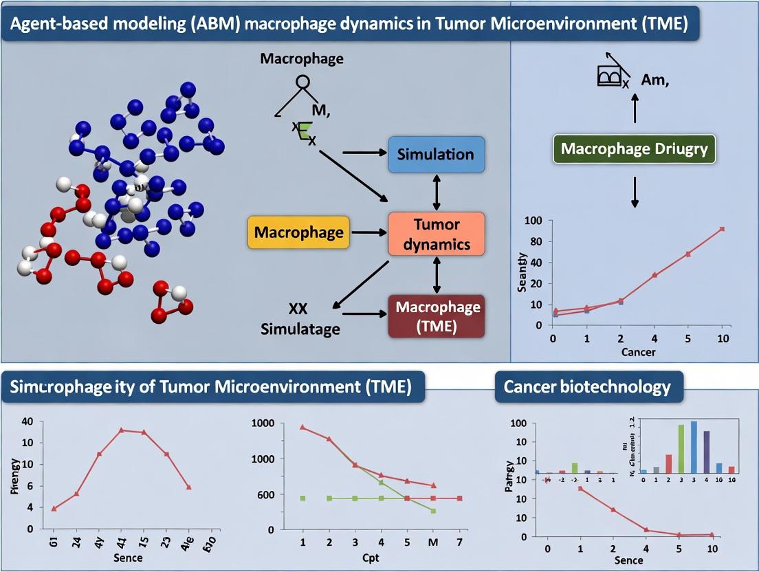
Decoding Tumor Microenvironment Complexity: A Guide to Agent-Based Modeling of Macrophage Dynamics for Cancer Research
This article provides a comprehensive guide for researchers and drug development professionals on applying agent-based modeling (ABM) to simulate macrophage dynamics within the tumor microenvironment (TME).
Decoding Tumor Microenvironment Complexity: A Guide to Agent-Based Modeling of Macrophage Dynamics for Cancer Research
Abstract
This article provides a comprehensive guide for researchers and drug development professionals on applying agent-based modeling (ABM) to simulate macrophage dynamics within the tumor microenvironment (TME). It explores the foundational role of macrophages (M1/M2 polarization) in cancer progression and immunotherapy response. Methodologically, it details the construction of ABM frameworks, from defining agent rules to parameterizing biological interactions. The guide addresses common computational challenges, calibration techniques, and optimization strategies for robust simulations. Finally, it examines validation protocols against experimental data and compares ABM with continuum and hybrid modeling approaches, evaluating their respective strengths in predicting therapeutic outcomes and informing novel treatment strategies.
Understanding the Players: The Foundational Role of Macrophages in the Tumor Microenvironment
Within the tumor microenvironment (TME), macrophages exhibit profound functional plasticity, moving beyond the classical M1 (pro-inflammatory, anti-tumoral) and M2 (anti-inflammatory, pro-tumoral) dichotomy to occupy a spectrum of polarization states. Understanding this heterogeneity is critical for Agent-Based Modeling (ABM) of tumor immunology. ABM simulations require discrete, rule-based definitions for macrophage agent phenotypes, driven by quantitative molecular signatures and local microenvironmental cues. These computational models integrate in vitro and in vivo data to predict macrophage dynamics, their impact on tumor progression, and therapeutic intervention points. This document provides current protocols and data frameworks to empirically define macrophage states for parameterizing and validating such ABMs.
Core Macrophage Phenotypes: Quantitative Signatures
The following tables consolidate key surface markers, cytokine profiles, and functional outputs for defining macrophage states, based on recent human and mouse studies. Data is essential for initializing agents in an ABM.
Table 1: Characteristic Surface Markers & Secretory Profiles
| Phenotype | Key Inducing Signals | High Expression (Surface) | High Secretion (Cytokines/Chemokines) | Low Secretion / Inhibition |
|---|---|---|---|---|
| M1-like | IFN-γ, LPS, GM-CSF | CD80, CD86, HLA-DR (human), MHC-II (mouse), CD64, FcγRI | TNF-α, IL-1β, IL-6, IL-12, IL-23, CXCL9, CXCL10 | IL-10, TGF-β, CCL17, CCL22 |
| M2-like | IL-4, IL-13, IL-10, TGF-β, M-CSF | CD163, CD206, CD200R, CD209 (DC-SIGN), CCR2, MerTK | IL-10, TGF-β, CCL17, CCL22, CCL18, CCL24 | IL-12, IL-23, TNF-α, CXCL9/10 |
| Spectrum/TAM* | Mixed: IL-4, IL-10, TGF-β, TME factors | Hybrid: Often CD206+, CD163+, with variable MHC-II | Mixed: IL-10, TNF-α, CCL2, CCL5, VEGF, MMP9 | Context-dependent; often suppressed IL-12 |
*TAM: Tumor-Associated Macrophage. Phenotype is highly context-dependent.
Table 2: Functional Outputs & Metabolic Pathways
| Phenotype | Primary Function in TME | Metabolic Signature | Key Transcription Factors | Pro/Anti-Tumoral Role |
|---|---|---|---|---|
| M1-like | Pathogen killing, antigen presentation, Th1 recruitment | Glycolysis, TCA cycle disruption, NO/ROS production | STAT1, NF-κB, IRF5, AP-1 | Typically Anti-Tumoral |
| M2-like | Tissue repair, angiogenesis, immune suppression | Oxidative Phosphorylation, Fatty Acid Oxidation | STAT3, STAT6, IRF4, PPARγ, KLF4 | Typically Pro-Tumoral |
| Spectrum/TAM | Matrix remodeling, metastasis, T-cell suppression, angiogenesis | Mixed/Adaptive; often lipid metabolism | Co-expression or dynamic switching of above factors | Predominantly Pro-Tumoral |
Detailed Experimental Protocols
Protocol 1:In VitroGeneration and Validation of Human Monocyte-Derived Macrophage Phenotypes
Purpose: To generate standardized M1, M2, and intermediate polarization states from primary human monocytes for downstream analysis or ABM parameter calibration.
Materials:
- Human CD14+ Monocytes: Isolated from PBMCs using magnetic-activated cell sorting (MACS).
- Culture Medium: RPMI-1640 supplemented with 10% heat-inactivated FBS, 1% Penicillin-Streptomycin, 2mM L-Glutamine.
- Polarizing Cytokines:
- M1: 100 ng/mL IFN-γ (PeproTech, #300-02) for 24h, followed by 10 ng/mL LPS (Sigma, #L4391) for final 24h.
- M2a: 20 ng/mL IL-4 (PeproTech, #200-04) + 20 ng/mL IL-13 (PeproTech, #200-13) for 48h.
- M2c: 20 ng/mL IL-10 (PeproTech, #200-10) for 48h.
- Tissue Culture Plates: 6-well or 12-well plates, non-tissue-culture-treated for low adherence.
Procedure:
- Monocyte Differentiation: Seed CD14+ monocytes at 1x10^6 cells/mL in culture medium supplemented with 50 ng/mL recombinant human M-CSF (PeproTech, #300-25). Culture for 6 days to differentiate into M0 macrophages, with fresh medium+ M-CSF added on day 3.
- Polarization: On day 6, replace medium with fresh cytokine-containing polarization medium as specified above. Culture for 24-48 hours.
- Validation by Flow Cytometry: a. Harvest cells using gentle scraping with PBS/EDTA. b. Stain with fluorescent antibody cocktails: - M1 Panel: Anti-CD80-APC, Anti-CD86-PE, Anti-HLA-DR-FITC. - M2 Panel: Anti-CD163-PerCP-Cy5.5, Anti-CD206-PE-Cy7. c. Acquire data on a flow cytometer and analyze median fluorescence intensity (MFI) ratios.
- Validation by Secretome Analysis: Collect supernatant. Use a multiplex Luminex or ELISA assay to quantify TNF-α/IL-12 (M1) and CCL17/IL-10 (M2) levels.
Protocol 2: Multiplex Immunofluorescence (mIF) for Spatial Profiling of Macrophages in Tumor Sections
Purpose: To quantify the density, phenotype, and spatial distribution of macrophage subsets within the intact TME for spatial ABM validation.
Materials:
- Formalin-Fixed, Paraffin-Embedded (FFPE) tumor tissue sections (5 µm).
- Antibody Panel: Opal Polychromatic IHC kit (Akoya Biosciences). Example panel:
- CD68 (Pan-macrophage, Opal 520)
- CD163 (M2-like, Opal 570)
- HLA-DR (M1-like, Opal 620)
- Cytokeratin (Tumor cells, Opal 690)
- DAPI (nuclei).
- Automated staining system (e.g., BOND RX, Leica Biosystems) or manual equipment for precise cycling.
Procedure:
- Deparaffinization & Antigen Retrieval: Bake slides, deparaffinize in xylene, rehydrate. Perform heat-induced epitope retrieval (HIER) in EDTA buffer (pH 9.0) for 20 min.
- Sequential Immunostaining: a. Apply primary antibody 1 (e.g., anti-CD68). Incubate, then apply corresponding HRP-polymer secondary. Detect with Opal 520 fluorophore. Apply microwave treatment to strip antibodies. b. Repeat step (a) sequentially for each marker in the panel (CD163, HLA-DR, Cytokeratin). c. Counterstain with DAPI and apply coverslip.
- Image Acquisition & Analysis: Scan slides using a multispectral imaging system (e.g., Vectra/Polaris, Akoya). Use image analysis software (inForm, HALO, QuPath) to: a. Perform spectral unmixing. b. Segment cells based on DAPI. c. Phenotype macrophages: CD68+ only, CD68+CD163+ (M2-like), CD68+HLA-DR+ (M1-like). d. Calculate densities and spatial metrics (e.g., distance of phenotypes to nearest tumor cell).
Signaling Pathways & ABM Logic Diagrams
Diagram Title: Macrophage Polarization Signaling to ABM Rule Logic
Diagram Title: Macrophage Phenotype Interconversion and Plasticity
The Scientist's Toolkit: Key Research Reagent Solutions
Table 3: Essential Materials for Macrophage Heterogeneity Research
| Item Name & Vendor Example | Function in Research | Application Notes for ABM Integration |
|---|---|---|
| Recombinant Human/Mouse Cytokines (PeproTech, R&D Systems) | Induce specific polarization states in vitro. | Provide quantitative dose-response data for ABM signaling rules. |
| Fluorochrome-conjugated Antibodies (BioLegend, BD Biosciences) | Phenotype characterization via flow cytometry. | MFI data quantifies population heterogeneity for model validation. |
| Multiplex Immunofluorescence Kits (Akoya Opal, Roche DISCOVERY) | Spatial phenotyping in tissue context. | Critical for defining spatial rules and neighborhood effects in ABMs. |
| Seahorse XF Analyzer Kits (Agilent) | Measure metabolic flux (glycolysis, OXPHOS). | Links phenotype to metabolic state, a key agent parameter. |
| Nanostring nCounter/PanCancer Immune Panel | Multiplex gene expression profiling. | Provides high-dimensional signature for defining discrete agent states. |
| Transwell/Co-culture Inserts (Corning) | Study macrophage-tumor cell cross-talk. | Generates data for cell-cell interaction rules in ABM. |
| Phospho-STAT1/STAT6 Flow Kits (Cell Signaling Tech) | Quantify signaling pathway activation. | Directly measures signal transduction, informing ABM state transition probabilities. |
This document provides structured experimental data and protocols to inform and validate parameters for Agent-Based Models (ABMs) simulating macrophage dynamics within the Tumor Microenvironment (TME). Understanding these discrete cellular interactions is critical for constructing rules governing agent behavior, state changes, and spatial relationships in computational simulations of therapeutic intervention.
Macrophage-Cancer Cell Crosstalk: Pro-Tumoral Polarization
Table 1: Key Mediators in Macrophage-Cancer Cell Dialogue
| Mediator | Source Cell | Target Cell | Primary Effect | Experimental Readout (Typical Range) |
|---|---|---|---|---|
| CSF-1 (M-CSF) | Cancer, Stroma | Macrophage | Promotes M2-like polarization, survival, migration | [CSF-1] in TME: 50-500 pg/mL (ELISA) |
| CCL2 (MCP-1) | Cancer, Stroma | Macrophage | Chemoattraction into TME | Macrophage migration increase: 150-300% (Boyden chamber) |
| EGF | Macrophage (M2) | Cancer Cell | Promotes cancer cell proliferation, invasion | Cancer cell proliferation increase: 40-80% (MTT assay) |
| TGF-β | Cancer, TAMs | Macrophage, T Cell | Induces M2 polarization; Suppresses T cell function | p-Smad2/3 increase in Macs: 5-10 fold (Western blot) |
Experimental Protocol: Assessing Macrophage-Mediated Cancer Cell Invasion
Title: Transwell Co-Culture Assay for Macrophage-Induced Cancer Cell Invasion
Purpose: To quantify the enhancement of cancer cell invasiveness following interaction with M2-polarized macrophages.
Materials:
- Transwell inserts (8.0 µm pore, Matrigel-coated).
- Serum-free and complete growth media.
- Recombinant human IL-4/IL-13 (for M2 polarization).
- Fluorescent cell tracker dyes (e.g., Calcein AM, CM-Dil).
- 4% Paraformaldehyde (PFA).
- Crystal violet solution or fluorescence plate reader.
Procedure:
- Macrophage Preparation: Differentiate human monocytes (THP-1 cells or primary) into M0 macrophages using PMA (100 nM, 24h). Polarize to M2 phenotype with IL-4 (20 ng/mL) and IL-13 (20 ng/mL) for 48 hours.
- Cancer Cell Labeling: Harvest target cancer cells (e.g., MDA-MB-231 for breast cancer). Label with Calcein AM (5 µM) for 1 hour at 37°C.
- Co-Culture Setup: Place M2 macrophages in the lower chamber of a 24-well plate in serum-free medium. Seed labeled cancer cells into the Matrigel-coated upper insert. Allow invasion for 24-48 hours at 37°C.
- Quantification: Remove non-invaded cells from the insert's interior with a cotton swab. Fix invaded cells on the membrane underside with 4% PFA (10 min). Stain with crystal violet (0.1% w/v) for 20 min or quantify directly via fluorescence.
- Analysis: Image 5 random fields per membrane under a microscope (20x). Count invaded cells manually or measure fluorescence intensity (Ex/Em ~494/517 nm for Calcein). Normalize to control (cancer cells without macrophages).
ABM Parameterization: Output provides a quantitative rate for the rule: "M2 macrophage presence increases probability of cancer agent invasion."
Signaling Pathway Diagram
Diagram Title: Macrophage-Cancer Cell Pro-Tumoral Crosstalk
Macrophage-T Cell Communication: Immunosuppression
Table 2: Immune Checkpoints and Suppressive Factors
| Interaction | Ligand (Source) | Receptor (Target) | Functional Outcome | Measurable Impact |
|---|---|---|---|---|
| PD-L1/PD-1 | PD-L1 (TAM, Cancer) | PD-1 (T cell) | T cell exhaustion, apoptosis | ↓ IFN-γ production by 60-80% |
| CD80/CTLA-4 | CD80 (TAM) | CTLA-4 (T cell) | Inhibits T cell activation | ↓ T cell proliferation by 50-70% |
| Arginase I Activity | TAM (M2) | Extracellular L-Arg | Depletes essential T cell nutrient | ↓ CD3ζ expression in T cells |
| IL-10 Secretion | TAM (M2), Treg | IL-10R (T cell) | Suppresses effector T cell function | ↓ TNF-α, IL-2 secretion |
Experimental Protocol: Measuring T Cell Suppression by Macrophages
Title: CFSE-Based T Cell Proliferation Suppression Assay
Purpose: To quantify the capacity of M2-polarized macrophages to inhibit CD4+ or CD8+ T cell proliferation in vitro.
Materials:
- Human peripheral blood mononuclear cells (PBMCs).
- CD3/CD28 T cell activation beads.
- CFSE cell proliferation dye.
- Recombinant human IFN-γ & LPS (for M1), IL-4 & IL-13 (for M2).
- Anti-human CD3 antibody (coating).
- Flow cytometer with 488 nm laser.
Procedure:
- Macrophage Generation & Polarization: Differentiate monocytes from PBMCs with M-CSF (50 ng/mL, 6 days). Polarize into M1 (IFN-γ 20 ng/mL + LPS 100 ng/mL, 24h) or M2 (IL-4/IL-13 20 ng/mL, 24h).
- T Cell Labeling: Isolate untouched T cells from PBMCs. Resuspend at 1x10^7 cells/mL in PBS/0.1% BSA. Add CFSE to final 5 µM, incubate 10 min at 37°C. Quench with 5x volume of cold complete media.
- Co-Culture: Seed polarized macrophages in a 96-well plate. Add CFSE-labeled T cells (1:1 to 1:5 macrophage:T cell ratio). Activate T cells with soluble anti-CD3 (1 µg/mL) or CD3/CD28 beads (1 bead per cell). Culture for 4-5 days.
- Flow Cytometry Analysis: Harvest non-adherent cells. Stain with anti-CD4-APC or anti-CD8-APC antibodies. Acquire on flow cytometer. Gate on live, CD4+ or CD8+ lymphocytes.
- Data Interpretation: Analyze CFSE dilution in the FL1 channel. Compare proliferation index (division cycles) of T cells co-cultured with M2 vs. M1 macrophages or T cells alone.
ABM Parameterization: Generates a suppression probability coefficient for the rule: "M2 macrophage agent reduces division rate of adjacent T cell agents."
Signaling Pathway Diagram
Diagram Title: Macrophage-Mediated T Cell Suppression Pathways
Macrophage-Stromal Cell Interactions: Remodeling the Niche
Table 3: Key Factors in Stromal Engagement
| Stromal Cell | Key Signal to Macrophage | Macrophage Response | TME Outcome | Measurement Technique |
|---|---|---|---|---|
| Cancer-Associated Fibroblast (CAF) | CXCL12, IL-6, CSF-1 | Recruitment, M2 polarization, Survival | Desmoplasia, Immune exclusion | Collagen deposition (Sirius Red, +50-200%) |
| Mesenchymal Stem Cell (MSC) | PGE2, TGF-β, IDO | Polarization to immunosuppressive phenotype | Enhanced angiogenesis, Metastasis | Microvessel density (CD31+ IHC) |
| Endothelial Cell | VEGF, SEMAPHORIN 6A | Pro-angiogenic (TIE2+ TAM) phenotype | Vasculature abnormalization, Metastasis | In vitro tube formation assay |
Experimental Protocol: 3D Spheroid Model of Macrophage-Stromal Interaction
Title: Generation of Multicellular Tumor Spheroids with CAFs and Macrophages
Purpose: To establish a 3D co-culture model for studying macrophage-stroma crosstalk and its impact on matrix remodeling.
Materials:
- Ultra-low attachment (ULA) 96-well round-bottom plates.
- Primary CAFs or fibroblast cell line (e.g., MRC-5).
- Cancer cell line of interest.
- Monocyte cell line (THP-1) or primary monocytes.
- Recombinant M-CSF.
- CellTracker dyes (different colors for each cell type).
- Confocal microscope.
- Collagen I matrix.
Procedure:
- Cell Preparation: Label CAFs with CellTracker Red (CMTPX), cancer cells with Green (CMFDA), and monocytes with Blue (Hoechst or similar). Differentiate monocytes to macrophages with M-CSF (50 ng/mL, 6 days).
- Spheroid Formation: Mix cells in desired ratio (e.g., 500 cancer cells: 200 CAFs: 100 macrophages per spheroid) in complete media. Seed 100 µL suspension per well in ULA plate. Centrifuge plate at 300xg for 3 min to aggregate cells. Culture for 72h to form compact spheroids.
- Matrix Embedding & Invasion: Carefully transfer individual spheroids into a pre-chilled droplet of collagen I gel (2 mg/mL) in a glass-bottom dish. Allow to polymerize at 37°C for 30 min. Overlay with complete medium.
- Imaging & Analysis: Image spheroids daily for 5-7 days using confocal microscopy. Track collective invasion into the matrix (spheroid area increase), macrophage positioning (edge vs. core), and matrix degradation (using fluorescent collagen).
- Molecular Analysis: Harvest multiple spheroids for RNA/protein extraction to analyze expression of MMPs (e.g., MMP2, MMP9), collagens, and cytokines.
ABM Parameterization: Provides spatial rules and probabilities for agent (macrophage) movement towards stromal elements and resultant matrix modification events.
Signaling Pathway Diagram
Diagram Title: Macrophage-Stromal Cell Network in TME
The Scientist's Toolkit: Research Reagent Solutions
Table 4: Essential Reagents for Studying Macrophage Interactions
| Reagent/Category | Example Product (Supplier) | Primary Function in Experiments |
|---|---|---|
| Polarization Cytokines | Recombinant Human IL-4, IL-13, IFN-γ, LPS (PeproTech, R&D Systems) | To generate in vitro M1 or M2 macrophage phenotypes from monocyte precursors. |
| Neutralizing/Antibodies | Anti-human CSF-1R, Anti-CCL2, Anti-PD-L1 (BioLegend, Bio X Cell) | To block specific interaction axes in co-culture experiments and assess functional contribution. |
| Flow Cytometry Panels | Anti-CD68, CD80, CD86, CD163, CD206, HLA-DR (Multiple suppliers) | To immunophenotype macrophage polarization states and purity before/after co-culture. |
| Transwell Systems | Corning Transwell Permeable Supports (with/without Matrigel) | To study chemotaxis (migration) and paracrine effects in compartmentalized co-cultures. |
| 3D Culture Matrices | Cultrex BME, Collagen I, Matrigel (Corning, R&D Systems) | To provide a physiologically relevant 3D environment for invasion and spheroid assays. |
| Live-Cell Imaging Dyes | CellTracker Probes, Calcein AM (Thermo Fisher) | To label distinct cell populations for tracking in co-cultures and spatial analysis. |
| Cytokine Quantification | DuoSet ELISA Kits (R&D Systems) or LEGENDplex Bead Arrays (BioLegend) | To measure concentrations of key signaling molecules (CSF-1, CCL2, IL-10, TGF-β) in conditioned media. |
| Small Molecule Inhibitors | CSF-1R inhibitor (PLX3397), CCR2 inhibitor (RS504393), ARG1 inhibitor (CB-1158) | To pharmacologically validate targets and generate data for modeling drug effects in ABMs. |
Application Notes: Integrating ABM with Experimental Immunology
Macrophage dynamics within the Tumor Microenvironment (TME) are a critical determinant of immunotherapy outcomes. Agent-based modeling (ABM) provides a computational framework to simulate the spatiotemporal interactions between tumor cells, macrophages (M1/M2 phenotypes), T cells, and therapeutic agents. These models can identify non-linear resistance mechanisms and predict response biomarkers.
Table 1: Key Quantitative Parameters for Macrophage ABM in TME
| Parameter | Typical Experimental Range | ABM Variable | Impact on Immunotherapy Response |
|---|---|---|---|
| M2:M1 Macrophage Ratio | 0.5 - 8.0 (Human NSCLC biopsy) | Polarization_Threshold |
High ratio correlates with anti-PD-1 resistance (r > 0.7) |
| CD8+ T Cell Proximity to M1 (μm) | <30 for productive activation | Interaction_Radius |
Distance >50μm reduces killing probability by 80% |
| CCR2 Ligand (CCL2) Concentration | 100-1500 pg/mL (plasma) | Chemokine_Field |
Levels >800 pg/mL predict poor response to CTLA-4 blockade |
| PD-L1 Expression on TAMs (MFI) | 10^3 - 10^5 (flow cytometry) | Immune_Checkpoint_State |
MFI >50,000 linked to T cell exhaustion in ABM simulations |
| Phagocytosis Rate (targets/cell/hour) | 0.5 - 5.0 (in vitro) | Efferocytosis_Rate |
Rate <1.0 allows for tumor cell accumulation in silico |
Experimental Protocols
Protocol 2.1: Ex Vivo Macrophage-Tumor Spheroid Co-culture for Therapy Screening
Purpose: To functionally assess the impact of macrophage subsets on tumor cell viability during immune checkpoint inhibitor (ICI) treatment.
Materials:
- Primary human monocyte-derived macrophages or murine bone marrow-derived macrophages (BMDMs).
- GFP-labeled tumor cell line (e.g., MC38, A375).
- Anti-PD-1/PD-L1 therapeutic antibodies (clinical grade).
- Low-attachment 96-well U-bottom plates.
- Live-cell imaging system (e.g., Incucyte).
- Flow cytometry antibodies: CD11b, F4/80, CD80, CD206, PD-L1, Live/Dead.
Procedure:
- Spheroid Generation: Seed 5x10^3 GFP+ tumor cells per well in U-bottom plates. Centrifuge at 300 x g for 3 min. Culture for 72h to form compact spheroids.
- Macrophage Polarization & Addition: Differentiate monocytes with M-CSF (50 ng/mL, 6 days). Polarize with IFN-γ+LPS (20 ng/mL+100 ng/mL, 24h) for M1 or IL-4 (20 ng/mL, 48h) for M2. Harvest and add 2x10^3 macrophages per spheroid well.
- Therapy Treatment: Add anti-PD-L1 antibody (10 µg/mL) or isotype control. Include wells with macrophages only and tumor-only controls.
- Longitudinal Imaging: Place plate in live-cell imager. Acquire phase contrast and GFP fluorescence images every 6 hours for 5 days.
- Endpoint Analysis: Gently dissociate spheroids with Accutase. Analyze by flow cytometry for macrophage phenotype and tumor cell death (Live/Dead stain). Calculate spheroid growth index: (GFP Area Day5/GFP Area Day0).
Data Interpretation: A response is defined as a >50% reduction in spheroid growth index in anti-PD-L1 wells co-cultured with M1 macrophages compared to isotype control. Resistance is indicated when M2 macrophages are present and no significant reduction occurs.
Protocol 2.2: Multiplex Immunofluorescence (mIF) for Spatial Profiling of Macrophage Dynamics
Purpose: To quantify macrophage spatial relationships and phenotypes within the TME of pre- and post-immunotherapy tumor sections.
Materials:
- Formalin-fixed, paraffin-embedded (FFPE) tumor tissue sections (4 µm).
- Opal 7-Color IHC Kit (Akoya Biosciences) or comparable.
- Primary antibodies: CD68 (pan-macrophage), CD163 (M2-like), HLA-DR (M1-like), CD8 (cytotoxic T cells), PD-L1, Pan-Cytokeratin (tumor), DAPI.
- Automated multiplex staining system (e.g., Vectra Polaris).
- Phenochart and inForm image analysis software.
Procedure:
- Deparaffinization & Antigen Retrieval: Bake slides at 60°C for 1h. Deparaffinize in xylene and rehydrate. Perform antigen retrieval in Tris-EDTA buffer (pH 9.0) using a pressure cooker.
- Sequential Staining Cycles: For each marker, apply primary antibody, then HRP-conjugated secondary, followed by Opal fluorophore (e.g., Opal 520, 570, 620, 690, 780). Perform microwave stripping between cycles to remove antibodies.
- Counterstaining & Coverslipping: After final cycle, apply DAPI, then mount with ProLong Diamond antifade.
- Image Acquisition: Scan slides using the Vectra Polaris at 20x magnification. Select at least 5 representative tumor regions (both invasive margin and core).
- Spatial Analysis: Use inForm software for cell segmentation (nuclear: DAPI, membrane/cytoplasm: respective markers). Phenotype cells based on marker combinations (e.g., CD68+CD163+HLA-DR- = M2). Export cell coordinates and phenotypes.
- Spatial Metrics Calculation: Use R package
spatstatto calculate:- Mixing Score: Average distance from each CD8+ T cell to the nearest CD68+ cell.
- Cluster Analysis: Ripley's K-function for M2 macrophage aggregation.
- Interface Score: Density of M1 macrophages within a 30µm border of tumor nests.
Visualizations
Title: Macrophage Polarization Pathways & Immunotherapy Links
Title: Integrated Experimental & ABM Workflow for TAM Dynamics
The Scientist's Toolkit
Table 2: Key Research Reagent Solutions for Macrophage-Immunotherapy Studies
| Reagent/Material | Vendor Example (Catalogue #) | Function in Research |
|---|---|---|
| Recombinant Human/Murine M-CSF | PeproTech (300-25) | Differentiation of monocytes/bone marrow progenitors into macrophages. |
| Opal 7-Color Automation IHC Kit | Akoya Biosciences (NEL821001KT) | Enables multiplex immunofluorescence on FFPE slides for spatial phenotyping. |
| CellTrace Violet / CFSE Cell Proliferation Kits | Thermo Fisher (C34557) | To label and track macrophage or tumor cell proliferation in co-cultures. |
| Mouse Anti-Human CD47 Blocking Antibody | BioLegend (323102) | To probe the "don't eat me" signal and enhance phagocytosis in functional assays. |
| Recombinant PD-1/PD-L1 Checkpoint Proteins | ACROBiosystems (PD1-H5259) | For binding studies or to generate checkpoint-equipped artificial target cells. |
| LIVE/DEAD Fixable Viability Dyes | Thermo Fisher (L34966) | Critical for flow cytometry to distinguish live/dead cells post-treatment. |
| CLODRONATE LIPOSOMES | Liposoma (CP-005-005) | In vivo macrophage depletion tool to establish their functional role in therapy. |
| Bioinformatics Tool: CIBERSORTx | https://cibersortx.stanford.edu/ | Deconvolutes bulk RNA-seq to estimate macrophage subset abundances from data. |
Why ABM? The Case for Discrete, Individual-Centric Modeling of Cellular Behaviors.
Traditional continuum models of the Tumor Microenvironment (TME), such as ordinary differential equations, treat cell populations as homogeneous averages. This approach fails to capture the spatial heterogeneity, stochastic cell-cell interactions, and emergent behaviors critical to macrophage dynamics and therapy response. Agent-Based Modeling (ABM) addresses this by simulating individual "agents" (e.g., macrophages, tumor cells) with defined rules, enabling the study of how local interactions give rise to complex system-level phenomena.
Key Advantages of ABM for Macrophage-TME Research
- Spatial Heterogeneity: Models nutrient, oxygen, and signal gradients that polarize macrophages into pro-tumor (M2-like) or anti-tumor (M1-like) phenotypes.
- Stochasticity: Captures intrinsic randomness in cell division, death, and phenotypic switching.
- Emergent Behavior: Allows for the observation of unplanned outcomes like the spontaneous formation of immunosuppressive niches or tumor cell escape.
- Precision Medicine Integration: Facilitates "digital twin" simulations by incorporating patient-specific data on cell densities and genetic profiles.
Application Notes: ABM of Macrophage-Mediated Immunotherapy Resistance
Recent studies (2023-2024) utilize ABM to dissect mechanisms of resistance to Immune Checkpoint Inhibitors (ICIs). A core finding is that ABMs predict macrophage phagocytic dysfunction, driven by the "Don't Eat Me" signal CD47-SIRPα and metabolic competition, as a pivotal resistance node.
Table 1: Summary of Key Quantitative Insights from Recent ABM Studies
| Study Focus | Key ABM Prediction | Validated Experimental Outcome | Impact on Drug Development |
|---|---|---|---|
| CD47 Blockade Failure | Resistance emerges from TME acidity impairing antibody binding affinity. | In vitro binding assays show >60% reduction in anti-CD47 affinity at pH 6.5 vs. 7.4. | Rationale for developing pH-sensitive CD47 antibodies. |
| Metabolic Competition | Tumor glycolytic rate outcompetes macrophages for glucose, suppressing M1 polarization. | PET imaging and IHC show inverse correlation between tumor SUVglucose and iNOS+ macrophages in murine models. | Supports combination therapy: ICIs + glycolytic inhibitors. |
| Spatial Hiding | Tumor cells located >100µm from a vascular niche are protected from macrophage phagocytosis. | Multiplex IHC analysis of patient samples confirms low phagocytosis in hypoxic, peri-necrotic regions. | Highlights need for drugs improving macrophage infiltration. |
Detailed Experimental Protocols for ABM Validation
Protocol 1: Validating ABM-Predicted pH-Dependent Antibody Binding
- Objective: To test the ABM-predicted loss of anti-CD47 antibody efficacy in acidic TME conditions.
- Materials: Recombinant human CD47 protein, therapeutic anti-CD47 antibody (e.g., Magrolimab), PBS buffers titrated to pH 7.4 and 6.5, Biacore SPR or Octet RED96 system.
- Procedure:
- Dilute CD47 protein to 5 µg/mL in PBS buffers at pH 7.4 and 6.5. Immobilize onto biosensor chips.
- Dilute anti-CD47 antibody in matching pH buffers at concentrations from 0.1 to 100 nM.
- Prime the instrument with the respective pH buffer.
- Perform association/dissociation kinetics measurements for each antibody concentration.
- Analyze data to calculate binding affinity (KD) at each pH.
- Expected Outcome: A significantly higher KD (weaker binding) at pH 6.5 compared to pH 7.4.
Protocol 2: Spatial Mapping of Macrophage Phagocytosis in Hypoxic Niches
- Objective: To correlate macrophage-tumor cell distances with phagocytosis markers, validating ABM spatial predictions.
- Materials: Multicellular tumor spheroid co-culture (macrophages + GFP-labeled tumor cells), Hypoxyprobe-1, Anti-Hypoxyprobe & Anti-CD68 antibodies, Confocal microscope.
- Procedure:
- Treat spheroids with 200 µM Hypoxyprobe-1 for 2 hours.
- Fix, permeabilize, and stain for Hypoxyprobe (hypoxia) and CD68 (macrophages).
- Acquire high-resolution z-stack images via confocal microscopy.
- Use image analysis software (e.g., Imaris, FIJI) to:
- Create a 3D distance map from each macrophage to all tumor cells.
- Quantify the GFP signal intensity inside CD68+ regions as a phagocytosis metric.
- Correlate phagocytosis metric with distance to the nearest hypoxic (Hypoxyprobe+) region.
- Expected Outcome: A significant negative correlation between phagocytosis efficiency and proximity to hypoxic cores.
Visualizing Key Pathways and Workflows
Title: ABM-Predicted Mechanism of CD47 Therapy Resistance
Title: ABM Validation Workflow for TME Hypotheses
The Scientist's Toolkit: Key Research Reagent Solutions
Table 2: Essential Reagents for Validating Macrophage ABM Predictions
| Reagent / Solution | Provider Examples | Function in ABM Validation |
|---|---|---|
| pH-Adjustable Cell Culture Media | Thermo Fisher (Gibco), Sigma-Aldrich | Mimics acidic TME conditions for in vitro functional assays. |
| Recombinant CD47 & SIRPα Proteins | R&D Systems, Sino Biological | Used in surface plasmon resonance (SPR) to measure binding kinetics under varied TME conditions. |
| Hypoxyprobe-1 (Pimonidazole HCl) | Hypoxyprobe, Inc. | Chemical probe for immunohistochemical detection of hypoxic regions in tissues/spheroids. |
| Multiplex IHC/IF Antibody Panels | Akoya Biosciences (PhenoCycler), Standard Antibodies | Enables spatial profiling of macrophage phenotypes (CD68, iNOS, CD206) with tumor and stroma markers. |
| 3D Tumor Spheroid/Microtumor Kits | Corning, Cultrex | Provides a physiologically relevant scaffold for co-culturing macrophages and tumor cells. |
| Live-Cell Metabolic Dyes (e.g., 2-NBDG) | Cayman Chemical, Thermo Fisher | Visualizes and quantifies glucose uptake competition between tumor cells and macrophages. |
Building the Digital Twin: A Step-by-Step Guide to ABM Construction for Macrophage-TME Simulations
This document outlines the core components for developing an Agent-Based Model (ABM) to simulate macrophage dynamics within the Tumor Microenvironment (TME). These models are crucial for hypothesis testing, identifying therapeutic targets, and understanding complex cell-cell interactions in cancer research and drug development.
Core Conceptual Components
Agent Definitions
Agents are autonomous entities with properties and behaviors. In a macrophage TME ABM, the primary agents are:
- Macrophages: Key properties include spatial position, polarization state (M1 or M2), cytokine secretion profile, phagocytic capacity, and motility. Rules govern state transitions (e.g., M1M2 repolarization) in response to environmental signals.
- Tumor Cells: Properties include proliferation rate, nutrient consumption, hypoxic state, and secretion of chemoattractants (e.g., CCL2) and cytokines (e.g., IL-4, IL-10, IL-13).
- T Cells: Properties include type (CD8+, CD4+ Treg), activation state, and cytokine secretion (e.g., IFN-γ).
- Endothelial Cells: Form the vascular network. Properties include spatial coordinates and rules for angiogenesis factor secretion.
Table 1: Core Agent Properties for a Macrophage TME ABM
| Agent Type | Key Intrinsic Properties | Key Behavioral Rules |
|---|---|---|
| Macrophage | Position, State (M0/M1/M2), Receptor Expression, Lifespan | Chemotaxis, Phagocytosis, Cytokine Secretion, State Switching |
| Tumor Cell | Position, Proliferation Cycle, Hypoxia Status, Nutrient Level | Proliferation, Apoptosis, Cytokine/CCL2 Secretion, Migration |
| CD8+ T Cell | Position, Activation State | Cytotoxicity (IFN-γ, Perforin), Chemotaxis, Proliferation |
| Blood Vessel | Start/End Coordinates, Permeability, Diameter | Sprouting (VEGF response), Nutrient/Oxygen Diffusion |
Environment Definition
The environment is the spatial and biochemical context in which agents interact.
- Spatial Grid: Typically a 2D or 3D lattice representing tissue. Resolution (µm per grid point) must be defined.
- Diffusible Fields: Continuous scalar fields representing concentrations of key molecules. These fields are updated dynamically.
- Nutrients: Oxygen, Glucose.
- Signaling Molecules: Cytokines (IFN-γ, IL-4, IL-10, TGF-β), Chemokines (CCL2, CXCL12), Growth Factors (VEGF, CSF-1).
- Extracellular Matrix (ECM): Can be modeled as a grid property affecting agent motility and creating physical barriers.
Table 2: Key Diffusible Environmental Fields in the TME
| Field Name | Source Agent(s) | Target/Effect | Typical Diffusion Coefficient (µm²/s)* |
|---|---|---|---|
| Oxygen | Blood Vessels | All Cells (Metabolism) | 1000 - 2000 |
| CCL2 | Tumor Cells, Stroma | Macrophages (Chemoattraction) | 10 - 20 |
| IFN-γ | T Cells, NK Cells | Macrophages (M1 Polarization) | 10 - 20 |
| IL-4/IL-13 | T Cells, Tumor Cells | Macrophages (M2 Polarization) | 10 - 20 |
| CSF-1 | Tumor Cells | Macrophages (Survival/Proliferation) | 10 - 20 |
| VEGF | Hypoxic Tumor Cells, M2 Macrophages | Endothelial Cells (Angiogenesis) | 10 - 20 |
*Values are approximate and model-dependent.
Rule Definition
Rules are functions that determine how agents perceive their local environment and update their state and properties at each time step (∆t). They are often probabilistic.
Movement Rule (Chemotaxis):
- Perception: Agent senses concentration gradient of a chemoattractant (e.g., CCL2, CSF-1) in neighboring grid points.
- Decision: Probability of moving toward higher concentration is calculated (e.g., using a biased random walk).
- Action: Agent position is updated.
State Transition Rule (Macrophage Polarization):
- Perception: Agent integrates local concentrations of IFN-γ and IL-4/IL-10.
- Decision: If [IFN-γ] > threshold1 AND [IFN-γ] > [IL-4], adopt M1 state with probability P1. If [IL-4] > threshold2 AND [IL-4] > [IFN-γ], adopt M2 state with probability P2.
- Action: Internal state property is updated, altering subsequent secretion and behavioral rules.
Secretion Rule:
- Condition: Agent is in a specific state (e.g., M1 macrophage).
- Action: Agent adds a defined quantity of a molecule (e.g., TNF-α) to its current grid location per time step.
Interaction Rule (Phagocytosis):
- Perception: Macrophage agent detects an apoptotic tumor cell agent within its interaction radius.
- Decision: Probability of phagocytosis is a function of macrophage state (M1 > M2) and opsonin presence.
- Action: If successful, the tumor cell agent is removed, and macrophage agent properties may change.
Protocol for Constructing a Core Macrophage ABM
Aim: To build a simplified ABM simulating macrophage recruitment and polarization in a 2D tumor spheroid.
Step 1: Platform Selection & Setup
- Select an ABM platform (e.g., NetLogo, CompuCell3D, Python/Mesa).
- Define world parameters: Grid size (e.g., 200x200 pixels, 1px = 5µm), boundary conditions (closed/toroidal), simulation duration (e.g., 1000 time steps, 1 step = 6 hours).
Step 2: Implement the Environment
- Initialize a central tumor region (high cell density).
- Initialize blood vessels at specific coordinates.
- Create dynamic fields for Oxygen, CCL2, IFN-γ, and IL-4. Set initial concentrations and diffusion-decay equations.
- Example Code Snippet (Conceptual):
oxygen_field.diffuse(rate=1500); oxygen_field.decay(rate=0.1)
- Example Code Snippet (Conceptual):
Step 3: Instantiate Agents
- Create tumor cell agents within the central region. Assign properties:
proliferation_clock=random(12-24),secretes_CCL2=True. - Create a population of macrophage precursor agents at random peripheral locations. Assign properties:
state="M0",speed=0.5,target_field="CCL2".
Step 4: Program Core Agent Rules
- Macrophage Movement: Implement a gradient-following algorithm for the target field.
- Macrophage Polarization: At each step, for each macrophage:
- Tumor Cell Proliferation: If
proliferation_clockreaches 0 and local oxygen > threshold, divide with probability P.
Step 5: Calibration & Validation
- Calibrate parameters (diffusion rates, thresholds, probabilities) against in vitro data (e.g., macrophage migration speed, cytokine half-life).
- Validate by comparing model output to a baseline experiment (e.g., expected distribution of M1/M2 macrophages in a control vs. IFN-γ-treated spheroid).
Step 6: Experimentation & Analysis
- Run the calibrated baseline simulation. Record metrics: M1/M2 ratio over time, macrophage infiltration depth, tumor cell count.
- Perform in silico experiments: Apply a "treatment" rule (e.g., introduce an agent that blocks CCL2 receptor) and compare outcomes to baseline.
Visualizing Key Relationships and Workflows
Diagram 1: The Core ABM Component Triad
Diagram 2: Macrophage State Transition Rules in the TME
The Scientist's Toolkit: Research Reagent Solutions
Table 3: Essential Reagents for Validating Macrophage ABM Parameters
| Reagent / Tool | Category | Primary Function in ABM Context |
|---|---|---|
| Recombinant Human/Mouse Cytokines (IFN-γ, IL-4, CSF-1, CCL2) | Biochemical Reagent | Calibrate secretion rates and polarization thresholds in rules. Used in in vitro dose-response experiments. |
| Transwell Migration Assay | Experimental System | Quantify macrophage chemotaxis speed and probability towards TME factors (e.g., CCL2). Informs movement rules. |
| Flow Cytometry Antibodies (CD80, CD206, iNOS, Arg1) | Detection Reagent | Quantify M1/M2 polarization states in vitro and in vivo. Provides validation data for state transition rules. |
| Hypoxia Chamber (1% O₂) | Environmental Control | Characterize tumor cell and macrophage behavior under low oxygen. Informs rules for hypoxia-driven secretion (VEGF, etc.). |
| Conditioned Media from Tumor Cell Lines | Complex Stimulus | Provides a holistic TME-mimetic signal to validate integrated model responses, beyond single cytokines. |
| siRNA / CRISPR-Cas9 for Gene Knockdown (e.g., CCR2, STAT1, STAT6) | Genetic Tool | Test specific rule logic by removing a signaling component and comparing model predictions to experimental outcomes. |
| Live-Cell Imaging Microscopy | Analysis Platform | Track macrophage movement, interaction duration, and spatial distribution in co-cultures. Critical for calibrating spatial rules. |
Within agent-based modeling (ABM) of macrophage dynamics in the tumor microenvironment (TME), accurate parameterization of cellular rates is critical for generating biologically plausible simulations. This protocol details strategies for sourcing quantitative rates for key macrophage behaviors—migration, polarization, phagocytosis, and cytokine secretion—from experimental literature, ensuring models reflect the complexity of the TME.
Sourcing and Summarizing Quantitative Rate Data
Table 1: Macrophage Migration Rates
Data compiled from in vitro and in vivo studies.
| Parameter | Reported Value (Mean ± SD or Range) | Experimental System | Measurement Technique | Key Reference (DOI) |
|---|---|---|---|---|
| Random Motility Speed (M0) | 0.2 - 0.5 µm/min | Human monocytes in collagen | Time-lapse microscopy | 10.1016/j.cell.2018.01.016 |
| Chemotactic Speed (M2, toward CCL2) | 0.8 - 1.5 µm/min | Murine BMDMs in 3D gel | Multiphoton imaging | 10.1038/s41586-019-0934-8 |
| Persistence Time | 10 - 30 min | Human MDMs on 2D substrate | Automated cell tracking | 10.1083/jcb.201903070 |
Table 2: Macrophage Polarization State Transition Rates
Based on cytokine exposure kinetics.
| Transition | Half-life / Time to Phenotype Shift | Polarizing Signal | Experimental Readout | Key Reference (DOI) |
|---|---|---|---|---|
| M0 → M1-like | 12 - 24 hours | LPS (100 ng/mL) + IFN-γ (20 ng/mL) | CD80/86 flow cytometry | 10.1016/j.immuni.2014.06.008 |
| M0 → M2-like | 24 - 48 hours | IL-4 (20 ng/mL) | CD206/Arg1 flow cytometry | 10.4049/immunohorizons.2000060 |
| M2-like → M1-like | 48 - 72 hours | TLR agonist re-stimulation | Cytokine secretion multiplex | 10.3389/fimmu.2019.01084 |
Table 3: Phagocytosis and Cytokine Secretion Rates
Per-cell rates under specified conditions.
| Process | Quantified Rate | Target / Stimulus | Assay | Key Reference (DOI) |
|---|---|---|---|---|
| Phagocytosis | 2 - 5 apoptotic cells/hour | Fluorescently-labeled apoptotic cells | Flow cytometry, confocal | 10.1126/science.1184929 |
| TNF-α Secretion | 0.5 - 2 pg/cell/hour (peak) | LPS (100 ng/mL) | ELISA, single-cell secretion assay | 10.1038/ni.1656 |
| IL-10 Secretion | 0.1 - 0.5 pg/cell/hour (peak) | Immune complexes + LPS | Multiplex bead array | 10.1172/JCI136646 |
Experimental Protocols for Parameter Quantification
Protocol 1: Quantifying Macrophage Migration in 3D Collagen Matrices
Objective: Measure random and chemotactic motility speeds for ABM parameterization.
Materials:
- Collagen I, rat tail (e.g., Corning)
- Ibidi µ-Slide Chemotaxis 3D
- Recombinant human CCL2/MCP-1
- Differentiated human monocyte-derived macrophages (MDMs)
- Live-cell imaging microscope with environmental chamber
Procedure:
- Prepare a 1.5 mg/mL collagen I working solution in cell culture medium on ice. Neutralize with 0.1N NaOH.
- Resuspend MDMs at 2x10⁵ cells/mL in the collagen solution. Pipette 10 µL into the central chamber of the µ-Slide.
- Allow gel polymerization at 37°C for 30 min. Fill reservoirs with medium ± 100 ng/mL CCL2.
- Acquire time-lapse phase-contrast images every 2 minutes for 8-12 hours at 37°C, 5% CO₂.
- Track individual cell centroids using automated software (e.g., TrackMate in Fiji/ImageJ).
- Calculate mean speed (total path length/time) and persistence (mean squared displacement analysis). Export data for ABM parameter input.
Protocol 2: Measuring Polarization Kinetics via Flow Cytometry
Objective: Determine time-dependent transition rates between phenotypic states.
Materials:
- Murine bone marrow-derived macrophages (BMDMs)
- Polarizing cytokines: LPS, IFN-γ, IL-4
- Fluorochrome-conjugated antibodies: anti-mouse CD80 (M1), CD206 (M2)
- Flow cytometer with time-stamp capability
Procedure:
- Seed BMDMs in 12-well plates at 5x10⁵ cells/well. Allow to adhere overnight.
- Add polarizing stimuli: M1 (LPS 100 ng/mL + IFN-γ 20 ng/mL) or M2 (IL-4 20 ng/mL). Maintain control wells.
- At defined timepoints (e.g., 6, 12, 24, 48, 72h), harvest cells by gentle scraping.
- Stain cells with viability dye and surface antibodies (CD80, CD206) for 30 min at 4°C.
- Acquire data on a flow cytometer. Gate on live, single cells.
- Calculate the percentage of CD80+ (M1) or CD206+ (M2) cells over time. Fit a logistic or exponential curve to determine the half-time (t½) for phenotype transition. This t½ informs the ABM state switch rate.
Protocol 3: Single-Cell Phagocytosis Kinetic Assay
Objective: Quantify the rate of phagocytic events per macrophage.
Materials:
- pHrodo Green E. coli BioParticles (Thermo Fisher)
- CellMask deep red plasma membrane stain
- Confocal or high-content imaging system
Procedure:
- Seed macrophages on a glass-bottom 96-well plate.
- Prepare pHrodo BioParticles according to manufacturer's instructions. pHrodo fluorescence increases dramatically in acidic phagosomes.
- Add CellMask stain to label cell membranes, then add opsonized pHrodo particles to wells.
- Immediately begin imaging at 37°C, acquiring both red (membrane) and green (phagocytosis) channels every 30 seconds for 2 hours.
- Using image analysis software, segment individual cells and detect the appearance of bright, punctate green fluorescence within the red cytoplasmic mask.
- Count the number of new phagocytic events per cell per unit time. Report as particles internalized per cell per hour.
Signaling and Workflow Visualizations
Title: Macrophage Polarization Signaling Pathways
Title: Workflow for Sourcing ABM Rate Parameters
The Scientist's Toolkit: Research Reagent Solutions
Table 4: Essential Reagents for Macrophage Rate Quantification
| Reagent / Material | Supplier Example | Function in Parameterization |
|---|---|---|
| Recombinant Cytokines (LPS, IFN-γ, IL-4, IL-13) | PeproTech, R&D Systems | Standardized polarizing stimuli to measure phenotype transition kinetics. |
| pHrodo BioParticles (E. coli, S. aureus, Zymosan) | Thermo Fisher Scientific | Fluorescent particles for real-time, quantitative phagocytosis assays. |
| CellTracker Dyes (CMFDA, CMTMR) | Thermo Fisher Scientific | Long-term cell labeling for tracking migration and persistence in co-cultures. |
| Ibidi µ-Slides (Chemotaxis, 3D) | Ibidi GmbH | Microfluidic slides for precise 2D/3D chemotaxis and migration experiments. |
| Collagen I, Rat Tail | Corning, Advanced BioMatrix | Hydrogel for 3D cell culture, mimicking the extracellular matrix for migration studies. |
| LIVE/DEAD Fixable Viability Dyes | Thermo Fisher Scientific | Critical for flow cytometry to gate on live cells during polarization time courses. |
| LegendPlex Bead-Based Immunoassays | BioLegend | Multiplex cytokine detection from supernatants to quantify secretion rates per cell. |
| Matrigel Matrix | Corning | Basement membrane extract for more physiologically complex 3D invasion assays. |
| FUCCI (Fluorescent Ubiquitination-based Cell Cycle Indicator) | MBL International | Reports cell cycle state, important for controlling proliferation rates in ABM. |
| Incucyte Live-Cell Analysis System | Sartorius | Enables automated, long-term kinetic imaging for migration and phagocytosis. |
This document provides detailed Application Notes and Protocols for implementing core cellular logic—proliferation, apoptosis, and phenotype switching—within an Agent-Based Modeling (ABM) framework. The focus is on simulating macrophage dynamics in the Tumor Microenvironment (TME) for therapeutic research. These protocols translate established biological mechanisms into computational rules, enabling in silico experimentation.
Core Biological Logic & Quantitative Parameters
The following tables summarize key quantitative data and thresholds derived from current literature, essential for parameterizing the ABM.
Table 1: Proliferation & Apoptosis Triggers and Rates in Macrophages
| Process | Key Trigger/Signal | Reported Rate/Probability | Critical Concentration (in vitro) | Primary Molecular Mediator |
|---|---|---|---|---|
| Proliferation (Local) | M-CSF (CSF-1) | 0.05 - 0.15 div/day [1] | 10-50 ng/mL CSF-1 | PI3K/Akt, MAPK pathway |
| Proliferation (Inhibition) | IFN-γ, TGF-β | Reduction by 60-80% [2] | 10 ng/mL IFN-γ | STAT1, SMAD |
| Apoptosis (Induction) | TNF-α, LPS, Nutrient deprivation | 20-40% over 24h [3] | 20 ng/mL TNF-α | Caspase-8/9, BAX/BAK |
| Apoptosis (Inhibition) | M-CSF, IL-10 | Reduction by 50-70% [4] | 25 ng/mL IL-10 | Akt, Bcl-2 |
Table 2: Macrophage Phenotype Switching Signals & Markers
| Phenotype | Polarizing Signal | Key Surface Marker | Secretory Profile | Typical TME Context |
|---|---|---|---|---|
| M1 (Pro-inflammatory) | IFN-γ + LPS | CD80, CD86, MHC-II High | High: TNF-α, IL-12, IL-1β, iNOS | Early tumor, immunogenic |
| M2 (Anti-inflammatory) | IL-4, IL-13, IL-10 | CD206, CD163, ARG1 | High: IL-10, TGF-β, VEGF, ARG1 | Established tumor, hypoxic core |
Experimental Protocols for Benchmarking Data
Protocol 3.1: In Vitro Macrophage Proliferation Assay (MTT)
- Purpose: Quantify proliferation rates under varying M-CSF and IFN-γ concentrations for ABM parameterization.
- Materials: Primary human monocyte-derived macrophages (MDMs) or murine bone marrow-derived macrophages (BMDMs), RPMI-1640 complete medium, recombinant human/murine M-CSF, recombinant IFN-γ, MTT reagent, DMSO, 96-well plate, CO2 incubator, plate reader.
- Procedure:
- Seed macrophages at 5x10^3 cells/well in a 96-well plate.
- After 24h, replace medium with treatments: (A) M-CSF gradient (0, 5, 20, 50 ng/mL), (B) Constant M-CSF (20 ng/mL) + IFN-γ gradient (0, 5, 20 ng/mL).
- Incubate for 72 hours at 37°C, 5% CO2.
- Add 10 μL MTT solution (5 mg/mL) per well. Incubate 4 hours.
- Carefully remove medium, add 100 μL DMSO to solubilize formazan crystals.
- Measure absorbance at 570 nm with a reference at 630 nm.
- Calculate relative proliferation: (Absorbance Treatment / Absorbance Control) * 100%.
Protocol 3.2: Flow Cytometry for Phenotype Identification
- Purpose: Generate quantitative data on phenotype distribution under polarizing conditions to validate switching logic.
- Materials: Macrophages, polarizing cytokines (IFN-γ/LPS for M1; IL-4/IL-13 for M2), flow cytometry buffer (PBS + 2% FBS), fluorochrome-conjugated antibodies (anti-human: CD80-FITC, CD206-PE, MHC-II-APC; isotype controls), fixation buffer, flow cytometer.
- Procedure:
- Polarize macrophages for 48 hours.
- Harvest cells, wash with cold PBS.
- Block Fc receptors with 5% BSA for 15 min on ice.
- Stain with surface antibody cocktail for 30 min in the dark on ice.
- Wash twice with flow buffer.
- Fix cells with 4% PFA for 15 min (optional for immediate analysis).
- Acquire data on flow cytometer (collect ≥10,000 events per sample).
- Analyze using double-gating strategy: single cells -> live cells -> marker expression. Calculate % positive and Median Fluorescence Intensity (MFI).
Computational Implementation & Signaling Logic Diagrams
Diagram 1: Core Macrophage Agent Decision Logic
Diagram 2: Intracellular Signaling for Phenotype Switching
The Scientist's Toolkit: Research Reagent Solutions
Table 3: Essential Reagents for Macrophage-TME Studies
| Reagent / Material | Supplier Examples | Function in Experimentation |
|---|---|---|
| Recombinant Human/Murine M-CSF (CSF-1) | PeproTech, R&D Systems | Essential for macrophage survival, differentiation, and in vitro proliferation assays. Key input for proliferation module. |
| Polarizing Cytokine Cocktails (IFN-γ, LPS, IL-4, IL-13) | BioLegend, Sigma-Aldrich | Used to generate stable M1 or M2 populations in vitro. Provides ground truth for phenotype switching logic. |
| Fluorochrome-conjugated Antibody Panels (CD80, CD86, CD206, CD163, MHC-II) | BD Biosciences, BioLegend | Enable quantification of surface phenotype via flow cytometry, critical for validating in-silico phenotype markers. |
| Phospho-specific Antibodies (p-STAT1, p-STAT6, p-Akt) | Cell Signaling Technology | Used in Western Blot or cytometry to track intracellular signaling pathway activity, informing logic gate thresholds. |
| Hypoxia Chamber / Chemicals (CoCl₂, DFO) | Baker Ruskinn, Sigma-Aldrich | Simulate hypoxic TME conditions in vitro to study its effect on apoptosis, proliferation, and phenotype switching. |
| Agent-Based Modeling Platform (NetLogo, CompuCell3D, AnyLogic) | Open Source / Commercial | Software environment for implementing, visualizing, and analyzing the described cellular logic rules in a simulated TME. |
| Live-Cell Imaging System (Incucyte) | Sartorius | Allows longitudinal, quantitative tracking of proliferation, apoptosis, and motility, generating kinetic data for model fitting. |
The tumor microenvironment (TME) is a complex ecosystem where tumor-associated macrophages (TAMs) play a pro-tumorigenic role, often promoted by the colony-stimulating factor 1 (CSF-1)/CSF-1 receptor (CSF-1R) axis. Simultaneously, the CD47 "don't eat me" signal, via interaction with SIRPα on macrophages, acts as a key innate immune checkpoint. Agent-based modeling (ABM) provides a computational framework to simulate the spatiotemporal dynamics of these interactions, predict therapeutic outcomes, and optimize combination strategies. This application note details protocols for integrating quantitative biological data into an ABM to simulate dual targeting of CSF-1/CSF-1R and CD47-SIRPα.
Table 1: Key Biological Parameters for ABM Input
| Parameter | Value Range (Reported) | Source/Cell Type | Notes for ABM |
|---|---|---|---|
| CSF-1R Expression | 5,000 - 50,000 receptors/cell | Human Monocyte/Macrophage | Density affects binding probability. |
| CSF-1/CSF-1R Kd | 10 - 100 pM | In vitro binding assays | Determines ligand-receptor interaction strength. |
| CD47 Expression on Tumor Cells | 20,000 - 500,000 molecules/cell | Various carcinoma lines (e.g., AML, breast) | High variability influences immune evasion. |
| SIRPα Expression on Macrophages | 10,000 - 100,000 molecules/cell | Human Macrophages | Polymorphisms affect CD47 binding affinity. |
| CD47-SIRPα Kd | 0.2 - 20 µM | Variant-dependent | High Kd (low affinity) is common in humans. |
| Macrophage Phagocytosis Rate (Baseline) | 0.1 - 2 events/cell/hour | In vitro co-culture | Base probability in the model before modulation. |
| CSF-1R Inhibitor (e.g., PLX3397) IC50 | 10 - 100 nM | Cell-based assays | Concentration for 50% pathway inhibition. |
| Anti-CD47 mAb (e.g., Magrolimab) EC50 for Phagocytosis | 0.1 - 10 µg/mL | In vitro phagocytosis assays | Concentration for half-maximal phagocytic boost. |
Table 2: Example ABM Simulation Output Metrics
| Output Metric | Description | Relevance to Therapy |
|---|---|---|
| TAM Density Change (%) | % reduction in TAM count in TME after therapy. | Measures efficacy of CSF-1R blockade. |
| Phagocytic Events per Macrophage | Average phagocytosis count per simulated hour. | Measures direct effect of CD47-SIRPα blockade. |
| Tumor Cell Clearance Rate | % tumor cell killing over simulation period. | Combined endpoint for therapeutic efficacy. |
| Spatial Heterogeneity Index | Measure of macrophage/tumor cell clustering. | Predicts resistance and recurrence patterns. |
Detailed Experimental Protocols for Data Generation
Protocol 1: In Vitro Quantification of Phagocytosis for Model Calibration
Objective: Generate quantitative dose-response data for anti-CD47 and CSF-1R inhibitor treatments to calibrate ABM rules.
Materials: See "Scientist's Toolkit" below.
Method:
- Macrophage Differentiation: Isolate PBMCs from healthy donor blood using Ficoll density gradient. Adhere monocytes for 2 hours. Culture adherent cells for 6-7 days in RPMI-1640 + 10% FBS + 100 ng/mL recombinant human M-CSF (CSF-1) to generate M2-polarized macrophages.
- Target Cell Labeling: Culture target tumor cells (e.g., Raji B-cell lymphoma or MDA-MB-231 breast cancer). Label with 5 µM CellTracker Green CMFDA dye for 45 minutes at 37°C. Wash 3x with PBS.
- Inhibitor Pre-treatment: Treat macrophages with CSF-1R inhibitor (e.g., PLX3397) at a range of concentrations (0, 10, 50, 100, 500 nM) for 24 hours.
- Phagocytosis Assay: Seed labeled tumor cells onto pre-treated macrophages at a 5:1 (tumor:macrophage) ratio. Add anti-CD47 monoclonal antibody (or isotype control) at concentrations from 0.01 to 50 µg/mL. Co-culture for 2-4 hours at 37°C.
- Quenching & Analysis: Wash wells gently. Add trypan blue (0.4%) to quench fluorescence of extracellular/adhered, but not internalized, tumor cells. Image using high-content microscopy (≥5 fields/well). Quantify phagocytosis as (number of green+ puncta inside macrophages) / (total number of macrophages).
- Data Fitting: Fit dose-response curves using a four-parameter logistic model to derive EC50/IC50 values for ABM parameterization.
Protocol 2: Flow Cytometry for Receptor Density Quantification
Objective: Measure CSF-1R and SIRPα expression on macrophages, and CD47 on tumor cells, for initializing agent properties in the ABM.
Method:
- Cell Staining: Harvest cells. Aliquot 1x10^6 cells per staining tube. Wash with FACS buffer (PBS + 2% FBS).
- Antibody Incubation: Resuspend cells in 100 µL FACS buffer containing fluorochrome-conjugated antibodies against CSF-1R (clone D12S), SIRPα (clone 15-414), or CD47 (clone CC2C6), or appropriate isotype controls. Incubate for 30 minutes at 4°C in the dark.
- Wash and Analyze: Wash cells twice, resuspend in 300 µL buffer. Analyze immediately on a flow cytometer calibrated with quantification beads (e.g., QuantiBRITE PE Beads).
- Quantification: Use bead standard curves to convert median fluorescence intensity (MFI) to approximate antibody binding capacity (ABC) or molecules of equivalent soluble fluorochrome (MESF). Input these distributions into the ABM.
Signaling Pathway and Experimental Workflow Diagrams
Diagram Title: CSF-1/CSF-1R and CD47-SIRPα Signaling and Therapeutic Blockade
Diagram Title: Agent-Based Modeling Simulation Workflow
The Scientist's Toolkit: Research Reagent Solutions
Table 3: Essential Materials for Featured Experiments
| Item | Example Product (Supplier) | Function in Protocol |
|---|---|---|
| Recombinant Human M-CSF (CSF-1) | PeproTech (300-25) | Differentiates monocytes into macrophages for in vitro assays. |
| CSF-1R Tyrosine Kinase Inhibitor | PLX3397 (Selleckchem, S7818) | Tool compound to block CSF-1R signaling in vitro and for modeling. |
| Anti-Human CD47 mAb (Blocking) | Magrolimab (Hu5F9-G4) clone (Bio X Cell, BE0392) | Blocks CD47-SIRPα interaction, promotes phagocytosis. |
| Fluorochrome-conjugated Anti-Human CD47 | APC anti-human CD47 (BioLegend, 323120) | Flow cytometry quantification of CD47 expression on tumor cells. |
| Fluorochrome-conjugated Anti-Human CSF-1R | PE anti-human CD115 (CSF-1R) (BioLegend, 347306) | Flow cytometry quantification of CSF-1R on macrophages. |
| QuantiBRITE PE Beads | BD Biosciences (340495) | Converts flow cytometry MFI to absolute receptor counts. |
| CellTracker Green CMFDA | Thermo Fisher Scientific (C7025) | Fluorescently labels live tumor cells for phagocytosis assays. |
| High-Content Imaging System | PerkinElmer Operetta CLS or equivalent | Automated imaging and quantification of phagocytosis events. |
| ABM Software Platform | AnyLogic, NetLogo, or custom Python (Mesa) | Framework for building and executing the spatial stochastic model. |
Application Notes
This protocol provides a standardized framework for analyzing data generated from agent-based modeling (ABM) simulations of macrophage dynamics within the tumor microenvironment (TME). The core objective is to extract quantitative metrics that characterize tumor progression, immune cell composition, and emergent spatial patterns. These metrics are critical for validating models against in vitro and in vivo data, generating testable hypotheses, and identifying potential therapeutic intervention points.
Core Quantitative Metrics for ABM TME Analysis
The following metrics should be calculated at each simulation time step or at specified checkpoints.
Table 1: Tumor Growth Dynamics Metrics
| Metric | Formula/Description | Interpretation |
|---|---|---|
| Total Tumor Cell Count | N_tumor(t) | Direct measure of tumor burden over time. |
| Net Growth Rate | (Ntumor(t) - Ntumor(t-Δt)) / Δt | Instantaneous rate of tumor expansion or regression. |
| Tumor Volume (Relative) | Calculated from a convex hull or bounding box around tumor cells. | Approximates physical tumor size and shape. |
| Proliferation:Death Ratio | (# tumor cell divisions) / (# tumor cell deaths) over interval. | Indicates balance between growth and attrition. |
Table 2: Immune Cell Population & Ratio Metrics
| Metric | Formula/Description | Interpretation |
|---|---|---|
| Macrophage Density | N_macrophage / Area(Region of Interest) | Density of total tumor-associated macrophages (TAMs). |
| M1:M2 Polarization Ratio | NM1 / NM2 | Functional phenotype balance; prognostic indicator. |
| Macrophage:Tumor Cell Ratio | Nmacrophage / Ntumor | Overall immune pressure on tumor. |
| Cytotoxic T Cell to Treg Ratio | NCD8+ / NTreg | Measure of immunosuppressive state within TME. |
| Phagocytosis Events | # tumor cells phagocytosed / (N_macrophage * Δt) | Functional output of macrophage activity. |
Table 3: Spatial Pattern & Interaction Metrics
| Metric | Formula/Description | Interpretation |
|---|---|---|
| Spatial Entropy (Cell Distribution) | -Σ (pi * log(pi)) for grid occupancy. | Quantifies disorder/randomness in cell placement. |
| Pair Correlation Function (g(r)) | Probability of finding a cell of type B at distance r from type A. | Reveals spatial clustering or exclusion between cell types. |
| Mean Distance to Nearest Neighbor | Average distance from a tumor cell to the closest immune cell. | Measures immune infiltration depth. |
| Macrophage Spatial Polarization Index | (M1 in periphery - M1 in core) / Total M1. | Describes compartmentalization of phenotypes. |
Experimental Protocols for Model Calibration & Validation
Protocol 1: Calibrating Macrophage Chemotaxis Parameters Using In Vitro Transwell Assay Data Objective: To calibrate ABM parameters governing macrophage migration towards tumor-secreted chemokines (e.g., CCL2).
- Source Experimental Data: Perform a Transwell migration assay. Seed macrophages in the upper chamber; place tumor cell-conditioned medium (or control) in the lower chamber. Incubate (e.g., 6-24h). Count migrated cells (via microscopy or cytometry).
- Quantify Output: Calculate chemotactic index = (# cells migrated to test) / (# cells migrated to control).
- ABM Calibration Workflow: a. In the ABM, define a virtual "Transwell" setup: a source of diffusing chemokine and agents (macrophages) with migration rules. b. Systematically vary the ABM's chemotaxis strength and persistence time parameters. c. For each parameter set, run the simulation matching the experimental duration and calculate the virtual chemotactic index. d. Use optimization algorithms (e.g., least squares) to identify the parameter set that minimizes the difference between simulated and experimental indices.
Protocol 2: Validating Spatial Metrics Against Multiplex Immunohistochemistry (mIHC) Objective: To validate ABM-predicted spatial patterns using quantitative mIHC data from tumor tissue sections.
- Source Experimental Data: Stain FFPE tumor sections with multiplex antibody panels (e.g., CD68, CD163, CD8, Pan-Cytokeratin, DAPI). Acquire whole-slide images using a multispectral imaging system.
- Image & Data Processing: a. Use image analysis software (e.g., QuPath, HALO) to perform cell segmentation and phenotyping. b. Export coordinate data (X, Y) and phenotype for each identified cell.
- Spatial Analysis: a. Calculate Pair Correlation Functions (g(r)) for key cell-cell interactions (e.g., M2 macrophages to tumor cells) from the mIHC data. b. Calculate Mean Nearest Neighbor Distances (e.g., cytotoxic T cells to tumor cells).
- ABM Validation: a. Run the ABM under conditions mimicking the in vivo context. b. At a comparable time point, export the coordinates and states of all agents. c. Compute the same spatial metrics (g(r), nearest neighbor) from the ABM output. d. Statistically compare (e.g., using Kolmogorov-Smirnov test on g(r) curves) the experimental and simulated spatial distributions.
Diagram Specifications and Visualizations
Title: ABM Output Analysis and Validation Workflow
Title: Key Signaling Pathways Driving Macrophage Polarization
The Scientist's Toolkit: Research Reagent Solutions
Table 4: Essential Reagents for Experimental Validation
| Item | Function in TME/Macrophage Research |
|---|---|
| Recombinant Human/Mouse Cytokines (e.g., CSF-1, IFN-γ, IL-4, IL-13) | Used to polarize macrophages to specific phenotypes (M1/M2) in vitro for functional assays and model calibration. |
| CCL2/MCP-1 Chemokine | Key chemoattractant for monocyte/macrophage migration. Essential for chemotaxis assay calibration. |
| Fluorescent Cell Tracking Dyes (e.g., CFSE, CellTracker) | Label specific cell populations in vitro to track proliferation, migration, and interactions over time. |
| Multiplex Immunohistochemistry/Immunofluorescence Antibody Panels | Enable simultaneous detection of multiple cell markers (CD68, CD163, CD8, etc.) on a single tissue section for spatial analysis. |
| Phagocytosis Assay Kits (e.g., pHrodo-labeled particles) | Quantify the phagocytic activity of macrophages in co-culture with tumor cells or target beads. |
| Selective Small Molecule Inhibitors (e.g., CSF-1R inhibitor, STAT6 inhibitor) | Pharmacological tools to perturb specific pathways in vitro and in vivo, providing data for model hypothesis testing. |
| Extracellular Matrix Hydrogels (e.g., Matrigel, Collagen I) | Provide a 3D scaffold for more physiologically relevant in vitro co-culture and invasion assays. |
Navigating Computational Challenges: Calibration, Sensitivity, and Scaling of Macrophage ABMs
Title: ABM Framework & Parameterization Sources for TME
Title: Protocol for Parameter Calibration & Iterative Validation
Common Pitfalls: Over-Parameterization, Unrealistic Rules, and Validation Debt
Application Notes on Agent-Based Modeling of Macrophage Dynamics in the TME
Context: Within the broader thesis on advancing Agent-Based Modeling (ABM) for tumor microenvironment (TME) research, these notes address critical methodological pitfalls that threaten model credibility and translational utility in simulating macrophage-tumor interactions.
Table 1: Common Macrophage Agent Rules & Associated Validation Debt
| Rule Category | Example Parameter/Rule | Typical Source | Validation Debt Risk | Suggested Empirical Constraint |
|---|---|---|---|---|
| Phenotype Switching | M1→M2 rate constant | In vitro cytokine stimulation | High (static conditions vs. dynamic TME) | Time-lapse imaging in 3D co-culture |
| Chemotaxis | CXCL12 sensitivity gradient threshold | Boyden chamber assays | Medium (2D vs. 3D matrix) | Microfluidic device with controlled gradient |
| Phagocytosis | Probability per contact with apoptotic cell | In vitro co-culture counts | High (ignores "don't eat me" signals) | Incucyte or similar real-time apoptosis/phagocytosis assay |
| Cytokine Secretion | IL-10 units per agent per time step | Bulk supernatant measurement | Very High (population average, not single-cell) | Single-cell secretomics or qPCR of key targets |
| Metabolic Adaptation | OCR/Glycolysis switch hypoxia threshold | Seahorse analyzer data | Medium (macrophage-specific TME metrics needed) | SCENITH metabolomics on TME-derived macrophages |
Table 2: Protocol for Pruning Over-Parameterized ABMs
| Step | Action | Tool/Technique | Acceptance Criterion |
|---|---|---|---|
| 1. Global Sensitivity Analysis (GSA) | Perform variance-based GSA (e.g., Sobol indices) on all parameters. | SALib (Python), UNCSIM | Identify >10 parameters with total-order index < 0.05. |
| 2. Rule Complexity Audit | Map decision trees for agent rules. Simplify redundant or non-mechanistic branches. | NetLogo, AnyLogic, custom code audit | No "ELSE IF" chain > 3 levels deep without empirical basis. |
| 3. Identifiability Analysis | Check if unique parameter sets produce identical outputs. | Profile likelihood, Monte Carlo sampling | Key output variance explained by <15 identifiable parameters. |
| 4. Cross-Validation | Split in silico experimental data (e.g., virtual knockout) into training/validation sets. | Custom simulation batches | R² > 0.7 for validation set predictions of core metrics (e.g., tumor cell count). |
Experimental Protocols for Model Constraining and Validation
Protocol A: Constraining Macrophage Phenotype Switching Rules with Ex Vivo Data
- Objective: To derive a realistic, data-driven rule for macrophage phenotypic state transitions within the TME for ABM implementation.
- Materials: See "Research Reagent Solutions" below.
- Method:
- Isolate CD14+ monocytes from healthy donor PBMCs using magnetic-activated cell sorting (MACS).
- Differentiate into M0 macrophages with 100 ng/mL M-CSF for 6 days.
- Seed M0 macrophages onto transwell inserts above patient-derived organotypic tumor slices (PDOTS) or 3D tumor spheroids.
- At time points T=0h, 24h, 48h, 72h, dissociate and perform multi-parametric flow cytometry (CD80, CD206, HLA-DR, MerTK) and single-cell RNA sequencing (10x Genomics).
- Use computational cytometry (e.g., Citrus algorithm) to identify clusters and transition probabilities between phenotypic states over time.
- Fit a continuous-time Markov chain model to the cluster proportions. The derived transition matrix provides direct parameters for the ABM state-change rules.
- ABM Integration: Replace simplified "IF cytokine > threshold THEN switch" rules with probabilistic state transition matrices informed by this ex vivo data.
Protocol B: Validating Spatial Predictions of ABM via Multiplexed Imaging
- Objective: To test ABM predictions of macrophage infiltration patterns and tumor cell killing in situ.
- Method:
- Run the calibrated ABM under simulated conditions matching a murine tumor model (e.g., MC38 colorectal carcinoma).
- Extract a key spatial prediction: e.g., "Macrophages with high SIRPα expression will be spatially excluded from tumor islets with high CD47 expression by >50µm."
- In the corresponding in vivo model, harvest tumors at endpoint, section, and perform cyclic immunofluorescence (CyCIF) or CODEX multiplex imaging for CD68, SIRPα, CD47, Keratin, and a cytotoxicity marker (e.g., cleaved caspase-3).
- Use image analysis software (QuPath, HALO) to quantify the average minimum distance between SIRPα+ macrophages and CD47+ tumor islets.
- Statistically compare the distribution of distances from the ABM-simulated virtual tissue and the real multiplexed imaging data using Kolmogorov-Smirnov test. A non-significant result (p > 0.05) supports model validity.
The Scientist's Toolkit: Research Reagent Solutions
| Item | Supplier (Example) | Function in Constraining/Validating ABM |
|---|---|---|
| Patient-Derived Organotypic Tumor Slices (PDOTS) | Generated in-house from surgical specimens | Maintains native TME architecture for ex vivo macrophage co-culture and rule validation. |
| µ-Slide Chemotaxis | ibidi GmbH | Enables high-resolution live imaging of macrophage chemotaxis to generate accurate gradient-following rules. |
| Incucyte Live-Cell Analysis System | Sartorius | Provides kinetic, label-free data on cell confluence, death, and phagocytosis for model time-course calibration. |
| IsoLight/Isolight SPRINT | IsoPlexis | Single-cell secretomics platform to quantify macrophage cytokine polyfunctionality, informing secretion rules. |
| CODEX Multiplex Imaging System | Akoya Biosciences | Allows >40-plex phenotyping and spatial analysis of TME sections for rigorous spatial validation of ABM outputs. |
| SALib (Sensitivity Analysis Library) | Open-source (Python) | Performs global sensitivity analyses to identify and eliminate non-influential parameters, combating over-parameterization. |
Title: Key Signaling Pathways Governing Macrophage Behavior in TME
Within the broader thesis on Agent-Based Modeling (ABM) of macrophage dynamics in the Tumor Microenvironment (TME), calibration is the critical process of tuning model parameters to ensure biological fidelity. This document provides Application Notes and Protocols for integrating in vitro and in vivo experimental data to parameterize and validate an ABM simulating macrophage polarization, recruitment, and tumor-immune interactions.
Core Calibration Workflow
The iterative calibration process leverages data from distinct sources to inform different parameter classes.
Diagram Title: Iterative ABM Calibration Workflow for TME Macrophages
Table 1: ExemplaryIn VitroData for Direct Parameterization
| Parameter Class | Experimental Assay | Example Quantitative Readout (Mean ± SD) | Derived ABM Parameter |
|---|---|---|---|
| Macrophage Polarization | qPCR of marker genes (M1: iNOS, TNF-α; M2: Arg1, CD206) | M1/M2 Ratio after LPS/IL-4 stimulation: 8.2 ± 1.5 vs 0.3 ± 0.1 | Probability of state transition per timestep |
| Chemotactic Migration | Transwell assay toward CCL2 or CSF-1 | Migration velocity: 1.2 ± 0.3 µm/min | Agent movement speed rule |
| Phagocytosis Rate | Flow cytometry (pHrodo-labeled tumor cells) | % Macrophages phagocytosing in 4h: 15 ± 4% | Phagocytosis event probability |
| Cytokine Secretion | Multiplex ELISA (e.g., IL-10, IL-12) | IL-10 pg/mL/10^6 cells/24h (M2): 450 ± 75 | Secretion rate per agent per step |
Table 2:In VivoData for Indirect Fitting and Validation
| Data Type | Measurement Technique | Typical Murine Model Data | ABM Output to Match |
|---|---|---|---|
| Tumor Growth Kinetics | Caliper measurements / BLI | Volume doubling time: 5-7 days | Simulated tumor cell count over time |
| Immune Cell Infiltration | Flow cytometry of dissociated tumor | %TAMs of CD45+ cells: 20-40% | Agent population ratios in simulated TME |
| Spatial Distribution | Multiplex IHC / Imaging Mass Cytometry | Distance of M2 TAMs from vasculature: <50 µm | Spatial correlation metrics in model grid |
Detailed Experimental Protocols
Protocol 4.1:In VitroMacrophage Polarization Assay for State Transition Rates
Purpose: Generate quantitative data on polarization kinetics to inform ABM state-switching rules. Materials: See "Research Reagent Solutions" below. Procedure:
- Differentiate human monocytes (from PBMCs) with 50 ng/mL M-CSF for 6 days to obtain M0 macrophages.
- Seed M0 macrophages in 12-well plates (2x10^5 cells/well). Stimulate in triplicate:
- M1: 100 ng/mL LPS + 20 ng/mL IFN-γ.
- M2: 20 ng/mL IL-4.
- Control: Media only.
- At time points (0, 6, 24, 48h), harvest cells for:
- RNA Extraction: Perform qPCR for NOS2 (M1) and MRC1 (M2). Calculate fold-change vs control.
- Surface Markers: Analyze via flow cytometry for CD80 (M1) and CD206 (M2).
- Data Analysis: Fit time-course data with a logistic function to derive the half-maximal polarization time (T50) and maximum fraction. The inverse of T50 can seed the ABM's probability of state change per simulated hour.
Protocol 4.2:In VivoTumor-Immune Contexture Profiling for Model Validation
Purpose: Obtain spatial and compositional data on TAMs from murine models to calibrate and validate the ABM's emergent behavior. Procedure:
- Tumor Implantation: Implant 5x10^5 syngeneic cancer cells (e.g., MC38, LLC) subcutaneously in C57BL/6 mice (n=10).
- Longitudinal Monitoring: Measure tumor dimensions with calipers every 2-3 days. Calculate volume: V = (length x width^2)/2.
- Endpoint Harvest: At volumes ~1000 mm^3, euthanize and harvest tumors.
- Single-Cell Suspension: Mechanically dissociate and enzymatically digest tumors (Collagenase IV/DNase I, 37°C, 30 min). Filter through a 70µm strainer.
- Immune Phenotyping: Stain cells with fluorophore-conjugated antibodies: CD45, F4/80, CD11b, Ly6C, MHC-II, CD206. Use flow cytometry to identify TAM subsets (e.g., F4/80+CD11b+).
- Spatial Analysis (Optional): Fix a separate tumor segment, embed in OCT, and section for IHC (anti-F4/80, anti-αSMA). Use digital pathology to quantify TAM proximity to vessels/necrosis.
- Data Integration: The composition (%TAMs, M1/M2 ratio) and growth curve are direct targets for ABM output matching via parameter fitting algorithms.
Signaling Pathways Governing Macrophage Dynamics in ABM
Key molecular pathways that should be abstracted into model rules.
Diagram Title: Core Signaling Pathways Abstracted in Macrophage ABM
The Scientist's Toolkit: Research Reagent Solutions
| Item | Function in Calibration Context | Example Product / Assay |
|---|---|---|
| Recombinant Human/Murine Cytokines | Polarize macrophages in vitro for state transition rate quantification. | PeproTech: IL-4, IFN-γ, LPS, CSF-1 (M-CSF). |
| Multiplex Cytokine ELISA Panels | Quantify secretory output of macrophages to parameterize cytokine field in ABM. | Bio-Plex Pro Human Cytokine 8-plex (Bio-Rad). |
| Live-Cell Imaging System | Track macrophage migration and interactions with cancer cells for motility rules. | Incucyte S3 (Sartorius) for time-lapse analysis. |
| Flow Cytometry Antibody Panels | Phenotype macrophage subsets from in vitro and in vivo sources. | Anti-human: CD14, CD80, CD206; Anti-mouse: F4/80, CD11b, Ly6C. |
| Spatial Biology Platform | Generate spatial distribution data of TAMs in TME for model validation. | GeoMx Digital Spatial Profiler (Nanostring) or CODEX (Akoya). |
| Parameter Optimization Software | Algorithmically fit ABM parameters to in vivo data. | Approximate Bayesian Computation (ABC) tools (e.g., ABCpy). |
Agent-based modeling (ABM) of macrophage dynamics within the tumor microenvironment (TME) involves numerous parameters governing cell behavior, signaling, and intercellular interactions. Global Sensitivity Analysis (GSA) is a critical methodology for systematically quantifying how uncertainty in model inputs (parameters) influences the uncertainty in key simulation outcomes, such as tumor size, macrophage polarization ratio, or drug efficacy. This protocol outlines the application of GSA within this specific research context to identify the most influential parameters, thereby guiding model refinement, experimental design, and target identification in drug development.
Key GSA Methods: Protocols and Application Notes
Variance-Based GSA (Sobol' Indices) Protocol
Objective: To compute first-order (Si) and total-order (STi) Sobol' indices, quantifying individual and interactive parameter effects on output variance.
Protocol:
- Parameter Space Definition: For k parameters, define plausible ranges (uniform distributions recommended) based on literature and preliminary simulations.
- Sample Matrix Generation: Generate two independent N × k sample matrices (A and B) using a quasi-random sequence (Sobol' sequence). N is the sample size (typically 1,000–10,000).
- Resampling Matrices: Create k hybrid matrices AB^(i), where column i is taken from B and all other columns from A.
- Model Evaluation: Run the ABM simulation for each row in matrices A, B, and all AB^(i). Record the output of interest Y for each run.
- Index Calculation:
- Compute total output variance: VY = Var(Y(A)).
- Compute first-order effect for parameter i: Vi = Var[E(Y | Xi)] ≈ (1/N) Σ Y(A)j * Y(AB^(i))j - [E(Y)]².
- First-order Sobol' index: Si = Vi / VY.
- Total-effect index: STi = 1 - [V-i / VY], calculated using matrices B and AB^(i).
Application Note: STi is crucial for identifying parameters involved in interactions. A high STi indicates a parameter is influential alone or via interactions.
Morris Screening Method Protocol
Objective: To perform an efficient, qualitative screening of influential parameters prior to more computationally intensive variance-based GSA.
Protocol:
- Parameter Discretization: Discretize each parameter's range into p levels.
- Trajectory Generation: Generate r random "trajectories" in the parameter space. Each trajectory starts at a random point, and each parameter is varied once along the trajectory.
- Elementary Effects (EE) Calculation: For each parameter change in a trajectory, compute the EE: EEi = [Y(X1,..., Xi-1, Xi+Δ, ...) - Y(X)] / Δ.
- Sensitivity Metrics: For each parameter i, compute the mean μ (approximating overall influence) and standard deviation σ (indicating nonlinearity/interactions) of its EE distribution from r trajectories.
- Interpretation: High μ indicates high influence. High σ indicates the parameter's effect is nonlinear or interactive.
Data Presentation: GSA Results from a Hypothetical Macrophage ABM
Table 1: Exemplar Sobol' Indices for Key ABM Outputs
| Parameter (Description) | Range | Output: Tumor Volume (Day 20) | Output: M2/M1 Ratio (Day 15) | ||
|---|---|---|---|---|---|
| Si (First-Order) | STi (Total-Order) | Si (First-Order) | STi (Total-Order) | ||
Prolif_Tumor (Tumor cell proliferation rate) |
[0.1, 0.5] /day | 0.62 | 0.65 | 0.01 | 0.05 |
Chemo_M1 (M1 chemoattractant secretion) |
[0.01, 1.0] a.u. | 0.08 | 0.12 | 0.45 | 0.50 |
Polarize_M2 (M2 polarization rate by IL-4) |
[1e-4, 1e-2] /cell/day | 0.05 | 0.25 | 0.30 | 0.55 |
Death_Mac (Macrophage apoptosis rate) |
[0.001, 0.05] /day | 0.10 | 0.15 | 0.15 | 0.20 |
Table 2: Exemplar Morris Screening Results (μ/σ)
| Parameter | μ* (Mean of | EE | ) | σ (Std. Dev. of EE) | Interpretation |
|---|---|---|---|---|---|
Prolif_Tumor |
1.25 | 0.15 | High, Linear influence | ||
Polarize_M2 |
0.85 | 0.80 | High, Nonlinear/Interactive | ||
Chemo_M1 |
0.60 | 0.20 | Moderate, Linear influence | ||
Death_Mac |
0.20 | 0.10 | Low influence |
The Scientist's Toolkit: Key Research Reagents & Solutions for GSA Validation
Table 3: Essential Reagents for Validating GSA-Predicted Dynamics
| Item/Reagent | Function in Experimental Validation | Example Product/Catalog |
|---|---|---|
| Recombinant Cytokines (IL-4, IL-13, IFN-γ) | To perturb polarization parameters in vitro; test model predictions on M1/M2 ratios. | PeproTech, #200-04 (IL-4), #300-02 (IFN-γ) |
| CSF-1 / M-CSF | To modulate macrophage survival and proliferation parameters. | R&D Systems, #216-MC-025 |
| CCL2 / MCP-1 | To test chemotaxis parameters in migration assays. | BioLegend, #579406 |
| Small Molecule Inhibitors (CSF-1R, STAT6) | To pharmacologically inhibit predicted high-sensitivity pathways (e.g., M2 polarization). | Selleckchem, PLX3397 (CSF-1Ri) |
| Multiplex Cytokine Bead Array | To quantify secreted factors from co-cultures, validating paracrine signaling loops in the ABM. | Bio-Rad, Bio-Plex Pro Human Cytokine 27-plex |
| Flow Cytometry Antibody Panels (CD80, CD206, F4/80, CD11b) | To quantify macrophage phenotype distributions, the core output of polarization dynamics. | BioLegend, Anti-mouse CD206 (MMR) APC, #141708 |
| Live-Cell Imaging & Analysis Software | To track cell motility and proliferation rates for direct parameter estimation. | Sartorius, Incucyte or Olympus, cellSens |
Visualizations
Diagram 1: GSA Workflow in ABM Research (93 chars)
Diagram 2: Key Signaling Pathways in Macrophage ABM (96 chars)
Within the context of a thesis on agent-based modeling (ABM) of macrophage dynamics in the tumor microenvironment (TME), scaling simulations to biologically relevant sizes presents significant computational challenges. This document outlines strategies and protocols for optimizing large-scale, 3D spatial simulations to enable the study of emergent, system-level behaviors in immuno-oncology research. Efficient scaling is critical for translating mechanistic ABM insights into hypotheses for therapeutic intervention.
Foundational Optimization Strategies
Table 1: Computational Optimization Techniques for Large-Scale ABM
| Technique | Description | Expected Performance Gain | Applicability to Macrophage ABM |
|---|---|---|---|
| Spatial Hashing & Neighborhood Lists | Pre-computing and caching neighbor agents to avoid O(n²) distance checks. | 50-90% reduction in step time | High - Essential for 3D chemotaxis and cell-cell contact. |
| Parallelization (Shared Memory) | Using OpenMP or similar to distribute agent updates across CPU cores. | ~Linear scaling with core count (to a point). | High - Agents are independent within a step. |
| Parallelization (Distributed) | Using MPI to split spatial domain across compute nodes (e.g., for >10⁷ agents). | Enables simulations impossible on single node. | Medium-High for massive, heterogeneous TME. |
| Adaptive Time-Stepping | Dynamically adjusting the simulation time-step based on system activity. | 30-70% reduction in total steps. | Medium - Must be careful with fast paracrine signaling. |
| Approximate, Hierarchical Search | Using Barnes-Hut or similar for long-range diffusive field solvers. | Reduces O(n²) to O(n log n). | High for metabolite (e.g., lactate, O₂) diffusion. |
| Efficient State Serialization | Binary, sparse, or incremental saving of simulation state. | 80-95% reduction in I/O time & storage. | Essential for all large-scale parameter sweeps. |
Protocol 1: Implementing Spatial Hashing for 3D Agent Neighborhood Queries
Objective: To efficiently find all macrophage and tumor cell agents within a specified interaction radius in a large, sparse 3D domain. Materials: Simulation codebase (e.g., C++, Python with NumPy), 3D spatial data structures. Procedure:
- Discretize the Simulation Domain: Overlay a 3D grid where each cube (voxel) has a side length equal to the maximum interaction radius
R. - Assign Agents to Voxels: At each time step or upon significant movement, map each agent's (x,y,z) coordinates to a unique voxel index:
voxel_idx = (floor(x/R), floor(y/R), floor(z/R)). - Build Hash Map: Create a hash table (e.g.,
std::unordered_mapin C++,dictin Python) where keys are voxel indices and values are lists of agent IDs contained in that voxel. - Query Neighborhood: For a given agent in voxel
V, only check for collisions/interactions with agents inVand its 26 neighboring voxels (the 3x3x3 region centered onV). - Update Strategy: Implement incremental updates; only rehash agents that have moved a distance ≥
Rsince their last hash assignment, or perform full rebuild everyksteps if many agents move rapidly. Validation: Profile simulation step time before and after implementation. The time complexity should shift from O(N²) towards O(N) for sparse systems.
Diagram 1: 3D Spatial Hashing Workflow
Scaling 3D Biochemical Field Solvers
Macrophage phenotype (M1/M2) is heavily influenced by diffusible signals (cytokines, metabolites) in the TME. Solving reaction-diffusion equations at scale is a primary bottleneck.
Table 2: Comparison of Field Solution Methods for Large 3D Domains
| Method | Principle | Scalability | Accuracy Trade-off | Best For |
|---|---|---|---|---|
| Finite Difference (FDM) | Discretize domain into a regular grid. | Poor for large 3D (O(n³) grid points). | High with fine grid. | Small, uniform tissue regions. |
| Finite Element (FEM) | Irregular mesh, finer where needed. | Better for complex geometry. | High with good meshing. | Vascularized TME with boundaries. |
| Cellular Automata on Lattice | Discrete concentrations on agent grid. | Good, but memory-intensive. | Low (discrete levels). | Qualitative, rapid screening. |
| Hybrid Agent-in-Field | Agents secrete/consume; field solved on separate solver. | Very High (decoupled scaling). | Medium-High. | Large-scale Macrophage ABM. |
| PDE-Solver Acceleration | Use of Fast Fourier Transform (FFT) or Multigrid methods. | Excellent for regular grids. | High. | Homogeneous diffusion domains. |
Protocol 2: Hybrid Agent-in-Field Solver for Cytokine Diffusion
Objective: To efficiently simulate the diffusion of cytokines (e.g., IL-10, TNF-α) and metabolites across a large 3D TME with thousands of secreting/consuming macrophage and tumor cell agents. Materials: Simulation framework, linear algebra/PDE solver library (e.g., PETSc, Eigen), high-performance computing (HPC) access. Procedure:
- Domain Discretization: Create a fixed, regular 3D Eulerian grid over the spatial domain. Grid resolution should be finer than a single cell diameter.
- Field Representation: Each diffusible factor is represented as a 3D array (concentration value at each grid point).
- Agent-to-Grid Mapping (Secretion): At each time step
Δt, for each secreting agent, add its secretion rate to the concentration of the grid cell(s) it occupies. Use area-weighted distribution if agent spans multiple grid cells. - Grid PDE Solution: Solve the diffusion equation
∂C/∂t = D∇²C + S - γCfor one global time step using an implicit method (e.g., Alternating Direction Implicit - ADI) for stability.Sis the secretion source term from Step 3,γis a decay constant. - Grid-to-Agent Mapping (Sensing): For each agent, interpolate the concentration of relevant fields at its precise spatial location from surrounding grid points (e.g., trilinear interpolation).
- Agent State Update: Use the sensed concentrations to update internal agent state (e.g., macrophage phenotype via a calibrated signaling network model). Validation: Conduct a mass conservation check. Compare gradient profiles against analytical solutions for a single point source.
Diagram 2: Hybrid Agent-Field Simulation Cycle
The Scientist's Toolkit: Research Reagent Solutions for ABM Validation
Table 3: Essential Wet-Lab Reagents for Validating Macrophage-TME ABM Predictions
| Reagent / Material | Function in Experimental Validation | Example Product/Catalog |
|---|---|---|
| Primary Human Monocytes | Source for deriving M0 macrophages, providing donor variability for model parameterization. | CD14+ Monocytes from peripheral blood (e.g., STEMCELL Tech #70025). |
| Polarization Cytokines | To induce defined M1 (IFN-γ + LPS) or M2 (IL-4/IL-13) phenotypes for in vitro calibration of agent rules. | Recombinant Human IFN-γ (PeproTech #300-02), IL-4 (#200-04). |
| 3D Spheroid/Tumor Co-culture Kit | To create a simplified, reproducible 3D TME for comparing simulation output to empirical data. | CellRaft AIR 3D Co-culture System, or Matrigel (Corning #356231). |
| Live-Cell Imaging Dyes | To track macrophage migration, tumor cell killing, and viability in 3D over time. CellTracker dyes, viability stains. | CellTracker Green CMFDA (Thermo Fisher C2925), Propidium Iodide. |
| Multiplex Cytokine Array | To measure the secretome (concentration fields) from co-cultures, providing data for field solver validation. | LEGENDplex Human Inflammation Panel (BioLegend #740809). |
| Hypoxia Marker | To validate simulated oxygen/metabolite gradients and their effect on macrophage behavior. | Pimonidazole HCl (Hypoxyprobe) or HIF-1α antibodies. |
| Pharmacologic Inhibitors | To perturb key pathways (e.g., CSF-1R) predicted by simulation as therapeutic targets, testing model predictions. | CSF-1R Inhibitor (PLX3397, Selleckchem #S7818). |
Workflow Integration & High-Performance Computing (HPC) Protocols
Protocol 3: Parameter Sweep on an HPC Cluster for Model Calibration
Objective: To systematically calibrate unknown model parameters (e.g., macrophage migration speed, cytokine secretion rates) against experimental data using large-scale parallel computation. Materials: Calibrated ABM code, HPC access with job scheduler (e.g., SLURM), parameter file templates, experimental reference data. Procedure:
- Define Parameter Space: Identify 5-10 key uncertain parameters. Define plausible bounds (min, max) for each based on literature.
- Design Sweep: Use a space-filling design (e.g., Latin Hypercube Sampling) to generate
N(e.g., 10,000) unique parameter sets. - Prepare Job Array: Write a batch script that takes a unique job array ID. The script uses this ID to select the corresponding parameter set from a master file.
- Configure Single Run: Each job will: a) Load its parameter set, b) Initialize the simulation, c) Run the simulation for a defined (scaled-down) biological time, d) Calculate a goodness-of-fit metric (e.g., MSE) between simulation output and target experimental data (e.g., % tumor killing, cytokine concentration), e) Write the parameter set and its fit score to a unique output file.
- Launch and Monitor: Submit the job array. Use cluster monitoring tools (
squeue,sacct) to track progress. - Gather and Analyze: After all jobs complete, concatenate results. Identify the top
kparameter sets with the best fit scores for further refinement and validation. Note: Each simulation in the sweep can use the optimized strategies (spatial hashing, hybrid solver) described above to minimize runtime per job.
Diagram 3: HPC Parameter Sweep Workflow
Implementing the described strategies for spatial indexing, hybrid field solving, and HPC utilization is essential for scaling macrophage ABM to realistic 3D TME volumes and cell densities. This enables researchers to move from conceptual models to in silico experiments that generate robust, quantitative hypotheses about tumor-immune dynamics, which can be rigorously tested using the parallel wet-lab toolkit. This synergistic approach accelerates the translation of ABM insights into drug development pipelines.
In Agent-Based Modeling (ABM) of macrophage dynamics within the Tumor Microenvironment (TME), reproducibility is not merely a best practice but a scientific necessity. The inherent complexity of ABM simulations—with stochastic elements, non-linear interactions, and multi-scale parameters—makes precise replication challenging. This document outlines application notes and protocols to ensure that computational experiments in macrophage-TME ABM research can be reliably reproduced, validated, and built upon by the wider research community, thereby accelerating therapeutic discovery.
Foundational Principles & Current Landscape
Reproducibility crises across computational sciences have highlighted systematic gaps. A 2023 meta-analysis of 500 published computational biology studies found that only 30% provided code, and a mere 15% provided data and code in a fully executable format. For ABM specifically, a 2024 survey of the Journal of Artificial Societies and Social Simulation revealed improved but still insufficient practices.
Table 1: Current State of Reproducibility in Computational Biology & ABM (2023-2024)
| Practice | General Computational Biology (%) | ABM/Simulation Studies (%) | Target for Macrophage-TME ABM (%) |
|---|---|---|---|
| Code Availability | 30 | 45 | 100 |
| Version Control Usage | 41 | 38 | 100 |
| Containerization | 12 | 18 | 100 |
| Parameter Tables | 55 | 70 | 100 |
| Complete Model Documentation | 22 | 35 | 100 |
| Archived Model & Data | 18 | 25 | 100 |
Protocol: Structured Code Management for ABM Development
Objective: To create a version-controlled, well-documented, and executable codebase for a macrophage-TME ABM.
Materials & Software:
- Git (version control system)
- GitHub, GitLab, or Bitbucket (repository hosting)
- Integrated Development Environment (e.g., VSCode, PyCharm)
- Python/NetLogo or other ABM platform
- A
requirements.txt(Python) or equivalent dependency file.
Procedure:
Repository Initialization:
- Create a new repository on a hosted platform (e.g., GitHub). Initialize with a
README.md, a.gitignorefile tailored to your language (e.g.,.pycfiles for Python,.classfiles for Java), and an appropriate open-source license (e.g., MIT, GPL-3.0).
- Create a new repository on a hosted platform (e.g., GitHub). Initialize with a
Project Structure:
- Adopt a consistent, modular directory structure.
Coding Standards:
- Use clear, consistent naming conventions.
- Document every function, class, and module using docstrings. Specify purpose, parameters, and return values.
- Implement unit tests (e.g., using
pytest) for core functions like agent state transitions (e.g., M1/M2 polarization logic) and cytokine concentration updates.
Version Control Workflow:
- Make atomic commits with descriptive messages (e.g., "FIX: corrected boundary condition in diffusion function", "FEAT: added phagocytosis probability rule").
- Use branches for developing new features (e.g.,
add-angiogenesis-module). - Employ pull requests for merging changes, requiring at least one review if working in a team.
Protocol: Comprehensive Model Documentation (ODD+D)
Objective: To thoroughly document the ABM following the ODD (Overview, Design concepts, Details) protocol, extended for TME-specific Details (ODD+D).
Procedure:
Overview:
- Purpose: State the model's goal: e.g., "To investigate the role of CSF-1R inhibition on macrophage polarization spatial patterns and tumor cell clearance in a 3D avascular TME."
- State Variables & Scales: Define all agent (macrophage, tumor cell) and environmental (cytokine concentration, ECM density) variables. Specify temporal and spatial scales (e.g., 1 step = 6 hours, 1 grid cell = 20μm²).
Design Concepts:
- Theoretical & Empirical Foundations: Cite biological principles (e.g., chemotaxis, phagocytosis). Link agent rules to in vitro/vivo data where possible.
- Agent Rules: Formally describe decision-making. Example: "Macrophage M1/M2 polarization state is updated each step based on local IFN-γ and IL-4/IL-13 concentrations using a logistic function threshold."
- Stochasticity: Identify where it is introduced (e.g., initial cell placement, movement direction, division probability).
Details (ODD+D Extension for TME):
- Initialization: Specify exact starting numbers, spatial layouts, and parameter values. Use a table.
- Input Data: List all external data used for parameterization (e.g., macrophage migration speed from Boyden chamber assays).
- Submodels:
- Chemotaxis: Provide the mathematical formula for movement probability bias.
- Cell-Cell Interaction: Define rules for tumor cell killing, phagocytosis success rate.
- Signaling Pathways: Document logic for intracellular pathway activation (e.g., LPS/TLR4 → NF-κB → M1 markers).
Protocol: Creating a Executable Model Archive
Objective: To package the model, dependencies, and data into a single, runnable archive.
Method A: Using Conda Environment
- Export the exact environment:
conda env export > environment.yml. - Archive the repository (
git archive) including theenvironment.ymland all necessary data files. - A user can recreate the environment with
conda env create -f environment.yml.
Method B: Using Docker Containerization
Write a
Dockerfilethat specifies the base image, copies code, installs dependencies, and sets the entry point.Build the image:
docker build -t macrophage-abm .- Share the image via Docker Hub or export it as a
.tarfile.
Visualizing Key Signaling Pathways in Macrophage-TME ABM
Diagram 1: Core Macrophage Polarization Signaling Logic
Diagram 2: Reproducible ABM Workflow Protocol
The Scientist's Toolkit: Research Reagent Solutions for ABM-TME Research
Table 2: Essential Digital & Research Reagents for Macrophage-TME ABM
| Item Category | Specific Tool/Reagent | Function in ABM-TME Research |
|---|---|---|
| ABM Platform | NetLogo, Mesa (Python), AnyLogic, CompuCell3D | Provides the core simulation environment for implementing agent rules and spatial dynamics. |
| Version Control | Git, GitHub Desktop | Tracks all changes to code and documentation, enabling collaboration and historical recovery. |
| Containerization | Docker, Singularity, Conda | Encapsulates the exact software environment (OS, libraries, code) to guarantee runnability. |
| Documentation | Markdown, Sphinx, Jupyter Notebooks | Creates human-readable documentation interleaved with executable code. |
| Parameter Source | PubMed, BioNumbers, Cell Migration Gateway, ImmuneDB | Provides empirically-derived quantitative data (e.g., diffusion coefficients, cell rates) to ground model parameters in biology. |
| Sensitivity Analysis | SALib (Python), R sensitivity package | Systematically tests which model parameters (e.g., chemokine secretion rate) most influence key outputs (e.g., tumor size). |
| Model Archive | Zenodo, Figshare, CoMSES Net | Provides a permanent, citable DOI for the complete model package (code, data, documentation). |
| Biomarker Reference | Cell surface proteins (CD80, CD86, CD206, CD163), cytokine kits (ELISA for TNF-α, IL-10) | Provides in vitro/vivo validation targets for ABM predictions on macrophage state and TME activity. |
Ensuring Predictive Power: Validating and Comparing ABM Against Data and Alternative Models
Agent-based models (ABMs) simulating macrophage dynamics within the tumor microenvironment (TME) require rigorous, multi-scale validation against experimental data. These models integrate rules for macrophage recruitment, polarization (M1/M2 states), phagocytosis, and cytokine signaling. Validation ensures computational predictions reflect biological reality, bridging in silico findings with actionable therapeutic insights for drug development. This protocol details systematic benchmarking against flow cytometry (single-cell protein), imaging (spatial context), and transcriptomics (bulk & single-cell RNA expression) data.
Validation metrics are categorized by scale and data modality. The following tables summarize target benchmarks for a typical macrophage ABM.
Table 1: Flow Cytometry-Derived Benchmarks
| Benchmark Parameter | Experimental Readout | Target ABM Output | Acceptable Deviation |
|---|---|---|---|
| M1/M2 Ratio | %CD80+CD86+ (M1), %CD206+CD163+ (M2) | Agent state count ratio | ≤15% absolute difference |
| Recruitment Index | Total macrophage count (F4/80+CD11b+) per mg tissue | Total agent count per simulated volume | ≤10% |
| Activation Level | Mean Fluorescence Intensity (MFI) of MHC-II | Agent-specific "activation" variable | Correlation R² ≥ 0.7 |
Table 2: Imaging-Derived Spatial Benchmarks
| Benchmark Parameter | Experimental Method (e.g., Multiplex IHC) | ABM Spatial Output | Validation Metric |
|---|---|---|---|
| Tumor Infiltration | Distance of macrophages from tumor boundary (µm) | Agent coordinates relative to simulated tumor core | Kolmogorov-Smirnov test D-statistic < 0.2 |
| Spatial Clustering | Ripley's K or L-function analysis | Point pattern analysis of agent locations | Permutation test p > 0.05 (null: no difference) |
| Cell-Cell Contact | % Macrophages in direct contact with T cells (PD-1/PD-L1 co-localization) | Agent neighbor analysis within interaction radius | ≤20% absolute difference |
Table 3: Transcriptomic-Derived Benchmarks
| Benchmark Parameter | Data Source (e.g., scRNA-seq) | ABM Molecular Output | Validation Method |
|---|---|---|---|
| Polarization Signature | M1 (IL1B, NOS2) vs. M2 (ARG1, MRC1) gene scores | Agent phenotype-specific "signature score" | Spearman rank correlation ρ ≥ 0.6 |
| Pathway Activity | GSVA scores for HIF-1, TGF-β, NF-κB pathways | Simulated pathway activity level per agent state | Linear regression slope 0.8-1.2 |
| Population Heterogeneity | Shannon entropy of cell clusters | Diversity of agent phenotypic states | ≤25% relative difference |
Detailed Experimental Protocols for Benchmark Data Generation
Protocol 3.1: Flow Cytometry for Macrophage Phenotyping in Murine Tumors
Objective: Quantify macrophage subsets and activation states from dissociated tumors for ABM state validation. Materials: See Scientist's Toolkit. Procedure:
- Tumor Dissociation: Excise 500 mg tumor tissue. Mechanically mince and enzymatically digest using a murine Tumor Dissociation Kit (e.g., Miltenyi) in a C-tube with the GentleMACS Octo Dissociator. Run program 37CmTDK_1.
- Cell Staining: Pass single-cell suspension through a 70µm strainer. Count cells.
- Surface Staining: Aliquot 1x10^6 cells. Block Fc receptors with anti-CD16/32 antibody (10 min, 4°C). Stain with fluorescent antibody cocktail (see Toolkit) in PBS/2% FBS for 30 min at 4°C, protected from light. Include viability dye (e.g., Zombie NIR).
- Intracellular Staining (Optional): Fix and permeabilize using Foxp3/Transcription Factor Staining Buffer Set. Stain for transcription factors (e.g., STAT1, STAT6) or cytokines.
- Data Acquisition & Analysis: Acquire on a 3-laser, 12-color flow cytometer (e.g., BD Fortessa). Collect ≥100,000 live single-cell events.
- Gating Strategy: Live cells → Single cells (FSC-A vs FSC-H) → CD45+ leukocytes → CD11b+F4/80+ macrophages → M1 (CD80+CD86+) vs. M2 (CD206+CD163+).
- Export population percentages and MFI values for direct comparison to ABM output.
Protocol 3.2: Multiplex Immunofluorescence (mIF) for Spatial Validation
Objective: Generate spatial maps of macrophage location and phenotype within the intact TME. Materials: See Scientist's Toolkit. Procedure:
- Tissue Preparation: Flash-freeze or FFPE-embed tumor samples. Section at 5µm thickness. Bake FFPE slides at 60°C for 1 hr.
- Multiplex Staining Cycle (Automated, e.g., Akoya Biosciences Phenocycler): a. Deparaffinization/Rehydration (FFPE only): Xylene and ethanol series. b. Antigen Retrieval: Heat-induced epitope retrieval (HIER) in pH6 or pH9 buffer. c. Staining: Incubate with primary antibody cocktail (e.g., CD68, CD163, CD8, Pan-CK, DAPI) for 2-3 hours. For iterative methods, apply fluorophore-conjugated secondary, image, then inactivate fluorescence with stripping buffer. Repeat with next antibody panel. d. Imaging: Acquire whole-slide, multi-channel images using a slide scanner (e.g., Vectra Polaris, Zeiss Axio Scan).
- Image & Spatial Analysis:
- Segment cells (e.g., QuPath, HALO) using DAPI for nuclei and membrane/cytoplasmic markers.
- Phenotype macrophages based on marker positivity (CD68+, CD163+).
- Calculate spatial metrics: Distance from tumor stroma boundary using masked images; perform Ripley's K-function using
spatstatin R; quantify cell-cell neighbor distances. Export coordinate and phenotype lists.
Protocol 3.3: Bulk & Single-Cell RNA Sequencing for Transcriptomic Benchmarks
Objective: Obtain gene expression signatures to validate ABM polarization rules and heterogeneity. Materials: See Scientist's Toolkit. Procedure (10x Genomics scRNA-seq Workflow):
- Cell Preparation: Generate single-cell suspension as in Protocol 3.1. Achieve >90% viability. Adjust concentration to 700-1200 cells/µl.
- Library Preparation: Use Chromium Next GEM Single Cell 3' Reagent Kit v3.1.
- Load cells, gel beads, and partitioning oil onto a Chromium Chip G.
- Perform GEM-RT, cleanup, cDNA amplification, and library construction per manufacturer's instructions.
- Include a sample for bulk RNA-seq (e.g., TRIzol extraction, poly-A selection) as a complementary dataset.
- Sequencing & Bioinformatic Analysis:
- Sequence on Illumina NovaSeq (scRNA-seq: 20,000 reads/cell; bulk: 30M paired-end reads).
- scRNA-seq Analysis (CellRanger -> Seurat/R): Map reads, generate feature-barcode matrices. Filter low-quality cells. Normalize, scale, and perform PCA. Cluster cells using Louvain algorithm. Identify macrophage clusters via canonical markers (Adgre1, Cd68). Calculate M1/M2 signature scores (AddModuleScore function). Export cluster proportions and per-cell signature scores.
- Bulk RNA-seq Analysis (Salmon -> DESeq2): Quantify transcripts. Perform GSVA or ssGSEA using hallmark gene sets (e.g., inflammatory response, TGF-β signaling) to get pathway activity scores for the sample.
Visualizations: Workflows & Pathways
Multi-Scale ABM Validation Workflow
Core Macrophage Polarization Signaling Pathways
The Scientist's Toolkit: Key Research Reagent Solutions
| Category | Item / Kit Name (Example) | Function in Validation Protocol |
|---|---|---|
| Tissue Processing | Miltenyi Biotec, Mouse Tumor Dissociation Kit | Gentle enzymatic blend for generating viable single-cell suspensions from solid tumors for flow/seq. |
| Flow Cytometry | BioLegend, Brilliant Stain Buffer | Mitigates fluorochrome polymer interactions, enabling high-parameter panels for macrophage phenotyping. |
| Multiplex Imaging | Akoya Biosciences, PhenoCode Signature Panels | Pre-optimized antibody panels for simultaneous detection of 6+ markers (e.g., CD68, CD163, PD-L1). |
| Spatial Analysis | Akoya/Zeiss, inForm or HALO AI Software | Advanced image analysis for cell segmentation, phenotyping, and spatial statistics generation. |
| scRNA-seq | 10x Genomics, Chromium Single Cell 3' Kit v3.1 | High-throughput, droplet-based partitioning for capturing transcriptomes of thousands of single cells. |
| Bulk RNA-seq | Illumina, Stranded mRNA Prep Kit | Efficient library preparation from bulk RNA for accurate gene expression quantification. |
| Data Analysis | R/Bioconductor, Seurat & spatstat Packages | Essential open-source tools for scRNA-seq analysis (Seurat) and spatial point pattern analysis (spatstat). |
This document outlines the application of agent-based modeling (ABM) for the predictive validation of novel immunotherapies within the broader thesis context of "Agent-based modeling of macrophage dynamics in the tumor microenvironment (TME)." Virtual clinical trials, powered by mechanistic ABM simulations, offer a platform for in silico hypothesis testing and trial design optimization prior to costly and lengthy human studies.
Core ABM Framework and Quantitative Benchmarks
The foundational ABM simulates key cellular interactions within a spatially resolved TME. The model is calibrated using quantitative data from in vitro and in vivo studies to establish biological plausibility.
Table 1: Core Agent Rules and Calibration Parameters
| Agent Type | Key States/Behaviors | Calibration Source & Quantitative Value | Model Parameter (Mean ± SD) |
|---|---|---|---|
| Tumor Cell | Proliferation, Apoptosis, MHC-I expression, PD-L1 expression | In vitro doubling time: 24-48hIn vivo PD-L1+ cells: 15-40% of tumor | Proliferation Rate: 0.03 /hBaseline PD-L1 Prob.: 0.25 |
| Cytotoxic T Cell | Activation, Exhaustion (PD-1+), Killing, Migration | In vitro killing capacity: 1-5 tumor cells/T cellTumor infiltration: 5-20% of CD45+ cells | Killing Probability: 0.7/stepExhaustion Threshold: 10 contacts |
| Macrophage (M1/M2) | Polarization, Phagocytosis, Cytokine Secretion, Suppression | M2/M1 Ratio in solid tumors: 3:1 to 10:1IL-10 secretion (M2): 500-2000 pg/ml | M2 Bias (TME signal): 0.65Suppression Radius: 2 grid units |
| Immunotherapy Agent | Checkpoint Blockade (anti-PD-1), TAM Depletion/Repolarization | Clinical PK: Anti-PD-1 C~trough~ ~10 µg/mlPreclinical: Anti-CSF-1R depletes >80% TAMs | PD-1 Block Efficacy: 0.6 (prob.)TAM Depletion Rate: 0.8 /day |
Detailed Experimental Protocols for Model Calibration
Protocol 2.1: In Vitro Macrophage Polarization & Cytokine Profiling for ABM Rule Definition
- Objective: To quantify cytokine signatures defining M1 (pro-inflammatory) and M2 (pro-tumor) states for accurate ABM agent rule-sets.
- Materials: Human monocyte cell line (THP-1) or primary monocytes, PMA, LPS/IFN-γ (M1 polarizers), IL-4/IL-13 (M2 polarizers), ELISA kits (TNF-α, IL-12, IL-10, TGF-β), flow cytometry antibodies (CD80, CD206).
- Method:
- Differentiate THP-1 cells with 100 nM PMA for 48h. Adherent macrophages are rested for 24h.
- Polarize in parallel: M1 (20 ng/mL IFN-γ + 100 ng/mL LPS), M2 (20 ng/mL IL-4 + 20 ng/mL IL-13) for 48h.
- Collect supernatant for ELISA. Perform cell staining for surface markers (CD80 for M1, CD206 for M2).
- Quantify cytokine concentrations and marker mean fluorescence intensity (MFI). Data directly informs the cytokine secretion profiles and state-switching rules of macrophage agents in the ABM.
Protocol 2.2: In Vivo Syngeneic Model Imaging for Spatial ABM Validation
- Objective: To capture spatial distributions of T cells and macrophages relative to tumor cells for validating the ABM's spatial architecture.
- Materials: C57BL/6 mice, MC38 or B16-F10 cells, anti-PD-1 antibody, multiplex immunofluorescence (mIF) panel (CD8, F4/80, CD206, PD-L1, Cytokeratin), confocal microscope.
- Method:
- Implant tumor cells subcutaneously. At ~100 mm³, randomize into control and anti-PD-1 treatment groups.
- Treat with anti-PD-1 (200 µg, i.p., twice weekly). Harvest tumors at defined endpoints.
- Process tissue for mIF. Acquire ≥5 fields of view per tumor using a confocal microscope.
- Use image analysis software to calculate cell densities and spatial metrics (e.g., distance of CD8+ T cells to nearest M2 macrophage). These metrics are statistically compared to ABM simulation outputs for validation.
Visualization of Key Systems
Immunotherapy Resistance Pathways in TME
Virtual Clinical Trial Simulation Workflow
The Scientist's Toolkit: Key Research Reagent Solutions
Table 2: Essential Reagents for TME and Immunotherapy Research
| Reagent/Category | Example Product/Specifics | Function in Experimental Protocols |
|---|---|---|
| Multiplex Immunofluorescence | Akoya Biosciences PhenoCycler-Fusion / CODEX | Enables spatial profiling of 40+ protein markers on a single tissue section. Critical for validating ABM spatial predictions. |
| High-Parameter Flow Cytometry | BD FACSymphony (28+ colors) | Deep immunophenotyping of dissociated tumors (T cell exhaustion states, macrophage polarization). Provides cell population data for ABM calibration. |
| Spatial Transcriptomics | 10x Genomics Visium, NanoString GeoMx | Maps gene expression within tissue architecture. Identifies regional crosstalk signaling pathways to inform ABM rules. |
| Humanized Mouse Models | PDX-derived or CD34+ humanized NSG mice (Jackson Lab) | In vivo testing of human-specific immunotherapies in a context with human immune components. Generates in vivo PK/PD data. |
| Cytokine Profiling Array | R&D Systems Proteome Profiler Array | Simultaneous semi-quantitative detection of 100+ cytokines/chemokines from conditioned media or serum. Defines macrophage secretome. |
| Live-Cell Imaging System | Sartorius Incucyte SX5 with immune cell modules | Real-time, label-free quantification of immune cell-mediated tumor cell killing in vitro. Provides dynamic data for model tuning. |
Computational modeling is essential for deciphering macrophage behavior within the complex Tumor Microenvironment (TME). This analysis compares Agent-Based Models (ABM) with Ordinary/Partial Differential Equation (ODE/PDE) models.
Core Characteristics and Applications
A side-by-side comparison of the two modeling paradigms is presented below.
Table 1: Comparison of ABM and ODE/PDE Modeling Approaches
| Feature | Agent-Based Model (ABM) | ODE/PDE Model |
|---|---|---|
| Granularity | Individual cell (high-resolution); tracks phenotype, spatial location, state. | Population-level averages (low-resolution); density/concentration fields. |
| Macrophage Representation | Autonomous agents with rules for polarization (M1/M2), migration, phagocytosis, cytokine secretion. | Continuous variables for M1/M2 population densities; reaction-diffusion equations for signals. |
| Spatial Dynamics | Explicit, discrete (e.g., lattice, off-lattice). Captures heterogeneity, cell-cell contacts. | Implicit (PDE) or non-existent (ODE). PDEs model diffusion of chemokines/oxygen. |
| Stochasticity | Intrinsic; captures heterogeneity and rare events (e.g., phenotypic switch). | Typically deterministic; stochastic PDEs are complex. |
| Key Outputs | Emergent spatial patterns, cellular heterogeneity, lineage tracing, rare event statistics. | Bulk population kinetics, concentration gradients, stability analysis. |
| TME Insight | Mechanistic: How cell interactions lead to spatial immune exclusion or tumor promotion. | Systemic: How overall cytokine levels affect global macrophage population shifts. |
| Computational Cost | High (scales with agent count). | Relatively low. |
| Example Software/Tools | NetLogo, PhysiCell, Compucell3D, NUFEB. | MATLAB, COPASI, R (deSolve), FEniCS. |
Recent publications provide quantitative benchmarks for model parameters and outcomes.
Table 2: Quantitative Data from Macrophage Dynamics Modeling Studies (2020-2024)
| Study Focus | ABM Findings (Key Metrics) | ODE/PDE Findings (Key Metrics) | Ref. Year |
|---|---|---|---|
| M1/M2 Polarization in Hypoxia | Critical hypoxia threshold for M2 switch: pO₂ < 12 mmHg. Spatial M2 aggregation near vessels < 100µm. | Bifurcation analysis shows hysteresis; M2 dominance sustained even if pO₂ normalizes. | 2023 |
| Phagocytosis & Checkpoint Blockade | Anti-CD47 efficacy drops from 85% to 40% when macrophage density < 5% of tumor cells. | ODE predicts optimal drug dose is 2x higher when IL-10 concentration > 5 ng/ml. | 2022 |
| Macrophage-Mediated Tumor Cell Invasion | Emergent finger-like invasion patterns require macrophage motility > 1.5 µm/min and TGF-β secretion. | PDE model identifies critical TGF-β diffusion coefficient (D > 100 µm²/s) for instability. | 2024 |
| CAR-Macrophage Therapy | Tumor clearance probability >90% requires CAR expression efficiency >70% and persistence > 14 days. | ODE identifies minimal required phagocytosis rate constant (k > 0.02 cell⁻¹ day⁻¹) for efficacy. | 2023 |
Experimental Protocols for Model Parameterization and Validation
Robust models require empirical data for parameters and validation. Below are detailed protocols for key experiments.
Protocol: Live-Cell Imaging for ABM Spatial Parameterization
Aim: Quantify macrophage migration and interaction dynamics with tumor spheroids to parameterize ABM rules. Workflow Diagram Title: Live-Cell Imaging Workflow for ABM Parameters
Detailed Protocol:
- Co-culture Setup:
- Generate GFP-labeled tumor cell spheroids (~300 µm diameter) using ultra-low attachment plates.
- Prepare primary human monocyte-derived macrophages (iMacs), stain cell membrane with CellMask Deep Red (1:1000, 10 min).
- Mix 1 spheroid with 5x10⁴ iMacs in 40 µL of ice-cold growth factor reduced Matrigel. Plate in 8-well chambered coverglass. Overlay with 200 µL complete medium.
- Allow gel to polymerize (37°C, 30 min). Add treatments (e.g., CSF-1, IFN-γ, LPS).
Confocal Imaging:
- Use a spinning disk confocal system with environmental chamber (37°C, 5% CO₂, humidified).
- Acquire z-stacks (20 µm, 5 µm steps) at multiple positions every 20 minutes for 48-72 hours using a 20x air objective (NA 0.8).
Image Analysis:
- Pre-processing: Apply Gaussian filter (σ=1) and subtract background in FIJI/ImageJ.
- Tracking: Use the TrackMate plugin. Set estimated blob diameter to 15 µm. Use the LoG detector. Link frames with a max distance of 25 µm. Filter tracks for duration > 6 time points.
- Feature Extraction: Export track statistics: mean speed, displacement, confinement ratio. Quantify macrophage-spheroid contact duration and number of contacts per cell.
Protocol: Bulk Cytokine Profiling for ODE/PDE Rate Constants
Aim: Measure cytokine secretion kinetics to derive production and decay rates for ODE/PDE models. Workflow Diagram Title: Cytokine Kinetics Assay for ODE Parameters
Detailed Protocol:
- Cell Stimulation: Seed primary human macrophages in a 96-well flat-bottom plate at 1x10⁵ cells/well in 200 µL serum-free medium (to avoid cytokine interference). After 2h, add polarizing stimuli (e.g., LPS 100ng/mL for M1, IL-4 20ng/mL for M2). Include unstimulated controls.
- Kinetic Sampling: At each time point (0, 2, 6, 12, 24, 48h), carefully collect 50 µL of supernatant from designated replicate wells (n=4). Immediately centrifuge at 300 x g for 5 min to pellet any cells, and transfer supernatant to a fresh plate. Store at -80°C. Note: Do not reuse wells after sampling.
- Cytokine Measurement: Thaw samples on ice. Use a high-sensitivity multiplex immunoassay (e.g., Meso Scale Discovery V-PLEX) per manufacturer's instructions to quantify TNF-α, IL-1β, IL-6, IL-10, TGF-β, CCL2, CCL18.
- Data Fitting: Fit concentration vs. time data for each cytokine to a simple production-decay model: dC/dt = α - βC. Use non-linear least squares regression (e.g., in Python with SciPy or MATLAB) to estimate the production rate α (pg/mL/10⁵ cells/h) and decay rate constant β (h⁻¹). Calculate half-life as ln(2)/β.
The Scientist's Toolkit: Essential Research Reagents and Solutions
Table 3: Key Reagent Solutions for Macrophage-TME Modeling Research
| Item | Function/Application in Modeling Context | Example Product/Catalog # |
|---|---|---|
| Primary Human Monocytes (CD14+) | Gold standard for generating in vitro macrophages for experiments that parameterize models. Isolated from PBMCs. | Miltenyi Biotec, CD14 MicroBeads (human). |
| Polarization Cytokine Kits | To generate standardized M1 (e.g., LPS, IFN-γ) and M2 (e.g., IL-4, IL-13) populations for in vitro assays. | PeproTech, Macrophage Generation & Polarization Kit. |
| Ultra-Low Attachment (ULA) Plates | For generating uniform, size-controlled tumor spheroids for co-culture and imaging experiments. | Corning, Costar 7007 (96-well ULA plate). |
| Growth Factor Reduced Matrigel | Provides a 3D, biologically relevant extracellular matrix for invasion and migration assays critical for spatial ABMs. | Corning, 356231. |
| Live-Cell Imaging Dyes | For non-perturbative, long-term tracking of cells in co-culture (e.g., membrane, cytoplasmic labels). | Thermo Fisher, CellTracker Deep Red (C34565). |
| High-Sensitivity Cytokine Assay | Quantifies low-concentration signaling molecules from small-volume samples for ODE rate parameter estimation. | Meso Scale Discovery, V-PLEX Proinflammatory Panel 1 (human). |
| Hypoxia Chamber/Mimetics | To experimentally simulate and parameterize macrophage responses to TME hypoxia, a critical model variable. | Coy Laboratory Products, Hypoxia Chambers; Cobalt(II) chloride. |
| Computational Tool: PhysiCell | Open-source, cross-platform ABM environment specifically designed for multicellular systems biology, including TME. | PhysiCell.org (Version 1.11.0 or later). |
| Computational Tool: COPASI | Software for simulating and analyzing ODE-based biochemical reaction systems, suitable for pathway modeling. | COPASI.org (Version 4.41). |
1. Introduction within Thesis Context This work is situated within a broader thesis exploring Agent-Based Modeling (ABM) simulation of macrophage dynamics in the Tumor Microenvironment (TME). Macrophage polarization (M1 anti-tumor vs. M2 pro-tumor) is a critical determinant of therapeutic outcome. A key thesis objective is to understand how systemic drug exposure translates into spatially heterogeneous effects on macrophage populations and, consequently, tumor progression. Traditional PK/PD models operate at a population level, averaging out spatial dynamics. Conversely, ABM excels at simulating local interactions but often lacks quantitative linkage to systemic pharmacokinetics. This Application Note details the hybrid integration of a PK/PD framework with a TME-focused ABM to bridge this scale gap, enabling the prediction of how dosing regimens influence emergent macrophage behavior and tumor-immune co-evolution.
2. Core Hybrid Model Architecture & Data
Table 1: Model Components and Their Integration
| Model Layer | Component | Mathematical Form/ABM Rule | Linkage Interface |
|---|---|---|---|
| PK (Systemic) | 2-Compartment Model | dC_p/dt = -k_el*C_p - k_12*C_p + k_21*C_t; dC_t/dt = k_12*C_p - k_21*C_t |
Plasma concentration (C_p) drives tumor interstitial concentration. |
| PD (Systemic/TME) | Drug Effect on Tumor Cell Proliferation | E_max * C_e^γ / (EC_50^γ + C_e^γ) |
C_e (effect-site conc.) derived from C_p and tumor vascular density in ABM grid. |
| ABM (TME Spatial) | Macrophage Agent State | State ∈ {M0, M1, M2}; Transition rules based on local [Drug], [IFN-γ], [IL-4], [CSF-1], proximity to Tumor Cells. | Local [Drug] mapped from PK model via a perfusion/diffusion field updated each ABM step. |
| ABM (TME Spatial) | Tumor Cell Agent | Phenotype: proliferation rate, drug sensitivity, cytokine secretion (e.g., IL-4, CSF-1). | Proliferation rate modulated by PD effect (E). Secretion rates influence macrophage rules. |
| Feedback Loop | Drug PK Modulation | Tumor burden reduction (from ABM) alters the volume of distribution (V_d) in the PK model over long timescales. |
Total tumor cell count from ABM is fed back to PK parameters every 7 model days. |
Table 2: Example Baseline Parameter Values for a CSF-1R Inhibitor Simulation
| Parameter | Symbol | Value | Unit | Source |
|---|---|---|---|---|
| PK: Clearance | CL |
0.5 | L/day | (Wijaya et al., J Pharmacokinet Pharmacodyn, 2023) |
| PK: Central Volume | V_c |
2.5 | L | (Wijaya et al., 2023) |
| PD: Max Effect (M2→M0) | E_max |
0.85 | – | Fitted from ex vivo data (Kumar et al., Cancer Immunol Res, 2022) |
| PD: EC50 (M2 Depletion) | EC_50 |
125 | nM | Fitted from ex vivo data (Kumar et al., 2022) |
| ABM: Grid Resolution | – | 20x20 | µm/px | Typical for cellular-scale ABM |
| ABM: M2 Secretion (IL-10) | S_IL10 |
0.1 | molecules/step | Calibrated to multiplex IHC data |
| ABM: Initial M1:M2 Ratio | – | 30:70 | % | Meta-analysis of murine carcinoma models (Lee et al., Front Immunol, 2023) |
3. Experimental Protocols for Model Calibration & Validation
Protocol 3.1: In Vivo PK/PD for Hybrid Model Parameterization Objective: Obtain time-concentration and target engagement data to fit PK and initial PD parameters.
- Dosing: Administer the drug of interest (e.g., CSF-1R inhibitor) to tumor-bearing mice (n=8/group) at 30 mg/kg orally.
- Serial Sampling: Collect blood samples (20 µL) via submandibular bleed at 0.25, 0.5, 1, 2, 4, 8, 12, and 24 hours post-dose. Centrifuge to obtain plasma.
- Tissue Harvest: At predefined timepoints (1, 4, 12h), euthanize cohorts (n=3/timepoint). Excise tumors, snap-freeze in LN₂ for drug concentration analysis (LC-MS/MS) and homogenize for phospho-target analysis (Western blot).
- Data Analysis: Fit plasma concentration-time data to a 2-compartment PK model using non-linear mixed-effects software (e.g., Monolix). Link tumor concentrations to plasma via an effect compartment. Fit phospho-CSF-1R inhibition data to an
E_maxmodel to deriveEC_50andE_maxfor proximal PD.
Protocol 3.2: Spatial Phenotyping for ABM Rule Calibration Objective: Quantify spatial distributions and phenotypes for initializing and validating the ABM.
- Multiplex Immunofluorescence (mIF): Section formalin-fixed, paraffin-embedded tumor samples. Perform sequential staining using Opal tyramide signal amplification with antibodies for: Pan-macrophage (CD68), M1-like (HLA-DR, iNOS), M2-like (CD163, ARG1), Tumor cells (cytokeratin), and vessels (CD31).
- Image Acquisition & Analysis: Scan slides using a Vectra Polaris or similar multispectral imager. Use inform or QuPath software for cell segmentation and phenotyping. Export data as cell lists containing (X, Y) coordinates and phenotype markers.
- Spatial Statistics: Calculate nearest-neighbor distances, clustering indices (Ripley's K), and cell-cell interaction counts (e.g., M2 macrophages within 20µm of tumor cells).
- ABM Initialization/Rule Validation: Use the observed cell coordinates and phenotype proportions to seed the initial ABM grid. Calibrate transition probabilities in the ABM rules (e.g., "Tumor cell within 2 grid spaces secretes IL-4, increasing M0→M2 probability by ΔP") until the simulated spatial statistics match the experimental histology data.
4. The Scientist's Toolkit
Table 3: Essential Research Reagent Solutions & Materials
| Item | Function in Hybrid Modeling Workflow | Example Product/Catalog # |
|---|---|---|
| In Silico Modeling Platform | Software for implementing coupled PK/PD-ABM simulations. | AnyLogic, NetLogo with R/Matlab extension, NVIDIA Clara Discovery. |
| PK/PD Modeling Software | For parameter estimation from in vivo data. | Monolix, NONMEM, Phoenix WinNonlin. |
| Multiplex IHC Antibody Panel | Enables simultaneous labeling of multiple cell phenotypes on a single tissue section for ABM calibration. | Akoya Biosciences Opal 7-Color Automation IHC Kit. |
| Spatial Transcriptomics Kit | (Advanced) Validates model-predicted cytokine/chemokine expression gradients in the TME. | 10x Genomics Visium Spatial Gene Expression. |
| Tumor Dissociation Kit | For generating single-cell suspensions from tumors for validating model-predicted cellular composition. | Miltenyi Biotec Tumor Dissociation Kit, human/mouse. |
| LC-MS/MS System | Gold standard for quantifying drug concentrations in plasma and tumor homogenates for PK model fitting. | SCIEX Triple Quad 6500+ LC-MS/MS System. |
5. Visualizations
Hybrid Model Data Flow
Agent Interactions & Drug Effect in TME
Within the broader thesis on agent-based modeling (ABM) of macrophage dynamics in the tumor microenvironment (TME), establishing rigorous evaluation metrics is paramount. This document details application notes and protocols for assessing the credibility and translational utility of computational models in preclinical oncology drug development.
Table 1: Core Metrics for ABM Credibility (Verification & Validation)
| Metric Category | Specific Metric | Target Value/Range | Measurement Method |
|---|---|---|---|
| Operational Verification | Code-to-Model Correspondence | >95% | Traceability matrix linking model rules to code functions. |
| Stochastic Reproducibility | CV < 15% across 100 runs | Coefficient of variation (CV) for key outputs (e.g., macrophage count). | |
| Data-Driven Validation | Histology Spatial Correlation | Pearson's r > 0.70 | Comparison of simulated vs. IHC-stained (e.g., CD68) macrophage spatial point patterns. |
| Population Dynamic Calibration | R² > 0.85, NRMSE < 20% | Fit of simulated time-series data (e.g., M1/M2 ratio) to in vitro co-culture data. | |
| Predictive Validation | Therapeutic Response Prediction | AUC-ROC > 0.80 | Ability to predict known in vivo outcome of CCR2 inhibitor in syngeneic mouse model. |
| Novel Therapeutic Insight | Qualitative/Mechanistic | Generation of a testable hypothesis (e.g., "Combination X will reduce TAM infiltration by Y%"). |
Table 2: Metrics for Translational Utility in Drug Development
| Utility Dimension | Metric | Description & Benchmark |
|---|---|---|
| Mechanistic Insight | Target Identification Strength | Model identifies a novel, druggable target (e.g., a specific macrophage receptor-ligand pair) with in silico knockdown showing >40% desired effect. |
| Candidate Triaging | In Silico Screen Efficiency | Model prioritizes 2 lead combination therapies from 50 candidates, with lead candidates showing >50% higher predicted efficacy than random selection. |
| Clinical Translation | Biomarker Prediction | Model predicts a pharmacodynamic biomarker (e.g., soluble mediator concentration) with a defined kinetic relationship to tumor growth inhibition. |
| Risk Reduction | Clinical Outcome Correlation | Simulated "virtual patient" cohort response rates correlate with Phase Ib clinical trial outcomes (Spearman's ρ > 0.60). |
Experimental Protocols for Model Calibration and Validation
Protocol 1: Spatial Calibration Using Multiplex Immunohistochemistry (mIHC)
Objective: To calibrate and validate the spatial distribution and phenotype of macrophages in the ABM against experimental TME data. Workflow:
- Tissue Acquisition & Staining: Use a syngeneic or PDX mouse model treated with control or therapeutic agent. Harvest tumors, section, and perform mIHC staining (Panel: CD68 [macrophages], CD206 [M2-like], MHC-II [M1-like], DAPI [nuclei]).
- Image Acquisition & Analysis: Acquire whole-slide images using a multiplex fluorescence scanner. Use image analysis software (e.g., QuPath, HALO) to perform cell segmentation and phenotyping.
- Data Extraction: Export spatial point patterns (cell centroids with phenotype labels) and calculate summary statistics: cell densities, M1/M2 ratio, neighborhood analysis (e.g., average distance from macrophages to T cells).
- Model Calibration: Initialize the ABM with simulated tissue geometry. Parameterize macrophage recruitment, polarization, and motility rules. Run simulations and extract identical spatial statistics.
- Comparison & Optimization: Use Approximate Bayesian Computation (ABC) or likelihood-free inference to adjust ABM parameters (e.g., chemokine sensitivity) to minimize the discrepancy between simulated and experimental spatial statistics (target: NRMSE < 20%).
Protocol 2:In VitroFunctional Validation of Predicted Mechanisms
Objective: To experimentally test a key mechanism (e.g., a paracrine signaling axis) predicted by the ABM to govern macrophage dynamics. Workflow:
- ABM Hypothesis Generation: Run virtual knockout/perturbation experiments in the calibrated ABM. Identify a critical predicted mediator (e.g., "Macrophage-derived IL-10 is necessary for fibroblast activation").
- Co-culture System Setup: Establish a transwell or direct 3D co-culture system with primary bone marrow-derived macrophages (BMDMs) and cancer-associated fibroblasts (CAFs).
- Perturbation: Treat co-cultures with an IL-10 neutralizing antibody or isotype control. Include a condition with recombinant IL-10 as a positive control.
- Endpoint Analysis:
- qPCR: Measure expression of CAF activation markers (α-SMA, FAP) and macrophage polarization markers (iNOS, Arg1).
- ELISA: Quantify IL-10 in supernatant to confirm neutralization.
- Imaging: Analyze CAF contractility or collagen remodeling in 3D gels.
- Validation Criterion: The experimental results should confirm the ABM prediction directionally (e.g., IL-10 blockade reduces CAF activation markers by >30%, p < 0.05).
Visualizations
Title: ABM Hypothesis-Driven Experimental Validation Workflow
Title: Key Macrophage-Centric Signaling in the TME (ABM Focus)
The Scientist's Toolkit: Research Reagent Solutions
Table 3: Essential Reagents for Macrophage-TME ABM Validation
| Reagent / Solution | Vendor Examples (Illustrative) | Function in Validation Protocol |
|---|---|---|
| Fluorescent-conjugated Antibodies for mIHC | BioLegend, Cell Signaling Tech, Abcam | Multiplex phenotyping of immune and stromal cells (CD68, F4/80, CD206, MHC-II, α-SMA, CD8) in tissue sections for spatial model calibration. |
| Image Analysis Software | Indica Labs HALO, Akoya Phenoptics, QuPath (open-source) | Quantitative, spatially-resolved cell segmentation, classification, and spatial statistics extraction from mIHC slides. |
| Primary Cell Culture Systems | STEMCELL Tech, ATCC, Cell Biologics | Source for BMDMs, CAFs, tumor cell lines to establish in vitro co-culture systems for functional mechanism testing. |
| Recombinant Cytokines & Neutralizing Antibodies | PeproTech, R&D Systems | To perturb key pathways (e.g., add rIL-10, block TGF-β) predicted by the ABM in co-culture validation experiments. |
| qPCR Assays & ELISA Kits | Thermo Fisher, Qiagen, R&D Systems | Quantification of gene expression (polarization markers, activation markers) and protein secretion for endpoint analysis. |
| 3D Extracellular Matrix (ECM) Gels | Corning Matrigel, Cultrex BME, Collagen I | Provide a physiologically relevant 3D environment for studying macrophage-CAF interactions and ECM remodeling. |
| Live-Cell Imaging Systems | Sartorris Incucyte, PerkinElmer Opera | Longitudinal, label-free or fluorescent tracking of cell motility and interactions in co-culture for dynamic model input. |
Conclusion
Agent-based modeling represents a powerful, mechanistic tool for unraveling the complex, non-linear dynamics of macrophages within the tumor microenvironment. By moving beyond population-average descriptions, ABM allows researchers to explore emergent behaviors, test mechanistic hypotheses, and perform in silico screens of therapeutic strategies targeting macrophage polarization and function. Successful implementation requires rigorous foundational knowledge, careful methodological construction, proactive troubleshooting, and robust validation against experimental data. Future directions include tighter integration with single-cell omics data for agent definition, coupling with AI for pattern discovery, and the development of standardized modules to accelerate community-wide model building. Ultimately, validated ABM simulations hold significant promise for de-risking drug development, identifying novel combination therapies, and progressing towards personalized digital twins in oncology.