Harnessing CRISPR Screens: A Comprehensive Guide to Discovering New Cancer Drug Targets
This article provides a detailed overview of CRISPR screening for cancer drug target discovery, tailored for researchers, scientists, and drug development professionals.
Harnessing CRISPR Screens: A Comprehensive Guide to Discovering New Cancer Drug Targets
Abstract
This article provides a detailed overview of CRISPR screening for cancer drug target discovery, tailored for researchers, scientists, and drug development professionals. It covers foundational principles, from the core mechanism of CRISPR-Cas9 to different screening modalities. It explores key methodologies, including pooled vs. arrayed screens and in vivo applications. Practical guidance is offered for common technical challenges and data interpretation. Finally, it examines target validation strategies and compares CRISPR screening to alternative technologies like RNAi. The article synthesizes how this transformative tool is accelerating the identification of novel, druggable vulnerabilities in cancer.
CRISPR Screening 101: Core Concepts and Types for Cancer Target Discovery
This technical guide details the molecular mechanism of the CRISPR-Cas9 system, from its prokaryotic immune function to its adaptation as a precise genome-editing tool. The content is framed within the thesis that CRISPR-Cas9 screening is a transformative methodology for systematic identification and validation of novel cancer drug targets. We provide in-depth protocols, quantitative data summaries, and essential resource toolkits for researchers engaged in oncology drug discovery.
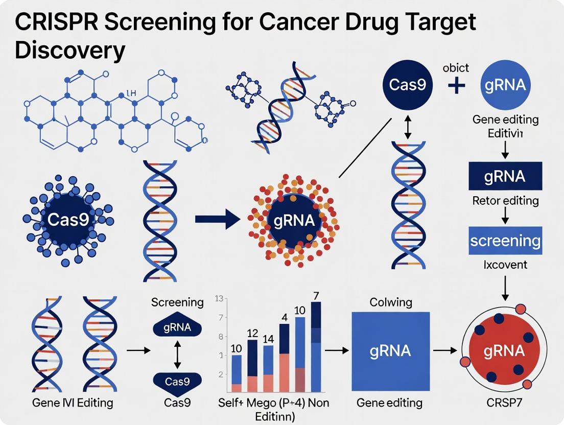
Clustered Regularly Interspaced Short Palindromic Repeats (CRISPR) and CRISPR-associated (Cas) proteins constitute an adaptive immune system in bacteria and archaea. It records fragments of invading viral DNA within the host genome, providing a heritable genetic memory. Upon re-infection, these sequences are transcribed and guide Cas nucleases to cleave complementary foreign DNA. The repurposing of the Type II CRISPR-Cas9 system from Streptococcus pyogenes has revolutionized genetic engineering due to its simplicity, comprising a single effector nuclease (Cas9) and a programmable guide RNA (gRNA).
In cancer research, the ability of CRISPR-Cas9 to create targeted gene knockouts enables genome-wide functional screening. This allows for the systematic identification of genes essential for cancer cell proliferation, survival, and drug resistance, directly informing target discovery pipelines.
Core Molecular Mechanism of CRISPR-Cas9
The engineered CRISPR-Cas9 system requires two core components:
- Cas9 Nuclease: An endonuclease that creates double-strand breaks (DSBs) in DNA.
- Guide RNA (gRNA): A chimeric RNA molecule combining the natural tractRNA and crRNA. Its 5' end contains a ~20 nucleotide spacer sequence complementary to the target DNA site.
The mechanism proceeds in three phases:
- Target Recognition: The gRNA directs Cas9 to a genomic locus complementary to its spacer sequence. Targeting requires a short Protospacer Adjacent Motif (PAM), typically 5'-NGG-3' for SpCas9, immediately downstream of the target.
- DNA Cleavage: Upon binding, Cas9 undergoes a conformational change, activating its two nuclease domains (HNH and RuvC). The HNH domain cleaves the complementary (target) strand, and the RuvC domain cleaves the non-complementary strand, generating a blunt-ended DSB.
- DNA Repair & Edit Outcome: The cell repairs the DSB via:
- Non-Homologous End Joining (NHEJ): An error-prone pathway that often introduces small insertions or deletions (indels), leading to gene knockouts.
- Homology-Directed Repair (HDR): In the presence of a donor DNA template, precise edits (e.g., point mutations, insertions) can be introduced.
Quantitative Data: CRISPR-Cas9 System Performance Metrics
The efficiency and specificity of CRISPR-Cas9 editing are critical for screening applications. Below are key quantitative benchmarks.
Table 1: Performance Characteristics of Common CRISPR-Cas9 Nucleases
| Nuclease Variant | PAM Sequence | Targeting Range* | Typical Editing Efficiency (in cultured cells) | Reported Off-Target Rate (Relative to SpCas9) | Primary Application in Screening |
|---|---|---|---|---|---|
| SpCas9 (Wild-type) | 5'-NGG-3' | 1 in 8 bp | 40-80% | 1.0 (Baseline) | Genome-wide knockout libraries |
| SpCas9-HF1 | 5'-NGG-3' | 1 in 8 bp | 20-60% | ~10-fold reduction | High-fidelity knockout screens |
| SpCas9-NG | 5'-NG-3' | 1 in 4 bp | 20-50% | Varies by target | Expanded target range screens |
| SaCas9 | 5'-NNGRRT-3' | 1 in 32 bp | 20-60% | ~10-fold reduction | In vivo delivery applications |
*Frequency in the human genome based on PAM requirement.
Table 2: Common Readouts in CRISPR-Cas9 Oncology Screens
| Screen Type | Library Size (Typical # of gRNAs) | Delivery Method | Primary Readout Technology | Key Metric (Hit Selection) |
|---|---|---|---|---|
| Knockout (Proliferation) | 70,000 - 100,000 | Lentiviral transduction | NGS of gRNA barcodes | Depletion/enrichment (log2 fold-change) |
| Activation (CRISPRa) | 30,000 - 70,000 | Lentiviral transduction | NGS of gRNA barcodes | Enrichment (log2 fold-change) |
| In Vivo | 5,000 - 30,000 | Lentiviral transduction + transplantation | NGS of gRNA from tumor vs. input | Tumor fitness score (enrichment ratio) |
Detailed Protocol: CRISPR-Cas9 Knockout Screening for Cancer Dependencies
This protocol outlines a standard genome-wide loss-of-function screen to identify genes essential for cancer cell viability.
Materials & Reagents
- Cell Line: A proliferative human cancer cell line (e.g., A549, HCT-116).
- CRISPR Library: A genome-wide lentiviral sgRNA library (e.g., Brunello or Brie library; ~4 sgRNAs/gene, 76,441 sgRNAs total).
- Packaging Plasmids: psPAX2 and pMD2.G.
- Transfection Reagent: Polyethylenimine (PEI) or Lipofectamine 3000.
- Selection Antibiotics: Puromycin.
- Media: Appropriate complete cell culture medium, serum, etc.
- Consumables: 15-cm tissue culture plates, multi-well plates, pipettes, cryovials.
Protocol Steps
Day 1-3: Library Virus Production (in HEK293T cells)
- Seed HEK293T cells in 15-cm plates to reach ~70% confluency the next day.
- Co-transfect cells with the library plasmid, psPAX2 (packaging), and pMD2.G (envelope) at a molar ratio of 3:2:1 using PEI.
- Change media 6 hours post-transfection.
- Harvest viral supernatant at 48 and 72 hours post-transfection. Pool, filter through a 0.45 µm filter, and concentrate using PEG-it virus precipitation solution or ultracentrifugation. Aliquot and store at -80°C.
Day 4-6: Cell Line Preparation & Transduction
- Titrate virus on the target cancer cell line to determine the volume needed to achieve a Multiplicity of Infection (MOI) of ~0.3, ensuring most cells receive a single viral integration.
- Seed 2 x 10^7 target cells. Transduce cells with the pre-titered library virus in the presence of polybrene (8 µg/mL).
- Change media 24 hours post-transduction.
Day 7-10: Selection and Expansion
- Begin puromycin selection (dose determined by kill curve) 48 hours post-transduction. Maintain selection for 5-7 days until all uninfected control cells are dead.
- Passage cells, maintaining a representation of at least 500 cells per sgRNA in the library at all times. This ensures library coverage. Harvest cells for the "T0" timepoint by centrifugation and pellet freezing.
Day 11-21: Screening & Harvest
- Continue to passage cells every 3-4 days for approximately 14 population doublings. This allows for the depletion of sgRNAs targeting essential genes.
- At the endpoint ("Tend"), harvest at least 5 x 10^7 cells by centrifugation and pellet freezing.
Day 22-30: Genomic DNA Extraction & NGS Library Prep
- Extract genomic DNA from T0 and Tend pellets using a maxi-prep kit (e.g., Qiagen Blood & Cell Culture DNA Maxi Kit). Ensure yield meets requirement for >500x coverage.
- Amplify integrated sgRNA sequences via two-step PCR. Step 1: Amplify sgRNA region from gDNA using primers containing partial Illumina adapter sequences. Step 2: Add full Illumina adapters and sample barcodes.
- Purify PCR products, quantify, and pool for sequencing on an Illumina NextSeq or HiSeq platform (minimum 300 reads per sgRNA).
Data Analysis
- Align sequencing reads to the reference sgRNA library.
- Count reads per sgRNA for T0 and Tend samples.
- Normalize counts and calculate log2 fold-change depletion for each sgRNA/gene using specialized software (e.g., MAGeCK or CERES).
The Scientist's Toolkit: Essential Research Reagents for CRISPR Screening
Table 3: Key Research Reagent Solutions for CRISPR-Cas9 Oncology Screens
| Item | Function & Role in Screening | Example Product/Resource |
|---|---|---|
| Validated sgRNA Libraries | Pre-designed, pooled collections of sgRNAs targeting the whole genome or specific gene families with optimized on-target efficiency. Essential for screen reproducibility. | Broad Institute GPP (Brunello, Brie), Addgene (GeCKO, KO). |
| High-Fidelity Cas9 Variant | Engineered Cas9 nuclease with reduced off-target cleavage, improving the specificity of phenotypic hits. | SpCas9-HF1, eSpCas9(1.1). |
| Lentiviral Packaging Mix | A system for producing replication-incompetent lentiviruses to deliver sgRNA and Cas9 components stably into target cells. | psPAX2 & pMD2.G plasmids, Lenti-X Packaging System. |
| Next-Generation Sequencing Kit | For amplifying and barcoding sgRNA sequences from genomic DNA to quantify their abundance pre- and post-selection. | Illumina Nextera XT, NEBNext Ultra II. |
| CRISPR Analysis Software | Specialized computational tools to process NGS read counts, normalize data, and identify significantly enriched/depleted genes. | MAGeCK, PinAPL-Py, CRISPRcloud. |
| Positive Control sgRNAs | sgRNAs targeting known essential genes (e.g., POLR2A, RPA3) to validate screening protocol and assay sensitivity. | Provided with commercial libraries. |
| Cell Viability/Proliferation Assay | To perform secondary validation of screen hits in low-throughput format (e.g., 96-well). | CellTiter-Glo, Incucyte live-cell imaging. |
In the pursuit of novel cancer drug targets, CRISPR-based functional genomics has become indispensable. The choice of screening modality—Knockout (CRISPRko), Activation (CRISPRa), or Interference (CRISPRi)—fundamentally shapes the biological questions answered and the targets identified. This guide provides a technical framework for selecting the optimal modality within the context of cancer target discovery, emphasizing experimental design, data interpretation, and translational relevance.
Core Modalities: Mechanisms and Applications
CRISPR Knockout (CRISPRko) utilizes Cas9 nuclease to create double-strand breaks, resulting in frameshift mutations and permanent gene disruption via non-homologous end joining (NHEJ). It is the gold standard for identifying essential genes and tumor vulnerabilities.
CRISPR Activation (CRISPRa) employs a catalytically dead Cas9 (dCas9) fused to transcriptional activators (e.g., VPR, SAM) to upregulate endogenous gene expression from the native locus. It is ideal for identifying tumor suppressors or genes conferring drug resistance when overexpressed.
CRISPR Interference (CRISPRi) uses dCas9 fused to transcriptional repressors (e.g., KRAB) to downregulate gene expression, typically via promoter or transcription start site binding. It offers a reversible, titratable knockdown, useful for studying essential gene networks and synthetic lethal interactions.
Quantitative Comparison of Modalities
Table 1: Key Characteristics of CRISPR Screening Modalities
| Feature | CRISPRko | CRISPRa | CRISPRi |
|---|---|---|---|
| Cas Protein | SpCas9 nuclease | dCas9-VPR, dCas9-SAM | dCas9-KRAB |
| Genetic Alteration | Permanent knockout | Sustained overexpression | Reversible knockdown |
| Primary Application | Loss-of-function screens | Gain-of-function screens | Tunable loss-of-function screens |
| Typical Library Size | ~70,000 sgRNAs (whole genome) | ~70,000 sgRNAs (whole genome) | ~70,000 sgRNAs (whole genome) |
| On-Target Efficacy | High (indels) | Moderate (2-10x upregulation) | High (~5-10x downregulation) |
| Off-Target Effects | Medium (DNA cleavage) | Low (epigenetic) | Low (epigenetic) |
| Best for Identifying | Essential genes, vulnerabilities | Tumor suppressors, resistance genes | Essential genes, synthetic lethality |
Table 2: Screening Performance Metrics in Cancer Cell Lines (Representative Data)
| Modality | False Discovery Rate (FDR) | Hit Concordance* | Screening Duration (weeks) | Key Validation Rate |
|---|---|---|---|---|
| CRISPRko | 1-5% | High (>80%) | 3-4 | 60-80% |
| CRISPRa | 5-15% | Moderate (50-70%) | 3-4 | 40-60% |
| CRISPRi | 2-8% | High (>75%) | 3-4 | 50-70% |
*Concordance of essential genes across similar cell lines.
Experimental Protocols
Protocol 1: Pooled CRISPRko Screen for Essential Genes
- Library Design & Cloning: Use the Brunello (4 sgRNAs/gene) or similar genome-wide library. Clone into lentiviral backbone (e.g., lentiCRISPRv2).
- Viral Production: Produce lentivirus in HEK293T cells via co-transfection of library plasmid with psPAX2 and pMD2.G.
- Cell Infection & Selection: Infect target cancer cells at low MOI (0.3-0.4) to ensure single integration. Select with puromycin (1-5 µg/mL) for 5-7 days.
- Screening & Passaging: Maintain cells at >500x library representation. Passage cells for 14-21 doublings. Harvest genomic DNA at T0 and Tfinal.
- NGS & Analysis: Amplify integrated sgRNA sequences via PCR. Sequence on Illumina platform. Analyze depletion/enrichment with MAGeCK or CERES.
Protocol 2: CRISPRa/i Screen with Inducible Systems
- Stable Cell Line Generation: Create a cancer cell line stably expressing dCas9-VPR (CRISPRa) or dCas9-KRAB (CRISPRi) under a doxycycline-inducible promoter.
- Library Transduction: Transduce with a sub-pooled sgRNA library targeting gene promoters (for CRISPRi) or ~200 bp upstream of TSS (for CRISPRa).
- Induction & Phenotyping: Induce with doxycycline (1 µg/mL) throughout the screen. Apply selective pressure (e.g., drug treatment) if needed.
- Harvest & Sequencing: Harvest cells at endpoints. Process gDNA and sequence sgRNA abundance. Analyze with MAGeCK MLE or similar.
Visualizing Modality Selection Logic
Diagram Title: Decision Logic for CRISPR Screening Modality Selection
Signaling Pathway Impact of Different Modalities
Diagram Title: Example: Modality Effects on a PI3K-AKT-mTOR Pathway
The Scientist's Toolkit: Key Research Reagent Solutions
Table 3: Essential Reagents for CRISPR Screening in Cancer Research
| Item | Function & Description | Example Product/Catalog |
|---|---|---|
| Genome-wide sgRNA Library | Pre-designed, pooled lentiviral library targeting all human genes. Enables genome-scale screening. | Broad Institute: Brunello (CRISPRko), Calabrese (CRISPRa), Dolcetto (CRISPRi). |
| Lentiviral Packaging Plasmids | Second/third-generation systems for safe, high-titer virus production. | psPAX2 (packaging), pMD2.G (VSV-G envelope). |
| dCas9 Effector Plasmids | Express dCas9 fused to transcriptional modulators for CRISPRa/i. | pLV-dCas9-VPR (Activation), pLV-dCas9-KRAB (Interference). |
| Validated Cell Line | A cancer cell line with high viral transduction efficiency and known genotype. | A549, K562, MCF-7, etc. |
| Next-Generation Sequencing Kit | For preparing sgRNA amplicon libraries from genomic DNA. | Illumina Nextera XT, NEBNext Ultra II. |
| Analysis Software | Computationally identifies significantly enriched/depleted sgRNAs/genes from NGS data. | MAGeCK, PinAPL-Py, CRISPRcloud. |
| Positive Control sgRNAs | Targeting essential genes (e.g., POLR2D) for assay validation. | Non-Targeting Control sgRNAs for baseline. |
The strategic selection of CRISPR modality directly influences the pipeline for target discovery. CRISPRko remains the workhorse for identifying non-redundant core essential genes that represent high-priority therapeutic vulnerabilities. CRISPRa excels in illuminating context-specific tumor suppressor networks and mechanisms of drug resistance. CRISPRi offers precision for dissecting dosage-sensitive genes and synthetic lethal pairs, particularly for undruggable oncogenes. Integrating findings from complementary modalities provides a robust, multi-dimensional validation of novel cancer targets, de-risking their progression into drug development pipelines. Future directions will involve more sophisticated in vivo screens and single-cell readouts to further refine target identification within the complex tumor microenvironment.
Within the modern paradigm of cancer drug target discovery, CRISPR screening has emerged as a transformative technology. This whitepaper details its application in three cornerstone areas: identifying synthetic lethal interactions, elucidating resistance mechanisms, and mapping essential genes. These goals are integral to developing targeted, durable, and less toxic cancer therapies.
Core Concepts in Context
Synthetic Lethality (SL) occurs when loss-of-function mutations in two genes are individually viable but lethal in combination. CRISPR screens systematically disrupt genes in a background of a specific cancer driver mutation (e.g., BRCA1) to find genes whose inhibition selectively kills cancer cells.
Resistance Mechanisms are studied via positive-selection CRISPR screens where cells are exposed to a therapeutic agent. Enriched sgRNAs reveal genes whose loss confers survival, pointing to potential drug targets for combination therapy.
Essential Genes are identified through genome-wide negative-selection screens. Depleted sgRNAs indicate genes required for cellular fitness, revealing core dependencies and vulnerabilities specific to cancer cell lineages.
Current Data Landscape
The table below summarizes representative quantitative outcomes from recent CRISPR screening studies in oncology.
Table 1: Key Quantitative Findings from Recent CRISPR Screens
| Goal | Cancer Model | Screen Type | Key Hit(s) | Validation Rate | Primary Readout |
|---|---|---|---|---|---|
| Synthetic Lethality | BRCA1-mutant Ovarian | Genome-wide KO | PALB2, RAD54L | ~85% (in vitro) | Cell Viability (ATP assay) |
| Resistance Mechanism | Melanoma on MAPKi | Genome-wide KO | NF2, PTEN | ~70% (in vitro/vivo) | Drug-surviving Fraction |
| Lineage Essentiality | AML vs. Healthy HSPCs | Genome-wide KO | MCL1, BCL2L1 | >90% (in vitro) | Fold-change depletion (NGS) |
| CRISPRi Transcriptional | KRAS-mutant NSCLC | Genome-wide CRISPRi | SLC33A1, CCNI | ~80% (in vitro) | Proliferation Rate |
Detailed Experimental Protocols
Protocol 1: Genome-wide Knockout Screen for Synthetic Lethality
Objective: Identify genes synthetically lethal with an oncogenic driver mutation.
- Library Design & Production: Utilize the Brunello or similar genome-wide sgRNA library (~4-6 sgRNAs/gene, ~75,000 sgRNAs total). Produce high-titer lentivirus.
- Cell Line Engineering: Use an isogenic pair: Parental (e.g., BRCA1 WT) and Mutant (e.g., BRCA1 KO) cell lines. Transduce cells at low MOI (~0.3) to ensure single sgRNA integration. Maintain >500x coverage per sgRNA.
- Selection & Expansion: Select transduced cells with puromycin (1-3 µg/mL, 3-7 days). Expand populations for >10 population doublings to allow phenotypic manifestation.
- Sample Collection & Sequencing:
- Harvest cells at Day 0 (post-selection) and Day ~21 (endpoint).
- Extract genomic DNA (gDNA) using a column-based method. Amplify sgRNA cassettes via PCR with barcoded primers.
- Sequence on an Illumina platform (75bp single-end).
- Data Analysis: Align reads to the reference library. Calculate sgRNA depletion/enrichment using MAGeCK or PinAPL-Py. Genes with significantly depleted sgRNAs specifically in the mutant background are candidate synthetic lethal hits.
Protocol 2: Positive-Selection Screen for Drug Resistance
Objective: Discover gene knockouts that confer resistance to a targeted therapy.
- Transduction & Selection: Follow Protocol 1 steps 1-3 using the relevant cancer cell line.
- Drug Treatment: Split cells into treatment and vehicle control arms. Treat with the drug at IC70-IC90 concentration.
- Passaging & Harvest: Maintain drug pressure, passaging cells as needed for 2-3 weeks. Harvest genomic DNA from treated and control populations when a clear survival fraction is evident.
- Sequencing & Analysis: Process as in Protocol 1. sgRNAs significantly enriched in the drug-treated arm relative to control indicate genes whose loss promotes resistance.
Visualizing Workflows and Pathways
Title: CRISPR Screening Experimental Workflow
Title: PARPi Synthetic Lethality with BRCA Loss
The Scientist's Toolkit: Research Reagent Solutions
Table 2: Essential Materials for CRISPR Screening
| Reagent / Tool | Function & Role in Screening |
|---|---|
| Genome-wide sgRNA Library (e.g., Brunello, Human GeCKOv2) | Pre-designed pooled library targeting all human genes with multiple sgRNAs per gene; the core screening reagent. |
| Lentiviral Packaging Mix (psPAX2, pMD2.G) | Third-generation system for producing high-titer, replication-incompetent lentivirus to deliver sgRNAs. |
| Puromycin Dihydrochloride | Selection antibiotic to eliminate non-transduced cells, ensuring a pure population of sgRNA-bearing cells. |
| Polybrene (Hexadimethrine Bromide) | Cationic polymer that increases viral transduction efficiency by neutralizing charge repulsion. |
| NucleoSpin Blood or Tissue Kit (Macherey-Nagel) | For high-quality, high-yield genomic DNA extraction from cell pellets, crucial for downstream NGS. |
| KAPA HiFi HotStart ReadyMix (Roche) | High-fidelity PCR enzyme for accurate, unbiased amplification of integrated sgRNA sequences from gDNA. |
| MiSeq or NextSeq System (Illumina) | Next-generation sequencing platform for deep sequencing of sgRNA amplicons to determine abundance. |
| MAGeCK (Bioinformatics Tool) | Computational pipeline for analyzing CRISPR screen data to identify significantly enriched or depleted genes. |
| CellTiter-Glo Luminescent Assay (Promega) | ATP-based viability assay for validating candidate hits in secondary, low-throughput assays. |
Within the paradigm of functional genomics for cancer drug target discovery, CRISPR-Cas9 screening has emerged as a cornerstone technology. This in-depth technical guide elucidates the three core components of a successful CRISPR screen: the design of single-guide RNA (sgRNA) libraries, methods for Cas9 delivery, and subsequent readout technologies. The integration of these elements enables the systematic identification of genes essential for cancer cell survival, drug resistance, and synthetic lethality, directly informing therapeutic development.
sgRNA Library Design and Selection
sgRNA libraries are curated collections of guide RNAs designed to target a specific set of genes genome-wide or within a pathway of interest.
Library Types and Key Quantitative Metrics
| Library Type | Target Scope | Typical sgRNA/Gene | Key Design Considerations | Primary Use Case in Cancer Research |
|---|---|---|---|---|
| Genome-wide | All annotated genes | 4-10 | Uniform on-target efficiency, minimization of off-target effects | Discovery of novel essential genes and vulnerabilities across cancer lineages. |
| Focused/Knockout | Subset (e.g., kinases, druggable genome) | 4-10 | High-confidence on-target scoring algorithms | Deep interrogation of specific gene families for target identification. |
| CRISPRi/a (Modulation) | Promoters/enhancers | 3-5 per TSS | Proximity to transcription start site (TSS) for CRISPRi/a | Identifying gene regulatory dependencies and non-coding vulnerabilities. |
| Custom | User-defined genes | 3-6 | Flexibility for validation or specific pathways | Follow-up validation and mechanistic studies on hit genes from primary screens. |
Protocol: A Typical Workflow for Arrayed sgRNA Library Cloning
- Design & Synthesis: sgRNA sequences are designed using predictive algorithms (e.g., Doench '16, MIT CRISPR Design Tool). Oligonucleotides are synthesized on an array.
- PCR Amplification: Oligo pool is amplified using primers adding flanking homology arms compatible with the lentiviral backbone (e.g., lentiGuide-Puro).
- Golden Gate Assembly: Amplified pool and linearized backbone are assembled using BsmBI-v2 restriction enzyme and T4 DNA Ligase in a one-pot reaction.
- Transformation & Pooling: The assembly reaction is transformed into E. coli (e.g., Endura Electrocompetent Cells) via electroporation. Colonies are scraped and pooled to ensure >200x coverage of the library.
- Plasmid Preparation: High-quality plasmid DNA is extracted from the bacterial pool using a maxiprep kit. The library is sequence-verified by NGS to confirm representation and uniformity.
Cas9 Delivery Systems
Effective delivery of the Cas9 nuclease and sgRNA library into the target cell population is critical for screen performance.
Delivery Method Comparison
| Delivery Method | Format | Editing Efficiency | Scalability | Suitability for In Vivo Screens | Key Challenges |
|---|---|---|---|---|---|
| Lentiviral Transduction | sgRNA (most common) or Cas9+sgRNA | High (>80% in permissive lines) | Excellent for large pools | Possible with barcoded models | Integration bias, variable titer, biosafety level 2. |
| Electroporation (RNP) | Pre-complexed Cas9 protein + sgRNA | Very High (~90-95%) | Moderate (arrayed screens) | Limited | Cytotoxicity, not suitable for pooled delivery, requires arrayed format. |
| Adenoviral (AV) or Adeno-associated (AAV) | Viral delivery of sgRNA/Cas9 | Moderate to High | Good | Excellent for in vivo | Packaging size constraints (AAV), immune response. |
| Stable Cas9 Expression | Cell line engineering | Consistent (100% Cas9+) | High once engineered | Excellent | Clonal variation, potential for Cas9 toxicity or adaptive responses. |
Protocol: Generating a Lentiviral sgRNA Library Pool
- Cell Seeding: Seed HEK293T (or Lenti-X) packaging cells in a 15cm dish to reach 70-80% confluency the next day.
- Transfection Complex: In a tube, combine:
- 20 µg sgRNA library plasmid (e.g., lentiGuide-Puro)
- 15 µg psPAX2 packaging plasmid
- 10 µg pMD2.G envelope plasmid
- Opti-MEM to 1.5 mL. In a separate tube, dilute 112 µL of PEI (1 mg/mL) in 1.5 mL Opti-MEM. Combine the two tubes, mix, and incubate 15-20 min at RT.
- Transfection & Harvest: Add complex dropwise to cells. Replace media with fresh DMEM + 10% FBS after 6-8 hours. Harvest viral supernatant at 48 and 72 hours post-transfection.
- Concentration: Pool supernatants, filter through a 0.45µm PES filter. Concentrate using Lenti-X Concentrator (1:3 ratio) or ultracentrifugation.
- Titering: Serially dilute virus on target cells with polybrene (8µg/mL). Apply selection (e.g., puromycin) 48hr later. Calculate TU/mL based on colony survival. Aim for a Multiplicity of Infection (MOI) of ~0.3 to ensure most cells receive a single sgRNA.
Readout Technologies
The choice of readout determines the type of biological question a screen can answer.
Readout Modality Comparison
| Readout Technology | Measurement | Screening Format | Key Data Output | Application in Cancer Target Discovery |
|---|---|---|---|---|
| Viability/Proliferation | Cell count/survival over time | Pooled (most common) | sgRNA fold-depletion/enrichment | Identify essential genes for tumor cell fitness. |
| Fluorescence-Activated Cell Sorting (FACS) | Protein expression (e.g., cell surface markers, reporters) | Pooled | sgRNA frequency in sorted populations | Identify regulators of pathways, differentiation states, or antigen presentation. |
| Barcode Sequencing (BarSeq) | Unique molecular identifiers (UMIs) attached to clones | Pooled | Clone abundance under selective pressure | High-resolution tracking of clonal dynamics in drug response. |
| Single-Cell RNA Sequencing (scRNA-seq) | Whole transcriptome + sgRNA identity | Pooled (CROP-seq, Perturb-seq) | Gene expression profiles per sgRNA | Uncover mechanistic gene networks and heterogeneous responses to perturbation. |
| Imaging-Based | Morphology, biomarker intensity, etc. | Arrayed | High-content image features | Identify genes affecting cell morphology, organelle function, or drug-induced phenotypes. |
Protocol: A Standard Pooled Viability Screen Workflow & NGS Sample Prep
- Infection and Selection: Infect target cancer cells (e.g., A549, MCF-7) stably expressing Cas9 with the lentiviral sgRNA library at MOI~0.3. Maintain at >500x library representation. Apply appropriate selection (e.g., puromycin 1-2µg/mL) for 3-7 days.
- Harvest Timepoints: Harvest a representative sample of cells (~500x coverage) as the "T0" reference timepoint. Continue passaging remaining cells for ~14-21 population doublings, maintaining representation. Harvest the final "Tend" sample.
- Genomic DNA (gDNA) Extraction: Use a bulk gDNA extraction kit (e.g., Qiagen Blood & Cell Culture Maxi Kit) from ~1e7 cells per sample. Quantify by Nanodrop/Qubit.
- sgRNA Amplification for Sequencing: Perform a two-step PCR.
- PCR1 (Add Sequencing Adaptors): In a 50µL reaction, use 2-4µg gDNA, primers amplifying the sgRNA constant region, and a high-fidelity polymerase. Cycle number should be minimized (~20 cycles) to avoid skewing.
- PCR2 (Add Sample Indexes & P5/P7): Use 5µL of purified PCR1 product with indexing primers. Run 10-12 cycles.
- Sequencing & Analysis: Pool purified PCR2 products, quantify, and sequence on an Illumina NextSeq (75bp single-end). Align reads to the library reference, count sgRNAs, and use MAGeCK or CRISPhieRmix to identify significantly depleted/enriched genes between T0 and Tend.
The Scientist's Toolkit: Research Reagent Solutions
| Item | Function & Role in CRISPR Screening |
|---|---|
| Lentiviral Backbone Plasmid (e.g., lentiGuide-Puro) | sgRNA expression vector containing U6 promoter, sgRNA scaffold, and puromycin resistance for selection. |
| Packaging Plasmids (psPAX2, pMD2.G) | Second-generation lentiviral system components for producing replication-incompetent viral particles in HEK293T cells. |
| Polybrene (Hexadimethrine bromide) | A cationic polymer that enhances viral transduction efficiency by neutralizing charge repulsion between virions and cell membrane. |
| Puromycin Dihydrochloride | Selection antibiotic that kills non-transduced cells, ensuring a population uniformly expressing the sgRNA library. |
| High-Fidelity Polymerase (e.g., Q5, KAPA HiFi) | Critical for accurate, low-bias amplification of sgRNA sequences from genomic DNA for NGS library preparation. |
| Next-Generation Sequencing Kit (Illumina) | For determining sgRNA abundance in pooled populations. Essential for calculating gene essentiality scores. |
| Cas9-Nuclease Expressing Cell Line | A genetically engineered cancer cell line stably expressing SpCas9, enabling direct sgRNA library transduction. |
| Biological Analysis Software (e.g., MAGeCK, CRISPhieRmix) | Computational tools for robust statistical analysis of screen data, identifying significantly enriched or depleted genes. |
Visualized Workflows and Pathways
Diagram Title: Pooled CRISPR Screen End-to-End Workflow
Diagram Title: CRISPR-Cas9 Mechanism to Screening Readout
Diagram Title: From Screen Hit to Target Validation Thesis
From Bench to Dataset: Executing Pooled, Arrayed, and In Vivo CRISPR Screens
This technical guide details a comprehensive CRISPR-Cas9 screening workflow, framed within the thesis that systematic functional genomics is the cornerstone of next-generation cancer drug target discovery. The protocol enables genome-wide identification of genes essential for cancer cell survival or drug response, translating genetic perturbations into actionable therapeutic hypotheses.
Design: Library and Experimental Planning
The design phase establishes the screening hypothesis and selects the appropriate CRISPR library.
Library Selection Criteria
CRISPR knockout (CRISPRko) libraries are standard for loss-of-function screening. Key quantitative metrics for common genome-wide human libraries are summarized below.
Table 1: Common Genome-Wide CRISPRko Libraries (Human)
| Library Name | # of sgRNAs | # of Genes Targeted | Avg. sgRNAs/Gene | Control sgRNAs | Primary Use Case |
|---|---|---|---|---|---|
| Brunello | 77,441 | 19,114 | 4 | 1,000 non-targeting | Genome-wide knockout |
| TKOv3 | 70,948 | 17,661 | 4 | 1,000 non-targeting | Essential gene profiling |
| Brie | 78,637 | 19,150 | 4 | 1,000 non-targeting | Dual-sgRNA for redundancy |
Experimental Design
A robust screen requires careful planning of biological replicates, sequencing depth, and controls.
- Replicates: Minimum of 3 biological replicates per condition to ensure statistical power.
- Cell Line Validation: Confirm Cas9 expression and activity via western blot (anti-Cas9) and SURVEYOR/T7E1 assay on a known essential gene (e.g., RPA3).
- Screen Arms: For drug target discovery, standard design includes:
- Proliferation Screen: Identify core fitness genes.
- Drug-Modifier Screen: Cells treated with sub-lethal dose of oncology compound vs. DMSO control.
Protocol 1.1: Cas9 Activity Validation (T7 Endonuclease I Assay)
- Transfect target cells with sgRNA targeting a known essential gene.
- After 72h, extract genomic DNA (gDNA) using a column-based kit.
- PCR-amplify the target region (300-500bp) from gDNA.
- Hybridize PCR products: Denature at 95°C for 10 min, re-anneal by ramping down to 25°C at -0.1°C/sec.
- Treat heteroduplex DNA with 5 units of T7E1 enzyme (NEB) at 37°C for 30 min.
- Analyze fragments on a 2% agarose gel. Cleaved bands indicate Cas9-mediated indel formation.
Transduction: Library Delivery and Representation
This phase involves introducing the sgRNA library into the cellular population at low multiplicity of infection (MOI) to ensure one sgRNA per cell.
Viral Production and Titration
Lentiviral vectors are the standard delivery method. Critical quantitative parameters:
- MOI: Aim for MOI of ~0.3 to minimize cells with multiple sgRNAs.
- Library Coverage: Maintain a minimum of 500 cells per sgRNA to prevent stochastic dropout. For the Brunello library (77k sgRNAs), this requires > 38.5 million transduced cells.
- Transduction Efficiency: Optimize to 30-50% using a fluorescent reporter (e.g., GFP).
Protocol 2.1: Large-Scale Lentiviral Production (in HEK293T)
- Seed 15 million HEK293T cells in a 15cm dish 24h prior.
- Co-transfect using PEI Max: 18 µg library plasmid (psgRNA), 12 µg psPAX2 (packaging), and 6 µg pMD2.G (VSV-G envelope).
- Change media 6h post-transfection.
- Harvest viral supernatant at 48h and 72h, filter through a 0.45µm PES filter, and concentrate via ultracentrifugation (25,000 rpm, 2h, 4°C) or PEG-it virus precipitation.
- Aliquot and titer on target cells via qPCR (Lentivirus qPCR Titer Kit) or puromycin selection.
Library Transduction and Puromycin Selection
Protocol 2.2: Pooled Library Transduction
- Harvest 200 million (for 500x coverage) target cells expressing Cas9.
- Transduce cells at MOI=0.3 in the presence of 8µg/mL polybrene via spinfection (1000g, 90 min, 32°C).
- At 24h post-transduction, replace with fresh medium.
- At 48h post-transduction, begin puromycin selection (dose predetermined by kill curve). Maintain selection for 5-7 days until all non-transduced control cells are dead.
Table 2: Critical Reagents for Transduction
| Reagent | Function | Example Product/Catalog # |
|---|---|---|
| sgRNA Library Plasmid | Encodes sgRNA and puromycin resistance | Addgene #73178 (Brunello) |
| Lentiviral Packaging Plasmid (psPAX2) | Provides gag, pol, rev, tat genes | Addgene #12260 |
| Envelope Plasmid (pMD2.G) | Provides VSV-G glycoprotein for pseudotyping | Addgene #12259 |
| Polybrene | Cationic polymer enhancing viral adhesion | Hexadimethrine bromide, Sigma H9268 |
| Puromycin Dihydrochloride | Selection antibiotic for transduced cells | Thermo Fisher A1113803 |
Diagram 1: Library Transduction and Selection Workflow
Selection: Applying Functional Pressure
The cell population is passaged under the experimental condition (e.g., drug treatment) to induce differential fitness based on sgRNA identity.
Screening Timeline and Sampling
- Proliferation Screen: Passage cells for 14-21 population doublings to allow fitness effects to manifest.
- Drug-Modifier Screen: Treat one arm with compound at IC10-IC20 concentration; maintain a parallel DMSO control arm.
- Sampling: Harvest a minimum of 50 million cells (representing >500x coverage) at the T0 timepoint (post-puromycin, pre-selection) and at the Tfinal endpoint. Extract and store gDNA at -80°C.
Protocol 3.1: Genomic DNA Extraction from Pelleted Cells
- Resuspend cell pellet (50M cells) in 5 mL PBS.
- Add 15 mL of Lysis Buffer (10 mM Tris-HCl pH8.0, 100 mM EDTA, 0.5% SDS, 20 µg/mL RNase A) and incubate at 37°C for 1h.
- Add 50 µg/mL Proteinase K and incubate at 55°C overnight.
- Perform phenol-chloroform-isoamyl alcohol extraction, followed by ethanol precipitation.
- Resuspend gDNA in TE buffer and quantify via Qubit dsDNA BR Assay. Expected yield: ~250 µg.
Sequencing: sgRNA Quantification and Analysis
sgRNA representation is quantified via NGS of amplified gDNA to determine enrichment/depletion.
sgRNA Amplification and Sequencing
This two-step PCR adds sequencing adapters and sample indices.
Protocol 4.1: Two-Step PCR for NGS Library Preparation Step 1 (Amplify sgRNA inserts from gDNA):
- Primers: Forward:
AATGATACGGCGACCACCGAGATCTACAC[i5]ACACTCTTTCCCTACACGACGCTCTTCCGATCTNNNNN*GTTTAAGAGCTAAGCTG*; Reverse:CAAGCAGAAGACGGCATACGAGAT[i7]GTGACTGGAGTTCAGACGTGTGCTCTTCCGATCT*ACAAGCATAGCAAGTTAAAATAAGG*. (Constant regions in italics). - Reaction: 10 µg gDNA, 2x KAPA HiFi HotStart ReadyMix, 0.5 µM primers. Cycle: 98°C 3min; 22 cycles of (98°C 20s, 63°C 30s, 72°C 30s); 72°C 5min. Step 2 (Add full Illumina adapters):
- Use 1 µL of purified PCR1 product as template with universal primers.
- Purify final library with SPRI beads, quantify via qPCR (KAPA Library Quant Kit), and pool for sequencing on an Illumina NextSeq (75bp single-end, ~100 reads per sgRNA).
Data Analysis Pipeline
Raw sequencing data is processed to generate gene-level fitness scores.
Table 3: Core Bioinformatics Pipeline Steps & Tools
| Step | Tool/Algorithm | Key Output |
|---|---|---|
| Demultiplexing | bcl2fastq (Illumina) |
Sample-specific FASTQ files |
| sgRNA Read Counting | MAGeCK count |
Count table for all sgRNAs per sample |
| Differential Abundance | MAGeCK test (RRA algorithm) |
Enriched/Depleted sgRNAs & genes |
| Pathway Enrichment | GSEA, Enrichr |
Pathways synthetic lethal with drug |
The core analysis generates a β-score (log2 fold-change) for each gene. Essential genes have negative β-scores; genes whose knockout confers drug resistance have positive β-scores in the drug-treated arm.
Diagram 2: Sequencing and Analysis Pipeline
The Scientist's Toolkit: Key Research Reagents
Table 4: Essential Reagent Solutions for CRISPR Screening
| Category | Item | Function | Critical Parameters |
|---|---|---|---|
| Library & Vectors | Genome-wide sgRNA Plasmid Library | Encodes the pool of targeting sgRNAs | Coverage, # of controls, vector backbone (lentiGuide) |
| Lentiviral Packaging Plasmids | Produces replication-incompetent virus | 2nd vs. 3rd generation (psPAX2/pMD2.G) | |
| Cell Culture | Cas9-Expressing Cell Line | Provides the nuclease for genome editing | Stable vs. inducible expression; activity validation |
| Polybrene / Protamine Sulfate | Enhances viral transduction efficiency | Cell line-specific optimization required | |
| Puromycin / Blasticidin | Selects for successfully transduced cells | Kill curve to determine minimal effective dose | |
| Molecular Biology | gDNA Extraction Kit | High-yield, high-purity gDNA from millions of cells | Scalability to >50M cells, removal of inhibitors |
| High-Fidelity PCR Master Mix | Accurate amplification of sgRNA loci from gDNA | Low error rate, amplification of complex pools | |
| Sequencing | Custom PCR Primers | Adds Illumina adapters & sample indices | Unique dual indices (UDI) to prevent index hopping |
| SPRI Beads | Size selection and purification of NGS libraries | Consistent bead-to-sample ratio for reproducibility |
This step-by-step workflow provides a robust framework for conducting CRISPR screens aimed at cancer drug target discovery. By systematically linking genetic perturbation to phenotypic fitness under therapeutic pressure, researchers can nominate high-confidence targets and illuminate novel synthetic lethal interactions, directly informing therapeutic development pipelines.
Within the paradigm of CRISPR screening for cancer drug target discovery, pooled screens represent a cornerstone high-throughput methodology. This approach enables the simultaneous evaluation of thousands to millions of genetic perturbations in a single, complex experiment, dramatically accelerating the identification of genes essential for cancer cell survival, drug resistance, and synthetic lethality. This technical guide details the advantages, core workflow, and implementation of pooled CRISPR screens in oncological research.
Advantages of Pooled Screening
Pooled screening offers distinct benefits for large-scale functional genomics.
Table 1: Comparison of Pooled vs. Arrayed Screening
| Feature | Pooled CRISPR Screening | Arrayed CRISPR Screening |
|---|---|---|
| Throughput | Very High (10^5 - 10^8 perturbations) | Moderate (10^1 - 10^4 perturbations) |
| Format | Mixed population in a single vessel | Each perturbation in a separate well |
| Cost Per Perturbation | Very Low | High |
| Primary Readout | Next-Generation Sequencing (NGS) | Imaging, Luminescence, Fluorescence |
| Complexity of Assay | Compatible with simple survival/proliferation | Compatible with complex, multi-parametric assays |
| Hit Deconvolution | Required post-screen via NGS | Directly known from well position |
| Typical Application | Genome-wide dropout screens, in vivo screens | High-content imaging, kinetic assays, secondary validation |
Detailed Workflow for a Pooled CRISPR-Cas9 Dropout Screen
The following protocol outlines a standard negative selection (dropout) screen to identify genes essential for cancer cell proliferation.
Library Design and Cloning
- Objective: To construct a lentiviral plasmid library containing single guide RNA (sgRNA) sequences targeting the gene set of interest (e.g., whole genome, kinome).
- Protocol:
- Select a published library (e.g., Brunello, GeCKO v2) or design a custom sgRNA library. Typically, 3-6 sgRNAs per gene are used for statistical robustness.
- Synthesize the oligo pool containing all sgRNA sequences flanked by cloning sequences.
- Perform a pooled cloning reaction to insert the oligo pool into the lentiviral sgRNA expression backbone via Golden Gate or similar assembly.
- Transform the reaction into highly competent E. coli and carry out a massive plasmid preparation to ensure >200x representation of the library to maintain diversity.
Lentiviral Production & Transduction
- Objective: To generate a population of cancer cells each harboring a single genetic perturbation.
- Protocol:
- Co-transfect HEK293T cells with the sgRNA library plasmid, psPAX2 (packaging), and pMD2.G (VSV-G envelope) plasmids using PEI or commercial transfection reagent.
- Harvest lentiviral supernatant at 48 and 72 hours, concentrate via ultracentrifugation, and titer on target cancer cells.
- Transduce the target cancer cell line (e.g., a pancreatic cancer line) at a low Multiplicity of Infection (MOI ~0.3) to ensure most cells receive only one sgRNA. Include puromycin selection to generate a stable cell pool.
Screening Experiment & Phenotypic Selection
- Objective: To apply selective pressure and allow differential proliferation based on sgRNA fitness effects.
- Protocol:
- Day 0 (T0): Harvest a baseline sample of at least 500 cells per sgRNA in the library for genomic DNA (gDNA) extraction. This serves as the reference point.
- Split the remaining transduced cell pool and culture for ~14-21 population doublings. For a drug target discovery screen, one arm can be treated with a chemotherapeutic agent, while the other serves as an untreated control.
- Endpoint (T14/T21): Harvest the final cell population from all experimental arms.
Next-Generation Sequencing & Data Analysis
- Objective: To quantify sgRNA abundance changes and identify significantly depleted or enriched genes.
- Protocol:
- Extract gDNA from all timepoint samples using a mass-scale method.
- Perform a two-step PCR to amplify the integrated sgRNA sequence from the gDNA and attach Illumina sequencing adapters and sample barcodes.
- Pool PCR products, sequence on an Illumina platform to obtain >500 reads per sgRNA.
- Align reads to the reference sgRNA library. Use computational pipelines (MAGeCK, BAGEL, CRISPRcleanR) to:
- Normalize read counts.
- Compare sgRNA abundance between baseline, control, and treated samples.
- Perform statistical testing to rank genes based on essentiality (beta score) and significance (FDR).
Table 2: Key Quantitative Metrics in a Typical Genome-Wide Dropout Screen
| Metric | Typical Value/Range | Purpose & Implication |
|---|---|---|
| Library Representation | >200x per sgRNA | Ensures statistical power and minimizes stochastic dropout. |
| Cell Coverage at Transduction | >500 cells per sgRNA | Maintains library complexity post-transduction. |
| Sequencing Depth | >500 reads per sgRNA | Enables accurate quantification of abundance changes. |
| False Discovery Rate (FDR) | < 0.05 (5%) | Threshold for statistical significance of candidate hits. |
| Gene Essentiality Score (β) | Negative value indicates essentiality | Magnitude correlates with degree of fitness defect. |
Signaling Pathways in CRISPR Screen Hit Validation
A common hit from a cancer dropout screen is a gene within a core survival pathway. The diagram below illustrates the canonical PI3K-AKT-mTOR pathway, frequently identified as essential in oncology screens.
Title: PI3K-AKT-mTOR Pathway in Cancer Cell Survival
Experimental Workflow for a Pooled CRISPR Screen
Title: Pooled CRISPR Screen Workflow from Library to Hits
The Scientist's Toolkit: Key Research Reagent Solutions
Table 3: Essential Materials for Pooled CRISPR Screening
| Item | Function in Screen | Example/Notes |
|---|---|---|
| Validated sgRNA Library | Defines the genetic perturbations tested. | Brunello (human genome-wide), kinome/subset libraries. Cloned into lentiGuide-Puro backbone. |
| Lentiviral Packaging Plasmids | Required for production of infectious lentiviral particles. | psPAX2 (packaging), pMD2.G (VSV-G envelope). |
| HEK293T Cells | Standard cell line for high-titer lentivirus production. | Readily transfectable, robust growth. |
| Polybrene (or equivalent) | A cationic polymer that enhances viral transduction efficiency. | Typically used at 4-8 µg/mL during transduction. |
| Puromycin (or other selector) | Antibiotic for selecting successfully transduced cells. | Critical to establish a pure population of CRISPR-modified cells. Dose must be pre-determined. |
| High-Quality gDNA Extraction Kit | For mass isolation of genomic DNA from cell pellets. | Must handle large sample sizes (≥10^7 cells) with high yield and minimal bias. |
| Herculase II Fusion DNA Polymerase | Robust polymerase for efficient amplification of sgRNAs from gDNA. | Used in the two-step PCR protocol for NGS library construction. |
| Illumina Sequencing Reagents | Platform-specific kits for cluster generation and sequencing. | MiSeq or NextSeq systems common for screen deconvolution. |
| Analysis Software/Pipeline | Computational tool for raw read processing, normalization, and hit calling. | MAGeCK, BAGEL, CRISPRcleanR. Requires R/Python environment. |
Within the broader thesis of CRISPR screening for cancer drug target discovery, functional validation remains a critical bottleneck. Pooled CRISPR screens excel at identifying genes essential for fitness but often lack the resolution for deep, multifaceted phenotypic analysis. Arrayed CRISPR screens, where each well contains a single, predefined genetic perturbation, enable high-content imaging, multi-parametric flow cytometry, and complex biochemical assays. This whitepaper details the application of arrayed screens for deep phenotyping in the functional validation of candidate cancer drug targets, providing technical protocols, data presentation standards, and essential resources.
Core Quantitative Data from Recent Arrayed Screening Studies
The following table summarizes key quantitative outcomes from recent arrayed CRISPR screening studies in cancer research, highlighting the depth of phenotyping achievable.
Table 1: Quantitative Outcomes from Recent Arrayed CRISPR Phenotypic Screens
| Study Focus (Year) | Phenotypic Readout | Key Metric | Value | Implication for Target Discovery |
|---|---|---|---|---|
| Synthetic Lethality in BRCA1-Mutants (2023) | High-Content Imaging (Nuclear γH2AX foci) | Hit Genes (Z-score > 3) | 42 | Identified 42 genes whose knockout induced DNA damage specifically in BRCA1-deficient cells. |
| Drug Combination Resistance (2024) | Multiplexed Flow Cytometry (Annexin V/pS6/Ki67) | % Reversal of Apoptosis | 65% | Knockout of BCL2L11 reversed drug-induced apoptosis by 65%, defining a key resistance mechanism. |
| Metastatic Potential (2023) | 3D Spheroid Invasion Assay | Mean Invasion Index Change | -2.8 ± 0.4 | KO of LIMK2 reduced invasion index by 2.8-fold, nominating it as a potential anti-metastatic target. |
| Senescence Bypass (2024) | SA-β-Gal & Secretome Analysis | Senescence Escape Rate | 18.3% | CDKN2A KO enabled 18.3% of oncogene-induced senescent cells to re-enter the cell cycle. |
Detailed Experimental Protocols
Protocol: Arrayed CRISPR-Cas9 Knockout for High-Content Imaging
Objective: To validate a gene candidate's role in maintaining genomic integrity using an arrayed, image-based readout. Materials: Arrayed sgRNA library (e.g., in lentiviral format), cancer cell line of interest, polybrene (8 µg/mL), puromycin (concentration determined by kill curve), PBS, formaldehyde (4%), Triton X-100 (0.5%), primary antibody (γH2AX), fluorescent secondary antibody, DAPI, high-content imaging system. Method:
- Reverse Transfection: Seed cells in 384-well imaging plates at 2,000 cells/well. Incubate for 6 hours.
- Viral Transduction: Add pre-arrayed lentiviral sgRNAs (MOI ~3-5) and polybrene directly to wells. Spinfect at 1000 × g for 1 hour at 32°C. Incubate at 37°C, 5% CO₂.
- Selection: After 48 hours, replace medium with puromycin-containing medium. Select for 72-96 hours.
- Phenotype Induction & Fixation: At day 7 post-transduction, treat cells with a sub-lethal dose of a DNA-damaging agent (e.g., 1 µM Camptothecin, 24h). Aspirate medium and fix cells with 4% formaldehyde for 15 min.
- Immunostaining: Permeabilize with 0.5% Triton X-100 for 10 min. Block with 5% BSA for 1 hour. Incubate with anti-γH2AX primary antibody (1:1000) overnight at 4°C. Incubate with fluorescent secondary antibody (1:500) and DAPI (1 µg/mL) for 1 hour at RT.
- Imaging & Analysis: Acquire 20+ fields/well using a 20x objective on a high-content imager. Use analysis software to segment nuclei (DAPI) and quantify γH2AX foci count/intensity per cell.
Protocol: Multiplexed Flow Cytometry Phenotyping from Arrayed Screens
Objective: To measure multiple cell states (apoptosis, cell cycle, signaling) in a single well from an arrayed screen. Materials: Arrayed CRISPR-edited cells in 96-well plate, trypsin/EDTA, PBS, formaldehyde (1.6%), methanol (100%), fluorochrome-conjugated antibodies (e.g., Annexin V-FITC, anti-pS6-PE, anti-Ki67-Alexa647), flow cytometry buffer (PBS + 1% FBS). Method:
- Cell Harvest: At assay endpoint, trypsinize cells, transfer to a V-bottom 96-well plate, and pellet at 300 × g for 5 min.
- Fixation & Permeabilization: For surface+intracellular staining: Resuspend in 1.6% formaldehyde for 10 min at RT. Wash with PBS. Resuspend in 100% ice-cold methanol and incubate 30 min on ice. Wash twice with flow cytometry buffer.
- Staining: Resuspend cell pellet in 50 µL flow cytometry buffer containing pre-titrated antibody cocktail. Incubate for 1 hour at RT in the dark.
- Acquisition: Wash cells, resuspend in PBS, and acquire data on a multiplexing-capable flow cytometer (e.g., 3-laser, 8-color). Collect a minimum of 5,000 events per well.
- Analysis: Use flow cytometry analysis software (e.g., FlowJo). Gate on single, live cells. Calculate median fluorescence intensity (MFI) for pS6, % positive for Annexin V and Ki67 for each sgRNA condition.
Signaling Pathways & Workflow Visualizations
Title: Arrayed CRISPR Screen Workflow for Target Validation
Title: DNA Damage Response Pathway & Screenable Node
The Scientist's Toolkit: Key Research Reagent Solutions
Table 2: Essential Reagents for Arrayed Phenotypic Screens
| Reagent / Material | Vendor Examples | Critical Function |
|---|---|---|
| Arrayed sgRNA Libraries | Horizon Discovery, Sigma (MISSION), Synthego | Pre-arrayed in plates; ensures defined perturbation per well for complex assays. |
| Lentiviral Packaging Mix | Thermo Fisher (Lenti-vpak), OriGene | Produces high-titer lentivirus for efficient, arrayed delivery of CRISPR components. |
| 384-Well Imaging Plates | Corning (CellCarrier-384 Ultra), Greiner Bio-One (µClear) | Optically clear, tissue-culture treated plates optimized for high-content microscopy. |
| High-Content Imaging System | PerkinElmer (Opera/Operetta), Molecular Devices (ImageXpress) | Automated microscopes for acquiring multi-parameter image data from arrayed plates. |
| Multiplex Flow Cytometry Antibody Panels | BioLegend, Cell Signaling Technology | Pre-optimized antibody cocktails for simultaneous detection of multiple cell states/proteins. |
| 3D Extracellular Matrix (ECM) | Corning (Matrigel), Cultrex (BME) | Hydrogels for establishing 3D spheroid or organoid models to assay invasion/growth. |
| Automated Liquid Handler | Beckman Coulter (Biomek), Tecan (Fluent) | Essential for consistent reagent dispensing, transfection, and staining in high-density plates. |
| Data Analysis Software (HCI) | Harmony (PerkinElmer), IN Carta (Sartorius) | Software to segment cells, extract features (morphology, intensity, texture), and analyze trends. |
In vivo CRISPR screening represents a paradigm shift in cancer drug target discovery research. Moving beyond traditional in vitro models, this approach interrogates gene function directly within the complex physiology of a living organism. Within the broader thesis of employing CRISPR screening for oncology target identification, this whitepaper focuses on the critical application of modeling the tumor microenvironment (TME) and the metastatic cascade. These processes are notoriously difficult to recapitulate in vitro, making in vivo screens indispensable for uncovering novel, context-dependent therapeutic vulnerabilities and mechanisms of treatment resistance.
Core Principles of In Vivo Screening for TME and Metastasis
In vivo screens for metastasis and TME interactions typically employ pooled CRISPR knockout (KO) or activation (CRISPRa) libraries delivered to tumor cells, which are then implanted into immunocompetent or immunodeficient mouse models. The readout is based on the relative abundance of each single-guide RNA (sgRNA) in the primary tumor versus metastatic sites (e.g., lungs, liver, bone marrow) or in tumor cells exposed to specific TME pressures (e.g., immune attack, hypoxia, nutrient starvation). Genes whose targeting enriches or depletes in these conditions represent candidate promoters or suppressors of metastasis or TME adaptation.
Quantitative Data from Key Studies
Recent studies have quantified the impact of various genetic perturbations on metastatic potential and TME interaction.
Table 1: Key Quantitative Findings from Recent In Vivo CRISPR Screens for Metastasis
| Study (Year) | Model System | Library Size | Key Hit Gene(s) | Fold-Change in Metastasis (vs. Primary) | Proposed Function in Metastasis |
|---|---|---|---|---|---|
| Chen et al. (2023) | Murine breast cancer (4T1) in BALB/c | 5,000 sgRNAs (Kinase/Phosphatase) | Ppp2r2b | 8.5x depletion in lung mets | Metastasis suppressor via modulating AKT signaling |
| Lawson et al. (2022) | Human PDAC in NSG mice | GeCKOv2 (~18,000 genes) | Kdm5a | 12.3x enrichment in liver mets | Promotes oxidative stress resistance in disseminating cells |
| Diamanti et al. (2024) | CRC organoids in liver metastasis model | Custom (TME-focused) | Socs1 | 6.7x enrichment in liver niche | Enables evasion of NK cell surveillance in liver |
Table 2: Common TME Pressures Interrogated by In Vivo Screens
| TME Pressure | Screening Strategy | Example Hit Genes | Validation Method |
|---|---|---|---|
| Immune Checkpoint Blockade (ICB) | Screen in anti-PD-1 treated vs. untreated hosts | Ptpn2, Adar1 | Single-cell RNA-seq of tumor infiltrating lymphocytes |
| Hypoxia | Compare sgRNA abundance in core vs. periphery of tumor | Hif1a, Vhl | Hypoxia probes & IHC |
| Nutrient Stress | Use reporters for glutamine or glucose deprivation | Slc1a5, Gls | Metabolomic profiling |
| Matrix Interaction | Isolate cells from tumor parenchyma vs. stroma | Itgb1, Mmp14 | 3D collagen invasion assay |
Detailed Experimental Protocols
Protocol 4.1: Pooled In Vivo Screen for Lung Metastasis
This protocol outlines a standard workflow for identifying genes regulating metastatic colonization of the lungs.
A. Library Preparation and Tumor Cell Engineering:
- Library Selection: Choose a genome-wide KO (e.g., Brunello, ~77,000 sgRNAs) or a custom, focused library targeting signaling pathways of interest.
- Viral Transduction: Produce high-titer lentivirus of the sgRNA library in HEK293T cells. Transduce the target cancer cell line (e.g., murine 4T1 or human MDA-MB-231) at a low MOI (~0.3) to ensure most cells receive a single sgRNA. Use puromycin selection for 5-7 days.
- Library Representation: Maintain a minimum of 500 cells per sgRNA during expansion to prevent library bottlenecking. Harvest 50 million cells as the "Pre-injection" reference sample (gDNA extracted).
B. In Vivo Selection:
- Implantation: Inject 2-5 million library-transduced cells into the appropriate site (orthotopic mammary fat pad for breast cancer, tail vein for experimental metastasis assay) of 6-8 week old mice (n=5-10 per group).
- Tumor Growth: Monitor primary tumor growth with calipers.
- Endpoint Harvest: At a predetermined endpoint (e.g., primary tumor volume ~1500 mm³ or 4 weeks post tail-vein injection), euthanize mice.
- Sample Collection: Aseptically resect primary tumors and metastatic lungs. Mechanically dissociate and enzymatically digest tissues to single-cell suspensions. Isolate tumor cells using fluorescence-activated cell sorting (FACS) for a relevant marker (e.g., GFP+ if cells are fluorescently tagged).
C. Next-Generation Sequencing (NGS) and Analysis:
- gDNA Extraction & PCR: Extract gDNA from Pre-injection, Primary Tumor, and Lung Metastasis cell pools. Amplify the integrated sgRNA cassette via a two-step PCR: Step 1 adds Illumina adapter sequences, Step 2 adds indexes for multiplexing.
- Sequencing: Pool PCR products and sequence on an Illumina NextSeq platform to achieve >500 reads per sgRNA.
- Bioinformatic Analysis:
- Align reads to the reference sgRNA library.
- Normalize sgRNA counts across samples.
- Use algorithms like MAGeCK (Model-based Analysis of Genome-wide CRISPR-Cas9 KO) or BAGEL (Bayesian Analysis of Gene Essentiality) to compare sgRNA abundance between Lung Metastasis and Primary Tumor samples.
- Identify significantly enriched or depleted genes (FDR < 0.1). Genes enriched in metastases are candidate metastasis promoters; depleted genes are candidate suppressors.
Protocol 4.2: Screening for Immune Evasion Genes Under ICB
This protocol details a screen to find genes whose loss sensitizes tumors to immune checkpoint blockade.
- Follow Protocol 4.1.A to generate library-expressing tumor cells.
- Implant cells into immunocompetent, syngeneic mouse models (e.g., C57BL/6 for murine MC38 cells).
- When tumors are palpable, randomize mice into two groups: Control (IgG treatment) and Treatment (anti-PD-1 antibody, 200 μg per dose, administered intraperitoneally every 3 days).
- Harvest tumors after several treatment cycles. Isolate tumor cells via FACS.
- Extract gDNA and prepare NGS libraries as in 4.1.C.
- Perform differential analysis comparing sgRNA abundance in the anti-PD-1 treated group versus the control group. Genes whose sgRNAs are significantly depleted in the treatment group are "synthetic lethal" with ICB, representing potential combination therapy targets.
Key Visualizations
In Vivo CRISPR Screening Core Workflow
TME Pressures Reveal Context-Dependent Vulnerabilities
Screening Models for Metastatic Cascade Steps
The Scientist's Toolkit: Essential Research Reagents
Table 3: Key Research Reagent Solutions for In Vivo CRISPR Screening
| Reagent / Material | Provider Examples | Function in Experiment |
|---|---|---|
| Genome-wide CRISPR KO Libraries (e.g., Brunello, mouse GeCKOv2) | Addgene, Sigma-Aldrich, Custom Array Synthesizers | Provides the pooled set of sgRNAs targeting all protein-coding genes for loss-of-function screening. |
| Lentiviral Packaging Plasmids (psPAX2, pMD2.G) | Addgene | Essential for producing replication-incompetent lentiviral particles to deliver the sgRNA library into target cells. |
| Polybrene (Hexadimethrine bromide) | Sigma-Aldrich | A cationic polymer that enhances viral transduction efficiency by neutralizing charge repulsion between virus and cell membrane. |
| Puromycin Dihydrochloride | Thermo Fisher, Sigma-Aldrich | Selective antibiotic for enriching transduced cells that express the sgRNA vector's puromycin resistance gene. |
| Collagenase/Dispase Enzymes | Roche, STEMCELL Technologies | Used for the enzymatic dissociation of solid primary tumors and metastatic tissues into single-cell suspensions for sorting. |
| FACS Antibodies & Cell Sorters | BioLegend, BD Biosciences, Sony | Fluorescently-labeled antibodies specific to tumor cell markers (e.g., anti-GFP, anti-human CD298) enable isolation of tumor cells from complex tissue digests. |
| gDNA Extraction Kits (Large Scale) | Qiagen (Blood & Cell Culture DNA Maxi Kit), Zymo Research | For high-yield, high-quality genomic DNA extraction from millions of pooled tumor cells prior to sgRNA amplification. |
| Q5 High-Fidelity DNA Polymerase | NEB | Critical for accurate, low-bias PCR amplification of the integrated sgRNA sequences from genomic DNA for NGS library preparation. |
| Illumina Sequencing Kits (NextSeq 500/550 High Output) | Illumina | Provides the chemistry for next-generation sequencing of the amplified sgRNA pool to determine their relative abundance. |
| Bioinformatics Software (MAGeCK, BAGEL, PinAPL-Py) | Open Source (GitHub) | Specialized computational pipelines for statistical analysis of sgRNA read counts to identify significantly enriched or depleted genes. |
Navigating Pitfalls: Best Practices for CRISPR Screen Design and Data Analysis
CRISPR-Cas9 functional genomics screens have become a cornerstone of modern cancer drug target discovery research. By enabling systematic interrogation of gene function across the genome, these screens promise to reveal novel therapeutic vulnerabilities. However, the translation of screening hits into robust, druggable targets is often confounded by persistent technical challenges. This in-depth guide examines three core technical hurdles—off-target effects, library coverage, and screen saturation—within the thesis that rigorous methodological optimization is paramount for generating biologically actionable data in oncology research.
Off-Target Effects: Specificity in Targeting
Off-target effects refer to unintended genetic modifications at sites with sequence similarity to the designed single guide RNA (sgRNA). These events can lead to false-positive or false-negative phenotypes, severely compromising screen validity, especially when seeking subtle fitness effects in cancer models.
Quantitative Analysis of Off-Target Rates
| Factor Influencing Off-Target Rate | Typical Impact (Range) | Key Mitigation Strategy |
|---|---|---|
| Cas9 Variant (SpCas9 vs. Hi-Fi/evo) | 50-90% reduction with engineered variants | Use high-fidelity Cas9 enzymes |
| sgRNA Design (Specificity Scores) | Up to 10-fold difference between high/low-score guides | Employ algorithms (e.g., CRISPick, ChopChop) with off-target prediction |
| Delivery Method (RNP vs. Lentivirus) | RNP can reduce off-targets by ~50% | Use ribonucleoprotein (RNP) electroporation for transient exposure |
| Cell Type (Division rate, repair pathways) | Variable; difficult to quantify | Include multiple negative control sgRNAs per screen |
Experimental Protocol: Off-Target Validation (CIRCLE-seq)
A definitive method for identifying off-target sites in vitro.
- Genomic DNA Isolation: Extract high-molecular-weight gDNA from target cell line.
- Chromatin Digestion & Circularization: Digest gDNA with a cocktail of restriction enzymes to create fragments. Ligate fragments into circles using T4 DNA ligase.
- In vitro Cleavage Reaction: Incubate circularized DNA with the Cas9:sgRNA ribonucleoprotein (RNP) complex of interest.
- Library Prep & Sequencing: Linearize the cleaved circles, add sequencing adapters, and perform high-throughput sequencing.
- Bioinformatic Analysis: Map sequence reads. Cleavage sites are identified as fragment ends with sequence homology to the sgRNA.
Title: CIRCLE-seq Workflow for Off-Target Identification
Library Coverage: Ensuring Statistical Power
Library coverage defines the depth of screening—the number of cells transduced per sgRNA and the number of sgRNAs per gene. Inadequate coverage leads to high sampling noise and an inability to distinguish true hits from stochastic dropout, a critical failure in discovering essential cancer dependencies.
Quantitative Guidelines for Library Coverage
| Parameter | Minimum Recommended Value | Rationale | ||
|---|---|---|---|---|
| Cells per sgRNA (at transduction) | 500-1000x | Ensures uniform representation, accounts for transduction efficiency variance. | ||
| sgRNAs per Gene | 3-10 (often 4-6) | Allows for statistical aggregation of gene-level phenotypes, controls for sgRNA efficacy variability. | ||
| Library Representation (Post-Selection) | > 200x read coverage per sgRNA | Required for robust statistical detection of differential abundance. | ||
| Fold-Change Detection Threshold | Typically > | 2 | log2 | Screen-specific; depends on biological effect size and noise. |
Experimental Protocol: Determining Minimum Cell Coverage
- Pilot Transduction: Transduce a small aliquot of cells with the full library at a range of MOIs (e.g., 0.2, 0.5, 0.8) to achieve ~30-50% infection efficiency.
- Selection & Sampling: Apply selection (e.g., puromycin) for 5-7 days. Harvest genomic DNA from a pilot sample representing 500x the library complexity (e.g., 500 cells per sgRNA * total sgRNAs).
- NGS & Analysis: Amplify the sgRNA locus and sequence. Calculate the percentage of sgRNAs recovered with >100 reads.
- Scale-Up: If >90% of sgRNAs are recovered, scale the main screen to use 1000x cells per sgRNA at the determined MOI. If recovery is low, increase the cell number for the main transduction.
Title: Workflow for Determining Optimal Screen Coverage
Screen Saturation: Achieving Phenotypic Penetrance
Screen saturation ensures that the perturbation has sufficient time and penetrance to manifest a measurable phenotype. Under-saturation leaves true genetic dependencies undetected, particularly for genes with slow-turnover proteins or in slower-cycling cancer cell populations.
Quantitative Parameters Influencing Saturation
| Biological Factor | Impact on Saturation Time | Experimental Adjustment |
|---|---|---|
| Protein Half-Life | Longer half-life = longer saturation time | Extend screen duration; consider CRISPR inhibition (CRISPRi) for faster knockdown. |
| Cell Doubling Time | Slower division = longer saturation time | Plan duration based on population doublings (≥5-10) rather than absolute days. |
| Phenotype Type (Viability vs. Signaling) | Signaling/functional phenotypes may manifest faster than viability. | Use endpoint assays (FACS, luminescence) at multiple time points. |
Experimental Protocol: Time-Course Saturation Analysis
- Setup: Transduce cells with the CRISPR library at high coverage. After selection, split cells into multiple parallel arms.
- Longitudinal Sampling: Harvest genomic DNA from one arm at multiple time points post-selection (e.g., Day 7, 14, 21, 28).
- NGS & Phenotype Scoring: Sequence sgRNA abundance at each time point. Calculate gene-level fitness scores (e.g., MAGeCK, CERES) for each time point.
- Saturation Analysis: Plot the number of significant essential genes detected versus time. Saturation is reached when the curve plateaus.
Title: Time-Course Protocol for Assessing Screen Saturation
Integrated Workflow for Robust Screening
The interplay between these challenges necessitates an integrated experimental design. A focus on specificity (off-target mitigation), depth (coverage), and time (saturation) maximizes the signal-to-noise ratio for target discovery.
Title: Integrated Strategy to Overcome Core CRISPR Screen Challenges
The Scientist's Toolkit: Research Reagent Solutions
| Reagent/Material | Function in CRISPR Screens | Key Consideration for Cancer Research |
|---|---|---|
| High-Fidelity Cas9 (e.g., SpCas9-HF1, eSpCas9) | Engineered protein variant with reduced non-specific DNA binding, lowering off-target cleavage. | Essential for genetically unstable cancer models where off-target effects can confound fitness signals. |
| Arrayed vs. Pooled sgRNA Libraries | Arrayed: sgRNAs in separate wells. Pooled: all sgRNAs delivered together. | Pooled libraries are standard for genome-wide screens. Arrayed is used for focused validation or phenotypic assays incompatible with sequencing. |
| Next-Generation Sequencing (NGS) Kits (e.g., Illumina) | Amplification and sequencing of the integrated sgRNA cassette to quantify abundance. | Read depth must exceed library complexity (≥200x). Multiplexing indexes allow parallel processing of many samples. |
| Bioinformatics Pipelines (e.g., MAGeCK, CERES) | Statistical analysis of sgRNA read counts to identify significantly enriched or depleted genes. | CERES models copy-number-specific effects, critical for aneuploid cancer cell lines. |
| Validated Control sgRNAs (Essential & Non-Targeting) | Essential controls (e.g., targeting core proteasome) confirm screen worked. Non-targeting controls define null distribution. | Cancer-type-specific core essentials (e.g., MYC in some cancers) can serve as positive controls. |
| Lentiviral Packaging Plasmids (psPAX2, pMD2.G) | Required for production of lentiviral particles to deliver the sgRNA and Cas9. | Use 3rd generation systems for improved safety. Titration is critical for achieving optimal MOI. |
| Puromycin or Other Selection Agents | Selects for cells successfully transduced with the CRISPR construct. | Concentration and duration of selection must be pre-optimized for each cancer cell line. |
Within modern cancer drug target discovery, CRISPR-Cas9 functional genomics screening is a cornerstone technology for identifying genes essential for cancer cell survival, proliferation, and drug response. The ultimate value of a screen hinges on the robustness of its hit-calling, a process fundamentally dependent on optimizing screen sensitivity through careful experimental design. This guide details the critical parameters—Multiplicity of Infection (MOI), replication strategy, and control design—that determine the statistical power and reliability of a CRISPR screen for cancer research.
Multiplicity of Infection (MOI): Balancing Representation and Multiplicity
MOI is defined as the ratio of transducing viral particles to target cells. Its optimization ensures each cell receives a single guide RNA (gRNA) without compromising library representation.
Key Considerations:
- Low MOI (e.g., ~0.3): Maximizes the fraction of cells with a single integration, minimizing confounding effects from multiple gRNA integrations. However, it requires a larger cell population to maintain full library coverage.
- High MOI (>1): Increases library coverage with fewer cells but leads to a higher fraction of cells receiving multiple gRNAs, complicating phenotype attribution.
The optimal MOI is a balance. A common target is an MOI of 0.3-0.5, ensuring >80% of transduced cells receive a single viral integration while maintaining >500x representation of each gRNA in the library.
Quantitative Impact of MOI on Library Coverage: Table 1: Effect of MOI on Transduction Outcomes and Library Representation (for a 100,000 gRNA library)
| Target MOI | Cells Transduced (%) | Cells with 0 gRNAs (%) | Cells with 1 gRNA (%) | Cells with >1 gRNA (%) | Minimum Cells for 500x Coverage* |
|---|---|---|---|---|---|
| 0.3 | ~26% | ~74% | ~86% of transduced | ~14% of transduced | ~19.2 million |
| 0.5 | ~39% | ~61% | ~78% of transduced | ~22% of transduced | ~12.8 million |
| 0.8 | ~55% | ~45% | ~67% of transduced | ~33% of transduced | ~9.1 million |
| 1.0 | ~63% | ~37% | ~60% of transduced | ~40% of transduced | ~7.9 million |
Calculated as: (Library Size * 500) / (MOI * Fraction with 1 gRNA). Assumes Poisson distribution.
Experimental Protocol: MOI Titer Determination
- Day -1: Seed cells in 6-well plate.
- Day 0: Prepare serial dilutions of lentiviral library stock (e.g., 1:10, 1:100, 1:1000) in cell culture medium containing polybrene (8 µg/mL). Replace cell medium with diluted virus.
- Day 1: Replace transduction medium with fresh growth medium.
- Day 3-4: Analyze transduction efficiency via fluorescence (if using an FFP reporter) or by flow cytometry for a surface marker. Calculate viral titer (TU/mL) using the formula:
Titer = (F * C * D) / V, where F is % fluorescence-positive cells, C is cell number at transduction, D is dilution factor, and V is virus volume (mL). - Calculate volume of virus needed for desired MOI:
Virus Volume = (MOI * Number of Cells) / Titer.
Replication: The Foundation of Statistical Significance
Biological and technical replicates are non-negotiable for distinguishing true genetic hits from stochastic noise, especially in complex phenotypes like drug resistance.
Replication Strategies:
- Biological Replicates: Independent cell cultures transduced and selected in parallel. Accounts for biological variability (e.g., passage effects, culture conditions).
- Technical Replicates: Same cell pool split after transduction. Primarily assesses technical variability in sample processing and sequencing.
Quantitative Guidance on Replication: Table 2: Recommended Replication Scheme Based on Screen Type and Goal
| Screen Context / Goal | Minimum Biological Replicates | Key Rationale |
|---|---|---|
| Primary, Discovery-Focused (e.g., fitness) | 3 | Provides sufficient power for robust variance estimation and hit calling using tools like MAGeCK or BAGEL. |
| Secondary, Validation-Focused (e.g., drug-gene interaction) | 4-6 | Increased power to detect smaller effect sizes and complex synthetic lethal interactions. |
| In Vivo Screening | At least 3 (mice/cohort) | Mandatory to account for immense inter-animal biological variability. |
| Pilot/Small-Scale Screen | 2 | Allows initial assessment of effect size and variability to power a larger screen. |
Control Design: Anchoring the Data
Effective controls calibrate the screen and enable rigorous statistical analysis.
Essential Control Classes:
- Non-Targeting Controls (NTCs): gRNAs with no perfect match in the genome. They model the null distribution of gRNA abundance changes, critical for false discovery rate (FDR) estimation. A minimum of 50-100 unique NTCs is recommended.
- Essential Gene Positive Controls: gRNAs targeting core essential genes (e.g., RPA3, PSMB2). Their depletion validates screen efficacy and dynamic range.
- Non-Essential Gene Negative Controls: gRNAs targeting "safe-harbor" or non-essential loci (e.g., AAVS1, HPRT). They serve as a reference for neutral phenotype.
- Benchmarking Controls: gRNAs with known, moderate phenotypes for calibrating effect size.
Experimental Protocol: Control gRNA Spike-In Control gRNAs are often cloned into the same library backbone. If using a custom library:
- Design and synthesize oligos for control gRNAs.
- Clone them alongside targeting gRNAs during library assembly via pooled oligo synthesis and cloning.
- After library amplification, sequence-validate to ensure control representation.
The Hit-Calling Workflow
Data analysis integrates all optimized parameters to identify high-confidence hits.
Title: CRISPR Screen Analysis Workflow from Sequencing to Validation
Pathway Integration for Cancer Target Discovery
Robust hits must be interpreted within the context of cancer signaling networks to prioritize druggable pathways.
Title: From Genetic Hits to Druggable Cancer Pathway Prioritization
The Scientist's Toolkit: Research Reagent Solutions
Table 3: Essential Reagents and Materials for CRISPR Screening
| Item | Function & Critical Specification |
|---|---|
| CRISPR Library (e.g., Brunello, Calabrese) | Pooled gRNA repository. Must have high uniformity, validated on-target efficiency, and include core control gRNAs. |
| Lentiviral Packaging Plasmids (psPAX2, pMD2.G) | For producing replication-incompetent lentivirus. Use 3rd generation systems for enhanced safety. |
| Polybrene (Hexadimethrine bromide) | A cationic polymer that enhances viral transduction efficiency by neutralizing charge repulsion. Typical use: 4-8 µg/mL. |
| Puromycin (or alternative selection agent) | Antibiotic for selecting successfully transduced cells. Critical: Determine kill curve (minimum 3-day dose) for each cell line prior to screening. |
| Next-Generation Sequencing Kit (e.g., Illumina) | For gRNA abundance quantification. Ensure read length covers the entire gRNA constant region. |
| Cell Viability Assay (e.g., ATP-based luminescence) | For orthogonal validation of hit genes in follow-up low-throughput assays. |
| sgRNA Expression Vector (e.g., lentiGuide-Puro) | Backbone for cloning and expressing individual gRNAs during validation. |
| Cas9-Expressing Cell Line | Stable cell line (e.g., derived from lentiCas9-Blast) expressing Cas9 nuclease, ensuring uniform cutting activity. |
The systematic discovery of genetic vulnerabilities in cancer cells is a cornerstone of modern oncology research. Within the broader thesis of employing CRISPR screening for cancer drug target discovery, the computational transformation of next-generation sequencing (NGS) data into reliable "hit lists" of essential genes is a critical step. This guide details two cornerstone algorithms—MAGeCK and CERES—that address key challenges in CRISPR screen analysis, namely batch effects and copy-number bias, enabling the confident identification of genes whose knockout inhibits tumor cell survival or growth.
Core Algorithms: MAGeCK and CERES
MAGeCK (Model-based Analysis of Genome-wide CRISPR/Cas9 Knockout)
MAGeCK is a comprehensive computational tool designed to identify positively and negatively selected sgRNAs and genes from CRISPR knockout screens. It employs a robust statistical model that accounts for the variance of sgRNA counts across samples.
Key Features:
- Variance Modeling: Uses a Negative Binomial model to account for over-dispersion in sgRNA read counts.
- RRA Algorithm: Employs the Robust Rank Aggregation (RRA) algorithm to rank sgRNAs and aggregate them at the gene level, reducing noise from ineffective sgRNAs.
- Multiple Comparisons: Handles multi-sample time-series and group comparison experiments (e.g., treatment vs. control).
Quantitative Performance Metrics: Recent benchmarks (2023-2024) comparing CRISPR screen analysis tools highlight the following consistent performance characteristics for MAGeCK:
Table 1: Benchmark Performance of MAGeCK (v0.5.9.5) in Simulated and Real Datasets
| Metric | Performance on High-Efficiency Screens | Performance on Noisy/Low-Coverage Data | Notes |
|---|---|---|---|
| Precision (Top 100 Hits) | 92-96% | 75-82% | Excellent signal-to-noise in ideal conditions. |
| Recall of Known Essentials | 85-90% | 70-78% | Reliably identifies core fitness genes. |
| False Discovery Rate (FDR) Control | Well-calibrated <5% FDR | Can be elevated (~10%) | Requires adequate replication. |
| Runtime (1000 samples) | ~45 minutes | ~45 minutes | Efficient scaling with sample number. |
CERES
Developed specifically for genome-scale CRISPR knockout screens, CERES addresses a major confounder in cancer cell lines: copy-number variation. It computationally estimates and removes the confounding effect of copy-number on sgRNA depletion scores, preventing the false identification of amplified non-essential genes as essential.
Key Innovation: CERES models sgRNA efficiency and gene-independent copy-number effect simultaneously. It decomposes the observed knockout effect into a gene-specific effect and a copy-number-specific effect, yielding a corrected gene fitness effect score.
Quantitative Impact of CERES Correction: Analysis of DepMap project data (release 23Q4) demonstrates the critical correction provided by CERES.
Table 2: Impact of CERES Correction on Hit List Accuracy in Cancer Cell Lines
| Cell Line Type | False Positives from Amp. Regions (Uncorrected) | False Positives after CERES | % Reduction |
|---|---|---|---|
| High-Copy Number (e.g., SCLC) | 35-50% of top hits | 5-10% of top hits | >80% |
| Diploid/Near-Diploid | 10-15% of top hits | 2-5% of top hits | ~70% |
| Highly Rearranged (e.g., Osteosarcoma) | 25-40% of top hits | 5-12% of top hits | ~75% |
Integrated Analysis Pipeline: A Detailed Protocol
The following protocol outlines a standard workflow for analyzing a CRISPR knockout screen from raw sequencing data to a final hit list, integrating both MAGeCK and CERES principles.
Experimental Protocol: From FASTQ to Hit List
I. Input Materials & Quality Control
- FASTQ Files: Paired-end sequencing reads from the screen.
- sgRNA Library File: A .txt file mapping each sgRNA sequence to its target gene.
- Sample Manifest: A .csv file describing each sample (condition, replicate, time point).
- Copy-Number Data (for CERES): Segment mean and minor allele data (e.g., from SNP arrays or WES) for the cell line used.
Protocol Steps:
Step 1: Read Alignment and sgRNA Counting
- Use
cutadaptortrim_galoreto remove adapter sequences.- Command:
cutadapt -a [ADAPTER] -o output.fastq input.fastq
- Command:
- Align reads to the sgRNA library reference using a lightweight aligner like
Bowtie 2.- Command:
bowtie2 -x sgRNA_lib_index -U trimmed.fastq -S output.sam
- Command:
- Count reads per sgRNA using a custom script or
mageck count.- Command:
mageck count -l library.txt -n sample_output --sample-label L1 --fastq sample.fastq
- Command:
Step 2: Quality Assessment (QA)
- Generate QA plots: Assess read distribution, sgRNA dropout, and replicate correlation.
- Calculate the fraction of reads mapping to the library (should be >70%).
- Verify the distribution of log2 read counts is similar across replicates.
Step 3: Gene Essentiality Scoring
- Option A (Standard): Run MAGeCK RRA for condition comparison.
- Command:
mageck test -k count_table.txt -t treatment_sample -c control_sample -n output_results --control-sgrna negative_control_sgrnas.txt
- Command:
- Option B (Copy-Number Correction): Implement a CERES-like pipeline.
- Generate initial sgRNA fold-change values (e.g., using
mageck mle). - Fit a LOESS or Gaussian Process model between sgRNA log-fold-change and the copy-number at its target genomic locus.
- Subtract the predicted copy-number effect to obtain corrected sgRNA scores.
- Aggregate corrected sgRNA scores to gene-level CERES scores.
- Generate initial sgRNA fold-change values (e.g., using
Step 4: Hit Calling and Prioritization
- Rank genes by their essentiality score (e.g., MAGeCK's beta score or CERES score) and associated FDR (MAGeCK) or posterior probability.
- Apply a significance threshold (typically FDR < 0.05 or 0.1 for discovery).
- Prioritize hits by:
- Consistency across replicates/analyses.
- Known biological relevance (pathway enrichment, e.g., via g:Profiler).
- Dependency concordance in public databases (e.g., DepMap).
- Druggability assessment (e.g., using DGIdb).
Step 5: Visualization and Reporting
- Generate a volcano plot (log2 fold-change vs. -log10 FDR).
- Create a ranked list plot (e.g., using MAGeCK's rank plot).
- Plot the copy-number effect correction (for CERES).
Visualization of Workflows and Relationships
Diagram 1: CRISPR Screen Analysis Pipeline Workflow
Diagram 2: CERES Model Decomposes sgRNA Effect
The Scientist's Toolkit: Research Reagent Solutions
Table 3: Essential Reagents and Materials for CRISPR Screening & Analysis
| Item Name | Provider Examples | Function in CRISPR Screen Workflow |
|---|---|---|
| Genome-Wide CRISPR Knockout Library (e.g., Brunello, TKOv3) | Addgene, Sigma-Aldrich | Provides pooled sgRNAs targeting all human genes for loss-of-function screening. |
| Lentiviral Packaging Mix (psPAX2, pMD2.G) | Addgene, Takara Bio | Essential plasmids for producing lentiviral particles to deliver the sgRNA library. |
| Polybrene / Hexadimethrine bromide | Sigma-Aldrich, Millipore | Enhances lentiviral transduction efficiency in target cells. |
| Puromycin / Blasticidin | Thermo Fisher, InvivoGen | Selection antibiotics for cells successfully transduced with the sgRNA library. |
| NGS Library Prep Kit (for sgRNA amplicons) | Illumina, NEB | Prepares the PCR-amplified sgRNA region from genomic DNA for high-throughput sequencing. |
| Cell Line Genomic DNA Extraction Kit | Qiagen, Macherey-Nagel | High-yield, pure genomic DNA extraction for sgRNA amplicon generation. |
| MAGeCK Software Suite | SourceForge (open-source) | Primary computational tool for alignment, counting, and statistical testing of screen data. |
| Copy-Number Variation Data (e.g., via SNP Array) | Affymetrix, Illumina | Genomic reference data required for CERES-like correction of copy-number bias. |
| DepMap Portal & CERES Score Data | Broad Institute, Sanger Institute | Public resource for benchmarking hit lists against genome-wide essentiality data from 1000+ cancer cell lines. |
Addressing False Positives/Negatives and Interpreting Gene Essentiality Scores
CRISPR-Cas9 knockout screening is a cornerstone of functional genomics in oncology, enabling genome-scale identification of genes essential for cancer cell survival and proliferation—potential therapeutic targets. The core analytical output is the gene essentiality score, a quantitative metric reflecting the degree to which a gene's loss affects cellular fitness. Accurate interpretation of these scores is critical for target prioritization but is confounded by biological and technical artifacts leading to false-positive (genes incorrectly deemed essential) and false-negative (essential genes missed) results. This guide details the sources of these errors and provides a framework for robust score interpretation within drug discovery pipelines.
False Positives:
- Off-Target Effects: CRISPR-Cas9 cleavage at genomic sites with sequence homology to the single-guide RNA (sgRNA).
- Copy Number Effects: sgRNAs targeting high-copy-number genomic regions induce DNA damage response, leading to proliferation defects independent of gene function.
- Phenotypic Noise: High proliferation variance in control cells can create artifactual essentiality signals.
- Toxic sgRNAs: Certain sgRNA sequences or genomic targets cause cell death irrespective of the targeted gene's function.
False Negatives:
- Incomplete Knockout: Inefficient cutting, in-frame mutations, or protein residual function can obscure essentiality.
- Genetic Redundancy/Compensation: Paralogous genes or pathway feedback mechanisms buffer the loss of the target gene.
- Screen Sensitivity: Insufficient library depth, low replication, or short duration fails to reveal fitness defects.
- Condition-Specific Essentiality: A gene may be essential only under specific physiological or stress conditions not modeled in the screen.
Quantitative Impact and Mitigation Strategies
Table 1: Common Artifacts, Their Estimated Impact, and Primary Mitigations
| Artifact | Typical Effect on Essentiality Score | Primary Mitigation Strategies |
|---|---|---|
| Off-Target Effects | False Positive Inflation | Use optimized, high-fidelity Cas9 variants (e.g., SpCas9-HF1). Employ computational off-target prediction and filter sgRNAs with >3 mismatches. |
| Copy Number Effects | False Positive Correlation (R~0.4-0.6) | Use copy-number-aware algorithms (e.g., CERES, BAGEL2) that correct for this confounding variable. |
| Incomplete Knockout | Attenuated Score (False Negative) | Use multi-sgRNA per gene (typically 4-10). Employ Cas9-ribonucleoprotein (RNP) delivery for rapid, potent cutting. |
| Screen Sensitivity | Increased Variance, False Negatives | Ensure high coverage (>500x per sgRNA). Perform robust biological replicates (n≥3). Use optimized viability assays (e.g., ATP-based). |
| Phenotypic Noise | Increased False Discovery Rate | Implement stringent negative control sgRNAs (e.g., targeting safe-harbor loci). Use robust statistical models (MAGeCK, drugZ). |
Experimental Protocols for Artifact Control
Protocol: Validation of Candidate Essential Genes Using Orthogonal Assays
Purpose: To confirm true essentiality post-screening, minimizing false positives. Materials: Candidate gene list, isogenic cell line of interest. Procedure:
- sgRNA-Independent Validation: Design siRNA or shRNA constructs targeting independent sequences of the candidate gene.
- Transfection/Transduction: Deliver siRNA (lipofection) or shRNA (lentivirus) into target cells in a 96-well format. Include non-targeting controls (NTC) and a known essential gene positive control (e.g., POLR2A).
- Proliferation/Viability Assay: At 72, 96, and 120 hours post-transfection, measure viability using a CellTiter-Glo luminescent assay.
- Data Analysis: Normalize luminescence to NTC. A true essential gene shows >50% reduction in viability relative to NTC across time points.
Protocol: Assessing On-Target Editing Efficiency
Purpose: To rule out false negatives due to inefficient knockout. Materials: Genomic DNA from screen endpoint, PCR reagents, TIDE or ICE analysis software. Procedure:
- Genomic DNA Extraction: Harvest cells from the final screen timepoint. Extract gDNA.
- PCR Amplification: Design primers flanking the Cas9 cut site (~500-800bp amplicon) for top candidate genes. Amplify target regions.
- Sanger Sequencing: Purify PCR products and submit for Sanger sequencing.
- Indel Analysis: Analyze sequencing chromatograms using the TIDE web tool (https://tide.nki.nl). Input control (unsorted) and experimental sample sequences. The tool reports indel frequency and spectrum. An editing efficiency of <70% suggests potential for false negatives.
Interpreting Gene Essentiality Scores: Algorithms and Confidence
Gene essentiality is not a direct measurement but a statistical inference. Common scores include:
- MAGeCK Score (β): A negative beta score from the MAGeCK MLE algorithm indicates essentiality. Statistical significance is provided via a p-value and False Discovery Rate (FDR).
- CERES Score: A value between 0 and -1, where scores closer to -1 indicate higher confidence essentiality. CERES corrects for copy-number and multi-sgRNA effects.
- BAGEL2 Bayes Factor (BF): A log-likelihood ratio comparing the likelihood of essentiality vs. non-essentiality. BF > 10 is strong evidence for essentiality.
Table 2: Interpretation Guidelines for Common Essentiality Scores
| Algorithm | Score Type | Threshold for Essentiality | Threshold for High-Confidence Hit (Example) |
|---|---|---|---|
| MAGeCK MLE | β (beta) | β < 0 | β < -1.0, FDR < 0.05 |
| CERES | CERES Score | Score < 0 | Score < -0.5, in multiple cell lines |
| BAGEL2 | Bayes Factor (BF) | BF > 0 (log10) | BF > 1.5 (log10) |
| drugZ | Z-score | Z < 0 | Z < -3.0, FDR < 0.05 |
Confidence Metrics: Always integrate multiple lines of evidence:
- Statistical Significance: FDR or adjusted p-value.
- Consistency: Essentiality across multiple sgRNAs targeting the same gene.
- Context: Is the gene a known pan-essential gene (e.g., ribosomal protein)? Use databases like DepMap to compare.
- Biological Plausibility: Does the gene fit within known oncogenic pathways?
Visualizations
CRISPR Screen Analysis and Curation Workflow
From Signal to Curated Essentiality Score
The Scientist's Toolkit: Research Reagent Solutions
Table 3: Essential Reagents and Resources for Robust CRISPR Screening
| Item | Function & Rationale |
|---|---|
| High-Fidelity Cas9 (e.g., SpCas9-HF1) | Mutant Cas9 variant with significantly reduced off-target cleavage, minimizing false positives. |
| Genome-Wide CRISPR Knockout Library (e.g., Brunello, TorontoKnockOut) | Optimized sgRNA libraries with 4-6 guides/gene, designed for minimal off-target effects. |
| Copy Number Data (e.g., from DepMap) | Genomic copy number profiles for cell lines used; essential input for correction algorithms like CERES. |
| CellTiter-Glo Luminescent Assay | Gold-standard ATP-based viability assay for quantifying proliferation/fitness in screen readouts. |
| Next-Generation Sequencing (NGS) Platform | For deep sequencing of sgRNA abundance pre- and post-screen. High depth (>500x) is critical. |
| Control sgRNAs (Non-Targeting & Core Essential) | Non-targeting controls for background; targeting pan-essential genes (e.g., POLR2A) for quality control. |
| Orthogonal Validation Tools (siRNA/shRNA) | Independent gene perturbation reagents to confirm hits without relying on original sgRNAs. |
| Analysis Software (MAGeCK, BAGEL2, PinAPL-Py) | Open-source computational pipelines specifically designed for robust essentiality scoring and statistical testing. |
From Hit to Candidate: Validating CRISPR Screen Targets and Comparative Technology Assessment
Within a comprehensive thesis on CRISPR screening for cancer drug target discovery, primary screens yield numerous candidate genetic vulnerabilities. The transition from a screening "hit" to a high-confidence, therapeutically actionable target requires rigorous, multi-layered validation. This guide details a sequential, orthogonal validation strategy designed to eliminate false positives, confirm target essentiality, and establish a foundational mechanistic understanding, thereby bridging the gap between initial discovery and preclinical development.
Orthogonal CRISPR Tool Validation
Primary pooled CRISPR-KO screens using Cas9 nucleases can be confounded by off-target effects, nuclease-induced toxicity, and chromatin context. Orthogonal gene perturbation tools are essential for confirmation.
Protocol 1.1: Validation with CRISPRi/a
- Objective: To repress (CRISPRi) or activate (CRISPRa) target gene expression using catalytically dead Cas9 (dCas9) fused to effector domains.
- Workflow:
- Lentiviral Library/Vector Production: Clone single-guide RNAs (sgRNAs) targeting the promoter or transcription start site of candidate genes into a lentiviral vector encoding dCas9-KRAB (for CRISPRi) or dCas9-VPR (for CRISPRa). Include non-targeting control sgRNAs.
- Cell Line Engineering: Stably express dCas9-effector fusion protein in the cancer cell line of interest.
- Infection & Selection: Transduce the engineered cell line with the validation sgRNA library at a low MOI (<0.3) to ensure single integration. Select with puromycin for 5-7 days.
- Phenotypic Assessment: Perform a competitive growth assay over 14-21 days. Monitor sgRNA abundance by sequencing genomic DNA at Day 0 and endpoint.
- Data Analysis: Depletion (CRISPRi) or enrichment (CRISPRa) of target-specific sgRNAs relative to controls confirms gene essentiality or oncogenic function, respectively.
Protocol 1.2: Validation with Base Editing or Prime Editing
- Objective: To introduce specific missense or knock-out mutations without double-strand breaks, using base editors (BE) or prime editors (PE).
- Workflow:
- sgRNA & Editor Design: Design sgRNAs to install premature stop codons (for KO) or specific point mutations (for functional studies) using BE or PE systems.
- Delivery: Co-transfect target cells with mRNA encoding the editor and synthetic sgRNA via electroporation or lipid nanoparticles.
- Validation: After 72-96 hours, assess editing efficiency via next-generation sequencing of the target locus and measure functional consequences (e.g., Western blot, cell viability assay).
Table 1: Comparison of Orthogonal CRISPR Validation Tools
| Tool | Core Mechanism | Primary Use in Validation | Key Advantage | Typical Validation Timeline |
|---|---|---|---|---|
| CRISPR-KO (Cas9) | Nuclease-induced DSB | Primary Screening | Complete gene disruption | 14-21 days |
| CRISPRi (dCas9-KRAB) | Epigenetic repression | Confirmation of essentiality | Minimal off-target toxicity, tunable | 14-21 days |
| CRISPRa (dCas9-VPR) | Transcriptional activation | Gain-of-function validation | Identifies oncogenes | 14-21 days |
| Base Editor (BE) | Chemical conversion of bases | Introduction of specific point mutations | No DSB, high precision | 7-14 days |
| Prime Editor (PE) | Reverse transcription of edit | Flexible sequence installation | Broadest range of edits, no DSB | 10-18 days |
Pharmacological Inhibition Validation
Genetic validation must be supported by pharmacological intervention to assess druggability and anticipate clinical translation.
Protocol 2.1: Small Molecule Inhibitor Dose-Response
- Objective: To determine the sensitivity of cancer cell models to clinically relevant or tool compounds targeting the candidate.
- Workflow:
- Compound Selection: Source a well-characterized inhibitor (e.g., from Selleckchem, MedChemExpress) with known target specificity.
- Cell Plating: Plate cells in 96-well or 384-well plates at optimal density.
- Compound Treatment: Treat cells with a 10-point, half-log serial dilution of the inhibitor (e.g., 10 µM to 0.3 nM). Include DMSO vehicle controls.
- Viability Assay: After 72-120 hours (or appropriate duration), measure cell viability using ATP-based (CellTiter-Glo) or resazurin-based assays.
- Data Analysis: Calculate half-maximal inhibitory concentration (IC50) and area under the curve (AUC) using software like GraphPad Prism.
Protocol 2.2: Combination Synergy Studies
- Objective: To evaluate if co-inhibition of the target with standard-of-care agents yields synergistic effects.
- Workflow:
- Matrix Design: Use a dose-response matrix combining the novel inhibitor with a standard chemotherapeutic or targeted agent.
- High-Throughput Screening: Automate plating and treatment using liquid handlers.
- Synergy Scoring: Analyze results using models like Bliss Independence or Loewe Additivity to calculate synergy scores.
Table 2: Quantitative Metrics from Pharmacological Validation
| Metric | Formula/Description | Interpretation in Cancer Target Validation |
|---|---|---|
| IC50 | Concentration causing 50% inhibition | Measures potency; lower IC50 indicates greater sensitivity. |
| AUC (Area Under Curve) | Integral of the dose-response curve | Broader measure of overall compound effect; lower AUC = greater efficacy. |
| Therapeutic Index (TI) | IC50 (normal cell) / IC50 (cancer cell) | Estimates selectivity for cancer cells; higher TI is preferred. |
| Bliss Synergy Score | EAB - (EA + EB - EA*E_B) | Score > 10 suggests significant synergistic interaction. |
| GR50 | Concentration for 50% growth rate inhibition | Normalizes for differential division rates; often more robust than IC50. |
Mechanistic Studies to Elucidate Mode of Action
Understanding how target loss inhibits cancer cell growth solidifies the biological rationale and can reveal biomarkers.
Protocol 3.1: Cell Cycle & Apoptosis Analysis
- Objective: To determine if target perturbation causes cell cycle arrest or induces programmed cell death.
- Workflow:
- Treatment: Treat cells with CRISPR-mediated knockout or pharmacological inhibitor for 48-72 hours.
- Staining: For cell cycle, fix and stain DNA with propidium iodide (PI). For apoptosis, stain with Annexin V-FITC and PI.
- Analysis: Analyze samples by flow cytometry. Quantify cell distribution in G1, S, G2/M phases or early/late apoptotic populations.
Protocol 3.2: Transcriptomic & Proteomic Profiling
- Objective: To identify downstream pathways and signaling networks altered by target inhibition.
- Workflow:
- Sample Collection: Harvest cells after genetic or pharmacological perturbation at multiple time points (e.g., 24h, 72h).
- RNA/DNA/Protein Extraction: Isolve total RNA (for RNA-seq), genomic DNA (for ATAC-seq), or proteins (for mass spectrometry).
- Data Generation & Bioinformatic Analysis: Perform RNA-seq, pathway enrichment analysis (GSEA, Ingenuity), and network mapping to identify key downstream effects.
Diagram Title: Mechanistic Study Workflow for Target Validation
The Scientist's Toolkit: Research Reagent Solutions
| Category | Item/Reagent | Function & Application in Validation |
|---|---|---|
| CRISPR Tools | lentiCRISPRv2 / lentiGuide-Puro | Lentiviral backbones for stable Cas9 and sgRNA expression. |
| dCas9-KRAB / dCas9-VPR | Lentiviral particles for stable CRISPRi/a cell line generation. | |
| BE4max / PE2 plasmids | Base and prime editor systems for precise genetic perturbation. | |
| sgRNA libraries (e.g., Brunello, Dolcetto) | Focused libraries for secondary validation screens. | |
| Pharmacological Agents | Tool compound inhibitors (e.g., SGC-CBP30, S63845) | Well-characterized molecules for specific target inhibition. |
| Clinical-stage compounds (from company pipelines) | Assess translational potential and relevant pharmacodynamics. | |
| CellTiter-Glo / Resazurin | Luminescent/fluorescent assays for high-throughput viability. | |
| Mechanistic Assays | Annexin V Apoptosis Kit (e.g., from BioLegend) | Flow cytometry-based detection of apoptotic cells. |
| Propidium Iodide / RNase A Solution | Staining for DNA content and cell cycle analysis by flow cytometry. | |
| TRIzol / RIPA Buffer | Reagents for simultaneous RNA/DNA/protein extraction. | |
| 10x Genomics Chromium | Platform for single-cell RNA-seq to assess heterogeneity. | |
| Delivery & Selection | Polybrene / Lipofectamine 3000 | Enhances lentiviral transduction or transfection efficiency. |
| Puromycin / Blasticidin | Antibiotics for selection of stably transduced cells. |
Diagram Title: Example Signaling Pathway for a Validated Target
This multi-step validation framework—utilizing orthogonal genetic tools, pharmacological corroboration, and deep mechanistic investigation—transforms primary CRISPR screening hits into robust, biologically understood candidates for cancer drug development. Implementing this sequential strategy within a thesis ensures the identification of targets with the highest potential for successful translation into novel therapeutics.
CRISPRi/a for Epigenetic and Non-Coding Region Target Discovery
Within the broader thesis of leveraging CRISPR screening for cancer drug target discovery, the functional interrogation of non-coding genomic elements and the epigenetic landscape presents a frontier. CRISPR interference (CRISPRi) and CRISPR activation (CRISPRa) have emerged as precise, scalable tools for perturbing gene regulation without altering the primary DNA sequence. This technical guide details their application in discovering novel therapeutic targets within epigenetic modifiers and non-coding regions such as enhancers, silencers, and long non-coding RNA (lncRNA) loci.
Core Mechanisms: CRISPRi vs. CRISPRa
CRISPRi utilizes a catalytically dead Cas9 (dCas9) fused to transcriptional repressor domains (e.g., KRAB) to induce targeted gene silencing via chromatin compaction. CRISPRa employs dCas9 fused to transcriptional activator complexes (e.g., VPR, SAM) to upregulate gene expression by recruiting histone acetyltransferases and other co-activators.
Table 1: Quantitative Comparison of CRISPRi/a Systems
| System | Core dCas9 Fusion | Typical Fold Change (Expression) | Optimal Targeting Region | Primary Application in Screening |
|---|---|---|---|---|
| CRISPRi | dCas9-KRAB | 0.1-0.3x (70-90% knockdown) | -50 to +300 bp from TSS | Essential gene identification, enhancer validation |
| CRISPRa | dCas9-VPR | 10-1000x activation | -200 to -50 bp from TSS | Oncogene activation, lncRNA functionalization |
| CRISPRa (SAM) | dCas9-VP64-p65-Rta | 100-5000x activation | Up to 1 kb upstream of TSS | Genome-wide activation screens |
Experimental Workflow for Pooled CRISPRi/a Screens
A detailed protocol for a genome-wide CRISPRi screen targeting epigenetic regulators and non-coding regions is as follows:
Protocol 3.1: Pooled Library Construction & Transduction
- Library Design: Design 5-10 sgRNAs per target. For non-coding regions, tile sgRNAs every 100-200 bp across putative regulatory elements (e.g., H3K27ac-marked regions). Include essential and non-targeting negative controls.
- Lentiviral Production: Generate lentivirus for the pooled sgRNA library in HEK293T cells using a 3-plasmid system (psPAX2, pMD2.G, and the lentiviral sgRNA vector). Concentrate virus via ultracentrifugation.
- Cell Transduction: Transduce the target cancer cell line (e.g., A549, MCF-7) at a low MOI (~0.3) to ensure single sgRNA integration. Maintain representation at >500 cells per sgRNA.
- Selection and Expansion: Apply puromycin selection (1-2 μg/mL) for 5-7 days post-transduction. Expand cells for ≥10 population doublings to ensure phenotypic manifestation.
Protocol 3.2: Screening, Sequencing & Analysis
- Phenotype Induction: For negative selection screens (e.g., identifying essential non-coding regions), harvest genomic DNA at the initial timepoint (T0) and after 14-21 population doublings (Tfinal). For positive selection (e.g., resistance), apply a selective pressure (e.g., drug treatment).
- sgRNA Amplification & Sequencing: PCR amplify the integrated sgRNA cassettes from genomic DNA using indexed primers. Sequence on an Illumina NextSeq platform to a minimum depth of 5 million reads per sample.
- Bioinformatic Analysis: Align reads to the library reference. Using tools like MAGeCK, calculate log2 fold-change and statistical significance (FDR) for each sgRNA and target region. Integrate with ATAC-seq or ChIP-seq data to prioritize hits.
Signaling Pathways in Epigenetic Regulation via CRISPRi/a
CRISPRi/a-mediated perturbation of epigenetic writers/readers/erasers impacts key oncogenic pathways.
Diagram 1: CRISPRi targeting epigenetic regulators disrupts oncogenic pathways.
The Scientist's Toolkit: Research Reagent Solutions
Table 2: Essential Reagents for CRISPRi/a Screening
| Item | Function & Description | Example Product/Catalog # |
|---|---|---|
| dCas9-KRAB Expression Vector | Stable expression of the repressive core; often includes puromycin resistance for selection. | pHR-dCas9-KRAB-P2A-Puro (Addgene #127966) |
| dCas9-VPR Activation Vector | Stable expression of the activation core; used for CRISPRa screens. | pHR-dCas9-VPR-P2A-Puro (Addgene #130830) |
| Genome-Wide sgRNA Library | Pre-designed, cloned pooled libraries targeting epigenetic factors or non-coding regions. | Human CRISPRi-v2 Non-coding Library (Addgene #150475) |
| Lentiviral Packaging Plasmids | For producing high-titer, replication-incompetent lentivirus. | psPAX2 (Addgene #12260), pMD2.G (Addgene #12259) |
| Next-Generation Sequencing Kit | For preparing sgRNA amplicon libraries from genomic DNA. | Illumina Nextera XT DNA Library Prep Kit |
| Cell Line-Specific Growth Media | Optimized media for maintaining screening-relevant cancer cell phenotypes. | ATCC-formulated media (e.g., RPMI-1640 + 10% FBS) |
| MAGeCK Software | Bioinformatics pipeline for analyzing screen data and identifying significant hits. | https://sourceforge.net/p/mageck/wiki/Home/ |
Case Study: Identifying a Novel Enhancer in PD-L1 Regulation
A recent screen using CRISPRi to tile the region surrounding the CD274 (PD-L1) locus in lung adenocarcinoma cells identified a critical enhancer 15 kb upstream.
Protocol 6.1: Validation of Non-Coding Hits
- CRISPRi/a Re-test: Clone individual sgRNAs targeting the candidate enhancer into the appropriate dCas9 vector. Transduce into naive cells.
- qRT-PCR/Western Blot: Measure PD-L1 mRNA and protein levels 7 days post-transduction. Expect ~60-80% reduction with CRISPRi.
- Hi-C/ChIP-qPCR: Confirm the physical looping interaction between the enhancer and CD274 promoter via Hi-C. Perform ChIP-qPCR for H3K27ac and dCas9-KRAB occupancy.
- Phenotypic Assay: Co-culture validated cells with activated Jurkat T-cells and measure T-cell apoptosis via flow cytometry (Annexin V/PI staining).
Diagram 2: Workflow for validating a non-coding screen hit.
Data Integration & Future Outlook
Integrating CRISPRi/a screen data with complementary -omics datasets is crucial.
Table 3: Multi-Omic Data Integration for Hit Prioritization
| Data Type | Integration Purpose | Tool/Method |
|---|---|---|
| ATAC-seq/ChIP-seq | Confirm screen hits overlap active (H3K27ac) or repressed (H3K27me3) chromatin. | BEDTools intersection |
| RNA-seq (Post-perturbation) | Distinguish direct transcriptional changes from secondary effects. | Differential expression analysis (DESeq2) |
| Hi-C/ChIA-PET | Validate physical looping between non-coding hits and candidate target gene promoters. | Fit-Hi-C, ChIA-PET2 |
| TCGA Cohorts | Assess clinical relevance: correlate target region epigenetics with patient survival. | Cox proportional hazards model |
The continued evolution of CRISPRi/a, including the development of inducible systems and more compact activators, will deepen our ability to map the functional cancer epigenome and non-coding genome, directly translating into novel druggable targets for oncology.
Within the field of cancer drug target discovery, functional genomics screening is indispensable for identifying genes essential for cancer cell survival, proliferation, and drug resistance. Two primary technologies dominate this landscape: RNA interference (RNAi) and Clustered Regularly Interspaced Short Palindromic Repeats (CRISPR). This whitepaper provides a direct technical comparison of their specificity, efficiency, and applications, framing the discussion within the context of CRISPR screening for target discovery.
Core Mechanism & Technical Basis
RNAi (typically shRNA): Utilizes the endogenous RNA-induced silencing complex (RISC). Delivered short hairpin RNAs (shRNAs) are processed into siRNAs that guide RISC to complementary mRNA transcripts, leading to their cleavage or translational repression, resulting in knockdown of the target protein.
CRISPR-Cas9 (Knockout): Employs a bacterially derived Cas9 nuclease complexed with a single guide RNA (sgRNA). The sgRNA directs Cas9 to a complementary genomic DNA sequence, where Cas9 induces a double-strand break (DSB). Error-prone repair via non-homologous end joining (NHEJ) leads to insertions/deletions (indels), resulting in frameshift mutations and permanent gene knockout.
Diagram 1: Core Mechanisms of RNAi and CRISPR-Cas9
Quantitative Comparison of Specificity & Efficiency
Table 1: Direct Comparison of Key Screening Parameters
| Parameter | RNAi (shRNA) | CRISPR-Cas9 (Knockout) | Implications for Cancer Target Discovery |
|---|---|---|---|
| Molecular Target | Cytoplasmic mRNA | Genomic DNA | CRISPR screens identify essential genomic loci directly. |
| Primary Effect | Transcript knockdown (typically 70-90%) | Gene knockout (100% in frame-disrupted clones) | CRISPR reduces false negatives from incomplete knockdown. |
| Duration of Effect | Transient (days to weeks) | Permanent, heritable | CRISPR enables long-term assays for phenotypes like senescence. |
| Off-Target Effects | High: Seed-sequence mediated miRNA-like dysregulation of multiple transcripts. | Moderate: gRNA-dependent; mismatches tolerated, especially distal to PAM. | CRISPR off-targets are more predictable and can be mitigated with improved designs. |
| On-Target Efficacy | Variable; depends on shRNA design, integration site, and target mRNA structure. | High and consistent; depends primarily on gRNA design and chromatin accessibility. | CRISPR provides more uniform and predictable knockout across library. |
| Screening Noise | Higher due to incomplete knockdown and off-targets. | Lower due to complete knockout and fewer off-targets. | CRISPR screens yield higher signal-to-noise, requiring fewer replicates. |
| Essential Gene Discovery | Prone to false negatives (ineffective shRNAs) and false positives (toxic shRNAs). | More robust identification with high concordance between gRNAs. | CRISPR is the gold standard for defining core/context-specific fitness genes. |
| Dose-Response Analysis | Possible via tunable promoters (e.g., Tet-On) but challenging. | Limited to complete knockout; requires base-editing or CRISPRi/a for modulation. | RNAi may better model pharmacologic inhibition gradients. |
Applications in Cancer Drug Target Discovery
Table 2: Application-Specific Suitability
| Application | Preferred Technology | Rationale & Protocol Considerations |
|---|---|---|
| Genome-Wide Loss-of-Function | CRISPR-Cas9 Knockout | Protocol: Lentiviral delivery of pooled sgRNA library (e.g., Brunello, Brie) into Cas9-expressing cancer cell line. Cells are cultured for ~14 population doublings. Genomic DNA is harvested, sgRNAs amplified & sequenced. Enrichment/depletion analysis identifies fitness genes. Superior for identifying core essential genes. |
| Vulnerability in Specific Context (e.g., drug treatment, hypoxia) | CRISPR-Cas9 Knockout | Protocol: Conduct screen as above, but under selective pressure (e.g., drug dose). Identifies synthetic lethal partners and resistance mechanisms with high specificity. |
| Kinetic or Acute Phenotypes (e.g., signaling changes) | RNAi (siRNA/siRNA pools) | Protocol: Reverse transfection of siRNA library into cancer cells. Phenotype (e.g., phospho-protein flow cytometry) assessed 72-96h post-transfection. Faster protein depletion than CRISPR. |
| Transcriptional Modulation (Activation/Repression) | CRISPR Activation (CRISPRa) / Interference (CRISPRi) | Protocol: Uses dCas9 fused to transcriptional effector (e.g., KRAB for i, VPR for a). Enables gain-of-function and partial knockdown screens without altering DNA sequence. Specificity superior to RNAi. |
| In Vivo Screening | CRISPR-Cas9 | Protocol: Pooled CRISPR cells are implanted in vivo, tumors harvested after weeks, and sgRNAs sequenced. Tolerates longer screening timeline; RNAi immune response is a confounder. |
| Target Validation (Secondary Screening) | Both (Orthogonal Confirmation) | Protocol: Use of multiple sgRNAs/shRNAs and/or pharmacologic inhibitors to confirm primary screen hits. CRISPR is preferred for knockout validation. |
Diagram 2: Technology Selection Workflow for Screening
The Scientist's Toolkit: Key Research Reagent Solutions
Table 3: Essential Materials for Functional Genomics Screening
| Reagent / Material | Function in Screening | Example/Note |
|---|---|---|
| Validated sgRNA/shRNA Library | Pre-designed, pooled collection targeting the genome or subset. | CRISPR: Broad Institute's Brunello library. RNAi: TRC shRNA library. Essential for uniform coverage. |
| Lentiviral Packaging System | Produces viral particles for efficient, stable genomic integration of constructs. | psPAX2 (packaging) & pMD2.G (VSV-G envelope) plasmids. Biosafety Level 2 required. |
| Stable Cas9-Expressing Cell Line | Provides constitutive Cas9 for CRISPR screens; eliminates need for co-delivery. | Generated via lentiviral transduction and blasticidin/puromycin selection. |
| Polybrene (Hexadimethrine Bromide) | Enhances viral transduction efficiency by neutralizing charge repulsion. | Typical working concentration: 4-8 µg/mL. |
| Puromycin / Other Antibiotics | Selects for cells successfully transduced with the viral vector containing resistance gene. | Critical step to establish a representative pooled population. |
| Next-Generation Sequencing (NGS) Platform | Quantifies sgRNA/shRNA abundance pre- and post-selection. | Illumina platforms are standard. Requires specific primers for library amplification. |
| Genomic DNA Extraction Kit (High-Yield) | Harvests genomic DNA from pooled cell populations for NGS library prep. | Must handle large cell numbers (e.g., >100 million) with high purity. |
| Screen Analysis Software/Pipeline | Statistical identification of significantly enriched/depleted guides/genes. | CRISPR: MAGeCK, CERES. RNAi: RIGER, HiTSelect. |
For the central thesis of cancer drug target discovery via functional screening, CRISPR-Cas9 knockout has largely superseded RNAi for genome-wide loss-of-function screens due to its superior specificity, efficiency, and consistency in identifying essential genes. RNAi remains relevant for studies of acute knockdown, dose-response modeling, or when working with non-dividing cells. The optimal approach may involve a primary CRISPR screen to identify candidate vulnerabilities, followed by orthogonal validation using RNAi or, increasingly, more precise CRISPR-based perturbations like base editing or CRISPRi, thereby building a robust pipeline for translating genetic dependencies into novel therapeutic targets.
Within the broader thesis of CRISPR screening for cancer drug target discovery, a critical challenge is the transition from high-throughput genetic perturbation data to the identification of high-confidence, clinically actionable targets. Individual CRISPR knockout screens generate long lists of candidate genes affecting phenotypes like cell proliferation or drug resistance. True translational potential, however, is unlocked by integrating these functional genomics datasets with multi-omics layers—including transcriptomics, proteomics, and epigenomics—to contextualize targets within oncogenic pathways, assess their mechanistic role, and prioritize those with predictive biomarkers. This guide details the technical framework for this integrative analysis.
Core Data Types and Integration Strategy
Effective integration requires harmonizing data from disparate sources. Key quantitative data types and their roles are summarized below.
Table 1: Core Data Types for Integrative Target Prioritization
| Data Type | Source/Assay | Key Metric | Role in Prioritization |
|---|---|---|---|
| CRISPR Functional Genomics | Pooled in vitro or in vivo screen | Gene Effect Score (e.g., CERES, MAGeCK), p-value | Identifies genes essential for survival/growth under specific conditions. |
| Transcriptomics | RNA-seq, Single-cell RNA-seq | Gene Expression (TPM, FPKM), Differential Expression | Correlates essentiality with expression; identifies overexpressed dependencies; infers pathway activity. |
| Proteomics | Mass Spectrometry (LC-MS/MS), RPPA | Protein Abundance, Phospho-site level | Confirms gene product is expressed; assesses post-translational activation; more direct link to function. |
| Epigenomics | ChIP-seq, ATAC-seq | Chromatin Accessibility, Histone Mark Peaks | Identifies regulatory context; links essential genes to super-enhancers or transcription factor networks. |
| Clinical & Biomarker Data | TCGA, CPTAC, Patient-Derived Models | Mutation Status, Copy Number Alteration, Clinical Outcome | Anchors findings in patient relevance; identifies genomic biomarkers for patient stratification. |
Experimental Protocols for Key Data Generation
Protocol: Genome-wide CRISPR-Cas9 Drop-out Screen
Objective: Identify genes essential for cancer cell proliferation in vitro. Materials: Brunello or similar genome-wide sgRNA library (~4 sgRNAs/gene), lentiviral packaging plasmids (psPAX2, pMD2.G), HEK293T cells, target cancer cell line, puromycin, genomic DNA extraction kit, NGS library prep kit. Procedure:
- Lentivirus Production: Co-transfect HEK293T cells with sgRNA library plasmid and packaging plasmids using PEI. Harvest virus-containing supernatant at 48h and 72h.
- Cell Infection & Selection: Infect target cells at low MOI (<0.3) to ensure single integration. Select with puromycin (2 µg/mL) for 72h. This is Day 0.
- Population Maintenance: Passage cells, maintaining a minimum representation of 500 cells per sgRNA to avoid bottleneck effects. Harvest cells at Day 0 (baseline) and Day ~14 (endpoint).
- Genomic DNA Extraction & NGS Library Prep: Isolate gDNA. Amplify integrated sgRNA sequences via PCR using indexed primers.
- Sequencing & Analysis: Sequence on Illumina platform. Align reads to library reference. Calculate depletion scores using MAGeCK (Li et al., 2014) or BAGEL2 for essential genes.
Protocol: Proteomic Profiling via Data-Independent Acquisition (DIA) Mass Spectrometry
Objective: Quantify global protein expression in CRISPR-modified vs. control cells. Materials: RIPA lysis buffer, trypsin, C18 StageTips, LC-MS/MS system, spectral library. Procedure:
- Sample Preparation: Lyse cells in RIPA buffer. Reduce (DTT), alkylate (IAA), and digest proteins with trypsin (1:50 w/w) overnight.
- Desalting: Desalt peptides using C18 StageTips.
- LC-MS/MS Analysis: Inject peptide sample onto a reversed-phase nanoLC column coupled to a high-resolution tandem mass spectrometer.
- DIA Acquisition: Fragment all ions in sequential isolation windows (e.g., 25 Da) covering the full mass range.
- Data Analysis: Process raw files using Spectronaut or DIA-NN, querying against a project-specific spectral library. Analyze differential protein abundance.
Integrative Analysis Workflow and Pathway Mapping
The core logic of data integration follows a convergent prioritization scheme.
Diagram Title: Convergent Multi-Omic Data Integration Workflow
A key step is mapping prioritized genes to dysregulated oncogenic pathways. Below is a generic representation of pathway analysis.
Diagram Title: Mapping a Prioritized Target to PI3K/AKT/mTOR Pathway
The Scientist's Toolkit: Research Reagent Solutions
Table 2: Essential Reagents and Tools for Integrative CRISPR-Omics Research
| Item | Function & Application | Example/Supplier |
|---|---|---|
| Genome-wide sgRNA Library | Provides guide RNAs for systematic gene knockout in CRISPR screens. | Broad Institute Brunello library (Human), Addgene. |
| Lentiviral Packaging System | Produces VSV-G pseudotyped lentivirus for efficient sgRNA delivery. | psPAX2 (packaging) & pMD2.G (envelope) plasmids. |
| Next-Generation Sequencing (NGS) Platform | Quantifies sgRNA abundance pre- and post-selection for fitness calculation. | Illumina NextSeq 550. |
| CRISPR Screen Analysis Software | Computes gene essentiality scores and statistical significance from NGS counts. | MAGeCK, BAGEL2, CERES (for copy-number correction). |
| Multi-Omic Integration Platform | Enables joint visualization and statistical analysis of disparate data types. | R/Bioconductor (e.g., moFA), Python (e.g., OmicsIntegrator), commercial (QIAGEN OmicSoft). |
| Pathway & Network Analysis Database | Places prioritized genes in biological context to infer mechanism. | KEGG, Reactome, STRING, MSigDB. |
| Public Omics Repository | Source for validation data from patient tumors and normal tissues. | The Cancer Genome Atlas (TCGA), DepMap, CPTAC. |
| Validated Antibodies for Western Blot/IHC | Confirms protein-level changes of target and pathway members post-knockout. | Cell Signaling Technology, Abcam (validate for specific application). |
| Pharmacological Inhibitors (if available) | Tests phenotypic consequence of target inhibition, mimicking therapeutic effect. | Selleckchem, MedChemExpress. |
Conclusion
CRISPR screening has fundamentally transformed the landscape of cancer drug target discovery, moving beyond single-gene studies to systematic, genome-wide interrogation of gene function. By mastering foundational concepts, executing rigorous methodologies, troubleshooting common pitfalls, and employing robust validation, researchers can reliably translate screening hits into high-confidence therapeutic candidates. While challenges in modeling tumor complexity and translating in vitro findings remain, the integration of CRISPR with other modalities—such as single-cell sequencing, in vivo models, and patient-derived organoids—represents the future frontier. This powerful convergence promises to accelerate the pipeline from genetic vulnerability to novel, effective cancer therapies, ultimately delivering more precise and personalized treatments to patients.