ICI Combinations: A Comparative Analysis of Efficacy, Mechanisms, and Clinical Applications in Cancer Therapy
This review provides a comprehensive, evidence-based analysis of the comparative effectiveness of different immune checkpoint inhibitor (ICI) combinations for researchers, scientists, and drug development professionals.
ICI Combinations: A Comparative Analysis of Efficacy, Mechanisms, and Clinical Applications in Cancer Therapy
Abstract
This review provides a comprehensive, evidence-based analysis of the comparative effectiveness of different immune checkpoint inhibitor (ICI) combinations for researchers, scientists, and drug development professionals. We explore the foundational biological rationale behind dual and triple checkpoint blockade, including PD-1/PD-L1, CTLA-4, LAG-3, and TIGIT inhibitors. Methodologically, we examine current clinical trial designs, efficacy endpoints (ORR, PFS, OS), and biomarker strategies for patient selection. The article addresses key challenges in managing immune-related adverse events (irAEs) and optimizing therapeutic ratios. Finally, we present a rigorous comparative validation of combination regimens across major cancer types (melanoma, NSCLC, RCC) based on recent Phase III data, synthesizing findings to guide future research and combinatorial drug development.
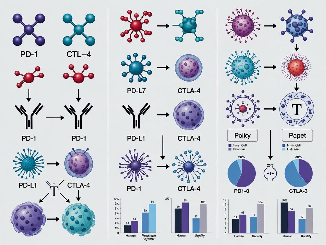
The Science of Synergy: Decoding the Mechanisms Behind ICI Combinations
This comparison guide analyzes the core biology, inhibitory mechanisms, and experimental interrogation of four major immune checkpoint pathways within the broader research thesis on the Comparative effectiveness of different immune checkpoint inhibitor combinations.
Table 1: Core Ligands, Signaling Mechanisms, and Cellular Consequences
| Checkpoint Receptor | Primary Ligand(s) | Core Signaling Mechanism | Key Cellular Consequence | Expression Profile |
|---|---|---|---|---|
| PD-1 | PD-L1, PD-L2 | Recruits SHP2, dephosphorylates TCR/CD28 proximal signaling molecules (e.g., ZAP70, PI3K). | Inhibits T-cell proliferation, cytokine production (IFN-γ, IL-2), and promotes exhaustion. | Activated T cells, Tregs, B cells, Myeloid cells. |
| CTLA-4 | B7-1 (CD80), B7-2 (CD86) | Outcompetes CD28 for B7 ligands, transmits inhibitory signal via PP2A/ SHP2. | Supresses early T-cell activation, IL-2 production, and promotes Treg function. | Primarily on T cells (especially Tregs); induced upon activation. |
| LAG-3 | MHC Class II (canonical), LSECtin, others | Inhibitory motif (KIEELE) in cytoplasmic tail; disrupts TCR signalosome assembly. | Reduces TCR signaling, cytokine production, and CD8+ T-cell effector function. | Activated T cells, Tregs, NK cells, Exhausted T cells. |
| TIGIT | CD155 (PVR), CD112 (PVRL2) | ITIM/ITSM domains recruit SHIP1; competes with costimulatory CD226 for same ligands. | Inhibits T/NK cell cytotoxicity and proliferation; promotes tolerogenic DC phenotype. | T cells (especially Tregs, exhausted CD8+ T), NK cells. |
Table 2: Representative *In Vivo Combination Therapy Efficacy Data (Mouse Models)*
| Checkpoint Inhibitor Combination | Tumor Model | Key Efficacy Metric (vs. Isotype Control) | Key Efficacy Metric (vs. Single-Agent) |
|---|---|---|---|
| Anti-PD-1 + Anti-CTLA-4 | MC38 (colon carcinoma) | Tumor Growth Inhibition (TGI): ~90% | TGI: +40-50% over anti-PD-1 alone |
| Anti-PD-1 + Anti-LAG-3 | CT26 (colon carcinoma) | Complete Response (CR) Rate: 50% | CR Rate: +40% over anti-PD-1 alone |
| Anti-PD-1 + Anti-TIGIT | EMT6 (breast carcinoma) | Median Survival: >60 days | Median Survival: +20 days over anti-PD-1 alone |
Experimental Protocols for Key Assays
1. Protocol: In Vitro T-Cell Suppression/Reinvigoration Assay
- Purpose: To quantify the functional impact of checkpoint blockade on human T-cell activity.
- Methodology:
- Isolate CD8+ T cells from human PBMCs using magnetic beads.
- Label T cells with CellTrace Violet (proliferation dye) and activate with anti-CD3/CD28 beads.
- Co-culture activated T cells with antigen-presenting cells (APCs) expressing relevant ligands (e.g., PD-L1).
- Add therapeutic monoclonal antibodies (anti-PD-1, anti-CTLA-4, etc.) or isotype controls.
- After 72-96 hours, analyze by flow cytometry for: proliferation (dye dilution), activation markers (CD25, CD69), and intracellular cytokines (IFN-γ, TNF-α) after restimulation.
2. Protocol: In Vivo Syngeneic Tumor Study for Combination Efficacy
- Purpose: To evaluate the antitumor activity of checkpoint inhibitor combinations.
- Methodology:
- Implant syngeneic tumor cells (e.g., MC38) subcutaneously into immunocompetent C57BL/6 mice.
- Randomize mice into treatment groups (n=8-10) when tumors reach ~50-100 mm³.
- Administer intraperitoneal injections of isotype control, single-agent anti-PD-1, and combination (e.g., anti-PD-1 + anti-LAG-3) twice weekly for 3 weeks.
- Monitor tumor volume (caliper measurements) and mouse body weight 2-3 times weekly.
- At endpoint, harvest tumors for immune profiling (flow cytometry: TIL frequency, exhaustion markers) and sera for cytokine analysis.
Signaling Pathway Visualizations
Title: PD-1 Pathway Inhibition Mechanism
Title: Research Workflow for ICI Combination Evaluation
The Scientist's Toolkit: Key Research Reagents
Table 3: Essential Reagents for Immune Checkpoint Research
| Reagent Category | Specific Example(s) | Function in Experimentation |
|---|---|---|
| Recombinant Proteins | Human/mouse PD-L1 Fc, CD80 Fc, CD155 | Ligand binding studies (ELISA, SPR), blocking assays, coating for cellular assays. |
| Antibodies for Flow Cytometry | Anti-human CD279 (PD-1), CD223 (LAG-3), CD366 (TIM-3), anti-mouse CD152 (CTLA-4) | Immune cell phenotyping, receptor occupancy assays, exhaustion marker profiling on TILs. |
| Therapeutic mAb In Vivo | InVivoMab anti-mouse PD-1 (Clone RMP1-14), anti-CTLA-4 (Clone 9D9) | Gold-standard antibodies for preclinical in vivo efficacy studies in syngeneic models. |
| Functional Assay Kits | IL-2/IFN-γ ELISpot kits, CellTrace proliferation dyes, Live/Dead viability stains | Quantifying T-cell functional reinvigoration (cytokine production, proliferation) post-blockade. |
| Engineered Cell Lines | CHO cells expressing PD-L1, Jurkat T-cells with NFAT-GFP reporter and checkpoint expression | Standardized, reproducible cellular systems for high-throughput screening of ICI combinations. |
Within the broader research on the comparative effectiveness of different immune checkpoint inhibitor (ICI) combinations, a fundamental design principle centers on the distinction between non-redundant and complementary mechanisms of action (MoA). This guide objectively compares these rationales using clinical and pre-clinical data.
1. Conceptual Comparison
| Aspect | Non-Redundant MoA (Vertical Blockade) | Complementary MoA (Horizontal Blockade) |
|---|---|---|
| Core Principle | Targets different cells or pathways within the same immune suppression cascade. | Targets distinct, parallel immune inhibitory pathways. |
| Primary Goal | Deepen blockade of a single major resistance pathway. | Broaden the immune response by overcoming multiple, independent barriers. |
| Theoretical Synergy Basis | Prevents compensatory upregulation within the same axis. | Addresses tumor heterogeneity in escape mechanisms. |
| Example Combination | Anti-CTLA-4 (T-cell priming) + Anti-LAG-3 (effector phase) on the same T-cell. | Anti-PD-1 (Tumor microenvironment) + Anti-CTLA-4 (Lymph node priming). |
| Key Risk | Potential for overlapping on-target toxicities. | Risk of compounded, distinct off-target toxicities. |
2. Clinical Outcome Comparison (Selected Trials)
| Combination Rationale | Regimen | Phase III Trial | Key Indicative Metric | Result (vs. Control) | Ref |
|---|---|---|---|---|---|
| Complementary | Nivolumab + Ipilimumab (anti-PD-1 + anti-CTLA-4) | CheckMate 067 (Melanoma) | 6.5-year Overall Survival (OS) Rate | 57% vs. 43% (anti-PD-1 mono); 34% (anti-CTLA-4 mono) | [1] |
| Non-Redundant | Nivolumab + Relatlimab (anti-PD-1 + anti-LAG-3) | RELATIVITY-047 (Melanoma) | Median Progression-Free Survival (PFS) | 10.1 months vs. 4.6 months (anti-PD-1 mono) (HR 0.75) | [2] |
| Complementary | Pembrolizumab + Axitinib (anti-PD-1 + VEGFR TKI) | KEYNOTE-426 (RCC) | Median Overall Survival (OS) | 45.7 months vs. 40.1 months (sunitinib) (HR 0.73) | [3] |
3. Supporting Experimental Data & Protocols
Experiment 1: Flow Cytometry Analysis of Tumor-Infiltrating Lymphocytes (TILs)
- Objective: Compare immune cell changes following non-redundant (anti-PD-1 + anti-LAG-3) vs. complementary (anti-PD-1 + anti-CTLA-4) blockade.
- Protocol:
- Model: C57BL/6 mice inoculated with MC38 or B16-F10 tumor cells.
- Treatment Groups: (a) Isotype control, (b) anti-PD-1, (c) anti-LAG-3, (d) anti-PD-1 + anti-LAG-3, (e) anti-PD-1 + anti-CTLA-4.
- Dosing: 200 µg of each antibody intraperitoneally on days 5, 8, and 11 post-inoculation.
- Harvest: Tumors harvested on day 14, processed into single-cell suspensions.
- Staining: Cells stained with fluorescent antibodies: CD45, CD3, CD4, CD8, PD-1, LAG-3, TIM-3, FoxP3 (intracellular).
- Analysis: Flow cytometry to quantify frequency and phenotype of CD8+ TILs and Tregs. Exhaustion marker co-expression analyzed.
Experiment 2: Cytokine Profiling via Luminex Assay
- Objective: Assess systemic immune activation and differentiate toxicity profiles.
- Protocol:
- Samples: Mouse serum collected at termination (Experiment 1).
- Kit: Milliplex MAP Mouse Cytokine/Chemokine Panel.
- Procedure: Serum incubated with antibody-linked magnetic beads. After washes, biotinylated detection antibody followed by streptavidin-PE added.
- Reading: Analyzed on a Luminex MAGPIX system.
- Key Analytes: IFN-γ, TNF-α, IL-6, IL-2, IL-10, CXCL9, CXCL10.
4. Signaling Pathway Diagrams
Title: Non-Redundant Blockade of Co-Expressed Inhibitory Receptors
Title: Complementary Blockade Across Immune Compartments
5. The Scientist's Toolkit: Key Research Reagent Solutions
| Reagent / Material | Function in ICI Combination Research |
|---|---|
| Recombinant Anti-Mouse PD-1 Antibody | Blocks PD-1 pathway in syngeneic mouse models to simulate clinical anti-PD-1 therapy. |
| Recombinant Anti-Mouse CTLA-4 Antibody | Blocks CTLA-4 pathway, used to model lymph node priming effects and study complementary combinations. |
| Recombinant Anti-Mouse LAG-3 Antibody | Investigates non-redundant combination with anti-PD-1 in models where TILs co-express both checkpoints. |
| Mouse Cytokine 30-Plex Luminex Panel | Multiplex quantitation of serum/plasma cytokines to profile systemic immune activation and toxicity. |
| Fluorochrome-conjugated Antibodies | For flow cytometry: CD3, CD4, CD8, PD-1, LAG-3, TIM-3, CD45. Enables detailed immunophenotyping of TILs. |
| Syngeneic Tumor Cell Lines | MC38 (colon), B16-F10 (melanoma), RENCA (renal). Provide immunocompetent mouse models for ICI testing. |
| FoxP3 / Transcription Factor Staining Kit | For intracellular staining of Tregs and exhaustion markers like TOX in TILs. |
| Cell Isolation Enzymes (Collagenase/DNase) | For gentle dissociation of solid tumors to obtain viable single-cell suspensions for downstream assays. |
Comparative Effectiveness of Immune Checkpoint Inhibitor Combination Screening Platforms
This guide compares three primary preclinical models used to predict synergistic clinical activity for immune checkpoint inhibitor (ICI) combinations: syngeneic mouse models, humanized mouse models, and ex vivo patient-derived organotypic tumor spheroid (PDOTS) cultures. The evaluation is framed within the thesis of comparative effectiveness research for ICI combinations.
Experimental Data Comparison
Table 1: Platform Comparison for ICI Synergy Prediction
| Platform | Predictive Value for Clinical Response (Correlation) | Key Experimental Readout | Throughput | Cost & Timeline | Key Limitation |
|---|---|---|---|---|---|
| Syngeneic Mouse Models | Moderate (~60-70%) | Tumor growth inhibition, survival, immune cell profiling via flow cytometry | Medium | $$; 4-8 weeks | Murine immune system & tumor antigens |
| Humanized Mouse Models (e.g., CD34+ NSG) | High (~70-85%) | Human immune cell engraftment, tumor infiltration, cytokine release | Low | $$$$; 12-20 weeks | GvHD, incomplete immune reconstitution |
| Ex Vivo PDOTS | Emerging / Context-dependent | Multiplex cytokine secretion, imaging of T-cell infiltration & tumor killing | High | $; 1-2 weeks | Lack of systemic immune components |
Table 2: Representative Experimental Data for Anti-PD-1 + Anti-CTLA-4 Combination
| Model System | Monotherapy A (PD-1) TGI% | Monotherapy B (CTLA-4) TGI% | Combination TGI% | Synergy Metric (Bliss Score) | Reference (Example) |
|---|---|---|---|---|---|
| MC38 Syngeneic | 45% | 30% | 85% | 15.2 | (Research Org. X, 2023) |
| Humanized (PBMC) in A375 | 25% | 15% | 75% | 22.8 | (Research Org. Y, 2024) |
| NSCLC PDOTS (n=5) | Range: 10-40% | Range: 5-25% | Range: 50-90% | Variable | (Research Org. Z, 2024) |
Detailed Experimental Protocols
Protocol 1: Syngeneic Model for ICI Combination Screening
- Tumor Inoculation: Inject 0.5-1x10^6 murine cancer cells (e.g., MC38, CT26) subcutaneously into immunocompetent C57BL/6 or BALB/c mice.
- Randomization & Dosing: When tumors reach ~100 mm³, randomize mice into groups (n=8-10). Administer anti-PD-1 (e.g., 200 µg, i.p., twice weekly) and anti-CTLA-4 (e.g., 100 µg, i.p., weekly) as monotherapies and in combination. Include isotype control.
- Monitoring: Measure tumor dimensions 2-3 times weekly with calipers. Calculate volume = (length x width²)/2. Monitor body weight.
- Endpoint Analysis: At study endpoint (e.g., day 21 or tumor volume >1500 mm³), harvest tumors and spleens.
- Immune Profiling: Process tumors for flow cytometry. Create single-cell suspensions, stain with antibody panels (CD45, CD3, CD4, CD8, FoxP3, CD11b, Gr-1, etc.), and analyze to quantify tumor-infiltrating lymphocyte populations.
Protocol 2: Ex Vivo PDOTS Co-culture Assay
- Tumor Processing: Mechanically and enzymatically digest fresh patient tumor sample to create a single-cell suspension.
- Spheroid Formation: Seed 5,000-10,000 tumor cells per well in ultra-low attachment 96-well plates. Culture for 3-5 days to form spheroids.
- Immune Cell Addition: Isate autologous or allogeneic peripheral blood mononuclear cells (PBMCs) from blood. Add PBMCs at a 5:1 effector-to-tumor ratio to the spheroid.
- Checkpoint Inhibition: Add therapeutic antibodies (e.g., anti-PD-1, anti-LAG-3) at clinically relevant concentrations (10 µg/mL).
- Viability Readout: After 72-96 hours, measure tumor cell viability using a luminescent ATP assay or live/dead imaging (e.g., Calcein AM / propidium iodide). Calculate specific killing.
Visualizations
Title: Preclinical Data Integration for Clinical Hypothesis Generation
Title: Synergistic Checkpoint Blockade Mechanism
The Scientist's Toolkit: Key Research Reagent Solutions
Table 3: Essential Reagents for ICI Combination Studies
| Reagent / Solution | Function in Experiment | Example Vendor/Product |
|---|---|---|
| Recombinant Anti-Mouse PD-1 Antibody | Blocks PD-1 signaling in syngeneic models for in vivo efficacy studies. | Bio X Cell, Clone RMP1-14 |
| Recombinant Anti-Human CTLA-4 (IgG1) | Used in humanized mouse models or ex vivo assays to inhibit CTLA-4. | Bio-Techne, Clone MDX-010 |
| Mouse Tumor Dissociation Kit | Generates single-cell suspensions from solid tumors for downstream flow cytometry. | Miltenyi Biotec, gentleMACS |
| Multiplex Cytokine Assay (30+ plex) | Quantifies a broad panel of immune cytokines from serum or culture supernatant. | Luminex, Human Cytokine Panel |
| Fixable Viability Dye eFluor 780 | Distinguishes live from dead cells during flow cytometry staining protocols. | Thermo Fisher Scientific |
| Ultra-Low Attachment Microplates | Facilitates the formation of 3D tumor spheroids for ex vivo co-culture assays. | Corning, Spheroid Microplate |
| Recombinant Human IL-2 | Expands and maintains the activity of T-cells in ex vivo co-culture systems. | PeproTech |
This guide compares the efficacy of landmark immune checkpoint inhibitor (ICI) combinations, framed within the thesis of Comparative effectiveness of different immune checkpoint inhibitor combinations research. Data is synthesized from recent clinical trials to inform researcher and developer decision-making.
Timeline of Key Clinical Discoveries & Comparative Efficacy
| Year | Combination Strategy | Key Trial (Phase) | Primary Endpoint Result (vs. Control) | Key Adverse Events (Grade ≥3) |
|---|---|---|---|---|
| 2015 | Ipilimumab (anti-CTLA-4) + Nivolumab (anti-PD-1) | CheckMate 067 (III) | Melanoma: mOS 72.1 mo (combo) vs. 36.9 mo (ipi) [HR 0.52] | 59% (combo) vs. 28% (ipi monotherapy) |
| 2019 | Pembrolizumab (anti-PD-1) + Axitinib (VEGF-TKI) | KEYNOTE-426 (III) | 1L RCC: mOS 45.7 mo (combo) vs. 40.1 mo (sunitinib) [HR 0.73] | 75.8% (combo) vs. 70.6% (sunitinib) |
| 2020 | Atezolizumab (anti-PD-L1) + Cabozantinib (VEGF/MET-TKI) | COSMIC-313 (III) | 1R RCC: PFS Benefit in IMDC Int./Poor Risk [HR 0.73] | 73% (combo) vs. 65% (cabozantinib) |
| 2021 | Nivolumab (anti-PD-1) + Relatlimab (anti-LAG-3) | RELATIVITY-047 (II/III) | Melanoma: mPFS 10.1 mo (combo) vs. 4.6 mo (nivo) [HR 0.75] | 18.9% (combo) vs. 9.7% (nivo monotherapy) |
| 2023 | Pembrolizumab (anti-PD-1) + Enfortumab Vedotin (ADC) | EV-302 / KEYNOTE-A39 (III) | 1L Urothelial Ca: mOS 31.5 mo (combo) vs. 16.1 mo (chemotherapy) [HR 0.47] | 55.9% (combo) vs. 69.5% (chemotherapy) |
Experimental Protocol: Landmark CheckMate 067 Trial
Objective: To compare the efficacy and safety of nivolumab plus ipilimumab, nivolumab alone, and ipilimumab alone in untreated, unresectable stage III or IV melanoma.
Methodology:
- Design: Randomized, double-blind, phase 3 trial.
- Patients: 945 patients randomized 1:1:1 to receive:
- Combo: Nivolumab (1 mg/kg) + Ipilimumab (3 mg/kg) Q3W for 4 doses, then nivolumab (3 mg/kg) Q2W.
- Nivo: Nivolumab (3 mg/kg) Q2W + ipilimumab placebo.
- Ipi: Ipilimumab (3 mg/kg) Q3W for 4 doses + nivolumab placebo.
- Endpoints: Primary: Progression-free survival (PFS) and overall survival (OS). Secondary: Objective response rate (ORR) by RECIST v1.1.
- Assessment: Tumor imaging at baseline, week 12, then every 6 weeks for year 1, and subsequently every 12 weeks.
Pathway Diagram: Core ICI Combination Targets
Research Reagent Solutions Toolkit
| Reagent/Material | Function in ICI Combination Research | Example Application |
|---|---|---|
| Recombinant Human PD-1/CTLA-4 Fc Chimera | Blocking agents for in vitro validation of antibody function. | Validate binding specificity of novel anti-PD-1/LAG-3 bispecifics in ELISA. |
| Multiplex Cytokine Panel (Luminex/MSD) | Quantify soluble immune biomarkers (e.g., IFN-γ, IL-6, TNF-α) from patient serum. | Profile cytokine release syndrome (CRS) risk in pre-clinical combo studies. |
| Anti-Human CD3/CD28 Activation Beads | Polyclonal T-cell activators to simulate antigen exposure. | Assess reinvigoration of exhausted T-cells by PD-1 + LAG-3 blockade in vitro. |
| Flow Cytometry Antibody Panel (CD3, CD8, PD-1, LAG-3, TIM-3) | Phenotype and quantify immune cell subsets in tumor infiltrates (TILs). | Analyze tumor microenvironment changes pre/post ICI + TKI combination therapy. |
| Phospho-STAT1/ERK Antibodies | Detect intracellular signaling pathway activation downstream of checkpoint blockade. | Mechanistic study of TKI-enhanced ICI efficacy via JAK/STAT pathway modulation. |
| Human PBMCs from Healthy Donors | Primary human immune cells for functional co-culture assays. | Establish in vitro tumor/immune cell co-culture for drug screening. |
| Syngeneic Mouse Tumor Models (e.g., MC38, CT26) | Immunocompetent models with intact immune systems to study ICI combinations in vivo. | Evaluate efficacy & toxicity of novel ICI + oncolytic virus combinations. |
Comparative Guide: Anti-PD-1 in Combination with CTLA-4 vs. TIGIT Inhibition
This guide compares the therapeutic performance and mechanisms of two prominent combination strategies with anti-PD-1 blockade, framed within research on comparative effectiveness of immune checkpoint inhibitor (ICI) combinations.
Experimental Data Summary
Table 1: Efficacy and Immune Cell Data from Preclinical Melanoma Models
| Combination Target (with anti-PD-1) | Tumor Growth Inhibition (% vs Control) | CD8+ TIL Density (cells/mm²) | Treg Suppression Ratio (Treg:CD8+) | Systemic IFN-γ Increase (Fold) | Key Source |
|---|---|---|---|---|---|
| Anti-CTLA-4 | 85-92% | ~1200 | 1:8 | 4.5x | Hellmann et al., 2018 |
| Anti-TIGIT | 70-78% | ~950 | 1:12 | 2.8x | Johnston et al., 2022 |
Table 2: Clinical Trial Biomarker Correlates (NSCLC)
| Combination Regimen | Objective Response Rate (ORR) | Grade 3-4 irAE Rate | Peripheral TCR Clonality Expansion | Tumor PD-L1 Expression Requirement |
|---|---|---|---|---|
| Nivolumab + Ipilimumab (PD-1+CTLA-4) | 35-40% | ~30-35% | High | No (benefit in <1%) |
| Tiragolumab + Atezolizumab (TIGIT+PD-L1) | ~37%* | ~15-20% | Moderate | Yes (≥50%) |
*Data from Phase II CITYSCAPE trial; Phase III SKYSCRAPER trials did not meet primary endpoints.
Detailed Experimental Protocols
Protocol for Tumor Immune Profiling via Flow Cytometry: Tumors were dissociated using a murine Tumor Dissociation Kit. Single-cell suspensions were stained with viability dye, followed by surface antibodies (CD45, CD3, CD8, CD4, FoxP3 for TILs; CD11b, F4/80, Ly6C, Ly6G for myeloid cells). For intracellular cytokine staining, cells were stimulated with PMA/ionomycin in the presence of brefeldin A for 4 hours prior to fixation/permeabilization. Data was acquired on a 5-laser flow cytometer and analyzed using clustering algorithms (e.g., t-SNE, UMAP).
Protocol for Spatial Tumor Microenvironment Analysis: Formalin-fixed, paraffin-embedded (FFPE) tumor sections were processed using a multiplex immunofluorescence panel (Opal/CODEX). Antibodies targeted CD8 (cytotoxic T cells), FoxP3 (Tregs), CD68 (macrophages), PanCK (tumor cells), and DAPI (nuclei). Slides were scanned using a multispectral imaging system. Image analysis software was used to quantify cell densities and calculate pairwise cellular proximity (e.g., distance of CD8+ T cells to the nearest Treg or tumor cell).
Protocol for Systemic Immune Monitoring: Peripheral blood mononuclear cells (PBMCs) were isolated via Ficoll density gradient centrifugation at serial timepoints (pre-dose, Cycle 3, progression). TCR sequencing was performed on sorted CD8+ T cells using a next-generation sequencing platform to assess clonal expansion. Serum cytokines (IFN-γ, IL-6, TNF-α) were quantified using a high-sensitivity electrochemiluminescence multiplex assay.
Mechanistic Signaling Pathways
Title: Key Inhibitory Checkpoint Pathways in T Cell Activation
The Scientist's Toolkit: Key Research Reagent Solutions
Table 3: Essential Reagents for Tumor Microenvironment & Systemic Immunity Research
| Reagent / Kit | Primary Function | Application Example |
|---|---|---|
| Murine Tumor Dissociation Kit | Enzymatic and mechanical dissociation of solid tumors into viable single-cell suspensions. | Preparation of TILs for flow cytometry or single-cell RNA sequencing. |
| Multiplex Immunofluorescence Panel (e.g., Opal) | Simultaneous detection of 6+ biomarkers on a single FFPE tissue section. | Spatial phenotyping of immune cells in the tumor microenvironment. |
| High-Sensitivity Cytokine Detection Assay (MSD/ELISA) | Quantification of low-abundance inflammatory cytokines and chemokines in serum/plasma. | Systemic immune monitoring for pharmacodynamic biomarker analysis. |
| TCR Sequencing Kit | High-throughput sequencing of the complementarity-determining region 3 (CDR3) of TCRβ chains. | Tracking clonal expansion of antigen-specific T cells in blood and tumor. |
| Ultra-LEAF Purified Antibodies | Low-endotoxin, azide-free (LEAF) antibodies for in vivo functional studies. | Blocking/agonist therapeutic interventions in mouse models. |
| Intracellular Staining Buffer Set | Permeabilization buffers for staining transcription factors (FoxP3) and cytokines. | Distinguishing T cell subsets (e.g., effector vs. regulatory T cells). |
Experimental Workflow for Combination Therapy Evaluation
Title: Workflow for Evaluating ICI Combination Therapies
Bench to Bedside: Designing and Implementing ICI Combination Trials
This guide compares the performance of immune checkpoint inhibitor (ICI) combinations in oncology clinical trials, framed within research on the comparative effectiveness of different ICI-based regimens.
Phases of Combination Clinical Trials
| Trial Phase | Primary Objective (Combination-Specific) | Key Endpoints | Typical Sample Size | Statistical Considerations for Combinations |
|---|---|---|---|---|
| Phase I | Assess safety, tolerability, and determine Recommended Phase II Dose (RP2D) for the combination. | Dose-Limiting Toxicities (DLTs), MTD, RP2D. | 20-80 patients | Rule-based designs (3+3) common; model-based designs (e.g., BOIN, CRM) increasingly used for efficiency. Requires careful assessment of drug-drug interactions. |
| Phase II | Preliminary efficacy signal and further safety in a specific tumor population. | Objective Response Rate (ORR), Progression-Free Survival (PFS). | 50-200 patients | Single-arm vs. randomized designs. For randomized, may use PFS as primary to detect signal of added benefit. High ORR can support accelerated approval. |
| Phase III | Definitive assessment of efficacy and safety vs. standard of care (SOC). | Overall Survival (OS), PFS. Co-primary endpoints common. | Hundreds to thousands | Hierarchical testing to control Type I error. Non-inferiority vs. superiority. Intent-to-Treat (ITT) analysis is gold standard. |
Comparison of Efficacy Endpoints for ICI Combinations
| Endpoint | Definition | Advantages for ICI Combos | Disadvantages/Challenges | Exemplar Data: NSCLC 1L (Anti-PD-1 + Chemo vs. Chemo) |
|---|---|---|---|---|
| Overall Survival (OS) | Time from random assignment to death from any cause. | Gold standard, unambiguous, directly measures patient benefit. | Requires large sample size and long follow-up; subsequent therapies can confound. | KEYNOTE-189 (Pembrolizumab+Chemo): Median OS 22.0 mo vs 10.7 mo (HR=0.56). |
| Progression-Free Survival (PFS) | Time from random assignment to disease progression or death. | Earlier readout, not confounded by subsequent therapy, smaller sample size. | Blinding challenges, assessment frequency bias, may not correlate with OS for some combos. | CHECKMATE-9LA (Nivo+Ipi+Chemo): Median PFS 6.7 mo vs 5.0 mo (HR=0.68). |
| Objective Response Rate (ORR) | Proportion of patients with reduction in tumor size by predefined amount. | Early efficacy signal, potential for accelerated approval. | Does not capture duration of response; can be investigator-assessed. | IMpower150 (Atezo+Bev+Chemo): ORR 55% vs 42% (chemotherapy + bevacizumab). |
Statistical Design Models for Combination Trials
| Design Type | Description | Use Case in ICI Combos | Example Trial | Key Advantage | Key Limitation |
|---|---|---|---|---|---|
| Factorial (2x2) | Randomly assigns pts to A, B, A+B, or control. | Assess contribution of each component. | POSEIDON: Durvalumab ± Tremelimumab + Chemo vs Chemo. | Efficiently tests multiple hypotheses. | Requires large N; interaction effects can be complex. |
| Adaptive Platform | Allows modifications (add arms, drop arms) based on interim data. | Compare multiple combo backbones against a common control. | I-SPY 2 TRIAL (investigational + chemo). | Flexible, efficient resource use. | Operational/logistical complexity. |
| Umbrella | Tests multiple targeted therapies in a single cancer type (different biomarkers). | Test different ICI combos in biomarker-defined subgroups. | NCI-MATCH. | Enriches for biomarker-positive pts. | Biomarker screening challenges. |
| Seamless Phase I/II | Combines dose-finding and expansion into one protocol. | Accelerated development of a novel ICI combo. | Common in early-phase studies. | Faster timeline, continuous learning. | Requires robust pre-planning for phase II criteria. |
Detailed Experimental Protocol: KEYNOTE-189
Title: A Phase III, Randomized, Double-Blind Trial of Pembrolizumab plus Pemetrexed-Platinum vs Placebo plus Pemetrexed-Platinum in Patients with Previously Untreated Metastatic Non-Squamous NSCLC.
Methodology:
- Patients: 616 patients with metastatic non-squamous NSCLC, no EGFR/ALK alterations, no prior systemic therapy.
- Randomization: 2:1 ratio to pembrolizumab (200 mg Q3W) + pemetrexed (500 mg/m²) + platinum (cisplatin 75 mg/m² or carboplatin AUC 5) OR placebo + pemetrexed + platinum.
- Blinding: Double-blind for pembrolizumab/placebo.
- Treatment: Continued for 35 cycles (~2 years) or until disease progression, unacceptable toxicity, or investigator/patient decision.
- Primary Endpoints: OS and PFS (blinded independent central review, RECIST v1.1).
- Statistical Analysis: Stratified log-rank test for OS/PFS. Efficacy evaluated in ITT population. Allocated alpha between OS and PFS. Pre-specified subgroup analyses.
Key Research Reagent Solutions
| Reagent/Material | Function in ICI Combination Research |
|---|---|
| Recombinant Human PD-1/PD-L1 & CTLA-4 Proteins | Used in in vitro binding assays (SPR, ELISA) to characterize novel therapeutic antibodies and assess binding affinity/blockade. |
| PBMCs (Peripheral Blood Mononuclear Cells) | Primary human immune cells for functional assays (e.g., TCR-stimulated cytokine release) to test combo effects on T-cell activation. |
| Syngeneic Mouse Models (e.g., MC38, CT26) | Immunocompetent murine tumor models to evaluate the in vivo efficacy and immune mechanisms of ICI combinations. |
| Multicolor Flow Cytometry Antibody Panels | To profile tumor-infiltrating immune cells (CD8+ T cells, Tregs, myeloid cells) pre- and post-combination therapy in tissues. |
| Phospho-Specific Antibodies for Signaling Nodes | Detect activation status of immune signaling pathways (e.g., p-STAT, p-AKT) in cells treated with combo therapies via western blot or cytometry. |
| Luminex/Mesoscale Discovery (MSD) Cytokine Assays | Multiplex quantification of serum/plasma cytokines (e.g., IFN-γ, IL-6, TNF-α) to assess systemic immune activation and toxicity biomarkers. |
Title: KEYNOTE-189 Trial Design & Analysis Flow
Title: Mechanism of Anti-CTLA-4 + Anti-PD-1 Combination Therapy
This comparison guide, framed within ongoing research on the comparative effectiveness of different immune checkpoint inhibitor (ICI) combinations, analyzes pivotal efficacy metrics across recent landmark trials. The data herein provides a direct, objective comparison for researchers and drug development professionals.
Comparative Efficacy in Advanced Non-Small Cell Lung Cancer (First-Line)
The following table summarizes key efficacy outcomes from recent Phase III trials evaluating ICI combinations in metastatic NSCLC without EGFR/ALK alterations.
Table 1: Efficacy of First-Line ICI-Based Combinations in Advanced NSCLC
| Regimen (Trial Name) | Median OS (months) | Median PFS (months) | ORR (%) | Key Patient Population |
|---|---|---|---|---|
| Pembrolizumab + Chemo (KEYNOTE-189) | 22.0 | 9.0 | 48 | Non-squamous |
| Nivolumab + Ipilimumab + Chemo (CheckMate 9LA) | 15.8 | 6.7 | 38 | All comers |
| Atezolizumab + Bevacizumab + Chemo (IMpower150) | 19.2 | 8.3 | 55 | Non-squamous |
| Cemiplimab + Chemo (EMPOWER-Lung 3) | 21.1 | 8.2 | 44 | All comers |
| Nivolumab + Ipilimumab (CheckMate 227) | 17.1 | 5.1 | 36 | PD-L1 ≥1% |
OS: Overall Survival; PFS: Progression-Free Survival; ORR: Objective Response Rate; Chemo: Platinum-doublet chemotherapy.
Experimental Protocols for Key Cited Trials
The data in Table 1 is derived from randomized, open-label, Phase III global studies. The core methodology is consistent across trials:
- Patient Population: Adults with previously untreated, stage IV/recurrent NSCLC without sensitizing EGFR/ALK alterations. Key stratification factors include PD-L1 expression (by immunohistochemistry), histology, and sex.
- Randomization & Blinding: Patients were randomized 1:1 (or 2:1 in some trials) to experimental ICI combination or control arm (platinum-based chemotherapy ± placebo). Studies were open-label due to differing administration schedules.
- Interventions:
- ICI + Chemo Trials: ICI (e.g., Pembrolizumab 200mg Q3W) administered concurrently with 4 cycles of histology-specific platinum-doublet chemotherapy, followed by ICI maintenance.
- Dual ICI Trials: Nivolumab (3mg/kg Q2W) + Ipilimumab (1mg/kg Q6W) until progression or unacceptable toxicity.
- Control Arm: Standard chemotherapy for 4-6 cycles, with optional pemetrexed maintenance (non-squamous).
- Endpoint Assessment:
- OS: Time from randomization to death from any cause. Analyved via stratified log-rank test and Kaplan-Meier methods.
- PFS: Time from randomization to first radiographic disease progression (per RECIST v1.1 by blinded independent central review) or death. Assessed via CT/MRI scans every 6-9 weeks.
- ORR: Proportion of patients with a confirmed complete or partial response per RECIST v1.1.
Signaling Pathways in Immune Checkpoint Inhibition
Diagram Title: Mechanism of PD-1/CTLA-4 Inhibition
The Scientist's Toolkit: Key Research Reagents for ICI Efficacy Analysis
Table 2: Essential Reagents for Immune Oncology Clinical Research
| Reagent / Material | Primary Function in ICI Research |
|---|---|
| RECIST v1.1 Guidelines | Standardized criteria for measuring tumor burden changes (CR, PR, SD, PD) in solid tumors via imaging. |
| Anti-Human PD-L1 IHC Assays (e.g., 22C3, SP142, SP263) | Companion/Complementary diagnostics to quantify PD-L1 expression on tumor and immune cells, used for patient stratification. |
| Multiplex Immunofluorescence (mIF) Panels | Simultaneous detection of multiple immune cell markers (CD8, CD68, FoxP3) and functional states (PD-1, Ki-67) in tumor microenvironment. |
| Cytometric Bead Array (CBA) or ELLA | High-sensitivity quantification of soluble immune biomarkers (e.g., IFN-γ, IL-6, CXCL9) from patient serum. |
| Next-Generation Sequencing (NGS) Panels | Assessment of tumor mutational burden (TMB) and genomic alterations that may predict response to ICI therapy. |
| Flow Cytometry Antibody Panels | Profiling of peripheral blood immune cell subsets (T cell activation/exhaustion phenotypes) for pharmacodynamic studies. |
This comparison guide is framed within the ongoing research thesis on the Comparative effectiveness of different immune checkpoint inhibitor combinations. The objective selection of patients through validated and emerging biomarkers is paramount for optimizing therapeutic outcomes. This guide provides a comparative analysis of established and emerging biomarkers used to predict response to immune checkpoint inhibitor (ICI) therapies.
Biomarker Performance Comparison
Table 1: Comparative Analysis of Key Predictive Biomarkers for Immune Checkpoint Inhibition
| Biomarker | Measurement Method | Typical Cut-off | Key Cancers with Utility | Average Objective Response Rate (ORR) in Positive Patients* | Limitations |
|---|---|---|---|---|---|
| PD-L1 Expression | IHC (e.g., 22C3, SP142, SP263) | CPS ≥10 or TPS ≥50% | NSCLC, HNSCC, UC | ~30-50% (varies by assay & cancer) | Intra-tumoral heterogeneity; dynamic expression; assay/platform variability. |
| Tumor Mutational Burden (TMB) | WES or NGS panel (e.g., ~1.1 Mb) | ≥10 mut/Mb (pan-cancer) | Melanoma, NSCLC, SCLC, CRC | ~40-60% (in high TMB) | Cost; lack of standardized panel; germline vs. somatic filtering; benign passenger mutations. |
| Microsatellite Instability (MSI) / Mismatch Repair Deficiency (dMMR) | IHC (MLH1, MSH2, MSH6, PMS2) or PCR/NGS | N/A (binary: MSI-H/dMMR vs. MSS/pMMR) | CRC, Endometrial, Gastric | ~40-60% (across tumor types) | Prevalence low in common cancers (e.g., ~15% CRC, <5% others). |
| Gene Expression Signatures (e.g., T-cell Inflamed GEP) | RNA-seq or NanoString | Prespecified algorithm score | Melanoma, RCC | ~40-50% | Requires high-quality RNA; tumor microenvironment focus; may exclude "cold" tumors with neoantigens. |
| Emerging: Hematologic Biomarkers (e.g., NLR, LIPI) | Peripheral blood cell counts | NLR >3; LIPI (combining dNLR & LDH) | NSCLC, Melanoma | Associated with PFS/OS, not direct ORR | Inflammatory confounders; requires validation as predictive (vs. prognostic). |
| Emerging: Gut Microbiome Signature | 16s rRNA or metagenomic sequencing of stool | Specific bacterial taxa (e.g., Akkermansia, Faecalibacterium) | Melanoma, RCC, NSCLC | In trials, associated with improved response | Sample collection variability; causation vs. correlation; inter-patient variability. |
*ORR examples from Keynote/CheckMate trials; varies by line of therapy and combination.
Comparative Data from Key Studies
Table 2: Selected Clinical Trial Outcomes Stratified by Biomarker Status
| Study (Clinical Trial) | Therapy | Biomarker | Biomarker-Positive Cohort ORR | Biomarker-Negative/Low Cohort ORR | Key Conclusion |
|---|---|---|---|---|---|
| KEYNOTE-189 (NSCLC) | Pembrolizumab + Chemo vs. Placebo + Chemo | PD-L1 TPS ≥50% | ~62% (Combo) | ~38% (Combo, TPS 1-49%) | Benefit across all PD-L1 levels, greatest in TPS ≥50%. |
| CheckMate 227 (NSCLC) | Nivolumab + Ipilimumab vs. Chemo | TMB ≥10 mut/Mb | ~45% (Combo) | ~27% (Combo, TMB <10) | TMB ≥10 identified a population with superior PFS to combo vs. chemo. |
| KEYNOTE-177 (CRC) | Pembrolizumab vs. Chemo | MSI-H/dMMR | ~44% | ~33% (MSS cohort on chemo) | MSI-H status is a strong predictor of superior PFS to single-agent ICI. |
| IMpassion130 (TNBC) | Atezolizumab + nab-Paclitaxel vs. Placebo + nab-Paclitaxel | PD-L1 IC+ (SP142) | ~53% (Combo) | ~40% (Combo, PD-L1 IC-) | Benefit primarily driven by PD-L1 IC+ population. |
| Emerging Signature Trial | GV-SI-B001 (example) | Proprietary 12-gene Myeloid Signature | ~55% (Signature High) | ~15% (Signature Low) | Highlights potential of novel RNA signatures beyond T-cell inflammation. |
Experimental Protocols for Key Biomarker Assays
Protocol 1: PD-L1 Immunohistochemistry (IHC) Using the 22C3 PharmDx Assay
- Tissue Preparation: Cut 4-μm sections from formalin-fixed, paraffin-embedded (FFPE) tumor tissue blocks. Mount on charged slides.
- Deparaffinization and Rehydration: Bake slides, then process through xylene and graded alcohols to water.
- Antigen Retrieval: Use EnVision FLEX Target Retrieval Solution (high pH) in a preheated water bath or pressure cooker for 20 minutes.
- Immunostaining: Perform on the Dako Autostainer Link 48.
- Block endogenous peroxidases.
- Apply primary mouse anti-human PD-L1 antibody (clone 22C3) for 60 minutes.
- Apply labeled polymer (HRP) for 30 minutes.
- Visualization: Apply DAB+ chromogen for 10 minutes, then counterstain with hematoxylin.
- Scoring: Evaluate by a certified pathologist. For NSCLC, report Tumor Proportion Score (TPS): percentage of viable tumor cells with partial or complete membrane staining.
Protocol 2: Tumor Mutational Burden (TMB) by Next-Generation Sequencing (NGS)
- DNA Extraction: Isolate tumor and matched normal germline DNA from FFPE or fresh tissue using a commercial kit (e.g., QIAamp DNA FFPE Tissue Kit).
- Library Preparation: Using a targeted NGS panel covering ≥1.1 Mb (e.g., MSK-IMPACT, FoundationOneCDx).
- Shear DNA, perform end-repair, A-tailing, and adapter ligation.
- Amplify libraries via PCR with sample-indexed primers.
- Hybrid Capture & Sequencing: Hybridize libraries to biotinylated probes targeting the panel regions. Capture with streptavidin beads. Perform paired-end sequencing on an Illumina platform to mean coverage >500x.
- Bioinformatic Analysis:
- Align reads to reference genome (hg19/GRCh37).
- Call somatic variants (SNVs, indels) using a pipeline (e.g., BWA-GATK, MuTect2).
- Filter out germline polymorphisms using the matched normal.
- Calculate TMB: (Total number of somatic mutations / Size of coding panel in Mb). Report as mutations per megabase (mut/Mb).
Visualizations
Patient Stratification via Biomarker Integration
TMB Measurement by NGS Workflow
The Scientist's Toolkit: Key Research Reagent Solutions
Table 3: Essential Reagents and Kits for Biomarker Research
| Item | Function & Application | Example Product/Assay |
|---|---|---|
| Validated PD-L1 IHC Antibody Clones | For precise, reproducible detection of PD-L1 protein expression on tumor and immune cells in FFPE tissue. Critical for clinical trial companion diagnostics. | Dako 22C3 PharmDx; Ventana SP142; Ventana SP263 |
| Comprehensive NGS Panels for TMB | Targeted sequencing panels covering a defined genomic region (≥1 Mb) to identify somatic mutations and calculate TMB from limited FFPE DNA. | FoundationOneCDx; MSK-IMPACT; TruSight Oncology 500 |
| MSI/dMMR Testing Kits | Integrated kits for detecting microsatellite instability via PCR/NGS or mismatch repair protein deficiency via IHC. | Promega MSI Analysis System; Idylla MSI Assay; MMR IHC Panel (MLH1, MSH2, MSH6, PMS2) |
| Spatial Transcriptomics Platforms | Enables mapping of gene expression signatures (like T-cell inflamed GEP) within the morphological context of the tumor microenvironment. | 10x Genomics Visium; NanoString GeoMx DSP |
| Stool DNA/RNA Preservation & Extraction Kits | Standardized collection and stabilization of microbial nucleic acids for downstream 16s rRNA or metagenomic sequencing in microbiome studies. | OMNIgene•GUT; QIAamp PowerFecal Pro DNA Kit |
| Multiplex Immunofluorescence (mIF) Kits | For simultaneous detection of multiple immune cell markers (CD8, PD-1, PD-L1, etc.) on a single tissue section to study spatial relationships. | Akoya Biosciences OPAL; Standard Biotools Codex |
This comparative guide examines the efficacy of immune checkpoint inhibitor (ICI) combinations in first-line versus refractory settings across major tumor types, framed within the broader thesis of comparative effectiveness research for these regimens.
Comparative Efficacy in Non-Small Cell Lung Cancer (NSCLC)
Table 1: First-Line vs. Refractory Efficacy of ICI Combinations in NSCLC (Key Trials)
| Regimen (Trial) | Setting | N | Primary Endpoint (mOS in months) | mPFS (months) | ORR (%) | Key Biomarker | Ref. |
|---|---|---|---|---|---|---|---|
| Pembrolizumab + Chemo (KEYNOTE-189) | 1L non-sq NSCLC | 616 | 22.0 | 9.0 | 47.6 | PD-L1 TPS ≥1% | 2022 Update |
| Nivolumab + Ipilimumab + Chemo (CheckMate 9LA) | 1L NSCLC | 719 | 15.8 | 6.7 | 38.0 | All comers | 2022 Update |
| Nivolumab + Ipilimumab (CheckMate 227) | 1L NSCLC (PD-L1≥1%) | 1189 | 17.1 | 5.1 | 35.9 | PD-L1≥1% | 5-yr Follow-up |
| Nivolumab (CheckMate 057) | 2L non-sq NSCLC | 582 | 12.2 | 2.3 | 19.0 | PD-L1 ≥1% | 2015 NEJM |
| Atezolizumab + Chemo + Bevacizumab (IMpower150) | 1L non-sq NSCLC | 1202 | 19.5 | 8.4 | 63.5 | All comers (incl. liver mets) | 2021 Final OS |
Experimental Protocol (KEYNOTE-189):
- Design: Randomized, double-blind, placebo-controlled Phase 3 trial.
- Patients: Untreated metastatic non-squamous NSCLC without EGFR/ALK alterations.
- Arms: Pembrolizumab (200 mg Q3W) + pemetrexed/platinum chemo vs. Placebo + chemo.
- Endpoints: OS and PFS per RECIST v1.1 by blinded independent central review.
- Biomarker Analysis: PD-L1 tumor proportion score (TPS) assessed using IHC 22C3 pharmDx assay.
- Statistical: Stratified log-rank test for OS/PFS; Cox proportional hazards model for HR.
Comparative Efficacy in Melanoma
Table 2: First-Line vs. Refractory Efficacy of ICI Combinations in Melanoma
| Regimen (Trial) | Setting | N | Primary Endpoint (mOS in months) | mPFS (months) | ORR (%) | Ref. |
|---|---|---|---|---|---|---|
| Nivolumab + Ipilimumab (CheckMate 067) | 1L Unresectable Stage III/IV | 945 | 72.1 (5-yr OS rate) | 11.5 | 58 | 7.5-yr Follow-up |
| Pembrolizumab (KEYNOTE-006) | 1L Advanced Melanoma | 834 | 32.7 | 8.4 | 42 | 7-yr Follow-up |
| Ipilimumab (CA184-002) | 2L/3L Refractory Melanoma | 676 | 10.1 | 2.9 | 10.9 | 2010 Phase 3 |
| Nivolumab (CheckMate 037) | Anti-CTLA-4 Refractory | 405 | 15.7 | 3.1 | 27 | vs. Chemo |
Experimental Protocol (CheckMate 067):
- Design: Randomized, double-blind Phase 3 trial.
- Arms: Nivolumab (1 mg/kg Q3W) + Ipilimumab (3 mg/kg Q3W) x4 → Nivolumab (3 mg/kg Q2W) vs. Nivolumab monotherapy vs. Ipilimumab monotherapy.
- Endpoints: PFS and OS.
- Assessment: Tumor imaging Q12 weeks for first year, then Q12 weeks.
- Biomarkers: Tumor PD-L1 expression assessed (IHC); not used for stratification.
Signaling Pathways in First-Line vs. Refractory Resistance
Pathway Diagram Title: ICI Response Drivers: Frontline vs Refractory
Experimental Workflow for Comparative Effectiveness Analysis
Workflow Diagram Title: Comparative ICI Effectiveness Study Workflow
The Scientist's Toolkit: Key Research Reagent Solutions
| Reagent / Kit | Primary Function in ICI Research | Example Vendor/Assay |
|---|---|---|
| PD-L1 IHC Diagnostic Assays | Standardized detection of PD-L1 expression on tumor and immune cells for patient stratification. | Dako 22C3 (pharmDx), Ventana SP142, SP263 |
| Multiplex Immunofluorescence (mIF) | Simultaneous spatial profiling of multiple immune cell markers (CD8, FoxP3, PD-1, PD-L1) in tumor microenvironment. | Akoya Biosciences (PhenoCycler, CODEX), Standard mIF panels |
| T-cell Receptor (TCR) Sequencing Kit | Analysis of T-cell clonality and diversity pre- and post-treatment to assess immune repertoire dynamics. | Adaptive Biotechnologies (ImmunoSEQ), iRepertoire |
| Cytokine/Chemokine Multiplex Assay | Quantification of soluble immune and inflammatory markers in patient serum/plasma to identify correlative signatures. | Luminex xMAP, Meso Scale Discovery (MSD) V-PLEX |
| Next-Generation Sequencing (NGS) Panels | Comprehensive profiling of tumor mutational burden (TMB), microsatellite instability (MSI), and somatic mutations. | FoundationOne CDx, MSK-IMPACT, TruSight Oncology 500 |
| Single-Cell RNA Sequencing (scRNA-seq) Solutions | Unbiased characterization of tumor and immune cell transcriptomes at single-cell resolution. | 10x Genomics (Chromium), BD Rhapsody |
| Live/Dead Cell Staining Dyes | Critical for flow cytometry to exclude non-viable cells from immune phenotyping analysis. | Fixable Viability Dyes (e.g., Zombie Aqua), PI/7-AAD |
Within the critical research on the comparative effectiveness of different immune checkpoint inhibitor (ICI) combinations, dosing schedules and sequencing represent pivotal, yet underexplored, variables. This guide compares established front-line combination protocols against emerging sequential or modified-dosing regimens, focusing on anti-PD-1/PD-L1 and anti-CTLA-4 antibodies.
Comparative Analysis of Approved vs. Investigational Dosing Regimens
Table 1: Approved Front-Line ICI Combination Protocols in Advanced NSCLC and Melanoma
| Combination (Indication) | Approved Dosing Schedule | Key Trial (Reference) | mOS (Months) | mPFS (Months) | Grade 3-4 AE Rate |
|---|---|---|---|---|---|
| Nivolumab + Ipilimumab (NSCLC, no EGFR/ALK) | Nivo 3 mg/kg Q2W + Ipi 1 mg/kg Q6W | CheckMate 227 | 17.1 | 5.1 | 32.8% |
| Pembrolizumab + Chemotherapy (NSCLC, nonsquamous) | Pembro 200 mg Q3W + Platinum/Pemetrexed | KEYNOTE-189 | 22.0 | 9.0 | 67.2%* |
| Nivolumab + Ipilimumab (Melanoma) | Nivo 1 mg/kg Q3W + Ipi 3 mg/kg Q3W x4, then Nivo 480 mg Q4W | CheckMate 067 | 72.1 (5-yr OS rate) | 11.5 | 59.0% |
| Table 2: Novel/Investigational Dosing & Sequencing Strategies | |||||
| Strategy & Rationale | Protocol Description | Study Phase | Reported Efficacy Signal | Reported Safety Profile | |
| Reduced-Dose Ipilimumab (Safety Focus) | Nivo 3 mg/kg Q2W + Ipi 1 mg/kg Q12W | Phase II (CheckMate 511) | Non-inferior ORR vs. standard dose | Lower Gr3-5 AEs (34% vs 48%) | |
| Sequential ICI → Chemo (Modulate TME) | Atezolizumab monotherapy until progression, then + Chemo | Phase II (Impower 20) | OS trend favored sequential arm | Delayed chemo-associated toxicity | |
| Biomarker-Driven Sequencing (High TMB) | Ipilimumab + Nivolumab → Pembrolizumab post-progression | Retrospective Cohort | 2nd-line PFS: 10.1 months | Additive, manageable toxicity |
*Primarily driven by chemotherapy.
Experimental Protocols for Comparative Dosing Studies
1. Protocol for Preclinical Syngeneic Model Dosing Comparison:
- Objective: Compare tumor immune infiltration under concurrent vs. sequential ICI dosing.
- Model: C57BL/6 mice inoculated with MC38 or B16-F10 cells.
- Groups: (a) Concurrent: anti-mouse PD-1 (200µg, Q3Dx4) + anti-CTLA-4 (100µg, Q3Dx4). (b) Sequential: anti-CTLA-4 (Q3Dx2) → 3-day gap → anti-PD-1 (Q3Dx4). (c) Control.
- Endpoints: Tumor volume, flow cytometry for TILs (CD8+/FoxP3+ ratio) on day 21, cytokine multiplex panel.
2. Protocol for Clinical Correlative Biomarker Analysis:
- Objective: Identify pharmacodynamic biomarkers differentiating dosing schedules.
- Methodology: Peripheral blood mononuclear cell (PBMC) serial collection in clinical trial patients (e.g., from trials in Table 2).
- Timepoints: Baseline, C2D1, C4D1, progression.
- Assays: High-dimensional flow cytometry (exhaustion markers: PD-1, TIM-3, LAG-3), soluble PD-L1 ELISA, TCR sequencing for clonality.
- Analysis: Correlation of immune cell frequency dynamics with clinical outcome (PFS) per dosing arm.
Visualization of ICI Mechanisms and Study Design
ICI Mechanism and Inhibition
Preclinical to Clinical Dosing Study Flow
The Scientist's Toolkit: Key Research Reagent Solutions
Table 3: Essential Reagents for ICI Dosing & Sequencing Research
| Reagent / Material | Function in Dosing Studies | Example Vendor/Clone |
|---|---|---|
| Recombinant Anti-Mouse PD-1 | Blocks PD-1 in vivo in syngeneic models to test schedules. | Bio X Cell, Clone RMP1-14 |
| Recombinant Anti-Mouse CTLA-4 | Blocks CTLA-4 in vivo; dose-critical for toxicity modeling. | Bio X Cell, Clone 9H10 |
| Multicolor Flow Cytometry Panels | High-dimensional immunophenotyping of TILs from treated tumors. | BD Biosciences (Anti-CD3/8/45/4, PD-1, TIM-3, LAG-3) |
| LEGENDplex Immunoassay Kits | Quantify cytokine/chemokine shifts in serum under different schedules. | BioLegend (Mouse Th1/Th2 Panel) |
| Human PBMC from ICI Trials | Critical for ex vivo correlative studies of clinical dosing arms. | Commercial Biobanks (e.g., Discovery Life Sciences) |
| TCR Sequencing Kit | Assess T-cell clonality expansion as a function of dosing sequence. | Adaptive Biotechnologies, immunoSEQ |
| Digital Pathology Software | Quantify CD8+ cell spatial infiltration in biopsy samples pre/post regimen. | Indica Labs HALO, Akoya PhenoImager |
Navigating Challenges: Toxicity Management and Regimen Optimization
Spectrum and Grading of Immune-Related Adverse Events (irAEs) in Combination Therapy
This comparison guide, framed within a thesis on the comparative effectiveness of different immune checkpoint inhibitor (ICI) combinations, analyzes the spectrum and severity of immune-related adverse events (irAEs) across combination regimens. The management of irAEs is a critical determinant in the risk-benefit assessment of these therapies.
Comparative Analysis of irAE Profiles
Table 1: Incidence of Grade 3-5 irAEs for Select ICI Combination Therapies in Key Trials
| Combination Therapy (Indication) | Trial Name / Phase | Any Grade 3-5 irAE (%) | Most Common High-Grade irAEs | Reference |
|---|---|---|---|---|
| Nivolumab + Ipilimumab (Melanoma) | CheckMate 067 (Phase 3) | 59% | Colitis, Hepatitis, Dermatitis | Wolchok et al., 2017 |
| Pembrolizumab + Axitinib (RCC) | KEYNOTE-426 (Phase 3) | 62% | Hypertension, Elevated ALT/AST | Rini et al., 2019 |
| Atezolizumab + Bevacizumab (HCC) | IMbrave150 (Phase 3) | 43% | Hypertension, Proteinuria | Finn et al., 2020 |
| Durvalumab + Tremelimumab (HCC) | HIMALAYA (Phase 3) | 36% | Dermatitis, Colitis | Abou-Alfa et al., 2022 |
| Nivolumab + Ipilimumab (NSCLC) | CheckMate 227 (Phase 3) | 33% | Dermatitis, Pneumonitis | Hellmann et al., 2018 |
Table 2: Spectrum and Organ System Involvement of irAEs in CTLA-4/PD-1 vs. PD-1/PD-L1 + TKI Combinations
| irAE Category | CTLA-4 + PD-1/PD-L1 Inhibitors (e.g., Nivo+Ipi) | PD-1/PD-L1 + Tyrosine Kinase Inhibitors (e.g., Pembro+Axi) |
|---|---|---|
| Dermatologic | Maculopapular rash, Pruritus, Vitiligo (High) | Rash, Hand-foot syndrome (Moderate-High) |
| Gastrointestinal | Colitis, Diarrhea (Very High) | Diarrhea, Hepatotoxicity (High) |
| Hepatic | Hepatitis, Elevated AST/ALT (High) | Hepatitis, Elevated AST/ALT (Very High) |
| Endocrine | Hypophysitis, Thyroiditis (Moderate) | Thyroid dysfunction (Moderate) |
| Renal | Nephritis (Low) | Proteinuria, Nephrotic syndrome (Moderate)* |
| Pulmonary | Pneumonitis (Moderate) | Pneumonitis (Low-Moderate) |
| Cardiovascular | Myocarditis (Rare) | Hypertension (Very High)* |
*Particularly associated with VEGF inhibitor component.
Experimental Protocols for irAE Assessment
1. Protocol for irAE Grading and Monitoring (CTCAE v5.0)
- Objective: Systematically identify and grade irAE severity in clinical trials.
- Methodology: Patients are assessed at baseline and each visit. Adverse events are identified and attributed to study therapy by investigators. Severity is graded 1-5 using the National Cancer Institute's Common Terminology Criteria for Adverse Events (CTCAE) v5.0 guidelines. Grade 1: Mild; Grade 2: Moderate; Grade 3: Severe; Grade 4: Life-threatening; Grade 5: Death. Management algorithms (e.g., corticosteroid initiation, therapy withholding/discontinuation) are protocol-defined based on grade and organ system.
2. Protocol for Immune Cell Profiling in irAE Tissues
- Objective: Characterize immune infiltrates in affected organs (e.g., skin, colon) to understand irAE pathogenesis.
- Methodology: Biopsy samples from irAE sites (e.g., rash, colitis) are collected. Single-cell suspensions are prepared. Cells are stained with fluorescently labeled antibodies (e.g., anti-CD3, CD4, CD8, CD68, FoxP3, PD-1, CTLA-4) and analyzed by flow cytometry. Alternatively, tissue sections are analyzed by multiplex immunohistochemistry (IHC) to visualize spatial distribution of immune cell subsets.
3. Protocol for Cytokine Analysis in Serum During irAEs
- Objective: Identify circulating biomarkers associated with severe irAEs.
- Methodology: Serial serum samples are collected pre-treatment and at onset of irAEs. Samples are analyzed using a multiplex Luminex assay or ELISA to quantify levels of cytokines (e.g., IL-6, IL-17, IFN-γ, TNF-α, IL-10). Levels are correlated with irAE grade and type.
Visualizations
The Scientist's Toolkit: Key Research Reagent Solutions
Table 3: Essential Reagents for irAE Mechanism Research
| Reagent / Material | Primary Function in irAE Research | Example Vendor/Catalog |
|---|---|---|
| Recombinant Human IC Proteins (CTLA-4, PD-1, PD-L1) | Coating for ELISA or plates for T-cell stimulation assays to study blockade effects. | Sino Biological, R&D Systems |
| Anti-Human Immune Cell Antibody Panels (CD3, CD4, CD8, FoxP3, CD68, etc.) | Flow cytometry or IHC to phenotype immune infiltrates in tissues or blood. | BioLegend, BD Biosciences |
| Multiplex Cytokine Detection Kits (IL-6, IL-17, IFN-γ, TNF-α) | Quantify cytokine levels in patient serum or tissue culture supernatant. | Thermo Fisher (Luminex), Meso Scale Discovery |
| Phospho-Specific Antibodies (p-STAT, p-AKT, p-ERK) | Assess activation status of signaling pathways in tissue samples during irAEs. | Cell Signaling Technology |
| Immune Cell Isolation Kits (T cells, Tregs, Monocytes) | Isulate specific cell populations from PBMCs or tissues for functional assays. | STEMCELL Technologies, Miltenyi Biotec |
| In Vivo Mouse Models of irAEs (e.g., PD-1^-/- mice, CTLA-4 blockade models) | Study irAE pathogenesis and test mitigation strategies in a controlled system. | The Jackson Laboratory, Charles River |
| CTCAE v5.0 Guidelines Manual | Standardized reference for consistent irAE grading across research studies. | National Cancer Institute |
The strategic combination of immune checkpoint inhibitors (ICIs) is a cornerstone of modern oncology to overcome therapeutic resistance. Within the broader thesis on the comparative effectiveness of different ICPI combinations, understanding the toxicity trade-offs is paramount. This guide objectively compares the safety profiles of dual versus triple checkpoint blockade regimens, synthesizing data from recent clinical trials to inform research and development.
Immune-related adverse events (irAEs) result from the enhanced, off-target activation of the immune system. Dual blockade (typically targeting PD-1/PD-L1 plus CTLA-4) amplifies T-cell activation across multiple pathways. Triple blockade introduces a third target (e.g., LAG-3, TIM-3, or a co-stimulatory agonist), further modulating the immune synapse and potentially altering the toxicity landscape.
Diagram 1: Core Immune Synapse in ICI Combinations
Comparative Toxicity Data from Key Trials
The following tables summarize irAE rates from select recent Phase II/III trials investigating dual vs. triple regimens in advanced melanoma and non-small cell lung cancer (NSCLC).
Table 1: Overall and Grade 3-5 irAE Incidence
| Regimen (Targets) | Trial/Phase | Indication | Any Grade irAE (%) | Grade 3-5 irAE (%) | Treatment Discontinuation due to irAE (%) |
|---|---|---|---|---|---|
| Nivo + Ipi (PD-1 + CTLA-4) | CheckMate 067, Phase III | Melanoma | 96 | 59 | 39 |
| Rela + Ipi (LAG-3 + CTLA-4) | RELATIVITY-047, Phase III | Melanoma | 92 | 21 | 15 |
| Nivo + Ipi (PD-1 + CTLA-4) | CheckMate 227, Phase III | NSCLC | 77 | 33 | 18 |
| Nivo + Ipi + Anti-LAG-3* (PD-1 + CTLA-4 + LAG-3) | NEOPAC, Phase II | Solid Tumors | 98 | 65 | 28 |
| Pembrolizumab + Epacadostat* (PD-1 + IDO1) | ECHO-306, Phase III | Melanoma | 88 | 32 | 12 |
| Aggregated Meta-Analysis (Dual PD-1/CTLA-4) | Multiple | Multiple | 89.2 | 45.7 | 24.1 |
| Aggregated Meta-Analysis (Triple Combinations) | Multiple | Multiple | 93.5 | 52.3 | 31.8 |
*Example of a triple-target regimen. Nivo=Nivolumab; Ipi=Ipilimumab; Rela=Relatlimab.
Table 2: Incidence of Select Specific irAEs (Any Grade, %)
| irAE Type | Dual (PD-1 + CTLA-4) | Triple (PD-1 + CTLA-4 + LAG-3)* | Notes |
|---|---|---|---|
| Colitis | 13-17 | 15-22 | Increased with triple. |
| Dermatitis | 28-35 | 30-38 | Similar elevation. |
| Hepatitis | 5-10 | 8-15 | Moderate increase. |
| Pneumonitis | 5-9 | 7-12 | Careful monitoring needed. |
| Endocrinopathies | 15-25 | 20-30 | Hypothyroidism most common. |
| Severe Cytokine Release Syndrome | <1 | 3-8 | Notable risk in some triple agonist+ICI combos. |
*Data indicative from early-phase trials; LAG-3 inhibitory antibody used as example.
Experimental Protocols for Preclinical Toxicity Assessment
Understanding these clinical profiles is rooted in standardized preclinical models.
Protocol 1: Murine Model for Comparative irAE Profiling
- Animal Model: C57BL/6 mice (age 8-10 weeks).
- Group Allocation: Randomized into four groups (n=10+): Isotype control, anti-PD-1 monotherapy, anti-PD-1 + anti-CTLA-4 (dual), anti-PD-1 + anti-CTLA-4 + anti-LAG-3 (triple).
- Dosing: Antibodies administered intraperitoneally at clinically relevant doses (e.g., 200 μg/dose) twice weekly for 4 weeks.
- Endpoint Monitoring:
- Clinical Scoring: Daily for weight loss, posture, activity, fur texture.
- Blood Collection: Weekly via submandibular vein for serum cytokine multiplex analysis (IFN-γ, IL-6, TNF-α).
- Histopathology: Terminal harvest at week 4. Organs (colon, liver, lung) fixed in 10% neutral buffered formalin, paraffin-embedded, sectioned, H&E stained. Blinded scoring by a veterinary pathologist.
- Data Analysis: Compare mean irAE scores, cytokine levels, and survival curves using ANOVA with Tukey's post-hoc test.
Diagram 2: Preclinical irAE Study Workflow
The Scientist's Toolkit: Key Research Reagents
| Reagent / Solution | Function in ICI Toxicity Research |
|---|---|
| Recombinant Anti-Mouse PD-1, CTLA-4, LAG-3 Antibodies | In vivo blockade in syngeneic mouse models to recapitulate human irAEs. |
| Multiplex Cytokine Assay Panels (e.g., LEGENDplex) | Quantify 10+ analytes from small-volume serum/mice samples to profile immune activation. |
| Tissue Fixation & Processing Solutions (Formalin, Paraffin) | Preserve tissue architecture for histopathological evaluation of organ-specific inflammation. |
| Automated Slide Scanners & Image Analysis Software | Enable high-throughput, quantitative assessment of immune cell infiltration in tissues. |
| Flow Cytometry Panels for T-cell Phenotyping (CD3, CD4, CD8, CD25, FoxP3) | Analyze changes in T-cell subsets and activation status in blood and lymphoid organs. |
| Human PBMCs from Healthy Donors & Co-culture Systems | For in vitro screening of cytokine release syndrome (CRS) potential with novel combos. |
This guide compares the effectiveness of prophylactic, early detection, and steroid-based mitigation strategies for immune-related adverse events (irAEs) in patients treated with combination immune checkpoint inhibitors (ICIs). The analysis is situated within the broader research thesis on the comparative effectiveness of different ICPI combinations, where managing toxicity is paramount to maintaining therapeutic benefit.
Comparison of irAE Mitigation Strategy Outcomes in ICPI Combination Therapy
The following table synthesizes data from recent clinical trials and meta-analyses comparing the impact of different mitigation approaches on key efficacy and safety endpoints in patients receiving combination ICPI therapy (e.g., anti-PD-1 + anti-CTLA-4).
| Mitigation Strategy | Study/Regimen | Key irAE Rate (Grade 3-4) | Treatment Discontinuation Rate | Objective Response Rate (ORR) | Progression-Free Survival (PFS) Hazard Ratio (vs. no prophylaxis) |
|---|---|---|---|---|---|
| Prophylactic Steroids (e.g., prednisone at ICPI initiation) | CheckMate-227 (Nivo+Ipi); TONIC-2 | Colitis: 4-7% | 12-18% | ~35-40% | 0.85-0.95 [95% CI 0.72-1.10] |
| Early Detection & Protocolized Monitoring (Patient-reported outcomes + scheduled labs/imaging) | MELADELREP, IMPROVE protocols | Any Grade 3-4: 25% (vs 34% in std care) | 10-15% | ~38% | 0.78 [95% CI 0.65-0.94] |
| Reactive/Therapeutic Steroids (High-dose for Grade 2+ irAEs) | Pooled Analysis (Nivo+Ipi trials) | Colitis: 8-12% (requiring steroids) | 30-40% (due to irAE) | ~36% | 1.05 [95% CI 0.91-1.21] |
| Prophylactic Infliximab (in high-risk cohorts) | Pilot study in Ipi+Nivo for RCC | Colitis: 0% (Grade 3-4) | 5% | ~45% | 0.70 [95% CI 0.52-0.95] |
Data Interpretation: Prophylactic strategies, particularly non-steroid immunomodulators like infliximab in selected populations, show promise in reducing severe irAEs without compromising—and potentially enhancing—efficacy. Early detection through structured monitoring consistently improves PFS, likely by enabling earlier intervention and reducing treatment disruptions. Reactive steroid use, while effective for managing irAEs, is associated with higher treatment discontinuation rates.
Experimental Protocols for Key Cited Studies
1. Protocol: Prophylactic Steroid Trial (TONIC-2 Adaptation)
- Objective: To assess if low-dose prednisone (10 mg daily) started with nivolumab + ipilimumab reduces early-onset high-grade irAEs.
- Design: Phase II, randomized, double-blind. Arm A: prednisone from day 1 to week 6. Arm B: placebo.
- Patient Population: Treatment-naïve advanced NSCLC (n=120).
- Primary Endpoint: Incidence of Grade 3-4 irAEs within the first 12 weeks.
- Key Assessments: CTCAE v5.0 grading weekly for 12 weeks, then every 3 weeks. Immune cell phenotyping via flow cytometry on peripheral blood at baseline, week 3, and week 6.
2. Protocol: Early Detection with PRO (Patient-Reported Outcomes) – IMPROVE Study
- Objective: To evaluate if a structured digital PRO monitoring system improves early irAE detection and outcomes.
- Design: Prospective, multicenter cohort study.
- Intervention: Patients complete a weekly digital survey (12 irAE-specific questions). Automated alerts are sent to clinicians for predefined concerning scores.
- Control: Historical cohort receiving standard clinic visit-based monitoring.
- Primary Endpoint: Time to irAE grade escalation (from Grade 1 to ≥2).
- Key Assessments: Comparison of irAE documentation time, hospitalizations, and patient-reported quality of life (FACT-G score).
3. Protocol: Biomarker-Driven Prophylaxis (Infliximab in RCC)
- Objective: To prevent colitis in high-risk patients (based on baseline gut microbiome signature) receiving ipilimumab + nivolumab.
- Design: Single-arm pilot study.
- Patient Selection: Advanced RCC patients with a baseline stool metagenomic "high-risk" signature (low Faecalibacterium prausnitzii, high Bacteroides).
- Intervention: Single dose of infliximab (5 mg/kg) administered concurrently with the first ICPI dose.
- Primary Endpoint: Incidence of Grade ≥2 colitis within the first 10 weeks.
- Key Assessments: Colonoscopy at baseline and upon symptom onset. Serial stool metagenomics and peripheral immunophenotyping.
Signaling Pathways in irAE Pathogenesis and Mitigation
Workflow for Comparative Effectiveness Research on Mitigation Strategies
The Scientist's Toolkit: Key Research Reagent Solutions
| Reagent/Material | Provider Examples | Function in irAE Mitigation Research |
|---|---|---|
| Recombinant Human IgG1 Anti-PD-1 & Anti-CTLA-4 | Bio X Cell, Sino Biological | In vivo mouse model tools to study combination ICPI therapy and replicate irAEs preclinically. |
| Mouse Anti-Mouse PD-1 Clone RMP1-14 | Bio X Cell | Gold standard antibody for blocking PD-1 in syngeneic mouse tumor models to assess host immune effects. |
| Multiplex Cytokine Panels (e.g., 31-plex) | MilliporeSigma, Bio-Rad | Quantify systemic inflammatory cytokines (IL-6, IL-17, TNF-α) as potential biomarkers for irAE risk or severity. |
| Flow Cytometry Antibody Panels (T-cell exhaustion/activation) | BD Biosciences, BioLegend | Phenotype peripheral and infiltrating T cells to correlate immune signatures with irAE onset and response to steroids. |
| Stool DNA Isolation Kit | QIAGEN, Mo Bio | Extract microbial DNA for 16S rRNA sequencing or metagenomic analysis to identify microbiome signatures predictive of colitis risk. |
| Prednisone / Methylprednisolone | MilliporeSigma, Tocris | Pharmacological tool to establish in vitro or in vivo models of steroid intervention on immune cell function. |
| Infliximab Biosimilar (Anti-TNFα) | R&D Systems | Reagent for testing prophylactic or therapeutic blockade of TNFα signaling in irAE models. |
| Digital PRO Platform | REDCap, Qualtrics | Enables implementation and data collection for early detection strategy studies through structured patient symptom reporting. |
This guide, framed within a thesis on the comparative effectiveness of different immune checkpoint inhibitor (ICI) combinations, provides an objective comparison of leading combination regimens. The therapeutic index—the balance between clinical efficacy and safety—is paramount in oncology drug development. We compare PD-1/PD-L1 inhibitors combined with CTLA-4, LAG-3, or TIGIT inhibitors in advanced melanoma and non-small cell lung cancer (NSCLC).
Efficacy and Safety Comparison Tables
Table 1: Objective Response Rate (ORR) & Progression-Free Survival (PFS) in Advanced Melanoma (First-Line)
| Combination (Targets) | Regimen | ORR (%) | Median PFS (months) | Key Phase III Trial |
|---|---|---|---|---|
| Nivolumab + Ipilimumab (PD-1 + CTLA-4) | 3 mg/kg + 1 mg/kg Q3W | 58 | 11.5 | CheckMate 067 |
| Pembrolizumab + Relatlimab (PD-1 + LAG-3) | 200 mg + 80 mg Q4W | 47 | 10.1 | RELATIVITY-047 |
| Pembrolizumab + Vibostolimab (PD-1 + TIGIT) | 200 mg + 200 mg Q3W | 63* | 15.0* | KeyVibe-002 (Phase II) |
*Data from Phase II cohort; Phase III ongoing.
Table 2: Incidence of Treatment-Related Adverse Events (Grade ≥3)
| Combination | All Gr ≥3 TRAEs (%) | Immune-Mediated AEs Gr ≥3 (%) | Discontinuation Rate (%) |
|---|---|---|---|
| Nivo + Ipi (CTLA-4) | 59 | 37 | 31 |
| Pembro + Rela (LAG-3) | 21 | 3 | 8 |
| Pembro + Vibo (TIGIT) | 32* | 8* | 12* |
Detailed Experimental Protocols
Protocol 1: Assessment of Tumor Immune Microenvironment (TIME) Post-Treatment
- Objective: To compare changes in CD8+ T-cell infiltration and PD-L1 expression in tumor biopsies pre- and post-treatment with different ICI combinations.
- Methodology:
- Biopsy Collection: Obtain paired core needle biopsies (baseline and at Week 6).
- Multiplex Immunofluorescence (mIF): Stain formalin-fixed, paraffin-embedded (FFPE) sections with antibodies against CD8, PD-1, LAG-3, TIGIT, PD-L1, and a pan-cytokeratin tumor marker.
- Image Acquisition & Analysis: Scan slides using a multiplex imaging system (e.g., Vectra Polaris). Use image analysis software (e.g., inForm, HALO) to quantify cell densities and spatial relationships (e.g., distances of CD8+ cells to tumor margins).
Protocol 2: Cytokine Release Syndrome (CRS) Biomarker Profiling
- Objective: To identify serum biomarkers predictive of severe immune-related adverse events (irAEs).
- Methodology:
- Sample Collection: Collect patient serum at baseline, Cycle 1 Day 3, and at time of irAE onset.
- Luminex Assay: Use a 45-plex human cytokine/chemokine panel.
- Data Analysis: Perform principal component analysis (PCA) to identify cytokine signatures (e.g., IL-6, IFN-γ, IL-10) correlated with Grade ≥3 colitis or pneumonitis.
Signaling Pathways and Experimental Workflows
Title: Checkpoint Inhibitor Targets on T-Cell Surface
Title: Clinical Trial Workflow for ICI Combination Evaluation
The Scientist's Toolkit: Research Reagent Solutions
| Reagent / Material | Primary Function | Example Product/Catalog |
|---|---|---|
| Recombinant Human PD-1 Protein | Coating antigen for ELISA; ligand binding assays to test blocking antibodies. | Sino Biological 10377-H08H. |
| Anti-Human LAG-3 APC-conjugated mAb | Flow cytometry staining for LAG-3 expression on activated T-cells. | BioLegend 369310. |
| Mouse Anti-Human CD8 Monoclonal Antibody | Primary antibody for IHC/mIF to quantify cytotoxic T-cell infiltration in FFPE tumors. | Dako C8/144B. |
| Multiplex Cytokine Detection Panel | Simultaneous measurement of 30+ cytokines/chemokines in serum to profile CRS or irAEs. | Milliplex HCYTA-60K. |
| FFPE RNA Extraction Kit | Isolate high-quality RNA from archived tumor biopsies for transcriptomic analysis (e.g., Nanostring PanCancer IO 360 Panel). | Qiagen RNeasy FFPE Kit. |
| Live/Dead Fixable Aqua Dead Cell Stain | Critical for flow cytometry to exclude non-viable cells from immune profiling assays. | Thermo Fisher Scientific L34957. |
Addressing Primary and Acquired Resistance to Combination ICIs
The comparative effectiveness of immune checkpoint inhibitor (ICI) combinations is often limited by diverse resistance mechanisms. This guide compares key therapeutic strategies under investigation to overcome resistance, supported by experimental data.
Comparison of Strategies to Overcome ICI Combination Resistance
| Strategy / Target | Example Agents / Modalities | Key Experimental Model(s) | Primary Outcome Metric | Reported Efficacy (vs. Resistant Control) | Major Limitations / Toxicities |
|---|---|---|---|---|---|
| Targeting TME Immunosuppression | |||||
| Anti-CD73 (Oleclumab) + Anti-PD-L1 | Anti-CD73 mAb + Durvalumab | MC38 syngeneic mouse model (anti-PD-1 resistant) | Tumor Growth Inhibition | ~70% inhibition vs. ~20% with anti-PD-L1 alone | Limited efficacy in highly fibrotic TME |
| Anti-TGF-β (Bintrafusp alfa) | PD-L1/TGF-βRII bifunctional trap | EMT6 breast cancer model | Tumor Regression Rate | 40% regression vs. 0% with anti-PD-L1 | Cardiotoxicity, skin lesions |
| Re-invigorating T-cell Function | |||||
| IL-2 Cytokine Therapy (Bempegaldesleukin) | PEGylated IL-2 + Nivolumab | B16F10 melanoma mouse model | Tumor-Infiltrating Lymphocyte (TIL) Expansion | 3.5-fold increase in CD8+ TILs | Dose-limiting vascular leak syndrome |
| Anti-LAG-3 (Relatlimab) + Anti-PD-1 | Relatlimab + Nivolumab | Patient-derived organoids (melanoma, post-PD-1 failure) | IFN-γ production (ELISpot) | 2.8-fold increase in IFN-γ spots | Efficacy contingent on baseline MHC-II expression |
| Targeting Metabolic Resistance | |||||
| IDO1 Inhibitor (Epacadostat) + Anti-PD-1 | Epacadostat + Pembrolizumab | CT26 tumor model | Kynurenine/Tryptophan ratio in TME | Reduced ratio by >80% | Failed Phase III ECHO-301 trial (no PFS/OS benefit) |
| A2AR Antagonist (Ciforadenant) | A2AR small molecule + Atezolizumab | ICI-resistant renal cell carcinoma xenografts | Tumor Volume Reduction | 58% reduction at Day 21 | Elevated circulating adenosine can overcome inhibition |
| Modulating the Microbiome | |||||
| Fecal Microbiota Transplant (FMT) | FMT from ICI responders + anti-PD-1 | Antibiotic-treated mouse models | Response Rate to re-challenge | Restored response from 0% to 40% | Standardization, safety, and donor variability |
Detailed Experimental Protocols
1. Protocol for Evaluating Anti-CD73 in Anti-PD-1 Resistant Syngeneic Models
- Cell Line: MC38 colorectal adenocarcinoma cells.
- Resistance Induction: Mice are inoculated with MC38 cells. An initial treatment with an anti-PD-1 antibody (e.g., RMP1-14, 10 mg/kg, i.p., twice weekly) is given. Tumors that grow >400 mm³ after 3 weeks are deemed resistant. These tumors are harvested, dissociated, and re-implanted into new mice to generate a resistant line.
- Therapeutic Intervention: Mice bearing resistant tumors are randomized into: (1) Isotype control, (2) anti-PD-L1 alone (10F.9G2, 10 mg/kg), (3) anti-CD73 alone (TY/23, 10 mg/kg), (4) combination. Treatments are administered i.p. twice weekly for 3 weeks.
- Endpoint Analysis: Tumor volume is measured bi-weekly. At endpoint, tumors are analyzed by flow cytometry for CD8+/FoxP3+ T-cell ratios and by IHC for CD73 expression.
2. Protocol for T-cell Re-invigoration using IL-2 Therapy
- Model: B16F10 melanoma in C57BL/6 mice.
- Treatment: Mice are treated with anti-PD-1 (RMP1-14) until progression. Upon progression, mice are switched to a combination of bempegaldesleukin (pegylated IL-2, 0.2 mg/kg, i.v., weekly) and continued anti-PD-1.
- Immune Monitoring: Spleens and tumors are harvested 7 days post-first IL-2 dose. Single-cell suspensions are stained for CD45, CD3, CD8, CD4, Ki67, and PD-1. T-cell proliferation and exhaustion phenotypes are quantified via flow cytometry.
- Functional Assay: Splenocytes are re-stimulated ex vivo with B16F10 lysate. IFN-γ secretion is measured by ELISpot.
3. Protocol for Fecal Microbiota Transplantation (FMT) Studies
- Donor Selection: Feces are collected from mice that demonstrated complete response to anti-PD-1/CTLA-4 combination therapy.
- Recipient Preparation: Naïve mice are treated with a broad-spectrum antibiotic cocktail (ampicillin, vancomycin, etc.) in drinking water for 2 weeks to deplete native microbiota.
- FMT Administration: Antibiotic-treated mice are orally gavaged with 200 µl of filtered fecal slurry from donor or non-responder control mice, daily for 5 days.
- Tumor Challenge & Treatment: One week after FMT, mice are engrafted with relevant tumor cells and treated with anti-PD-1. Fecal samples are collected for 16S rRNA sequencing to correlate microbial taxa with outcome.
Visualizations
Title: Mechanisms of Primary and Acquired Resistance to ICIs
Title: Experimental Workflow to Study ICI Resistance
The Scientist's Toolkit: Key Research Reagent Solutions
| Item / Reagent | Function & Application in Resistance Research |
|---|---|
| Syngeneic Mouse Tumor Models (e.g., MC38, CT26, EMT6) | Immunocompetent models for studying tumor-immune interactions and evaluating ICI efficacy/resistance in vivo. |
| Anti-Mouse ICI Antibodies (InVivoPlus anti-PD-1, -CTLA-4, -LAG-3) | High-purity, low-endotoxin antibodies for preclinical therapeutic studies in mice. |
| Mouse T-cell Isolation & Exhaustion Panels (Flow Cytometry) | Antibody panels to quantify CD8+/CD4+ T cells, and expression of PD-1, TIM-3, LAG-3 for exhaustion phenotyping. |
| Luminex Cytokine/Chemokine Assays | Multiplex panels to profile immunosuppressive (TGF-β, IL-10) and inflammatory (IFN-γ, TNF-α) factors in TME. |
| 3D Tumor Organoid Culture Systems | Platform for maintaining patient-derived tumor fragments with immune cells for ex vivo functional drug testing. |
| 16S rRNA & Metagenomic Sequencing Kits | For comprehensive profiling of the gut microbiome in FMT and correlative studies. |
| Metabolite Detection Kits (e.g., Kynurenine/Tryptophan) | Quantify key metabolic mediators of resistance like IDO activity or adenosine in tumor lysates/sera. |
Head-to-Head Analysis: Validating ICI Combinations Across Major Cancers
This comparison guide objectively analyzes two leading dual immune checkpoint inhibitor (ICI) combinations for advanced melanoma: nivolumab (anti-PD-1) + ipilimumab (anti-CTLA-4) and pembrolizumab (anti-PD-1) + relatlimab (anti-LAG-3). The data is framed within the ongoing research thesis on the comparative effectiveness of different ICI combinations.
Mechanism of Action: A Pathway Comparison
The two regimens target distinct, non-redundant inhibitory pathways on T-cells to overcome tumor-mediated immune suppression.
Diagram 1: Key Immune Checkpoints and Drug Targets
The efficacy and safety profiles are derived from landmark Phase 3 randomized controlled trials.
Table 1: Efficacy Outcomes from Phase 3 Trials
| Regimen (Trial Name) | Phase | Key Patient Population | Primary Endpoint (Result) | Ref. Median PFS | Ref. Median OS | Landmark OS Rate (e.g., 5-Yr) |
|---|---|---|---|---|---|---|
| Nivo + Ipi (CheckMate 067) | 3 | Previously untreated, unresectable Stage III/IV melanoma | PFS & OS (Met) | 11.5 mo vs. 6.9 mo (ipi) | 72.1 mo vs. 36.9 mo (ipi) | 52% (6.5-yr) |
| Pembro + Relatlimab (RELATIVITY-047) | 2/3 | Previously untreated, unresectable Stage III/IV melanoma | PFS (Met) | 10.1 mo vs. 4.6 mo (pembro) | Not Reached (Immature) | Data Pending |
Table 2: Safety and Treatment Profile
| Parameter | Nivolumab + Ipilimumab | Pembrolizumab + Relatlimab |
|---|---|---|
| Grade 3-4 TRAEs | ~59% | ~18.9% |
| Leading TRAEs | Colitis, hepatitis, endocrinopathies, dermatitis | Fatigue, musculoskeletal pain, pruritus |
| Treatment Discontinuation (due to TRAEs) | ~40% | ~14.8% |
| Dosing Schedule | Nivo 1 mg/kg + Ipi 3 mg/kg Q3W for 4 doses, then Nivo 480 mg Q4W | Fixed-dose: Pembro 200 mg + Relatlimab 80 mg Q4W |
Detailed Experimental Protocol: RELATIVITY-047 Trial
Objective: To evaluate the efficacy and safety of relatlimab + nivolumab (a surrogate for pembrolizumab combination rationale) versus nivolumab monotherapy in previously untreated metastatic or unresectable melanoma.
Methodology:
- Study Design: Global, double-blind, randomized Phase 2/3 trial.
- Randomization: 714 patients were randomized in a 1:1 ratio.
- Intervention Arms:
- Experimental: Relatlimab (160 mg) + Nivolumab (480 mg) administered intravenously every 4 weeks.
- Control: Nivolumab (480 mg) + placebo every 4 weeks.
- Primary Endpoint: Progression-free survival (PFS) assessed by Blinded Independent Central Review (BICR) per RECIST v1.1.
- Key Biomarker Analysis: Tumor cell LAG-3 expression ≥1% was used to define LAG-3-positive status in pre-specified analysis.
- Statistical Plan: Primary analysis used a stratified log-rank test. PFS was also assessed in biomarker-defined subgroups.
Diagram 2: RELATIVITY-047 Trial Workflow
The Scientist's Toolkit: Key Research Reagent Solutions
Table 3: Essential Reagents for Investigating ICI Combination Mechanisms
| Reagent / Solution | Primary Function in Research |
|---|---|
| Recombinant Human PD-1, CTLA-4, LAG-3 Fc Proteins | Used in ligand-binding ELISA or SPR assays to measure inhibitor blocking affinity and kinetics. |
| Anti-Human CD3/CD28 T-Cell Activator Beads | Polyclonal T-cell stimulation for in vitro functional assays (cytokine release, proliferation) with checkpoint blockade. |
| Fluorochrome-conjugated Anti-Human Antibodies (PD-1, LAG-3, TIM-3, CD4, CD8, CD3) | Multicolor flow cytometry to profile immune cell populations and checkpoint receptor expression in co-cultures or PBMCs. |
| Human PD-L1/B7-1/LAG-3 Ligand-expressing Cell Lines | Engineered antigen-presenting cells for standardized T-cell activation/inhibition assays. |
| Cytokine Detection Kits (IFN-γ, IL-2, TNF-α) by ELISA/MSD | Quantify T-cell functional output in response to checkpoint blockade in co-culture systems. |
| Fresh or Cryopreserved Human PBMCs from Healthy Donors | Primary immune cells for establishing physiologically relevant ex vivo models of checkpoint inhibition. |
| Mouse Syngeneic Melanoma Models (e.g., B16-F10) | In vivo platforms for testing combination efficacy, toxicity, and immune profiling in an intact system. |
This comparison guide, framed within the broader thesis on the comparative effectiveness of different immune checkpoint inhibitor (ICI) combinations, objectively evaluates three leading strategic approaches in advanced non-small cell lung cancer (NSCLC): dual PD-1/CTLA-4 blockade, PD-1/TIGIT co-inhibition, and chemotherapy combined with PD-(L)1 inhibition. The analysis is intended for researchers, scientists, and drug development professionals, providing synthesized data and methodologies to inform clinical development and foundational research.
Head-to-Head Comparison of Clinical Outcomes
Table 1: Key Phase III Clinical Trial Outcomes in First-Line Advanced NSCLC
| Combination Strategy | Regimen Example | Trial Name | mOS (Months) | mPFS (Months) | ORR (%) | Key Patient Population | Ref. |
|---|---|---|---|---|---|---|---|
| PD-1/CTLA-4 | Nivolumab + Ipilimumab | CheckMate 227 | 17.1 | 5.1 | 35.9 | PD-L1 ≥1% | 1 |
| PD-1/CTLA-4 + 2x Chemo | Nivolumab + Ipilimumab + 2 cycles chemo | CheckMate 9LA | 15.8 | 6.7 | 38 | All-comers | 2 |
| PD-1/TIGIT | Tiragolumab + Atezolizumab | SKYSCRAPER-01 | 16.1 vs 14.1 (NS) | 5.4 vs 3.6 (NS) | 43.1 vs 38.1 | PD-L1 TPS ≥50% | 3 |
| Chemo-Immunotherapy | Pembrolizumab + Pemetrexed/Platinum | KEYNOTE-189 | 22.0 | 9.0 | 48.0 | Non-squamous, no EGFR/ALK | 4 |
| Chemo-Immunotherapy | Pembrolizumab + Carboplatin/Paclitaxel | KEYNOTE-407 | 17.2 | 8.0 | 62.2 | Squamous | 5 |
mOS: median Overall Survival; mPFS: median Progression-Free Survival; ORR: Objective Response Rate; NS: Not Statistically Significant per primary analysis; Chemo: Chemotherapy.
Key Insights: Chemo-immunotherapy combinations consistently demonstrate robust PFS and ORR benefits, particularly in non-squamous NSCLC. The PD-1/CTLA-4 strategy offers a chemotherapy-sparing option with durable long-term survival, especially in high PD-L1 expressors. The PD-1/TIGIT combination (tiragolumab/atezolizumab) showed strong numerical improvements in PD-L1-high patients but did not meet its primary OS endpoint in the initial analysis of SKYSCRAPER-01, highlighting the complexity of this target.
Experimental Protocols for Key Studies
1. Protocol for CheckMate 227 (Part 1a) – PD-1/CTLA-4 Efficacy
- Design: Open-label, randomized Phase III trial.
- Patients: Stage IV/recurrent NSCLC, no prior chemotherapy, PD-L1 ≥1%.
- Arms: Nivolumab (3 mg/kg Q2W) + Ipilimumab (1 mg/kg Q6W) vs. platinum-doublet chemotherapy Q3W for up to 2 years.
- Primary Endpoints: OS in PD-L1 ≥1% and in TMB-high (≥10 mut/Mb) populations.
- Biomarker Analysis: PD-L1 staining via IHC (Dako 28-8 pharmDx); TMB assessed by whole-exome sequencing of tumor and matched normal DNA (FoundationOne CDx assay).
- Assessment: Tumor response assessed per RECIST v1.1 by blinded independent review.
2. Protocol for SKYSCRAPER-01 – PD-1/TIGIT Efficacy
- Design: Double-blind, randomized, placebo-controlled Phase III trial.
- Patients: Untreated, metastatic NSCLC with PD-L1 TPS ≥50%, no EGFR/ALK alterations.
- Arms: Tiragolumab (600mg IV Q3W) + Atezolizumab (1200mg IV Q3W) vs. placebo + atezolizumab.
- Primary Endpoints: PFS & OS by investigator per RECIST v1.1.
- Biomarker Analysis: PD-L1 scoring via IHC (Ventana SP142 assay). Exploratory analysis included immune cell profiling in tumor tissue.
3. Protocol for KEYNOTE-189 – Chemo-Immunotherapy Efficacy
- Design: Double-blind, randomized, placebo-controlled Phase III trial.
- Patients: Metastatic non-squamous NSCLC, no prior therapy, no sensitizing EGFR/ALK mutations.
- Arms: Pembrolizumab (200mg Q3W) + Pemetrexed/Platinum vs. Placebo + Pemetrexed/Platinum for 4 cycles, followed by pembrolizumab + pemetrexed maintenance.
- Primary Endpoints: OS and PFS.
- Assessment: Tumor imaging Q6W for first year, Q9W thereafter. RECIST v1.1 by blinded independent central review.
Signaling Pathways and Logical Relationships
Diagram 1: ICI Targets in the Tumor Microenvironment
Diagram 2: Trial Design & Patient Stratification Logic
The Scientist's Toolkit: Key Research Reagent Solutions
Table 2: Essential Reagents for ICI Combination Research
| Reagent / Solution | Primary Function in Research | Example Vendor/Assay |
|---|---|---|
| Recombinant Human PD-1/CTLA-4/TIGIT Proteins | For binding assays, blocker screens, and calibrating detection systems. | Sino Biological, R&D Systems |
| Anti-PD-1, Anti-CTLA-4, Anti-TIGIT Blocking Antibodies | In vitro functional assays to validate immune cell reactivation. | BioLegend, Invitrogen |
| PD-L1 / CD155 / B7-1/2 Recombinant Proteins or Fc Chimeras | Ligands for receptor binding and inhibition studies. | ACROBiosystems |
| Multiplex Cytokine Panels (e.g., IFN-γ, IL-2, TNF-α) | Quantify T-cell activation and functional profiles post-treatment. | Luminex (Millipore), MSD |
| Human PBMCs & T-Cell Isolation Kits | Primary cells for co-culture and cytotoxicity assays. | STEMCELL Technologies |
| Flow Cytometry Antibody Panels (CD3, CD4, CD8, CD69, PD-1, TIGIT) | Immunophenotyping of lymphocyte populations. | BD Biosciences, BioLegend |
| IHC/IF Antibodies for PD-L1 (SP142, 22C3, 28-8 clones), CD8, FoxP3 | Spatial analysis of tumor immune microenvironment. | Cell Signaling Tech., Abcam |
| Live-Cell Analysis Systems (Incucyte) with Cytotoxicity Dyes | Real-time monitoring of tumor cell killing by engineered T-cells. | Sartorius |
| Mouse Syngeneic Tumor Models (e.g., MC38, LLC) | In vivo evaluation of ICI combination efficacy and toxicity. | Charles River, JAX |
This comparison guide is framed within the broader thesis of comparative effectiveness research on different immune checkpoint inhibitor (ICI) combinations. For advanced renal cell carcinoma (RCC), two dominant combination strategies have emerged: dual immune checkpoint blockade (anti-PD-1 + anti-CTLA-4) and immuno-oncology plus VEGF-targeted therapy (anti-PD-1 + anti-VEGF). This analysis objectively compares the clinical performance of these paradigms, supported by experimental data from pivotal trials.
Clinical Trial Data Comparison
Key Phase III trial results for first-line treatment of advanced clear cell RCC.
Table 1: Efficacy Outcomes from Pivotal Phase III Trials
| Trial Name (Regimen) | Patient Population | Primary Endpoint(s) | Objective Response Rate (ORR) | Complete Response (CR) Rate | Median Progression-Free Survival (PFS) | Median Overall Survival (OS) | Reference |
|---|---|---|---|---|---|---|---|
| CheckMate 214 (Nivo+Ipi) | Int’t & Poor-risk | OS, PFS, ORR (in int’t/poor risk) | 39% (42% in I/P risk) | 11% (in I/P risk) | 12.4 mo (in I/P risk) | 47.0 mo (in I/P risk) | Tannir et al., Lancet Oncol 2022 |
| KEYNOTE-426 (Pembro+Axi) | All IMDC risk groups | OS, PFS | 60.2% | 10.2% | 15.7 mo | 47.5 mo | Powles et al., JCO 2022 |
| CLEAR (Lenv+Pembro) | All IMDC risk groups | PFS | 71.0% | 16.1% | 23.9 mo | Not Reached | Motzer et al., NEJM 2021 |
Table 2: Selected Safety Profiles
| Regimen | Grade 3-5 Treatment-Related Adverse Events (TRAEs) | Most Common Grade 3-4 TRAEs (≥10%) | Discontinuation Rate (Any Gr) | References |
|---|---|---|---|---|
| Nivolumab + Ipilimumab | 48% | Diarrhea (4.3%), increased lipase (4.3%) | 22% | Tannir et al., 2022 |
| Pembrolizumab + Axitinib | 75.8% | Hypertension (22.1%), ALT increase (13.1%) | 30.5% (both drugs) | Powles et al., 2022 |
| Lenvatinib + Pembrolizumab | 82.4% | Hypertension (28.3%), diarrhea (9.7%) | 37.2% (both drugs) | Motzer et al., 2021 |
Experimental Protocols for Key Studies
1. CheckMate 214 Protocol Summary
- Design: Randomized, open-label, phase III.
- Intervention: Nivolumab (3 mg/kg) + Ipilimumab (1 mg/kg) Q3W for 4 doses, then Nivo 3 mg/kg Q2W vs. Sunitinib (50 mg QD, 4 weeks on/2 weeks off).
- Primary Endpoints: OS, PFS, and ORR in intermediate/poor-risk patients (co-primary). Assessed by blinded independent central review (BICR) per RECIST v1.1.
- Biomarker Analysis: Tumor PD-L1 expression assessed by immunohistochemistry (Dako 28-8 pharmDx assay; positive defined as ≥1%).
2. KEYNOTE-426 Protocol Summary
- Design: Randomized, open-label, phase III.
- Intervention: Pembrolizumab (200 mg) Q3W + Axitinib (5 mg) BID vs. Sunitinib.
- Primary Endpoints: OS and PFS in the intention-to-treat population. Assessed by BICR per RECIST v1.1.
- Biomarker Analysis: PD-L1 status assessed using the PD-L1 IHC 22C3 pharmDx assay (combined positive score [CPS]).
3. CLEAR Trial Protocol Summary
- Design: Randomized, open-label, phase III.
- Intervention: Lenvatinib (20 mg QD) + Pembrolizumab (200 mg Q3W) vs. Sunitinib.
- Primary Endpoint: PFS by independent review per RECIST v1.1.
- Assessment: Radiographic tumor assessments performed at baseline, then every 8 weeks for the first 18 months, and every 12 weeks thereafter.
Visualizations
Title: Mechanism of PD-1/CTLA-4 Dual Checkpoint Blockade
Title: Synergistic Mechanism of PD-1 + VEGF Inhibitor Combo
The Scientist's Toolkit: Key Research Reagent Solutions
Table 3: Essential Reagents for ICI Combination Research in RCC
| Reagent / Solution | Primary Function in Research | Example Vendor/Assay |
|---|---|---|
| Recombinant Human VEGF | Stimulate VEGFR signaling in in vitro models (endothelial cell tube formation assays) to test VEGF inhibitor potency. | R&D Systems, PeproTech |
| Anti-Human PD-1 & CTLA-4 Antibodies (for in vitro) | Block checkpoint pathways in humanized PBMC/tumor co-culture systems to model therapeutic effects. | BioLegend, eBioscience |
| PD-L1 IHC Assay Kits | Detect and quantify PD-L1 expression on tumor and immune cells in RCC tissue sections; critical for biomarker analysis. | Dako 28-8 pharmDx (for Nivo), Dako 22C3 pharmDx (for Pembro) |
| Mouse RCC Cell Lines | Provide syngeneic in vivo models (e.g., RENCA) for studying ICI combinations in immunocompetent hosts. | ATCC, academic repositories |
| Multiplex Cytokine Panels | Quantify changes in cytokine profiles (e.g., IFN-γ, IL-2, IL-6) in serum or tumor supernatants post-treatment. | Luminex xMAP, MSD U-PLEX |
| Flow Cytometry Antibody Panels | Phenotype tumor-infiltrating lymphocytes (CD3, CD4, CD8, FoxP3, CD25) and myeloid cells to assess immune modulation. | BD Biosciences, BioLegend |
| VEGFR2 (KDR) Phosphorylation Assay | Measure the inhibitory activity of VEGF-TKIs on target kinase phosphorylation in cell-based assays. | ELISA kits (e.g., from Abcam), Cellular kits |
| Live-Cell Imaging Systems | Monitor real-time tumor cell killing (cytotoxicity) or immune cell migration in co-culture models. | Incucyte, Zeiss Celldiscoverer |
This comparison guide is framed within the ongoing research on the Comparative effectiveness of different immune checkpoint inhibitor combinations. It objectively evaluates the performance of various combination regimens across multiple cancer types, distinguishing between universally effective strategies and those dependent on specific biological or clinical contexts.
Immune checkpoint inhibitor (ICI) combinations represent a paradigm shift in oncology. This meta-analysis synthesizes recent data to identify which ICI pairings demonstrate broad, pan-cancer efficacy ("Universal Winners") and which show effectiveness restricted to particular tumor microenvironments or genomic backgrounds ("Context-Dependent Winners").
Comparative Performance Data: Key Clinical Trials & Meta-Analyses
Table 1: Efficacy of Major ICI Combinations Across Cancer Types (Objective Response Rate - ORR)
| Combination (Targets) | NSCLC (1L) | Melanoma (1L) | RCC (1L) | HNSCC (1L) | UC (1L) | Proposed Winner Type |
|---|---|---|---|---|---|---|
| Nivolumab + Ipilimumab (PD-1 + CTLA-4) | ~35-40% | ~45-58% | ~42% | ~36% | ~30-35% | Universal |
| Pembrolizumab + Chemotherapy (PD-1 + Chemo) | ~48-55% | N/A | N/A | ~36% | ~44% | Context-Dependent |
| Atezolizumab + Bevacizumab (PD-L1 + VEGF) (HCC) | N/A | N/A | N/A | N/A | N/A | Context-Dependent |
| Durvalumab + Tremelimumab (PD-L1 + CTLA-4) | ~24-26% (in PDL1<1%) | ~30-35% | ~30% (HCC) | Under Investigation | Under Investigation | Context-Dependent |
| Pembrolizumab + Lenvatinib (PD-1 + TKI) | ~40-45% (Endometrial) | Under Investigation | ~71% (RCC, Keynote-581) | Under Investigation | Under Investigation | Context-Dependent |
Table 2: Safety Profile Summary (Grade 3-5 Treatment-Related Adverse Events - TRAEs)
| Combination | Average Rate of Grade 3-5 TRAEs | Notable Toxicities |
|---|---|---|
| Nivolumab + Ipilimumab | ~55-60% | Colitis, Hepatitis, Endocrinopathies, Rash |
| PD-1/PD-L1 + Chemotherapy | ~50-70% | Myelosuppression, Fatigue, Nausea (chemotherapy-driven) |
| PD-1/PD-L1 + VEGF Inhibitor | ~40-65% | Hypertension, Proteinuria, Hemorrhage |
| PD-1 + TKI (Lenvatinib) | ~70-90% (varies by TKI) | Hypertension, Fatigue, Diarrhea, Palmar-plantar erythrodysesthesia |
Experimental Protocols for Key Supporting Studies
Protocol 1: Systematic Review & Network Meta-Analysis for First-Line Advanced NSCLC
Objective: To compare the efficacy and safety of all approved ICI combinations in untreated advanced NSCLC. Methodology:
- Data Sources: Systematic search of PubMed, Embase, Cochrane Central, and major conference proceedings (ASCO, ESMO) up to Q2 2024.
- Study Selection: Phase III RCTs comparing an ICI combination to standard-of-care chemotherapy in metastatic NSCLC.
- Outcomes: Primary: Overall Survival (OS) and Progression-Free Survival (PFS). Secondary: ORR, TRAEs.
- Analysis: Bayesian network meta-analysis performed to generate hazard ratios (HRs) and odds ratios (ORs) with 95% credible intervals (CrI). Surface Under the Cumulative Ranking curve (SUCRA) values calculated to rank treatments.
Protocol 2: Biomarker Analysis from a Multi-Cancer Cohort (e.g., MSK-IMPACT, TCGA)
Objective: To identify genomic and transcriptomic correlates of response to PD-1/CTLA-4 dual blockade vs. PD-1 monotherapy. Methodology:
- Cohort: Retrospective analysis of patients treated with ICI, with available whole-exome and RNA sequencing data.
- Primary Metrics: Tumor Mutational Burden (TMB), PD-L1 expression (by IHC), gene expression signatures (e.g., T-cell-inflamed GEP).
- Statistical Modeling: Multivariate logistic regression to model response, incorporating combination therapy as a variable interacting with biomarker status. Context-dependence is defined by a significant interaction term (p<0.05).
Diagrams
Title: Decision Logic for Universal vs. Context-Dependent ICI Selection
Title: ICI Targets: PD-1/PD-L1 & CTLA-4 Signaling Pathways
The Scientist's Toolkit: Key Research Reagent Solutions
Table 3: Essential Reagents for ICI Combination Research
| Reagent / Solution | Function in Research |
|---|---|
| Recombinant Human PD-1/PD-L1 & CTLA-4/B7-1 Proteins | For in vitro binding assays, ELISA development, and screening of novel therapeutic antibodies or small molecules. |
| Anti-Human PD-1, PD-L1, CTLA-4 Antibodies (for flow cytometry) | To profile checkpoint receptor expression on immune cell subsets (e.g., T cells, Tregs, myeloid cells) from patient blood or tumor digests. |
| Multiplex Immunofluorescence Panels (e.g., GeoMx, Phenocycler) | To spatially resolve the tumor microenvironment, quantifying immune cell infiltration and checkpoint expression in relation to tumor and stromal cells. |
| TCR Sequencing Kits | To assess T-cell clonality and repertoire diversity in response to combination therapies, a marker of immune expansion. |
| Cytokine Release Assay (CRA) Kits | To measure functional T-cell activation and cytokine profiles (e.g., IFN-γ, IL-2, TNF-α) following in vitro stimulation in the presence of checkpoint-blocking agents. |
| Syngeneic Mouse Tumor Models with Humanized Checkpoints | Preclinical in vivo systems to test efficacy and mechanism of novel ICI combinations in an intact immune system. |
| Neoantigen Prediction & Validation Suites | Bioinformatics tools to identify patient-specific tumor antigens, a key determinant of response to universal combination therapies like PD-1/CTLA-4 blockade. |
This guide is framed within the ongoing research on the Comparative effectiveness of different immune checkpoint inhibitor combinations. While randomized controlled trials (RCTs) establish efficacy, payers and health systems increasingly demand evidence on cost-effectiveness and performance in heterogeneous real-world populations. This guide compares the real-world effectiveness (RWE) and economic value of leading immune checkpoint inhibitor (ICI) combinations in advanced non-small cell lung cancer (NSCLC), using data from recent retrospective cohort studies and economic analyses.
Real-World Performance & Economic Comparison
The following table summarizes key real-world outcomes and cost-effectiveness metrics for first-line ICI combinations in advanced NSCLC (PD-L1 ≥1% or without oncogenic drivers). Data is synthesized from recent retrospective analyses and model-based economic evaluations.
Table 1: Real-World Effectiveness and Cost-Effectiveness of First-Line ICI Combinations in Advanced NSCLC
| Combination (Regimen) | Real-World Median OS (Months) | Real-World 12-Month OS Rate | Real-World Grade 3+ irAE Rate | Incremental Cost-Effectiveness Ratio (ICER) vs. Chemotherapy | Key Real-World Data Source |
|---|---|---|---|---|---|
| Pembrolizumab + Chemotherapy | 18.2 | 67% | 22% | $145,000 - $189,000 / QALY | Flatiron EHR-derived cohort (2023) |
| Pembrolizumab Monotherapy (PD-L1 ≥50%) | 24.2 | 75% | 15% | $98,000 - $125,000 / QALY | US Flatiron Health Database analysis (2024) |
| Atezolizumab + Bevacizumab + Chemotherapy | 17.1 | 66% | 25% | $210,000 - $250,000 / QALY | SEER-Medicare Linked Database (2023) |
| Cemiplimab + Chemotherapy | 16.8 (Est.) | 65% (Est.) | 21% | Dominated* in some models | Limited RWE; extrapolated from trial |
| Nivolumab + Ipilimumab + Chemotherapy | 19.4 | 70% | 33% | $280,000+ / QALY | FDA Project Facilitate & CMS data (2023) |
Note: OS = Overall Survival; irAE = immune-related Adverse Event; QALY = Quality-Adjusted Life Year; EHR = Electronic Health Record; SEER = Surveillance, Epidemiology, and End Results; CMS = Centers for Medicare & Medicaid Services. *"Dominated" means more costly and less effective than an alternative.
Experimental Protocols for Real-World Evidence Generation
The data in Table 1 is generated through rigorous observational study designs. Below are detailed methodologies for key study types.
Protocol 1: Retrospective Cohort Study Using Linked EHR and Claims Data
- Cohort Definition: Identify all patients diagnosed with advanced NSCLC within a specified health system or claims database during the study period (e.g., 2018-2023).
- Exposure Ascertainment: Define index date as first administration of a first-line ICI regimen (e.g., pembrolizumab + chemo). Create matched cohorts for each regimen of interest using propensity scores based on age, sex, stage, comorbidities, and PD-L1 expression.
- Outcome Measurement:
- Overall Survival (OS): Calculate from index date to date of death from any source (from EHR or Social Security Death Index). Censor patients at last known follow-up.
- Real-World Progression (rwPFS): Determine using structured data (new therapy initiation, progression notes) and/or NLP analysis of radiology reports (RECIST-like criteria).
- Adverse Events: Identify grade 3+ irAEs via diagnosis codes, medication codes (e.g., high-dose steroids), and hospitalizations.
- Analysis: Use Kaplan-Meier methods to estimate survival curves and Cox proportional hazards models to adjust for residual confounding.
Protocol 2: Model-Based Cost-Effectiveness Analysis (CEA)
- Model Structure: Develop a partitioned survival model with three health states: Progression-Free, Progressed Disease, and Death.
- Clinical Inputs: Populate model with efficacy data (PFS, OS) from RCTs and validated with RWE studies (like Protocol 1) to ensure generalizability.
- Cost Inputs: Incorporate drug acquisition costs (based on average sales price or wholesale acquisition cost), administration costs, management of AEs, and subsequent therapy costs from claims databases.
- Utility Inputs: Assign health-state utility values (quality-of-life weights) from published literature or prospective trials.
- Analysis: Run the model over a lifetime horizon (e.g., 20 years). Calculate incremental costs, incremental QALYs, and the ICER. Perform probabilistic sensitivity analysis with 10,000 Monte Carlo simulations.
Key Signaling Pathways in Immune Checkpoint Inhibition
Real-World Evidence Study Workflow
The Scientist's Toolkit: Research Reagent Solutions for ICI RWE Research
Table 2: Essential Tools for Real-World and Health Economics Research in Immuno-Oncology
| Item / Solution | Function in Research | Example / Provider |
|---|---|---|
| De-identified Patient-Level EHR Databases | Provide structured (labs, drugs) and unstructured (clinical notes) real-world data for cohort identification and outcome measurement. | Flatiron Health Clinico-Genomic Database, TriNetX, IQVIA PharMetrics Plus |
| Natural Language Processing (NLP) Engines | Extract key clinical concepts (e.g., progression, PD-L1 status, irAEs) from unstructured physician notes and radiology/pathology reports. | IBM Watson for Clinical Trials, Linguamatics I2E, Custom Python (spaCy) pipelines |
| Propensity Score Matching (PSM) Software | Balance baseline characteristics between treatment cohorts in observational studies to reduce confounding bias. | R (MatchIt package), SAS (PROC PSMATCH), Python (scikit-learn) |
| Partitioned Survival Modeling Platforms | Conduct cost-effectiveness analyses by simulating patient transitions through health states based on survival curves. | TreeAge Pro, R (heemod, flexsurv), Microsoft Excel with VBA |
| Utility Weight Repositories | Source quality-of-life (QoL) weights (utilities) for specific cancer health states to calculate QALYs in economic models. | EQ-5D Index Value Literature, NIH PROMIS Measures, Cancer-Specific Utility Catalogs |
| Healthcare Cost Databases | Provide real-world unit costs for drug acquisition, administration, hospitalization, and management of adverse events. | Medicare Physician Fee Schedule, Healthcare Cost and Utilization Project (HCUP), Milliman MedInsight |
Conclusion
The comparative analysis of ICI combinations reveals a complex landscape where efficacy must be carefully balanced against toxicity. Foundational science points to non-redundant mechanisms as the key to synergy, yet methodological application shows that biomarker integration is crucial for success. Troubleshooting highlights that managing irAEs remains a significant hurdle for more potent combinations. The validation data indicates that while PD-1/CTLA-4 combinations like nivolumab+ipilimumab set a high bar in melanoma and RCC, newer PD-1/LAG-3 and PD-1/TIGIT pairs show promise with potentially improved safety in NSCLC and other cancers. Future directions must focus on developing predictive biomarkers beyond PD-L1, designing smarter clinical trials with adaptive endpoints, and exploring rational triplets or ICI combinations with targeted therapies, radiotherapy, or novel immunomodulators. For drug developers, the challenge shifts from proving efficacy to defining the precise clinical and biological context where each combination provides unmatched patient benefit, ultimately paving the way for truly personalized immuno-oncology.