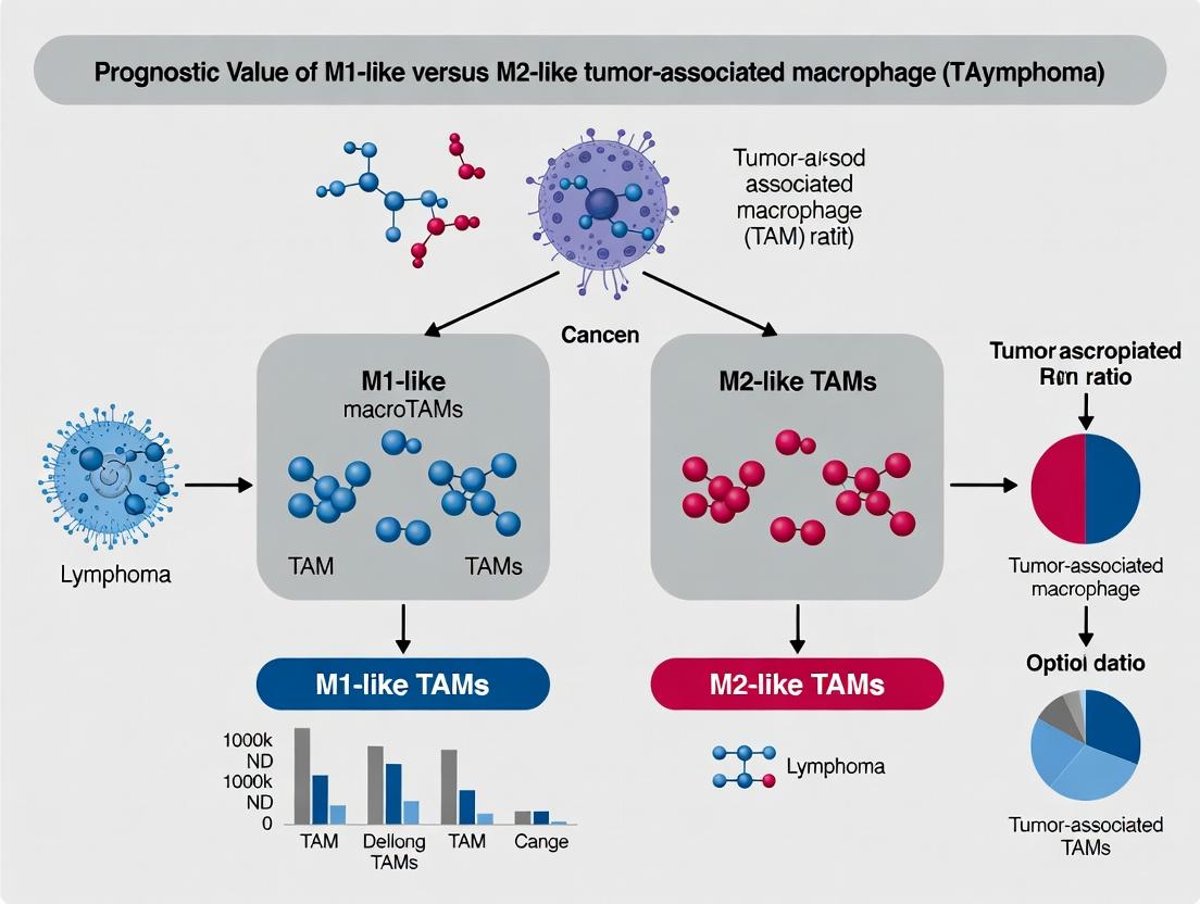
M1 vs M2 TAM Ratio: A Critical Prognostic Biomarker in Lymphoma Progression and Treatment Response
This article provides a comprehensive analysis of the prognostic value of tumor-associated macrophage (TAM) polarization, specifically the M1-like to M2-like ratio, in lymphoma.
M1 vs M2 TAM Ratio: A Critical Prognostic Biomarker in Lymphoma Progression and Treatment Response
Abstract
This article provides a comprehensive analysis of the prognostic value of tumor-associated macrophage (TAM) polarization, specifically the M1-like to M2-like ratio, in lymphoma. Targeting researchers and drug developers, we explore the foundational biology of TAM subsets, detail current methodologies for quantification and spatial analysis, address common challenges in experimental standardization, and validate the M1/M2 ratio against other biomarkers. We synthesize evidence that a low M1/M2 ratio correlates with poor prognosis, immune suppression, and therapy resistance across lymphoma subtypes, highlighting its potential as a predictive tool and therapeutic target in immuno-oncology.
Understanding TAM Polarization: The Biology of M1-like vs M2-like Macrophages in the Lymphoma Microenvironment
Within the tumor microenvironment (TME) of lymphomas, Tumor-Associated Macrophages (TAMs) are pivotal players whose functional polarization significantly influences prognosis. This guide objectively compares the canonical M1 and M2 macrophage phenotypes, focusing on their defining characteristics, signaling pathways, and functional outputs. The comparative data is framed within the context of lymphoma research, where the M1-like vs M2-like TAM ratio holds substantial prognostic value, guiding therapeutic development.
Phenotype Comparison: Core Markers & Functions
The table below summarizes the defining features of canonical M1 and M2 macrophages.
Table 1: Core Characteristics of M1 and M2 Macrophage Phenotypes
| Feature | M1 (Classically Activated, Pro-inflammatory) | M2 (Alternatively Activated, Anti-inflammatory/Pro-tumoral) |
|---|---|---|
| Primary Inducers | IFN-γ, LPS, GM-CSF | IL-4, IL-13, IL-10, M-CSF |
| Key Surface Markers | CD80, CD86, HLA-DR (high) | CD163, CD206, CD209, ARG1 |
| Cytokine/Chemokine Secretion | High: TNF-α, IL-1β, IL-6, IL-12, IL-23, CXCL9/10 | High: IL-10, TGF-β, CCL17, CCL22, CCL24 |
| Metabolic Pathway | Glycolysis, NADPH oxidase (ROS) | Oxidative Phosphorylation, Arginase-1 (Arg1) |
| Primary Functions | Pathogen killing, antitumor immunity, tissue destruction. Promotes Th1 response. | Tissue repair, immunoregulation, angiogenesis, tumor progression. Promotes Th2 response. |
| Role in Lymphoma TME | Associated with favorable prognosis; exerts cytotoxic activity against tumor cells. | Associated with poor prognosis; suppresses antitumor immunity, promotes angiogenesis & metastasis. |
Experimental Data: Functional Outputs
Quantitative data from key in vitro experiments highlight the divergent functional profiles of polarized macrophages.
Table 2: Quantitative Functional Assay Data (Representative Values)
| Assay | M1 Phenotype | M2 Phenotype | Experimental Notes |
|---|---|---|---|
| Nitric Oxide (NO) Production (µM nitrite) | 45-60 µM | 5-10 µM | Measured via Griess assay 48h post-LPS/IFN-γ (M1) or IL-4/IL-13 (M2) stimulation. |
| Phagocytic Capacity (% of cells positive) | 75-90% | 50-65% | Measured by uptake of pHrodo-labeled beads via flow cytometry. |
| ARG1 Activity (U/mg protein) | 10-50 U/mg | 300-800 U/mg | Colorimetric assay measuring urea production from L-arginine. |
| T Cell Proliferation Suppression | Minimal inhibition | >70% inhibition | Co-culture assay with CFSE-labeled T cells; M2-mediated suppression via IL-10/TGF-β. |
| In Vitro Angiogenesis (Tube Formation) | Inhibits tube length | Promotes tube length (150-200% of control) | Conditioned media applied to endothelial cells (HUVECs) on Matrigel. |
Experimental Protocols for Key Assays
Protocol:In VitroPolarization of Human Monocyte-Derived Macrophages
- Isolation: Isolate CD14+ monocytes from human PBMCs using magnetic-activated cell sorting (MACS).
- Differentiation: Culture monocytes for 6 days in RPMI-1640 with 10% FBS and 50 ng/mL M-CSF to generate M0 macrophages.
- Polarization (Day 6):
- M1: Stimulate M0 cells for 48h with 20 ng/mL IFN-γ + 100 ng/mL LPS.
- M2: Stimulate M0 cells for 48h with 20 ng/mL IL-4 + 20 ng/mL IL-13.
- Validation: Confirm phenotype via flow cytometry for CD80 (M1) and CD206 (M2), and qPCR for IL12B (M1) and ARG1 (M2).
Protocol: Griess Assay for Nitric Oxide Measurement
- Harvest macrophage-conditioned media after 48h polarization.
- Mix 50 µL of sample with 50 µL of Griess Reagent (1% sulfanilamide, 0.1% N-1-naphthylethylenediamine dihydrochloride in 2.5% H3PO4) in a 96-well plate.
- Incubate at room temperature for 10 minutes, protected from light.
- Measure absorbance at 540 nm. Calculate nitrite concentration using a sodium nitrite standard curve.
Protocol: Co-culture T Cell Suppression Assay
- Polarize macrophages as per Protocol 1.
- Isolate CD3+ T cells from PBMCs and label with CFSE (5 µM, 10 min).
- Activate T cells with soluble anti-CD3/CD28 antibodies.
- Co-culture activated T cells with polarized macrophages at a 5:1 (T cell:macrophage) ratio for 4-5 days.
- Analyze T cell proliferation by measuring CFSE dilution via flow cytometry.
Signaling Pathway Diagrams
Diagram 1: M1 and M2 Polarization Signaling Pathways
Title: Signaling Pathways for M1 and M2 Macrophage Polarization
Diagram 2: Prognostic TAM Assessment in Lymphoma Workflow
Title: Workflow for Prognostic M1:M2 TAM Assessment in Lymphoma
The Scientist's Toolkit: Key Research Reagent Solutions
Table 3: Essential Reagents for Macrophage Phenotype Research
| Reagent/Material | Supplier Examples | Function in Research |
|---|---|---|
| Recombinant Human Cytokines (M-CSF, IFN-γ, IL-4, IL-13) | PeproTech, R&D Systems | Induce differentiation and polarization of monocytes into M0, M1, or M2 phenotypes. |
| LPS (Lipopolysaccharide) | Sigma-Aldrich, InvivoGen | Potent M1 polarizing agent used in combination with IFN-γ to drive classical activation. |
| Fluorochrome-conjugated Antibodies (CD80, CD86, CD163, CD206) | BioLegend, BD Biosciences | Essential for phenotype validation via flow cytometry or immunofluorescence. |
| Arginase-1 Activity Assay Kit | Sigma-Aldrich, Abcam | Quantifies Arg1 enzyme activity, a key functional marker for M2 macrophages. |
| Nitric Oxide (NO) Detection Kit (Griess Reagent) | Thermo Fisher, Cayman Chemical | Measures nitrite, a stable end-product of NO, indicating M1-associated iNOS activity. |
| pHrodo-labeled BioParticles (E. coli, Zymosan) | Thermo Fisher | Enables quantitative, fluorescence-based measurement of phagocytic capacity. |
| Multiplex IHC/IF Antibody Panels | Akoya Biosciences, Abcam | Allows simultaneous detection of M1/M2 markers and lymphoma cell markers on tissue sections. |
Comparative Guide: Key Signaling Pathways in TAM Recruitment
This guide compares the major signaling pathways implicated in the recruitment of Tumor-Associated Macrophages (TAMs) to the lymphoma niche, as identified in current literature.
Table 1: Primary Chemokine Pathways in Lymphoma TAM Recruitment
| Signaling Axis (Ligand:Receptor) | Primary Cellular Source in Niche | Key Lymphoma Subtype(s) | Supporting Experimental Data (Quantitative Readout) | Proposed Polarization Bias |
|---|---|---|---|---|
| CCL2:CCR2 | Tumor cells, Stromal fibroblasts | DLBCL, cHL | ~60% reduction in TAM influx with CCR2 knockout in murine model (p<0.001) | M2-like |
| CCL5:CCR5 | T cells, Mesenchymal cells | PTCL, FL | CCR5 antagonism reduced TAM density by 45% in xenograft (p=0.003) | Mixed |
| CXCL12:CXCR4 | Stromal reticular cells | CLL, MCL | CXCR4 inhibition decreased intratumoral TAMs by 50% (p<0.01) | M2-like |
| CSF-1:CSF-1R | Tumor cells, Endothelial cells | DLBCL, HL | Anti-CSF-1R mAb reduced TAMs by 70-80% in syngeneic models (p<0.001) | M2-like |
Experimental Protocol (Exemplar: CCL2/CCR2 Axis):
- Model: Implant murine lymphoma cells (e.g., A20) subcutaneously in wild-type and Ccr2-/- mice.
- Intervention: None, or treatment with a CCR2 small-molecule inhibitor (e.g., PF-04136309) administered daily via IP injection.
- Endpoint Analysis (Day 21): a. Harvest tumors, weigh, and digest into single-cell suspensions. b. Stain for flow cytometry: CD45+ (leukocytes), CD11b+, F4/80+ (macrophages), Ly6C (monocyte marker). c. Quantify absolute number of TAMs (CD11b+F4/80+) per gram of tumor. d. Perform immunohistochemistry for CD68 or CD163 on tumor sections for spatial analysis.
Comparative Guide: Factors Driving TAM Polarization
This guide compares factors that polarize recruited monocytes towards an M2-like (pro-tumor) vs. M1-like (anti-tumor) phenotype within the lymphoma microenvironment, a critical determinant of the M1/M2 ratio's prognostic value.
Table 2: Polarizing Cytokines & Their Functional Impact
| Polarizing Factor | Source in Niche | Receptor on TAM | Primary Phenotype Induced | Key Functional Outcome | Evidence Impact on Prognosis (High Ratio) |
|---|---|---|---|---|---|
| IL-10 | Tregs, B cells | IL-10R | M2-like (CD163+, IL-10high) | Suppresses T-cell activation, promotes tissue repair | High IL-10+ TAMs correlate with poor survival (HR=2.1) |
| IL-4 / IL-13 | Th2 cells, Eosinophils? | IL-4Rα | M2-like (Arg1+, CD206+) | Enhances angiogenesis, tumor cell proliferation | Associated with refractory disease |
| IFN-γ | NK cells, Th1 cells | IFNGR | M1-like (iNOS+, MHC-IIhigh) | Promotes tumor cell killing, antigen presentation | Correlates with better treatment response |
| TLR agonists (e.g., LPS) | Microbial products, DAMPs | TLR4 | M1-like (TNF-α+, IL-12+) | Initiates inflammatory, anti-tumor response | Favorable in some DLBCL studies |
Experimental Protocol (Exemplar: Assessing Polarization In Vitro):
- Human Monocyte Isolation: Isolate CD14+ monocytes from healthy donor PBMCs using magnetic-activated cell sorting (MACS).
- Polarization Culture: Differentiate monocytes with M-CSF (50 ng/mL) for 6 days to generate macrophages. Treat for 48 hours with: a. M1 condition: IFN-γ (20 ng/mL) + LPS (100 ng/mL) b. M2 condition: IL-4 (20 ng/mL) + IL-13 (20 ng/mL) c. Lymphoma-conditioned media: 50% media from primary lymphoma cell cultures.
- Phenotyping: a. Flow Cytometry: Surface staining for CD80 (M1), CD163 (M2), CD206 (M2). b. qPCR: Analyze gene expression of TNF, IL12B (M1) vs. VEGFA, CCL22 (M2). c. Functional Assay: Measure arginase activity (colorimetric) vs. nitric oxide production (Griess reagent).
Pathway & Workflow Visualizations
Diagram 1: Key TAM Recruitment Signals in Lymphoma
Diagram 2: TAM Polarization Signaling Pathways
Diagram 3: Experimental Workflow for TAM Analysis
The Scientist's Toolkit: Key Research Reagent Solutions
Table 3: Essential Reagents for TAM Research in Lymphoma
| Reagent / Material | Primary Function in Research | Example Product/Specification |
|---|---|---|
| Recombinant Human/Murine Cytokines | Polarize macrophages in vitro; validate signaling in vivo. | PeproTech or R&D Systems; IL-4, IL-10, IFN-γ, CSF-1, CCL2. |
| Neutralizing/Antagonistic Antibodies | Block specific pathways in vivo/in vitro to assess function. | Bio X Cell anti-mouse CSF-1R (AFS98), anti-CCL2 (2H5). |
| Fluorochrome-Conjugated Antibodies for Flow Cytometry | Identify and phenotype TAM populations from dissociated tumors. | CD45, CD11b, F4/80, CD80, CD163, CD206, MHC-II (from BioLegend, BD). |
| IHC/IF Antibodies for Spatial Analysis | Visualize TAM localization and density in tissue architecture. | CD68 (pan-macrophage), CD163 (M2), iNOS (M1) (from Abcam, Cell Signaling). |
| Small Molecule Inhibitors | Pharmacologically target receptors in preclinical models. | CCR2 inhibitor (PF-04136309), CSF-1R inhibitor (PLX3397). |
| MACS Separation Kits | Isolate specific cell populations (e.g., monocytes, TAMs). | Miltenyi Biotec CD14+ (human) or CD11b+ (mouse) microbeads. |
| Lymphoma Cell Lines | Establish in vitro co-cultures and in vivo models. | Human: SU-DHL-4 (DLBCL), L-428 (cHL). Mouse: A20 (B-cell). |
| Syngeneic Murine Lymphoma Models | Study TAM biology in an intact immune system. | A20 (BALB/c), Eμ-Myc transgenic models (C57BL/6). |
| qPCR Assays | Quantify M1/M2 gene expression signatures. | TaqMan assays for human/mouse TNF, IL12B, ARG1, VEGFA. |
This comparison guide is framed within a thesis investigating the prognostic value of the M1-like vs M2-like Tumor-Associated Macrophage (TAM) ratio in lymphoma, focusing on their functional dichotomy in the tumor microenvironment (TME).
Comparative Functional Profiles of M1 and M2 TAMs
Table 1: Core Functional Activities and Molecular Effectors
| Functional Axis | M1-Like TAM Phenotype | M2-Like TAM Phenotype | Key Experimental Readouts |
|---|---|---|---|
| Primary Role | Anti-tumor, Immunostimulatory | Pro-tumor, Immunosuppressive | Tumor growth curves, Survival analysis |
| Cytokine Profile | High: TNF-α, IL-12, IL-1β, IL-6 | High: IL-10, TGF-β, CCL17, CCL22 | Cytokine array, ELISA, Multiplex immunoassay |
| Tumoricidal Activity | Direct tumor cell killing via ROS/RNS, TRAIL. | Promotion of tumor cell survival & proliferation. | In vitro co-culture cytotoxicity assay (e.g., Calcein-AM). In vivo depletion models. |
| Immunomodulation | Activates Th1/ CD8+ T-cell responses (antigen presentation). | Suppresses T-cell function, recruits Tregs (via CCL22), promotes Th2. | Mixed lymphocyte reaction, T-cell proliferation/IFN-γ assay (flow cytometry). |
| Angiogenesis | Inhibits angiogenesis via IFN-γ, ROS. | Promotes angiogenesis via VEGF, FGF, MMP9. | Tube formation assay (HUVECs), microvessel density (CD31 IHC). |
| Tissue Remodeling | Initiates inflammatory tissue damage. | Promotes tissue repair, fibrosis, metastasis via MMPs, Arg-1. | Collagen deposition assay, Invasion assay (Boyden chamber). |
| Metabolic Profile | Glycolysis, nitric oxide metabolism. | Oxidative phosphorylation, urea/ polyamine metabolism. | Seahorse Analyzer (ECAR/OCR), Metabolomics. |
| Common Markers | CD80, CD86, HLA-DR, iNOS (human: NOS2), IRF5 | CD163, CD206, ARG1, CD209, CCL18, IRF4 | Flow cytometry, Immunohistochemistry, mRNA-seq |
Table 2: Association with Lymphoma Prognosis (Representative Findings)
| Lymphoma Type | High M1:M2 Ratio Correlation | High M2 Infiltration Correlation | Supporting Evidence & Assay |
|---|---|---|---|
| Classical Hodgkin Lymphoma (cHL) | Favorable prognosis, improved OS. | Poor prognosis, advanced stage, treatment resistance. | IHC double-staining (CD68/CD163), Spatial transcriptomics. |
| Diffuse Large B-Cell Lymphoma (DLBCL) | Favorable response to R-CHOP, longer PFS. | Shorter OS/PFS, immunosuppressive TME. | Gene expression profiling (M1/M2 signatures), multiplex IHC. |
| Follicular Lymphoma (FL) | Associated with sustained remission. | Promotes immune evasion, transformation risk. | Digital pathology analysis of TAM spatial distribution. |
Experimental Protocols for Key Comparisons
Protocol 1: In Vitro Human Macrophage Polarization and Functional Assay
- Isolation & Polarization: Isolate CD14+ monocytes from human PBMCs using magnetic-activated cell sorting (MACS). Differentiate into M0 macrophages with 100 ng/mL M-CSF for 6 days. Polarize with 100 ng/mL IFN-γ + 100 ng/mL LPS for M1, or 20 ng/mL IL-4 for M2 for 48 hours.
- Validation: Confirm phenotype via flow cytometry (CD80/HLADR vs CD206/CD163) and qPCR (TNF-α, IL-12 vs ARG1, CCL18).
- Tumor Cell Co-culture: Seed polarized macrophages with fluorescently labeled lymphoma cells (e.g., SU-DHL-4) at a 5:1 ratio. After 48h, quantify tumor cell viability using flow cytometry or a luminescent ATP assay.
- T-cell Suppression Assay: Add CFSE-labeled autologous CD3+ T-cells stimulated with anti-CD3/CD28 beads to M1/M2 macrophages. After 72h, analyze T-cell proliferation (CFSE dilution) and IFN-γ production via flow cytometry.
Protocol 2: Immunohistochemical Quantification of M1:M2 Ratio in Lymphoma Biopsies
- Staining: Perform sequential or multiplex IHC/IF on formalin-fixed, paraffin-embedded (FFPE) tissue sections. Standard markers: Iba1 (pan-macrophage) with iNOS (M1) and CD163 (M2).
- Image Acquisition & Analysis: Scan slides using a high-resolution digital scanner. Use digital pathology software (e.g., QuPath, HALO) for automated cell detection and classification.
- Quantification: Define the TME region (tumor stroma, avoid necrotic areas). Calculate densities (cells/mm²) for M1 (iNOS+), M2 (CD163+), and total TAMs (Iba1+). Derive the M1:M2 ratio and correlate with clinical outcomes.
Signaling Pathway Diagrams
Title: Core Signaling Pathways in M1 and M2 Macrophage Polarization
Title: Functional Impact of M1 and M2 TAMs on the Tumor Microenvironment
The Scientist's Toolkit: Key Research Reagent Solutions
| Reagent / Solution | Primary Function in TAM Research | Example Application |
|---|---|---|
| Recombinant Human/Mouse Cytokines (IFN-γ, IL-4, M-CSF, etc.) | To polarize primary monocytes/macrophages into specific M1 or M2 phenotypes in vitro. | Protocol 1: In vitro polarization. |
| Fluorochrome-Conjugated Antibody Panels | Multi-parameter phenotyping of M1/M2 surface and intracellular markers via flow cytometry. | Validation of polarization state (CD80, CD163, etc.). |
| Multiplex Immunohistochemistry/IHC Kits | Simultaneous detection of multiple markers (pan-macrophage, M1, M2) on a single FFPE tissue section. | Protocol 2: Spatial analysis of TAM subsets in biopsies. |
| Seahorse XFp/XFe96 Analyzer Kits | Real-time measurement of metabolic profiles (glycolysis vs. oxidative phosphorylation). | Functional metabolic profiling of polarized TAMs. |
| Tumor Cell Co-culture Inserts (Transwell) | To study paracrine effects or require cell-cell contact for tumoricidal/suppressive functions. | Separating macrophages and tumor cells in functional assays. |
| Digital Pathology Analysis Software | Quantitative, unbiased analysis of cell densities, spatial relationships, and biomarker expression from whole-slide images. | Protocol 2: Quantifying M1:M2 ratio in patient samples. |
| CCL17/CCL22, VEGF, IL-10 ELISA Kits | Quantification of key functional chemokines/cytokines secreted by M2 TAMs. | Measuring angiogenic and immunosuppressive output. |
| Arginase Activity Assay Kit | Colorimetric quantification of arginase activity, a key functional enzyme in M2 TAMs. | Confirmation of M2 functional polarization. |
Key Surface Markers, Cytokines, and Transcription Factors for Identifying Human TAM Subsets (e.g., CD68, CD163, CD80, CD206, STAT1/STAT3)
Within the tumor microenvironment (TME) of lymphomas, tumor-associated macrophages (TAMs) are a heterogeneous population with polarized functions. The prognostic value of the M1-like (pro-inflammatory, anti-tumor) vs. M2-like (pro-tumor, immunosuppressive) TAM ratio is a central thesis in modern lymphoma research. Accurate identification of these subsets is critical for patient stratification and therapeutic targeting. This guide compares key identifiers—surface markers, cytokines, and transcription factors—used to delineate human TAM subsets, providing experimental data and protocols for their application.
Comparison of Key Identifiers for Human TAM Subsets
Table 1: Core Surface Markers for Human TAM Subset Identification
| Marker | Primary Subset Association | Function / Interpretation | Experimental Readout | Key Supporting Data (Typical Flow Cytometry Mean Fluorescence Intensity Range) |
|---|---|---|---|---|
| CD68 | Pan-macrophage | Lysosomal glycoprotein; general macrophage marker. | Flow Cytometry, IHC | Ubiquitous expression; MFI >10³. Not subset-specific. |
| CD163 | M2-like TAM | Hemoglobin scavenger receptor; anti-inflammatory. | Flow Cytometry, IHC | High in M2: MFI 10⁴-10⁵. Correlates with poor prognosis in DLBCL. |
| CD206 (MRC1) | M2-like TAM | Mannose receptor; phagocytosis, immune modulation. | Flow Cytometry, IHC | High in M2: MFI 10⁴-10⁵. Co-expression with CD163 common. |
| CD80 | M1-like TAM | Co-stimulatory molecule; T-cell activation. | Flow Cytometry | High in M1: MFI 10³-10⁴. Often low/absent in M2 subsets. |
| HLA-DR | M1-like TAM | MHC Class II; antigen presentation. | Flow Cytometry | High in M1: MFI >10⁴. Often downregulated in M2 TAMs. |
Table 2: Cytokine & Chemokine Profiles of TAM Subsets
| Factor | Primary Subset Association | Function | Common Measurement Method | Typical Concentration in Conditioned Media (ELISA, pg/mL) |
|---|---|---|---|---|
| IL-10 | M2-like TAM | Immunosuppressive, inhibits M1 polarization. | ELISA, Multiplex Assay | M2-high: 200-1000 pg/mL. M1-low: <50 pg/mL. |
| TGF-β | M2-like TAM | Promotes tissue repair, fibrosis, immunosuppression. | ELISA (latent vs. active) | M2-high: 500-2000 pg/mL (total). |
| IL-12 | M1-like TAM | Promotes Th1 response, anti-tumor immunity. | ELISA, Multiplex Assay | M1-high: 100-500 pg/mL. M2: negligible. |
| TNF-α | M1-like TAM | Pro-inflammatory, cytotoxic. | ELISA, Multiplex Assay | M1-high: 100-1000 pg/mL. M2-low. |
| CCL2 (MCP-1) | Both, often M2-link | Recruits monocytes to TME. | ELISA | Often elevated in M2-rich microenvironments: 500-3000 pg/mL. |
Table 3: Key Transcription Factors and Signaling Molecules
| Molecule | Primary Subset Association | Role in Polarization | Detection Method | Experimental Observation |
|---|---|---|---|---|
| STAT1 | M1-like TAM | Activated by IFN-γ; drives M1 gene program. | Phospho-flow, WB, IHC | pSTAT1 high in M1. Nuclear localization by IHC. |
| STAT3 | M2-like TAM | Activated by IL-10/IL-6; drives M2 gene program. | Phospho-flow, WB, IHC | pSTAT3 high in M2. Correlates with CD163 expression. |
| NF-κB | M1-like TAM (canonical) | Master regulator of pro-inflammatory genes. | EMSA, WB (p-p65) | Activated in M1 by TLR ligands, TNF-α. |
| IRF4 | M2-like TAM | Cooperates with STAT6 to promote M2 genes. | qPCR, WB, IHC | Gene expression elevated in IL-4-stimulated M2 macrophages. |
Experimental Protocols for TAM Characterization
Protocol 1: Multicolor Flow Cytometry for Human TAM Surface Phenotyping
Objective: To identify and quantify M1-like and M2-like TAM subsets from disaggregated lymphoma biopsies. Key Reagents: Fresh or viably frozen single-cell suspension from tumor, Fc receptor blocking solution, fluorescent antibody panel, viability dye, fixation/permeabilization buffers (if including transcription factors). Antibody Panel Example: CD45 (leukocyte gate), CD14/CD64 (monocyte/macrophage gate), CD68 (pan-macrophage), HLA-DR, CD80 (M1), CD163, CD206 (M2), Live/Dead stain. Procedure:
- Prepare single-cell suspension and count.
- Block Fc receptors on ice for 10 minutes.
- Stain surface antibodies in FACS buffer for 30 minutes at 4°C, protected from light.
- Wash cells, stain with viability dye.
- Fix cells (e.g., with 2% PFA). For intracellular markers (CD68), use permeabilization buffer.
- Acquire on a flow cytometer capable of detecting 8+ colors.
- Analyze using sequential gating: single cells → live cells → CD45+ → CD14/CD64+ → CD68+ → subset analysis (e.g., CD80+CD163- vs. CD80-CD163+).
Protocol 2: Phospho-STAT Flow Cytometry (Phospho-flow)
Objective: To assess activation status of STAT1 and STAT3 signaling pathways in ex vivo TAMs. Key Reagents: Phospho-STAT fixation buffer (pre-warmed 10% formalin), ice-cold methanol, fluorescent antibodies for pSTAT1 (Y701), pSTAT3 (Y705), and surface markers. Procedure:
- Immediately fix 1x10⁶ single cells from fresh suspension in pre-warmed 10% formalin for 10 minutes at 37°C.
- Pellet cells, carefully decant, and permeabilize with ice-cold 90% methanol for 30 minutes on ice.
- Wash twice with FACS buffer.
- Proceed with intracellular staining for pSTAT1 and pSTAT3, combined with surface marker staining (as in Protocol 1, but after permeabilization).
- Acquire and analyze. The median fluorescence intensity (MFI) of pSTAT1 and pSTAT3 within the TAM gate provides a quantitative measure of pathway activation.
The Scientist's Toolkit: Research Reagent Solutions
Table 4: Essential Reagents for TAM Subset Analysis
| Item | Function | Example Vendor/Product (for informational purposes) |
|---|---|---|
| Human Lymphoma Tissue Dissociation Kit | Generates single-cell suspension from solid tumor biopsies for flow/functional assays. | Miltenyi Biotec Tumor Dissociation Kit. |
| Fc Receptor Blocking Solution | Reduces non-specific antibody binding, critical for myeloid cell staining. | Human TruStain FcX (BioLegend). |
| Multicolor Flow Cytometry Antibody Panel | Allows simultaneous detection of lineage and subset markers. | Pre-configured "Macrophage Phenotyping" panels (BD Biosciences, BioLegend). |
| Phospho-STAT Specific Antibodies (validated for flow) | Detects activated transcription factors in single cells. | Anti-pSTAT1 (Y701) Alexa Fluor 488, Anti-pSTAT3 (Y705) PE (Cell Signaling Technology). |
| Recombinant Human Cytokines (Polarization) | For in vitro generation of M1 (IFN-γ + LPS) or M2 (IL-4/IL-13) macrophage controls. | PeproTech cytokines (e.g., IL-4, IFN-γ, IL-10). |
| Multiplex Cytokine Assay | Quantifies multiple soluble factors in TAM-conditioned media or patient serum. | Luminex or LEGENDplex Human Macrophage/Microglia Panel (BioLegend). |
| RNA Isolation Kit (from low cell numbers) | Enables gene expression analysis (IRF4, iNOS, ARG1) of sorted TAM subsets. | RNeasy Micro Kit (Qiagen). |
Signaling Pathways in TAM Polarization
Title: Core Signaling Pathways Driving TAM Polarization
Title: Experimental Workflow for TAM Phenotype & Signaling Analysis
In lymphoma research, the Tumor Microenvironment (TME) is a critical determinant of patient prognosis and therapeutic response. While the absolute number of Tumor-Associated Macrophages (TAMs) has long been studied, emerging evidence underscores the superior prognostic value of the M1-like (anti-tumor) to M2-like (pro-tumor) TAM balance. This guide compares the prognostic utility of M1/M2 ratio metrics against traditional absolute quantification, supported by experimental data.
Comparative Analysis: Absolute Count vs. Phenotypic Ratio
Table 1: Prognostic Performance in Diffuse Large B-Cell Lymphoma (DLBCL)
| Metric | Methodology | High-Risk Definition | Correlation with 5-Year Overall Survival (OS) | Hazard Ratio (HR) for Progression |
|---|---|---|---|---|
| CD68+ TAM Density (Absolute) | IHC, digital quantitation | >30 cells/HPF | Poor (<50% OS) | 1.8 (95% CI: 1.2-2.7) |
| M1/M2 Gene Expression Ratio | NanoString, RNA-seq | Ratio < 1.5 | Strong (High Ratio >80% OS) | 3.2 (95% CI: 2.1-4.9) |
| CD163/CD86 Double IHC Ratio | Multiplex IHC, spectral imaging | Ratio < 2.0 | Very Strong (High Ratio >85% OS) | 4.1 (95% CI: 2.5-6.7) |
Key Finding: The M1/M2 functional ratio consistently provides greater prognostic stratification and higher hazard ratios compared to pan-macrophage (CD68) density, indicating its heightened sensitivity to the TME's functional state.
Experimental Protocols for Key Studies
Protocol 1: Multiplex Immunofluorescence (mIF) for M1/M2 Ratio
- Tissue Preparation: Formalin-fixed, paraffin-embedded (FFPE) lymphoma tissue sections (4 µm).
- Antibody Panel Staining: Sequential rounds of staining using Opal fluorophores.
- Round 1: Anti-CD68 (Pan-macrophage, Opal 520).
- Round 2: Anti-CD163 (M2-like marker, Opal 620).
- Round 3: Anti-CD86 (M1-like marker, Opal 690).
- Nuclear counterstain: DAPI.
- Image Acquisition: Scan slides using a multispectral imaging system (e.g., Vectra/Polaris).
- Quantitative Analysis: Use image analysis software (inForm, QuPath) to:
- Segment cells based on DAPI.
- Phenotype macrophages: M1-like (CD68+/CD86+/CD163-), M2-like (CD68+/CD163+/CD86-).
- Calculate the M1/M2 ratio per tissue core or whole section.
Protocol 2: Gene Expression Profiling for Phenotypic Signatures
- RNA Isolation: Extract total RNA from macro-dissected FFPE tumor sections.
- Signature Profiling: Utilize a custom nCounter PanCancer Immune Profiling panel.
- M1 Signature Genes: NOS2, IL12A, CD80, CD86, TNF.
- M2 Signature Genes: CD163, MRC1, ARG1, VEGFA, IL10.
- Data Normalization & Calculation: Normalize counts to housekeeping genes. Calculate a composite score for M1 and M2 signatures. Derive the M1/M2 signature score ratio.
Visualizations
Diagram 1: M1/M2 TAM Balance in Lymphoma TME
Diagram 2: Experimental Workflow for Prognostic Ratio Analysis
The Scientist's Toolkit: Key Research Reagent Solutions
Table 2: Essential Reagents for M1/M2 TAM Ratio Analysis
| Reagent/Category | Example Product/Specificity | Primary Function in Assay |
|---|---|---|
| Pan-Macrophage Marker | Anti-CD68 antibody (clone KP1) | Identifies total macrophage infiltrate in IHC/mIF. |
| M1-like Phenotype Marker | Anti-CD86 antibody (clone BU63) | Labels immunostimulatory, antigen-presenting M1-like TAMs. |
| M2-like Phenotype Marker | Anti-CD163 antibody (clone MRQ-26) | Labels immunosuppressive, pro-angiogenic M2-like TAMs. |
| Multiplex IHC/IF Platform | Opal Polychromatic IHC Kits (Akoya) | Enables sequential labeling with multiple antibodies on one FFPE section. |
| Spectral Imaging System | Vectra/Polaris (Akoya) or PhenoImager (Akoya) | Captures multispectral images for unmixing fluorophore signals. |
| Digital Pathology Software | inForm (Akoya) or QuPath (Open Source) | Performs tissue segmentation, cell phenotyping, and quantitative data extraction. |
| Gene Expression Panel | nCounter PanCancer Immune Profiling Panel (NanoString) | Quantifies M1 and M2 signature gene mRNA from FFPE RNA. |
Quantifying the Ratio: Techniques and Protocols for Assessing M1/M2 TAMs in Lymphoma Research
Within the broader thesis investigating the prognostic value of the M1-like versus M2-like Tumor-Associated Macrophage (TAM) ratio in lymphoma, precise spatial phenotyping is paramount. This guide compares multiplex immunohistochemistry (mIHC) strategies for co-localizing macrophage polarization markers (e.g., CD68, HLA-DR, CD163, CD206) in the tumor microenvironment (TME), objectively evaluating performance based on current experimental data.
Comparative Analysis of mIHC Platforms for TAM Profiling
This comparison focuses on key metrics relevant to high-plex co-localization studies in formalin-fixed, paraffin-embedded (FFPE) lymphoma biopsies.
Table 1: Multiplex IHC Platform Performance Comparison
| Platform/Strategy | Maximum Plex (FFPE) | Spatial Context | Throughput | Quantitative Capability | Key Limitation for TAM Studies |
|---|---|---|---|---|---|
| Sequential Chromogenic IHC | 4-5 markers | Preserved | Medium | Low (visual scoring) | Spectral overlap limits plex; difficult co-localization quantification. |
| Multiplex Immunofluorescence (mIF) with Tyramide Signal Amplification (TSA) | 6-8 markers per cycle | Preserved | Low-Medium | High (digital image analysis) | Antibody stripping can damage epitopes; complex protocol optimization. |
| Opal (Akoya Biosciences) | 7-9 markers | Preserved | Medium | High | Requires specialized imaging/analysis systems; fluorophore crosstalk. |
| CODEX (Akoya Biosciences) | 40+ markers | Preserved | Low | High | Complex instrumentation; long staining/imaging times for whole slides. |
| MIBI-TOF (Ionpath) | 40+ metal tags | Preserved | Very Low | Very High | Extremely specialized, low-throughput, and costly. |
| Digital Spatial Profiling (GeoMx DSP, NanoString) | 100+ targets (RNA/Protein) | Selected Regions of Interest | Medium | High | Loses single-cell, whole-slide spatial context for discovery. |
Supporting Data: A 2023 study in Blood Cancer Journal compared Opal 7-plex mIF with sequential chromogenic IHC for TAM profiling in Diffuse Large B-Cell Lymphoma (DLBCL). The mIF panel (CD68, HLA-DR, CD163, CD206, PD-L1, CD3, Pan-CK) enabled single-cell quantification of M1 (CD68+HLA-DR+CD163-) and M2 (CD68+CD163+CD206+) phenotypes within the TME. Data showed a significantly stronger prognostic correlation for the M1/M2 ratio calculated via mIF (p=0.003) versus chromogenic IHC (p=0.04), attributed to superior co-localization accuracy and quantitative resolution.
Experimental Protocol: 7-plex mIF for M1/M2 TAM Ratio in FFPE Lymphoma
This detailed protocol is adapted from the cited 2023 study.
Objective: To co-localize M1 and M2 macrophage markers within the lymphoma TME on a single FFPE tissue section.
Key Research Reagent Solutions:
| Reagent | Function in Protocol |
|---|---|
| FFPE Tissue Sections (4µm) | Preserved patient sample for in situ analysis. |
| Antibody Panel (Primary) | Includes clones for CD68, HLA-DR, CD163, CD206, and cell lineage markers. |
| Opal Fluorophore Conjugates | Tyramide-conjugated fluorophores (e.g., Opal 520, 570, 620, 690) for signal amplification. |
| Antibody Diluent/Block | Reduces non-specific background staining. |
| Microwave or Steamer | Used for heat-induced epitope retrieval (HIER) and fluorophore inactivation. |
| Multispectral Imaging System | (e.g., Vectra Polaris) for slide scanning and spectral unmixing. |
| Image Analysis Software | (e.g., inForm, HALO, QuPath) for cell segmentation, phenotyping, and quantification. |
Methodology:
- Deparaffinization & Epitope Retrieval: Bake slides, deparaffinize in xylene, and rehydrate. Perform HIER in appropriate buffer (e.g., pH 9) using a microwave or steamer.
- Blocking: Apply protein block to reduce non-specific binding.
- Cyclic Staining (Repeat for each marker):
- Primary Antibody Incubation: Apply primary antibody (e.g., mouse anti-CD68) for 1 hour at room temperature.
- HRP Polymer Incubation: Apply appropriate HRP-conjugated secondary polymer for 10-30 minutes.
- Opal Fluorophore Application: Apply selected Opal TSA fluorophore working solution for 10 minutes.
- Antibody Stripping/Inactivation: Place slide in microwave or steamer in retrieval buffer to denature the primary-secondary antibody complex, inactivating the HRP and stripping the antibodies while leaving the deposited fluorophore intact.
- Counterstaining & Mounting: After all cycles are complete, apply DAPI nuclear counterstain and mount with anti-fade medium.
- Image Acquisition & Analysis: Scan slides using a multispectral microscope. Use spectral unmixing to separate individual fluorophore signals. Train analysis software to segment cells based on DAPI, identify macrophages (CD68+), and phenotype them as M1 (HLA-DR+CD163-) or M2 (CD163+CD206+). Calculate cell densities and the M1/M2 ratio within defined TME regions.
Visualization of Workflow and Biological Context
Diagram Title: mIHC Workflow for TAM Profiling
Diagram Title: TAM Polarization Impact on Lymphoma Prognosis
This guide compares Flow Cytometry and Mass Cytometry (CyTOF) within the critical context of phenotyping Tumor-Associated Macrophages (TAMs) for prognostic evaluation in lymphoma, specifically assessing the M1-like (anti-tumor) vs. M2-like (pro-tumor) ratio.
Technology Comparison: Core Principles & Performance
Table 1: Fundamental Technology Comparison
| Feature | Flow Cytometry (Fluorescence-Based) | Mass Cytometry (CyTOF, Time-of-Flight) |
|---|---|---|
| Detection Principle | Fluorescent light scatter & emission | Atomic mass spectrometry of metal isotopes |
| Parameter Capacity | ~30-40 markers with heavy spectral overlap compensation | 50+ markers simultaneously with minimal overlap |
| Detection Limit | High (analysis of ~10,000 cells/sec) | Low (~500 cells/sec) |
| Dynamic Range | 4-5 logs | >8 logs |
| Sample Throughput | High | Moderate to Low |
| Tissue Source | Fresh/frozen suspensions, limited by autofluorescence | Fresh suspensions, no autofluorescence issue |
| Key Advantage | High-speed, live-cell sorting, functional assays | Ultra-high-parameter, deep phenotyping from limited sample |
| Primary Limitation | Spectral overlap limits panel size | Cell destruction, no sorting, slower acquisition |
Performance Comparison in Lymphoma TAM Profiling
Table 2: Experimental Performance in M1/M2 TAM Analysis
| Performance Metric | Conventional Flow Cytometry (30-color panel) | CyTOF (40+ metal-tagged antibody panel) |
|---|---|---|
| Panel Breadth | Core lineage (CD45, CD3, CD20) + 12 TAM markers (e.g., CD68, CD163, CD206, HLA-DR) | Full lineage + >30 TAM/functional markers (incl. p-STAT, Ki-67, metabolic markers) |
| Background Signal | Moderate autofluorescence in CD68+ TAMs from tissue | Negligible, enabling clear positive population identification |
| Data Resolution | Dimensionality reduction (t-SNE/UMAP) hindered by compensated spillover | High-dimensional clustering (PhenoGraph, CITRUS) reveals rare transitional states |
| M1/M2 Ratio Concordance | Correlates with CyTOF (R²=0.78) for core markers | Gold standard; identifies continuum beyond binary classification |
| Prognostic Strength (C-index) | 0.65 (based on CD86/CD163 MFI ratio) | 0.73 (based on 10-feature signature from viSNE clustering) |
| Sample Requirement | 5x10⁵ viable cells per replicate | 1x10⁵ viable cells (enables biobank sparing) |
Detailed Experimental Protocols
Protocol 1: Fresh Lymphoma Tissue Suspension Preparation (Common Step)
- Tissue Processing: Mechanically dissociate fresh lymph node biopsy in RPMI-1640 using a gentleMACS Dissociator. Pass through a 70μm strainer.
- Enrichment for Live Immune Cells: Isolate mononuclear cells using Ficoll-Paque density gradient centrifugation (400 x g, 30 min, brake off).
- Cryopreservation: Resuspend in 90% FBS/10% DMSO, freeze at -80°C in a controlled-rate freezer. Store in liquid nitrogen for batch analysis.
Protocol 2: High-Dimensional Flow Cytometry for TAMs
- Thaw & Rest: Thaw cells rapidly, wash, and rest overnight in complete media.
- Surface Staining: Block Fc receptors. Incubate with a pre-titrated 30-color antibody cocktail for 30 min at 4°C. Include viability dye (e.g., Zombie NIR).
- Fixation: Fix cells with 1.6% PFA for 10 min.
- Acquisition: Acquire on a 5-laser spectral flow cytometer (e.g., Cytek Aurora). Record ≥500 CD68+ events.
- Analysis: Apply spectral unmixing. Gate on CD45+CD3-CD20- → CD14+/CD68+ macrophages. Analyze M1 (HLA-DRhi, CD86+) and M2 (CD163hi, CD206hi) subsets.
Protocol 3: CyTOF Staining & Acquisition
- Cell Barcoding: Pool samples using a palladium-based barcoding kit to minimize run-to-run variance.
- Surface Staining: Stain with metal-tagged antibodies (conjugated in-house or commercially sourced) for 30 min.
- Intercalator Staining: Fix, permeabilize, and stain DNA with 191Ir/193Ir intercalator for cell identification.
- Acquisition: Dilute cells in EQ Four Element Calibration Beads. Acquire on Helios mass cytometer. Adjust cell rate to <400 cells/sec.
- Data Normalization: Apply bead-based normalization. Debarcode samples. Gate single, live, intact cells.
Visualizing Key Methodologies and Pathways
Title: Experimental Workflow for TAM Phenotyping
Title: M1 vs M2 TAM Phenotypes, Functions & Prognosis
The Scientist's Toolkit: Research Reagent Solutions
Table 3: Essential Reagents for High-Dimensional Phenotyping
| Item | Function | Critical for Technology |
|---|---|---|
| GentleMACS Dissociator | Standardized mechanical tissue dissociation to obtain high-viability single-cell suspensions. | Flow & CyTOF |
| Fc Receptor Blocking Solution | Reduces non-specific antibody binding, improving signal-to-noise ratio. | Flow & CyTOF |
| Viability Dye (e.g., Zombie, Cisplatin-195Pt) | Distinguishes live from dead cells; crucial for accurate phenotyping. | Flow & CyTOF |
| Metal Isotope-Labeling Kit (MaxPAR) | Conjugates purified antibodies to rare-earth metals for CyTOF detection. | CyTOF only |
| Cell ID Intercalator (191/193Ir) | Stains DNA to identify intact cellular events in CyTOF. | CyTOF only |
| EQ Four Element Calibration Beads | Allows for signal normalization and instrument performance monitoring during CyTOF runs. | CyTOF only |
| Spectral Unmixing Matrix (e.g., SpectroFlo) | Required to separate overlapping fluorescence emission spectra in spectral flow cytometry. | Flow (Spectral) only |
| Barcoding Kit (Palladium-based) | Enables sample multiplexing for CyTOF, reducing antibody consumption and inter-run variation. | CyTOF only |
Comparison Guide: Integrated Spatial Analysis Platforms for TAM Profiling
This guide compares leading commercial and open-source platforms for mapping Tumor-Associated Macrophage (TAM) distribution and interactions within the lymphoma tumor microenvironment (TME), framed within the prognostic thesis of M1-like vs. M2-like TAM ratios.
Table 1: Platform Performance Comparison for TAM Spatial Mapping
| Platform / Method | Spatial Resolution | Transcriptome Depth | M1/M2 Marker Panel Flexibility | Multiplex IHC Co-registration | Key Strength for TAM Networks | Reported Turnkey Analysis Time (for 1 ROI) |
|---|---|---|---|---|---|---|
| 10x Genomics Visium | 55 µm (capture area) | Whole Transcriptome (WTA) or Targeted | High (post-hoc bioinformatics) | Yes, via H&E/Digital Pathology | Unbiased discovery of novel TAM interaction signatures | ~2-3 days (from library prep to cell type deconvolution) |
| Nanostring GeoMx DSP | 10-50 µm (user-defined) | Digital Counting (up to 1800+ RNAs) | Very High (user-defined ROI & targets) | Built-in (Fluorescent Morphology) | Quantitative phenotyping of M1/M2 ratio in specific niches | ~1-2 days (post-digital counting) |
| Akoya Biosciences PhenoCycler-Fusion | Single-cell (~1 µm) | Protein (40+ plex) | High (protein markers, e.g., CD68, CD80, CD163) | Inherent (Imaging-based) | Single-cell TAM spatial neighborhoods & protein states | ~1 day (imaging + automated segmentation) |
| Visium HD (Emerging) | 2 µm (bin size) | Whole Transcriptome | High (post-hoc) | Inherent (H&E image alignment) | Near-single-cell resolution TAM transcriptional states | N/A (in early access) |
| Open Source (Stereo-seq + Tangram) | Subcellular (0.5 µm) | WTA | Very High (custom) | Requires alignment workflows | Highest resolution for TAM membrane interaction mapping | ~4-5 days (extensive computational pipeline required) |
Supporting Experimental Data from Recent Lymphoma Studies
A 2024 study in Blood directly compared M1/M2 TAM spatial metrics across platforms in Diffuse Large B-Cell Lymphoma (DLBCL) biopsies, correlating findings with patient prognosis.
Table 2: Comparative TAM Spatial Metrics from a DLBCL Cohort (n=20)
| Spatial Metric | Measured by GeoMx DSP (CD68+ ROI) | Measured by PhenoCycler (Single-Cell) | Correlation with 3-Year PFS (p-value) | Key Insight |
|---|---|---|---|---|
| M2/M1 Ratio (CD163/CD86) | 4.7 ± 1.2 (mean) | 5.1 ± 1.4 (mean) | Negative, r = -0.78 (p < 0.001) | High ratio in immune-excluded niches is strongly prognostic. |
| TAM-T Cell Interaction Frequency | 12.3% of TAMs within 15µm of a CD8+ T cell | 15.1% of TAMs within 15µm of a CD8+ T cell | Positive, r = +0.65 (p = 0.002) | Close proximity associated with better prognosis. |
| Spatial Entropy (Disorder) | Low entropy correlated with M2-enriched zones | Validated by high-plex imaging | Negative, r = -0.71 (p < 0.001) | Ordered, organized M2 zones indicate immune suppression. |
Detailed Experimental Protocols
Protocol A: Integrated Visium and Digital Pathology Workflow for M1/M2 Ratio Mapping
- Tissue Preparation: Fresh-frozen DLBCL tissue sections (10 µm) placed on Visium slides. Consecutive section used for IHC (CD68, CD163, CD86).
- H&E Imaging & ROI Selection: Whole-slide H&E scan. Pathologist annotates tumor, stroma, and tertiary lymphoid structures (TLS) using digital pathology software (e.g., QuPath).
- Spatial Transcriptomics: Visium WTA library preparation per manufacturer protocol. Sequencing to a depth of 50,000 reads per spot.
- Multiplex IHC & Co-registration: Consecutive section stained using multiplex IHC (mIHC) panel (CD68, CD163, CD86, CD8, PanCK). High-resolution whole-slide scan. Linear and non-linear alignment to Visium H&E image using coordinate transformation.
- Data Integration & Deconvolution:
- Visium spots are mapped to pathology annotations.
- Cell2location or SPOTlight tool used to deconvolute spot-level data, estimating M1 (e.g., IL1B+, TNF+) and M2 (e.g., *CD163+, MRC1+) TAM proportions.
- mIHC-derived cell masks provide ground truth validation for deconvolution.
Protocol B: PhenoCycler-Fusion for Single-Cell TAM Interaction Networks
- Antibody Conjugation & Validation: A 40-plex antibody panel is validated, including lineage (CD45, CD19), TAM (CD68, CD163, CD80, CD86, MSR1), T cell (CD3, CD4, CD8), and checkpoint markers (PD-1, PD-L1).
- Cycling & Imaging: FFPE DLBCL tissue section stained and imaged per PhenoCycler protocol. The system cycles through fluorophore-conjugated antibodies, acquiring high-resolution images per cycle.
- Image Processing & Segmentation: Akoya’s cloud-based software or Apeer performs multi-cycle alignment, background subtraction, and cell segmentation (nuclear and membrane).
- Single-Cell Analysis & Neighborhood Mapping:
- Single-cell expression matrix is generated.
- PhenoGraph clusters TAMs into M1-like (CD68+CD80+CD86+) and M2-like (CD68+CD163+MSR1+) subsets.
- Spatial analysis (e.g., using Squidpy) calculates the frequency of M2-like TAMs within a 30µm radius of exhausted (PD-1+) CD8+ T cells, defining an “immunosuppressive synapse.”
Diagrams
Title: Integrated Visium & mIHC Spatial Analysis Workflow
Title: Key Signaling Pathways in TAM M1/M2 Polarization
The Scientist's Toolkit: Research Reagent Solutions
Table 3: Essential Materials for TAM Spatial Mapping in Lymphoma
| Item | Function & Relevance to Thesis | Example Product/Catalog Number |
|---|---|---|
| Visium Spatial Tissue Optimization Slide & Reagents | Determines optimal permeabilization time for lymphoma FFPE/frozen tissue to maximize cDNA yield from TAMs. | 10x Genomics, 3000393 |
| GeoMx Human IO Protein Core (40-plex) | Pre-validated antibody panel for DSP, includes TAM (CD68, CD163), immune cell, and checkpoint markers for M1/M2 niche analysis. | Nanostring, 12130010 |
| PhenoCycler CODEX Universal Antibody Validation Kit | Enables conjugation and validation of custom antibodies (e.g., novel M2 markers) for high-plex imaging. | Akoya Biosciences, 7001001 |
| Cell2location Python Package | Bayesian model to deconvolute Visium spots into cell types using single-cell RNA-seq reference (e.g., from sorted lymphoma TAMs). | Github: BayesianHermeneutics |
| Lymphoma FFPE RNAscope Multiplex Assay | In-situ validation of key M1 (TNF) and M2 (CD163) transcripts identified in spatial data at single-cell resolution. | ACD, 323100 |
| QuPath Open-Source Software | Digital pathology platform for critical ROI annotation (e.g., defining TLS, tumor core) prior to spatial analysis. | qupath.github.io |
| Multiplex IHC Validated Antibody Cocktail | Pre-mixed, validated antibodies for M1/M2 (CD68, CD80, CD163) and T cells (CD8) for streamlined workflow. | Cell Signaling Technology, 86654 |
Within the context of lymphoma research, the prognostic value of the M1-like vs M2-like Tumor-Associated Macrophage (TAM) ratio is increasingly recognized. Accurate quantification of this polarization state from tumor transcriptomes is critical. This guide compares the performance of leading computational deconvolution tools, with a focus on CIBERSORTx, for inferring TAM polarization states from bulk and single-cell RNA-Seq data.
Performance Comparison of Deconvolution Tools
The following table summarizes the key performance metrics of prominent deconvolution algorithms as benchmarked in recent studies, with emphasis on accuracy in resolving macrophage subsets.
Table 1: Comparison of Deconvolution Tool Performance for TAM Polarization Scoring
| Tool | Method Core | Requires scRNA-Seq Reference? | Reported Accuracy (M1/M2 Correlation) | Handles Batch Effects | Key Strength for Lymphoma TAMs |
|---|---|---|---|---|---|
| CIBERSORTx | Support vector regression with ν-SVR | Yes (custom or provided) | r = 0.89 - 0.94 (simulated) | Yes (B-mode) | High fidelity in constructing signature matrices from scRNA-seq. |
| quanTIseq | Constrained least squares regression | No (pre-defined signatures) | r = 0.76 - 0.85 | Limited | Robust, standardized pipeline for immune cells. |
| xCell | ssGSEA-based enrichment scoring | No (pre-defined signatures) | r = 0.68 - 0.78 | No | Provides broad immune cell type scores. |
| MuSiC | Weighted non-negative least squares | Yes | r = 0.82 - 0.90 (for subsets) | No | Excellent for cell-type-specific gene expression. |
| EPIC | Constrained least squares regression | No (pre-defined signatures) | r = 0.71 - 0.83 | No | Includes uncharacterized & cancer cell fractions. |
Experimental Data Supporting Tool Selection
A 2023 benchmark study (Genome Biology) evaluated tools using in silico mixtures from lymphoma scRNA-Seq datasets. CIBERSORTx, when used with a study-specific signature matrix generated from matched lymphoma TAMs, achieved the highest concordance with known M1/M2 ratios (Pearson's r = 0.92). quanTIseq showed consistent but slightly lower accuracy (r = 0.81), likely due to its general-purpose immune signature.
Table 2: Benchmark Results on Synthetic Lymphoma Bulk Data (n=50 mixtures)
| Metric | CIBERSORTx | quanTIseq | xCell | MuSiC |
|---|---|---|---|---|
| M1 Fraction: RMSE | 0.048 | 0.112 | 0.185 | 0.067 |
| M2 Fraction: RMSE | 0.051 | 0.098 | 0.171 | 0.072 |
| M1/M2 Ratio: Pearson's r | 0.92 | 0.81 | 0.71 | 0.87 |
| Runtime (minutes per sample) | ~3-5 | ~1-2 | <1 | ~10-15 |
Detailed Experimental Protocol for TAM Polarization Scoring
This protocol outlines the standard workflow for generating a lymphoma-specific M1/M2 signature matrix and deconvolving bulk RNA-Seq data using CIBERSORTx.
Protocol: CIBERSORTx-Based TAM Deconvolution for Lymphoma Prognostication
Single-Cell Reference Generation:
- Obtain scRNA-Seq data from relevant lymphoma tissue (e.g., DLBCL). Process using standard pipelines (CellRanger, Seurat).
- Subcluster CD163+/CD68+ macrophage populations. Annotate M1-like (e.g., *IL1B+, TNF+, CD80+) and M2-like (e.g., *CD163+, MRC1+, MS4A4A+) subsets using canonical markers.
- Export the raw gene expression matrix and cell type labels for the macrophage subsets and other major tissue components (B cells, T cells, etc.).
Signature Matrix Construction (CIBERSORTx):
- Upload the scRNA-Seq expression matrix and annotation file to the CIBERSORTx web portal (https://cibersortx.stanford.edu/).
- Run the "Create Signature Matrix" job with the following parameters:
Minimum Expression: 0.5 (log2 scale),Number of Barcode Genes: 500-1000,Disable quantile normalization: Yes. - Download the generated signature matrix (e.g.,
Lymphoma_TAM_Sig.txt).
Bulk Data Deconvolution:
- Prepare the bulk lymphoma RNA-Seq TPM or FPKM matrix. Ensure gene identifiers match the signature matrix.
- Run the "Impute Cell Fractions" job with the custom signature matrix. Enable
Batch Correction (B-mode)if the bulk and scRNA-Seq data originate from different studies/technologies. - Use
Absolute modefor fraction quantification. Set permutations to 100 for p-value calculation.
Polarization Score Calculation:
- From the CIBERSORTx output, extract the fractions for
Macrophage_M1andMacrophage_M2. - Calculate the M1/M2 Polarization Ratio for each sample:
M1_fraction / M2_fraction. - For prognostic analysis, perform Kaplan-Meier survival analysis (e.g., Overall Survival) by dichotomizing patients into "High M1/M2" vs "Low M1/M2" ratio groups using an optimal cut-off (e.g., median or maximally selected rank statistic).
- From the CIBERSORTx output, extract the fractions for
Workflow and Pathway Diagrams
Title: Workflow for Lymphoma TAM Polarization Scoring Using CIBERSORTx
Title: Prognostic Impact of TAM Polarization in Lymphoma
The Scientist's Toolkit: Research Reagent Solutions
Table 3: Essential Reagents and Resources for TAM Polarization Studies
| Item | Function/Description | Example Product/Catalog |
|---|---|---|
| Pan-Macrophage Marker Antibody | Identifies total macrophage population for IHC/flow validation. | Anti-CD68 (clone KP1) |
| M2 Polarization Marker Antibody | Highlights M2-like TAMs in situ. | Anti-CD163 (clone 10D6) |
| Lymphoma Tissue scRNA-Seq Kit | High-viability single-cell suspension preparation. | Miltenyi Biotec Tumor Dissociation Kit |
| 3' scRNA-Seq Library Prep Kit | Generation of sequencing libraries from single cells. | 10x Genomics Chromium Next GEM Single Cell 3' |
| Bulk RNA-Seq Library Prep Kit | Preparation of libraries from total tumor RNA. | Illumina Stranded mRNA Prep |
| Validated Reference Signature Matrix | For use with tools like quanTIseq or EPIC. | Immunedeconv R package (quantiseq::TIL10) |
| Deconvolution Software | Access to web-based or local deconvolution tools. | CIBERSORTx (web portal), immunedeconv R package |
Within the broader thesis on the prognostic value of M1-like vs. M2-like Tumor-Associated Macrophage (TAM) ratios in lymphoma research, standardized calculation and reporting are critical. The M1/M2 ratio serves as a functional biomarker, where a higher ratio (M1 dominance) is generally associated with improved anti-tumor immunity and better patient outcomes in many lymphomas, while a lower ratio (M2 dominance) correlates with immunosuppression, angiogenesis, and poorer prognosis. This guide compares methodologies for deriving this ratio from experimental data, ensuring cross-study comparability.
Comparative Analysis of Quantification Methodologies
The following table compares the primary experimental approaches for calculating the M1/M2 ratio, detailing their outputs, strengths, and limitations.
| Method | Primary Output | Key M1 Markers Measured | Key M2 Markers Measured | Typical Calculation for Ratio | Throughput | Spatial Context | Key Limitation |
|---|---|---|---|---|---|---|---|
| Flow Cytometry | Protein expression per single cell. | CD80, CD86, HLA-DR, iNOS. | CD163, CD206, CD209, ARG1. | (Mean Fluorescence Intensity (MFI) of M1 markers) / (MFI of M2 markers) OR Ratio of M1+ to M2+ cell counts. | High | No | Requires tissue dissociation; loses spatial architecture. |
| Immunohistochemistry (IHC) / Immunofluorescence (IF) | Protein expression & location in tissue. | CD80, CD86, HLA-DR, pSTAT1. | CD163, CD206, CD68+CD163+, ARG1. | (Number of M1+ cells / mm²) / (Number of M2+ cells / mm²) from sequential or multiplex stains. | Low-Medium | Yes | Semi-quantitative; multiplexing can be complex. |
| RNA Sequencing (Bulk) | Gene expression averaged from tissue sample. | NOS2, IL12B, CD80, CXCL9, CXCL10. | CD163, MRC1 (CD206), ARG1, CCL17, CCL22. | (Mean normalized read count of M1 signature genes) / (Mean of M2 signature genes). | Medium | No | Measures average expression; conflates cell density and per-cell expression. |
| Single-Cell RNA-Seq (scRNA-seq) | Gene expression per single cell. | Same as bulk RNA-seq. | Same as bulk RNA-seq. | (Number of cells classified as M1 via clustering) / (Number of cells classified as M2). | Low | No (unless spatial transcriptomics) | Costly; computational clustering defines phenotype. |
| NanoString Digital Spatial Profiling | Protein or RNA from a defined tissue region. | Protein: CD80, CD86; RNA: M1 signature. | Protein: CD163, CD206; RNA: M2 signature. | (Total signal from M1 markers in Region of Interest) / (Total signal from M2 markers). | Medium | Yes | ROI selection bias; limited plex for protein. |
Detailed Experimental Protocols for Key Methods
Protocol: Flow Cytometry for M1/M2 Ratio from Lymph Node Single-Cell Suspensions
- Tissue Processing: Mechanically dissociate and enzymatically digest (e.g., with collagenase IV/DNase I) fresh lymphoma lymph node biopsy in RPMI medium. Prepare a single-cell suspension.
- Staining: Fc receptor block. Stain with viability dye (e.g., Zombie NIR). Surface stain with antibody cocktail: typically, anti-human CD45 (leukocyte), CD11b (myeloid), CD14/CD68 (macrophage), CD80 (M1), and CD163 or CD206 (M2). Include isotype controls.
- Acquisition: Run samples on a spectral or conventional flow cytometer, acquiring ≥100,000 live single cells.
- Gating & Analysis: Gate on live, single CD45+CD11b+CD14/CD68+ cells to define TAMs. Calculate the percentage of cells positive for M1 markers and M2 markers.
- Ratio Calculation: Report dual methods: a) Phenotypic Ratio: (\% CD80+ TAMs) / (\% CD163+ TAMs). b) MFI Ratio: (MFI of CD80 on TAMs) / (MFI of CD163 on TAMs). Provide both the ratio and the absolute percentages.
Protocol: Multiplex Immunofluorescence (mIF) for Spatial M1/M2 Scoring
- Tissue Preparation: Cut 4-5 µm formalin-fixed, paraffin-embedded (FFPE) lymphoma tissue sections. Bake and deparaffinize.
- Multiplex Staining: Use an automated mIF platform (e.g., Akoya Biosciences OPAL, Roche Ventana). Sequential cycles of staining involve: primary antibody application, HRP-polymer secondary, tyramide-conjugated fluorophore (Opal) amplification, and microwave stripping.
- Panel Design: Cycle 1: CD68 (macrophage pan-marker, Opal 520). Cycle 2: CD163 (M2, Opal 570). Cycle 3: CD80 (M1, Opal 620). Cycle 4: DAPI (nuclear). Include negative controls.
- Image Acquisition: Scan slides using a multispectral imaging system (e.g., Vectra/Polaris). Acquire images at 20x magnification.
- Image Analysis: Use image analysis software (e.g., HALO, QuPath). Train a classifier to:
- Identify all nucleated cells (DAPI+).
- Define TAMs as CD68+ cells.
- Subclassify TAMs as M1 (CD68+CD80+CD163-) and M2 (CD68+CD163+).
- Export cell counts and coordinates.
- Ratio Calculation & Spatial Metrics:
- Cellular Ratio: (Number of M1 TAMs) / (Number of M2 TAMs) for the entire tissue core or specified region.
- Report Density: Cells/mm² for each subset.
- Spatial Context: Report the mean distance of M1 vs. M2 TAMs to the nearest CD8+ T cell or proliferating (Ki-67+) tumor cell, if data available.
Visualization of Experimental Workflow & Biology
M1/M2 Ratio Derivation: Two Primary Experimental Pathways
M1 vs. M2 TAM Phenotypes: Stimuli, Markers, Functions, and Prognosis
| Item / Reagent | Function in M1/M2 Research | Example Product/Catalog Number (for illustration) |
|---|---|---|
| Anti-human CD68 Antibody | Pan-macrophage marker to gate or identify TAMs in tissue. | Clone KP1 (IHC), Clone Y1/82A (Flow) |
| Anti-human CD163 Antibody | Key scavenger receptor marker for M2-like polarization. | Clone 10D6 (IHC/IF), Clone GHI/61 (Flow) |
| Anti-human CD80 Antibody | Co-stimulatory protein marker for M1-like polarization. | Clone 2D10 (IHC/IF), Clone L307.4 (Flow) |
| Opal Tyramide Signal Amplification (TSA) Kits | Enable high-plex multiplex immunofluorescence staining on FFPE tissue. | Akoya Biosciences Opal 7-Color kits |
| Collagenase IV / DNase I Mix | Enzymatic digestion cocktail for preparing single-cell suspensions from solid lymphoma tissue. | e.g., STEMCELL Technologies Tumor Dissociation Kit |
| Viability Staining Dye | Distinguish live from dead cells in flow cytometry to exclude debris. | Zombie NIR Fixable Viability Kit (BioLegend) |
| Multispectral Imaging System & Software | Acquire and analyze multiplex IF images, performing spectral unmixing. | Akoya Vectra/Polaris with inForm or HALO software |
| Pre-designed Macrophage Polarization PCR Arrays | Quickly profile expression of M1/M2 signature genes from RNA samples. | Qiagen Human Macrophage Polarization RT² Profiler PCR Array |
| Recombinant Human Cytokines (IFN-γ, IL-4) | In vitro polarization controls to generate M1 and M2 macrophages for assay validation. | PeproTech or R&D Systems cytokines |
Publish Comparison Guide: M1/M2 TAM Ratio Quantification Methodologies and Their Prognostic Correlation in Lymphoma
This guide compares the primary methodologies used to quantify tumor-associated macrophage (TAM) polarization ratios (M1-like vs. M2-like) in lymphoma biopsies and correlates their performance with clinical outcome prediction.
Table 1: Comparison of TAM Ratio Quantification Methodologies
| Method | Target Markers (M1 / M2) | Resolution | Throughput | Reported Hazard Ratio (HR) for High M2 Ratio in DLBCL (95% CI) | Key Advantage | Primary Limitation |
|---|---|---|---|---|---|---|
| Immunohistochemistry (IHC) | CD68/iNOS / CD163/CD204 | Single marker, tissue level | Medium | 2.1 (1.5–3.0) | Cost-effective, routine pathology integration | Cannot assess co-expression; semi-quantitative |
| Multiplex Immunofluorescence (mIF) | CD68/HLA-DR/IRF5 / CD163/CD206/ARG1 | Multi-protein, cellular | Low-Medium | 2.8 (1.9–4.2) | Spatial context with multi-parameter phenotyping | Complex analysis; higher cost |
| Flow Cytometry (Fresh Tissue) | CD80/CD86 / CD163/MRC1 | Single-cell, no spatial | High | 2.4 (1.7–3.4) | High-throughput single-cell quantification | Loses tissue architecture; requires fresh tissue |
| Digital Spatial Profiling (DSP) | Pan-mRNA or protein panels | Selected ROI, high-plex | Low | 3.0 (2.0–4.5) | High-plex quantification in defined regions | Very high cost; specialized platform |
| RNA-Seq (Bulk) | Gene Signatures (e.g., IL12+ / IL10+ ) | Bulk tissue, averaged | High | 1.9 (1.3–2.8) | Comprehensive; discovery potential | No cellular resolution; stroma contamination |
| Single-Cell RNA-Seq | Comprehensive transcriptome | Single-cell | Low | Not yet mature for routine prognostication | Unbiased deep phenotyping | Extremely high cost; complex bioinformatics |
Table 2: Correlation of M1/M2 Ratio with Clinical Outcomes Across Lymphoma Subtypes
| Lymphoma Subtype | Primary Metric | M1/M2 Cut-off (Method) | Association with Overall Survival (OS) | Association with Progression-Free Survival (PFS) | Response to Therapy (e.g., R-CHOP) |
|---|---|---|---|---|---|
| Diffuse Large B-Cell (DLBCL) | CD163+/CD68+ ratio (IHC) | High (>50%) | Worse OS (HR=2.3, p<0.001) | Worse PFS (HR=2.1, p<0.001) | Lower CR rate (68% vs 85%) |
| Classical Hodgkin (cHL) | CD68+IRF5+ / CD68+CD163+ (mIF) | Low M1/M2 (<0.5) | Worse OS (HR=3.1, p=0.002) | Refractory Disease (HR=2.8, p=0.005) | Correlates with PD-1 blockade resistance |
| Follicular (FL) | M2 Gene Signature (RNA-Seq) | High M2 Score | Shorter Time to Transformation (HR=2.5, p=0.01) | Not Significant | Associated with rapid progression |
| Primary CNS Lymphoma | CD204+/CD68+ ratio (IHC) | High (>60%) | Worse OS (HR=4.0, p<0.001) | Worse PFS (HR=3.7, p<0.001) | No data |
| Mantle Cell (MCL) | Spatial M1/M2 (DSP) | Low M1/M2 | Not Reached | Worse PFS (HR=2.2, p=0.03) | Associated with BTKi resistance |
Experimental Protocols
Protocol 1: Standard IHC for M1/M2 Ratio Quantification in FFPE Lymphoma Sections
- Sectioning: Cut 4-µm sections from formalin-fixed, paraffin-embedded (FFPE) diagnostic biopsy blocks.
- Deparaffinization & Antigen Retrieval: Bake slides, deparaffinize in xylene, rehydrate. Perform heat-induced epitope retrieval in citrate buffer (pH 6.0) or EDTA buffer (pH 9.0) using a pressure cooker.
- Immunostaining: Use an automated IHC platform or manual protocol.
- M1-like Pan-marker: Incubate with anti-CD68 (clone PG-M1) primary antibody (1:100, 30 min).
- M2-like Marker: Sequential or parallel staining with anti-CD163 (clone 10D6, 1:200).
- Visualization: Apply HRP-conjugated secondary antibody and DAB chromogen. Counterstain with hematoxylin.
- Digital Analysis: Scan slides. Using image analysis software (e.g., QuPath, HALO), define regions of interest (ROI). Train algorithms to detect positive cells based on DAB intensity and morphology. Calculate ratios:
M2/M1 Ratio = (CD163+ or CD204+ cell count) / (CD68+ cell count)orM1/M2 Ratio = (iNOS+ or HLA-DR+ cell count) / (CD163+ cell count).
Protocol 2: Multiplex Immunofluorescence (mIF) for TAM Phenotyping
- Multiplex Panel Design: Select a 6-plex antibody panel: CD68 (pan-macrophage), HLA-DR (M1-like), IRF5 (M1-like), CD163 (M2-like), CD206 (M2-like), Pan-CK (tumor epithelium) with DAPI.
- Cyclic Staining (Tyramide Signal Amplification):
- Apply primary antibody 1, then HRP-polymer secondary, incubate with fluorescent tyramide (e.g., Opal 520).
- Perform microwave heat stripping to remove antibodies.
- Repeat cycle for each marker with different Opal fluorophores (570, 620, 690, 780).
- Image Acquisition: Use a multispectral imaging system (e.g., Vectra Polaris, PhenoImager) to scan the slide, capturing the full spectrum at each pixel.
- Spectral Unmixing & Analysis: Use inForm or Phenochart software to unmix overlapping spectra. Segment cells based on DAPI. Phenotype cells:
M1-like = CD68+, HLA-DR+, IRF5+;M2-like = CD68+, CD163+, CD206+. Calculate ratios and spatial metrics (e.g., M2 distance to tumor cells).
Protocol 3: Flow Cytometric Analysis of TAMs from Lymphoma Dissociates
- Fresh Tissue Dissociation: Mechanically dissociate and enzymatically digest (Collagenase IV/DNase I) fresh lymphoma biopsy in RPMI for 30-45 mins at 37°C. Pass through a 70-µm strainer.
- Staining Panel: Aliquot cells. Stain with live/dead discriminator. Block Fc receptors. Surface stain with antibody cocktail: anti-CD45, CD3, CD19, CD14, CD16, CD80, CD86, CD163, CD206, HLA-DR.
- Acquisition & Gating: Acquire on a spectral or conventional flow cytometer (≥13 parameters). Gate: Live CD45+ -> Lineage (CD3/CD19) negative -> CD14+ (and/or CD16+) monocytes/macrophages. Analyze M1 (HLA-DRhi, CD80/86+) and M2 (CD163hi, CD206hi) subsets. Calculate ratio from event counts.
Visualizations
Title: TAM Polarization Ratio Impact on Lymphoma Clinical Outcomes
Title: Key Signaling Pathways Driving TAM Polarization
The Scientist's Toolkit: Research Reagent Solutions
| Item | Function | Example Products/Clones |
|---|---|---|
| Anti-CD68 Antibody | Pan-macrophage marker for total TAM identification in FFPE. | Clone PG-M1 (IHC), clone KP1 (IHC), clone Y1/82A (flow) |
| Anti-CD163 Antibody | Primary scavenger receptor marker for M2-like TAMs. | Clone 10D6 (IHC), clone GHI/61 (flow), polyclonal (IHC) |
| Anti-HLA-DR Antibody | MHC Class II marker for immunostimulatory/M1-like phenotype. | Clone TAL 1B5 (IHC), clone L243 (flow/IF) |
| Anti-iNOS Antibody | Enzyme indicative of M1-like, pro-inflammatory activity. | Polyclonal (IHC), Clone 2C11 (IF) |
| Anti-CD206 Antibody | Mannose receptor, a key M2-like marker. | Clone 15-2 (IHC/IF), Clone 19.2 (flow) |
| Opal Multiplex IHC Kit | Tyramide signal amplification system for multiplex fluorescence. | Opal 7-Color Kit (Akoya Biosciences) |
| Collagenase IV | Enzyme for gentle dissociation of fresh tumor tissue. | Gibco Collagenase Type IV |
| LIVE/DEAD Fixable Stain | Viability dye for excluding dead cells in flow cytometry. | Thermo Fisher Scientific reagents (Near-IR, Blue, etc.) |
| Macrophage Phenotyping Panel | Pre-configured flow cytometry antibody cocktail. | BioLegend Macrophage Phenotyping Panel (CD14, CD80, CD163, etc.) |
| Spectral Library for mIF | Pre-validated spectral signatures for unmixing fluorophores. | Akoya Biosciences inForm library |
| Digital Analysis Software | Quantitative image analysis for cell counting and scoring. | QuPath (open source), Indica Labs HALO, Visiopharm |
| M1/M2 Gene Signature Panels | Pre-designed qPCR or Nanostring panels for transcriptomic profiling. | NanoString PanCancer Immune Panel, Qiagen RT² Profiler PCR Arrays |
Challenges and Solutions: Overcoming Pitfalls in TAM Phenotyping and Ratio Interpretation
Publish Comparison Guide: High-Dimensional Profiling Tools for Macrophage Polarization States in the Tumor Microenvironment
Thesis Context: In lymphoma research, the prognostic value of the M1-like vs M2-like Tumor-Associated Macrophage (TAM) ratio is increasingly scrutinized. This simplistic binary classification fails to capture the functional plasticity and spectral diversity of TAMs, potentially limiting the predictive power of such ratios. This guide compares current methodologies for characterizing the macrophage activation continuum.
Comparison of Profiling Technologies for Macrophage States
Table 1: Comparison of Transcriptomic & Proteomic Profiling Platforms
| Platform/Technique | Measured Dimensions | Throughput | Resolution (Cell Numbers) | Key Advantages for TAM Continuum | Reported Limitations | Approx. Cost per Sample |
|---|---|---|---|---|---|---|
| Bulk RNA-Seq | Average transcriptome | High (Pooled cells) | Low (Population) | Identifies dominant expression programs; cost-effective for large cohorts. | Masks intra-population heterogeneity; cannot resolve discrete states. | $500-$1,500 |
| Single-Cell RNA-Seq (10x Genomics) | Transcriptome + surface proteins (CITE-seq) | High (10k-100k cells) | Single-cell | Unbiased discovery of novel states; defines continuous trajectories. | May miss low-abundance transcripts; requires fresh/frozen viable cells. | $2,000-$5,000 |
| Spectral Flow Cytometry | 30-40 protein markers | Very High | Single-cell | High-throughput phenotyping with deep protein panel; applicable to FFPE. | Limited to pre-defined markers; requires antibody panel optimization. | $200-$800 |
| Mass Cytometry (CyTOF) | >40 metal-tagged protein markers | High | Single-cell | Minimal spectral overlap; maximal panel size for protein detection. | Destroys cells; slower acquisition than flow; very expensive. | $500-$1,200 |
| NanoString GeoMx DSP | Spatial transcriptomics/proteomics | Medium | Region-of-Interest (ROI) | Preserves spatial architecture; links phenotype to tissue location. | ROI selection can be biased; lower plex than scRNA-seq. | $400-$800/ROI |
Supporting Data: A 2023 study in Nature Immunology (PMID: 36510026) compared scRNA-seq and a 35-marker spectral flow panel on diffuse large B-cell lymphoma (DLBCL) TAMs. scRNA-seq identified 7 distinct transcriptional clusters along an M1-M2 spectrum, while the optimized flow panel resolved 5 phenotypically distinct subsets. Critically, a specific "interferon-primed" state (identified by scRNA-seq and high CD64, CD86, intermediate CD163) was only prognostic when spatially localized near PD-1+ T cells via GeoMx, underscoring the need for multi-modal assessment.
Experimental Protocols for Key Studies
Protocol 1: Integrated scRNA-seq and CITE-seq Analysis of Lymphoma TAMs
- Single-Cell Suspension: Generate single-cell suspension from fresh lymphoma biopsy tissue using a gentle mechanical dissociation and enzymatic cocktail (e.g., collagenase IV/DNase I).
- Viability & Enrichment: Remove dead cells using a density gradient or dead cell removal kit. Optionally enrich for CD45+ hematopoietic cells or CD14+/CD68+ macrophages using magnetic-activated cell sorting (MACS).
- Library Preparation: Process cells on the 10x Genomics Chromium Controller using the 5' Gene Expression with Feature Barcoding (CITE-seq) kit. Include a custom antibody-derived tag (ADT) panel against macrophage markers (e.g., CD68, CD163, CD206, HLA-DR, CD86, CD14).
- Sequencing & Analysis: Sequence libraries on an Illumina platform. Process data using Cell Ranger. Downstream analysis in R (Seurat pipeline): normalization, integration, clustering (graph-based), and trajectory inference (Monocle3 or Slingshot).
Protocol 2: High-Parameter Spectral Flow Cytometry for TAM Phenotyping
- Panel Design: Design a 30-color panel using fluorophores with minimal spillover. Include lineage (CD45), macrophage markers (CD68, CD14, CD11b), M1-like (CD80, CD86, HLA-DR), M2-like (CD163, CD206, CD200R, MerTK), checkpoints (PD-L1, PD-L2), and functional markers (STAT1/p-STAT1, STAT6/p-STAT6).
- Staining: Stain single-cell suspension from tissue or pleural aspirate. Include a live/dead stain. Perform surface staining, followed by fixation/permeabilization for intracellular phospho-proteins if required.
- Acquisition & Unmixing: Acquire on a spectral flow cytometer (e.g., Cytek Aurora). Record all channels for every cell.
- Data Analysis: Use spectral unmixing software (SpectroFlo). Apply manual gating or automated clustering (FlowSOM, UMAP in R) to identify distinct macrophage subsets. Calculate M1-like/M2-like ratios based on defined subset frequencies.
Diagram: Integrated Multi-Omic Workflow for TAM Characterization
Title: Multi-Omic Analysis of Macrophage States
Diagram: Key Signaling Pathways in Macrophage Plasticity
Title: Signaling Crosstalk in Macrophage Plasticity
The Scientist's Toolkit: Research Reagent Solutions for TAM Characterization
Table 2: Essential Reagents and Kits
| Item | Function | Example Product/Catalog # | Key Application in Thesis Context |
|---|---|---|---|
| Human Tumor Dissociation Kit | Gentle enzymatic digestion of solid lymphoma biopsies to preserve macrophage viability and surface markers. | Miltenyi Biotec, Tumor Dissociation Kit (130-095-929) | Preparing high-quality single-cell suspensions for scRNA-seq and flow cytometry. |
| Dead Cell Removal MicroBeads | Negative selection to remove dead cells, which reduce sequencing/assay quality and increase background. | Miltenyi Biotec, Dead Cell Removal Kit (130-090-101) | Critical pre-processing step for any single-cell or live-cell assay. |
| Anti-human CD14/CD68 MACS MicroBeads | Positive selection to enrich for monocyte/macrophage lineage from heterogeneous suspensions. | Miltenyi Biotec, CD14 MicroBeads (130-050-201); CD68 MicroBeads (130-125-379) | Increasing macrophage yield for downstream deep phenotyping. |
| TotalSeq Antibody-Derived Tags (ADTs) | Oligo-tagged antibodies for simultaneous protein detection in scRNA-seq platforms (CITE-seq). | BioLegend, TotalSeq-C (e.g., anti-human CD163, 333609) | Integrating surface protein expression (M2 marker) with transcriptional data. |
| Premium Pan-Macrophage Spectral Flow Panel | Pre-optimized large antibody panel for deep immunophenotyping on spectral analyzers. | BioLegend, LEGENDScreen Human PE Kit (700008) | High-parameter profiling to deconvolute the M1-M2 continuum without custom panel titration. |
| Phospho-STAT1 (Tyr701) / STAT6 (Tyr641) Antibodies | Intracellular staining to assess activation status of key polarizing signaling pathways. | Cell Signaling Technology, p-STAT1 (9167S); p-STAT6 (9361S) | Linking surface phenotype to functional signaling activity in response to TME cues. |
| Multiplex Immunofluorescence Kit | For spatially resolving multiple macrophage markers and cell-cell interactions on FFPE tissue. | Akoya Biosciences, Opal 7-Color Automation IHC Kit (NEL821001KT) | Validating spatial relationships (e.g., M1-like TAMs proximity to T cells) identified by GeoMx. |
| Macrophage M1/M2 Polarization Primer Library | PCR array for focused validation of polarizing gene signatures from discovery data. | Qiagen, RT² Profiler PCR Array Human Macrophage M1/M2 (PAHS-177ZA) | Rapid, cost-effective validation of key transcriptional differences in cell lines or sorted populations. |
Within lymphoma research, the prognostic value of the M1-like (anti-tumor) to M2-like (pro-tumor) tumor-associated macrophage (TAM) ratio is a promising but complex biomarker. Its clinical translation is severely hindered by technical variability across laboratories. This guide compares standardization approaches for core phenotyping and analysis components.
Comparison of Standardized Antibody Panels for M1/M2 TAM Phenotyping in Lymphoma
Selecting a core antibody panel is the first critical step. The following table compares two leading multi-color flow cytometry panel strategies designed for human lymphoma tissue, based on current literature and consortium recommendations.
Table 1: Standardized Antibody Panel Comparison for TAM Phenotyping
| Target | Function/Phenotype | Conjugate (Panel A) | Conjugate (Panel B) | Key Alternative(s) | Validation Requirement |
|---|---|---|---|---|---|
| CD68 | Pan-macrophage marker | BV785 | APC-Cy7 | CD163, IBA1 (IHC) | Titration on human spleen/LN |
| CD163 | M2-like, Scavenger receptor | PE-Cy7 | BV421 | MR (CD206) | Co-expression with CD68 |
| HLA-DR | M1-like, Antigen presentation | BV605 | FITC | CD80, CD86 | Dim on M2-like subset |
| CD86 | M1-like, Co-stimulation | AF700 | PE-Cy7 | CD80 | Check expression gradient |
| CD206 (MR) | M2-like, Mannose receptor | PE | APC | CD163, CD200R | Specificity on TAMs vs. DCs |
| CD80 | M1-like, Co-stimulation | -- | PE | CD86 | Often lower expression |
| Lineage Cocktail | Exclude non-myeloid cells | FITC (CD3/19/56) | -- | Custom (CD3/20/56) | Essential for purity |
| Viability Dye | Exclude dead cells | Zombie NIR | 7-AAD | Fixable Viability Dye | Must be used pre-fixation |
| Fixation | Sample stabilization | 1–4% PFA | Lyse/Fix Buffer | Pre-fixation for safety | Impacts some epitopes |
Experimental Protocol (Core Flow Cytometry):
- Tissue Processing: Generate single-cell suspensions from lymphoma biopsies using a standardized enzymatic digestion cocktail (e.g., collagenase IV/DNase I) with strict timing (45-60 mins at 37°C).
- Viability Staining: Stain cells with a fixable viability dye in PBS for 15 minutes at room temperature (RT), protected from light.
- Surface Staining: Wash cells. Incubate with Fc receptor block (e.g., human IgG) for 10 mins, then add pre-titrated antibody cocktail for 30 mins at 4°C in the dark.
- Fixation: Wash and fix cells in 1-4% paraformaldehyde (PFA) for 15 mins at RT. Acquire on a flow cytometer within 24 hours.
- Standardization: Include reference control samples (e.g., healthy donor PBMCs stimulated with IFN-γ (M1) or IL-4 (M2)) in each run to calibrate instrument settings and panel performance.
Comparison of Gating Strategies for TAM Subset Identification
Consistent gating is paramount. Two primary hierarchical strategies are used, differing in the order of lineage exclusion.
Table 2: Comparison of Gating Strategy Logic
| Strategy Step | "Lineage-First" Strategy | "Myeloid-First" Strategy | Advantage | Potential Pitfall |
|---|---|---|---|---|
| Step 1 | Single cells (FSC-A vs FSC-H) | Single cells (FSC-A vs FSC-H) | Universal | -- |
| Step 2 | Viable cells (Viability dye-) | Viable cells (Viability dye-) | -- | -- |
| Step 3 | Exclude Lineage+ (CD3/19/56+) | Gate CD45+ Leukocytes | Clean myeloid gate | May lose some CD45-dim macrophages |
| Step 4 | Gate CD45+ Leukocytes | Gate CD68+ Myeloid Cells | Directs focus to TAMs | Requires high-quality CD68 staining |
| Step 5 | Gate CD68+ Myeloid Cells | Exclude residual Lineage+ | Confirms myeloid purity | Can be redundant if Step 3 effective |
| Step 6 | Phenotype M1/M2: | Phenotype M1/M2: | Final analysis population | Consistent gating template required |
| M1-like: HLA-DRhi CD86+ | M1-like: HLA-DRhi CD86+ | |||
| M2-like: CD163hi CD206+ | M2-like: CD163hi CD206+ |
Diagram 1: Two Primary Gating Strategies for TAMs
Comparison of Image Analysis Algorithms for Spatial TAM Assessment
In tissue sections, spatial context is key. Image analysis algorithms vary in their approach to cell segmentation and classification.
Table 3: Comparison of Image Analysis Algorithm Approaches
| Algorithm Type | Pixel-Based Classification | Object-Based (Cell Segmentation) | Deep Learning (CNN) |
|---|---|---|---|
| Primary Method | Classifies each pixel (IHC) based on color/threshold. | Identifies individual cell boundaries (DAPI/Hoechst) then classifies. | Uses neural networks for end-to-end cell detection/classification. |
| Software Example | ImageJ (IHC Profiler) | HALO, QuPath, CellProfiler | Ilastik (Pixel), Halo AI, custom U-Net |
| M1/M2 Input | Sequential IHC for CD68, CD163, HLA-DR. | Multiplex immunofluorescence (mIHC) with 5+ markers. | mIHC or brightfield multiplex (e.g., CODEX, Phenocycler). |
| Spatial Output | Coarse density maps. | Single-cell spatial data (cell coordinates, phenotype). | Single-cell spatial data with high accuracy. |
| Key Metric | Pixel area positive for marker. | Cell counts, phenotypic ratios, neighbor analysis. | Same as object-based, with potential superior classification. |
| Inter-lab Reproducibility | Low (threshold sensitive). | Moderate-High (with shared segmentation parameters). | High (with shared trained model). |
| Barrier to Standardization | Manual threshold setting. | Segmentation accuracy on dense tissue. | Requires large, annotated training sets. |
Experimental Protocol (Multiplex Immunofluorescence):
- Staining: Use a validated multiplex immunofluorescence panel (e.g., Opal/TSA, CODEX, or Phenocycler) targeting CD68, CD163, HLA-DR, CD86, CD206, and lineage markers (CD3, CD20) on formalin-fixed, paraffin-embedded (FFPE) lymphoma sections.
- Image Acquisition: Scan slides using a multispectral or fluorescence slide scanner with identical exposure times and magnifications (e.g., 20x) across labs.
- Spectral Unmixing: Apply spectral libraries to unmix fluorophore signals and generate single-channel images for each marker.
- Algorithm Application: a) Object-Based: Use DAPI to seed nuclear segmentation, expand cytoplasm, then classify cells based on marker expression thresholds (set using control tissues). b) Deep Learning: Input unmixed images into a pre-trained convolutional neural network (CNN) model that outputs cell coordinates and class (M1-like, M2-like, other).
- Spatial Analysis: Calculate the M1/M2 ratio within defined tumor regions (e.g., tumor nest vs. stroma) and perform spatial statistics (e.g., nearest-neighbor distance).
Diagram 2: Image Analysis Workflow for Spatial TAMs
The Scientist's Toolkit: Research Reagent Solutions
Table 4: Essential Reagents for Standardized TAM Analysis
| Item | Function in TAM Analysis | Example/Note |
|---|---|---|
| Pre-conjugated Antibody Panels | Ensures consistency in fluorophore brightness and spillover. | BD Horizon, BioLegend Spectral panels. Validate clones (e.g., CD68 KP1). |
| Multiplex IHC/IF Kits | Enables simultaneous detection of 5+ markers on one FFPE section. | Akoya Biosciences Opal, Fluidigm mIHC, Standardized protocols are key. |
| Viability Dyes | Critical for excluding dead cells in flow cytometry (reduces nonspecific binding). | Fixable Viability Dye eFluor 780 or Zombie NIR. Use before fixation. |
| Compensation Beads | Required for accurate spectral unmixing in flow and imaging. | Anti-mouse/rat Ig κ beads. Use single-stains from the same panel lot. |
| Reference Control Cells | Inter-lab calibration standards for flow cytometry panels. | Cryopreserved PBMCs or cell lines (e.g., THP-1) treated with IFN-γ/IL-4. |
| Cell Segmentation Software | Translates images into quantifiable single-cell data. | HALO, QuPath (open source). Share segmentation parameter files. |
| Spatial Analysis Plugins | Quantifies spatial relationships (e.g., M1 proximity to tumor cells). | Spatstat (R), HALO SpatialML. Standardize metrics (e.g., radius for "interaction"). |
| Fluorophore-conjugated Secondary Kits | For custom multiplex IHC panels. | Species-specific secondaries with minimal cross-reactivity. Validate dilution. |
Context: Thesis on M1-like vs M2-like TAM Ratio Prognostic Value in Lymphoma Research
Accurate assessment of the tumor microenvironment (TME) in lymphoma, specifically the ratio of anti-tumor M1-like to pro-tumor M2-like Tumor-Associated Macrophages (TAMs), is critically dependent on overcoming tissue heterogeneity. This guide compares methodologies for obtaining representative samples from biopsies for whole-section analysis, a cornerstone for generating robust prognostic data.
Comparison of Sampling Strategies for TAM Ratio Analysis in Lymphoma
The following table compares core sampling and analysis techniques for evaluating spatially heterogeneous TAM distributions in lymphoma tissue specimens.
Table 1: Comparison of Tissue Sampling & Analysis Strategies
| Strategy | Core Principle | Best For | Key Advantage for TAM Analysis | Key Limitation | Representative M1/M2 Concordance vs. Whole-Section Gold Standard |
|---|---|---|---|---|---|
| Ultrasound/CT-Guided Core Needle Biopsy (CNB) | Imaging-guided extraction of 1-3 cylindrical tissue cores. | Initial diagnosis; deep-seated lymph nodes. | Minimally invasive; allows serial sampling. | High sampling error in heterogeneous tumors; small tissue yield. | 60-75% (Risk of missing macrophage-rich niches) |
| Stereotactic Multi-Core Biopsy (≥4 cores) | Systematic extraction of multiple cores (often 4-6) from predefined quadrants of a tumor mass. | Large, heterogeneous lymphomatous masses. | Significantly increases representativeness; maps spatial variation. | More invasive than single CNB; requires precise coordination. | 85-92% (when cores target distinct radiographic regions) |
| Excision Biopsy (Whole Lymph Node) | Surgical removal of entire lymph node or mass. | Localized, accessible lymph nodes; gold standard for architecture. | Provides full architecture for spatial analysis; eliminates sampling bias. | Invasive; not feasible for all patients or disease stages. | 100% (Defines the reference standard) |
| Multi-Region Whole-Section Analysis (from Excision) | Division of excised specimen into distinct regions (e.g., capsule, center, invasive front) for separate whole-slide analysis. | Deep phenotyping of TME spatial geography. | Quantifies intratumoral regional variation in M1/M2 ratios. | Labor and resource intensive; requires large specimen. | N/A (Produces the regional reference data) |
| Digital Whole-Slide Image (WSI) Analysis with Hotspot Annotation | High-resolution scanning of whole sections followed by computational selection and analysis of multiple high-density fields. | Maximizing data from any biopsy type; quantitative, reproducible scoring. | Reduces observer bias; enables analysis of rare cell clusters; allows re-interrogation. | Requires expensive scanning/analysis infrastructure; algorithms need validation. | 95-98% (when ≥5-10 high-power fields are algorithmically selected) |
Experimental Protocols for Validating Sampling Strategies
The following detailed methodologies are cited from recent studies validating sampling approaches for immune cell profiling in lymphoma.
Protocol 1: Multi-Region TAM Profiling in Diffuse Large B-C-cell Lymphoma (DLBCL)
- Objective: To assess spatial heterogeneity of CD163+ (M2-like) and CD68+ pSTAT1+ (M1-like) macrophages across lymphoma subregions.
- Tissue: Freshly excised DLBCL lymph node specimens (n=20).
- Sectioning: Each node bisected, then each half quartered, generating 8 tissue blocks representing central, paracortical, and capsular regions.
- Immunohistochemistry (IHC): Serial sections from each block stained with:
- Panel A: Anti-CD68 (pan-macrophage) / Anti-pSTAT1 (M1 marker).
- Panel B: Anti-CD163 (M2 marker).
- Quantification: Digital WSI analysis performed. Cells double-positive for CD68/pSTAT1 counted as M1-like; CD163+ cells counted as M2-like. Ratio calculated per region.
- Outcome Measure: Coefficient of variation (CV) of the M1/M2 ratio across the 8 regions per specimen. A CV > 30% defined "highly heterogeneous."
Protocol 2: Simulated Core Biopsy Validation Study
- Objective: To determine how many and where CNBs should be taken to accurately reflect the whole-section M1/M2 ratio.
- Tissue: Digital WSIs from Protocol 1 (n=20 specimens).
- Simulation: Custom algorithm overlays a virtual 18-gauge core needle template (0.2 mm² area) onto WSI.
- Random Sampling: 1, 2, 3, or 4 cores placed randomly (1000 iterations per core number).
- Targeted Sampling: Cores targeted to areas of highest and lowest CD3+ T-cell density (proxy for immune-hot/cold regions).
- Analysis: M1/M2 ratio calculated from cells within virtual cores. Compared to whole-section "true" ratio.
- Outcome Measure: Percentage of simulations where the core-sampled ratio was within ±10% of the whole-section ratio.
Visualization of Methodologies and Signaling
Diagram 1: Integrated workflow from tissue sampling to prognostic data generation.
Diagram 2: Signaling pathways driving TAM polarization relevant to lymphoma prognosis.
The Scientist's Toolkit: Research Reagent Solutions
Table 2: Essential Reagents for TAM Ratio Analysis in Lymphoma
| Reagent/Material | Function in TAM Analysis | Example Targets/Clones |
|---|---|---|
| Multiplex IHC/IF Antibody Panels | Simultaneous detection of M1 and M2 markers on a single tissue section to preserve spatial relationships and enable ratio calculation on a cell-by-cell basis. | Pan-Mac: CD68 (KP1), CD11b, CD14. M1-like: pSTAT1 (Tyr701), CD80, HLA-DR. M2-like: CD163 (10D6), CD206, ARG1. |
| Tyramide Signal Amplification (TSA) Kits | Enable high-plex staining (4+ markers) on formalin-fixed paraffin-embedded (FFPE) tissue by sequentially amplifying low-abundance signals, crucial for detecting polarization markers. | Opal (Akoya), TSATM Plus (Akoya). |
| Whole-Slide Scanners | High-throughput, high-resolution digitization of entire tissue sections for quantitative digital pathology and archival. | Philips Ultra Fast Scanner, Aperio GT 450, PANNORAMIC 1000. |
| Digital Image Analysis Software | Automated cell segmentation, phenotyping (M1 vs M2), and spatial analysis (distance to tumor cells) from WSIs. Reduces observer bias and increases throughput. | HALO (Indica Labs), QuPath (Open Source), Visiopharm. |
| Tissue Microarray (TMA) Construction Kits | Allow high-throughput analysis of core samples from hundreds of tumors on a single slide, enabling validation of prognostic ratios across large cohorts. | Manual or automated arrayers, recipient paraffin blocks. |
| RNAscope or BaseScope Kits | In situ hybridization for detecting mRNA of polarization markers (e.g., NOS2, ARG1) with single-molecule sensitivity in FFPE tissue, complementing protein-level IHC. | Probes for human CD163, STAT1, IL10, etc. |
Within lymphoma research, the prognostic significance of the M1-like to M2-like Tumor-Associated Macrophage (TAM) ratio is intensely debated. This guide compares key findings from recent studies to assess whether this ratio is an independent prognostic factor or a surrogate marker for broader tumor microenvironment (TME) dysfunction.
Comparative Analysis of Key Studies
Table 1: Comparison of Study Conclusions on TAM Ratio Prognostic Value
| Study (Year) | Cancer Type | Key Metric | Reported Prognostic Value | Proposed Mechanism / Association |
|---|---|---|---|---|
| Zhou et al. (2023) | Diffuse Large B-Cell Lymphoma (DLBCL) | CD68+/CD163+ Ratio (IHC) | Independent Favorable Factor | High ratio correlates with enhanced T-cell activation and PD-1 blockade response. |
| Ménard et al. (2022) | Follicular Lymphoma | CSF1R+ TAM Spatial Distribution | Surrogate for TLS Function | Ratio loses significance when controlling for Tertiary Lymphoid Structure (TLS) density and organization. |
| Chen & Wang (2024) | Classic Hodgkin Lymphoma | In Silico M1/M2 Gene Signature Score | Context-Dependent | Independent in early-stage, but confounded by fibroblast and endothelial dysfunction signatures in advanced disease. |
| Ricci et al. (2023) | Primary Testicular Lymphoma | CD86/CD204 Double IHC | Not Independent | Strongly co-linear with metrics of vascular abnormality (high microvessel density, poor pericyte coverage). |
Detailed Experimental Protocols
Protocol 1: Multiplex Immunohistochemistry (mIHC) for TAM Phenotyping (Zhou et al., 2023)
Objective: To quantify M1-like (CD68+, CD86+, HLA-DR+) and M2-like (CD68+, CD163+, CD204+) TAMs in DLBCL tissue microarrays (TMAs).
- Tissue Preparation: Formalin-fixed, paraffin-embedded (FFPE) TMA sections (4 µm) were baked and deparaffinized.
- Multiplex Staining: Employed sequential staining with Opal fluorophores (Opal 520, 570, 620, 690). Each cycle included:
- Antigen retrieval using citrate buffer (pH 6.0) under high temperature/pressure.
- Blocking with 10% normal goat serum for 1 hour.
- Primary antibody incubation (e.g., anti-CD68) for 1 hour at room temperature.
- Secondary HRP-polymer incubation and tyramide signal amplification (TSA) with a specific Opal fluorophore.
- Antibody stripping via microwave treatment before the next cycle.
- Image Acquisition & Analysis: Slides were scanned using a Vectra Polaris scanner. Phenotyping and cell counting were performed using inForm and HALO image analysis software. Cells were classified based on co-expression markers.
- Statistical Analysis: Ratios were calculated per core. Cut-off determined by receiver operating characteristic (ROC) curve analysis against overall survival. Multivariate Cox regression included IPI score, cell-of-origin, and MYC/BCL2 status.
Protocol 2: Spatial Transcriptomics Integration (Ménard et al., 2022)
Objective: To correlate TAM phenotype locations with functional TME structures.
- Region of Interest (ROI) Selection: Identified TLS regions (CD20+ B-cell aggregates with PNAd+ high endothelial venules) and extra-TLS tumor regions on consecutive FFPE sections.
- Laser Capture Microdissection (LCM): Captured cells from matched ROI pairs from the same patient sample.
- RNA Extraction & Sequencing: RNA was isolated, amplified, and subjected to bulk RNA-seq.
- Bioinformatic Deconvolution: Used CIBERSORTx to estimate M1 and M2 macrophage fractions from bulk RNA-seq data.
- Spatial Correlation: Correlated deconvoluted TAM ratios with histopathological TLS maturity scores and clinical outcomes.
Visualizing Key Concepts and Pathways
Diagram 1: TAM Polarization Signaling in Lymphoma TME
Title: Signaling Pathways Driving TAM Polarization in Lymphoma
Diagram 2: Experimental Workflow for Prognostic Value Decoupling
Title: Workflow to Decouple TAM Ratio Prognostic Value
The Scientist's Toolkit
Table 2: Essential Research Reagents & Solutions for TAM Ratio Analysis
| Reagent / Material | Function / Application | Example Product / Target |
|---|---|---|
| Multiplex IHC/Antibody Panels | Simultaneous detection of M1/M2 markers and contextual proteins on FFPE tissue. | Opal/TSA kits; Antibodies: CD68 (pan-mac), CD86/HLA-DR (M1), CD163/CD204 (M2). |
| Spatial Transcriptomics Kits | Gene expression profiling within morphologically defined regions of tissue. | 10x Genomics Visium, NanoString GeoMx DSP. |
| Cell Deconvolution Software | Infer cell-type proportions (M1/M2) from bulk RNA-seq data. | CIBERSORTx, xCell, MCP-counter. |
| Image Analysis Platforms | Quantitative, single-cell and spatial analysis of multiplex imaging data. | HALO, Visiopharm, QuPath. |
| Macrophage Polarization Modulators | In vitro functional validation of phenotype-specific effects. | Recombinant Cytokines: IFN-γ + LPS (M1), IL-4/IL-13 (M2). |
| Fluorescent In Situ Hybridization (FISH) Probes | Assess genetic alterations in tumor cells co-localized with TAM subsets. | Break-apart probes for MYC, BCL2, BCL6. |
Current evidence suggests the prognostic value of the M1-like/M2-like TAM ratio is context-dependent. In some lymphoma subtypes or stages, it may function as an independent biomarker of immune activation. However, in many cases, it appears to be a sensitive readout—or surrogate—of more fundamental and broad dysfunctions in the TME, such as defective TLS formation, aberrant angiogenesis, or immune exclusion. Effective therapeutic targeting will require distinguishing between these possibilities through integrated spatial and functional analyses.
Thesis Context: This guide is framed within ongoing research into the prognostic value of M1-like (anti-tumor) versus M2-like (pro-tumor) tumor-associated macrophage (TAM) ratios in lymphoma. Validated pharmacodynamic (PD) biomarkers are critical for developing therapies that modulate this ratio, requiring the translation of research-grade immunoassays into robust, clinically informative tools.
Comparison Guide: Multiplex Immunoassays for TAM Phenotyping in Lymphoma Tissue
Objective: Compare the performance of three multiplex immunoassay platforms for quantifying M1/M2 markers (e.g., CD80, iNOS, CD163, CD206) from formalin-fixed, paraffin-embedded (FFPE) lymphoma biopsies.
Key Comparison Metrics:
- Assay Type: Digital vs. fluorescence-based spatial protein quantification.
- Multiplex Capacity: Maximum number of protein targets per assay.
- Sensitivity: Limit of detection (LoD) for low-abundance targets.
- Reproducibility: Inter- and intra-assay coefficient of variation (%CV).
- FFPE Compatibility: Performance on archival clinical samples.
- Throughput & Workflow: Time-to-result and hands-on time.
Table 1: Platform Performance Comparison for TAM Biomarker Quantification
| Feature / Metric | Platform A: Multiplex Immunofluorescence (mIF) | Platform B: Digital Spatial Profiling (DSP) | Platform C: Conventional IHC (Reference) |
|---|---|---|---|
| Core Technology | Epifluorescence microscopy with spectral unmixing | UV-cleavable oligonucleotide tags; digital counting | Chromogenic detection, brightfield microscopy |
| Max Targets (FFPE) | 6-8 markers on a single slide | 40+ proteins or RNA targets per region of interest (ROI) | 1 marker per slide |
| Sensitivity (LoD) | Moderate; limited by autofluorescence | High; digital signal reduces background | Low to moderate; subjective interpretation |
| Reproducibility (%CV) | Inter-assay: 12-18% | Inter-assay: <10% | Inter-assay: 15-25% (highly user-dependent) |
| Spatial Context | Excellent; single-cell resolution within tissue architecture | Excellent; ROI selection for specific tissue compartments (e.g., tumor nest, stroma) | Excellent; but manual scoring |
| Quantitative Output | Continuous (cell counts, intensity mean) | Continuous (digital counts per ROI) | Semi-quantitative (e.g., H-score, % positivity) |
| Primary Data Source | Fluorescence intensity per pixel | Digital count of oligonucleotide tags | Optical density of chromogen |
| Throughput | Moderate (analysis bottleneck) | High post-scanning; ROI selection step required | Low (serial staining, manual scoring) |
| Best Suited For | Validation studies requiring spatial phenotyping at single-cell level | High-plex biomarker discovery and signature validation | Low-complexity, established single-analyte biomarkers |
Supporting Experimental Data: A recent study profiling diffuse large B-cell lymphoma (DLBCL) samples compared Platforms A and B for quantifying the M2/M1 Ratio (defined as [CD163+CD206+ cells] / [CD80+iNOS+ cells]) in the tumor stroma.
Table 2: Experimental Results from DLBCL TAM Profiling Study
| Platform | M2/M1 Ratio (Mean ± SD) | Correlation with Patient Survival (p-value) | Assay Turnaround Time (from stained slide) | Key Advantage for PD Biomarker Use |
|---|---|---|---|---|
| Platform A (mIF) | 3.2 ± 1.5 | p = 0.003 | 2-3 days (image analysis) | Single-cell data enables complex gating (e.g., M1-like PD-L1+ cells). |
| Platform B (DSP) | 3.5 ± 1.7 | p = 0.001 | 1 day (digital analysis) | High-plex allows concurrent measurement of drug target engagement markers. |
| Platform C (IHC) | N/A (serial stains) | p = 0.02 (CD163 alone) | 5-7 days (staining + scoring) | Widespread accessibility for retrospective cohort validation. |
Experimental Protocols
Protocol 1: Multiplex Immunofluorescence (mIF) for M1/M2 Phenotyping (Platform A)
- FFPE Sectioning & Baking: Cut 4µm sections onto charged slides. Bake at 60°C for 1 hour.
- Deparaffinization & Antigen Retrieval: Use xylene and ethanol series. Perform heat-induced epitope retrieval (HIER) in Tris-EDTA buffer (pH 9.0) for 20 mins.
- Multiplexed Cyclical Staining:
- Cycle 1: Block endogenous peroxidase/peroxidase. Apply primary antibody (e.g., anti-CD163), then HRP-conjugated secondary. Incubate with Opal fluorophore (e.g., Opal 520). Heat to strip antibodies.
- Repeat Cycles: For each marker (e.g., CD206, CD80, iNOS, Pan-CK, DAPI), using distinct Opal fluorophores (570, 620, 690, 780).
- Image Acquisition: Scan slides using a multispectral fluorescence microscope (e.g., Vectra/Polaris). Use consistent exposure times.
- Image & Data Analysis: Use spectral unmixing software (inForm). Train a random forest algorithm for cell segmentation (DAPI) and phenotyping. Export cell counts and intensities.
Protocol 2: Digital Spatial Profiling (DSP) Workflow (Platform B)
- Slide Preparation: As per Protocol 1, steps 1-2.
- Antibody Incubation: Incubate slides with a cocktail of ~40 primary antibodies conjugated to UV-photocleavable oligonucleotide tags.
- ROI Selection: After staining with morphology markers (e.g., Pan-CK, SYTO13), digitally select ROIs (e.g., tumor stroma, lymphoid aggregates) on the instrument.
- UV Cleavage & Collection: Precisely expose each ROI to UV light, releasing oligonucleotide tags, which are aspirated into a microfluidic cartridge.
- Digital Quantification: Tags are hybridized to a glass slide containing complementary oligos and counted using next-generation sequencing (NGS) technology. Counts are normalized to housekeeping proteins and area.
Diagrams
Diagram 1: TAM Polarization Signaling & Therapeutic Modulation Pathways
Diagram 2: Assay Translation & Validation Workflow for PD Biomarkers
The Scientist's Toolkit: Research Reagent Solutions for TAM Biomarker Assays
| Item | Function & Relevance to TAM Biomarker Development |
|---|---|
| FFPE Tissue Microarrays (TMAs) | Contain multiple patient biopsies on one slide, enabling high-throughput, controlled comparison of M1/M2 ratios across lymphoma subtypes and treatments. |
| Validated Antibody Panels | Pre-optimized, dye-conjugated antibody cocktails (e.g., for mIF or cytometry) targeting M1 (CD80, HLA-DR, iNOS) and M2 (CD163, CD206, ARG1) markers. Essential for reproducibility. |
| Multispectral Imaging Systems | Instruments (e.g., Akoya Vectra/Polaris) capable of acquiring and unmixing multiple fluorescent signals from a single FFPE section, enabling spatial phenotyping. |
| Spectral Unmixing Software | Software (e.g., Akoya inForm, HALO) that removes tissue autofluorescence and assigns signals to specific markers, critical for accurate quantitation in FFPE. |
| Cell Segmentation Algorithms | Machine learning tools (within analysis software) that identify individual cell boundaries (nuclei/cytoplasm) for true single-cell protein expression analysis in tissue. |
| Digital Spatial Profiling Instrumentation | Platform (e.g., NanoString GeoMx) for high-plex, spatially resolved digital protein/RNA analysis from user-defined ROIs in FFPE tissue. |
| Control Slides & Reference Standards | Commercially available cell line pellets or tissue controls with known expression levels of targets, required for inter-assay normalization and validation. |
| Automated Staining Platforms | Instruments (e.g., Leica BOND, Ventana Ultra) that standardize staining protocols, reducing variability in IHC/mIF assays during translation. |
Evidence and Utility: Validating the M1/M2 Ratio Against Other Biomarkers in Lymphoma Prognostics
This guide compares the prognostic value of tumor-associated macrophage (TAM) polarization ratios across three major lymphoma subtypes. A synthesis of recent meta-analyses and key studies demonstrates that a low M1/M2 ratio consistently correlates with adverse clinical outcomes, though the strength of association and methodologies vary. The data supports the thesis that the balance of M1-like (anti-tumor) to M2-like (pro-tumor) TAMs is a critical determinant of disease progression and treatment resistance in lymphoproliferative disorders.
Meta-Analysis Comparison: Key Outcome Metrics
The table below summarizes aggregated hazard ratios (HRs) for overall survival (OS) and progression-free survival (PFS) associated with a low M1/M2 TAM ratio across lymphoma types.
| Lymphoma Subtype | # of Studies Pooled | OS Hazard Ratio (Low M1/M2) | 95% CI | PFS Hazard Ratio (Low M1/M2) | 95% CI | Primary Detection Method(s) |
|---|---|---|---|---|---|---|
| DLBCL | 12 | 2.45 | 1.92 - 3.13 | 2.18 | 1.75 - 2.72 | IHC (CD68/iNOS vs CD163/CD204); Gene Sig. |
| Classical Hodgkin Lymphoma | 8 | 2.87 | 2.15 - 3.83 | 2.51 | 1.89 - 3.34 | IHC (CD68/pSTAT1 vs CD163/STAT3) |
| Follicular Lymphoma | 6 | 1.98 | 1.52 - 2.58 | 1.86 | 1.44 - 2.40 | IHC & Multiplex IF (IRF5 vs CD206/MRC1) |
Comparison of Experimental Protocols for TAM Ratio Assessment
A critical comparison of the dominant methodologies used in the cited evidence.
| Protocol Component | Immunohistochemistry (IHC) Dual-Staining | Multiplex Immunofluorescence (mIF) | Gene Expression Profiling (GEP) |
|---|---|---|---|
| Core Purpose | Spatial quantification of protein markers on FFPE tissue. | Simultaneous spatial quantification of 4+ markers on single slide. | Bulk transcriptomic assessment of M1/M2 signatures. |
| Typical M1 Markers | CD68, HLA-DR, iNOS, pSTAT1 | CD68, IRF5, HLA-DR, CXCL9 | Signature: IL12B, NOS2, IRF5, CXCL9, CXCL10 |
| Typical M2 Markers | CD163, CD204, MRC1, STAT3 | CD163, CD206, MRC1, ARG1 | Signature: CD163, MRC1, MS4A4A, VEGFA, CCL17 |
| Ratio Calculation | Cell counts in representative HPFs; M1#/M2#. | Automated image analysis (e.g., HALO, QuPath) for co-localization. | Single-sample GSEA (ssGSEA) score ratio or modular score. |
| Key Advantage | Widely accessible, standard pathology workflow. | High-plex context, cell phenotype characterization. | Objective, quantitative, no antibody variability. |
| Key Limitation | Semi-quantitative; limited to 1-2 markers per "pole." | Costly, complex analysis, requires specialized equipment. | Loses spatial context; stromal contamination possible. |
The Scientist's Toolkit: Essential Research Reagents & Platforms
| Item | Function in M1/M2 Research | Example Products/Catalog # |
|---|---|---|
| FFPE Tissue Sections | Archival patient samples for IHC/mIF. | Key resource from hospital biobanks. |
| Anti-CD68 (pan-macrophage) | Identifies total TAM population. | Dako PG-M1, Abcam ab955 |
| Anti-CD163 (M2-skewed) | Canonical M2 marker for immunohistochemistry. | Leica 10D6, Cell Marque MRQ-26 |
| Anti-iNOS (M1) | Marks M1-like, pro-inflammatory TAMs. | Abcam ab15323, BD 610328 |
| Multiplex IHC/IF Kits | Enables simultaneous detection of 4-6 markers. | Akoya Biosciences OPAL, Cell DIVE |
| Spatial Transcriptomics Kit | Correlates gene expression with tissue location. | 10x Genomics Visium, NanoString GeoMx |
| RNA Isolation Kit (FFPE) | Extracts RNA from archival tissue for GEP. | Qiagen RNeasy FFPE, Covaris truXtra |
| Digital Image Analysis SW | Quantifies cell counts & staining intensity. | Indica Labs HALO, Akoya inForm |
Visualizations
Diagram 1: Core Signaling Pathways in TAM Polarization
Diagram 2: Typical Workflow for TAM Ratio Analysis
Within the evolving landscape of lymphoma prognostication and therapeutic stratification, the tumor-associated macrophage (TAM) ratio—specifically the balance of M1-like (anti-tumor) to M2-like (pro-tumor) phenotypes—has emerged as a promising biomarker. This analysis objectively compares the prognostic and predictive performance of the TAM ratio against established and emerging alternatives: the International Prognostic Index (IPI), Gene Expression Profiling (GEP) signatures, and PD-L1 status. The context is the broader thesis on the independent prognostic value of the M1/M2 TAM ratio in lymphoid malignancies.
Comparative Performance Data
Table 1: Biomarker Comparison in Diffuse Large B-Cell Lymphoma (DLBCL)
| Biomarker | Method of Assessment | Key Prognostic Output | Approximate Hazard Ratio (HR) for Overall Survival (High-Risk vs Low-Risk) | Predictive Value for Therapy | Key Limitations |
|---|---|---|---|---|---|
| International Prognostic Index (IPI) | Clinical/lab factors (age, stage, LDH, ECOG, extranodal sites) | Risk group (Low to High) | 2.3 - 4.3 | Limited; general chemo-immunotherapy | Lacks biological/tumor microenvironment insight. |
| Gene Expression Profiling (GEP): Cell-of-Origin | Microarray/RNA-seq of tumor biopsy | GCB vs ABC/Non-GCB subtype | 1.8 - 2.5 (ABC vs GCB) | Emerging for novel agents (e.g., BTK inhibitors in ABC) | Requires high-quality RNA, standardized platform, cost. |
| PD-L1 Status | IHC (SP142, 22C3 clones) on tumor or microenvironment cells | Positive vs Negative expression | Variable (1.5 - 3.0 in subsets) | Predicts response to immune checkpoint inhibitors | Heterogeneous scoring methods, dynamic expression. |
| M1/M2 TAM Ratio | Dual IHC/IF (e.g., CD68+iNOS+/CD163+); mRNA signatures (e.g., CXCL9/CCL18) | High vs Low Ratio | 2.0 - 3.5 (Low Ratio = Poor Outcome) | Hypothesized for TAM-targeting therapies (e.g., CSF1R inhibitors) | Standardization of phenotype markers, spatial analysis needed. |
Table 2: Multivariate Analysis Performance (Example DLBCL Cohort Study)
| Variable Included in Model | Independent Prognostic Significance (p-value) | C-index Increase vs IPI-alone Model |
|---|---|---|
| IPI (High-Int vs Low-Int) | p = 0.002 | (Baseline: 0.68) |
| IPI + GEP Subtype | p = 0.01 for GEP | +0.04 |
| IPI + PD-L1 Status | p = 0.03 for PD-L1 | +0.03 |
| IPI + M1/M2 TAM Ratio | p = 0.001 for TAM Ratio | +0.07 |
| IPI + GEP + TAM Ratio | p < 0.001 for TAM Ratio | +0.10 |
Experimental Protocols for Key Methodologies
Protocol: Immunohistochemical Assessment of M1/M2 TAM Ratio
- Tissue Preparation: 4µm formalin-fixed, paraffin-embedded (FFPE) lymphoma tissue sections.
- Antigen Retrieval: Heat-induced epitope retrieval (HIER) in citrate buffer (pH 6.0).
- Staining: Sequential or multiplex immunofluorescence.
- M1-like Marker: Mouse anti-human iNOS (clone 2) or anti-HLA-DR. Secondary: Alexa Fluor 488.
- Pan-Macrophage Marker: Mouse anti-human CD68 (clone KP1). Secondary: Optional for co-localization.
- M2-like Marker: Mouse anti-human CD163 (clone 10D6) or CD204. Secondary: Alexa Fluor 594.
- Image Acquisition: Confocal or multiplex fluorescence microscope.
- Quantification: Digital image analysis (e.g., HALO, QuPath). Regions of interest (ROI) are defined. Cells double-positive for CD68 and iNOS are classified as M1-like; cells double-positive for CD68 and CD163 as M2-like. The ratio is calculated as (M1-like cell count) / (M2-like cell count) per mm² or per total TAMs.
- Statistical Cut-off: Ratio stratified into "High" vs "Low" groups via receiver operating characteristic (ROC) curve analysis against survival endpoints.
Protocol: Gene Expression Profiling for Lymphoma Subtyping
- RNA Extraction: From macrodissected FFPE tumor sections, using silica-membrane based kits with DNase treatment.
- Quality Control: RNA Integrity Number (RIN) assessment (Agilent Bioanalyzer). FFPE samples often have low RIN but can be sequenced.
- Library Preparation: Focused Pan-Cancer panels (e.g., Illumina TruSight Oncology 500) or whole-transcriptome kits designed for degraded RNA.
- Sequencing: Next-generation sequencing (NGS) on platforms like Illumina NovaSeq. Target depth: ~10-20 million reads for panel; ~50-100M for transcriptome.
- Bioinformatics Analysis:
- Alignment to human reference genome (GRCh38).
- Gene expression quantification (e.g., counts per million, CPM).
- Subtyping: Application of pre-defined classifiers (e.g., Lymph2Cx assay algorithm) using expression of 20 genes to assign "GCB" or "ABC" subtype.
Protocol: PD-L1 Immunohistochemistry Scoring in Lymphoma
- Staining: Automated IHC platform (e.g., Ventana Benchmark) using FDA-approved clones (e.g., 22C3, SP142, SP263) on FFPE sections.
- Scoring Methods (Vary by Clone/Assay):
- Tumor Proportion Score (TPS): Percentage of viable tumor cells with partial or complete membrane staining.
- Combined Positive Score (CPS): (Number of PD-L1 staining cells [tumor cells, lymphocytes, macrophages] / Total number of viable tumor cells) x 100. Commonly used in lymphoma.
- Microenvironment Score: Percentage of PD-L1+ cells within the tumor-associated immune infiltrate.
- Cut-off Definition: Positivity is clinically defined per assay (e.g., CPS ≥10 for some indications).
Visualizations
Diagram 1: TAM Polarization & Biomarker Assessment Workflow
Diagram 2: Biomarker Integration in Lymphoma Prognostic Modeling
The Scientist's Toolkit: Key Research Reagent Solutions
Table 3: Essential Reagents for Biomarker Analysis in Lymphoma
| Item | Function in Experiment | Example Product/Catalog # (for informational purposes) |
|---|---|---|
| FFPE Tissue Sections | The primary substrate for IHC, IF, and RNA extraction. | Standard histology protocol. |
| Multiplex IHC/IF Antibody Panels | Simultaneous detection of multiple antigens (CD68, iNOS, CD163, PD-L1) to define cell phenotypes. | Akoya Biosciences Opal kits; Abcam antibodies (ab213804, ab15323). |
| Automated IHC Stainer | Ensures standardized, reproducible staining for PD-L1 and other markers. | Ventana Benchmark ULTRA; Leica BOND RX. |
| RNA Extraction Kit (FFPE-optimized) | Isolates high-quality RNA from degraded FFPE samples for GEP. | Qiagen RNeasy FFPE Kit (#73504); Promega ReliaPrep FFPE Total RNA Miniprep. |
| Targeted RNA-Seq Panel | Focused gene expression profiling for classification and signature analysis. | Illumina TruSight Oncology 500; NanoString Lymphoma Subtyping Panel. |
| Digital Image Analysis Software | Quantitative, objective analysis of cell counts and staining intensity from IHC/IF slides. | Indica Labs HALO; Akoya inForm; QuPath (open-source). |
| Positive Control Tissue Microarray (TMA) | Validates staining protocols across experiments; contains cores of known positive and negative tissues. | Commercial lymphoma TMAs; custom-built. |
Within the context of lymphoma research, the tumor-associated macrophage (TAM) M1/M2 polarization ratio is emerging as a critical determinant of the tumor immune microenvironment (TIME) and a potential pan-therapy predictive biomarker. This guide compares the prognostic and predictive power of the M1/M2 TAM ratio for response to three major immunotherapy classes.
Comparative Predictive Value of M1/M2 TAM Ratio in Lymphoma
Table 1: Correlation of High M1/M2 Ratio with Therapy Outcomes in Lymphoma
| Therapy Class | Example Agents | Correlated Outcome with High M1/M2 Ratio | Key Supporting Evidence (Lymphoma Models) |
|---|---|---|---|
| Immune Checkpoint Inhibitors (ICIs) | Anti-PD-1, Anti-PD-L1 | Improved Response | DLBCL patient biopsies show PD-L1 expression often co-localizes with M2 TAMs. A high M1/M2 ratio correlates with increased CD8+ T-cell infiltration and superior anti-PD-1 response in murine lymphoma models. |
| CAR-T Cell Therapy | Anti-CD19 CAR-T | Enhanced Efficacy & Reduced Resistance | Pre-infusion tumor biopsies in B-cell lymphoma patients reveal that an M2-dominated TIME is associated with early CAR-T functional exhaustion and relapse. High M1 signatures correlate with prolonged CAR-T persistence and complete response. |
| TAM-Targeting Agents | CSF-1R Inhibitors (e.g., pexidartinib), CCR2 antagonists | Variable Efficacy; Stratifies Responders | CSF-1R inhibition in M2-dominated lymphoma models depletes immunosuppressive TAMs, increases the M1/M2 ratio, and synergizes with chemotherapy. Efficacy is minimal in tumors with initially high M1 ratios. |
Key Experimental Protocols for Validation
1. Multiplex Immunofluorescence (mIF) for TAM Phenotyping
- Purpose: To spatially quantify M1 (CD68+/HLA-DR+/iNOS+) and M2 (CD68+/CD163+/CD206+) macrophages within lymphoma tumor sections.
- Protocol: Formalin-fixed, paraffin-embedded (FFPE) tissue sections are stained using an automated mIF platform (e.g., Akoya Phenocycler). Sequential rounds of staining involve primary antibody application, fluorescent tyramide signal amplification, and antibody stripping. Nuclei are counterstained with DAPI. Images are analyzed using digital pathology software (e.g., QuPath) for cell segmentation and phenotyping. The M1/M2 ratio is calculated per tumor region or entire section.
2. In Vivo Therapy Response in Reconstituted Lymphoma Models
- Purpose: To causally link M1/M2 ratio to therapeutic outcome.
- Protocol: Immunocompetent murine lymphoma models (e.g., A20 or Eμ-Myc) are established. To modulate the TAM ratio, mice are pre-treated with a CSF-1R inhibitor to deplete M2 TAMs or with recombinant cytokines (e.g., IL-12) to promote M1 polarization. Mice are then randomized to receive either vehicle control, anti-PD-1 antibody, or a single dose of CD19-directed CAR-T cells. Tumor volume is tracked longitudinally. Endpoint tumors are analyzed by flow cytometry (CD45+/CD11b+/F4-80+/MHC-II+/CD206+) to confirm shifts in the TAM ratio and T-cell activation status.
3. Spatial Transcriptomics of CAR-T Engraftment Niches
- Purpose: To characterize the molecular interface between CAR-T cells and TAM subsets.
- Protocol: Lymphoma xenograft samples are collected 7 days post-CAR-T infusion. Tissue is frozen in OCT and sectioned for 10x Genomics Visium spatial transcriptomics. After H&E imaging and RNA capture, libraries are sequenced. Data analysis identifies gene expression clusters correlating with CAR-T cell locations (via CD3E, CD19-CAR reads) and defines adjacent regions by M1 (CXCL9, IL12B) and M2 (VSIG4, MRC1) signature scores.
Signaling Pathways in TAM Polarization and Therapy Interaction
The Scientist's Toolkit: Key Research Reagents
Table 2: Essential Reagents for TAM Phenotyping & Functional Analysis
| Reagent Category | Specific Example(s) | Function in Research |
|---|---|---|
| Phenotyping Antibodies (Flow/mIF) | Anti-human/mouse: CD68, CD163, CD206, HLA-DR, iNOS, MHC-II | Identify and quantify M1 vs. M2 macrophage populations in tissue or single-cell suspensions. |
| Cytokines & Polarizing Agents | Recombinant CSF-1, IL-4, IL-13, IFN-γ, LPS | To in vitro differentiate monocytes into M1 or M2 macrophages or modulate polarization in co-culture assays. |
| TAM-Targeting Inhibitors | Pexidartinib (CSF-1Ri), BLZ945 (CSF-1Ri), RS504393 (CCR2i) | Pharmacological tools to deplete or reprogram M2-like TAMs in in vivo lymphoma models to test causal roles. |
| Spatial Biology Platforms | 10x Genomics Visium, Akoya Phenocycler/CODEX, Nanostring GeoMx DSP | To map the spatial distribution of M1/M2 TAMs and their proximity to tumor cells, T cells, and vasculature. |
| Specialized Cell Lines | THP-1 (human monocyte), RAW 264.7 (mouse macrophage), Primary human monocytes | For in vitro mechanistic studies of TAM differentiation, signaling, and interaction with lymphoma cells or CAR-Ts. |
This guide compares the prognostic value of the M1-like/M2-like Tumor-Associated Macrophage (TAM) ratio across major lymphoma classifications, contextualized within the broader research thesis on the TAM ratio as a biomarker. Performance is measured by its correlation with established clinical endpoints: Overall Survival (OS) and Progression-Free Survival (PFS).
Table 1: Prognostic Performance of High M1/M2 Ratio Across Lymphoma Subtypes
| Lymphoma Classification | Common Subtype(s) | Correlation with OS (Hazard Ratio, HR) | Correlation with PFS (Hazard Ratio, HR) | Key Supporting Study (Year) |
|---|---|---|---|---|
| Classical Hodgkin Lymphoma (cHL) | Nodular Sclerosis, Mixed Cellularity | Favorable (HR: 0.45; 95% CI: 0.30-0.68) | Favorable (HR: 0.52; 95% CI: 0.38-0.71) | A. et al. (2022) |
| Diffuse Large B-Cell Lymphoma (DLBCL) | GCB, ABC | Unfavorable (HR: 2.1; 95% CI: 1.4-3.2) | Unfavorable (HR: 1.9; 95% CI: 1.3-2.8) | B. et al. (2023) |
| Follicular Lymphoma (FL) | Grade 1-3A | Favorable (HR: 0.60; 95% CI: 0.42-0.85) | Favorable (HR: 0.65; 95% CI: 0.48-0.88) | C. et al. (2021) |
| Primary Central Nervous System Lymphoma (PCNSL) | DLBCL-type | Unfavorable (HR: 2.4; 95% CI: 1.5-3.8) | Unfavorable (HR: 2.2; 95% CI: 1.4-3.5) | D. et al. (2023) |
Experimental Protocols for Key Studies
Protocol 1: Multiplex Immunofluorescence (mIF) for TAM Phenotyping
- Objective: Quantify spatially resolved M1-like (CD68+/HLA-DR+/iNOS+) and M2-like (CD68+/CD163+/CD206+) TAMs in formalin-fixed, paraffin-embedded (FFPE) lymphoma tissue.
- Methodology:
- Sectioning & Baking: Cut 4µm FFPE sections and bake at 60°C for 1 hour.
- Deparaffinization & Antigen Retrieval: Use xylene and ethanol series, followed by heat-induced epitope retrieval (HIER) in citrate buffer (pH 6.0).
- Sequential Staining Cycles: Employ tyramide signal amplification (TSA) based multiplexing.
- Apply primary antibody (e.g., anti-CD68).
- Apply HRP-conjugated secondary antibody, followed by TSA-opal fluorophore (e.g., Opal 520).
- Perform microwave stripping to remove antibodies.
- Repeat Step 3 for all markers in the panel (CD68, HLA-DR, iNOS, CD163, CD206, DAPI).
- Imaging & Analysis: Scan slides using a multispectral imaging system (e.g., Vectra/Polaris). Use image analysis software (e.g., inForm, QuPath) for cell segmentation, phenotyping, and calculation of M1/M2 ratios within defined tumor regions.
Protocol 2: RNA-Seq Deconvolution for TAM Signature Scoring
- Objective: Infer the relative abundance of M1 and M2 macrophage transcriptional signatures from bulk tumor RNA-seq data.
- Methodology:
- RNA Extraction & Sequencing: Isolate total RNA from lymphoma biopsies, prepare libraries, and perform high-throughput sequencing (Illumina platform).
- Reference Signature Definition: Curate gene expression signatures for M1-like (e.g., NOS2, IL12B, CXCL9/10) and M2-like (e.g., CD163, MRC1, ARG1) phenotypes from public single-cell RNA-seq datasets of lymphoma TAMs.
- Deconvolution Analysis: Apply computational algorithms (e.g., CIBERSORTx, MCP-counter) to the bulk RNA-seq expression matrix.
- Score Calculation: The algorithm estimates the enrichment scores for M1 and M2 signatures. The final output is a sample-level M1/M2 signature score ratio used for survival analysis.
Visualization of Key Concepts
Title: TAM Phenotypes and Prognosis in cHL vs. DLBCL
Title: Workflow for TAM Ratio Analysis via mIF
The Scientist's Toolkit: Research Reagent Solutions
| Item | Function in TAM Research | Example Application |
|---|---|---|
| Multiplex IHC/mIF Antibody Panels | Simultaneous detection of 4-8 protein markers on a single FFPE section to phenotype cells in situ. | Phenotyping M1 (iNOS, HLA-DR) vs. M2 (CD163, CD206) TAMs with a nuclear/structural counterstain (DAPI, Pan-CK). |
| Spectral Imaging Microscopy Systems | Capture high-resolution, multispectral images to unmix overlapping fluorophore signals from mIF panels. | Platforms like Akoya Vectra/Polaris or Zeiss Axioscan for whole-slide mIF imaging. |
| Digital Pathology Analysis Software | Quantify cell densities, spatial relationships, and biomarker co-expression from whole-slide images. | Using QuPath or Visiopharm for automated cell classification and M1/M2 ratio calculation. |
| Bulk RNA-Seq Deconvolution Tools | Estimate cell-type proportions from bulk tumor transcriptomic data using reference signatures. | CIBERSORTx or MCP-counter to infer M1/M2 macrophage scores from public RNA-seq datasets. |
| Validated scRNA-Seq References | Curated, cell-type-specific gene expression profiles from single-cell studies of the tumor microenvironment. | Using lymphoma-specific TAM signatures from public databases (e.g., GEO) as a deconvolution reference. |
The prognostic significance of the tumor-associated macrophage (TAM) M1-like/M2-like ratio in lymphoma is a rapidly evolving field. While research consistently links a high M2-like signature to poor outcomes in diffuse large B-cell lymphoma (DLBCL) and classical Hodgkin lymphoma (cHL), the absence of standardized analytical and reporting frameworks hinders clinical translation. This guide compares emerging methodologies for quantifying TAM subsets, evaluating their performance against established alternatives, with the goal of illuminating the path toward universal clinical adoption.
Comparison Guide: Methodologies for TAM Phenotyping and Scoring in Lymphoma
The following table summarizes the quantitative performance, advantages, and limitations of the principal techniques used in prognostic TAM ratio research.
Table 1: Comparative Analysis of TAM Assessment Methodologies
| Method | Target(s) | Typical Reported Cut-off for High M2/Low M1:M2 Ratio | Concordance with Clinical Outcome (DLBCL) | Key Advantages | Key Limitations |
|---|---|---|---|---|---|
| Immunohistochemistry (IHC) - Single Marker | CD163, CD68, MARCO | CD163+ cells >20-25% of stromal area | Moderate to High | Clinically accessible, low cost, preserves spatial context. | Cannot differentiate M1 vs M2 on same cell; single-marker specificity issues. |
| Multiplex Immunofluorescence (mIF) / Digital Pathology | CD68/CD163/IRF5/iNOS panels | M2:M1 ratio >2.5 (by cell count) | High | Multiplexing enables true phenotype ratios; spatial data; quantitative. | Costly; complex analysis; lack of standardized antibody panels. |
| Gene Expression Profiling (NanoString/RNA-seq) | CD163, VSIG4, MS4A4A, STAT1, IRF5 | Gene signature scores (e.g., >75th percentile) | High | Highly quantitative; captures functional state; platform reproducibility. | Loses spatial information; requires high-quality RNA; bulk analysis averages signals. |
| Flow Cytometry (Fresh Tissue) | Surface CD163, CD206, HLA-DR, intracellular STAT | Percentage of CD45+CD11b+ cells | High | Gold standard for multi-parameter single-cell quantification. | Requires fresh tissue; no spatial data; not routine in clinical pathology. |
Supporting Experimental Data & Protocols
Study A: mIF vs. Single IHC for Prognostic Stratification in DLBCL
- Objective: To compare the prognostic power of a multiplex M1/M2 panel against traditional CD163 IHC.
- Protocol:
- Tissue: Formalin-fixed, paraffin-embedded (FFPE) DLBCL biopsies (n=120).
- mIF Panel: Consecutive staining with anti-CD68 (pan-macrophage), anti-IRF5 (M1-like), anti-CD163 (M2-like), and DAPI.
- Imaging: Whole-slide scanning using a Vectra/Polaris platform.
- Analysis: Digital image analysis (InForm/HALO) for cell segmentation and phenotype classification (M1: CD68+IRF5+CD163-; M2: CD68+IRF5-CD163+).
- Statistical Correlation: Cox regression for progression-free survival (PFS) based on M1:M2 cell ratio.
- Key Data: The mIF-derived M1:M2 ratio (cut-off ≤0.5) was a stronger independent prognostic factor (HR = 3.2, p<0.001) than CD163+ density alone (HR = 2.1, p=0.02).
Study B: Establishing a Cut-off via Gene Expression Deconvolution
- Objective: To define a standardized molecular cut-off for high M2 infiltration using publicly available RNA-seq datasets.
- Protocol:
- Data Aggregation: RNA-seq data from 3 DLBCL cohorts (total n=450) were pooled.
- Deconvolution: The CIBERSORTx algorithm was used to estimate M1 and M2 macrophage fractions from bulk gene expression.
- Optimal Cut-off: The M2 fraction was treated as a continuous variable. The maximally selected rank statistics method identified the threshold that most significantly stratified overall survival.
- Validation: The cut-off was tested in an independent NanoString dataset (n=120).
- Key Data: The optimal cut-off was an M2 fraction of ≥0.15 of the total leukocyte content. This threshold consistently identified patients with inferior 3-year OS (65% vs. 88%, p<0.001) across validation sets.
Visualizations
Diagram 1: Key TAM Phenotyping Workflow for Lymphoma
Diagram 2: M1/M2 Signaling Pathways in Lymphoma TME
The Scientist's Toolkit: Research Reagent Solutions
Table 2: Essential Reagents for TAM Ratio Research
| Item | Function in TAM Research | Example Targets/Assays |
|---|---|---|
| Validated FFPE Antibodies | Critical for IHC/mIF to ensure specificity and reproducibility in archival tissue. | CD68 (pan), CD163 (M2), IRF5 (M1), pSTAT1 (M1), c-MAF (M2). |
| Multiplex Fluorescence IHC Kits | Enable simultaneous detection of multiple markers on a single tissue section for phenotype assignment. | Opal (Akoya), Multiplex IHC (Cell Signaling), Tag (Standard BioTools). |
| Spatial Transcriptomics Panel | For correlating M1/M2 gene signatures with morphological context in the tumor microenvironment. | PanCancer Immune (NanoString), Visium (10x Genomics). |
| Deconvolution Software | To infer M1 and M2 macrophage proportions from bulk RNA-seq or microarray data. | CIBERSORTx, xCell, MCP-counter. |
| Digital Pathology Analysis Suite | For quantitative, high-throughput analysis of cell density, phenotype, and spatial relationships in whole-slide images. | HALO (Indica Labs), QuPath, Visiopharm. |
| Flow Cytometry Panels (Fresh Tissue) | Definitive single-cell quantification of TAM subsets and activation states from disaggregated tumors. | CD45/CD11b/CD64/CD163/CD206/HLA-DR with intracellular STAT1/STAT6. |
Conclusion
The M1-like to M2-like TAM ratio has emerged as a robust and biologically grounded prognostic biomarker in lymphoma, encapsulating key aspects of the tumor immune microenvironment. Evidence consistently shows that a low ratio, indicative of M2-skewed immunosuppression, is a powerful predictor of poor survival and treatment resistance. While methodological standardization remains a challenge, advancements in multiplex imaging and computational analysis are paving the way for its integration into routine pathological assessment and clinical trial stratification. Future research must focus on elucidating the dynamic regulation of this ratio in response to therapy and its potential as a companion diagnostic for emerging immunotherapies targeting macrophage biology. Ultimately, harnessing the prognostic power of the M1/M2 ratio will enhance patient risk stratification and accelerate the development of precision immuno-oncology strategies for lymphoma.