Navigating the Immune Storm: A Comprehensive Guide to Managing irAEs from Immune Checkpoint Inhibitors in Cancer Therapy
This article provides a systematic overview for researchers, scientists, and drug development professionals on the management of immune-related adverse events (irAEs) stemming from Immune Checkpoint Inhibitors (ICIs).
Navigating the Immune Storm: A Comprehensive Guide to Managing irAEs from Immune Checkpoint Inhibitors in Cancer Therapy
Abstract
This article provides a systematic overview for researchers, scientists, and drug development professionals on the management of immune-related adverse events (irAEs) stemming from Immune Checkpoint Inhibitors (ICIs). It explores the foundational immunology of irAEs, details current clinical assessment and management protocols, examines strategies for optimizing treatment and mitigating severe toxicities, and evaluates novel biomarkers and therapeutic approaches. By synthesizing the latest research and guidelines, this resource aims to inform both preclinical development and clinical trial design to improve patient safety and therapeutic efficacy.
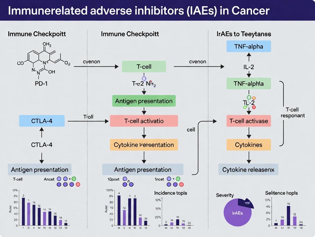
Understanding irAEs: Mechanisms, Spectrum, and Incidence of Immune Toxicity from Checkpoint Blockade
Technical Support Center
Troubleshooting Guide & FAQs
Q1: In our mouse model, we observe no irAEs after anti-CTLA-4 administration, despite successful tumor regression. What are potential experimental issues?
- A: This suggests a failure to break peripheral tolerance. Key troubleshooting steps:
- Verify Model & Timing: Ensure you are using a genetically competent (non-immunodeficient) strain. The onset of irAEs is often dose- and time-dependent. Extend the observation period and consider a higher or additional dose.
- Check Treg Depletion/Modulation: For CTLA-4 blockade, assess Treg population dynamics in target organs (e.g., colon, liver) via flow cytometry (FoxP3+ CD4+). Inadequate Treg inhibition may prevent autoimmunity.
- Confirm Target Engagement: Use a competitive binding ELISA to verify that your administered antibody effectively blocks the CTLA-4/B7 interaction in vivo.
- Evaluate Antigen Presence: irAEs often require pre-existing self-antigen presentation. Consider models with tissue-specific antigen expression or co-administration of a relevant self-antigen peptide.
Q2: When quantifying inflammatory cytokines in serum post-anti-PD-1 therapy, what is the optimal panel and timepoint to capture early signs of specific organ toxicity (e.g., colitis vs. pneumonitis)?
- A: A targeted, organ-informed approach is best. See the table below for guidance.
| Target Organ (irAE) | Key Cytokines/Chemokines to Quantify (Mouse) | Key Cytokines/Chemokines to Quantify (Human) | Suggested Early Sampling Timepoint Post-Dose |
|---|---|---|---|
| Colitis | IFN-γ, TNF-α, IL-17A, IL-6, CXCL10 | IFN-γ, TNF-α, IL-17, IL-6, calprotectin (stool) | 7-10 days |
| Pneumonitis | IFN-γ, IL-6, CXCL9, CXCL10 | IFN-γ, IL-6, CXCL9, CXCL10, KL-6 (serum) | 14-21 days |
| Hepatitis | IFN-γ, IL-6, CXCL10, ALT/AST (enzymes) | IFN-γ, IL-6, CXCL10, ALT/AST | 21-28 days |
| General Inflammation | IL-2, IL-1β, CRP (mouse analog) | IL-2, IL-1β, C-Reactive Protein (CRP) | 7 days |
Protocol: Multiplex Cytokine Assay (Luminex)
- Sample Collection: Collect serum via retro-orbital or cardiac puncture (mouse) or standard venipuncture (human). Centrifuge at 10,000xg for 10min at 4°C.
- Assay Setup: Thaw MILLIPLEX MAP kit reagents. Prepare standards in 7-point serial dilution.
- Plate Loading: Add 25µL of standards, controls, and samples to the pre-wetted 96-well filter plate. Add 25µL of antibody-immobilized beads.
- Incubation: Seal plate, incubate overnight at 4°C on a plate shaker.
- Washing: Wash 2x with wash buffer using a vacuum manifold.
- Detection: Add 25µL of biotinylated detection antibody, incubate 1hr. Add 25µL Streptavidin-PE, incubate 30min.
- Reading: Wash, resuspend in sheath fluid, read on a Luminex analyzer (e.g., MAGPIX). Analyze data with xPONENT software.
Q3: How can we experimentally distinguish between irAEs driven by direct T-cell attack on tissue vs. those driven by a cytokine storm?
- A: Employ a combination of histology and cellular assays.
- Histopathology: Perform H&E staining of affected tissue. Direct T-cell attack shows dense lymphocytic infiltrate directly damaging parenchyma. Cytokine storm may show more diffuse inflammation, edema, and hemorrhage with a mixed immune infiltrate.
- Immunofluorescence/IHC: Stain for Granzyme B+ CD8+ T cells in direct contact with target cells (e.g., colonic epithelium, hepatocytes). Their presence indicates direct cytotoxicity.
- T-cell Depletion Experiment: Administer a depleting anti-CD8 antibody in vivo. If symptoms abate, direct cytotoxicity is implicated. If not, the primary driver may be CD4+ T cells or innate immunity via cytokines.
- Serum Transfer: Transfer serum from ICI-treated mice with severe irAEs into naive mice. If symptoms rapidly replicate, a soluble factor (cytokine) is likely dominant.
Diagram: ICI Mechanism Breaking Peripheral Tolerance
Title: ICI Disruption of Peripheral Tolerance Leading to irAEs
The Scientist's Toolkit: Key Research Reagent Solutions
| Item/Category | Example Product/Catalog # | Primary Function in irAE Research |
|---|---|---|
| ICI Therapeutic Antibodies (Mouse) | InVivoPlus anti-mouse PD-1 (CD279), InVivoPlus anti-mouse CTLA-4 (CD152) | For in vivo blockade of checkpoint pathways to induce irAE phenotypes in preclinical models. |
| Fluorochrome-conjugated Antibodies for Flow Cytometry | Anti-mouse CD3, CD4, CD8, FoxP3, CD45, PD-1, CTLA-4, Tim-3, LAG-3 | To profile immune cell subsets, activation, and exhaustion status in blood, lymphoid organs, and target tissues. |
| Multiplex Cytokine/Chemokine Panel | MILLIPLEX MAP Mouse Cytokine/Chemokine Magnetic Bead Panel | To quantify a broad spectrum of inflammatory mediators in serum or tissue homogenates simultaneously. |
| Tissue Dissociation Kit | Miltenyi Biotec Tumor Dissociation Kit (gentleMACS) | For generating single-cell suspensions from solid organs (colon, lung, liver) for downstream cellular analysis. |
| In Vivo T-cell Depletion Antibodies | InVivoPlus anti-mouse CD8α, InVivoPlus anti-mouse CD4 | To functionally validate the role of specific T-cell subsets in driving or mitigating irAEs. |
| Histology Reagents | Formalin, Paraffin, H&E Staining Kit, Antibodies for IHC (e.g., anti-Granzyme B) | For morphological assessment and detection of cytotoxic immune cells within target tissues. |
| ELISA for Checkpoint Molecule Engagement | DuoSet IC ELISA for Human/B7-1 (CD80) Binding | To verify functional blocking activity of ICI antibodies in vitro or measure soluble checkpoint levels in vivo. |
Technical Support Center for irAE Research
Troubleshooting Guide: In Vitro & Ex Vivo Models
Issue 1: Poor T-cell activation in PBMC co-culture assay to model cytokine release.
- Potential Cause: Insufficient antigen presentation or suboptimal checkpoint inhibitor antibody concentration.
- Solution: Titrate the anti-PD-1/PD-L1 antibody (0.1-10 µg/mL). Validate target engagement with flow cytometry for phosphorylated ERK in T-cells. Ensure antigen-presenting cells express adequate MHC levels.
- Protocol: Isolate PBMCs from healthy donors. Co-culture with tumor cell lines expressing relevant tumor antigens at a 10:1 (PBMC:Tumor) ratio. Add titrated ICI. Measure IFN-γ in supernatant by ELISA at 24, 48, and 72 hours.
Issue 2: High background apoptosis in cardiomyocyte cell line treated with ICI-conditioned media.
- Potential Cause: Non-specific cytotoxicity from excessive inflammatory cytokines in conditioned media.
- Solution: Pre-dilute the conditioned media (1:2 to 1:10) in fresh cardiomyocyte maintenance media. Include a neutralizing anti-TNF-α antibody control.
- Protocol: Generate conditioned media from activated PBMC cultures with/without ICI for 48h. Treat human iPSC-derived cardiomyocytes with 50% conditioned media for 24h. Quantify apoptosis via Caspase-3/7 activity assay, normalized to total cell count.
Issue 3: Inconsistent histopathology scoring in murine colitis model.
- Potential Cause: Inadequate blinding or non-standardized region selection for scoring.
- Solution: Implement a double-blind scoring system using established criteria (e.g., Geboes Score adapted for mice). Define specific longitudinal sections of the colon (proximal, mid, distal) for analysis.
- Protocol: C57BL/6 mice treated with anti-CTLA-4. Harvest colon, roll into "Swiss rolls," fix, section, and H&E stain. Score for inflammatory infiltrate, crypt dropout, and ulceration on a 0-4 scale per defined parameter by two independent, blinded pathologists.
Frequently Asked Questions (FAQs)
Q1: What are the recommended positive controls for validating an ex vivo skin explant model of ICI-induced dermatitis? A: Use pre-treated explants from mice with established contact dermatitis (e.g., using dinitrofluorobenzene) or spike cultures with a known cytokine cocktail (e.g., IL-17, IFN-γ, IL-6) to mimic the inflammatory milieu. Anti-CD3/CD28 bead stimulation of resident T-cells can also serve as an activation control.
Q2: Which immune cell markers are most relevant for flow cytometry analysis in a murine model of ICI-induced myocarditis? A: Focus on cardiac tissue infiltrates. A core panel should include: CD45+ (leukocytes), CD3+ (T-cells), with subsets CD4+ and CD8+. Include CD11b+Ly6G+ (neutrophils), CD11b+Ly6C+ (monocytes/macrophages), and FoxP3+ within CD4+ (Tregs). Intracellular staining for TNF-α and IFN-γ in T-cells is critical.
Q3: How do I distinguish ICI-induced hepatitis from viral flare in a patient-derived organoid model? A: Incorporate the following controls: (1) Organoids from healthy donors, (2) Organoids treated with ICI alone, (3) Organoids exposed to relevant viral antigens (e.g., HBV surface antigen). Key readouts include: PCR for viral load, ELISA for granzyme B (T-cell mediated cytotoxicity), and ALT release (hepatocyte damage). A viral flare will show significant increase in viral load alongside cytotoxicity.
Q4: What is the optimal time window to assess pneumonitis in a mouse model after ICI initiation? A: The onset is typically subacute. Perform longitudinal analyses at Day 7, 14, and 21 post-first ICI dose. Day 14 often shows peak infiltration. Monitor daily for clinical signs (dyspnea, weight loss). Analyze bronchoalveolar lavage fluid (BALF) for immune cell counts and cytokines, followed by lung histopathology at endpoint.
Table 1: Spectrum of Select irAEs from Anti-PD-1/PD-L1 Therapy
| Organ System | Toxicity (Grade 3-4) | Median Onset (Weeks) | Key Mediators (Experimental) |
|---|---|---|---|
| Skin | Rash/Dermatitis | 2-4 | CD8+ T-cells, IL-17, IFN-γ |
| Gastrointestinal | Colitis | 6-8 | Lamina propria CD4+/CD8+ T-cells, Fecal microbiota diversity ↓ |
| Hepatic | Hepatitis | 6-12 | Liver CD8+ T-cells, IL-6, TNF-α |
| Pulmonary | Pneumonitis | 8-12 | Pulmonary CD4+ T-cells (Th1, Th17), BALF IFN-γ ↑ |
| Cardiac | Myocarditis | 4-8 | Cardiac muscle CD8+ T-cells, Troponin I/T, anti-striated muscle abs |
Experimental Protocol: Detailed Methodology for irAE Murine Model Characterization
Protocol Title: Multi-parameter Flow Cytometric Analysis of Cardiac Infiltrate in ICI-Induced Myocarditis.
- Model Induction: Inject C57BL/6 mice intraperitoneally with 200 µg anti-PD-1 antibody (clone RMP1-14) and 250 µg anti-CTLA-4 antibody (clone 9D9) on days 0, 3, and 6.
- Tissue Harvest: Euthanize mice on day 21. Perfuse heart with 10 mL cold PBS. Excise heart, mince finely with scalpels.
- Digestion: Incubate tissue in 2 mL digestion media (RPMI + 2 mg/mL Collagenase II + 40 U/mL DNase I) for 45 minutes at 37°C with agitation.
- Single-Cell Suspension: Pass through a 70 µm cell strainer, wash with FACS buffer (PBS + 2% FBS).
- Staining: Block Fc receptors with anti-CD16/32. Stain with surface antibody cocktail (CD45, CD3, CD4, CD8, CD11b, Ly6G, Ly6C) for 30 mins at 4°C. For intracellular cytokines, stimulate cells with PMA/ionomycin for 4h with brefeldin A, then fix/permeabilize before staining for IFN-γ, TNF-α, FoxP3.
- Analysis: Acquire on a flow cytometer capable of detecting ≥10 colors. Use counting beads for absolute cell number quantification per mg of heart tissue. Analyze with FlowJo software.
The Scientist's Toolkit: Research Reagent Solutions
Table 2: Essential Reagents for irAE Mechanistic Research
| Item | Function & Application | Example/Clone |
|---|---|---|
| Recombinant Human IL-2 | Expands & maintains antigen-specific T-cell clones for adoptive transfer models. | Proleukin |
| Anti-Mouse PD-1 (RMP1-14) | Blocks PD-1 in vivo to induce irAEs in murine models. | Clone RMP1-14 |
| Anti-Human CD3/CD28 Dynabeads | Polyclonal T-cell activator for positive control in cytokine release assays. | Gibco |
| iPSC-Derived Cardiomyocytes | Human-relevant in vitro model for cardiotoxicity screening. | Cellular Dynamics |
| Multiplex Cytokine Panel | Simultaneous quantification of 20+ cytokines from limited serum/BALF samples. | Luminex ProcartaPlex |
| TruCount Absolute Counting Beads | For absolute immune cell counts in flow cytometry of tissue infiltrates. | BD Biosciences |
| 16s rRNA Sequencing Kit | Profiles gut microbiome changes associated with ICI-colitis. | Illumina MiSeq |
| Phosflow Antibodies (pERK, pSTAT) | Detects intracellular signaling downstream of immune checkpoint engagement. | BD Phosflow |
Pathway & Workflow Visualizations
Title: ICI Mechanism: Therapeutic Effect vs. irAE Onset
Title: Workflow for Systemic irAE Characterization In Vivo
Title: Proposed Cellular Mechanism of Tissue Damage in irAEs
Technical Support Center: Troubleshooting irAE Incidence & Risk Factor Research
FAQs & Troubleshooting Guides
Q1: In our retrospective cohort study, the incidence rates of colitis for anti-PD-1 agents are significantly lower than published literature. What are potential sources of this data discrepancy? A: Common sources include:
- Case Ascertainment: Relying solely on diagnostic codes (e.g., ICD-10) underestimates incidence. Implement a protocol combining codes, corticosteroid prescription surges, and endoscopic biopsy reports.
- Grading Criteria Inconsistency: Ensure uniform application of CTCAE (Common Terminology Criteria for Adverse Events) v5.0 grading across your data review team. Subclinical findings (Grade 1) are often missed in real-world data.
- Onset Window Definition: Published trials often use "from first dose to 90-100 days post-last dose." Verify your study's observation window aligns with this.
Q2: Our multi-variable regression model for pneumonitis risk factors is unstable, with wide confidence intervals for key covariates like pre-existing lung disease. How can we improve the model? A: This suggests potential overfitting or rare event issues.
- Event per Variable (EPV) Rule: Ensure you have ≥10-15 events for each candidate variable. For rare irAEs like pneumonitis, consider Firth's penalized-likelihood logistic regression to reduce small-sample bias.
- Variable Coding: Avoid categorizing continuous variables (e.g., age) arbitrarily. Use splines or clinically relevant thresholds.
- Confounder Specification: Explicitly define and measure confounding variables like baseline steroid use, prior thoracic radiation, and performance status (ECOG score).
Q3: When comparing irAE incidence between anti-PD-1 and anti-CTLA-4 drug classes, how should we handle patients on combination therapy? A: Combination therapy presents a distinct risk profile and must be analyzed separately to avoid confounding.
- Define three clear cohorts for comparison: Monotherapy Anti-PD-1/L1, Monotherapy Anti-CTLA-4, and Combination Therapy.
- In primary analysis, exclude combination patients. Use them in a secondary, descriptive analysis.
- For time-to-event analysis (e.g., time to first Grade ≥2 irAE), censor patients at the start of a subsequent, different therapy.
Q4: What is the gold-standard method to establish causality between an ICI and an irAE like myocarditis for our case series? A: Use a standardized causality assessment framework. The WHO-UMC (Uppsala Monitoring Centre) system or Naranjo scale, adapted for oncology, is recommended.
- Protocol: For each case, systematically document:
- Temporal sequence (onset relative to ICI dosing).
- Dechallenge (improvement after ICI held/steroids initiated).
- Rechallenge (recurrence upon re-initiation—rarely intentional).
- Alternative etiologies ruled out (e.g., troponin elevation from sepsis, other cardiotoxic drugs).
- Known pathological mechanism (lymphocytic infiltration on biopsy is definitive).
Experimental Protocol: High-Dimensional Immune Phenotyping for irAE Risk Prediction
Objective: To identify pre-treatment peripheral immune cell subsets associated with subsequent development of severe (Grade ≥3) irAEs using mass cytometry (CyTOF).
Methodology:
- Sample Collection: Collect peripheral blood mononuclear cells (PBMCs) from patients prior to first ICI dose. Process within 4 hours using Ficoll-Paque density gradient centrifugation. Cryopreserve in liquid nitrogen.
- Panel Design: Design a 40-parameter CyTOF panel targeting T cell (activation, exhaustion, memory), B cell, myeloid, and innate lymphoid cell lineages. Include lineage markers (CD3, CD4, CD8, CD19, CD14, CD56), checkpoint receptors (PD-1, CTLA-4, LAG-3, TIM-3), and functional markers (Ki-67, CD38, HLA-DR).
- Staining & Acquisition: Thaw PBMCs, viability stain with cisplatin, surface stain with metal-tagged antibodies, fix, and acquire on a CyTOF instrument. Normalize data using bead standards.
- Data Analysis: Use automated clustering (e.g., FlowSOM, PhenoGraph) to identify cell populations. Perform differential abundance analysis between patients who did vs. did not develop severe irAEs (Mann-Whitney U test, FDR correction). Validate findings in an independent cohort.
Data Presentation Tables
Table 1: Incidence Rates of Select irAEs by ICI Drug Class (Pooled Clinical Trial Data)
| irAE Type | Anti-PD-1/L1 Monotherapy (%) | Anti-CTLA-4 Monotherapy (%) | Combination Therapy (%) | Grade ≥3 (Combination) (%) |
|---|---|---|---|---|
| Colitis | 1.0 - 2.5 | 8.0 - 12.0 | 9.0 - 14.0 | 7.0 - 9.0 |
| Pneumonitis | 2.0 - 5.0 | 1.0 - 2.5 | 6.0 - 10.0 | 2.0 - 4.0 |
| Hepatitis | 1.0 - 3.0 | 4.0 - 8.0 | 10.0 - 15.0 | 5.0 - 8.0 |
| Hypophysitis | <1.0 | 5.0 - 10.0 | 5.0 - 8.0 | 1.0 - 3.0 |
| Rash | 15.0 - 25.0 | 20.0 - 35.0 | 35.0 - 50.0 | 3.0 - 5.0 |
Table 2: Validated Patient-Specific Risk Factors for irAEs
| Risk Factor | Associated irAE(s) | Odds/Hazard Ratio (Approx.) | Evidence Level |
|---|---|---|---|
| Preexisting Autoimmune Disease | Any irAE, Colitis, Arthritis | OR: 1.5 - 2.5 | Meta-analysis |
| Chronic Obstructive Pulmonary Disease | Pneumonitis | HR: 2.0 - 3.5 | Retrospective Cohort |
| Combination ICI Therapy | Multiple (See Table 1) | HR: 1.5 - 4.0* | Pooled Trial Data |
| High ICI Dose (CTLA-4) | Colitis, Hypophysitis | Dose-dependent | Phase I/II Data |
| Gut Microbiome Signature | Colitis, Response | Specific taxa enrichment | Prospective Cohort |
*Varies by specific irAE.
Visualizations
The Scientist's Toolkit: Research Reagent Solutions
| Reagent/Material | Function in irAE Research |
|---|---|
| Recombinant Human PD-1/CTLA-4 Fc Chimeras | Used in competitive binding assays to quantify levels of anti-drug antibodies or to block pathways in in vitro T cell activation assays. |
| Multiplex Cytokine Panels (e.g., 35-plex) | Quantify a broad profile of inflammatory cytokines (IFN-γ, IL-6, IL-17, etc.) from patient serum to identify irAE-associated signatures. |
| Phospho-Specific Flow Cytometry Antibodies | Assess signaling pathway activation (pSTAT1, pSTAT3, pAKT) in immune cell subsets from patient PBMCs pre- and post-ICI. |
| Human Immune Cell Co-culture Systems | In vitro models to study ICI-induced T cell-mediated killing of organ-specific cells (e.g., cardiomyocytes, hepatocytes). |
| DNA Methylation & ATAC-Seq Kits | Profile epigenetic changes in immune cells associated with irAE susceptibility and progression. |
| 16S rRNA / Shotgun Metagenomic Sequencing Kits | Analyze gut microbiome composition as a predictive biomarker for colitis and other irAEs. |
Technical Support Center
Troubleshooting Guides & FAQs
Q1: In our murine model, why are we observing a delayed onset of colitis compared to the published median of 6 weeks post-ICI initiation?
- A: Delayed onset can be influenced by multiple factors. First, verify the genetic background of your mice; C57BL/6 strains typically show onset at 5-7 weeks, while BALB/c may exhibit different kinetics. Second, check the dosing regimen. Sub-optimal dosing (e.g., lower antibody concentration or extended intervals between doses) can delay immune activation. Third, consider the microbiome. Mice with a diverse, "dirty" microbiome often develop irAEs sooner than germ-free or antibiotic-treated cohorts. Troubleshooting Protocol: 1) Confirm anti-PD-1/anti-CTLA-4 antibody clone, concentration, and injection schedule (e.g., 200 µg i.p., twice weekly). 2) Perform fecal 16s rRNA sequencing on a sentinel cohort to characterize baseline microbiome. 3) Include a positive control group using a published protocol with known kinetics.
Q2: Our flow cytometry data from blood samples during pneumonitis is inconsistent. How can we reliably track immune cell kinetics?
- A: Blood sampling alone may not capture tissue-localized kinetics. Pneumonitis is driven by lung-infiltrating T cells and macrophages. Troubleshooting Protocol: Implement serial bronchoalveolar lavage (BAL) alongside peripheral blood collection. For BAL: anesthetize mouse, cannulate trachea, lavage lung with 0.8 mL cold PBS x3. Process BAL fluid and blood for: 1) Surface staining (CD45, CD3, CD4, CD8, PD-1, TIM-3), 2) Intracellular staining for cytokines (IFN-γ, TNF-α) after PMA/ionomycin stimulation, and 3) Myeloid panel (CD11b, CD11c, F4/80, Ly6G, Ly6C). Use counting beads for absolute quantification. Time points should be weekly and at symptom onset.
Q3: When monitoring hepatotoxicity via ALT/AST, what threshold constitutes a significant irAE event versus background fluctuation in our model?
- A: Use both fold-change and absolute value criteria. A consistent threshold is a ≥3-fold increase from baseline and an absolute value exceeding 100 U/L. See Table 1 for detailed species-specific thresholds.
Q4: How do we distinguish early-onset, acute irAEs from late-onset, chronic ones in preclinical studies for mechanistic work?
- A: Establish clear temporal definitions and distinct sampling protocols. In mice, Early-onset (< 2 months) is often driven by pre-existing auto-reactive T cells. Late-onset (> 2 months) may involve epitope spreading or persistent inflammation. For mechanistic studies: 1) Perform T-cell receptor sequencing on infiltrating lymphocytes at both time points. 2) Profile serum cytokines (e.g., IL-6, IL-17, CXCL10) longitudinally. Late-onset events often show sustained elevation of fibrotic markers like TGF-β.
Q5: The duration of dermatitis in our model is highly variable. What are the key experimental variables to control?
- A: Key variables are drug half-life, scoring system objectivity, and skin sampling time. Troubleshooting Protocol: 1) Use the same ICI antibody lot with verified in vivo half-life (~10 days for most IgG2a). 2) Implement a blinded, quantitative scoring system (e.g., 0-3 scale for erythema, scaling, thickness). 3) For histology, biopsy at peak severity AND during resolution. Fix in formalin for H&E (inflammatory grade) and also snap-freeze for RNA extraction (cytokine profiling).
Data Presentation
Table 1: Characteristic Onset and Duration of Common irAEs in Preclinical Models
| irAE Organ System | Typical Onset Post-ICI Initiation (Mouse Model) | Typical Duration (Without Intervention) | Key Clinical/Lab Marker |
|---|---|---|---|
| Colitis | 5 - 7 weeks | 2 - 4 weeks (can be progressive) | Weight loss >15%, diarrhea, histology (immune infiltrate) |
| Dermatitis | 2 - 4 weeks | 1 - 3 weeks | Clinical score, histology (epidermal thickness, infiltrate) |
| Hepatitis | 3 - 5 weeks | 1 - 2 weeks (transaminase elevation) | Serum ALT/AST (≥3x baseline) |
| Pneumonitis | 6 - 12 weeks | Can be chronic (>8 weeks) | Histology (alveolitis, fibrosis), BAL immune cell count |
| Myocarditis | 1 - 4 weeks | Often acute and severe | Histology (myocyte necrosis, CD3+ T cell infiltrate), Troponin I |
Table 2: Kinetics of Key Serum Biomarkers in ICI-Treated Mice
| Biomarker | Peak Elevation Relative to irAE Onset | Correlation with irAE Severity | Assay Recommendation |
|---|---|---|---|
| IL-6 | Precedes (3-5 days) and peaks at onset | High | Multiplex Luminex (serum) |
| CXCL10 | Concurrent with clinical onset | Moderate to High | ELISA (serum, BAL fluid) |
| TNF-α | Variable, often early | Moderate | Multiplex Luminex (serum, tissue homogenate) |
| Troponin I | Concurrent with clinical signs of myocarditis | High for cardiac events | High-sensitivity ELISA (serum) |
| Anti-nuclear Abs (ANA) | Late (>8 weeks), chronic phases | Low in mice, higher in human context | Indirect Immunofluorescence (serum) |
Experimental Protocols
Protocol 1: Longitudinal Monitoring of ICI-Induced Colitis in Mice Objective: To assess the onset, severity, and duration of colitis. Materials: See "Scientist's Toolkit" below. Method:
- Dosing: Administer anti-mouse PD-1 (clone RMP1-14) and CTLA-4 (clone 9D9) antibodies intraperitoneally at 200 µg each, twice weekly for 3 doses.
- Clinical Scoring: Weigh mice 3x weekly. Score stool consistency (0: normal, 1: soft, 2: diarrhea) and monitor for rectal prolapse.
- Endpoint Sampling: Euthanize cohorts at weeks 3, 5, 7, and 9 post-first dose (n=5/group). Include isotype control group.
- Tissue Collection: Isolate colon, measure length, and roll into a "Swiss roll". Fix in 10% neutral buffered formalin for 24h.
- Histopathology: Paraffin-embed, section, H&E stain. Score histology blindly (0-4) for inflammatory infiltrate, crypt loss, and mucosal hyperplasia.
- Flow Cytometry: Prepare single-cell suspension from lamina propria (using collagenase/DNase digestion). Stain for T cells (CD45, CD3, CD4, CD8, RORγt, FoxP3) and myeloid cells.
Protocol 2: Cytokine Profiling for irAE Kinetics Objective: To quantify systemic and tissue-specific cytokine changes during irAE development. Method:
- Serum Collection: Collect blood via retro-orbital bleed at pre-defined intervals (e.g., baseline, weekly). Allow clotting, centrifuge at 10,000xg for 10 min, store serum at -80°C.
- Tissue Homogenization: Snap-freeze target organs (e.g., liver, lung). Homogenize 30mg tissue in 300µL PBS with protease inhibitors using a bead homogenizer. Centrifuge at 12,000xg for 15 min at 4°C, collect supernatant.
- Multiplex Assay: Use a commercial mouse 10-plex cytokine panel (e.g., from Bio-Rad or Millipore) measuring IFN-γ, IL-1β, IL-2, IL-4, IL-6, IL-10, IL-17A, MCP-1, TNF-α, CXCL10. Follow manufacturer's protocol for the magnetic bead-based assay. Read on a Luminex instrument.
- Data Analysis: Normalize tissue cytokine levels to total protein concentration (BCA assay). Plot concentration over time for each analyte.
Diagrams
Title: ICI Mechanism and irAE Onset Pathways
Title: Experimental irAE Kinetics Workflow
The Scientist's Toolkit: Research Reagent Solutions
| Item | Function & Application in irAE Kinetics Studies |
|---|---|
| Anti-mouse PD-1 (clone RMP1-14) | In vivo blocking antibody to inhibit PD-1 signaling, inducing immune activation and irAEs in murine models. |
| Anti-mouse CTLA-4 (clone 9D9) | In vivo blocking antibody to inhibit CTLA-4, often used in combination with anti-PD-1 to model severe/early-onset irAEs. |
| Luminex Multiplex Assay Mouse Panel | For simultaneous quantification of multiple cytokines (e.g., IL-6, IFN-γ, TNF-α) from low-volume serum or tissue homogenate samples. |
| Collagenase Type VIII & DNase I | Enzyme cocktail for digesting solid tissues (e.g., colon, lung) to generate single-cell suspensions for flow cytometry. |
| Fluorochrome-conjugated Antibodies (CD45, CD3, CD4, CD8, FoxP3) | Essential for immunophenotyping immune cell subsets in blood, lymphoid organs, and inflamed tissues via flow cytometry. |
| TruSeq T-Cell Receptor (TCR) Sequencing Kit | To track clonal expansion and dynamics of T-cell populations over the course of irAE development and resolution. |
| Automated Serum Chemistry Analyzer | For high-throughput, precise measurement of liver enzymes (ALT/AST) and other biomarkers of organ damage. |
The Role of Microbiome and Genetic Predisposition in irAE Development
Technical Support Center: Troubleshooting & FAQs
This support center provides guidance for experimental challenges in investigating microbiome and genetic factors in immune-related adverse events (irAEs) from Immune Checkpoint Inhibitor (ICI) therapy. Content is framed within the thesis: "Managing immune-related adverse events (irAEs) from ICIs: An integrated approach targeting host predisposition and microbial modulation."
Frequently Asked Questions (FAQs)
Q1: During 16S rRNA sequencing of stool samples from ICI-treated patients, we get low taxonomic resolution (e.g., only to family level). How can we improve resolution to genus/species? A1: Low resolution is often due to targeting only the V1-V3 or V4 hypervariable regions. Use a full-length 16S rRNA gene sequencing approach (PacBio, Oxford Nanopore) or shift to shotgun metagenomic sequencing. Ensure primers (e.g., 27F/1492R for full-length) are validated. For established V4 datasets, supplement with a targeted qPCR assay for specific genera of interest (e.g., Akkermansia, Bacteroides).
Q2: Our GWAS on irAE susceptibility yields candidate SNPs in non-coding regions. How do we functionally validate their role in immune cell regulation? A2: Map SNPs to chromatin accessibility (ATAC-seq) and histone modification (ChIP-seq) data from relevant immune cells (e.g., CD8+ T cells, Tregs). Use CRISPR/Cas9 to create isogenic cell lines with risk vs. protective alleles in primary human T cells. Assess downstream effects on gene expression (RNA-seq), protein binding (EMSA), and T-cell activation/phenotype via flow cytometry (CD69, PD-1, CTLA-4).
Q3: When colonizing germ-free mice with patient-derived microbiota, we observe inconsistent irAE phenotypes. What are key controls for fecal microbiota transplantation (FMT) studies? A3: Inconsistency often stems from donor sample viability, host diet, and cage effects.
- Donor: Use freshly processed or cryopreserved stocks with documented viability. Pool samples from multiple donors with same irAE phenotype to create a consortium.
- Recipients: House germ-free mice in isolators, maintain on identical autoclaved diet. Co-house recipients of same experimental group to normalize microbiota.
- Verification: Confirm engraftment via pre-ICI stool sequencing at day 7-14 post-FMT.
Q4: How can we mechanistically link a specific bacterial taxa to T-cell infiltration in a specific organ (e.g., colon) during irAE development? A4: Employ a multi-omics approach:
- Spatial Correlation: Perform 16S seq on mucosal scraping from affected organ and correlate with immunohistochemistry (IHC) for CD3+/CD8+ cells from adjacent tissue section.
- Bacterial Isolation: Isolate the candidate bacterium anaerobically from patient stool.
- Gnotobiotic Model: Monocolonize germ-free mice with the isolate, administer ICI, and compare T-cell recruitment (flow cytometry on lamina propria lymphocytes) to germ-free controls.
- Metabolite Screening: Analyze bacterial supernatant by LC-MS for immunomodulatory metabolites (e.g., short-chain fatty acids, bile acid derivatives). Test purified metabolites in in vitro T-cell assays.
Key Experimental Protocols
Protocol 1: Longitudinal Metagenomic Analysis for irAE Prediction Objective: To identify microbial species and pathways predictive of irAE onset from pre-treatment stool samples.
- Sample Collection: Collect stool from patients prior to first ICI infusion (Day 0) and at regular intervals (e.g., Weeks 3, 6, 12). Immediately snap-freeze in liquid nitrogen and store at -80°C.
- DNA Extraction: Use bead-beating mechanical lysis with a kit designed for hard-to-lyse bacteria (e.g., QIAamp PowerFecal Pro DNA Kit). Include extraction controls.
- Shotgun Sequencing: Library prep with Illumina DNA Prep. Sequence on Illumina NovaSeq to a minimum depth of 10 million paired-end 150bp reads per sample.
- Bioinformatics: Process with the Human Microbiome Project QIIME2 pipeline. Remove human reads with KneadData. Perform taxonomic profiling using MetaPhIAn3 and functional profiling with HUMAnN2. Normalize to copies per million (CPM).
- Statistical Modeling: Use MaAsLin2 for longitudinal analysis. Build a Random Forest classifier (scikit-learn in Python) using species-level abundance at Day 0 to predict irAE development (binary outcome). Validate with nested cross-validation.
Protocol 2: Functional Validation of a Host Genetic Variant Using CRISPR-Cas9 in Primary T Cells Objective: To assess the impact of a non-coding irAE-associated SNP on T-cell gene expression and function.
- Cell Source: Isolate CD4+ naïve T cells from healthy donor PBMCs using magnetic negative selection.
- CRISPR RNP Electroporation: Design two crRNAs targeting near the SNP. Complex with HiFi Cas9 protein and transfect into cells via electroporation (Neon System, 1600V, 10ms, 3 pulses). Include a non-targeting crRNA control.
- Genotype Editing Verification: Harvest genomic DNA 48h post-electroporation. Perform PCR on the target region and sequence via Sanger. Use TIDE analysis to quantify editing efficiency.
- Phenotypic Assay: Differentiate edited T cells under Treg-polarizing (TGF-β, IL-2) or Th17-polarizing (IL-1β, IL-6, IL-23) conditions for 5 days. Analyze by:
- Flow cytometry: FOXP3, CTLA-4 (Tregs); RORγt, IL-17A (Th17).
- Secreted cytokines: Multiplex Luminex assay on supernatant.
- Gene Expression: RT-qPCR for target gene near SNP.
Summarized Quantitative Data
Table 1: Selected Genetic Variants Associated with Increased irAE Risk
| Gene/Region | SNP ID | Associated irAE | Odds Ratio (95% CI) | P-value | Study Cohort (N) |
|---|---|---|---|---|---|
| CTLA4 | rs231775 | Colitis, Hypophysitis | 1.45 (1.21-1.74) | 4.2 x 10^-5 | Multi-center (1245) |
| HLA-DRB1*11:01 | - | Pneumonitis | 3.12 (2.05-4.75) | 8.7 x 10^-8 | Japanese (1114) |
| TNFRSF1A | rs1800693 | Any Grade 3+ irAE | 2.01 (1.45-2.78) | 1.1 x 10^-5 | European (867) |
| FCGR2B | rs1050501 | Skin Toxicity | 1.82 (1.33-2.49) | 1.4 x 10^-4 | Pan-cancer (932) |
Table 2: Microbial Taxa Associated with irAE Risk in Pre-Treatment Stool
| Taxonomic Group | Association Direction (irAE Risk) | Reported Effect Size (Fold-Change) | Primary irAE Linked | Sequencing Method |
|---|---|---|---|---|
| Bacteroides fragilis | Decreased | 0.3x (Lower Abundance) | Colitis | Shotgun Metagenomics |
| Akkermansia muciniphila | Increased | 4.1x (Higher Abundance) | Pneumonitis | 16S rRNA (V4) |
| Faecalibacterium prausnitzii | Decreased | 0.5x (Lower Abundance) | Arthritis | Shotgun Metagenomics |
| Bifidobacterium longum | Decreased | 0.4x (Lower Abundance) | Colitis | 16S rRNA (V3-V4) |
Signaling & Workflow Diagrams
Title: Integrated Multi-Omics Workflow for irAE Risk Prediction
Title: Microbial Metabolite Modulates Tregs and Inflammation
Title: Genetic Risk Allele Impairs Treg Function
The Scientist's Toolkit: Research Reagent Solutions
| Item | Function & Application in irAE Research |
|---|---|
| Gnotobiotic Mouse Lines | Germ-free or defined-flora mice for causal testing of human microbiota in irAE models. Essential for FMT studies. |
| Cryopreservation Media for Stool | Specialized media (e.g., with glycerol) for long-term viability storage of donor stool samples for reproducible FMT. |
| Humanized Immune System Mice | NSG mice engrafted with human PBMCs or hematopoietic stem cells to study human-specific immune responses to ICIs. |
| Mucin-Coated Culture Plates | For enriching and cultivating oxygen-sensitive mucolytic bacteria (e.g., Akkermansia) from patient samples. |
| HLA Tetramers (for ICI antigens) | To track and isolate ICI-reactive T-cell clones from patients experiencing irAEs, linking genetics to immune response. |
| Neutralizing Antibodies (anti-IL-6, anti-IL-17) | Used in in vivo models to validate cytokine-driven mechanisms of specific irAEs suggested by microbiome data. |
| Selective Bacterial Growth Media | e.g., Bacteroides Bile Esculin agar for selective growth of Bacteroides species from complex communities. |
| CITE-Seq Antibody Panels | For combined single-cell RNA and surface protein sequencing (scRNA-seq + protein) to deeply phenotype immune cells in irAE tissues. |
Clinical Management Frameworks: Assessment, Grading, and Evidence-Based Intervention Protocols
Technical Support Center: Troubleshooting irAE Assessment
FAQ & Troubleshooting Guide
Q1: How do I distinguish between a Grade 2 and Grade 3 colitis event using CTCAE v5.0 criteria?
A: The key distinction lies in the intensity of symptoms and the intervention required.
- Grade 2 Colitis: Symptoms increase from baseline (e.g., more frequent stools, abdominal pain, mucus/blood in stool). It limits instrumental Activities of Daily Living (ADL).
- Grade 3 Colitis: Severe symptoms, limiting self-care ADL. This includes severe abdominal pain, peritoneal signs, fever, or ileus. Hospitalization for clinical management is typically required.
Q2: When grading rash, what is the operational definition of "Body Surface Area (BSA) involvement"?
A: The "rule of palms" is the standard clinical estimate, where the patient's palm (including fingers) represents approximately 1% of their BSA. Use this to quantify:
- Grade 1: <10% BSA involvement with or without symptoms (e.g., erythema, pruritus).
- Grade 2: 10-30% BSA involvement.
- Grade 3: >30% BSA involvement.
Q3: How should I grade laboratory abnormalities (e.g., AST/ALT elevation) that are asymptomatic?
A: Grade strictly based on the laboratory value multiples above the Upper Limit of Normal (ULN), as per CTCAE v5.0 tables. Symptoms do not modify the grade for pure lab abnormalities.
Q4: A patient on an ICI develops dyspnea. How do I protocolize the workup to differentiate between pneumonitis (an irAE) and disease progression?
A: Follow this diagnostic workflow:
- Immediate Action: Hold ICI and administer supplemental O2 if needed.
- Initial Workup: Obtain high-resolution CT chest, pulse oximetry, and full pulmonary function tests (including DLCO).
- Exclude Alternatives: Perform infectious workup (sputum culture, PCR for respiratory pathogens, serum procalcitonin). Consider cardiac evaluation (ECHO, BNP) if indicated.
- Confirmatory Evidence: Bronchoscopy with BAL for cell differential (lymphocytosis supports pneumonitis) and microbiological studies. Consider biopsy in focal lesions.
- Grade According to CTCAE v5.0: Based on imaging findings, symptom severity (rest vs. exertion), and oxygen requirement.
Table 1: Common irAEs: CTCAE v5.0 Grading and Initial Management Actions
| irAE Category | Grade 1 (Mild) | Grade 2 (Moderate) | Grade 3 (Severe) | Grade 4 (Life-threatening) | Recommended Action (Grade-based) |
|---|---|---|---|---|---|
| Colitis | Asymptomatic; clinical or diagnostic observations only | Abdominal pain; mucus or blood in stool | Severe pain; peritoneal signs; hospitalization indicated | Life-threatening consequences; urgent intervention indicated | 1: Monitor. 2: Hold ICI, start corticosteroids. 3-4: Permanently discontinue ICI, high-dose IV corticosteroids. |
| Pneumonitis | Radiographic changes only | Symptomatic, limiting instrumental ADL | Symptoms limiting self-care ADL; oxygen indicated | Life-threatening respiratory compromise | 1: Monitor. 2: Hold ICI, consider corticosteroids. 3-4: Permanently discontinue ICI, high-dose IV corticosteroids +/- immunosuppressants. |
| Hepatitis | AST/ALT ≤3x ULN AND/OR Total Bilirubin ≤1.5x ULN | AST/ALT >3-5x ULN AND/OR Bilirubin >1.5-3x ULN | AST/ALT >5-20x ULN AND/OR Bilirubin >3-10x ULN | AST/ALT >20x ULN AND/OR Bilirubin >10x ULN | 1: Monitor. 2: Hold ICI, consider corticosteroids. 3-4: Permanently discontinue ICI, high-dose IV corticosteroids. |
| Rash | Covering <10% BSA with or without symptoms | Covering 10-30% BSA | Covering >30% BSA; oral corticosterosticosteroids indicated | Life-threatening consequences | 1: Topical therapies. 2: Hold ICI, topical/oral corticosteroids. 3-4: Permanently discontinue ICI, systemic corticosteroids. |
Experimental Protocols
Protocol 1: Systematic irAE Identification and Grading in Preclinical Models
Objective: To consistently identify and grade irAE-like toxicities in murine models treated with combination ICIs.
Materials: See "Research Reagent Solutions" below. Methodology:
- Cohort Setup: Randomize mice into control, anti-PD-1, anti-CTLA-4, and combination therapy groups (n=10/group).
- Dosing: Administer antibodies via intraperitoneal injection per established dosing schedule (e.g., 200 µg anti-PD-1, 100 µg anti-CTLA-4, twice weekly for 3 weeks).
- Daily Monitoring: Weigh animals daily. Score for clinical signs using a standardized sheet: piloerection, posture, activity, skin rash, and diarrhea (scale 0-3).
- Bi-weekly Blood Collection: Collect ~100 µL via submandibular vein for serum chemistry (ALT, AST) and cytokine profiling (IFN-γ, IL-6, TNF-α via ELISA).
- Tissue Harvest: Euthanize at endpoint (or humane endpoint). Collect and preserve liver, colon, lung, and heart in 10% formalin (for H&E) and RNAlater (for transcriptomics).
- Histopathology: Blind scoring of H&E slides by a veterinary pathologist using a semi-quantitative scale (0=None, 1=Minimal, 2=Mild, 3=Moderate, 4=Severe) for inflammation, necrosis, and immune cell infiltration.
- Grading Translation: Map clinical, serum, and histopath scores to approximate human CTCAE grades (e.g., >15% weight loss = Grade 3; severe lymphocytic infiltration in liver = Grade 3-4 hepatitis).
Protocol 2: In Vitro T-cell Activation Assay for irAE Mechanism
Objective: To assess the differential activation of primary human T-cells in response to ICI exposure, modeling initial steps of autoimmunity.
Methodology:
- PBMC Isolation: Isolate PBMCs from healthy donor blood using Ficoll density gradient centrifugation.
- T-cell Stimulation: Coat 96-well plates with anti-CD3 (1 µg/mL) and soluble anti-CD28 (2 µg/mL). Add PBMCs (2x10^5/well).
- ICI Treatment: Add clinical-grade anti-PD-1 (e.g., Nivolumab, 10 µg/mL) or anti-CTLA-4 (e.g., Ipilimumab, 10 µg/mL) to treatment wells. Include isotype control.
- Incubation: Culture for 72 hours at 37°C, 5% CO2.
- Readouts:
- Proliferation: Measure via CFSE dilution using flow cytometry on CD4+ and CD8+ gates.
- Cytokine Release: Harvest supernatant at 48h. Quantify IFN-γ, IL-2, IL-17, and GM-CSF via multiplex ELISA.
- Activation Markers: Stain cells at 24h for CD25, CD69, and PD-1 using flow cytometry.
- Data Analysis: Compare fold-changes in proliferation and cytokine release in ICI-treated wells vs. isotype control. Correlate hyper-activation profiles with cytokines linked to specific irAEs (e.g., IL-17 with colitis).
Visualizations
TITLE: ICI Mechanism and irAE Risk Pathway
TITLE: irAE Clinical Assessment and Management Workflow
The Scientist's Toolkit: Research Reagent Solutions
| Item / Reagent | Function in irAE Research | Example Catalog # / Note |
|---|---|---|
| InVivoMab anti-mouse PD-1 (CD279) | Blocks PD-1 in murine models to induce ICI effects and potential irAEs. | Bio X Cell, BE0273 |
| InVivoMab anti-mouse CTLA-4 (CD152) | Blocks CTLA-4 for combinatorial checkpoint inhibition studies. | Bio X Cell, BE0164 |
| InVivoMab rat IgG2a isotype control | Critical control for antibody experiments in vivo. | Bio X Cell, BE0089 |
| Mouse AST/ALT ELISA Kit | Quantifies liver transaminases for grading hepatitis in preclinical models. | Abcam, ab263882 / ab282882 |
| Mouse IFN-gamma ELISA Kit | Measures key Th1 cytokine elevated in many irAE pathologies. | BioLegend, 430804 |
| LIVE/DEAD Fixable Viability Dyes | For flow cytometry to exclude dead cells during immune profiling. | Thermo Fisher, L34955/L34957 |
| Anti-human CD3/CD28 Antibodies | For polyclonal stimulation of T-cells in mechanistic in vitro assays. | Stemcell Tech, 10970/10971 |
| Human Cytokine/Chemokine Panel | Multiplex assay to profile broad cytokine storms associated with irAEs. | Milliplex, HCYTA-60K |
| RNAlater Stabilization Solution | Preserves tissue RNA for subsequent transcriptomic analysis of affected organs. | Thermo Fisher, AM7020 |
| Formalin-Fixed Paraffin-Embedding (FFPE) Kit | Standard for preparing tissue for histopathological grading of irAEs. | Various suppliers |
Technical Support Center: Managing irAEs in Preclinical & Clinical Research
FAQ & Troubleshooting Guide
Q1: In our murine model, administration of corticosteroids to mitigate an ICI-induced colitis-like irAE is blunting the anti-tumor efficacy of the PD-1 inhibitor. How can we troubleshoot this? A: This is a common experimental challenge. The primary issue is often the timing and dose of corticosteroid intervention.
- Troubleshooting Steps:
- Review Intervention Timing: Delay corticosteroid initiation. Implement a detailed clinical scoring system (see Table 1) and only initiate prednisolone (e.g., 10 mg/kg/day) when a moderate score is sustained for 48 hours, mimicking clinical guidelines.
- Taper Protocol: Avoid abrupt cessation. After 5-7 days of therapeutic dose, implement a 10-14 day taper (reduce dose by ~20% every 2-3 days).
- Efficacy Monitoring: Track tumor volume alongside irAE scores. Use a control group with delayed/tapered steroids versus immediate/high-dose steroids. The goal is to separate the therapeutic window for irAE management from anti-tumor activity.
- Experimental Protocol (Murine Colitis Model on ICI Therapy):
- Induction: Inject MC38 colon adenocarcinoma cells subcutaneously into C57BL/6 mice. Begin anti-PD-1 treatment (200 µg, i.p., every 3 days) once tumors are palpable (~50 mm³).
- Monitoring: Daily weight measurement and clinical irAE scoring (Table 1).
- Intervention Trigger: Initiate prednisolone-containing diet (or daily i.p. injection) when the irAE score is ≥6 for two consecutive days.
- Taper: Maintain therapeutic dose for 6 days, then mix medicated with standard chow to create a step-down dose over 12 days.
- Endpoints: Tumor volume/weight, colon histology (H&E scoring), and flow cytometry of tumor and lamina propria lymphocytes.
Q2: We are quantifying cytokine release syndrome (CRS) biomarkers in patient serum. What are the expected reference ranges for key cytokines pre- and post-ICI, and how are they modulated by corticosteroid treatment? A: Corticosteroids broadly suppress cytokine transcription. Expected directional changes are summarized below.
Table 1: Key Cytokine Levels in ICI-related CRS and Corticosteroid Effect
| Analyte | Baseline (Pre-ICI) | During Grade 2 CRS | Post-Corticosteroid (1-2 days) | Primary Source |
|---|---|---|---|---|
| IL-6 | <10 pg/mL | ↑↑↑ (100-1000+ pg/mL) | ↓↓↓ (>70% reduction target) | Activated T cells, Macrophages |
| IFN-γ | <5 pg/mL | ↑↑ (50-200 pg/mL) | ↓↓ | Activated CD8+ T cells |
| TNF-α | <5 pg/mL | ↑ (20-100 pg/mL) | ↓↓ | Macrophages, T cells |
| IL-10 | <5 pg/mL | ↑↑ (Variable) | Variable | Regulatory B/T cells |
Q3: What is the detailed protocol for assessing the impact of corticosteroids on immune cell populations via flow cytometry in treated models? A:
- Sample Collection: Spleen, tumor, blood, and target organ (e.g., colon, lung).
- Staining Panel: Include markers for: T cell subsets (CD3, CD4, CD8, PD-1, Tim-3, LAG-3), Tregs (CD4, CD25, FoxP3), myeloid cells (CD11b, Ly6C, Ly6G, MHC-II), and activation (CD69, Ki-67).
- Protocol Steps:
- Prepare single-cell suspensions (mechanical dissociation + collagenase IV for tumors/colon).
- Lyse RBCs using ACK buffer.
- Perform surface staining in PBS + 2% FBS for 30 min at 4°C.
- For intracellular staining (FoxP3, Ki-67, cytokines), fix/permeabilize using the FoxP3/Transcription Factor Staining Buffer Set.
- Acquire data on a ≥13-parameter flow cytometer.
- Critical Note: Include viability dye (e.g., Zombie NIR) to exclude dead cells. Corticosteroids induce apoptosis, significantly increasing dead cell debris.
- Expected Result: Corticosteroids will decrease frequencies of activated (CD69+, Ki-67+) CD4+ and CD8+ T cells and may increase the relative frequency of Tregs within the CD4+ compartment.
Q4: Which supportive care fundamentals are non-negotiable in our preclinical models when studying high-grade irAEs? A: Adherence to ethical and translational supportive care is critical.
- Hydration & Nutrition: Provide subcutaneous saline (0.5-1 mL daily) for mice showing >15% weight loss or diarrhea. Offer hydrogel packs and moistened chow on the cage floor.
- Analgesia: For colitis, hepatitis, or dermatitis models, administer buprenorphine SR (1 mg/kg, s.c., every 72 hours) as prescribed by your animal protocol.
- Monitoring Schedule: At minimum, twice-daily checks for severe models. Use the objective scoring system below.
Table 2: Preclinical irAE Clinical Scoring System (Example: Colitis)
| Score | Weight Loss | Activity/Posture | Stool Consistency |
|---|---|---|---|
| 0 | <5% | Normal | Normal pellets |
| 1 | 5-10% | Mildly lethargic | Soft, formed |
| 2 | 10-15% | Lethargic, hunched | Loose stool |
| 3 | >15% | Moribund | Watery diarrhea |
- Action Threshold: Score of 2 sustained for 48 hours triggers intervention. A score of 3 may require euthanasia per humane endpoints.
The Scientist's Toolkit: Research Reagent Solutions
Table 3: Essential Reagents for irAE Mechanistic & Therapeutic Studies
| Reagent / Material | Function & Application | Example Vendor / Catalog |
|---|---|---|
| Anti-mouse PD-1 (clone RMP1-14) | Induces checkpoint blockade; generates irAE models in susceptible strains. | Bio X Cell, BE0146 |
| Prednisolone acetate | Synthetic corticosteroid for in vivo intervention in irAE models. | Sigma-Aldrich, P60004 |
| Recombinant mouse IL-6 | Positive control for CRS/cytokine assays; validates blockade. | PeproTech, 216-16 |
| FoxP3 / Transcription Factor Staining Buffer Set | Essential for intracellular staining of Tregs and cytokines. | Thermo Fisher, 00-5523-00 |
| Mouse IL-6 ELISA Kit | Quantifies a key biomarker for CRS and response to steroids. | R&D Systems, M6000B |
| Collagenase IV, DNase I | Tissue dissociation for immune profiling of affected organs. | Worthington, CLS-4 / Sigma, DN25 |
| LIVE/DEAD Fixable Viability Dyes | Critical for excluding dead cells in flow cytometry post-steroid treatment. | Thermo Fisher, L34955 |
Visualizations
Diagram 1: Corticosteroid Mechanism in irAE Management
Diagram 2: Preclinical irAE Management & Assessment Workflow
Technical Support Center: Troubleshooting Guide & FAQs
This support center is designed within the context of research on managing immune-related adverse events (irAEs) from Immune Checkpoint Inhibitors (ICIs). It addresses common experimental and clinical translation challenges when investigating second-line immunosuppressants for refractory irAEs.
FAQs & Troubleshooting
Q1: In our murine model of ICI-induced colitis, mycophenolate mofetil (MMP) fails to reduce histopathological score compared to control, despite promising literature. What could be the issue? A: This often relates to pharmacokinetic mismatches or delayed intervention.
- Troubleshooting Steps:
- Verify Dosing & Bioavailability: MMF is a prodrug. Ensure conversion to active mycophenolic acid (MPA) is occurring. Consider administering MPA directly. For mice, typical MMF doses range from 100-200 mg/kg/day orally, divided. Confirm dose via plasma MPA level assay if possible.
- Timing of Initiation: In refractory models, therapy is often started after colitis is established. Ensure your intervention window mimics the clinical "refractory" state (e.g., start MMF 3-5 days after anti-PD-1 administration in a sensitized model).
- Endpoint Sensitivity: Histopathology may lag behind molecular changes. Incorporate flow cytometry for lamina propria lymphocytes (CD4+ T cells, CD8+ T cells, Tregs) and measure inflammatory cytokines (IFN-γ, TNF-α, IL-17) in tissue homogenates.
Q2: When testing infliximab in a cell-based assay to neutralize TNF-α-driven macrophage activation, we observe high variability in NO production readouts. How can we standardize this? A: Variability often stems from the source of TNF-α and macrophage sensitization state.
- Troubleshooting Steps:
- Standardize TNF-α Source: Use recombinant human TNF-α from a single, high-quality batch. Pre-titrate the TNF-α concentration to establish a consistent, sub-maximal activation signal (e.g., EC80) for your macrophage line (e.g., THP-1 derived).
- Control Macrophage Polarization: Use a defined protocol to differentiate and polarize macrophages towards an M1 state. A sample protocol is below.
- Include Robust Controls: Include a TNF-α only (no infliximab) control and a maximal inhibition control (e.g., potent TNF-α inhibitor). Use an endotoxin-free assay medium.
Q3: Our flow cytometry data on T-cell subsets from irAE-affected tissue, post-immunosuppressant treatment, shows inconsistent staining for FoxP3. What are the critical fixation/permeabilization steps? A: FoxP3 staining is highly sensitive to protocol deviations.
- Troubleshooting Steps:
- Use a Dedicated FoxP3 Staining Buffer Kit: Do not rely on homemade buffers for this transcription factor.
- Fixation Time is Critical: Fix cells with the recommended fixative (often containing paraformaldehyde) for exactly the time specified in the kit protocol (typically 30-60 min at 4°C). Over-fixation destroys epitopes.
- Permeabilization Consistency: After washing, cells must be thoroughly permeabilized. Ensure the permeabilization buffer is fresh and the incubation time/temperature is strictly followed. Always include a fluorescence-minus-one (FMO) control for FoxP3.
Experimental Protocols
Protocol 1: Differentiating and Polarizing THP-1 Monocytes for Infliximab Response Assay
- Objective: Generate consistent M1-polarized macrophages for TNF-α neutralization studies.
- Materials: THP-1 cell line, RPMI-1640 + 10% FBS, PMA (Phorbol 12-myristate 13-acetate), Recombinant human TNF-α, Infliximab, Griess Reagent for Nitric Oxide (NO) detection.
- Method:
- Seed THP-1 cells at 2.5 x 10^5 cells/mL in 96-well plates.
- Differentiate into macrophages by adding 100 ng/mL PMA for 48 hours.
- Carefully wash wells twice with warm medium to remove non-adherent cells and PMA.
- Rest cells in fresh medium without PMA for 24 hours.
- Polarize towards M1 by adding 20 ng/mL IFN-γ and 100 ng/mL LPS for 24 hours.
- Experimental Treatment: Pre-incubate serial dilutions of infliximab (e.g., 0.1-100 µg/mL) with a fixed concentration of TNF-α (e.g., 50 ng/mL) in serum-free medium for 30 min at 37°C. Add this mixture to the polarized macrophages for an additional 24 hours.
- Collect supernatant and measure nitrite concentration using the Griess assay as a proxy for NO production.
Protocol 2: Assessing Mycophenolate Mofetil Efficacy in a Refractory Colitis Model
- Objective: Evaluate MMF as escalation therapy in a murine model of anti-CTLA-4 induced colitis refractory to steroids.
- Materials: C57BL/6 mice, anti-CTLA-4 antibody, Dexamethasone, Mycophenolate Mofetil (oral gavage formulation), Clinical Disease Activity Index (CDI) scoring sheets.
- Method:
- Induce colitis via intraperitoneal injection of anti-CTLA-4 antibody (e.g., 10 mg/kg, days 0, 3, 7).
- Monitor daily for weight loss, stool consistency, and occult blood to establish a CDI.
- At a predefined moderate disease threshold (e.g., CDI > 6), initiate first-line therapy with intraperitoneal dexamethasone (e.g., 1 mg/kg/day) for 5 days.
- Define Refractoriness: Mice showing <15% improvement in CDI after 5 days of dexamethasone are deemed "steroid-refractory."
- Escalation Therapy: Randomize refractory mice to receive either vehicle or MMF (e.g., 150 mg/kg/day by oral gavage) for 7-10 days.
- Endpoint Analysis: Record daily CDI. Sacrifice mice for colon collection. Measure colon length/weight ratio. Process tissue for: H&E scoring (blinded), cytokine analysis (Luminex), and immune profiling by flow cytometry.
Data Presentation: Comparative Profiles of Key Immunosuppressants
Table 1: Pharmacological & Experimental Use Parameters for Refractory irAE Agents
| Parameter | Mycophenolate Mofetil (MMF) | Infliximab |
|---|---|---|
| Primary Mechanism | Inhibits inosine monophosphate dehydrogenase (IMPDH), depleting guanosine nucleotides for lymphocyte proliferation. | Chimeric monoclonal antibody that binds and neutralizes soluble and transmembrane TNF-α. |
| Key Biomarker | Plasma Mycophenolic Acid (MPA) trough level (Target: 1.0-3.5 µg/mL in transplant). | Serum drug levels & anti-drug antibodies (ATI). |
| Typical In Vivo Dose (Murine) | 100-200 mg/kg/day, orally, divided. | 10 mg/kg, intraperitoneal, every 1-2 weeks. |
| Time to Onset (Clinical) | Days to weeks. | Rapid (hours to days). |
| Common irAE Indications | Refractory hepatitis, colitis, myocarditis. | Refractory colitis, severe arthritis, uveitis. |
| Critical Experiment Control | Vehicle + MPA-spiked plasma for assay validation. | Isotype control antibody & TNF-α-only stimulation control. |
| Major Research Safety Concern | Profound lymphopenia, increased infection risk in models. | Reactivation of latent infections (e.g., M. tuberculosis in models). |
Visualizations
Diagram 1: Mechanism of Action in Refractory irAE Management
Diagram 2: Key Signaling Pathways & Drug Targets
The Scientist's Toolkit: Research Reagent Solutions
Table 2: Essential Reagents for Investigating Immunosuppressants in irAE Models
| Reagent / Solution | Function & Application | Key Consideration |
|---|---|---|
| Recombinant Human/Mouse TNF-α | Standardized ligand for in vitro macrophage activation and neutralization assays (e.g., with infliximab). | Ensure high specific activity and endotoxin-free status. Aliquot to avoid freeze-thaw cycles. |
| Mycophenolic Acid (MPA) Standard | HPLC or ELISA standard for quantifying active drug metabolite levels in plasma/serum from in vivo MMF studies. | Critical for pharmacokinetic/pharmacodynamic (PK/PD) correlation. |
| Anti-CTLA-4 Antibody (Clone 9D9) | Inducer of colitis and other irAEs in murine models, creating a platform for testing refractory case therapies. | Dose and schedule vary by model; monitor weight and diarrhea closely. |
| FoxP3 Staining Buffer Set | For reliable intracellular staining of regulatory T cells (Tregs) in tissue infiltrates post-treatment. | Must be compatible with surface marker antibodies used. Do not substitute permeabilization buffers. |
| Griess Reagent Kit | Quantifies nitrite (NO metabolite) in macrophage supernatants as a readout for TNF-α-driven inflammation. | Prepare fresh or use stabilized commercial kits. Run a standard curve with every assay. |
| Multiplex Cytokine Panel (e.g., 25-plex) | Profiles broad cytokine/chemokine changes in tissue homogenates or serum to assess immune modulation by drugs. | Choose panels relevant to irAEs (IFN-γ, IL-6, IL-17, TNF-α, GM-CSF). |
Technical Support Center: Troubleshooting irAE Research Protocols
FAQs & Troubleshooting Guides
Q1: In our murine model of ICI-induced colitis, we are not observing a consistent clinical disease score (CDS) despite confirmed anti-CTLA-4 administration. What are the primary troubleshooting steps? A1: This is a common protocol deviation. Follow this checklist:
- Verify Dosing & Agent: Confirm the anti-mouse CTLA-4 clone (e.g., 9D9) concentration, vehicle, and route (intraperitoneal is standard). Ensure proper storage and reconstitution of the antibody.
- Check Animal Model: The C57BL/6 background is most susceptible. Ensure mice are age-matched (8-12 weeks). Spontaneous colitis in control groups suggests an environmental pathogen; results may be confounded.
- Monitor Precisely: The CDS integrates weight loss, stool consistency, and occult blood. Weigh daily. Use a standardized scoring sheet (see Table 1). Inconsistent scoring between researchers is a major source of error.
- Histology Correlation: Sacrifice an outlier mouse. Even with a low CDS, histology (H&E stain of the distal colon) may reveal significant immune infiltrate, indicating a subclinical phenotype.
Q2: When isolating lymphocytes from lung tissue for flow cytometry analysis in pneumonitis models, our cell viability is consistently below 70%. How can we optimize this? A2: Low viability is often due to enzymatic and mechanical stress during lung digestion.
- Optimize Digestion Cocktail: Use a combination of Collagenase IV (1.5 mg/mL) and DNAse I (0.1 mg/mL) in RPMI. Minced lung tissue should be digested for 45-60 minutes at 37°C with gentle agitation.
- Gentle Mechanical Disruption: After digestion, disrupt tissue through a 70µm cell strainer using the plunger of a syringe. Rinse thoroughly. Avoid mashing tissue aggressively.
- Use a Viability Stain: Incorporate a live/dead fixable dye (e.g., Zombie Aqua) prior to surface antibody staining to accurately gate out dead cells in your final analysis.
- Process Controls Simultaneously: Always process cells from a healthy control mouse in parallel to isolate protocol failure from a true biological effect of severe pneumonitis.
Q3: For profiling thyroiditis, what is the recommended frequency and panel for assessing murine serum, and how do we correlate it with histopathology? A3: Thyroid dysfunction often precedes overt histologic change.
- Serum Collection Schedule: Bleed mice at baseline (pre-ICI), then weekly for 4-6 weeks. Small volume submandibular bleeds are sufficient.
- Essential Analytes: Measure TSH (Thyroid Stimulating Hormone) as the most sensitive marker of dysfunction. Pair with free T4. A rising TSH with low/normal T4 indicates developing hypothyroidism. Commercial murine ELISA kits are available.
- Terminal Correlation: At endpoint, perfuse the mouse, then carefully harvest the thyroid gland (located bilaterally adjacent to the trachea). Fix in formalin for H&E staining. Score histology for immune infiltration (e.g., 0-3 scale: none, mild, moderate, severe). Correlate the final histology score with the kinetic serum hormone profile.
Q4: Our cytokine multiplex assay from colitis colon homogenates shows high background and poor standard curves. What are the critical steps to improve data quality? A4: Homogenate samples are challenging due to high protein and lipid content.
- Homogenization Buffer: Use a compatible lysis buffer (e.g., PBS with 1% Triton-X and a protease inhibitor cocktail). Centrifuge homogenates at 12,000g for 15 minutes at 4°C and collect the clear supernatant.
- Sample Dilution: Predilute samples (1:2 or 1:4) in the assay's provided diluent or PBS to mitigate matrix effects. Re-run any samples with values exceeding the standard curve's top.
- Standard Curve Preparation: Always prepare the standard curve fresh in the same matrix as your samples (e.g., a pool of control homogenate supernatant). This corrects for matrix interference.
- Plate Washing: Use a calibrated plate washer. Insufficient washing is the most common cause of high background. Increase wash cycles to 4-5 times.
Table 1: Clinical Disease Score (CDS) for Murine ICI-Colitis
| Parameter | Score 0 | Score 1 | Score 2 | Score 3 | Score 4 |
|---|---|---|---|---|---|
| Weight Loss | <1% | 1-5% | 5-10% | 10-15% | >15% |
| Stool Consistency | Normal | Soft but formed | Loose stool | Diarrhea | Watery Diarrhea |
| Fecal Blood | Negative | Trace (Hemoccult+) | Grossly Positive | - | - |
Table 2: Key Serum Markers for Common irAEs in Preclinical Models
| irAE | Primary Analyte | Typely Assay | Expected Change vs. Control | Complementary Analysis |
|---|---|---|---|---|
| Colitis | IL-6, IFN-γ, TNF-α | Multiplex (Homogenate) | 5-20 fold increase | Histology (Inflammatory score) |
| Pneumonitis | KL-6, SP-D | ELISA (Serum/BALF) | 2-5 fold increase | Micro-CT, Lung Histology |
| Thyroiditis | TSH, free T4 | ELISA (Serum) | TSH ↑, fT4 ↓ or normal | Thyroid Histology (Infiltration) |
| Adrenalitis | ACTH, Corticosterone | ELISA (Serum) | ACTH ↑, Corticosterone ↓ | Adrenal Gland Histology |
Experimental Protocols
Protocol 1: Histopathological Scoring of ICI-Colitis Objective: To quantitatively assess the severity of colitis in H&E-stained colon sections. Method:
- Tissue Preparation: Swiss-roll the entire colon, fix in 10% neutral buffered formalin for 24h, paraffin-embed, and section at 5µm. Stain with H&E.
- Scoring Parameters: Score 3 independent, blinded fields per sample.
- Inflammatory Infiltrate: (0-3): None, mild, moderate, severe.
- Crypt Architecture: (0-3): Normal, distortion, severe distortion/regeneration.
- Mucosal Ulceration: (0-3): None, focal, multifocal, extensive.
- Crypt Abscesses: (0-1): Absent or present.
- Calculation: Sum scores for each parameter. Total score ranges from 0-10.
Protocol 2: Bronchoalveolar Lavage (BAL) for Pneumonitis Immune Profiling Objective: To collect airway immune cells and protein for analysis. Method:
- Cannulation: Euthanize mouse, expose trachea. Insert a blunted 22G needle secured with suture.
- Lavage: Slowly instill 0.8mL of ice-cold PBS + 2% FBS + 1mM EDTA into the lungs. Gently massage the chest and withdraw the fluid. Repeat 3x (total ~2.2mL recovered).
- Processing: Pool BAL fluid, centrifuge at 500g for 5min at 4°C.
- Cell Pellet: Resuspend in FACS buffer for flow cytometry (count, viability, stain for CD45, CD11b, CD11c, Siglec-F, Ly6G, CD3).
- Supernatant: Store at -80°C for cytokine/protein analysis (e.g., KL-6, IL-17).
Protocol 3: Indirect Immunofluorescence for Anti-Pituitary Antibodies Objective: To detect serum autoantibodies in models of hypophysitis. Method:
- Substrate Preparation: Use commercial primate pituitary substrate slides. Bring to room temperature.
- Serum Incubation: Dilute test murine serum 1:20 in PBS. Apply 50µL to a well, incubate in a humid chamber for 30min at RT.
- Washing: Rinse slides in PBS, then place in PBS bath for 10min with gentle agitation.
- Detection: Apply FITC-conjugated anti-mouse IgG secondary antibody (1:100 dilution). Incubate for 30min at RT in the dark.
- Mounting & Imaging: Wash as before, mount with antifade medium containing DAPI. Image using a fluorescence microscope. Positive staining will show specific cytoplasmic patterns in pituitary cells.
Visualizations
ICI Colitis: Key Immune Cell & Cytokine Axis
Preclinical Pneumonitis Assessment Workflow
Diagnostic Logic for ICI-Induced Endocrinopathies
The Scientist's Toolkit: Research Reagent Solutions
| Reagent/Kit | Primary Function in irAE Research | Example Use Case |
|---|---|---|
| Anti-mouse CTLA-4 (Clone 9D9) | Blocks inhibitory CTLA-4 signal on T-cells, inducing autoimmunity. | Induction of colitis in C57BL/6 mice. |
| Anti-mouse PD-1 (Clone RMP1-14) | Blocks PD-1/PD-L1 interaction, enhancing effector T-cell function. | Used alone or in combo for pneumonitis models. |
| Murine TSH ELISA Kit | Quantifies thyroid-stimulating hormone in serum. | Diagnosing ICI-induced thyroiditis. |
| Luminex Multiplex Cytokine Panel | Simultaneously measures 20+ cytokines/chemokines in small volumes. | Profiling inflammation in colon or lung homogenates. |
| Collagenase IV / DNAse I Mix | Enzymatic digestion of solid tissues for single-cell suspension. | Isolation of lung lymphocytes for flow cytometry. |
| Zombie Aqua Fixable Viability Kit | Distinguishes live from dead cells in flow cytometry experiments. | Accurate immunophenotyping of inflammatory infiltrates. |
| Hematoxylin & Eosin (H&E) Stain | Standard histology stain for visualizing tissue morphology and infiltrate. | Scoring colitis, pneumonitis, or endocrine organ inflammation. |
| Fluorochrome-conjugated Antibodies (CD45, CD3, CD4, CD8, etc.) | Cell surface and intracellular marker detection for immunophenotyping. | Characterizing immune subsets in tissue or blood via flow cytometry. |
ICI Dose Modification and the Critical Decision of Treatment Hold vs. Permanent Discontinuation
Troubleshooting Guides & FAQs
Q1: In my preclinical ICI combination model, I am observing severe hepatotoxicity. How do I determine if this is a direct drug effect or an immune-related adverse event (irAE) requiring a treatment hold protocol?
A: Differentiating on-target irAEs from off-target toxicities is critical. Implement this protocol:
- Histopathological Analysis: Sacrifice cohort animals. Perform H&E staining on liver tissue. Key irAE indicators include immune cell infiltrates (CD8+ T cells, macrophages), hepatocellular necrosis, and endothelialitis. Compare to control animals (ICI monotherapy, combination vehicle).
- Serum Biomarker Kinetics: Collect serial blood samples. Measure ALT, AST, bilirubin. A rapid, sharp rise post-dose suggests direct toxicity. A delayed, sustained elevation (e.g., post-cycle 2-3) is more characteristic of irAEs.
- Cytokine Profiling: Analyze serum via Luminex or MSD assay. Elevations in IFN-γ, IL-6, IL-17, and TNF-α support an immune-mediated mechanism.
- Treatment Hold Test: Upon Grade 3 elevation (CTCAE v5.0), hold both ICI and combination agent. Administer high-dose corticosteroids (e.g., prednisone 1-2 mg/kg equivalent). If biomarkers normalize rapidly, it strongly indicates an irAE. Resume treatment with caution only after confirming a clear causal link.
Q2: What are the definitive clinical chemistry and histopathological criteria for escalating from treatment hold to permanent discontinuation in a mouse model of ICI-induced colitis?
A: Permanent discontinuation in research models is advised when irreversible or life-threatening toxicity is confirmed. Use this decision matrix:
| Parameter | Treatment Hold Criteria (Grade 2-3) | Permanent Discontinuation Criteria (Grade 4/Life-Threatening) |
|---|---|---|
| Weight Loss | 10-20% from baseline | >20% with clinical signs (dehydration, hunched posture) |
| Diarrhea/Stool Score | Moderate, semi-formed stools (score 2-3) | Severe, watery diarrhea with perianal soiling (score 4) persisting >72h after steroid rescue |
| Colon Histology | Moderate immune infiltrate, focal crypt dropout | Transmural inflammation, extensive crypt destruction, ulceration |
| In Vivo Imaging | Localized luminescence (if using luciferase+ T cells) | Diffuse, intense signal suggesting systemic dissemination |
| Response to Rescue | Improvement with corticosteroids within 5 days | No improvement or worsening despite high-dose steroids |
Experimental Protocol for Assessment:
- Monitor: Daily weights, stool consistency score (0-4 scale), and activity.
- At Hold Trigger (e.g., >10% weight loss): Initiate prednisolone (10 mg/kg/day, IP) for 5 days.
- Evaluate at Day 5: If parameters worsen or show no improvement, euthanize for endpoint histology. This constitutes a Grade 4 irAE, mandating protocol-defined discontinuation. Collect colon, small intestine, and mesenteric lymph nodes for flow cytometry (immune infiltrate phenotyping).
Q3: What is the standard workflow for investigating the mechanism of a pneumonitis irAE to inform dose modification strategies?
A: The following experimental workflow integrates histology, bulk RNA-seq, and flow cytometry.
Diagram Title: irAE Pneumonitis Mechanistic Investigation Workflow
Q4: How do I design a study to test a prophylactic corticosteroid taper to prevent recurrence of an irAE upon ICI rechallenge?
A: This requires a controlled, multi-arm study with precise monitoring.
Experimental Protocol:
- Induction Phase: Treat all animals with ICI until a predefined, reversible Grade 2 irAE is achieved (e.g., rash, Grade 2 colitis).
- Hold & Rescue Phase: Hold ICI. Treat all animals with high-dose steroids (e.g., dexamethasone 5 mg/kg) until irAE resolves to Grade ≤1.
- Randomization & Rechallenge Phase: Randomize into 3 arms (n=10-15/arm).
- Arm A (Control Rechallenge): Resume ICI at full dose, no prophylaxis.
- Arm B (Prophylaxis Taper): Resume ICI at full dose + prophylactic steroid taper (e.g., dexamethasone starting at 2 mg/kg, tapering over 14 days).
- Arm C (Dose-Reduced Rechallenge): Resume ICI at 50-75% original dose, no prophylaxis.
- Primary Endpoint: Time to irAE recurrence of Grade ≥3 severity.
- Key Biomarkers: Monitor serum cytokines and tissue-specific autoantibodies weekly. Perform terminal immune phenotyping of the affected organ.
| Study Arm | ICI Dose on Rechallenge | Prophylaxis | Primary Outcome Measured |
|---|---|---|---|
| Control | 100% | None | Baseline recurrence rate/severity |
| Steroid Taper | 100% | Dexamethasone taper (14-day) | Efficacy of prophylaxis |
| Dose-Reduced | 50-75% | None | Efficacy of dose modification alone |
The Scientist's Toolkit: Research Reagent Solutions
| Reagent / Material | Function in irAE Research |
|---|---|
| Anti-mouse PD-1 (CD279) Clone RMP1-14 | Key ICI for preclinical syngeneic models to induce/study irAEs. |
| InVivoMAb anti-mouse CTLA-4 (CD152) Clone 9D9 | Another common ICI, often used in combination to increase irAE incidence and severity. |
| Recombinant Mouse IFN-γ | Used for in vitro stimulation or as positive control to validate IFN-γ pathway activation in suspected irAE tissues. |
| Foxp3 / Transcription Factor Staining Buffer Set | Essential for intracellular staining of Tregs (Foxp3) to assess immunosuppressive population changes during irAEs. |
| Mouse IL-6, IFN-γ, TNF-α ELISA MAX Deluxe Kits | Quantify key irAE-associated cytokines in serum or tissue homogenate. |
| Collagenase D, DNase I | Enzymatic cocktail for preparing single-cell suspensions from affected organs (colon, lung, liver) for flow cytometry. |
| Isoflurane, USP | Standard inhalant anesthetic for safe performance of bronchoalveolar lavage (BAL) in murine pneumonitis models. |
| Prednisolone Acetate (for injection) | The cornerstone rescue therapy in irAE models. Used to test reversal of toxicity and in rechallenge prophylaxis studies. |
| LIVE/DEAD Fixable Viability Dyes | Critical for excluding dead cells during high-parameter flow cytometry of inflamed, potentially necrotic tissues. |
Mitigating Severe Toxicity: Prophylaxis, Early Detection, and Personalized Management Approaches
Technical Support Center: Troubleshooting & FAQs for irAE Prophylaxis Studies
FAQ: Pre-Medication Protocols Q1: What is the recommended corticosteroid pre-medication regimen to prevent infusion-related reactions (IRRs) with ICIs, and users report variability in efficacy. How should we adjust? A1: Standard prophylaxis for IRR is dexamethasone 20mg IV (or equivalent) 30 minutes pre-infusion. If breakthrough IRR occurs, ensure administration timing is exact and consider patient-specific factors like prior IRR history. For recurrent events, a tiered approach is recommended:
- Grade 1-2 IRR: Pre-medicate with added diphenhydramine 25-50mg IV and acetaminophen 650mg PO.
- Grade ≥3 or recurrent IRR: Consider permanent ICI discontinuation per ASCO guidelines. Verify that reactions are not misattributed IRRs but true irAEs, which require different management.
Q2: In our trial of prophylactic steroids for preventing colitis, we observe confounding weight gain and hyperglycemia. How do we distinguish prophylaxis side effects from early irAEs? A2: This is a common confounding issue. Implement a strict monitoring schedule to differentiate:
- Timing: Prophylaxis side effects typically occur within days of steroid initiation. True steroid-refractory colitis usually develops after several weeks.
- Monitoring: Use the scheduled assessments below, particularly fecal calprotectin, which is specific for GI inflammation.
- Action: If symptoms arise with stable calprotectin and CRP, lean toward steroid side effect. If symptoms arise with rising calprotectin/CRP, suspect breakthrough colitis and escalate endoscopic evaluation.
Q3: Our protocol for thyroiditis monitoring shows inconsistent detection of subclinical cases. Is our schedule optimal? A3: A baseline and every-6-weeks schedule may miss early onset. Implement this refined protocol based on recent clinical trials:
- Baseline: TSH, free T4, anti-thyroid antibodies.
- During Weeks 1-12: Check TSH/free T4 every 3 weeks (high-risk period).
- After Week 12: Check every 6-12 weeks until treatment ends.
- For anti-PD-1/PD-L1 monotherapy: The risk persists longer; maintain 6-week checks.
- If TSH becomes abnormal, check T4 immediately and initiate thyroid hormone replacement per algorithm.
Experimental Protocols for Cited Studies
Protocol 1: Prophylactic Topical Steroids for Checkpoint Inhibitor-Related Pruritus Objective: To assess the efficacy of prophylactic betamethasone valerate 0.1% cream in preventing grade ≥2 pruritus in patients on anti-PD-1 therapy. Methodology:
- Randomization: 1:1 to prophylactic arm or standard care (reactive treatment) arm.
- Intervention: Prophylactic arm applies betamethasone cream to entire body (excluding face/genitals) once daily, starting on day 1 of ICI therapy and continuing for 6 weeks.
- Assessment: Weekly patient-reported itch NRS (Numerical Rating Scale, 0-10) and clinician-assessed CTCAE v5.0 grade for pruritus.
- Endpoint: Primary: Incidence of grade ≥2 pruritus within 12 weeks. Secondary: Time to onset, use of rescue systemic steroids.
- Monitoring: Weekly skin exams for infection, atrophy, striae.
Protocol 2: Monitoring for Subclinical Myocarditis with Troponin and ECG Objective: To detect subclinical immune myocarditis using a high-sensitivity troponin I (hsTnI) and ECG monitoring schedule. Methodology:
- Baseline: 12-lead ECG, echocardiogram, hsTnI.
- Schedule: hsTnI and ECG at every ICI infusion cycle (typically Q2W, Q3W, or Q4W).
- Action Algorithm:
- Normal (hsTnI <99th %ile, normal ECG): Continue monitoring.
- Troponin Elevation Only (hsTnI ≥99th %ile, normal ECG): Repeat hsTnI in 24-48 hours. If rising, order cardiac MRI and cardiology consult.
- New ECG Changes (any new arrhythmia, conduction delay, ST/T change): Immediate cardiology consult, echo, and cardiac MRI irrespective of troponin.
- Confirmation: Suspected cases undergo endomyocardial biopsy per institutional standards.
Data Presentation: Monitoring Schedules & Efficacy
Table 1: Standardized Prophylactic Pre-Medication Regimens
| Target irAE | Pre-Medication Agent | Dose & Route | Administration Timing | Supported By |
|---|---|---|---|---|
| Infusion Reaction | Dexamethasone | 20 mg IV | 30 min pre-infusion | NCCN/ASCO Guidelines |
| Infusion Reaction* | Diphenhydramine + Acetaminophen | 25-50 mg IV + 650 mg PO | 30 min pre-infusion | Clinical Practice |
| Colitis (High-Risk) | Budesonide | 9 mg PO daily | Start Day 1 of ICI, continue for 8 wks | Phase II RCT Data |
| Pruritus (Pre-emptive) | Betamethasone Valerate Cream | 0.1% topical to body | Apply once daily for 6 wks | Phase II RCT Data |
| Contrast for CT (ICI pts) | Methylprednisolone | 40 mg IV | 12h & 2h pre-contrast | Radiology Guidelines |
*For patients with prior IRR.
Table 2: Recommended irAE Prophylaxis Monitoring Schedule
| System / irAE | Baseline Workup | Monitoring Frequency (Weeks) | Key Action Thresholds |
|---|---|---|---|
| Endocrine (Thyroid) | TSH, fT4, Anti-TPO Ab | 3, 6, 12, then q12 | TSH >10 mIU/L or symptomatic; start levothyroxine |
| Endocrine (Pituitary) | AM Cortisol, ACTH | 6, 12, then symptom-driven | AM cortisol <3 µg/dL; start hydrocortisone |
| Gastrointestinal | Fecal Calprotectin*, CRP | 4, 8, 12, then q8-12 | Calprotectin >100 µg/g + symptoms → colonoscopy |
| Cardiac | ECG, hsTnI, Echo | Before each infusion (q2-4w) | hsTnI >99th %ile; New ECG changes → Cardiac MRI |
| Dermatologic | Full Body Skin Exam | 3, 6, then q12 | New grade 2 rash → start topical steroids |
| Hepatic | ALT, AST, ALP, Bilirubin | 4, 8, 12, then q8-12 | ALT/AST >3x ULN (Grade 2) → hold ICI + start steroids |
Not yet standard of care but used in trials. *Echo baseline for high-risk patients (prior cardiac disease, combo ICI).
The Scientist's Toolkit: Research Reagent Solutions
| Item / Reagent | Function in irAE Prophylaxis Research |
|---|---|
| High-Sensitivity Troponin I (hsTnI) Assay | Detects minute myocardial injury for subclinical myocarditis monitoring. |
| Fecal Calprotectin ELISA Kit | Quantifies neutrophil-driven GI inflammation to predict/confirm colitis. |
| Anti-TPO / Anti-Tg Antibody ELISA | Identifies patients at higher risk for immune-related thyroiditis. |
| CTCAE v5.0 Criteria Handbook | Standardized grading of adverse events for consistent trial endpoint assessment. |
| Recombinant IL-6 / IL-6R Assay | Research tool to investigate cytokine profiles predictive of certain irAEs. |
| Programmed Death-Ligand 1 (PD-L1) IHC Assay | Baseline tumor biomarker that may correlate with specific irAE risk profiles. |
| Corticosteroid Receptor Alpha Antibody | For IHC/IF studies of tissue samples to study steroid resistance mechanisms. |
Visualizations
Diagram 1: Prophylaxis Decision Path for ICI Colitis
Diagram 2: Myocarditis Monitoring & Response Workflow
Technical Support Center: Troubleshooting & FAQs for Biomarker Assays in irAE Research
This support center provides guidance for common experimental challenges in profiling cytokines, autoantibodies, and the T-cell receptor (TCR) repertoire within the context of Immune-Related Adverse Events (irAEs) from Immune Checkpoint Inhibitor (ICI) therapy.
Frequently Asked Questions (FAQs)
Q1: In our cytokine multiplex immunoassay (e.g., Luminex), we observe high background and poor standard curves. What are the primary causes and solutions? A1: This is often due to bead aggregation or matrix interference.
- Troubleshooting Steps:
- Vortex and Sonicate: Ensure bead stocks are vortexed for 60 seconds and sonicated in a bath sonicator for 30 seconds before use to break aggregates.
- Check Filter Plates: Use recommended non-binding hydrophilic Durapore (PVDF) filter plates. Hydrophobic PTFE plates can cause uneven bead distribution.
- Optimize Wash Buffer: Add 0.05% Tween-20 to the wash buffer if not present, and ensure thorough washing (3x) with a vacuum manifold or magnet.
- Use Diluent with Matrix: Prepare standards and dilute samples in the same matrix as your samples (e.g., 1:2 or 1:4 dilution of normal serum or plasma in assay buffer) to normalize matrix effects.
Q2: Our autoantibody array or ELISA shows inconsistent reproducibility between technical replicates of the same patient serum. How can we improve consistency? A2: Inconsistency typically stems from improper sample handling or assay condition variability.
- Troubleshooting Steps:
- Sample Prep: Ensure all samples are centrifuged at >10,000 x g for 10 minutes after thawing to remove fibrinogens or microparticles.
- Blocking: Use an optimized blocking buffer (e.g., 3% BSA in PBS + 0.1% Tween-20) for at least 2 hours at room temperature. For novel arrays, test commercial protein array blocking buffers.
- Incubation Uniformity: Always perform serum incubations on a horizontal microplate shaker (500-700 rpm) to ensure even binding.
- Detection: If using a biotin-streptavidin system, prepare the streptavidin-conjugate (e.g., HRP, fluorophore) fresh from a concentrated stock and do not reuse.
Q3: During TCRβ repertoire sequencing (bulk RNA-seq), our samples show very low T-cell content/diversity. Is this a library prep or sample issue? A3: This could be either. Follow this decision tree.
- Troubleshooting Steps:
- Assay Input Quality: Check the RNA Integrity Number (RIN) of your extracted RNA. A RIN >7.0 is critical. Use a Bioanalyzer or TapeStation.
- Assess T-cell Fraction: Prior to library prep, quantify the expression of a pan-T-cell marker (e.g., CD3E) by RT-qPCR to confirm the presence of T-cells. Low CD3E Ct values (>28 cycles) indicate a low T-cell fraction.
- Control Use: Always include a positive control (e.g., healthy donor PBMC RNA) and a negative control (e.g., HEK293 cell line RNA) in the same library prep batch. If the positive control fails, the library prep is at fault.
- Amplification Cycles: For low-input samples, consult your kit manual to determine if increasing the PCR amplification cycles by 2-4 is recommended, but be aware this may increase PCR bias.
Q4: We are trying to correlate cytokine spikes with specific TCR clonotype expansion. What is the best method for temporally aligning these two data types from limited volume serial samples? A4: Use a matched sample aliquot protocol for synchronized analysis.
- Recommended Protocol:
- Sample Collection: For each serial blood draw (e.g., pre-treatment, Cycle 2, at irAE onset), split the sample immediately after processing.
- Aliquot for Cytokines: Centrifuge one part for plasma/serum. Aliquot into 3-4 single-use vials and freeze at -80°C to avoid freeze-thaw cycles for repeat assays.
- Aliquot for TCR-seq: Isolate PBMCs from the other part using Ficoll gradient. Cryopreserve at least 2x10^6 cells per vial in multiple vials using a controlled-rate freezer. Use one vial per time point for simultaneous RNA extraction and TCR-seq library construction in a single batch to minimize technical batch effects.
Detailed Experimental Protocols
Protocol 1: High-Sensitivity Cytokine Profiling via Electrochemiluminescence (MSD Platform)
- Principle: Capture antibodies spotted on carbon electrodes bind cytokines, which are detected by ruthenium-conjugated detection antibodies, emitting light upon electrochemical stimulation.
- Steps:
- Plate Preparation: Block MSD MULTI-ARRAY plate with 150 μL/well of MSD Blocker A for 30 min with shaking.
- Sample/Standard Incubation: Add 25 μL of standard (diluted in diluent) or sample (neat or 1:2 dilution) per well. Incubate for 2 hours with shaking.
- Detection Antibody Incubation: Add 25 μL of SULFO-TAG labeled detection antibody mixture. Incubate for 2 hours with shaking.
- Readout: Add 150 μL/well of MSD GOLD Read Buffer B. Read immediately on the MSD instrument within 10 minutes.
- Critical Note: Do not use azide-containing buffers as they interfere with the electrochemiluminescence reaction.
Protocol 2: Autoantibody Screening using Human Proteome Microarrays
- Principle: Patient serum is incubated on slides printed with thousands of human proteins. Bound autoantibodies are detected with a fluorescent anti-human IgG secondary antibody.
- Steps:
- Array Blocking: Block microarray slides with 4°C pre-chilled 1% BSA in PBST (PBS + 0.1% Tween-20) for 1 hour at 4°C.
- Serum Incubation: Dilute serum 1:200 in blocking buffer. Apply 70-100 μL to each array under a lifter slip. Incubate for 90 minutes at 4°C on a level rocker.
- Washing: Wash 3x for 5 minutes each in 50 mL of PBST in Coplin jars with gentle agitation.
- Detection: Incubate with Alexa Fluor 647-conjugated goat anti-human IgG (1:2000 in blocking buffer) for 90 minutes at 4°C in the dark.
- Scanning: Wash as in step 3, rinse in deionized water, dry by centrifugation, and scan with a microarray scanner at 635 nm.
Protocol 3: TCRβ Repertoire Sequencing from PBMC RNA
- Principle: RNA is reverse transcribed, and TCRβ CDR3 regions are amplified using a multiplex PCR system with sample barcodes for NGS.
- Steps (Adapted from the MIKEY-seq protocol):
- cDNA Synthesis: Use 200-500 ng of total RNA for first-strand cDNA synthesis with a TCR V-region-specific primer mix and a template-switching oligo (TSO).
- PCR1 - Target Amplification: Perform the first PCR using a forward primer binding to the TSO sequence and a reverse primer binding to the constant region of TCRβ (TRBC). Use 12-14 cycles.
- PCR2 - Indexing & Library Completion: Clean up PCR1 product. Use 2-5 μL in a second PCR with forward and reverse primers containing full Illumina P5/P7 adapters and unique dual indices (i5/i7). Use 18-20 cycles.
- Purification & Sequencing: Size-select libraries (~400-600 bp) using SPRI beads. Quantify by qPCR, pool equimolar amounts, and sequence on an Illumina MiSeq (2x300 bp) or NovaSeq (2x150 bp) platform.
Data Presentation
Table 1: Performance Characteristics of Key Biomarker Assay Platforms
| Assay Type | Platform Example | Approx. Dynamic Range | Sample Volume Required | Key Advantage for irAE Research |
|---|---|---|---|---|
| Multiplex Cytokine | Electrochemiluminescence (MSD) | 3-4 logs, fg/mL-pg/mL | 25-50 μL | Superior sensitivity for low-abundance cytokines (e.g., IL-6, IL-17) |
| Multiplex Cytokine | Bead-based Flow (Luminex) | 3-3.5 logs, pg/mL-ng/mL | 50 μL | High-throughput for >30-plex panels; good for screening |
| Autoantibody Profiling | Protein Microarray | 3-4 logs (relative fluorescence) | 2-5 μL (per dilution) | Unbiased, high-content screening for novel autoantibodies |
| Autoantibody Validation | Chemiluminescence Immunoassay (CLIA) | 4-5 logs, relative light units | 10-20 μL | High-throughput, quantitative validation of candidate autoantibodies |
| TCR Repertoire | Bulk RNA-seq (5' RACE) | Detects clonotypes at >0.01% frequency | 200 ng RNA | Preserves native paired αβ chain information; no V-gene bias |
Table 2: Example Biomarker Signatures Associated with Specific irAEs
| irAE Type | Elevated Cytokines | Associated Autoantibodies | TCR Repertoire Feature | Reported Predictive Power (AUC)* |
|---|---|---|---|---|
| Colitis | IL-1β, IL-6, IL-17, CXCL10 | pANCA, Anti-enterocyte antibodies | Pre-treatment expansion of gut-homing (α4β7+) CD8+ clonotypes | 0.78-0.85 (Cytokine panel) |
| Pneumonitis | IL-6, G-CSF, CXCL9, HGF | Anti-MDA5, Anti-KS | Increased TCR richness pre-treatment with subsequent sharp collapse at onset | 0.82-0.90 (CXCL9 + G-CSF) |
| Myocarditis | IL-1RA, IL-16, CXCL10 | cTnI-specific autoantibodies, Anti-STEAP1 | Clonal expansion shared between heart tissue and blood | 0.85-0.95 (Troponin + IL-1RA) |
| Rash/Pruritus | IL-4, IL-13, CCL17, CCL22 | IgE autoantibodies (rare) | Skewing towards Th2/Th22-associated Vβ segments | 0.70-0.80 (IL-4/CCL17 ratio) |
*AUC values from representative recent studies (2022-2024). Performance varies by cohort and assay.
Visualizations
Diagram Title: Biomarker Dynamics from ICI Treatment to irAE Onset
Diagram Title: Integrated Workflow for irAE Biomarker Discovery
The Scientist's Toolkit: Research Reagent Solutions
| Category | Item Name/Example | Function & Critical Note |
|---|---|---|
| Sample Prep | Human XL Cytokine Luminex Discovery Assay | Pre-configured 45-plex bead panel for screening; optimal for >50μL serum/plasma samples. Avoid freeze-thaw cycles. |
| Sample Prep | Protease Inhibitor Cocktail (e.g., cOmplete) | Essential for preserving cytokine and autoantibody targets in plasma/serum during processing. Add immediately post-collection. |
| Assay Control | Multiplex Assay Quality Controls (Bio-Rad) | Lyophilized human serum with known analyte values. Critical for inter-plate normalization and longitudinal study data alignment. |
| Autoantibody | HuProt Human Proteome Microarray v4.0 | >21,000 full-length human proteins for unbiased autoantibody discovery. Requires specialized array scanners. |
| TCR-seq | SMARTer Human TCR a/b Profiling Kit (Takara Bio) | 5' RACE-based kit for unbiased TCR amplification from as little as 10 ng total RNA. Minimizes V-gene primer bias. |
| TCR-seq | Magnetic CD8+/CD4+ T Cell Isolation Kit (Miltenyi) | For isolating T-cell subsets from PBMCs prior to RNA extraction, enabling subset-specific TCR repertoire analysis. |
| Data Analysis | MiXCR Immunogenomics Analysis Software | Standalone tool for fast, accurate TCR/BCR sequencing data processing from raw reads to clonotype tables. |
| Data Analysis | Clustergrammer Visualization Tool | Web-based tool for generating interactive heatmaps of integrated cytokine/autoantibody data for cohort analysis. |
Managing Steroid-Refractory and Life-Threatening irAEs (e.g., Grade 3/4)
Technical Support Center: Troubleshooting & FAQs
Q1: My in vitro T-cell activation assay shows hyper-proliferation after ICI exposure, but how do I model and troubleshoot steroid-refractoriness in this system?
A: To model steroid-refractoriness, pre-treat your human PBMC-derived T-cells with a high-dose glucocorticoid (e.g., 1µM Methylprednisolone) for 24 hours prior to anti-CD3/CD28 stimulation + ICI (e.g., anti-PD-1). Refractoriness is indicated if proliferation (CFSE dilution) or IFN-γ release (ELISA) is suppressed by <50% compared to a non-steroid treated control.
- Troubleshooting Guide:
- Issue: No proliferation in any condition.
- Check: Viability of PBMCs, activity of stimulation antibodies.
- Issue: Steroids suppress proliferation in all conditions (no refractory model).
- Check: Glucocorticoid concentration may be too high. Titrate from 0.01µM to 10µM.
- Issue: High variability between donors.
- Check: Use PBMCs from at least 5 different donors. Include a positive control (Cyclosporin A) for suppression.
- Issue: No proliferation in any condition.
Q2: When establishing a murine model of steroid-refractory myocarditis, what are the critical checkpoints for confirming the grade 3/4 phenotype before administering rescue therapies?
A: Confirm these sequential parameters indicative of life-threatening (Grade 3/4) disease:
Table 1: Murine Model Checkpoints for Grade 3/4 Myocarditis
| Checkpoint | Assessment Method | Grade 3/4 Threshold | Timing Post-ICI |
|---|---|---|---|
| Weight Loss | Daily gravimetric analysis | >20% from baseline | Days 10-14 |
| Echocardiography | Left Ventricular Ejection Fraction (LVEF) | LVEF < 40% | Day 14 |
| Serum Biomarkers | cTn-I (Cardiac Troponin-I) ELISA | >10 ng/mL | Day 14 |
| Histology | H&E and CD3+ IHC of heart tissue | Extensive leukocytic infiltrate (>50 cells/HPF) | Endpoint (Day 15-16) |
- Protocol: C57BL/6 murine model of ICI-myocarditis.
- Immunization: Day 0: Inject 200µg α-myosin heavy chain peptide (Myh6 614-629) emulsified in Complete Freund's Adjuvant subcutaneously.
- ICI Administration: Days 7 & 10: Inject 200µg anti-PD-1 and anti-CTLA-4 antibodies intraperitoneally.
- Steroid Challenge: Days 12-14: Administer high-dose methylprednisolone (20 mg/kg/day i.p.). Refractoriness is confirmed if parameters in Table 1 continue to deteriorate.
- Rescue Therapy: Day 15: Administer experimental agent (e.g., anti-TNF-α, abatacept).
Q3: What are the key experimental pathways to map when investigating mechanisms of refractory colitis, and what are the essential reagents?
A: Focus on the non-canonical, steroid-resistant inflammatory pathways often implicated in refractory irAEs.
The Scientist's Toolkit: Key Reagents for Refractory irAE Mechanistic Studies
| Reagent/Solution | Function | Example Catalog # |
|---|---|---|
| Phospho-STAT1 (Tyr701) Antibody | Detects activation of a key steroid-resistance associated JAK-STAT pathway. | CST #9167 |
| Recombinant Human GM-CSF | Stimulates myeloid inflammation; used in assays to model alternative pathways. | PeproTech #300-03 |
| InVivoMAb anti-mouse TNF-α | Therapeutic-grade antibody for in vivo rescue studies in colitis models. | Bio X Cell #BE0058 |
| Foxp3 / Transcription Factor Staining Buffer Set | For flow cytometry analysis of Treg populations in refractory tissue. | ThermoFisher #00-5523-00 |
| LanthaScreen GR Competitive Binding Assay | High-throughput assay to test if patient-derived irAE sera affect glucocorticoid receptor binding. | ThermoFisher #PV4892 |
Q4: For flow cytometry analysis of infiltrating lymphocytes in refractory irAE tissues, what is the essential gating strategy to identify pathogenic subsets?
A: Use a sequential, high-parameter gating strategy to exclude artifacts and identify rare populations.
Q5: What are the current clinical-grade second-line agents for steroid-refractory irAEs, and what are their associated research targets?
A: These agents target specific immune pathways beyond broad glucocorticoid suppression.
Table 2: Rescue Therapies for Steroid-Refractory irAEs
| Agent | Primary Mechanism | Most Supported irAEs | Key Research Target/Assay |
|---|---|---|---|
| Infliximab (anti-TNF-α) | Neutralizes soluble TNF-α, induces apoptosis of TNF-α+ cells. | Colitis, Hepatitis | Serum TNF-α ELISA; tissue TNF-α IHC. |
| Tocilizumab (anti-IL-6R) | Blocks IL-6 receptor, inhibiting JAK/STAT3 pro-inflammatory signaling. | Myocarditis, Pneumonitis, Arthritis | Phospho-STAT3 flow cytometry; serum IL-6 levels. |
| Abatacept (CTLA-4-Ig) | Inhibits T-cell co-stimulation (CD80/CD86 blockade). | Myocarditis, Hepatitis, Pneumonitis | In vitro T-cell co-stimulation assay with CFSE. |
| Vedolizumab (anti-α4β7) | Gut-selective lymphocyte trafficking inhibition. | Colitis | Flow cytometry for α4β7 integrin on patient T-cells. |
| Mycophenolate Mofetil | Inhibits lymphocyte proliferation via IMPDH. | Hepatitis, Pneumonitis, Nephritis | In vitro lymphocyte proliferation assay (thymidine uptake). |
The Challenge of Chronic irAEs and Long-Term Immunosuppression
Technical Support Center: Troubleshooting Chronic irAE Research Models
FAQ & Troubleshooting Guides
Q1: In our murine model, chronic colitis induced by anti-CTLA-4 persists despite treatment cessation. What are the key pathways to investigate for irreversible tissue remodeling?
A: Focus on profiling the fibroblast and tissue-resident memory T cell (Trm) compartments. Persistent activation of the IL-23/Th17 axis and TGF-β-mediated fibroblast activation are often implicated. Recommended experimental protocol:
- Isolate lamina propria mononuclear cells (LPMCs) from colon tissue.
- Perform flow cytometry using panels for: CD4+ IL-17A+ IFNγ- (Th17), CD4+ IL-17A+ IFNγ+ (Th1/17), CD90+ Collagen I+ fibroblasts, and CD69+ CD103+ CD8+ Trm cells.
- Sort key populations for RNA-seq analysis.
- Treat a parallel cohort with anti-IL-23p19 (e.g., 500 µg, twice weekly) starting at symptom onset to assess pathway necessity.
Q2: When establishing a model of chronic checkpoint inhibitor-induced pneumonitis, what are the critical histological validation steps beyond H&E?
A: Standard H&E scoring (0-5 scale for inflammation and fibrosis) must be supplemented with:
- Trichrome Stain: Quantify collagen deposition (blue area) using image analysis software (e.g., ImageJ).
- Immunohistochemistry (IHC): Use antibodies against α-SMA (activated myofibroblasts), CD3 (T-cells), and F4/80 (macrophages).
- Hydroxyproline Assay: Perform this colorimetric assay on homogenized lung tissue to biochemically quantify total collagen content. Report data as µg hydroxyproline per mg of lung tissue.
Q3: Our in vitro T-cell exhaustion assay fails to show recovery after PD-1 blockade withdrawal. How can we optimize the chronic stimulation protocol?
A: The likely issue is the induction of terminal exhaustion. Modify your protocol:
- Human CD8+ T-cell Isolation: Use negative selection beads from PBMCs.
- Chronic Stimulation: Use plate-bound anti-CD3 (5 µg/mL) and soluble anti-CD28 (2 µg/mL) for 7 days. Include IL-2 (100 IU/mL).
- "Rescue" Attempt: On day 7, split cells. For the test group, replace stimuli with IL-7 (10 ng/mL) and IL-15 (10 ng/mL) for an additional 7 days. Maintain control group in original stimulus.
- Assessment: Compare groups via flow cytometry for PD-1, TIM-3, LAG-3 expression and IFN-γ production upon rechallenge.
Q4: What are the most relevant biomarkers to track in patient serum for monitoring chronic irAE activity versus infection during long-term immunosuppression?
A: Differentiating flare from infection is critical. The following table summarizes key biomarkers:
| Biomarker | Indication of Chronic irAE Flare | Indication of Infection | Notes |
|---|---|---|---|
| CK (Creatine Kinase) | Elevated in chronic myositis | Typically normal | Specific for muscle involvement. |
| CRP / ESR | Moderately elevated | Often severely elevated | Non-specific; trend is more informative. |
| IL-6 | May be elevated | Often very high (e.g., >100 pg/mL) | |
| Autoantibodies (e.g., ANA, dsDNA) | May appear or titers increase | Typically unchanged | Suggests autoimmune phenomena. |
| Procalcitonin | Usually normal (<0.5 ng/mL) | Elevated in bacterial infections | High negative predictive value. |
Experimental Protocol for Cytokine Profiling: Use a multiplex luminex assay (e.g., 25-plex human cytokine panel) on serial serum samples collected at baseline, during flare symptoms, and post-treatment. Normalize values to each patient's baseline.
The Scientist's Toolkit: Key Research Reagent Solutions
| Item | Function & Application in Chronic irAE Research |
|---|---|
| Anti-mouse PD-1/CTLA-4 clones (RMP1-14 & 9D9) | Induce irAEs in C57BL/6 mice. Administer intraperitoneally (200 µg each, 3 doses every 3 days). |
| Recombinant murine IL-23 protein | To sustain chronic inflammation in colitis models via osmotic pump delivery (1 µg/day for 14 days). |
| Phosflow antibodies (pSTAT1, pSTAT3) | For intracellular flow cytometry to assess JAK/STAT pathway activity in patient or murine PBMCs during immunosuppressive therapy. |
| Collagenase IV / DNase I digestion kit | For robust dissociation of fibrotic tissue (lung, colon, liver) for single-cell suspension preparation. |
| FOXP3/Transcription Factor Staining Buffer Set | Essential for accurate staining of Tregs (CD4+ CD25+ FOXP3+) in inflamed tissues. |
| Luminex Magnetic Bead Panel (e.g., TGF-β, IL-6, IL-17A) | Quantify soluble mediators in serum, plasma, or tissue culture supernatant from chronic irAE models. |
Experimental Workflow & Pathway Visualizations
Title: Chronic irAE Development & Management Workflow
Title: JAK-STAT Pathway in irAEs & Drug Action
Technical Support Center: Troubleshooting irAE Rechallenge Research
Frequently Asked Questions (FAQs) & Troubleshooting Guides
Q1: What are the key clinical risk factors for severe irAE recurrence upon ICI rechallenge? A: The primary risk factors are the grade and type of the initial irAE. Rechallenge after Grade 4 (life-threatening) irAEs is generally not recommended, except in highly selected cases. The recurrence risk is organ-specific.
| Initial irAE | Typical Recurrence Rate upon Rechallenge | Common Severity of Recurrence | Recommended Action in Research Protocols |
|---|---|---|---|
| Colitis | 20-35% | Often similar or higher grade | Mandatory baseline colonoscopy; prophylactic budesonide considered. |
| Hepatitis | 5-15% | Variable, can be severe | Close LFT monitoring (q1-2wk); consider steroid taper overlap. |
| Pneumonitis | 25-50% | Frequently severe (≥G3) | High-risk; often permanent discontinuation advised. Baseline HRCT required. |
| Endocrinopathies | <5% (if managed) | Usually controlled with hormone replacement | Rechallenge generally safe with stable hormone supplementation. |
| Dermatitis | 15-30% | Typically mild (G1-2) | Topical steroids preemptively; rarely a contraindication. |
Data synthesized from recent meta-analyses (2022-2024).
Q2: Our in vivo model of ICI-induced myocarditis recapitulates irAE but shows high variability. How can we standardize the rechallenge protocol? A: High variability often stems from inconsistent timing or dosage between initial challenge and rechallenge.
- Detailed Protocol:
- Model Induction: Use C57BL/6 mice. Inject anti-PD-1 (200 µg) and anti-CTLA-4 (100 µg) intraperitoneally on days 0, 3, and 6.
- irAE Confirmation: Monitor weight, echocardiography (EF<50% indicates cardiotoxicity), and serum troponin I (>1.5 ng/mL) by day 10-14.
- Treatment & Taper: Administer methylprednisolone (2 mg/kg/day) for 7 days upon G2+ toxicity, then taper over 7 days.
- Rechallenge Trigger: Wait for full clinical recovery (normal EF, troponin) + 7-day steroid-free washout.
- Standardized Rechallenge: Administer a single lower-dose ICI (e.g., 100 µg anti-PD-1 only) on rechallenge day (R-Day).
- Endpoint Analysis: Sacrifice cohorts on R+3, R+7, R+14 for histology (H&E, CD3+ T-cell infiltration scoring), cytokine panel (IFN-γ, IL-17, TNF-α via Luminex), and repeat echocardiography.
Q3: How do we functionally validate the role of Tregs in mitigating irAE recurrence using flow cytometry? A: A multi-parameter panel is required to dissect Treg dynamics.
- Detailed Protocol:
- Sample Collection: Isolate PBMCs (human) or splenocytes/lymph nodes (mouse) pre-rechallenge and at peak irAE recurrence.
- Cell Stimulation: Use PMA/ionomycin with brefeldin A for 4-6 hours to assess cytokine production.
- Staining Panel:
- Live/Dead: Zombie NIR fixable dye.
- Surface: CD4, CD25, CD127 (low expression marks Tregs), PD-1, CTLA-4.
- Intracellular: FoxP3 (after fixation/permeabilization), IFN-γ, IL-10.
- Gating Strategy: Live cells > CD4+ > CD25+CD127- > FoxP3+ for Tregs. Analyze FoxP3+MFI and %Tregs of CD4+. Within Tregs, assess CTLA-4 expression and IL-10 production.
- Functional Assay: In vitro suppression assay: Sort Tregs (CD4+CD25+) from post-rechallenge mice and co-culture with CFSE-labeled effector T cells (Teffs) from a naïve donor. Measure Teff proliferation (CFSE dilution) via flow cytometry.
Q4: What are the essential reagents for establishing a patient-derived organoid (PDO) model to test ICI rechallenge ex vivo? A: Here is a toolkit for setting up this advanced model.
Research Reagent Solutions: irAE Rechallenge PDO Model
| Item | Function | Example/Product Code |
|---|---|---|
| Matrigel, Growth Factor Reduced | Provides 3D extracellular matrix scaffold for organoid growth. | Corning #356231 |
| Advanced DMEM/F-12 | Base medium for intestinal/organoid culture. | Gibco #12634010 |
| Recombinant Noggin | BMP inhibitor critical for stem cell niche maintenance. | PeproTech #120-10C |
| Recombinant R-spondin-1 | WNT pathway agonist for epithelial stem cell proliferation. | PeproTech #120-38 |
| Recombinant EGF | Stimulates epithelial cell growth and organoid formation. | PeproTech #AF-100-15 |
| Y-27632 (ROCK inhibitor) | Prevents anoikis during initial plating; enhances cell survival. | Tocris #1254 |
| Anti-human PD-1/PD-L1 mAb | For ICI challenge in co-culture systems. | nivolumab/pembrolizumab analogs for research |
| Peripheral Blood Mononuclear Cells (PBMCs) | Autologous immune cell source for co-culture. | Isolated from patient via Ficoll-Paque |
| LIVE/DEAD Viability/Cytotoxicity Kit | Quantifies organoid cell death post-ICI rechallenge. | Thermo Fisher #L3224 |
Pathway & Workflow Visualizations
Title: Clinical Decision Pathway for ICI Rechallenge After irAE
Title: ICI Mechanism of Action & Key Signaling Pathways
Evaluating Biomarkers, Novel Therapeutics, and the Efficacy-Safety Trade-Off Across ICI Classes
Technical Support Center: Troubleshooting Guides & FAQs
This support center addresses common experimental and analytical challenges in the validation of predictive biomarkers for immune-related adverse events (irAEs) from Immune Checkpoint Inhibitors (ICIs).
FAQ 1: Sample Collection & Handling
- Q: What are the critical pre-analytical variables for circulating biomarker studies (e.g., cytokines, ctDNA) that most impact assay reproducibility?
- A: Key variables are time from collection to processing, centrifugation speed/temperature, and freeze-thaw cycles. For cytokine stability, process plasma/serum within 2 hours at 4°C. Use EDTA tubes for most assays. For cell-based assays (e.g., PBMC immunophenotyping), use heparin or CPT tubes and process within 8 hours. Document all variables meticulously.
FAQ 2: Multiplex Immunoassay Troubleshooting
- Q: We observe high background or poor standard curves in our multiplex cytokine/chemokine assays. What are the primary corrective steps?
- A: First, ensure samples are properly centrifuged to remove debris. Re-optimize the sample dilution factor in the assay buffer to mitigate matrix effects. Check reagent temperatures; all components must be at room temperature before use. If the problem persists, validate with a spike-and-recovery experiment to assess interference.
FAQ 3: Genomic DNA Sequencing for HLA Alleles
- Q: Our NGS pipeline for HLA typing (e.g., HLA-DRB115:01) has low read depth in certain exons. How can we improve coverage?
- A: This often indicates poor primer binding or GC-rich regions. Use a library preparation kit specifically validated for HLA gene amplification. Increase PCR cycle number slightly (e.g., from 18 to 22) during target enrichment. Verify the use of appropriate positive control DNA with known HLA alleles.
FAQ 4: Flow Cytometry Panel Design
- Q: How do we minimize spectral overlap when designing a 12+ color panel for T-cell exhaustion/exhaustion markers (e.g., PD-1, TIM-3, LAG-3) on patient PBMCs?
- A: Use a spectral analyzer tool (e.g., Fluorofinder, Cytobank). Place bright fluorochromes (e.g., PE, APC) on low-abundance markers. Assign Pacific Blue and FITC to highly expressed markers (e.g., CD3, CD4). Always perform a single-stained compensation control for each lot of antibody.
Experimental Protocol: Validating a Circulating Protein Biomarker
- Title: ELISA Protocol for Quantifying Pre-Treatment Serum IL-6 in ICI Patients.
- 1. Sample Prep: Thaw patient serum samples on ice. Centrifuge at 10,000xg for 10 minutes at 4°C to remove precipitates. Dilute samples 1:4 in the provided assay diluent.
- 2. Assay Run: Load 100µL of standards, controls, and diluted samples in duplicate onto the pre-coated ELISA plate. Incubate 2 hours at room temperature (RT). Wash 4x with 300µL wash buffer.
- 3. Detection: Add 100µL biotinylated detection antibody. Incubate 1 hour at RT. Wash 4x. Add 100µL HRP-streptavidin. Incubate 30 minutes at RT, protected from light. Wash 4x.
- 4. Development & Analysis: Add 100µL TMB substrate. Incubate 15 minutes at RT. Stop with 50µL stop solution. Read absorbance at 450nm within 30 minutes. Generate a 4-parameter logistic (4PL) standard curve to calculate concentrations.
Data Presentation: Validation Status of Select Predictive Biomarkers
Table 1: Analytical & Clinical Validation Status of Candidate irAE Biomarkers
| Biomarker Class | Specific Biomarker | Associated irAE(s) | Analytical Validation Stage | Reported Odds Ratio / Hazard Ratio (95% CI) | Clinical Utility Stage |
|---|---|---|---|---|---|
| Genetic | HLA-DRB115:01 | ICI-colitis, pneumonitis | CLIA-validated NGS assays | OR: 2.1 (1.5–3.0) for colitis | Pre-Clinical / Exploratory |
| Serologic (AutoAb) | Anti-CD74 IgG | ICI-pneumonitis | Research-use only ELISA | HR: 3.4 (1.7–6.8) for pneumonitis | Retrospective Validation |
| Cellular | CD21lo B cell frequency | Severe irAEs (multiple) | Multi-center flow cytometry SOP | OR: 4.5 (2.1–9.6) for severe irAEs | Prospective Cohort Testing |
| Transcriptomic | IFN-γ Gene Signature | ICI-colitis, hepatitis | RNA-seq/Nanostring SOP | HR: 2.8 (1.6–4.9) for GI/hepatic irAEs | Retrospective Validation |
| Microbiomic | Bacteroides spp. Abundance | ICI-colitis | Metagenomic sequencing | OR: 0.3 (0.1–0.7) for colitis (protective) | Pre-Clinical / Exploratory |
The Scientist's Toolkit: Research Reagent Solutions
Table 2: Essential Reagents for irAE Biomarker Research
| Item | Function / Application | Example (Research-Use Only) |
|---|---|---|
| Human Cytokine 30-Plex Panel | Simultaneously quantify 30 circulating inflammatory proteins from low-volume serum/plasma. | Luminex Performance Panel |
| Stable Isotope-Labeled Peptides | Internal standards for absolute quantification of candidate protein biomarkers via LC-MS/MS. | SpikeTides TQL |
| Multiplex IHC/IF Antibody Panel | Spatial profiling of immune cell subsets (Tregs, macrophages) and PD-L1 in irAE tissue biopsies. | Akoya CODEX/Phenocycler |
| TCR β-seq Library Kit | Track clonal T-cell expansion in blood and tissue pre- and post-ICI therapy. | ImmunoSEQ Assay |
| gDNA Extraction Kit (Blood/Tissue) | High-yield, high-purity DNA for germline and somatic NGS (HLA, GWAS). | QIAamp DNA Mini Kit |
| Viability Dye (Fixable) | Exclude dead cells in high-parameter flow cytometry of PBMCs or tissue digests. | Zombie NIR |
| 16S rRNA Gene Sequencing Kit | Profile gut microbiome composition from stool samples pre-ICI treatment. | Illumina 16S Metagenomic |
Pathway & Workflow Diagrams
Diagram 1: irAE Biomarker Development Pipeline
Diagram 2: Proposed HLA-Mediated irAE Pathogenesis
Technical Support Center: Troubleshooting irAE Research
FAQ & Troubleshooting Guide
Q1: In our murine model, we observe rapid-onset, severe colitis with anti-CTLA-4 monotherapy that doesn't match the reported human incidence. What could be the cause? A: This is commonly due to microbiome composition. Murine models, particularly in specific-pathogen-free facilities, have a different gut flora that can exacerbate colitis. Implement the following protocol:
- Co-housing: Co-house your experimental mice with mice from a different vendor/colony for 2-3 weeks prior to dosing to normalize microbiota.
- Fecal Microbiota Transfer (FMT): Perform FMT from human donors or different mouse cohorts via oral gavage of homogenized fecal pellets.
- Antibiotic Cocktail: As a control, pre-treat a group with a broad-spectrum antibiotic cocktail (e.g., ampicillin, vancomycin, neomycin, metronidazole) in drinking water for 2 weeks to deplete microbiota and confirm its role.
Q2: When assessing pneumonitis histologically, how do we differentiate ICI-induced injury from infectious pneumonia? A: A multi-modal assessment protocol is required:
- Histology Staining Panel: Perform H&E, Masson's Trichrome (for fibrosis), and CD3/CD8 immunohistochemistry (for T-cell infiltration).
- Microbiological PCR: On lung homogenate, run a multiplex PCR panel for common murine pathogens (e.g., Mycoplasma pulmonis, Pneumonia Virus of Mice).
- BALF Analysis: Perform bronchoalveolar lavage (BAL). Centrifuge; use supernatant for cytokine multiplex assay (focus on IFN-γ, IL-17, IL-6) and pellet for differential cell count (dominant lymphocytes suggest irAE).
Q3: Our flow cytometry data from tumor-draining lymph nodes shows inconsistent Treg depletion with anti-CTLA-4. What are the critical gating controls? A: The key is accurate Treg identification. Follow this staining and gating protocol:
- Staining Panel: Viability Dye | CD45 | CD3 | CD4 | CD25 | FoxP3 (intranuclear). Include an Fc block step.
- Critical Controls:
- Fluorescence Minus One (FMO) for FoxP3 and CD25 to set correct positive gates.
- Isotype Control for FoxP3 antibody.
- Unstained Control.
- Gating Hierarchy: Singlets → Live Cells → CD45+ → CD3+ → CD4+ → Plot CD25 vs. FoxP3. True Tregs are FoxP3+ CD25high. Do not gate on CD25 alone.
Q4: For cytokine release syndrome (CRS) modeling with combination therapy, what are the optimal timepoints for serum cytokine peak detection? A: Cytokine dynamics are rapid. Use this serial sampling protocol in mice:
- Baseline: Pre-dose (Day 0).
- Post-Dose: 6 hours, 24 hours, 72 hours, Day 7.
- Sampling Method: Retro-orbital or submandibular bleed (≤100 µl per timepoint).
- Key Analytes (via Luminex/ELISA): IL-6, IFN-γ, TNF-α, IL-2, GM-CSF. IL-6 peak typically occurs at 6-24 hours post first dose with combination therapy.
Q5: How do we functionally validate hepatocyte injury is immune-mediated and not direct drug toxicity? A: Implement an adoptive T-cell transfer experiment.
- Donor Mice: Treat donor mice with ICI (e.g., anti-PD-1/CTLA-4 combo).
- T-cell Isolation: Harvest splenic CD8+ T-cells via magnetic-activated cell sorting (MACS) from treated and naive donors.
- Recipient Mice: Use congenic markers (e.g., CD45.1/CD45.2) or inject into RAG1-/- mice (lacking lymphocytes).
- Transfer & Monitor: Inject 5-10 million T-cells intravenously. Monitor serum ALT/AST every 3 days. Elevated transaminases in recipients receiving T-cells from ICI-treated donors confirms immune-mediated injury.
Quantitative Toxicity Profile Data
Table 1: Incidence of Selected Grade 3-4 Immune-Related Adverse Events (irAEs) from Clinical Trials
| irAE | Anti-PD-1/L1 Monotherapy (%) | Anti-CTLA-4 (Ipilimumab) (%) | Anti-PD-1 + Anti-CTLA-4 Combination (%) | Common Onset (Weeks) |
|---|---|---|---|---|
| Colitis | 1.0 - 1.5 | 8.0 - 12.0 | 10.0 - 15.0 | 6-8 (CTLA-4); 8-12 (PD-1) |
| Hepatitis | 1.0 - 2.0 | 2.0 - 5.0 | 5.0 - 10.0 | 6-12 |
| Pneumonitis | 2.0 - 4.0 | 0.5 - 1.0 | 5.0 - 8.0 | 8-16 |
| Dermatitis | 1.0 - 2.0 | 3.0 - 5.0 | 8.0 - 12.0 | 3-6 |
| Endocrinopathy | 0.5 - 1.5 | 3.0 - 5.0 | 5.0 - 10.0 | Varies (Thyroid: 6-12; Hypophysitis: 6-12) |
| Nephritis | 0.5 - 1.0 | 1.0 - 2.0 | 2.0 - 5.0 | 8-14 |
Table 2: Common In Vivo Models for Specific irAEs
| irAE | Preferred Model | Inducing Agent(s) | Key Readouts | Notes |
|---|---|---|---|---|
| Colitis | C57BL/6 mouse | Anti-CTLA-4, Combination | Colon histology score, weight loss, fecal lipocalin-2 | Microbiome-dependent. |
| Pneumonitis | PD-1 deficient mouse or +MCA-induced sarcoma | Anti-PD-1 (in tumor context) | Histology, BALF cell differential, SpO2 | Often requires tumor presence for robust model. |
| Hepatitis | HBV-transgenic mouse or +OVA challenge | Anti-PD-1/L1, Combination | Serum ALT/AST, liver histology (CD8+ infiltrate) | Antigen-specific model (OVA) is more reproducible. |
| Myocarditis | HLA-DR4 transgenic mouse + α-myosin peptide | Anti-PD-1 | Cardiac MRI, troponin, histology (CD8+ infiltrate) | Rare but severe; model is antigen-specific. |
Experimental Protocols
Protocol 1: Histopathological Scoring of ICI-Induced Colitis (Murine)
- Tissue Collection: Inflamed colon segment fixed in 10% neutral buffered formalin for 24h.
- Processing & Sectioning: Paraffin embedding, 5 µm sections.
- Staining: H&E.
- Scoring (0-12 scale): Score 3 parameters (0-4 each):
- Inflammatory Cell Infiltrate: (0=None, 4=Transmural).
- Crypt Damage: (0=None, 4=Complete loss).
- Ulceration/Erosion: (0=None, 4=Extensive).
- Blinded Review: Minimum of 3 fields/section by two independent pathologists.
Protocol 2: Multiplex Cytokine Assay from Mouse Serum
- Sample Prep: Collect blood in serum separator tubes. Allow clot for 30min at RT. Centrifuge at 2000xg for 10min. Aliquot and store at -80°C.
- Assay: Use a commercially available magnetic bead-based multiplex panel (e.g., LEGENDplex).
- Procedure: Dilute serum 1:2. Follow kit instructions. Incubate with antibody-bead mix for 2h, then detection antibody for 1h, then SA-PE for 30min (all in dark).
- Analysis: Run on a flow cytometer with high-throughput sampler (HTS). Use kit-specific analysis software to interpolate concentrations from standard curves.
Diagrams
Title: Progression from ICI Administration to Clinical irAE
Title: PD-1/L1 vs. CTLA-4 Inhibitory Signaling Pathways
The Scientist's Toolkit: Research Reagent Solutions
| Reagent/Material | Function in irAE Research | Example Product/Catalog # |
|---|---|---|
| Anti-mouse PD-1 (Clone RMP1-14) | Blocks PD-1 in vivo to model anti-PD-1 therapy and combination toxicity. | BioXCell, BE0146 |
| Anti-mouse CTLA-4 (Clone 9H10) | Blocks CTLA-4 in vivo to model ipilimumab-like toxicity. | BioXCell, BE0164 |
| LEGENDplex Mouse Inflammation Panel | Multiplex assay for simultaneous quantification of 13 key serum cytokines (IL-6, IFN-γ, etc.). | BioLegend, 740446 |
| FoxP3 / Transcription Factor Staining Buffer Set | Essential for reliable intracellular staining of Treg marker FoxP3. | Thermo Fisher, 00-5523-00 |
| Collagenase/Dispase for Tissue Digestion | Enzymatic digest of lung/liver tissue for single-cell suspension and immune cell analysis. | Roche, 11097113001 |
| Anti-mouse CD45.1 & CD45.2 Antibodies | Congenic markers for adoptive T-cell transfer experiments to track donor vs. host cells. | BioLegend, 110724 (CD45.1), 109824 (CD45.2) |
| Luminex xMAP Instrumentation | Platform for running multiplex cytokine/chemokine bead-based assays. | Luminex, MAGPIX or FLEXMAP 3D |
| Precision-cut Tissue Slices (PCTS) System | Ex vivo culture of liver/lung slices to study tissue-specific immune toxicity. | Brendon Scientific, VF-300 |
Technical Support Center
FAQs & Troubleshooting for irAE Mechanism & Therapeutic Agent Research
Q1: Our murine model of checkpoint inhibitor-induced colitis shows high variability in disease severity after anti-CTLA-4 administration. What are the key checkpoint points to ensure model consistency? A: Variability often stems from gut microbiome differences, dosing schedule, and mouse strain. Key troubleshooting steps:
- Pre-conditioning: Co-house mice for 2-3 weeks prior to study to normalize microbiome.
- Agent Preparation: Always prepare monoclonal antibodies (e.g., anti-mouse CTLA-4, clone 9D9) fresh in sterile PBS, avoid repeated freeze-thaw cycles, and confirm concentration via UV spectrophotometry.
- Dosing Protocol: Administer via intraperitoneal injection at a consistent time of day. A common protocol is 10 mg/kg, twice per week for 3 weeks. Monitor weight daily; a >15% loss is a typical humane endpoint.
- Validation: Always include a cohort treated with an IgG isotype control. Confirm target engagement via flow cytometry of splenic Tregs (should show increased activation/ proliferation).
Q2: When evaluating the efficacy of a novel IL-6R inhibitor in mitigating myocarditis, our flow cytometry data on cardiac infiltrates is confounded by high autofluorescence. How can we resolve this? A: Cardiac tissue, especially after injury, is prone to autofluorescence. Implement this protocol:
- Tissue Processing: Perfuse the heart thoroughly with cold PBS before harvest. Use a gentle dissociation kit (e.g., Miltenyi Biotec Heart Dissociation Kit) to preserve cell surface markers.
- Staining Blockade: Incubate cells with True-Stain Monocyte Blocker or equivalent Fc receptor blocker for 15 min on ice.
- Fluorophore Selection: Avoid FITC and Alexa Fluor 488. Use bright, far-red markers (e.g., APC, AF700) for key targets (CD45, CD3, CD8, CD68).
- Autofluorescence Correction: Include an unstained control and a single-stained control for each fluorophore. Use compensation beads. Post-acquisition, tools like FlowJo’s "Autofluorescence Correction" algorithm can digitally subtract signal.
Q3: We are establishing a macrophage repolarization assay to test a CSF-1R inhibitor. What are the critical controls, and how do we quantify M1-to-M2 transition reliably? A:
- Experimental Workflow:
- Isolate human monocytes from PBMCs using CD14+ magnetic beads.
- Differentiate into M0 macrophages with 100 ng/mL GM-CSF for 6 days.
- Polarize to M1 state with 20 ng/mL IFN-γ + 100 ng/mL LPS for 48 hours.
- Treatment: Add the CSF-1R inhibitor (e.g., PLX3397) concurrently with 20 ng/mL IL-4 to induce repolarization for a further 48 hours.
- Critical Controls:
- Negative: M0 macrophages (GM-CSF only).
- M1 Control: M1-polarized, no treatment.
- M2 Control: M0 macrophages treated with IL-4 only (no prior M1 polarization).
- Vehicle Control: M1 macrophages treated with IL-4 + DMSO.
- Quantification: Use a multiplexed approach:
- Flow Cytometry: Surface markers: CD80 (M1) vs. CD206 (M2).
- qPCR: Gene expression: IL1B, TNF (M1) vs. ARG1, MRC1 (M2). Data should be presented as fold-change relative to the M1 control.
Q4: In our phospho-STAT3 ELISA for a JAK/STAT inhibitor study, we are getting inconsistent readings between replicates. What could be the issue? A: Inconsistency in phospho-protein assays is often due to sample handling. Follow this strict protocol:
- Cell Lysis: After treatment, remove media immediately and lyse cells directly on the plate with ice-cold lysis buffer containing fresh phosphatase and protease inhibitors. Scrape and transfer to a pre-chilled microtube.
- Processing: Centrifuge at 12,000g for 10 minutes at 4°C. Transfer supernatant to a new tube immediately.
- Assay: Perform protein quantification (BCA assay). Load equal protein amounts into the phospho-STAT3 ELISA plate immediately. Do not freeze-thaw lysates more than once. Ensure all kit reagents are equilibrated to room temperature as per manufacturer instructions.
Research Reagent Solutions Toolkit
| Reagent/Category | Example Product/Catalog # | Function in irAE Research |
|---|---|---|
| Immune Checkpoint Agonists/Antagonists | Recombinant anti-mouse CTLA-4 (Clone 9D9), Bio X Cell BE0164 | Induce irAEs (e.g., colitis) in preclinical murine models. |
| Cytokine & Signaling Inhibitors | Tocilizumab (anti-IL-6R), R&D Systems 10946-RM-100; Ruxolitinib (JAK1/2 inhibitor), Selleckchem S1378 | Test as therapeutic agents to suppress specific inflammatory pathways (e.g., cytokine release syndrome, hepatitis). |
| Phospho-Specific Antibody Panels | Phospho-STAT3 (Tyr705) (D3A7) XP Rabbit mAb, Cell Signaling #9145 | Detect activation of signaling pathways targeted by immunomodulators in tissue lysates or via flow cytometry. |
| Multiplex Cytokine Assay | LEGENDplex Human Inflammation Panel 1, BioLegend 740809 | Profile broad cytokine changes in patient serum or culture supernatant to identify irAE biomarkers & drug response. |
| Flow Cytometry Antibody Panels (Murine irAE) | Anti-mouse CD45, CD3, CD4, CD8, FoxP3, CD11b, Ly-6G, Ly-6C | Phenotype immune cell infiltration in affected organs (e.g., colon, lung, heart) in irAE models. |
| Cell Dissociation Kits (Tissue-Specific) | Heart Dissociation Kit, mouse and rat, Miltenyi Biotec 130-098-373 | Generate single-cell suspensions from solid organs for downstream analysis while preserving cell viability and surface markers. |
Table 1: Selected Novel Immunomodulators in Active Clinical Trials for irAE Management (Data from Recent Search)
| Therapeutic Agent | Target/Mechanism | Phase | Primary irAE Indication(s) | Notable Trial Identifier / Status |
|---|---|---|---|---|
| Corticosteroid-Sparing Agents | ||||
| Tofacitinib | JAK1/3 inhibitor | II | Refractory immune-related colitis, dermatitis | NCT04768504 (Active) |
| Baricitinib | JAK1/2 inhibitor | II | Refractory immune-related hepatitis, pneumonitis | NCT05328063 (Recruiting) |
| IL-6 Pathway Inhibitors | ||||
| Tocilizumab | IL-6 receptor mAb | II | Severe or corticosteroid-refractory irAEs (various) | Multiple, incl. NCT03999749 |
| Olokizumab | Anti-IL-6 mAb | I/II | Cytokine release syndrome, severe irAEs | Early-phase trials ongoing |
| T-Cell Directed Therapies | ||||
| Abatacept | CTLA-4 Ig (inhibits T-cell co-stimulation) | II | Severe ICI-induced myocarditis | NCT05335928 (Active) |
| Alefacept | LFA-3/IgG1 fusion (depletes CD2+ T cells) | I/II | Refractory checkpoint inhibitor colitis | Pilot studies reported |
| Antibody-Depleting Agents | ||||
| Inebilizumab | Anti-CD19 mAb | II | Neurologic irAEs (e.g., encephalitis) | Derived from neurologic autoimmunity trials |
| Microbiome-Targeted | ||||
| Fecal Microbiota Transplantation (FMT) | Microbiome modulation | II | Refractory immune-related colitis | NCT04038619 (Completed) |
| VE800 | Defined bacterial consortium | I/II | To reduce incidence of irAEs (with ICI) | NCT04208958 (Completed) |
Experimental Protocol: Evaluating a JAK Inhibitor in a Murine Model of ICI-Induced Myocarditis
Objective: To assess the efficacy of a JAK inhibitor (e.g., Ruxolitinib) in preventing cardiac dysfunction and immune infiltration in a murine model of anti-PD-1 + anti-CTLA-4 induced myocarditis.
Materials:
- Mice: 8-10 week old C57BL/6J mice.
- Agents: Anti-mouse PD-1 (clone RMP1-14), anti-mouse CTLA-4 (clone 9D9), Ruxolitinib.
- Instruments: Echocardiography system (e.g., Vevo 3100), flow cytometer.
Method:
- Induction & Treatment: Mice are randomized into 3 groups (n=10): (1) Control: IgG isotype; (2) ICI-only: Anti-PD-1 (10 mg/kg) + anti-CTLA-4 (10 mg/kg) i.p., days 0, 3, 7; (3) ICI + Ruxolitinib: ICI as in Group 2 + Ruxolitinib (60 mg/kg) orally, daily from day 0 to day 14.
- Monitoring: Daily weight and clinical scoring. Echocardiography performed at baseline and day 14 to measure left ventricular ejection fraction (LVEF).
- Terminal Analysis (Day 15):
- Serum is collected for troponin-I ELISA.
- Hearts are harvested: one-half snap-frozen for RNA/protein analysis; one-half digested into a single-cell suspension.
- Flow Cytometry: Cardiac cells are stained with: Live/Dead fixable dye, CD45, CD3, CD4, CD8, CD11b, Ly-6G, F4/80. Analyze for infiltrating leukocyte populations.
- Histopathology: Perfuse and fix remaining heart tissue in 10% formalin for H&E and CD3 immunohistochemistry staining. Score for inflammation.
Signaling Pathway Diagrams
Technical Support Center: Troubleshooting Preclinical irAE Models
This technical support center provides solutions for common experimental challenges in preclinical research focused on decoupling ICI efficacy from immune-related adverse events (irAEs). The content is framed within the thesis context of managing irAEs from immune checkpoint inhibitor (ICI) research.
FAQs & Troubleshooting Guides
Q1: In our murine model of colitis induced by anti-CTLA-4 therapy, we observe high variability in disease severity between animals, confounding our assessment of therapeutic interventions. What are the key factors to control? A: High variability often stems from gut microbiome composition. Implement the following:
- Housing: Cohouse experimental animals for at least 2 weeks prior to treatment to normalize microbiota.
- Diet: Use a consistent, standardized rodent diet. Avoid changing diet batches mid-study.
- Pathogen Surveillance: Regularly screen for Helicobacter spp. and other common murine pathogens via sentinel animals.
- Standardized Scoring: Use a detailed histopathology scoring system (e.g., from 0-5 based on inflammatory infiltrate, crypt loss, and architectural distortion) blinded to treatment groups.
Q2: When establishing a model of ICI-induced myocarditis using PD-1 knockout mice, what are the critical endpoints and how can we distinguish it from other cardiac pathologies? A: Key endpoints include:
- Functional: Echocardiography to measure ejection fraction and fractional shortening. A drop >10% is indicative.
- Serological: Measure cardiac troponin I (cTnI) and creatine kinase-MB (CK-MB) weekly.
- Histopathological: Gold-standard confirmation requires histology of heart tissue. Look for CD8+ T-cell and macrophage infiltrates, along with cardiomyocyte necrosis. Crucially, perform staining for viral capsid proteins (e.g., Coxsackievirus) to rule out viral myocarditis.
Q3: Our in vitro T-cell activation assay, designed to test toxicity-mitigating compounds, shows inconsistent results. What are the potential sources of error? A: Inconsistency typically arises from T-cell isolation or culture conditions.
- Isolation: Use negative selection kits for higher purity. Always check viability (trypan blue) and purity (flow cytometry for CD3+) post-isolation. Aim for >95% viability and >90% purity.
- Antigen-Presenting Cells (APCs): Use irradiated splenocytes or professional APCs from a consistent genetic background. Titrate the APC:T-cell ratio (start at 1:1 to 1:5).
- Stimulation: Use plate-bound anti-CD3/ soluble anti-CD28 for polyclonal stimulation. Ensure antibody concentrations are optimized and consistent across plates.
- Readout: For activation, measure CD69/CD25 expression by flow cytometry at 24-48 hours. For proliferation, use CFSE dilution analyzed at 72-96 hours.
Q4: We are investigating the role of the IL-6/JAK/STAT3 pathway in irAE pathogenesis. Which preclinical model is most suitable for testing a JAK inhibitor? A: The collagen-induced arthritis (CIA) model in DBA/1 mice treated with anti-PD-1 is highly responsive to IL-6/JAK/STAT3 inhibition. Monitor:
- Clinical Scoring: Paw swelling and redness (score 0-4 per paw).
- Histology: Ankle joint sections scored for synovitis, pannus formation, and bone erosion.
- Biomarkers: Serum levels of IL-6, phospho-STAT3 in joint lysates.
Table 1: Common Preclinical Models for Studying ICI-Induced irAEs
| irAE Organ | Common Model | Inducing Agent(s) | Key Readouts | Time to Onset |
|---|---|---|---|---|
| Colitis | C57BL/6 or BALB/c | Anti-CTLA-4 (e.g., 9D9) | Weight loss, histology score, fecal lipocalin-2 | 10-21 days |
| Hepatitis | C57BL/6 | Anti-PD-1 + Anti-CTLA-4 | Serum ALT/AST, liver histology (immune foci) | 14-28 days |
| Myocarditis | PD-1 KO; or WT + anti-PD-1 | Genetic or mAb (RMP1-14) | Ejection fraction (echo), cTnI, heart histology | 4-6 weeks |
| Pneumonitis | FcγR-humanized mice | Anti-PD-1 (human IgG4) | Micro-CT imaging, lung histology (alveolitis), SpO2 | 3-5 weeks |
| Arthritis | DBA/1 mice + ICI | Anti-PD-1 in CIA model | Clinical arthritis score, ankle histology | Accelerated vs. CIA alone |
Table 2: Therapeutic Targets in Preclinical Development for Mitigating irAEs
| Therapeutic Target | Proposed Mechanism | Experimental Agent | Model Tested | Reported Outcome (Efficacy vs. Toxicity) |
|---|---|---|---|---|
| IL-6/JAK/STAT3 | Inhibits inflammatory cytokine signaling | JAK inhibitor (Tofacitinib) | Anti-CTLA-4 colitis | Reduced colitis, preserved anti-tumor memory. |
| GM-CSF | Depletes pro-inflammatory monocytes | Anti-GM-CSF mAb | Anti-PD-1 pneumonitis | Attenuated lung inflammation, maintained CD8+ TIL function. |
| Treg Depletion/Modulation | Local Treg enhancement | IL-2-anti-IL-2 complexes (JES6-1) | Anti-PD-1 myocarditis | Increased cardiac Tregs, reduced myocarditis, sustained tumor control. |
| Microbiome Modulation | Fecal microbiota transplantation (FMT) | FMT from ICI-resistant mice | Anti-CTLA-4 colitis | Abrogated colitis, restored response in ICI-refractory tumors. |
| PPAR-γ Agonists | Metabolic reprogramming of T cells | Pioglitazone | Anti-PD-1 hepatitis | Reduced liver damage, enhanced intra-tumoral CD8+ T-cell fitness. |
Experimental Protocols
Protocol 1: Histopathological Scoring of ICI-Induced Colitis
- Tissue Harvest: At endpoint, flush colon with cold PBS, open longitudinally, and Swiss-roll. Fix in 10% neutral buffered formalin for 24h.
- Processing & Staining: Paraffin-embed, section at 5µm, and stain with Hematoxylin and Eosin (H&E).
- Blinded Scoring: Score 3-5 longitudinal sections per mouse by two independent pathologists. Use a scale of 0-5:
- 0: No inflammation.
- 1: Mild, scattered inflammatory infiltrate.
- 2: Moderate infiltrate with limited crypt loss.
- 3: Severe infiltrate with frequent crypt loss (≤50%).
- 4: Severe crypt loss (50-90%).
- 5: Complete crypt loss and ulceration (>90%).
- Analysis: Report the mean score ± SEM per treatment group.
Protocol 2: In Vivo Tumor Control / irAE Dual Assessment
- Model Setup: Implant syngeneic tumor cells (e.g., MC38) subcutaneously in C57BL/6 mice. Allow tumors to establish (~50 mm³).
- Treatment: Administer ICI (e.g., 200 µg anti-PD-1, i.p., days 0, 3, 7) ± experimental irAE-mitigating agent.
- Monitoring:
- Efficacy: Measure tumor volume (calipers) 3x weekly. Calculate tumor growth inhibition.
- Toxicity: Monitor body weight daily. Score for specific irAEs (e.g., diarrhea, posture). At endpoint, collect serum for organ-specific enzymes (ALT, cTnI) and target organs for histology.
- Immune Profiling: Harvest tumors and affected organs. Process to single-cell suspension for flow cytometry analysis of immune infiltrates (T cells, Tregs, myeloid cells).
Diagrams
Title: Decoupling Efficacy from Toxicity: Core Challenge and Strategies
Title: Integrated Preclinical Model Workflow
Title: Key Inflammatory Pathways and Therapeutic Blockade
The Scientist's Toolkit: Research Reagent Solutions
| Reagent / Material | Supplier Examples | Function in irAE Research |
|---|---|---|
| Anti-mouse CTLA-4 (clone 9D9) | Bio X Cell, InVivoMab | Induces colitis and other irAEs in wild-type mice; gold standard for CTLA-4 blockade models. |
| Anti-mouse PD-1 (clone RMP1-14) | Bio X Cell, InVivoMab | Induces pneumonitis, myocarditis (in prone models); used for PD-1 blockade. |
| Recombinant IL-2/JES6-1 Complexes | Custom synthesis or in-house | Expands regulatory T cells (Tregs) in vivo to test their role in suppressing specific irAEs. |
| Anti-mouse Ly6G/Ly6C (Gr-1) Antibody | Bio X Cell | Depletes granulocytes to investigate their role in initiating tissue damage during irAEs. |
| LIVE/DEAD Fixable Viability Dyes | Thermo Fisher | Critical for flow cytometry to exclude dead cells during immune profiling of inflamed tissues. |
| Mouse Cytokine/Chemokine Multiplex Assay | Luminex (Millipore), LEGENDplex | Quantifies panels of serum or tissue cytokine levels (e.g., IL-6, IFN-γ, TNF-α) for irAE biomarker discovery. |
| Collagenase/Dispase Mix | Roche, Sigma | For digesting solid organs (heart, liver, lung) to single-cell suspensions for immune cell analysis. |
| Fecal Lipocalin-2 ELISA Kit | R&D Systems, Antibodies-Online | Non-invasive biomarker for monitoring intestinal inflammation in colitis models. |
| Isoflurane Anesthesia System | VetEquip, Harvard Apparatus | Essential for safe and humane performance of in vivo imaging (e.g., echocardiography, micro-CT). |
Technical Support Center: irAE Data Analysis & Protocol Troubleshooting
FAQ & Troubleshooting Guides
Q1: In our real-world evidence (RWE) study on irAE incidence, we are observing significantly higher rates of pneumonitis compared to the pivotal clinical trial. How do we validate if this is a true signal versus a data artifact?
A: This is a common discrepancy. Follow this validation protocol:
- Data Source Audit: Verify the source of RWE diagnosis (ICD-10 codes vs. clinical narrative). Conduct a manual chart review of a random sample (e.g., n=50) to confirm pneumonitis via chest imaging and exclusion of infection.
- Cohort Refinement: Apply the exact clinical trial eligibility criteria to your RWE cohort using propensity score matching. Recalculate incidence.
- Protocol: For chart review, use a standardized abstraction form based on Common Terminology Criteria for Adverse Events (CTCAE) v5.0 definitions. Calculate inter-rater reliability (Cohen's kappa).
Q2: Our meta-analysis of clinical trials shows low-grade colitis, but RWE suggests more severe presentations. How can we design an experiment to investigate this pathophysiological gap?
A: This indicates a potential difference in patient biology or management delay. Implement a translational profiling study.
- Sample Collection: Obtain colon biopsy samples (paraffin-embedded or fresh frozen) from (a) RWE patients with severe irAE-colitis, (b) clinical trial patients with low-grade colitis, and (c) healthy controls.
- Experimental Protocol: Perform multiplex immunohistochemistry (IHC) for T-cell subsets (CD8, CD4, FOXP3) and myeloid inflammation markers (CD68, CD163). Use RNA sequencing on fresh tissue to compare gene expression signatures.
- Analysis: Use differential expression analysis (DESeq2) and pathway enrichment (GSEA) to identify unique inflammatory pathways in severe RWE cases.
Q3: We are unable to reconcile mortality rates for irAE myocarditis between trial and RWE databases due to inconsistent follow-up. What methodology can standardize this?
A: Implement an analysis using Restricted Mean Survival Time (RMST) over a fixed, clinically relevant period (e.g., 90 days post-ICI initiation).
- Protocol: For both data sources, define index date (ICI start), event (death), and censor date (last follow-up or study end). Truncate all data at 90 days.
- Analysis: Calculate RMST for each cohort. The difference in RMST represents the average survival time difference within the 90-day window, which is less sensitive to follow-up variation than hazard ratios.
- Sensitivity Analysis: Repeat the RMST analysis at 60 and 120 days to check robustness.
Data Comparison Tables
Table 1: Reported Incidence of Select irAEs: Clinical Trials vs. Real-World Evidence
| irAE Type | Median Incidence in RCTs (Range) | Median Incidence in RWE (Range) | Noted Gaps & Potential Reasons |
|---|---|---|---|
| Colitis | 1.5% (0.5-3.0%) | 3.8% (1.5-9.0%) | Broader patient eligibility, later detection in RWE. |
| Pneumonitis | 2.1% (1.0-5.0%) | 4.5% (2.0-12.0%) | Inclusion of patients with pre-existing lung conditions in RWE. |
| Hypothyroidism | 6.5% (3.0-12.0%) | 10.2% (7.0-20.0%) | Asymptomatic cases detected only via routine RWE lab monitoring. |
| Myocarditis | 0.2% (0.1-0.5%) | 0.8% (0.3-1.5%) | Under-reporting in trials; higher sensitivity of troponin screening in RWE. |
Table 2: Key Methodological Discrepancies Affecting Data
| Factor | Clinical Trial Data | Real-World Evidence | Impact on irAE Reporting |
|---|---|---|---|
| Patient Population | Homogeneous, strict criteria. | Heterogeneous, comorbidities common. | RWE shows broader spectrum, higher severity. |
| Detection Method | Protocol-mandated, scheduled. | Clinical symptom-driven, variable. | Trials catch asymptomatic; RWE may miss mild cases. |
| Follow-up | Fixed, intensive, finite duration. | Variable, often incomplete. | RWE may better capture late-onset irAEs. |
| Data Capture | Prospective, curated (CTCAE). | Retrospective, from clinical notes/codes. | RWE subject to coding inaccuracy, under-documentation. |
Experimental Protocols
Protocol 1: Validating an irAE Signal in RWE Using Chart Review
- Objective: Confirm true incidence of an irAE (e.g., hepatitis) identified via ICD-10 codes in an electronic health record (EHR) database.
- Materials: EHR access, standardized data abstraction form, statistical software.
- Method:
- Identify all patients with the ICD-10 code of interest after ICI initiation.
- Take a random sample (power calculation recommended) for manual review.
- Abstract data: lab values (ALT, AST, bilirubin), imaging reports, hepatology consult notes, and treatments administered (steroids, mycophenolate).
- Apply CTCAE v5.0 criteria to grade each case.
- Calculate Positive Predictive Value (PPV): (Confirmed Cases / Total Sampled Cases) * 100.
Protocol 2: Multiplex IHC for T-cell Profiling in irAE Tissue
- Objective: Characterize immune cell infiltration in irAE-affected tissue (e.g., skin rash).
- Materials: FFPE tissue sections, multiplex IHC kit (e.g., Akoya Biosciences OPAL, Roche Ventana), antibodies for CD8, CD4, FOXP3, PD-1, pan-cytokeratin, DAPI.
- Method:
- Bake slides, deparaffinize, and perform antigen retrieval.
- Apply first primary antibody (e.g., CD8), then HRP-conjugated secondary, followed by OPAL fluorophore (e.g., 520nm).
- Perform microwave treatment to strip antibodies.
- Repeat steps 2-3 for each marker in sequence (CD4->620nm, FOXP3->690nm).
- Counterstain with DAPI, mount, and image using a multispectral microscope.
- Use image analysis software (e.g., HALO, inForm) for cell segmentation and phenotyping.
Visualizations
Diagram 1: irAE Data Generation & Analysis Workflow
Diagram 2: Key Signaling Pathways in irAE Colitis
The Scientist's Toolkit: Key Research Reagent Solutions
| Reagent / Material | Function in irAE Research |
|---|---|
| Multiplex IHC Panels (e.g., for T-cells, macrophages) | Simultaneously visualize multiple immune cell phenotypes in FFPE tissue to characterize infiltrates. |
| Cytokine Bead Array (CBA) or Luminex Assay | Profile dozens of cytokines/chemokines from patient serum to identify irAE-associated signatures. |
| Single-Cell RNA Sequencing (scRNA-seq) Kits | Profile transcriptomes of individual cells from irAE tissue or blood to discover novel cell states. |
| Programmed Cell Death Ligand-1 (PD-L1) IHC Assays | Standardized assay to measure PD-L1 expression in tumor or irAE tissue as a potential biomarker. |
| Recombinant Immune Checkpoint Proteins (e.g., PD-1-Fc, CTLA-4-Fc) | Used as tools in in vitro assays to block interactions and study specific pathway effects. |
| Peripheral Blood Mononuclear Cells (PBMCs) from irAE Patients | Primary cells for functional assays (e.g., T-cell proliferation, activation markers) ex vivo. |
Conclusion
Effective management of irAEs is integral to the successful clinical application of ICIs and represents a critical frontier in immuno-oncology. This synthesis underscores that a deep understanding of underlying immunology (Intent 1) must inform structured clinical protocols (Intent 2), which are further refined through proactive mitigation and personalized strategies (Intent 3). The ongoing validation of biomarkers and comparison of therapeutic approaches (Intent 4) are essential for decoupling antitumor efficacy from immune toxicity. Future directions must prioritize the development of predictive diagnostics, targeted immunomodulators with less blunt effects than steroids, and standardized guidelines for rechallenge. For drug developers, designing next-generation ICIs and combination regimens with improved safety profiles, informed by these insights, will be paramount to advancing the field and improving patient outcomes.