Next-Generation Biomarkers and AI: Strategies for Enhancing Sensitivity and Specificity in Early Disease Detection
This article synthesizes the latest methodologies and validation frameworks for improving the sensitivity and specificity of early detection tests, with a focus on cancer and other diseases.
Next-Generation Biomarkers and AI: Strategies for Enhancing Sensitivity and Specificity in Early Disease Detection
Abstract
This article synthesizes the latest methodologies and validation frameworks for improving the sensitivity and specificity of early detection tests, with a focus on cancer and other diseases. It explores foundational principles of multi-cancer early detection (MCED) tests, innovative approaches like liquid biopsies and methylation sequencing, and strategies for optimizing performance in real-world clinical scenarios. Drawing from recent multi-center clinical trials and comparative studies, we provide a critical analysis of troubleshooting common pitfalls, integrating AI-driven models, and establishing rigorous validation protocols. The content is tailored for researchers, scientists, and drug development professionals seeking to advance the frontiers of diagnostic technology and translate promising biomarkers into clinically viable tools.
The Critical Imperative: Why Sensitivity and Specificity are Cornerstones of Early Detection
Core Concepts and Definitions
What are the fundamental metrics used to evaluate a diagnostic test? The performance of a diagnostic test is primarily evaluated using four key metrics: Sensitivity, Specificity, Positive Predictive Value (PPV), and Negative Predictive Value (NPV). These metrics are derived from a 2x2 contingency table that compares the test results against a "gold standard" diagnosis [1] [2].
- Sensitivity is the proportion of truly diseased individuals who are correctly identified as positive by the test [1] [3]. It answers the question: "If a person has the disease, how likely is the test to be positive?"
- Specificity is the proportion of truly non-diseased individuals who are correctly identified as negative by the test [1] [3]. It answers the question: "If a person is healthy, how likely is the test to be negative?"
- Positive Predictive Value (PPV) is the probability that an individual with a positive test result truly has the disease [4] [5].
- Negative Predictive Value (NPV) is the probability that an individual with a negative test result truly does not have the disease [4] [5].
Table 1.1: The Diagnostic Test 2x2 Contingency Table
| Actual Condition (Gold Standard) | |||
|---|---|---|---|
| Test Result | Disease Present | Disease Absent | |
| Positive | True Positive (TP) | False Positive (FP) | PPV = TP / (TP + FP) |
| Negative | False Negative (FN) | True Negative (TN) | NPV = TN / (TN + FN) |
| Sensitivity = TP / (TP + FN) | Specificity = TN / (TN + FP) |
Formulas and Calculation Methods
How are Sensitivity, Specificity, PPV, and NPV calculated? The formulas for these metrics are based on the values in the 2x2 table [1] [2].
Sensitivity = True Positives / (True Positives + False Negatives) Specificity = True Negatives / (True Negatives + False Positives) PPV = True Positives / (True Positives + False Positives) NPV = True Negatives / (True Negatives + False Negatives)
Predictive values can also be calculated using sensitivity, specificity, and the prevalence of the disease in the population [6] [7]: PPV = (Sensitivity × Prevalence) / [ (Sensitivity × Prevalence) + (1 – Specificity) × (1 – Prevalence) ] NPV = (Specificity × (1 – Prevalence)) / [ (Specificity × (1 – Prevalence)) + (1 – Sensitivity) × Prevalence ]
Worked Example from Recent Research
A 2025 study evaluating the Carcimun test, a multi-cancer early detection method, provides a clear example [8]. The study involved 64 cancer patients and 108 non-cancer participants (80 healthy, 28 with inflammatory conditions). Using a predefined cut-off value, the results were:
- True Positives (TP): 58
- False Negatives (FN): 6
- True Negatives (TN): 106
- False Positives (FP): 2
Table 2.1: Performance Metrics of the Carcimun Test (2025 Study)
| Metric | Calculation | Result |
|---|---|---|
| Sensitivity | 58 / (58 + 6) | 90.6% |
| Specificity | 106 / (106 + 2) | 98.1% |
| PPV | 58 / (58 + 2) | 96.7% |
| NPV | 106 / (106 + 6) | 94.6% |
This example demonstrates a test with high performance across all metrics, effectively identifying cancer patients while minimizing false positives and negatives, even in the presence of inflammatory conditions [8].
The Interplay Between Metrics and Disease Prevalence
Why does a test perform differently in different populations? Sensitivity and specificity are generally considered intrinsic properties of a test and are relatively stable across populations [2] [5]. In contrast, Positive and Negative Predictive Values are highly dependent on the prevalence of the disease in the tested population [1] [4] [5].
As prevalence decreases:
- PPV decreases because even with a high-specificity test, the number of false positives increases relative to true positives.
- NPV increases because a negative result is more likely to be correct in a largely healthy population.
As prevalence increases:
- PPV increases because a positive result is more likely to be a true positive.
- NPV decreases because the chance of a false negative missing an actual case becomes higher.
Table 3.1: Impact of Disease Prevalence on Predictive Values (Assuming 90% Sensitivity and Specificity)
| Disease Prevalence | Positive Predictive Value (PPV) | Negative Predictive Value (NPV) |
|---|---|---|
| 1% | 8.3% | 99.9% |
| 10% | 50.0% | 98.9% |
| 50% | 90.0% | 90.0% |
This relationship is critical for researchers designing screening protocols for the general population versus diagnostic tests for high-risk cohorts [5].
Troubleshooting Guide and FAQs
Frequently Asked Questions from the Research Bench
Q1: Our new assay has high sensitivity but low specificity. What are the potential causes and solutions?
- Problem: A high false positive rate suggests the test is detecting signals that are not specific to the target condition [3].
- Troubleshooting Steps:
- Reagent Specificity: Re-evaluate the specificity of your antibodies or primers. Consider performing cross-reactivity assays.
- Threshold Optimization: The cut-off value between positive and negative may be set too low. Re-analyze your data using a Receiver Operating Characteristic (ROC) curve to find a more optimal balance between sensitivity and specificity [2] [8].
- Sample Contamination: Implement stricter contamination controls during sample processing.
- Interfering Substances: Test for potential interferents in your sample matrix (e.g., hemoglobin, lipids).
Q2: How can we improve the Positive Predictive Value of our early detection test?
- Problem: Low PPV leads to too many false alarms, which is a major concern in population screening [5].
- Solutions:
- Increase Specificity: The most direct way to improve PPV is to increase the test's specificity, as this reduces the number of false positives [1].
- Target High-Risk Populations: Apply the test in cohorts with a higher disease prevalence, as PPV naturally increases with prevalence [5].
- Sequential Testing: Use your test as an initial screen and follow up on positive results with a second, highly specific confirmatory test.
Q3: What is the relationship between sensitivity/specificity and likelihood ratios?
- Answer: Likelihood ratios (LRs) combine sensitivity and specificity into a single metric that quantifies how much a test result will shift the probability of disease [1].
- Positive Likelihood Ratio (LR+): = Sensitivity / (1 - Specificity). A high LR+ indicates a positive test is strongly associated with the disease.
- Negative Likelihood Ratio (LR-): = (1 - Sensitivity) / Specificity. A low LR- indicates a negative test is strongly associated with being disease-free. LRs are useful for clinicians as they are independent of prevalence and can be used in Bayesian calculations [1].
Q4: Our validation study shows high accuracy, but what is the critical difference between PPV/NPV and Sensitivity/Specificity?
- Answer: The key difference is their dependency on disease prevalence and the question they answer [4] [5].
- Sensitivity/Specificity: Answer "What is the probability of the test result, given the true disease status?" They are stable test characteristics.
- PPV/NPV: Answer "What is the true disease status, given the test result?" They are clinical relevance metrics that vary with the population's disease prevalence.
Essential Research Reagent Solutions
Table 5.1: Key Materials and Reagents for Diagnostic Test Development
| Reagent / Material | Function in Assay Development |
|---|---|
| Gold Standard Reference | The benchmark method (e.g., biopsy, PCR, advanced imaging) used to definitively determine the true disease status for validation [2]. |
| Validated Antibodies / Probes | High-affinity, high-specificity binding molecules for detecting the target analyte. Critical for minimizing cross-reactivity and false positives. |
| Positive & Negative Control Samples | Well-characterized samples used in every assay run to ensure consistency, monitor performance, and detect drift or contamination. |
| Blocking Agents | Proteins or other substances used to block non-specific binding sites on surfaces, reducing background noise and improving specificity. |
| Signal Amplification Systems | Enzymes, polymers, or nanoparticles that enhance the detection signal, which is crucial for achieving high sensitivity in early-stage disease. |
| Standardized Sample Collection Kits | Ensures sample integrity and minimizes pre-analytical variability, which can significantly impact test performance metrics. |
Visualizing Diagnostic Test Relationships
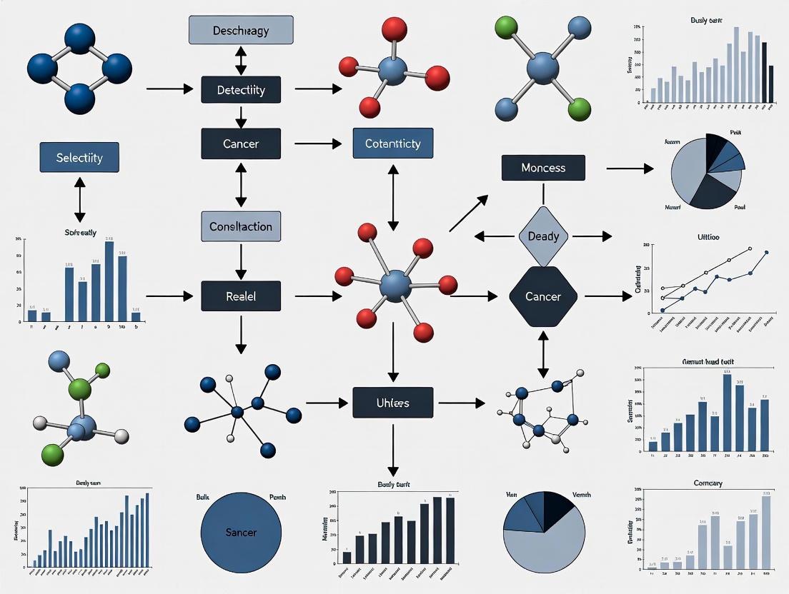
The following diagram illustrates the logical relationship between a test result, its performance metrics, and the clinical questions they help answer.
Diagram 6.1: Diagnostic Test Metrics Logic Flow
The Global Burden of Disease and the Limitations of Traditional Screening Methods
The Global Burden of Disease (GBD) study represents the largest and most comprehensive worldwide epidemiological observational study to quantify health loss from diseases, injuries, and risk factors across populations, over time [9]. By systematically identifying the biggest health problems, GBD research helps governments and scientists prioritize resources and advocate for improved health interventions [9]. A critical area of focus is early disease detection, where the performance of screening methods is paramount. The limitations of traditional screening methods, particularly in balancing sensitivity (correctly identifying true positives) and specificity (correctly identifying true negatives), present significant challenges to maximizing global health outcomes. This technical support center provides targeted guidance for researchers developing and validating improved early detection methodologies.
FAQs & Troubleshooting Guides
How can I improve the specificity of an existing screening test without losing sensitivity?
Problem: A standard screening test has high sensitivity but generates too many false positives, leading to unnecessary, invasive, and costly follow-up procedures for patients.
Solution: Implement a sequential testing strategy using a second, complementary biomarker. This "believe-the-negative" rule requires a positive result on both the standard test and the second confirmatory test to be considered a final positive [10].
Experimental Protocol:
- Step 1: Apply the standard screening test (Test A) to all study participants.
- Step 2: Only for participants who test positive on Test A, perform the second, innovative biomarker test (Test B).
- Step 3: The final positive classification is defined as positivity on both Test A and Test B.
- Step 4: Calculate the relative False Positive Rate (rFPR) and relative True Positive Rate (rTPR) to evaluate the combination test's performance [10]:
- rFPR:
P(Y_B = + | Y_A = +, non-diseased). This estimates the reduction in false positives. - rTPR:
P(Y_B = + | Y_A = +, diseased). This estimates the preservation of true positives.
- rFPR:
The goal is for the rFPR to be substantially less than 1 (indicating reduced false positives) while the rTPR remains close to 1 (indicating maintained sensitivity) [10].
Visualization: The following diagram illustrates the sequential testing workflow and its impact on subject classification.
Problem: For clinical deployment, your model must operate with very high specificity (e.g., >95%) to minimize false alarms, but its sensitivity at this strict threshold is unacceptably low, even though the overall Area Under the ROC Curve (AUC) is good.
Solution: Use the AUCReshaping technique during model fine-tuning. This method actively reshapes the ROC curve by boosting the weights of misclassified positive samples specifically within the high-specificity Region of Interest (ROI) [11].
Experimental Protocol:
- Step 1: Train your deep learning model on your dataset using a standard loss function.
- Step 2: Identify your Region of Interest (ROI), typically the false positive rate (FPR) range of 2-5% (specificity of 95-98%) [11].
- Step 3: During the fine-tuning stage, apply the AUCReshaping function. This function:
- Identifies positive samples (e.g., cancer cases) that are misclassified when the model operates at the high-specificity threshold.
- Adaptively increases the weight of these difficult-to-classify positive samples in the loss function.
- Iteratively focuses the model's learning on these samples, effectively "boosting" their importance [11].
- Step 4: Validate the model's performance, confirming that sensitivity within the predefined high-specificity ROI has improved.
Visualization: The diagram below contrasts standard model training with the AUCReshaping fine-tuning process.
How do I validate a new multi-cancer early detection (MCED) test in a clinically relevant way?
Problem: Initial validation of a novel MCED test shows high accuracy, but the study population did not include individuals with inflammatory or other non-cancerous conditions that could cause false positives.
Solution: Conduct a prospective, single-blinded study that includes cohorts of healthy individuals, cancer patients, and, critically, a control group of patients with inflammatory conditions or benign tumors [8].
Experimental Protocol:
- Step 1: Cohort Recruitment. Recruit three distinct participant groups:
- Group 1: Healthy volunteers.
- Group 2: Patients with verified malignancy (various cancer types, stages I-III).
- Group 3: Patients with verified inflammatory conditions (e.g., fibrosis, sarcoidosis, pneumonia) or benign tumors [8].
- Step 2: Sample Analysis. Analyze plasma samples (or other relevant biospecimens) from all participants using the novel MCED test. Perform all measurements in a blinded manner (the personnel conducting the test should be unaware of the sample's clinical status) [8].
- Step 3: Statistical Analysis. Calculate key performance metrics by comparing the test results against the confirmed clinical diagnoses:
- Sensitivity, Specificity, Positive Predictive Value (PPV), Negative Predictive Value (NPV).
- Use ANOVA with post-hoc tests to check for statistically significant differences in test values between the three groups [8].
Table 1: Performance Metrics of a Novel MCED Test (Example from Carcimun Test Study)
| Metric | Result | Interpretation |
|---|---|---|
| Accuracy | 95.4% | Overall correctness of the test. |
| Sensitivity | 90.6% | Effectively identifies cancer patients. |
| Specificity | 98.2% | Effectively rules out healthy individuals and those with inflammation. |
| Positive Predictive Value (PPV) | Reported | Proportion of true positives among all positive tests. |
| Negative Predictive Value (NPV) | Reported | Proportion of true negatives among all negative tests. |
Source: Adapted from [8]
The Scientist's Toolkit: Research Reagent Solutions
Table 2: Essential Materials for Featured Early Detection Experiments
| Research Reagent / Solution | Function / Application |
|---|---|
| Carcimun Test | A novel pancancer test that detects conformational changes in plasma proteins through optical extinction measurements at 340 nm, used as a biomarker for general malignancy [8]. |
| Vara MG (AI Software) | A CE-certified medical device incorporating deep learning for mammography screening. It provides normal triaging (flagging low-risk exams) and a safety net (alerting radiologists to highly suspicious findings they may have missed) [12]. |
| Indiko Clinical Chemistry Analyzer | A platform used for precise spectrophotometric measurement of optical density in plasma samples, crucial for assays like the Carcimun test [8]. |
| AUCReshaping Algorithm | A computational function used during deep learning model fine-tuning to reshape the ROC curve and improve sensitivity at pre-defined high-specificity operating points [11]. |
| Free Prostate-Specific Antigen (fPSA) Biomarker | Used as a second-line test in combination with the standard total PSA test to improve specificity in prostate cancer screening and reduce unnecessary biopsies [10]. |
Key Experimental Protocols in Detail
Protocol 1: Optical Extinction Measurement for Protein Conformation (Carcimun Test)
This protocol details the core wet-lab procedure for a novel protein-based cancer detection test [8].
- Sample Preparation: Add 70 µl of 0.9% NaCl solution to a reaction vessel, followed by 26 µl of blood plasma, resulting in a total volume of 96 µl.
- Dilution: Add 40 µl of distilled water (aqua dest.), adjusting the NaCl concentration to 0.63%. The total volume is now 136 µl.
- Incubation: Incubate the mixture at 37°C for 5 minutes to achieve thermal equilibration.
- Baseline Measurement: Perform a blank measurement at 340 nm to establish a baseline optical density.
- Acidification: Add 80 µl of 0.4% acetic acid (AA) solution (containing 0.81% NaCl). The final volume is 216 µl, with 0.69% NaCl and 0.148% acetic acid.
- Final Measurement: Perform the final absorbance measurement at 340 nm using a clinical chemistry analyzer (e.g., Indiko from Thermo Fisher Scientific).
Protocol 2: Implementing AI-Supported Double Reading in Mammography Screening
This protocol describes the real-world integration of an AI system into a standard double-reading workflow for mammograms, as implemented in the large-scale PRAIM study [12].
- Image Acquisition: Obtain standard four-view mammograms (craniocaudal and mediolateral oblique for each breast) from screening participants.
- AI Pre-classification: Process all mammograms through the AI system (e.g., Vara MG). The system preclassifies exams into two streams:
- Normal Triage: Exams deemed highly unsuspicious are tagged as "normal" in the radiologists' worklist.
- Safety Net: Exams deemed highly suspicious are flagged for the safety net feature.
- First Read (AI-Supported): The first radiologist reads the exam using an AI-supported viewer.
- For "normal" triaged cases, the radiologist can read with increased efficiency.
- If the radiologist initially interprets a "safety net" case as unsuspicious, an alert prompts them to review the AI's suggested suspicious region.
- Second Read (Independent): A second radiologist reads the exam independently, blind to the first reader's decision and AI results (unless they also choose to use the AI viewer).
- Consensus Conference: If either reader deems the case suspicious, a consensus conference is held with at least the two readers and a head radiologist.
- Recall Decision: The final decision to recall the patient for further diagnostic assessment is made in the consensus conference.
The Paradigm Shift to Multi-Cancer Early Detection (MCED) Tests
Technical Support Center: Troubleshooting Guides & FAQs
This technical support center provides resources for researchers and scientists working to improve the sensitivity and specificity of Multi-Cancer Early Detection (MCED) tests. The following guides address specific experimental challenges encountered during MCED assay development and validation.
Frequently Asked Questions (FAQs)
Q1: What are the primary biomarker classes used in MCED assays, and how do they compare? MCED tests primarily analyze tumor-derived components in blood, with the main biomarker classes being cell-free DNA (cfDNA) features and proteins [13] [14].
- cfDNA Mutations: Analysis of somatic mutations in circulating tumor DNA (ctDNA). This approach can be limited by the fact that some early-stage tumors shed very little DNA, and the same mutations can occur in different cancer types, making it challenging to identify the tissue of origin [14].
- cfDNA Methylation Patterns: This method examines epigenetic modifications, specifically DNA methylation. Cancer cells exhibit distinct methylation patterns that are often tissue-specific. This is currently one of the most promising approaches, as it can simultaneously detect the presence of cancer and predict its tissue of origin (Cancer Signal Origin) with high accuracy [15] [14].
- cfDNA Fragmentomics: This technique analyzes the fragmentation patterns of cfDNA, including fragment size and distribution. Tumor-derived DNA often has different fragmentation characteristics than DNA from healthy cells [13] [16].
- Protein Biomarkers: Measurement of circulating proteins associated with cancer. When used alone, proteins may lack sensitivity for early-stage detection, but they can enhance performance when combined with DNA-based biomarkers [14] [17].
Q2: Our MCED assay is showing good specificity but low overall sensitivity, particularly for Stage I cancers. What strategies can we implement? Low sensitivity for early-stage tumors is a common challenge, primarily due to low ctDNA shed [14]. Consider these troubleshooting strategies:
- Multi-Analyte Integration: Combine several biomarker classes instead of relying on a single one. For example, the CancerSEEK test simultaneously analyzes 16 cancer gene mutations and 8 protein biomarkers, which increased its sensitivity from 43% to 69% compared to using mutations alone [13]. The Cancerguard test also uses a multi-biomarker approach, combining DNA methylation and protein biomarkers [17].
- Optimize Bioinformatic Classifiers: Employ advanced machine learning algorithms trained on large, diverse datasets. The Galleri test uses targeted methylation sequencing and machine learning to achieve a sensitivity of 51.5% across multiple cancers while maintaining a specificity of 99.5% [13]. Continual refinement of these algorithms is crucial for improving performance [14] [16].
- Pre-analytical Variables: Standardize blood collection, plasma processing, and DNA extraction protocols. Inconsistent handling can lead to cfDNA degradation and variable results [15].
Q3: How can we assess the real-world clinical performance of our MCED test during development? Beyond initial clinical validation studies, real-world performance should be evaluated in large, prospective cohorts. Key performance metrics to track include:
- Cancer Signal Detection Rate (CSDR): The proportion of tests that return a positive result in a screened population. In a real-world study of over 111,000 individuals, the CSDR was 0.91% [15].
- Positive Predictive Value (PPV): The proportion of positive test results that are true cancers. This is a critical metric for understanding the clinical utility of a positive signal. In a study of an MCED test, the empirical PPV was 49.4% for asymptomatic individuals [15].
- Cancer Signal Origin (CSO) Prediction Accuracy: The rate at which the test correctly identifies the tissue of origin. High accuracy is essential to guide efficient diagnostic workups. Current tests have demonstrated CSO accuracy of approximately 87% to 90% in real-world settings [16] [15].
The following workflow outlines a generalized protocol for developing and validating an MCED test, integrating the key concepts from the FAQs above.
Diagram Title: MCED Test Development and Analysis Workflow
Q4: What is the recommended follow-up protocol for a positive MCED test result in a clinical study? There are no universally established guidelines. However, recent studies propose that a positive MCED test should trigger a diagnostic workup guided by the predicted Cancer Signal Origin (CSO) [15] [17].
- CSO-Guided Workup: The high accuracy of CSO prediction (e.g., ~87%) allows clinicians to initiate targeted imaging and specialist referrals [15]. For example, a CSO prediction of "lung" would lead to a low-dose CT scan.
- Imaging-Based Diagnostic Resolution: Some protocols suggest a comprehensive imaging workflow starting with a contrast-enhanced CT of the chest, abdomen, and pelvis, potentially followed by a PET-CT scan if needed [17]. This approach is designed to locate the cancer efficiently.
- Time to Diagnosis: Studies show that with a CSO-guided approach, the median time from the MCED result to a confirmed cancer diagnosis can be under 40 days [15].
Performance Data Comparison
The table below summarizes the performance characteristics of selected MCED tests as reported in clinical and real-world studies, providing a benchmark for researchers.
| MCED Test | Technology/Assay | Reported Sensitivity | Reported Specificity | Key Detectable Cancers |
|---|---|---|---|---|
| Galleri [13] [15] | Targeted Methylation Sequencing | 51.5% (overall); 39% (Stage I) [14] | 99.5% [13] | >50 cancer types [13] |
| CancerSEEK [13] | Multiplex PCR (16 genes) + Protein Immunoassay (8 proteins) | 62% (overall) [13] | >99% [13] | Lung, breast, colorectal, pancreatic, gastric, hepatic, esophageal, ovarian cancers [13] |
| Cancerguard [17] | DNA Methylation + Protein Biomarkers | 68% for high-mortality cancers (e.g., pancreatic, lung) [17] | 97.4% [17] | >50 cancer types and subtypes [17] |
| Shield (for CRC) [13] | Genomic mutations, methylation, DNA fragmentation | 83% for colorectal cancer (overall); 65% (Stage I) [13] | - | Colorectal cancer [13] |
| Real-World MCED (n=111,080) [15] | Targeted Methylation Sequencing | - | - | 32 cancer types diagnosed; 74% were cancers without USPSTF-recommended screening [15] |
The Scientist's Toolkit: Key Research Reagent Solutions
This table details essential materials and their functions for developing MCED tests, based on methodologies from established assays.
| Research Reagent / Material | Function in MCED Assay Development |
|---|---|
| Cell-free DNA (cfDNA) Extraction Kits | Isolation of high-quality, intact cfDNA from blood plasma samples is a critical pre-analytical step. Performance can vary between kits. |
| Bisulfite Conversion Reagents | For methylation-based assays (e.g., Galleri). These chemicals convert unmethylated cytosines to uracils, allowing methylated regions to be identified via sequencing [14]. |
| Targeted Methylation Panels | Custom probe sets designed to capture over 100,000 methylated regions in the genome, enabling sensitive detection of cancer-specific epigenetic signatures [13] [14]. |
| Multiplex PCR Panels | For mutation-based assays (e.g., CancerSEEK). Allows simultaneous amplification of multiple genomic regions (e.g., 1,900 positions in 16 genes) from a small sample volume [13]. |
| Immunoassay Kits (e.g., ELISA) | For quantifying protein biomarkers (e.g., the 8 proteins in CancerSEEK). Flow microsphere-based assays may offer advantages in reproducibility and dynamic range [18] [14]. |
| Next-Generation Sequencing (NGS) | Platform for high-throughput sequencing of captured DNA libraries (e.g., methylation-enriched or amplicon libraries). Essential for generating the primary data for machine learning analysis [13] [14]. |
The development of MCED tests represents a significant shift in cancer screening. The integration of multiple biomarker classes, advanced sequencing, and machine learning is key to improving sensitivity and specificity. As research progresses, standardizing experimental protocols and validation pathways will be crucial for translating these technologies into clinical practice.
Troubleshooting Guide: Addressing Common Experimental Challenges
This guide addresses frequent technical issues encountered during biomarker research for early cancer detection, providing targeted solutions to enhance the sensitivity and specificity of your assays.
Low ctDNA Detection Sensitivity in Early-Stage Cancers
Problem: Inability to reliably detect ctDNA at low variant allele frequencies (<0.1%), particularly in early-stage disease or minimal residual disease (MRD) monitoring [19].
Solutions:
- Implement Pre-analytical Enrichment: Utilize bead-based or enzymatic size selection to enrich for short cfDNA fragments (90-150 bp) characteristic of tumor-derived DNA. This can increase fractional abundance of ctDNA by several folds [19].
- Adopt Structural Variant-Based Assays: Transition from SNV-targeting to structural variant (SV) analysis. SV-based assays identify tumor-specific rearrangements (translocations, insertions, deletions) with breakpoint sequences unique to the tumor, achieving parts-per-million sensitivity [19].
- Leverage Nanomaterial-Based Sensors: Employ magnetic nano-electrode platforms using Fe₃O₄–Au core–shell particles for both PCR substrates and electrochemical modifications, achieving attomolar sensitivity within 7 minutes of PCR [19].
- Utilize Phased Variant Approaches: Implement methods like PhasED-seq that target multiple single-nucleotide variants on the same DNA fragment to improve detection sensitivity [19].
High Background Noise in Methylation Profiling
Problem: Excessive background interference in DNA methylation analysis, reducing signal-to-noise ratio and specificity.
Solutions:
- Optimize Bisulfite Conversion: Ensure complete conversion while minimizing DNA degradation. Include unmethylated and methylated controls to monitor conversion efficiency [20].
- Select Appropriate Detection Technology: Choose methods based on your application:
- Microarrays (Illumina Infinium BeadChip): For cost-effective, genome-wide coverage at predefined CpG sites [20].
- Whole-genome bisulfite sequencing (WGBS): For comprehensive, single-base resolution methylation mapping [20].
- Reduced representation bisulfite sequencing (RRBS): For cost-efficient analysis of CpG-rich regions [20].
- Long-read sequencing (Oxford Nanopore, PacBio): For detecting methylation patterns alongside structural variations without bisulfite conversion [20].
- Implement Effective Normalization: Use bioinformatics tools to correct for batch effects and platform-specific biases. Include technical replicates across batches [20].
Protein Biomarker Specificity Challenges
Problem: Limited specificity of individual protein tumor markers, leading to false positives in non-malignant conditions [21].
Solutions:
- Develop Multi-marker Panels: Combine multiple protein markers to increase diagnostic accuracy. For example, a panel including CEA, CYFRA 21-1, neuron-specific enolase (NSE), SCC, and ProGRP demonstrated 88.5% sensitivity and 82% specificity in detecting lung cancer, outperforming individual markers [21].
- Integrate Multi-analyte Approaches: Combine protein biomarkers with ctDNA mutations and methylation profiles. Tests like CancerSEEK have demonstrated enhanced performance by analyzing multiple biomarker classes simultaneously [22].
- Implement AI-Enhanced Analysis: Utilize machine learning algorithms to identify subtle patterns in multi-analyte data. The OncoSeek test integrates seven protein tumor markers with clinical data using AI, achieving 58.4% sensitivity and 92.0% specificity across multiple cancer types [23].
Sample Quality and Pre-analytical Variability
Problem: Inconsistent results due to pre-analytical factors including sample collection, processing, and storage.
Solutions:
- Standardize Blood Collection Protocols: Use consistent blood collection tubes (preferably specialized cfDNA tubes), processing times (within 2-4 hours of collection), and centrifugation conditions (dual-centrifugation protocol) [19] [24].
- Select Appropriate Liquid Biopsy Source: Choose sample type based on cancer type:
- Control Sample Integrity: Monitor cfDNA concentration and fragment size distribution. Implement quality control metrics including DNA integrity number and PCR amplification efficiency [24].
Frequently Asked Questions (FAQs)
What are the key advantages of DNA methylation biomarkers over mutation-based approaches?
DNA methylation offers several distinct advantages for early cancer detection:
- Early Emergence: Methylation alterations often appear early in tumorigenesis and remain stable throughout tumor evolution [24].
- Structural Stability: The DNA double helix provides enhanced stability compared to single-stranded nucleic acids, offering better resistance to degradation during sample processing [24].
- Enrichment Mechanism: Methylated DNA fragments demonstrate relative enrichment in cfDNA due to nucleosome interactions that protect them from nuclease degradation [24].
- Tissue-of-Origin Prediction: Methylation patterns are tissue-specific, enabling accurate prediction of the cancer's primary location [20].
How can I improve the sensitivity of ctDNA detection for minimal residual disease monitoring?
Enhancing MRD detection sensitivity requires a multi-faceted approach:
- Fragmentomics: Leverage the size difference between ctDNA (shorter fragments) and normal cfDNA. Size selection of shorter fragments can increase ctDNA fractional abundance and reduce required sequencing depth [19].
- Error-Suppressed Sequencing: Implement molecular barcoding and duplicate removal to distinguish true mutations from PCR and sequencing errors [19].
- Personalized Assays: Develop patient-specific assays targeting multiple clonal mutations or structural variants identified in tumor tissue [19].
- Multi-analyte Approach: Combine ctDNA with other biomarkers like protein markers or methylation patterns to create a more robust detection signal [22].
What computational approaches can enhance biomarker specificity?
Machine learning and AI methods significantly improve biomarker performance:
- Traditional ML: Support vector machines, random forests, and gradient boosting effectively classify cancer subtypes based on methylation patterns across tens to thousands of CpG sites [20].
- Deep Learning: Multilayer perceptrons and convolutional neural networks capture nonlinear interactions between CpGs for tumor subtyping and tissue-of-origin classification [20].
- Foundation Models: Transformer-based models like MethylGPT and CpGPT, pretrained on large methylome datasets, offer robust cross-cohort generalization and contextually aware CpG embeddings [20].
- AI-Empowered Integration: Algorithms that combine multiple biomarker types (proteins, ctDNA, methylation) with clinical data demonstrate enhanced specificity, as shown in tests like OncoSeek [23].
Experimental Protocols for Key Methodologies
Protocol 1: Ultrasensitive ctDNA Detection Using Structural Variant Analysis
Principle: Identify tumor-specific chromosomal rearrangements with breakpoint sequences unique to individual tumors [19].
Procedure:
- Tumor Whole Genome Sequencing: Sequence tumor tissue (80-100x coverage) and matched normal DNA to identify patient-specific structural variants.
- Breakpoint Selection: Select 3-5 clonal SVs with balanced allele frequencies for multiplexed detection.
- Probe Design: Create hybrid-capture probes or multiplex PCR primers targeting breakpoint junctions.
- Library Preparation from Plasma:
- Extract cfDNA from 2-5 mL plasma using silica-membrane columns.
- Quantify by fluorometry; require ≥10 ng cfDNA for analysis.
- Prepare sequencing libraries with molecular barcodes.
- Target Enrichment: Hybridize libraries with custom probes; capture for 16-24 hours.
- Sequencing: Sequence on Illumina platform (minimum 50,000x raw coverage).
- Bioinformatic Analysis:
- Align reads to reference genome.
- Identify breakpoint-spanning reads using custom algorithms.
- Calculate variant allele frequency from duplicate-deduplicated reads.
Expected Outcomes: Detection sensitivity of 0.001% VAF with >99% specificity for MRD monitoring [19].
Protocol 2: Targeted DNA Methylation Analysis Using Bisulfite Sequencing
Principle: Bisulfite conversion of unmethylated cytosines to uracils while methylated cytosines remain unchanged, allowing methylation status determination [20].
Procedure:
- DNA Extraction and Quality Control:
- Extract DNA from plasma, tissue, or cells.
- Quantify using fluorometry; assess integrity by capillary electrophoresis.
- Bisulfite Conversion:
- Use commercial bisulfite conversion kit.
- Incubate 500 ng-1 μg DNA in bisulfite reagent (98°C for 10 minutes, 64°C for 2.5 hours).
- Desalt and purify converted DNA.
- Library Preparation:
- Repair DNA ends and add adapters with unique molecular identifiers.
- Amplify with 8-12 PCR cycles using methylation-aware polymerases.
- Target Enrichment:
- Hybridize with biotinylated probes targeting 50-100 CpG islands.
- Capture with streptavidin beads; wash stringently.
- Sequencing: Sequence on Illumina platform (minimum 1000x coverage per CpG).
- Bioinformatics Analysis:
- Align to bisulfite-converted reference genome using specialized aligners (Bismark, BSMAP).
- Calculate methylation percentage at each CpG site.
- Perform differential methylation analysis between case and control groups.
Expected Outcomes: Quantitative methylation values for each targeted CpG with sensitivity to detect 1% methylated alleles in background of unmethylated DNA [20].
Performance Comparison of Biomarker Detection Technologies
Table 1: Analytical Performance of ctDNA Detection Technologies
| Technology | Limit of Detection | VAF Range | Multiplexing Capacity | Key Applications |
|---|---|---|---|---|
| ddPCR | 0.01%-0.1% | 0.01%-50% | Low (1-4 targets) | Monitoring known mutations, resistance detection [19] |
| Structural Variant Assays | 0.001% | 0.001%-100% | Medium (5-20 targets) | MRD, early detection [19] |
| Nanomaterial Sensors | Attomolar | N/A | Low | Point-of-care detection, rapid screening [19] |
| Targeted NGS Panels | 0.1% | 0.1%-100% | High (50-500 genes) | Comprehensive profiling, therapy selection [19] |
Table 2: DNA Methylation Analysis Platforms Comparison
| Platform | Resolution | Coverage | Cost per Sample | Ideal Use Cases |
|---|---|---|---|---|
| Infinium MethylationEPIC | Single CpG | 850,000 CpG sites | Medium | Biomarker discovery, large cohort studies [20] |
| Whole-Genome Bisulfite Sequencing | Single base | >20 million CpGs | High | Comprehensive discovery, novel biomarker identification [20] |
| RRBS | Single base | ~2 million CpGs | Medium-high | Cost-effective discovery, CpG island coverage [20] |
| Targeted Bisulfite Sequencing | Single base | Custom (50-10,000 CpGs) | Low-medium | Clinical validation, focused panels [20] |
Table 3: Protein Biomarker Performance in Multi-Cancer Detection
| Biomarker | Associated Cancers | Sensitivity Range | Specificity | Notes |
|---|---|---|---|---|
| CEA | Colorectal, lung, breast | 30-50% (CRC) | ~90% | Limited early-stage sensitivity [21] |
| CA-125 | Ovarian | ~50% (early stage) | ~90% | Elevated in benign conditions [22] |
| AFP | Hepatocellular carcinoma | ~60% (with ultrasound) | ~90% | Used in high-risk screening [21] |
| Multi-protein Panel (OncoSeek) | 14 cancer types | 38.9-83.3% (by type) | 92.0% | AI-enhanced, 7-protein panel [23] |
Experimental Workflows and Signaling Pathways
Diagram 1: ctDNA Analysis Workflow for Early Detection
ctDNA Analysis Workflow: Comprehensive process from sample collection to clinical reporting
Diagram 2: Multi-Analyte Integration for Enhanced Specificity
Multi-Analyte Integration: Combining biomarker classes with AI for enhanced detection
The Scientist's Toolkit: Essential Research Reagents and Materials
Table 4: Key Research Reagent Solutions for Biomarker Development
| Reagent/Material | Function | Key Considerations | Example Applications |
|---|---|---|---|
| cfDNA Extraction Kits | Isolation of cell-free DNA from plasma/serum | Yield, fragment preservation, inhibitor removal | All liquid biopsy applications [24] |
| Bisulfite Conversion Kits | Chemical conversion of unmethylated cytosines | Conversion efficiency, DNA damage minimization | Methylation analysis [20] |
| Molecular Barcodes | Unique sequence identifiers for error correction | Complexity, read length requirements | Ultrasensitive mutation detection [19] |
| Methylation-Specific PCR Primers | Amplification of methylated/unmethylated sequences | Specificity, annealing temperature optimization | Targeted methylation validation [20] |
| Capture Probes | Hybridization-based target enrichment | Sensitivity, off-target rate, coverage uniformity | Targeted sequencing [19] |
| Quality Control Assays | Assessment of DNA quantity/quality | Sensitivity, reproducibility, input requirements | All applications [24] |
| Reference Standards | Controls for assay validation | Allelic frequency, matrix effects, stability | Assay development and QC [19] |
Frequently Asked Questions
What are the key performance metrics for MCED tests, and how do they currently perform? The key performance metrics are sensitivity (or cancer signal detection rate), specificity, positive predictive value (PPV), and cancer signal origin (CSO) prediction accuracy. Current real-world data from over 111,000 individuals shows an overall cancer signal detection rate of 0.91% [25]. In a large interventional study (PATHFINDER 2), the Galleri test demonstrated a specificity of 99.6% (false positive rate of 0.4%) and a positive predictive value of 61.6% [26]. The test correctly identified the origin of the cancer signal in 87% to 92% of cases, which helps guide diagnostic workups [25] [26].
What is the difference between a test's sensitivity and its PPV, and why does it matter for screening? Sensitivity is the probability that the test will be positive when cancer is present. Positive Predictive Value (PPV) is the probability that a person with a positive test result actually has cancer [27]. For population screening, a high PPV is critical because it minimizes the number of false positives, thus reducing unnecessary, invasive, and costly diagnostic procedures and associated patient anxiety [10]. MCED tests are designed to have high specificity to keep the false positive rate low when used for broad screening [25].
Which cancers do MCED tests detect, and how does this impact their utility? MCED tests are designed to detect a wide range of cancers. For example, the Galleri test can detect more than 50 cancer types [26]. A key benefit is their ability to detect cancers for which no standard screening exists. When added to standard USPSTF-recommended screenings (for breast, cervical, colorectal, and lung cancers), the Galleri test increased the cancer detection rate more than seven-fold. Approximately three-quarters of the cancers it detected are types that currently lack recommended screening tests [26].
What are the major evidence gaps preventing widespread implementation of MCED tests? Despite promising early data, major evidence gaps remain. No MCED test has yet been approved by the FDA or endorsed by major clinical practice guidelines [27] [28]. Crucially, it is not yet known whether MCED testing reduces cancer-specific mortality, as data from large, randomized controlled trials (RCTs) is still pending [27]. Other unknowns include the optimal testing interval, the impact on adherence to existing cancer screenings, and the full range of potential harms, such as overdiagnosis [28].
What should be the follow-up process for a positive MCED test result? A positive MCED test result requires confirmation with standard diagnostic methods and is not a definitive cancer diagnosis [28]. The test's CSO prediction is intended to guide the subsequent diagnostic workup. In clinical studies, this workflow led to a diagnostic resolution in a median of 39.5 to 46 days [25] [26]. Follow-up involves imaging and procedures targeted to the predicted organ system, such as CT scans for a predicted lung CSO or colonoscopy for a predicted colorectal CSO.
Troubleshooting Guides
Issue: Interpreting a Positive MCED Test Result in an Asymptomatic Patient
- Problem: A patient with a positive MCED test result has no symptoms. How should a researcher or clinician validate this finding?
- Solution:
- Do not consider the MCED result a diagnosis. It is a screening signal that requires confirmation [28].
- Initiate a targeted diagnostic workup based on the predicted Cancer Signal Origin (CSO). Rely on the test's high CSO prediction accuracy (e.g., 92% in PATHFINDER 2) [26].
- Utilize established imaging and procedures. For example, use CT imaging for a lung CSO, mammography or breast MRI for a breast CSO, and endoscopy or colonoscopy for an upper GI or colorectal CSO.
- Monitor the time to diagnosis. In clinical settings, the median time from a positive result to diagnosis is around 40-46 days [25] [26]. A prolonged, non-targeted workup may delay confirmation.
- Underlying Principle: The empirical PPV for asymptomatic individuals is approximately 49.4%, meaning about half of the positive tests will be true positives and the other half false positives [25]. A structured, CSO-guided pathway is essential for efficiency.
Issue: Managing a False Positive MCED Test Result
- Problem: A patient underwent an invasive diagnostic procedure following a positive MCED test, but no cancer was found.
- Solution:
- Acknowledge the inherent risk. All screening tests have a false positive rate. The false positive rate for the Galleri test in the PATHFINDER 2 study was 0.4% [26].
- Document the outcome. This contributes to real-world evidence on test performance and diagnostic pathways.
- Counsel the patient. Discuss the meaning of the result, emphasizing that it indicates no cancer was detected by the definitive diagnostic workup and that the initial signal could have been a false positive.
- Reinforce the importance of continued routine screening. A negative MCED result does not eliminate the need for guideline-recommended screenings for breast, cervical, colorectal, and other cancers [26] [28].
- Underlying Principle: The goal of MCED test design is to maximize specificity to minimize false positives, but they cannot be eliminated entirely. The potential for false positives must be part of the pre-test discussion with patients [25] [10].
Issue: Integrating MCED Tests with Standard Cancer Screening in Research Protocols
- Problem: How should MCED tests be positioned relative to established, single-cancer screenings in a clinical study design?
- Solution:
- Frame MCED as a complementary test, not a replacement. Study protocols should mandate that participants continue with all USPSTF-recommended screenings (e.g., mammography, colonoscopy) [26] [28].
- Measure adherence. A key study outcome should be whether the introduction of an MCED test impacts participation rates in standard screenings.
- Analyze data separately. Report performance metrics for cancers with existing screenings and for those without separately to clarify the added value of the MCED test.
- Underlying Principle: MCED tests are intended to expand the landscape of detectable cancers, not to supersede existing, validated methods. Their greatest potential utility lies in detecting the ~70% of deadly cancers that currently lack screening options [26].
Performance Benchmark Data
The following tables consolidate key quantitative performance data from recent large-scale studies on the Galleri MCED test.
Table 1: Key Performance Metrics from Recent MCED Studies
| Metric | Real-World Cohort (n=111,080) [25] | PATHFINDER 2 Interventional Study (n=23,161) [26] |
|---|---|---|
| Cancer Signal Detection Rate | 0.91% | 0.93% |
| Specificity | Not explicitly stated | 99.6% |
| False Positive Rate | Inferred from CSDR | 0.4% |
| Positive Predictive Value (PPV) | 49.4% (asymptomatic) | 61.6% |
| Cancer Signal Origin (CSO) Accuracy | 87% | 92% |
| Time to Diagnosis (Median) | 39.5 days | 46 days |
Table 2: Cancer Detection by Stage in the PATHFINDER 2 Study [26]
| Cancer Stage | Percentage of Cancers Detected by Galleri |
|---|---|
| Stage I | Included in 53.5% |
| Stage II | (Stages I & II combined) |
| Stage III | Included in 69.3% |
| Stage IV | (Stages I-III combined) |
Experimental Protocols
Protocol 1: Analytical Validation of an MCED Test
- Objective: To determine the analytical sensitivity, specificity, and limit of detection of an MCED assay.
- Methodology:
- Sample Preparation: Use commercially available reference materials and cell-free DNA (cfDNA) samples from patients with known cancer diagnoses and healthy controls.
- Library Preparation & Sequencing: Extract cfDNA from plasma samples. Perform bisulfite conversion to preserve methylation patterns. Create sequencing libraries and conduct targeted next-generation sequencing (NGS) focusing on methylation regions [25].
- Data Analysis: Process sequencing data through a proprietary bioinformatics pipeline. Use a pre-trained machine learning classifier to distinguish cancer from non-cancer signals based on methylation patterns and to predict the CSO [25].
- Key Measurements:
- Limit of Detection (LoD): The lowest concentration of tumor-derived cfDNA that can be reliably detected.
- Analytical Specificity: The test's ability to remain negative when testing non-cancerous samples and samples with common benign conditions.
Protocol 2: Clinical Validation in a Screening Population (e.g., PATHFINDER 2 Design)
- Objective: To evaluate the clinical performance and safety of an MCED test in an intended-use population.
- Methodology:
- Study Design: Prospective, multi-center, interventional study [26].
- Participant Recruitment: Enroll adults (e.g., ≥50 years) with no clinical suspicion of cancer. A broad, average-risk screening population is targeted [26].
- Testing: Perform a single blood draw for the MCED test. Participants and their providers receive the test results.
- Outcome Ascertainment: For participants with a "Cancer Signal Detected" result, a guided diagnostic workup is initiated based on the predicted CSO. Clinical follow-up continues for at least 12 months to capture any subsequent cancer diagnoses [26].
- Key Measurements:
- Positive Predictive Value (PPV): (Number of true positive cancers / number of total positive tests).
- Specificity: (Number of true negatives / number of total negative tests).
- CSO Prediction Accuracy: (Number of correct CSO predictions / number of true positive cancers with a confirmed origin).
- Time to Diagnostic Resolution: Time from result to confirmed diagnosis.
- Safety Endpoints: Number of invasive procedures performed in participants with and without cancer.
MCED Test Workflow
The following diagram illustrates the core workflow of a targeted methylation-based MCED test, from blood draw to result.
MCED Test Workflow: From blood draw to result.
The Scientist's Toolkit: Research Reagent Solutions
Table 3: Essential Materials for MCED Test Development and Validation
| Item | Function in MCED Research |
|---|---|
| Cell-free DNA Blood Collection Tubes | Stabilizes nucleated blood cells during sample transport to prevent genomic DNA contamination and preserve the integrity of cfDNA [25]. |
| cfDNA Extraction Kits | Isolves short-fragment, circulating cell-free DNA from plasma samples for downstream molecular analysis [25]. |
| Bisulfite Conversion Reagents | Chemically converts unmethylated cytosine residues to uracil, allowing methylation patterns to be read as sequence differences during sequencing [25]. |
| Targeted Methylation Sequencing Panels | A set of probes designed to capture and sequence specific genomic regions known to have differential methylation patterns in cancer cells [25]. |
| Methylated & Unmethylated Control DNA | Provides a reference for assessing the efficiency of bisulfite conversion and the accuracy of the methylation calling bioinformatics pipeline. |
| Bioinformatics Pipeline (Software) | A suite of computational tools for processing raw sequencing data, aligning sequences, quantifying methylation, and applying a classification algorithm [25]. |
| Banked Plasma Biobanks | Collections of well-annotated plasma samples from individuals with and without cancer, essential for training and validating the machine learning models [25]. |
Innovative Technologies in Practice: From Liquid Biopsies to AI-Driven Diagnostics
Liquid biopsy is a minimally invasive approach that analyzes circulating biomarkers in bodily fluids, primarily blood, to provide real-time information on tumor dynamics, treatment response, and disease progression [29] [30]. Unlike traditional tissue biopsies, liquid biopsy allows for repeated sampling and longitudinal monitoring of cancer, making it particularly valuable for early detection and monitoring minimal residual disease [31]. The most widely studied biomarkers in cancer management include circulating tumor DNA (ctDNA), circulating microRNAs (miRNAs), circulating tumor cells (CTCs), and various proteins such as cytokines [29] [31]. Each biomarker class offers unique advantages and faces distinct technical challenges in detection and analysis, which this technical support center aims to address.
Table: Key Liquid Biopsy Biomarkers and Their Characteristics
| Biomarker Class | Primary Composition | Key Advantages | Major Technical Challenges |
|---|---|---|---|
| Circulating Tumor DNA (ctDNA) | Tumor-derived fragmented DNA | Short half-life enables real-time monitoring; Directly reflects tumor genetics [31] | Low abundance in early-stage disease; Requires highly sensitive detection methods [30] |
| Circulating microRNAs (miRNAs) | Small non-coding RNAs (~22 nucleotides) | High stability in circulation; Early epigenetic alterations [29] | Lack of universal normalizers; Methodological variability between studies [29] |
| Circulating Tumor Cells (CTCs) | Intact cells from primary/metastatic tumors | Complete cellular information; Functional studies possible [31] | Extreme rarity (1 CTC per million blood cells); Epithelial-mesenchymal transition changes markers [31] [30] |
| Serum Cytokines | Inflammatory proteins (e.g., TNF-α, IL-6, IL-10) | Reflect tumor microenvironment and systemic inflammation [29] | Lack of standardization; High variability across platforms [29] |
Detailed Experimental Protocols
Protocol for ctDNA Analysis using ddPCR
Principle: Digital Droplet PCR (ddPCR) partitions samples into thousands of nanoliter-sized droplets, allowing absolute quantification of rare mutant alleles with high sensitivity [29].
Reagents and Equipment:
- Cell-free DNA collection tubes (e.g., Streck Cell-Free DNA BCT)
- Plasma preparation tubes
- QIAamp Circulating Nucleic Acid Kit (Qiagen)
- ddPCR Supermix for Probes (Bio-Rad)
- Target-specific primer-probe sets
- QX200 Droplet Generator (Bio-Rad)
- QX200 Droplet Reader (Bio-Rad)
Procedure:
- Blood Collection and Processing: Collect blood in cell-free DNA BCT tubes. Process within 6 hours of collection. Centrifuge at 800 × g for 10 minutes at room temperature to separate plasma. Transfer plasma to microcentrifuge tubes and centrifuge at 14,000 × g for 10 minutes to remove residual cells [32].
cfDNA Extraction: Use QIAamp Circulating Nucleic Acid Kit according to manufacturer's instructions. Elute DNA in 50 μL of elution buffer. Quantify using Qubit dsDNA HS Assay Kit [32].
Droplet Generation: Prepare 20 μL reaction mixture containing 10 μL of 2× ddPCR Supermix, 1 μL of each primer-probe set (900 nM primers, 250 nM probe final concentration), 5 μL of template DNA, and nuclease-free water. Generate droplets using QX200 Droplet Generator [32].
PCR Amplification: Transfer droplets to a 96-well plate. Seal the plate and perform PCR amplification with the following conditions: 95°C for 10 minutes; 40 cycles of 94°C for 30 seconds and 55-60°C (assay-specific) for 60 seconds; 98°C for 10 minutes. Ramp rate: 2°C/second.
Droplet Reading and Analysis: Read plate using QX200 Droplet Reader. Analyze data with QuantaSoft software. Set threshold between positive and negative droplets based on controls. Calculate mutant allele frequency using the formula: (Number of mutant-positive droplets / Total number of droplets) × 100 [32].
Troubleshooting Tip: If droplet generation efficiency is low, ensure all reagents are at room temperature and check for particulate matter in samples. Filter samples if necessary.
Protocol for Circulating miRNA Profiling using RT-qPCR
Principle: Reverse transcription quantitative PCR (RT-qPCR) enables sensitive detection and quantification of circulating miRNAs, which are promising biomarkers for early cancer detection [29] [33].
Reagents and Equipment:
- miRNeasy Serum/Plasma Kit (Qiagen)
- miScript II RT Kit (Qiagen)
- miScript SYBR Green PCR Kit (Qiagen)
- Platform-specific miRNA assays (e.g., miR-21, miR-29a, miR-34a for cervical cancer [29])
- Thermal cycler with real-time PCR capability
Procedure:
- RNA Extraction: Use miRNeasy Serum/Plasma Kit according to manufacturer's instructions. Add 1 volume of QIAzol Lysis Reagent to 1 volume of plasma. Vortex and incubate for 5 minutes. Add chloroform (0.2 volumes), shake vigorously, and centrifuge at 12,000 × g for 15 minutes at 4°C. Transfer aqueous phase and add 1.5 volumes of 100% ethanol. Transfer to RNeasy MinElute column and proceed with manufacturer's protocol. Elute in 14 μL RNase-free water [29].
Reverse Transcription: Use miScript II RT Kit. Prepare 20 μL reaction containing 4 μL of miScript Reverse Transcriptase Mix, 4 μL of 5× miScript RT Buffer, 12 μL of template RNA. Incubate at 37°C for 60 minutes, then 95°C for 5 minutes. Store at -20°C [29].
qPCR Amplification: Use miScript SYBR Green PCR Kit. Prepare 25 μL reactions containing 12.5 μL of 2× QuantiTect SYBR Green PCR Master Mix, 2.5 μL of 10× miScript Universal Primer, 2.5 μL of 10× miScript Primer Assay, 2.5 μL of template cDNA, and 5 μL of RNase-free water. Run in triplicate with the following conditions: 95°C for 15 minutes; 40 cycles of 94°C for 15 seconds, 55°C for 30 seconds, and 70°C for 30 seconds [29].
Data Analysis: Use the 2^(-ΔΔCt) method for relative quantification. Normalize to spiked-in synthetic miRNAs (e.g., cel-miR-39) or stable endogenous controls (e.g., U6 snRNA) [29].
Troubleshooting Tip: If amplification efficiency is low, check RNA integrity and ensure reverse transcription reagents are fresh. Include no-template controls to detect contamination.
Frequently Asked Questions (FAQ) & Troubleshooting
Q1: Our ctDNA assays consistently show low variant allele frequency (VAF) detection in early-stage cancer samples. How can we improve sensitivity?
A: Low VAF (<0.1%) is a common challenge in early-stage cancers [30]. Consider these approaches:
- Implement Ultra-Sensitive Assays: New comprehensive genomic profiling assays like Northstar Select demonstrate a 95% limit of detection at 0.15% VAF for SNV/Indels, detecting 51% more pathogenic variants than on-market CGP assays [32].
- Optimize Blood Collection: Use specialized cell-free DNA collection tubes and process samples within 6 hours to prevent genomic DNA contamination [30].
- Increase Plasma Input: Increase plasma volume from 2-4 mL to 10 mL for low-shedding tumors, though this requires adjustments to extraction protocols [32].
- Utilize Molecular Barcodes: Implement unique molecular identifiers (UMIs) to distinguish true mutations from PCR errors [32].
Q2: We observe high variability in circulating miRNA results between sample batches. What normalization strategies do you recommend?
A: Normalization is critical for reproducible miRNA quantification [29]:
- Use Multiple Normalization Methods: Combine spiked-in synthetic miRNAs (e.g., cel-miR-39) with stable endogenous controls identified through stability algorithms (e.g., NormFinder, geNorm).
- Standardize Pre-analytical Conditions: Control for factors affecting miRNA levels: time from collection to processing, fasting status, and hemolysis [29].
- Employ Consistent RNA Isolation Methods: Use the same commercial kits across all samples and ensure consistent elution volumes [29].
- Validate Reference Genes: Test potential reference genes in your specific sample matrix; no universal normalizer exists for circulating miRNAs [29].
Q3: Our CTC recovery rates are suboptimal, particularly for mesenchymal phenotypes. How can we improve recovery?
A: CTC isolation is challenging due to heterogeneity and epithelial-mesenchymal transition (EMT) [31] [30]:
- Combine Enrichment Methods: Use both positive selection (EpCAM-based) and negative selection (CD45 depletion) strategies [31].
- Implement Size-Based Filtration: For mesenchymal CTCs that downregulate EpCAM, use size-based platforms like ScreenCell that capture CTCs based on physical properties rather than surface markers [30].
- Explore Novel Capture Technologies: Emerging technologies like protein corona disguised immunomagnetic beads (PIMBs) demonstrate improved CTC enrichment with leukocyte depletion of approximately 99.996% [30].
- Optimize Blood Draw Volume: Increase blood draw volume from 7.5 mL to 20-30 mL for rare CTC detection, with appropriate anticoagulant adjustments [31].
Q4: What emerging technologies show promise for improving liquid biopsy sensitivity for early detection?
A: Several advanced approaches are enhancing detection capabilities:
- Fragmentomics: Analyzing cfDNA fragment patterns (size, distribution) can detect cancers with high sensitivity without requiring mutation information, working with as little as 1 ng of cell-free DNA [34].
- Personalized Assays: Ultrasensitive assays like NeXT Personal using patient-specific variant panels can detect ctDNA as low as two parts per million, identifying recurrence up to a year before imaging [34].
- Multimodal Integration: Combining multiple biomarker classes (ctDNA, CTCs, exosomes) provides a more comprehensive tumor view and improves overall sensitivity [30].
- Artificial Intelligence: AI algorithms can integrate complex liquid biopsy data to enhance diagnostic accuracy and predictive power [30].
Research Reagent Solutions
Table: Essential Research Reagents for Liquid Biopsy Applications
| Reagent/Category | Specific Examples | Primary Function | Key Considerations |
|---|---|---|---|
| Blood Collection Tubes | Streck Cell-Free DNA BCT, PAXgene Blood cDNA Tubes | Preserve sample integrity | Different stabilizers affect downstream applications; Choose based on target analyte [30] |
| Nucleic Acid Extraction Kits | QIAamp Circulating Nucleic Acid Kit, miRNeasy Serum/Plasma Kit | Isolate ctDNA, cfRNA, miRNAs | Recovery efficiency varies by fragment size; Validate for your specific targets [29] [32] |
| Library Prep Kits | AVENIO ctDNA kits, NEBNext Ultra II DNA | Prepare sequencing libraries | Molecular barcoding reduces errors; Input requirements vary [32] |
| PCR Reagents | ddPCR Supermix, miScript SYBR Green PCR Kit | Target amplification & quantification | Probe vs. SYBR Green affects specificity; Digital PCR enables absolute quantification [29] [32] |
| CTC Enrichment Platforms | CellSearch System, ScreenCell filters | Isolate and enumerate CTCs | FDA-cleared vs. research-use-only; EpCAM-dependent vs. label-free [31] [30] |
Experimental Workflows and Signaling Pathways
Liquid Biopsy Experimental Workflow
Biomarker Selection Decision Pathway
For researchers focused on improving the sensitivity and specificity of early detection methods, choosing the right DNA methylation profiling technique is paramount. Bisulfite conversion has long been the gold standard for differentiating methylated cytosines from unmethylated ones. However, emerging enzymatic conversion methods now offer a powerful alternative, particularly for analyzing challenging clinical samples like circulating tumor DNA (ctDNA) and formalin-fixed paraffin-embedded (FFPE) tissue. This technical support center provides a detailed comparison, troubleshooting guides, and FAQs to help you navigate these technologies and optimize your experiments for maximum sensitivity in early cancer detection research.
Technology Comparison: Bisulfite vs. Enzymatic Conversion
The following table summarizes the core differences between these two foundational methods.
| Feature | Bisulfite Conversion | Enzymatic Conversion |
|---|---|---|
| Basic Principle | Chemical conversion using sodium bisulfite under high temperature and low pH to deaminate unmethylated C to U [35] [36] | Sequential enzymatic reactions (e.g., TET2 oxidation + APOBEC deamination) to convert unmodified C to U [35] [36] |
| DNA Damage | High, causes DNA fragmentation and depyrimidination [35] [37] | Low, gentle reaction preserves DNA integrity [36] [37] |
| DNA Input | Typically μg-level for mammalian genomes [36] | Can be as low as 10-100 ng, suitable for low-input samples [36] |
| CpG Detection | Fewer unique CpGs detected, especially at low coverage [37] | Superior detection of more unique CpGs at the same sequencing depth [35] [37] |
| GC Bias | Skewed GC content representation and biased genome coverage [37] | More uniform genome coverage and normalized GC bias plots [37] |
| Best For | Routine samples with ample, high-quality DNA | Fragmented, low-input, or precious samples (e.g., cfDNA, FFPE, single-cell) [36] |
Frequently Asked Questions (FAQs)
Q1: Which conversion method provides better sensitivity for detecting early-stage cancer biomarkers in liquid biopsies?
Enzymatic conversion often holds an advantage for liquid biopsy applications. Its gentler treatment results in longer DNA fragments and higher library yields from circulating cell-free DNA (cfDNA), which is naturally fragmented and scarce. This allows for more unique sequencing reads and robust detection of tumor-derived DNA, a critical factor for early-stage cancer when the tumor DNA signal in the blood is very low [35]. However, one study using ddPCR found that bisulfite conversion provided higher DNA recovery post-conversion [38]. The optimal choice can depend on your specific downstream analysis (sequencing vs. PCR).
Q2: My bisulfite-converted libraries have low complexity and high duplication rates. What is the cause and how can I fix this?
This is a common issue rooted in the extensive DNA fragmentation caused by bisulfite treatment [37]. The harsh conditions degrade a significant portion of your DNA sample, reducing the diversity of unique DNA molecules available for sequencing. To mitigate this:
- Increase DNA Input: If possible, start with more input DNA to compensate for losses.
- Use Post-Bisulfite Adapter Tagging (PBAT): Ligate adapters after bisulfite conversion to improve library yields from damaged DNA [35] [37].
- Consider Enzymatic Conversion: Switching to a method like EM-seq is the most effective way to eliminate this problem, as it preserves DNA integrity and significantly improves library complexity [35] [36].
Q3: Why is my DNA recovery so low after enzymatic conversion, and how can I improve it?
While enzymatic conversion is gentler on DNA, recovery can be low due to sample loss during the protocol's multiple cleanup steps using magnetic beads [38]. To enhance recovery:
- Optimize Bead Cleanup: Test different magnetic bead-to-sample ratios. Increasing the ratio (e.g., from 1.8x to 3.0x) can significantly improve DNA recovery by capturing more of the smaller fragments [38].
- Compare Kits: Some studies note that the full NEBNext EM-seq kit may provide slightly better recovery than the standalone conversion module [38].
- Ensure Bead Quality: Avoid over-drying the magnetic bead pellet, as this can lead to inefficient resuspension and sample loss [39].
Troubleshooting Common Experimental Issues
Problem 1: Low Library Yield
Potential Causes and Solutions:
- Cause: Poor DNA Input Quality
- Solution: Check DNA integrity and purity. Re-purify samples if contaminants (phenol, salts) are present, and use fluorometric quantification (e.g., Qubit) for accuracy over UV absorbance [39].
- Cause: Inefficient Adapter Ligation
- Solution: Titrate the adapter-to-insert molar ratio to find the optimal balance. Ensure fresh ligase and buffer, and maintain proper reaction temperature [39].
- Cause: Overly Aggressive Size Selection
Problem 2: Incomplete Cytosine Conversion
Potential Causes and Solutions:
- Cause: Impure DNA Input (for Bisulfite)
- Solution: Ensure DNA is pure before conversion. Particulate matter can interfere. Centrifuge the conversion reagent and use only the clear supernatant [40].
- Cause: Suboptimal Reaction Conditions
- Solution: For bisulfite, strictly follow the protocol for temperature, pH, and incubation time. For enzymatic methods, ensure enzymes and co-factors are fresh and stored correctly.
- Verification: Always include unmethylated control DNA (e.g., lambda phage) in your conversion reaction to calculate the conversion efficiency, which should be >99.5% [35] [38].
Experimental Workflow for Enhanced Sensitivity
The following diagram illustrates a generalized workflow for enzymatic methylation sequencing, which is particularly suited for sensitive applications.
Detailed Protocol: Target-Enriched Enzymatic Methylation Sequencing (TEEM-Seq)
This protocol is designed for high-sensitivity methylation profiling from low-input and FFPE samples [41].
DNA Fragmentation and Quality Control
- Fragment genomic DNA to an average insert size of 240-290 bp using a focused-ultrasonicator (e.g., Covaris).
- Assess DNA quality and quantity using a fluorometric assay (e.g., Qubit) and fragment analyzer.
Enzymatic Methyl-Seq Library Construction
- Construct the library from fragmented DNA using a commercial kit (e.g., NEBNext Enzymatic Methyl-seq Kit).
- Quantify the constructed library and evaluate its size distribution.
Targeted Enrichment
- Pool an equal amount of DNA from multiple libraries (e.g., 8-plex).
- Perform target enrichment using a comprehensive panel (e.g., Twist Human Methylome panel, covering ~3.98 million CpG sites) following the manufacturer's protocol.
Sequencing and Data Analysis
- Sequence the final enriched libraries on an Illumina platform (e.g., NovaSeq6000) with 150 bp paired-end reads.
- Process data through a bioinformatic pipeline (e.g., Trim Galore for adapter trimming, bwa-meth for alignment, MethylDackel for methylation calling).
The Scientist's Toolkit: Key Research Reagents
| Reagent / Kit | Function | Application Context |
|---|---|---|
| NEBNext Enzymatic Methyl-seq Kit | Library prep and enzymatic conversion of 5mC and 5hmC [35] [41] | Whole-genome and targeted methylation sequencing with minimal DNA damage. |
| EZ-96 DNA Methylation-Gold Kit | High-efficiency bisulfite conversion of DNA [35] | Gold-standard bisulfite conversion for sequencing or array-based methods. |
| Twist Human Methylome Panel | Target enrichment covering 3.98 million CpGs [41] | Focusing sequencing power on biologically relevant regions for cost-effective, deep sequencing. |
| AMPure XP Beads | Magnetic beads for size selection and clean-up [38] | Post-conversion and post-enrichment purification; critical for maximizing DNA recovery. |
| Lambda Phage DNA | Unmethylated control DNA for conversion efficiency spike-in [35] | Essential quality control to calculate and validate cytosine-to-uracil conversion efficiency. |
Advanced Applications: Haplotype Methylation Scoring for Ultra-Sensitive Detection
For the highest sensitivity in detecting trace amounts of tumor DNA, moving beyond average methylation levels is key. Highly Methylated Haplotype (HMH) profiling analyzes the co-methylation patterns of multiple CpGs on a single DNA molecule. Cancer-derived DNA often contains molecules where all or most CpGs in a region are fully methylated, a pattern rarely found in normal tissue [42].
Workflow:
- Perform bisulfite sequencing on a targeted gene panel.
- Analyze sequencing data at single-molecule resolution.
- Calculate an HMH score: the proportion of sequencing reads where all (or a threshold number of) CpG sites are methylated.
- This method can significantly outperform median methylation scoring, with one study reporting sensitivity for invasive cervical cancer detection increasing from 78.0% (median method) to 89.9% (haplotype method) at high specificity [42].
Technical Support Center: Troubleshooting Guides and FAQs
This technical support center provides resources for researchers working on novel methods for detecting protein conformational changes, with a focus on improving the sensitivity and specificity of early disease detection. The guidance below addresses common experimental challenges related to the Carcimun test and other advanced techniques.
Troubleshooting Guide
Table 1: Common Experimental Issues and Solutions
| Problem Area | Specific Issue | Potential Cause | Recommended Solution |
|---|---|---|---|
| Sample Preparation | Inconsistent plasma extinction values (Carcimun-test) | Presence of acute or chronic inflammation; improper plasma handling [43] | Exclude participants with inflammation (validate via CRP/fibrinogen tests); standardize centrifugation (3000 rpm for 5 min) and use EDTA tubes [43]. |
| Sample Preparation | Low signal in SHG experiments | Non-fluid or non-uniform supported lipid bilayer; loss of protein function upon labeling [44] | Image bilayer pre-experiment to confirm fluidity; confirm labeled protein retains enzymatic activity comparable to wild-type [44]. |
| Assay Sensitivity | Low test sensitivity (High false negatives) | Suboptimal extinction cut-off value; conformational changes not detected [43] | Re-evaluate cut-off using ROC curve analysis; confirm assay detects known conformational states (e.g., open/closed MBP) [44]. |
| Assay Specificity | Low test specificity (High false positives) | Interference from non-malignant conditions; ligand-induced conformational noise [43] | Strictly exclude samples with inflammatory conditions [43]; for binder design, use multimodal inverse folding (ABACUS-T) to maintain functional conformations [45]. |
| Data & Analysis | Poor accuracy in computational predictions | Over-reliance on a single static protein structure [45] | Use models that incorporate multiple backbone conformational states and evolutionary data from MSA [46] [45]. |
Frequently Asked Questions (FAQs)
Q1: Our Carcimun-test results show high specificity but lower than expected sensitivity. What are the first parameters we should investigate? First, verify that your plasma sample handling protocol is exact. Even minor deviations in centrifugation speed or time can affect protein conformations. Crucially, re-screen all samples for latent inflammation using secondary markers like C-reactive protein and fibrinogen, as inflammation is a primary confounder. Finally, re-calibrate the extinction cut-off value using a fresh standard curve, as the predefined value of 120 may require optimization for your specific population and analyzer [43].
Q2: How can we improve the specificity of a conformational detection assay to reduce false positives? A powerful strategy is the "believe-the-negative" rule, which requires positivity on two sequential tests. This combination can dramatically reduce the false positive rate. Furthermore, when designing protein-based sensors, utilize advanced computational models like ABACUS-T. This model integrates multiple backbone states and evolutionary information, which helps preserve functionally essential dynamics and avoids designs that are hyper-stable but functionally inactive, a common source of false readings [10] [45].
Q3: What are the best resources for accessing data on known protein dynamic conformations for our computational models? Several specialized molecular dynamics (MD) databases are invaluable. For general proteins, consult ATLAS. For transmembrane protein families like GPCRs, use GPCRmd. If your research involves coronaviruses, the SARS-CoV-2 proteins database provides relevant trajectories. These resources offer high-quality MD simulation data that capture protein flexibility beyond static structures [46].
Q4: When using second-harmonic generation (SHG) to study conformational change, how do we confirm that a measured signal change corresponds to a real structural motion? Validation is a multi-step process. First, ensure your labeled protein retains wild-type-like function and ligand-binding affinity. Second, correlate the direction and magnitude of the SHG signal change with known conformational states from techniques like X-ray crystallography. A signal change upon adding a ligand known to induce a large conformational shift (e.g., maltose binding to MBP) is a strong positive control [44].
Experimental Protocols
Protocol 1: Carcimun-Test for Detecting Cancer-Associated Protein Conformational Changes
This protocol is adapted from the prospective, single-blinded study by Salat et al. (2022) [43].
- 1. Sample Collection: Draw a 9 mL blood sample from the participant's cubital vein into a K3-EDTA tube.
- 2. Plasma Separation: Centrifuge the blood sample at 3,000 rpm for 5 minutes at room temperature to separate plasma.
- 3. Sample Blinding: Transfer the plasma to new, unlabeled tubes to ensure the subsequent analysis is single-blinded.
- 4. Protein Conformation Assay: Subject the plasma samples to a defined, patented biochemical process to induce and reveal conformational changes in specific plasma proteins. Analyze the processed samples in duplicate using a clinical chemistry analyzer (e.g., Thermo Scientific Konelab PRIME 60).
- 5. Data Analysis: Record the measurements in milli extinction units. A sample is considered positive for malignancy-associated conformational changes if the mean extinction value exceeds the cut-off of 120.
Key Exclusion Criteria: Participants with acute/chronic inflammation, fever, autoimmune diseases, or recent (14 days) contrast medium exposure must be excluded to ensure specificity [43].
Protocol 2: Detecting Ligand-Induced Conformational Changes via Second-Harmonic Generation (SHG)
This protocol is based on the work presented in PMC4547196 [44].
- 1. Surface Preparation: Clean glass slides with Piranha solution. Apply a silicone gasket to create wells. Prepare small unilamellar vesicles (SUVs) containing Ni-NTA lipids. Incubate the SUVs with NiCl₂, then add them to the wells to form a supported lipid bilayer. Wash to remove unbound vesicles.
- 2. Protein Tethering:
- Labeling: Conjugate the protein of interest (e.g., MBP, CaM, DHFR) with an SHG-active dye (e.g., SHG1-SE for amine groups).
- Binding: Incubate the labeled protein in the wells for at least 1 hour to allow His-tagged proteins to bind to the Ni-NTA bilayer. Wash away excess protein.
- 3. SHG Measurement: Irradiate the tethered protein with a pulsed fundamental laser beam. Collect the emitted second-harmonic light.
- 4. Ligand Addition: Introduce the ligand of interest to the well and monitor the SHG signal in real-time. A change in signal intensity indicates a ligand-induced reorientation of the dye, reporting on the protein's conformational change.
- 5. Validation: Confirm the observed signal change is specific by competing with imidazole, which disrupts His-tag binding.
Table 2: Performance Metrics of the Carcimun-Test (n=307 participants) [43]
| Performance Metric | Value (%) | Description |
|---|---|---|
| Sensitivity | 88.8% | Correctly identified cancer patients. |
| Specificity | 91.2% | Correctly identified healthy individuals. |
| Accuracy | 90.0% | Overall correct classification rate. |
| Positive Predictive Value (PPV) | 92.0% | Probability that a positive test indicates cancer. |
| Negative Predictive Value (NPV) | 87.0% | Probability that a negative test indicates health. |
Table 3: Research Reagent Solutions
| Reagent / Tool | Function / Application |
|---|---|
| SHG-Active Dyes (e.g., SHG1-SE) | Label proteins for Second-Harmonic Generation studies; changes in dye orientation report on conformational changes [44]. |
| Ni-NTA Supported Lipid Bilayers | Provide a biomimetic surface for tethered protein conformational studies, enabling protein immobilization via His-tags [44]. |
| Clinical Chemistry Analyzer | Measures optical extinction of processed plasma samples in the Carcimun-test to detect conformational shifts [43]. |
| ABACUS-T Computational Model | A multimodal inverse folding tool that uses multiple backbone states and MSA data to redesign proteins, enhancing stability while preserving functional conformations [45]. |
| Molecular Dynamics (MD) Databases (e.g., ATLAS, GPCRmd) | Provide pre-computed simulation trajectories of protein dynamics for analysis and training computational models [46]. |
Workflow Visualizations
Carcimun Test Workflow
SHG Conformational Detection
Multi-modal analysis, the integrated study of genomic, epigenomic, and proteomic data, is transforming early detection research. By combining these biological layers, researchers can achieve a more comprehensive view of disease biology, leading to significant improvements in the sensitivity and specificity of diagnostic tests [47]. This approach helps pinpoint biological dysregulation to specific pathways, enabling the identification of highly specific biomarkers that might be missed when analyzing a single data type [47]. The following guides and FAQs provide technical support for implementing these powerful methodologies.
Troubleshooting Guides & FAQs
Frequently Asked Questions
FAQ 1: What are the most significant challenges when integrating multi-omics datasets, and how can we address them?
A primary challenge is data harmonization; datasets often come from different cohorts or labs with varying formats, scales, and biological contexts [47]. To address this:
- Employ advanced computational methods for data harmonization to unify disparate datasets [47].
- Use network integration techniques, where multiple omics datasets are mapped onto shared biochemical networks to improve mechanistic understanding [47].
FAQ 2: Why might my multi-omics analysis lack sensitivity for detecting early-stage disease?
Sensitivity can be low in early-stage disease because biomarker signals, such as circulating tumor DNA (ctDNA), can be in very low abundance in the bloodstream [8]. Furthermore, estimates of sensitivity can be overly optimistic depending on the study design (e.g., using clinically diagnosed cases rather than pre-clinical samples) [48].
- Solution: Leverage the complementary nature of multi-omics. For example, integrate epigenomic data (like methylation patterns) with proteomic signals to capture different aspects of the disease biology that may be present earlier [47] [8].
FAQ 3: Our team is proficient in single-omics analysis. What is the biggest shift in mindset required for multi-modal analysis?
The biggest shift is moving away from analyzing data types in siloed workstreams. Instead of correlating results after individual analyses, the optimal approach is to interweave omics profiles into a single dataset for higher-level analysis from the start [47]. This requires purpose-built analytical tools designed for multi-omics data rather than using separate pipelines for each data type [47].
Troubleshooting Common Experimental Issues
Problem: Inconsistent or non-reproducible results in a liquid biopsy MCED (Multi-Cancer Early Detection) assay.
This is a common issue that can stem from various points in the experimental workflow. Follow this structured troubleshooting process [49]:
Identify the Problem: Precisely define the issue—for example, "high false-positive rates in samples from patients with inflammatory conditions."
List All Possible Explanations: Consider all potential causes:
- Sample Quality: Degradation of cell-free DNA or plasma proteins.
- Reagent Issues: Lot-to-lot variability or improper storage of reagents.
- Protocol Non-adherence: Deviations from the established sample processing protocol.
- Background Signal: Interference from non-cancerous biological processes, such as inflammation [8].
Collect Data: Systematically review your process.
- Controls: Check the results from positive and negative control samples.
- Reagents: Confirm expiration dates and storage conditions for all kits and reagents.
- Procedure: Review your laboratory notebook against the standard operating procedure to identify any modifications or missed steps [49].
Eliminate Explanations: Based on the collected data, rule out causes. For instance, if controls performed as expected, the reagents and core protocol are likely not the issue.
Check with Experimentation: Design an experiment to test remaining hypotheses. If inflammation is a suspected cause, spike healthy plasma samples with defined inflammatory markers and re-run the assay to measure the impact on the false-positive rate [8].
Identify the Cause: After experimentation, you may find that your assay's specificity is compromised by certain inflammatory conditions. The solution would be to refine the algorithm or assay conditions to better distinguish between malignant and inflammatory signals [8].
Multi-Cancer Early Detection (MCED) Test Performance
The following table summarizes key performance metrics from a study on the Carcimun test, an MCED test that detects conformational changes in plasma proteins, showcasing the potential of a multi-analyte approach [8].
Table 1: Performance Metrics of the Carcimun MCED Test in a Cohort Including Inflammatory Conditions
| Participant Group | Number of Participants | Mean Extinction Value | Sensitivity | Specificity | Accuracy |
|---|---|---|---|---|---|
| Healthy Individuals | 80 | 23.9 | |||
| Cancer Patients (Stages I-III) | 64 | 315.1 | 90.6% | 98.2% | 95.4% |
| Inflammatory Conditions/Benign Tumors | 28 | 62.7 |
Data adapted from a prospective study evaluating the Carcimun test [8].
Detailed Experimental Protocol: Protein Conformational Change Assay
This protocol is adapted from a study evaluating a novel blood-based MCED test, which integrates analysis of multiple protein signals [8].
Objective: To detect cancer-specific conformational changes in plasma proteins through optical extinction measurements.
Materials: [8]
- Sample Material: Blood plasma from participants.
- Reagents: 0.9% NaCl solution, distilled water (aqua dest.), 0.4% acetic acid (AA) solution (containing 0.81% NaCl).
- Equipment: Indiko Clinical Chemistry Analyzer (Thermo Fisher Scientific) or equivalent spectrophotometer.
- Labware: Reaction vessels, pipettes and tips.
Methodology: [8]
- Sample Preparation: Add 70 µl of 0.9% NaCl solution to the reaction vessel.
- Plasma Addition: Add 26 µl of blood plasma, creating a total volume of 96 µl with a final NaCl concentration of 0.9%.
- Dilution: Add 40 µl of distilled water, bringing the total volume to 136 µl and adjusting the NaCl concentration to 0.63%.
- Incubation: Incubate the mixture at 37°C for 5 minutes for thermal equilibration.
- Baseline Measurement: Perform a blank absorbance measurement at 340 nm to establish a baseline.
- Acidification: Add 80 µl of 0.4% acetic acid (AA) solution, resulting in a final volume of 216 µl with 0.69% NaCl and 0.148% acetic acid.
- Final Measurement: Perform the final absorbance measurement at 340 nm.
- Analysis: Calculate the extinction value. A cut-off value (e.g., 120 from prior studies) is used to differentiate between healthy and cancer subjects [8].
Important: All measurements must be performed in a blinded manner, where the personnel conducting the assay are unaware of the clinical diagnosis of the samples [8].
Visual Workflows for Multi-Omics Analysis
Multi-Omics Data Integration Workflow
Multi-Omics Integration Pathway
Liquid Biopsy MCED Experimental Workflow
Liquid Biopsy MCED Workflow
The Scientist's Toolkit: Research Reagent Solutions
Table 2: Essential Materials for Multi-Omics Early Detection Research
| Item | Function |
|---|---|
| Cell-Free DNA (cfDNA) Extraction Kits | To isolate and purify circulating tumor DNA (ctDNA) from blood plasma samples for genomic and epigenomic (e.g., methylation) analysis [47] [8]. |
| Next-Generation Sequencing (NGS) Kits | For comprehensive genomic, transcriptomic, and epigenomic profiling (e.g., whole genome sequencing, RNA-seq, methylation sequencing) [47]. |
| Protein Assay Kits | To detect and quantify protein biomarkers or conformational changes, adding a crucial layer of proteomic data [8]. |
| Liquid Biopsy Collection Tubes | Specialized tubes for stabilizing blood samples to prevent degradation of biomarkers like cfDNA and proteins before processing [47]. |
| Multi-Omic Analysis Software | Purpose-built computational tools and platforms designed to ingest, interrogate, and integrate diverse omics data types into a unified model [47]. |
Leveraging Artificial Intelligence and Deep Learning for Pattern Recognition in Complex Datasets
Troubleshooting Guide: Common Deep Learning Model Issues
1. My model performs well on training data but poorly on unseen validation data. What is happening?
This is a classic sign of overfitting. Your model has learned the details and noise of the training data to an extent that it negatively impacts its performance on new data.
- Solutions:
- Regularization: Apply L1 (Lasso) or L2 (Ridge) regularization to penalize large weights in the model [50].
- Dropout: Randomly drop units (neurons) from the neural network during training to prevent complex co-adaptations on training data [50].
- Early Stopping: Monitor the validation loss during training and stop the process once performance on the validation set starts to degrade [50].
- Data Augmentation: Artificially expand your training set by creating modified versions of your existing data through transformations such as rotation, scaling, and flipping [51] [50].
2. My model fails to learn the underlying patterns in the data, showing poor performance on both training and validation sets. What can I do?
This problem, known as underfitting, indicates your model is too simple to capture the underlying structure of the data.
- Solutions:
- Increase Model Complexity: Add more layers or more neurons to your network to increase its capacity to learn [50].
- Train Longer: Increase the number of training epochs to allow the model more time to learn the patterns [50].
- Experiment with Optimizers: Try different optimization algorithms, such as Adam, RMSprop, or Adagrad, which can sometimes help the model converge more effectively [50].
3. The training process is extremely slow. How can I speed it up?
Slow training can be caused by large model sizes, large datasets, or inefficient hyperparameters.
- Solutions:
4. I am getting NaN or infinite values in my loss during training. How do I resolve this?
Numerical instability often arises from issues with gradient calculations, inappropriate activation functions, or an excessively high learning rate.
- Solutions:
- Gradient Clipping: Cap the values of the gradients during backpropagation to prevent them from becoming excessively large [52].
- Weight Initialization: Use proper initialization schemes like Xavier or He initialization to set the initial weights of your network appropriately [50].
- Adjust Learning Rate: Reduce your learning rate to take smaller steps during optimization [52].
5. My model's performance is lower than reported in a reference paper. How should I debug this?
Reproducibility issues can stem from implementation bugs, subtle hyperparameter choices, or dataset differences [52].
- Debugging Strategy:
- Start Simple: Begin with a simple, well-known architecture to establish a baseline [52].
- Overfit a Single Batch: Try to drive the training error on a single, small batch of data arbitrarily close to zero. Success here helps confirm that your model can learn and that the forward and backward passes are implemented correctly [52].
- Compare Line-by-Line: If an official implementation is available, compare your code with the reference implementation line-by-line to identify discrepancies in model architecture, data preprocessing, or loss functions [52].
Frequently Asked Questions (FAQs)
Q1: What should I do if I have a small or imbalanced dataset for training?
- A: Several techniques can help mitigate data limitations:
- Data Augmentation: Create new training examples by applying label-preserving transformations to your existing data [50].
- Transfer Learning: Utilize a pre-trained model (e.g., ResNet, MobileNetV2) that was developed for a similar task on a large dataset. You can then fine-tune this model on your specific, smaller dataset [51] [50].
- Oversampling/Undersampling: Balance your dataset by oversampling the minority classes or undersampling the majority classes [50].
Q2: How can I select the right model architecture for my pattern recognition problem?
- A: A sensible strategy is to start simple and gradually increase complexity [52].
- For image data (e.g., medical scans), start with a Convolutional Neural Network (CNN) like LeNet or a small custom CNN, then consider more advanced architectures like ResNet [51] [52].
- For sequential or time-series data (e.g., patient records), start with a simple LSTM with one hidden layer [51] [52].
- For other structured data, begin with a fully-connected network with one hidden layer [52].
Q3: The "black box" nature of deep learning models is a concern for clinical adoption. Are there methods to make models more interpretable?
- A: Yes, the field of Explainable AI (XAI) is dedicated to this problem. Techniques like Gradient-weighted Class Activation Mapping (Grad-CAM) and Saliency Maps can highlight which regions of an input image (e.g., a dermoscopic lesion or MRI scan) were most influential in the model's decision, providing visual explanations that clinicians can understand and verify [53].
Q4: What are some common bugs that are hard to spot in deep learning code?
- A: Some of the most common and silent bugs include [52]:
- Incorrect tensor shapes leading to silent broadcasting.
- Incorrect input pre-processing (e.g., forgetting to normalize data or using inconsistent normalization).
- Incorrect loss function implementation (e.g., using softmax outputs with a loss that expects logits).
- Incorrectly setting the model to train or evaluation mode, which affects layers like dropout and batch normalization.
Experimental Protocols & Performance Data
High-Accuracy Models in Medical Detection
The following table summarizes methodologies and performance metrics from recent high-impact studies, demonstrating the application of deep learning for improving sensitivity and specificity in early disease detection.
Table 1: High-Performance AI Models in Disease Detection
| Disease Focus | Model Architecture | Dataset(s) Used | Key Preprocessing Steps | Reported Accuracy | Key Strengths |
|---|---|---|---|---|---|
| Alzheimer's Disease [51] | Hybrid LSTM & FNN for structured data; ResNet50 & MobileNetV2 for MRI | NACC, ADNI | Feature selection (90th percentile variability), Data augmentation (rotation, flipping, zooming) | 99.82% (Structured), 96.19% (MRI) | Captures temporal dependencies & static patterns; Uses multimodal data |
| Skin Melanoma [53] | Explainable Xception-based Model | Two distinct dermatology datasets | Artifact/hair removal, contrast enhancement, median filtering, resizing & scaling | 95.23% & 96.48% | Incorporates XAI (Grad-CAM), robust across datasets |
| Various Cancers [54] | CHIEF (Convolutional Neural Network) | 19,400 images from 32 global datasets | Standardized annotation, data curation, quality control | ~94% | High precision across 11 cancer types; validated on diverse, independent datasets |
| General Disease Detection [55] | Ensemble of ML and DL models | Synthesis of 16 disease studies | Data cleaning, normalization, feature selection | Varies by disease | Highlights flexibility and interpretability of ML vs. high accuracy of DL |
Detailed Experimental Protocol: Early Melanoma Detection
This protocol details the methodology from the study on explainable melanoma detection, providing a reproducible template for similar image-based classification tasks [53].
1. Data Acquisition and Curation:
- Datasets: Use publicly available dermoscopic image datasets such as HAM10000.
- Curation: Assemble a combined dataset from multiple sources to ensure diversity in skin tones, lesion types, and imaging conditions. This is critical for model generalizability.
2. Data Preprocessing Pipeline:
- Artifact Removal: Identify and remove images with significant bubbles, rulers, or other obstructions.
- Hair Removal: Apply digital filtering techniques to minimize the obscuring effect of hair.
- Color Normalization: Standardize color balances across images from different sources.
- Contrast Enhancement: Improve the visibility of lesion borders and internal patterns.
- Resizing and Scaling: Uniformly resize all images to the input dimensions required by the model (e.g., 224x224 pixels). Pixel values are typically scaled to a [0, 1] or [-1, 1] range.
3. Model Architecture and Training:
- Base Model: Utilize the Xception architecture as a feature extractor.
- Customization (Proposed Model):
- Add a Global Average Pooling layer after the base model to reduce parameters.
- Incorporate Batch Normalization layers to stabilize and accelerate training.
- Use Dropout layers (e.g., rate of 0.5) to prevent overfitting.
- Use a combination of ReLU and Swish activation functions in dense layers.
- Final layer uses Softmax activation for multi-class classification.
- Compilation: Use an optimizer like Adam and a loss function like categorical cross-entropy.
- Training: Train the model on the preprocessed training set, using a separate validation set for monitoring and early stopping.
4. Model Interpretation:
- Explainable AI (XAI): Apply Grad-CAM to generate heatmaps that visually indicate the image regions most influential in the model's classification decision. This step is crucial for clinical validation and trust.
Workflow and Pathway Visualizations
Pattern Recognition Model Development Workflow
Detailed Data Preprocessing Pipeline
Troubleshooting Decision Pathway
The Scientist's Toolkit: Research Reagent Solutions
Table 2: Essential Tools for AI-Powered Pattern Recognition Research
| Tool / Resource | Category | Function / Application | Example Use Case |
|---|---|---|---|
| NACC Dataset [51] | Data | Provides comprehensive, longitudinal clinical, demographic, and cognitive data for Alzheimer's Disease research. | Training models to predict progression from Mild Cognitive Impairment (MCI) to Alzheimer's. |
| ADNI Dataset [51] | Data | A multimodal dataset including MRI, PET, and genetic data for Alzheimer's Disease. | Training and validating neuroimaging-based deep learning models for early detection. |
| HAM10000 [53] | Data | A large, public collection of dermoscopic images of common pigmented skin lesions. | Developing and benchmarking AI models for automated melanoma detection. |
| Pre-trained Models (ResNet, MobileNetV2, Xception) [51] [53] | Model | Networks pre-trained on large datasets (e.g., ImageNet), enabling transfer learning to overcome data limitations. | Fine-tuning Xception for dermoscopic image classification with a small, specialized dataset. |
| Grad-CAM & Saliency Maps [53] | Software (XAI) | Generate visual explanations for decisions from CNN-based models, increasing interpretability and trust. | Identifying if a melanoma classifier is correctly focusing on the lesion itself or spurious surrounding features. |
| Python with TensorFlow/PyTorch | Software | Open-source, widely-used libraries for building and training deep learning models. | Implementing custom neural network architectures and training pipelines. |
| Data Augmentation Pipelines [51] [50] | Technique | Artificially increase dataset size and diversity via transformations, improving model robustness. | Applying random rotations, flips, and zooms to medical images to make the model invariant to these variations. |
| Batch Normalization & Dropout Layers [50] [53] | Model Component | Techniques to stabilize and regularize training, accelerating convergence and reducing overfitting. | Incorporating Dropout layers in a dense classifier head to prevent overfitting on a small dataset. |
Conquering Real-World Clinical Hurdles: Strategies for Minimizing False Positives and Negatives
Frequently Asked Questions (FAQs)
FAQ 1: What are the primary biological factors contributing to low ctDNA abundance in early-stage cancer patients? The low abundance of ctDNA in early-stage cancers stems from several factors. The tumor burden is small, meaning fewer tumor cells are present to undergo apoptosis or necrosis and release DNA [56]. Furthermore, tumors at early stages have a lower rate of cell turnover [57]. The released ctDNA is also rapidly eliminated from the bloodstream by liver macrophages and circulating nucleases, with a half-life estimated to be between 16 minutes and 2.5 hours [56] [58]. In early-stage disease, tumor-derived DNA can constitute less than 0.025% of the total circulating cell-free DNA (cfDNA), presenting a significant detection challenge [56].
FAQ 2: How does tumor heterogeneity impact the accuracy of liquid biopsy results? Tumor heterogeneity leads to a diverse population of cancer cells with different genomic profiles. A single tissue biopsy may not capture this full diversity, and similarly, the ctDNA shed into the bloodstream may not be representative of all tumor subclones [59]. This can result in false negatives if the assay targets a mutation not present in the shed DNA, or an underestimation of tumor mutational burden (TMB) [59]. The small percentage of mutations found only in blood (bTMB) or only in tissue (tTMB) can have significant predictive power, indicating that these biomarkers are not always equivalent but rather complementary [59].
FAQ 3: What pre-analytical steps are critical for maximizing ctDNA yield and integrity? The pre-analytical phase is crucial for reliable ctDNA analysis. Key steps include:
- Blood Collection: Using specialized blood collection tubes (BCTs) containing cell-stabilizing preservatives (e.g., Streck, PAXgene) is recommended. These prevent the release of genomic DNA from white blood cells and allow for sample stability at room temperature for up to 7 days, unlike conventional EDTA tubes which require processing within 2-6 hours [56].
- Plasma Processing: A double centrifugation protocol is standard. The first, low-speed spin (380–3,000 g) removes cells, and the second, high-speed spin (12,000–20,000 g) removes cellular debris, yielding clean plasma [56].
- Plasma Storage: Plasma should be stored at -80°C. For quantitative analysis, freeze-thaw cycles must be minimized by storing plasma in small fractions [56].
FAQ 4: What advanced sequencing methods help distinguish low-frequency mutations from technical artifacts? To overcome errors from PCR and sequencing, methods utilizing Unique Molecular Identifiers (UMIs) are essential. UMIs are molecular barcodes tagged onto individual DNA fragments before amplification, allowing bioinformatic correction of PCR duplicates and errors [57]. Advanced versions include:
- Duplex Sequencing: This gold-standard method sequences both strands of a DNA duplex independently; true mutations are only called if found in the same position on both strands [57].
- SaferSeqS and CODEC: These are more recent, highly efficient methods that provide ultra-high accuracy for detecting low-frequency variants, with CODEC reportedly achieving 1000-fold higher accuracy than conventional NGS [57].
Troubleshooting Guides
Issue 1: Consistently Low ctDNA Yield
Problem: Inability to isolate sufficient ctDNA for robust analysis from patient blood samples. Solution:
- Verify Blood Collection and Handling:
- Increase Input Plasma Volume: For cases of ultra-low abundance, such as minimal residual disease (MRD) detection, consider increasing the plasma volume processed from the standard 4-10 mL to 25 mL or more [56].
- Stimulate ctDNA Release (Experimental): Investigate pre-blood draw procedures to transiently increase ctDNA concentration. Studies have shown that applying localized irradiation [56], ultrasound (e.g., "sonobiopsy" for brain tumors [56]), or mechanical stress (e.g., mammography [56]) 6-24 hours before sampling can enhance ctDNA release.
Issue 2: High Background Noise from Wild-Type DNA
Problem: High levels of wild-type cell-free DNA obscure the signal from low-abundance mutant ctDNA alleles. Solution:
- Optimize DNA Extraction: Use silica membrane-based extraction kits (e.g., QIAamp Circulating Nucleic Acids Kit), which have been shown to yield more ctDNA than magnetic bead-based methods [56].
- Employ Ultra-Sensitive Assays: Utilize digital PCR (ddPCR) or targeted NGS panels with error-correction capabilities (e.g., CAPP-Seq, Safe-SeqS) designed to detect variant allele frequencies below 0.1% [60] [57].
- Explore Multi-Modal Analysis: Incorporate fragmentomics or methylation analysis. ctDNA fragments have characteristic sizes and methylation patterns that differ from normal cfDNA. Tools like "Oncoder" use deep learning on methylation data to more accurately capture tumor signals from a high background of normal cfDNA [61].
Issue 3: Inconsistent Results Due to Tumor Heterogeneity
Problem: Liquid biopsy results vary over time or fail to detect known mutations due to spatial and temporal heterogeneity of the tumor. Solution:
- Adopt a Tumor-Informed Approach: For MRD monitoring, first sequence the primary tumor tissue to identify a set of patient-specific mutations. Then, design a custom NGS panel to track these multiple mutations in plasma, which increases the likelihood of detecting a heterogeneous disease [57].
- Use Large Panels for Untargeted Analysis: When a tumor tissue sample is unavailable, use comprehensive NGS panels that cover several hundred genes. This increases the chance of detecting mutations from various tumor subclones [58].
- Longitudinal Monitoring: Perform serial liquid biopsies over time instead of relying on a single time point. This allows for tracking the evolution of subclonal populations and the early emergence of resistance mutations [59] [57].
Experimental Protocols for Key Methodologies
Protocol 1: High-Sensitivity ctDNA Extraction from Plasma
Objective: To isolate high-quality ctDNA from blood plasma with minimal contamination and maximal recovery for downstream NGS or PCR applications. Materials:
- Kits: QIAamp Circulating Nucleic Acid Kit (Qiagen) [56]
- Equipment: Microcentrifuge, water bath or thermomixer, vacuum manifold, spectrophotometer (e.g., Nanodrop) or fluorometer (e.g., Qubit)
Procedure:
- Plasma Preparation: Thaw frozen plasma samples on ice. Centrifuge at 16,000 × g for 10 minutes at 4°C to remove any precipitated cryoproteins. Transfer the supernatant to a new tube.
- Lysis: Mix the plasma sample thoroughly with Buffer ACL (contained in the kit) and Proteinase K. Incubate the mixture at 60°C for 30 minutes.
- Binding: Add Buffer ACB to the lysate, mix, and incubate on ice for 5 minutes. This step conditions the sample for binding to the silica membrane.
- Column Purification: Apply the mixture to the QIAamp Mini column. Centrifuge at 6,000 × g for 1 minute. Discard the flow-through.
- Washing: Wash the column by adding Buffer AW1. Centrifuge and discard flow-through. Repeat the wash with Buffer AW2.
- Elution: Place the column in a clean 1.5 mL microcentrifuge tube. Add nuclease-free water or Buffer AVE directly onto the membrane. Incubate at room temperature for 3-5 minutes, then centrifuge to elute the purified ctDNA.
- Quality Control: Quantify the ctDNA using a fluorometric method (Qubit) for accuracy and assess fragment size distribution using a Bioanalyzer or TapeStation.
Protocol 2: Droplet Digital PCR (ddPCR) for Mutation Detection
Objective: To absolutely quantify a known low-frequency mutation (e.g., KRAS G12D) in a ctDNA sample. Materials:
- Reagents: ddPCR Supermix for Probes (Bio-Rad), target-specific FAM/HEX probe assays, Droplet Generation Oil
- Equipment: QX200 Droplet Generator, C1000 Touch Thermal Cycler, QX200 Droplet Reader
Procedure:
- Reaction Setup: Prepare a 20 µL reaction mix containing ddPCR Supermix, the specific mutation assay (FAM-labeled) and a reference assay (HEX-labeled, e.g., for a wild-type sequence), and the ctDNA template.
- Droplet Generation: Transfer the reaction mix to a DG8 cartridge. Add Droplet Generation Oil and place the cartridge in the QX200 Droplet Generator. This will create ~20,000 nanoliter-sized water-in-oil droplets.
- PCR Amplification: Carefully transfer the generated droplets to a 96-well PCR plate. Seal the plate and run the PCR with a standard thermal cycling protocol optimized for the assay.
- Droplet Reading: Place the plate in the QX200 Droplet Reader. The reader streams each droplet one by one past a two-color optical detection system.
- Data Analysis: Use the QuantaSoft software to analyze the fluorescence amplitude of each droplet. The software clusters the droplets into mutant-positive (FAM+), wild-type-positive (HEX+), both, or negative. The concentration of the target mutation (copies/µL) is calculated using Poisson statistics.
Data Presentation
Table 1: Comparison of Key ctDNA Analysis Technologies
| Technology | Key Principle | Sensitivity | Throughput | Primary Application | Example Kits/Platforms |
|---|---|---|---|---|---|
| Digital PCR (dPCR) | Partitions sample into thousands of reactions for absolute quantification of known mutations. | Very High (≈0.01%) | Low | Monitoring known mutations, MRD detection [57] | Bio-Rad ddPCR, QIAcuity |
| Targeted NGS with UMIs | Uses molecular barcodes for error correction in focused gene panels. | High (≈0.1%) | Medium | Tumor-informed MRD, profiling known drivers [57] [58] | Signatera, CAPP-Seq |
| Whole Exome/Genome Sequencing (WES/WGS) | Sequences all exons or the entire genome. | Low to Medium | Very High | Discovery, comprehensive TMB and heterogeneity assessment [59] | FoundationOne CDx, Various WGS platforms |
| Methylation-Based Analysis | Detects tumor-specific DNA methylation patterns using sequencing or arrays. | High (with advanced bioinformatics) | Medium to High | Cancer early detection, tissue-of-origin determination [60] [61] | Oncoder (Deep Learning Tool) |
Table 2: Essential Research Reagent Solutions for ctDNA Analysis
| Reagent/Material | Function | Key Considerations |
|---|---|---|
| Cell-Free DNA BCT Tubes | Stabilizes blood cells during transport/storage, prevents release of genomic DNA. | Essential for multi-center trials; allows room temp storage for ~7 days [56]. |
| Silica-Membrane cfDNA Kits | Solid-phase extraction and purification of ctDNA from plasma. | Higher yield compared to magnetic bead methods; consistent performance [56]. |
| Unique Molecular Indexes (UMIs) | Molecular barcodes for NGS library prep to tag original DNA molecules. | Critical for distinguishing true low-frequency mutations from PCR/sequencing errors [57]. |
| Targeted NGS Panels | Probes for capturing and sequencing a predefined set of cancer-associated genes. | Balance between coverage/sensitivity and cost; size varies from dozens to hundreds of genes [59] [58]. |
Workflow and Pathway Visualizations
Differentiating Malignancy from Inflammatory Conditions and Benign Tumors
Frequently Asked Questions (FAQs) for Researchers
FAQ 1: What are the major challenges in differentiating malignancies from benign mimickers like xanthogranulomatous cholecystitis in imaging studies?
A primary challenge is the significant overlap in imaging features between malignant and benign conditions, which can lead to misdiagnosis. This may result in unnecessarily extensive resections for benign cases or inadequate oncological treatment for malignancies. Utilizing preoperative histological confirmation through methods like image-guided percutaneous biopsy can significantly improve diagnostic accuracy and support appropriate surgical decision-making. These biopsies have demonstrated high diagnostic accuracy (96.4% in one study) and can effectively differentiate between conditions like gallbladder cancer and xanthogranulomatous cholecystitis. [62]
FAQ 2: How can we address the low abundance of circulating tumor DNA (ctDNA) in early-stage cancer detection?
The low abundance of ctDNA in early-stage cancers is a significant challenge for liquid biopsy approaches, potentially leading to false negatives. One innovative solution is to explore alternative biomarkers beyond ctDNA. For instance, the Carcimun test detects conformational changes in plasma proteins through optical extinction measurements, offering a more universal marker for general malignancy. This method has demonstrated high sensitivity (90.6%) and specificity (98.2%) in a cohort that included individuals with inflammatory conditions, showing robustness in real-world clinical scenarios where inflammation can confound results. [8]
FAQ 3: What biases should we consider when estimating the sensitivity of a new cancer early detection biomarker?
It is crucial to understand that sensitivity estimates vary depending on the phase of biomarker development and should be clearly distinguished. Clinical sensitivity (Phase II), estimated from clinically diagnosed cases, is generally optimistic. Archived-sample sensitivity (Phase III) can be optimistic near clinical diagnosis but may become pessimistic at longer look-back intervals. Prospective empirical sensitivity (Phases IV & V) is often optimistic when the sojourn time is long relative to the screening interval. Using clear terminology for these different sensitivity measures is essential for a realistic assessment of diagnostic performance and for accurately predicting potential screening benefit. [48]
FAQ 4: Can immunotherapy be a viable strategy for treating benign tumors?
Yes, immunotherapy represents a promising and underexplored avenue for treating benign tumors. Benign tumors often result from specific, consistent genetic alterations that create steady targets for directed therapies. Their relatively slow growth and limited metastatic activity provide a broader treatment window compared to malignancies. Strategies can include transgenic T cells, bispecific antibodies, and vaccines targeting tumor-specific antigens (e.g., a MUC1 vaccine for colorectal adenomas). These approaches can potentially offer effective treatment while circumventing the need for more invasive conventional surgeries. [63]
Experimental Protocols & Data
Protocol 1: Image-Guided Percutaneous Gallbladder Biopsy
This protocol is adapted from a retrospective study investigating indeterminate gallbladder lesions with hepatic infiltration. [62]
- Objective: To obtain core tissue samples from radiologically indeterminate gallbladder lesions for definitive histological diagnosis.
- Materials:
- Medical imaging device (e.g., Ultrasound or CT scanner)
- Sterile biopsy kit (core needle, 16-18 gauge)
- Local anesthetic
- Specimen containers with formalin
- Methodology:
- Patient Positioning & Planning: Position the patient based on the chosen imaging modality (e.g., supine or oblique). Use real-time imaging to identify the target lesion and plan a safe, transheptic needle path to access the gallbladder lesion.
- Asepsis & Anesthesia: Sterilize the puncture site and administer local anesthesia to the skin, subcutaneous tissue, and liver capsule.
- Needle Insertion: Under continuous image guidance, advance the core biopsy needle through the liver parenchyma and into the target gallbladder lesion.
- Sample Acquisition: Fire the biopsy needle to obtain a core tissue sample. Obtain multiple passes (typically 2-3) to ensure adequate tissue yield.
- Sample Handling: Carefully place the tissue cores into specimen containers filled with 10% neutral buffered formalin for fixation and transport to pathology.
- Post-procedural Care: Apply pressure to the site and monitor the patient for any immediate complications.
Protocol 2: Carcimun Test for Multi-Cancer Early Detection
This protocol details the procedure for using the Carcimun test to detect conformational changes in plasma proteins. [8]
- Objective: To measure optical extinction values in blood plasma to differentiate cancer patients from healthy individuals and those with inflammatory conditions.
- Materials:
- Indiko Clinical Chemistry Analyzer (or equivalent spectrophotometer)
- Blood collection tubes (EDTA or heparin)
- Centrifuge
- Micro-pipettes and tips
- Reaction vessels
- 0.9% NaCl solution
- Distilled water (aqua dest.)
- 0.4% Acetic Acid (AA) solution (with 0.81% NaCl)
- Methodology:
- Plasma Preparation: Collect peripheral blood in anticoagulant tubes. Centrifuge at recommended speed and duration to separate plasma. Aliquot the plasma.
- Sample Preparation: In a reaction vessel, add 70 µl of 0.9% NaCl solution. Then, add 26 µl of the blood plasma sample, resulting in a total volume of 96 µl.
- Dilution: Add 40 µl of distilled water to the mixture, bringing the total volume to 136 µl.
- Incubation: Incubate the prepared sample at 37°C for 5 minutes to achieve thermal equilibration.
- Blank Measurement: Perform a blank absorbance measurement at 340 nm to establish a baseline.
- Acidification: Add 80 µl of the 0.4% acetic acid solution to the sample.
- Final Measurement: Perform the final absorbance measurement at 340 nm.
- Analysis: Calculate the extinction value. A pre-defined cut-off value (e.g., 120) is used to classify samples as positive or negative.
Quantitative Performance Data of Diagnostic Methods
The table below summarizes key performance metrics from recent studies on diagnostic methods for differentiating malignancy. [62] [8]
| Diagnostic Method | Study Focus | Sensitivity | Specificity | Overall Accuracy | Key Finding |
|---|---|---|---|---|---|
| Image-Guided Percutaneous Gallbladder Biopsy [62] | Differentiating GBC from benign gallbladder diseases | 75.0% | 100.0% | 96.4% | Safe and effective; no major complications reported. |
| Carcimun Test (Plasma Protein Conformation) [8] | Differentiating cancer from healthy states and inflammation | 90.6% | 98.2% | 95.4% | Effectively distinguished cancer from inflammatory conditions. |
The Scientist's Toolkit: Research Reagent Solutions
| Research Reagent / Material | Function / Explanation |
|---|---|
| Core Biopsy Needle (16-18G) | Used to obtain tissue cores for histological examination from suspected lesions, providing material for definitive diagnosis. [62] |
| Carcimun Test Kit | A proprietary reagent kit used to prepare plasma samples for optical extinction measurement, which detects malignancy-associated conformational changes in plasma proteins. [8] |
| Anti-MUC1 x anti-CD3 Bispecific Antibody | A bispecific antibody that engages T-cells (via CD3) and directs them to target cells expressing MUC1, a tumor-associated antigen found in some adenomas and cancers. [63] |
| Poly-ICLC Adjuvant | An immune adjuvant used in vaccine development (e.g., with a MUC1 vaccine) to enhance the body's immune response against the target antigen. [63] |
| Indiko Clinical Chemistry Analyzer | A spectrophotometric analyzer used to measure optical density at specific wavelengths (340 nm) for tests like the Carcimun assay. [8] |
Technical Workflows and Pathways
Diagnostic Decision Pathway for Indeterminate Lesions
Carcimun Test Experimental Workflow
Frequently Asked Questions (FAQs)
Q1: How can model compression techniques directly improve the sensitivity and specificity of an early detection model?
Model compression enhances sensitivity (true positive rate) and specificity (true negative rate) primarily through improved generalization and reduced overfitting. Pruning removes unnecessary parameters that may have learned noise in the training data rather than true signal, forcing the model to focus on the most relevant features for detection [64] [65]. Quantization can provide a similar regularizing effect by introducing minimal noise that helps the model become more robust to slight input variations [66] [67]. This results in models that perform more consistently on unseen clinical data, maintaining high sensitivity for true cases while reducing false positives that compromise specificity.
Q2: My model's accuracy drops significantly after pruning. What might be causing this and how can I address it?
Aggressive pruning that removes too many parameters or critical weights is a common cause of accuracy loss [68]. To address this:
- Implement iterative pruning: Instead of pruning once, gradually remove weights over multiple training cycles with fine-tuning between cycles [68] [65]. This allows the network to adapt to the sparser architecture.
- Adjust your pruning criteria: Magnitude-based pruning may remove important small weights. Consider using more sophisticated criteria that account for the weight's impact on final output [64].
- Validate with your target metric: For early detection, monitor sensitivity and specificity separately during pruning, not just overall accuracy. You may need to adjust your pruning strategy if one metric degrades faster than the other [69].
- Try structured pruning: For some architectures, removing entire neurons or filters (structured pruning) may preserve the model's representational capacity better than unstructured weight pruning [64] [68].
Q3: What are the practical considerations when choosing between post-training quantization and quantization-aware training for a medical imaging model?
The choice depends on your accuracy requirements, computational resources, and deployment timeline:
- Post-training quantization (PTQ) is faster and requires no retraining, making it suitable for rapid deployment. However, it may cause greater accuracy loss, which could be critical for sensitive diagnostic applications [66] [67].
- Quantization-aware training (QAT) incorporates quantization during training, resulting in better accuracy but requiring more computational resources and training time [67]. For medical imaging where preserving sensitivity is crucial, QAT is generally preferred despite the additional overhead.
For early detection models where high sensitivity is paramount, QAT typically provides better results, as the model can learn to compensate for quantization errors during training [67].
Q4: How can I determine the optimal balance between model compression and maintained performance for my specific early detection task?
Finding the optimal balance requires systematic experimentation:
- Establish baseline metrics for both performance (sensitivity, specificity) and efficiency (model size, inference speed) [68].
- Apply compression gradually while continuously monitoring both your key performance metrics and efficiency gains.
- Use the following table as a starting point for expected relationships between compression levels and model characteristics:
Table: Relationship Between Compression Intensity and Model Characteristics
| Compression Intensity | Expected Model Size Reduction | Potential Impact on Sensitivity | Recommended Use Cases |
|---|---|---|---|
| Low (20-30% pruning or 8-bit quantization) | 20-40% | Minimal (<2% change) | Production models requiring highest accuracy |
| Medium (40-60% pruning or mixed precision) | 50-70% | Moderate (2-5% change) | Real-time processing with constrained resources |
| High (70-90% pruning or lower-bit quantization) | 75-90% | Significant (>5% change) | Extreme edge devices with strict limitations |
The optimal balance is application-specific. For early detection of serious conditions where false negatives are critical, err on the side of less compression [69].
Troubleshooting Guides
Issue: Model Performance Degradation After Quantization
Symptoms
- Decreased sensitivity or specificity after applying quantization
- Unusual output patterns or confidence scores
- Increased inference time on certain hardware
Diagnosis Steps
- Quantify the performance drop precisely by measuring sensitivity and specificity separately before and after quantization.
- Analyze layer-wise sensitivity to identify which layers are most affected by quantization using tools that profile quantization sensitivity [67].
- Check for outlier values in activations that may be poorly handled by lower precision, a common issue in models with non-standardized inputs [67].
Solutions
- For post-training quantization:
- For quantization-aware training:
- Increase the fine-tuning epochs after quantization
- Adjust the learning rate schedule (often a reduced learning rate works best)
- Verify that the quantization simulation matches your target deployment environment
Issue: Unacceptable Inference Speed Despite Model Compression
Symptoms
- Model fails to meet real-time processing requirements
- High latency on target deployment hardware
- CPU/GPU utilization remains high after compression
Diagnosis Steps
- Profile inference time by layer to identify bottlenecks
- Verify hardware compatibility with your compression approach (e.g., some hardware doesn't efficiently execute sparse models from unstructured pruning)
- Check batch size and memory bandwidth utilization
Solutions
- Combine compression techniques: Apply both pruning and quantization for cumulative benefits [66]
- Switch to structured pruning: If using unstructured pruning, transition to structured pruning (removing entire neurons/filters) which typically provides better speedup on general hardware [64] [68]
- Optimize model architecture: Consider architecture modifications like depthwise separable convolutions that are inherently more efficient [70]
- Implement model sparsity: Use frameworks that can leverage sparsity for actual computational savings rather than just size reduction [70]
Experimental Protocols for Model Compression in Early Detection Research
Protocol 1: Sensitivity-Preserving Pruning for Diagnostic Models
Objective: Systematically prune a model while maintaining diagnostic sensitivity above a critical threshold (e.g., >95%).
Materials and Reagents Table: Key Research Reagent Solutions
| Reagent/Tool | Function | Example Specifications |
|---|---|---|
| Pre-trained Detection Model | Baseline for compression | Model trained on annotated medical image dataset |
| Pruning Framework | Implements pruning algorithms | TensorFlow Model Optimization Toolkit or PyTorch Pruning |
| Validation Dataset | Measures performance preservation | Curated medical images with confirmed diagnoses |
| Hardware Accelerator | Speeds up experimentation | GPU with ≥8GB VRAM |
Methodology
- Establish baseline performance on the validation set, with particular attention to sensitivity metrics.
- Implement iterative magnitude pruning:
- Start with a small pruning percentage (10-15%)
- After each pruning step, fine-tune the model on the training data
- Monitor sensitivity on the validation set after each fine-tuning epoch
- Establish a stopping criterion based on sensitivity (e.g., stop if sensitivity drops >3% from baseline)
- Apply increasing pruning rates until reaching the stopping criterion
- Document the maximum achievable pruning rate while maintaining acceptable sensitivity
Protocol 2: Quantization-Aware Training for Robust Early Detection
Objective: Implement quantization-aware training to maintain model performance at reduced precision.
Materials and Reagents Table: Quantization Research Reagents
| Reagent/Tool | Function | Example Specifications |
|---|---|---|
| Full-Precision Model | Starting point for QAT | FP32 trained model |
| QAT Framework | Simulates quantization during training | TensorFlow QAT API or PyTorch FX Graph Mode Quantization |
| Calibration Dataset | Adjusts quantization ranges | Representative subset of training data |
| Deployment Hardware | Target inference environment | Mobile device or edge processor with quantized operation support |
Methodology
- Prepare the pre-trained FP32 model for quantization-aware training
- Insert fake quantization nodes after weights and activations to simulate quantization effects during training
- Fine-tune the model with quantization simulation enabled:
- Use a lower learning rate (typically 0.1-0.01× original rate)
- Train for sufficient epochs to recover any quantization-induced accuracy loss
- Convert to truly quantized model for deployment after QAT completion
- Validate the final quantized model extensively on the test set, with special attention to edge cases that are critical for early detection
Performance Comparison of Compression Techniques
Table: Comparative Analysis of Compression Techniques for Early Detection Models
| Compression Technique | Typical Model Size Reduction | Inference Speed Improvement | Impact on Sensitivity | Impact on Specificity | Implementation Complexity |
|---|---|---|---|---|---|
| Weight Pruning | 50-90% [68] | 10-50%* [68] | Moderate decrease (2-8%) [68] | Mild decrease (1-5%) [68] | Low to Medium [65] |
| Structured Pruning | 40-70% [64] | 30-60% [64] | Mild decrease (1-4%) [64] | Mild decrease (1-3%) [64] | Medium [68] |
| 8-bit Quantization | 75% [66] | 100-200% [67] | Minimal decrease (0.5-2%) [67] | Minimal decrease (0.5-2%) [67] | Low [66] |
| Knowledge Distillation | 50-90% [66] | 50-200% | Variable (highly dependent on teacher model) [66] | Variable (highly dependent on teacher model) [66] | High [70] |
| Low-Rank Factorization | 50-80% [66] | 30-70% | Moderate decrease (3-10%) [66] | Moderate decrease (2-8%) [66] | High [70] |
Note: Actual speed improvement from pruning varies significantly based on hardware support for sparse operations [68].
Advanced Techniques for Sensitivity-Preserving Compression
Combined Compression Workflow
For optimal results in early detection applications, combine multiple compression techniques in a carefully sequenced workflow:
Sensitivity Monitoring Framework
When compressing early detection models, implement continuous sensitivity monitoring:
- Class-wise performance tracking - Monitor sensitivity for each diagnostic category separately
- Confidence calibration checks - Ensure the compressed model's confidence scores remain calibrated
- Edge case validation - Pay special attention to performance on borderline cases that are critical for early detection
- Statistical significance testing - Verify that performance changes are not statistically significant for your application context [69]
This comprehensive approach ensures that compressed models maintain their clinical utility while achieving the efficiency needed for widespread deployment in early detection systems.
Technical Support Center
This technical support center provides troubleshooting guides and FAQs for researchers implementing text-mining and automation workflows to enhance the sensitivity of early detection methods in drug development.
Frequently Asked Questions (FAQs)
Q: What are the most critical steps to ensure high sensitivity in my text-mining workflow for data screening? A: High sensitivity relies on a robust Named Entity Recognition (NER) process and comprehensive data preprocessing. Ensure you use specialized taggers for biomedical texts (e.g., Reflect for proteins/chemicals, BeCAS for diseases/pathways) and apply TF-IDF filtering to capture significant, non-tagged biological terms. This combination helps minimize false negatives by ensuring relevant entities and concepts are not missed during document processing [71].
Q: My automated workflow is failing to process documents correctly. What could be the issue? A: This is often a data quality or integration problem. First, verify the consistency and format of your input data, especially if pulling from multiple sources like CRMs or internal databases. Second, check the connectors or APIs between your tools (e.g., n8n, Make) and your data sources for errors or timeouts. Finally, review the conditional logic in your workflow to ensure it correctly handles all possible data scenarios, including missing or anomalous values [72].
Q: How can I validate that my automated screening tool is maintaining specificity while maximizing sensitivity? A: Implement a feedback loop. Manually review a gold-standard subset of results to calculate true positive, false positive, true negative, and false negative rates. Use this to fine-tune the parameters of your clustering algorithms (e.g., similarity metrics) and the stop-word lists in your text-mining pipeline. This process of continuous validation and adjustment is key to balancing sensitivity and specificity [71] [72].
Q: Our document clustering results are inconsistent. Which similarity metrics are most effective for biomedical text? A: The performance of similarity metrics can vary with your specific dataset. However, common and effective choices for document clustering of biomedical literature include the Tanimoto coefficient, simple cosine similarity, and Pearson correlation coefficient. We recommend testing multiple metrics on a validated sample of your data to determine which one yields the most biologically meaningful clusters for your research context [71].
Q: We are considering an open-source automation tool. What are the key technical factors for an enterprise deployment? A: For an enterprise deployment, prioritize tools that offer:
- Self-Hosting: The ability to host the platform on your own infrastructure for data privacy and security, offered by tools like n8n and Windmill [73].
- Extensibility: Support for custom code (JavaScript, Python) and APIs to build missing integrations or complex logic [73].
- Scalability & Governance: A distributed engine for performance, along with built-in logging, permission controls, and role-based access to maintain security and compliance [74] [73].
Troubleshooting Guides
Problem: Poor Clustering Performance in Document Analysis
Symptoms
- Drugs or documents with known similarities are not being grouped together.
- Clusters lack coherence and do not reflect expected biological relationships.
Investigation and Resolution
| Step | Action | Expected Outcome |
|---|---|---|
| 1 | Verify NER Tagging: Check if key entities (proteins, diseases, chemicals) are being correctly identified and mapped to standard identifiers (e.g., ENSEMBL, PubChem). | Tagged terms are consistent and accurate across the document corpus. |
| 2 | Review Significant Terms: Examine the "Significant Terms" list generated by TF-IDF. Manually check if critical biological process words are present or filtered out. | The list contains relevant, non-generic biological terms. |
| 3 | Adjust Similarity Metric: Experiment with different similarity metrics (e.g., switch from Cosine Similarity to Tanimoto coefficient). | Cluster cohesion improves, and known associations appear in the same group. |
| 4 | Re-evaluate Data Sources: Ensure the text fields being mined (e.g., "Mechanism of Action," "Pharmacodynamics") are rich in relevant information. | Input data is confirmed to be appropriate for the research question. |
Problem: Automation Workflow Breaks with Unstructured Data
Symptoms
- Workflow stops or throws errors when encountering PDFs or free-text fields.
- Data is not correctly extracted from documents and passed to subsequent steps.
Investigation and Resolution
| Step | Action | Expected Outcome |
|---|---|---|
| 1 | Identify Failure Point: Use the platform's logging tools (e.g., in Windmill or n8n) to find the exact node or step where the workflow fails. | The specific task causing the error is identified. |
| 2 | Incorporate an AI Step: Integrate a dedicated AI step or microservice for document processing. Use tools with built-in NLP capabilities to convert PDFs to text and summarize or extract key data points. | Unstructured documents are successfully parsed into structured, actionable data. |
| 3 | Add Error Handling: Configure the workflow to handle exceptions, such as routing failed documents to a review queue instead of stopping the entire process. | Workflow is more robust and can handle edge cases gracefully. |
| 4 | Validate Data Output: Check the structure and format of the data output from the AI step to ensure it matches what the next step in the workflow expects. | Data flows seamlessly between different applications and services. |
Experimental Protocols & Data
Methodology for a Text-Mining Workflow (Based on DrugQuest)
This protocol outlines the steps for clustering drug records from a database like DrugBank to discover novel associations [71].
- Query and Data Selection: A user query is executed against the database using simple Boolean operators (AND/OR). The search is performed on high-value text fields: "Description," "Indication," "Pharmacodynamics," and "Mechanism of Action."
- Named Entity Recognition (NER) and Tagging: The textual content of the retrieved records is processed using NER services.
- Reflect Tagging Service: Identifies and maps proteins/genes and chemicals to ENSEMBL and PubChem identifiers, respectively.
- BeCAS Tagging Service: Identifies and maps diseases/disorders and pathways to UMLS and NCBI BioSystems identifiers, respectively.
- Identification of Significant Terms: The remaining, untagged text is processed to find biologically significant words.
- TF-IDF Scoring: The Term Frequency-Inverse Document Frequency score is calculated for each word.
- Filtering: Words with low TF-IDF scores and words from a custom "stop word" list (e.g., articles, prepositions) are removed. The remaining words are designated "Significant Terms."
- Vectorization: Each DrugBank record is represented by a binary vector indicating the presence or absence of every tagged term and Significant Term.
- Document Clustering: A user-selected partitional clustering algorithm (e.g., K-means) and similarity metric (e.g., Tanimoto, Cosine Similarity) are applied to the vectors to group the records.
- Visualization and Analysis: Results are visualized using tag clouds and clustered drug lists, allowing researchers to explore the terms that define each cluster and infer new drug associations.
Comparison of AI Workflow Automation Tools
The table below summarizes key automation tools to help technical teams select the right platform for scaling their data screening pipelines [74] [73] [72].
| Tool | Primary Strength | Key AI/Automation Features | Ideal User | Pricing Model (Starts At) |
|---|---|---|---|---|
| n8n | Open-source flexibility & strong community [73] | 400+ integrations, JavaScript/Python code injection, AI nodes [73] | Technical teams needing customizable, self-hosted solutions [73] | Free (self-host); Paid cloud plans [73] |
| Make | Visual interface for complex, multi-branch workflows [74] | Advanced branching logic, data transformations, plug-in AI support [74] | Teams needing fine-grained workflow design without heavy coding [74] | Free plan; Paid plans ~$9/month [74] |
| Microsoft Power Automate | Deep integration with Microsoft 365/Azure ecosystem [74] | Copilot (natural language), RPA, process mining, AI Builder [74] [72] | Enterprise teams standardized on Microsoft tools [74] | Free tier; User-based paid plans [74] |
| Kissflow | No-code platform for business user empowerment [72] | Intelligent routing, NLP form processing, predictive analytics dashboards [72] | Organizations prioritizing rapid deployment and business-led automation [72] | Information Not Provided |
| UiPath | Market-leading Robotic Process Automation (RPA) with AI [72] | Advanced document understanding, process/task mining, computer vision [72] | Enterprises with complex document processing needs [72] | Information Not Provided |
Essential Research Reagent Solutions
This table details key digital "reagents" or tools for building a sensitive text-mining and automation pipeline.
| Item / Tool | Function in the Workflow |
|---|---|
| Reflect Tagging Service [71] | Performs Named Entity Recognition (NER) to identify proteins/genes and chemicals in text, mapping them to standard database identifiers to resolve synonym ambiguity. |
| BeCAS Tagging Service [71] | Performs NER to identify diseases/disorders and pathways in text, mapping them to standardized ontologies (UMLS, NCBI BioSystems). |
| British National Corpus (BNC) [71] | A reference corpus of English language used to filter out common, low-significance words based on their frequency, improving the signal-to-noise ratio in text analysis. |
| Tanimoto Coefficient [71] | A similarity metric used during document clustering to quantify the overlap of terms between two drug records, helping to group similar items. |
| TF-IDF (Term Frequency-Inverse Document Frequency) [71] | A numerical statistic that identifies the importance of a word to a document in a collection. It is used to filter out insignificant terms and select "Significant Terms". |
Workflow Visualization
Text-Mining Workflow for Drug Association Discovery
AI-Augmented Automation Workflow
Troubleshooting Guides and FAQs
Common Experimental Challenges
Q: Our assay has high sensitivity but is producing too many false positives, compromising specificity. What are the primary areas to investigate?
A: High false positive rates often originate from sample contamination, non-specific probe binding, or suboptimal threshold settings in your detection system. Focus on refining your washing protocols, adjusting hybridization temperatures, and validating your cut-off values using a well-characterized sample set.
Q: What steps can we take to improve detection sensitivity for low-abundance biomarkers without increasing background noise?
A: To enhance sensitivity while controlling noise, consider the following steps:
- Pre-concentrate your sample using specialized kits to increase target density.
- Switch to an enzymatic conversion method for methylation sequencing, which can reduce DNA damage and improve detection rates by up to 15% in promoter CpG islands compared to traditional bisulfite conversion [75].
- Optimize your probe design to ensure high specificity and uniformity in custom capture panels.
Q: How can we systematically validate improvements in both sensitivity and specificity during assay development?
A: Employ a stratified sampling method with expert review to create a robust gold standard dataset [76]. This dataset allows you to calculate sensitivity and specificity accurately. Continuously test your assay against this dataset while making incremental adjustments to your protocol, monitoring how changes impact both parameters.
Best Practices for Robust Assays
Q: Why is a custom target capture panel often preferable to whole-genome sequencing for early detection research?
A: Custom panels allow researchers to focus sequencing resources on specific, biologically relevant targets, increasing the depth of coverage and the confidence of detection for low-concentration analytes. One study demonstrated that a custom panel for hepatocellular carcinoma (HCC) detection enabled the capture of thousands of regions with better coverage, which was not feasible with broader methods [75].
Q: In the context of liquid biopsies, how can methylation sequencing improve both sensitivity and specificity?
A: Methylation sequencing provides an epigenetic layer of information. It can identify characteristic methylation patterns associated with malignancy (improving sensitivity) and trace cell-free DNA (cfDNA) back to its tissue of origin (improving specificity) [75]. This dual capability makes it a powerful tool for early detection.
Experimental Protocols & Data
The following table summarizes data from a study that successfully balanced sensitivity and specificity for early cancer detection.
Table 1: Performance Comparison of Early HCC Detection Methods [75]
| Detection Method | Sensitivity for Stage I & II HCC | Key Components |
|---|---|---|
| HelioLiver Test | 75.7% | Custom target enrichment panel, enzymatic methylation sequencing, protein immunoassay, demographic data |
| GALAD (Gold Standard) | 64.9% | Ultrasound with multiphasic MRI, demographic data, and protein measures (AFP) |
| AFP Test | 57.0% | Immunoassay for alpha-fetoprotein protein levels |
Detailed Method: Enzymatic Methylation Sequencing Workflow
This protocol is adapted from research demonstrating improved sensitivity and specificity for detecting hepatocellular carcinoma (HCC) in liquid biopsies [75].
Objective: To identify characteristic methylation patterns in cell-free DNA (cfDNA) for early cancer detection with high sensitivity and specificity.
Materials:
- Research Reagent Solutions:
- Twist Methylation Detection System: Uses enzymes (not bisulfite) for methylated cytosine conversion, minimizing DNA damage [75].
- Custom Target Enrichment Panel (e.g., HelioLiver): A pool of biotinylated oligonucleotide probes designed to hybridize and capture specific methylation targets of interest.
- Magnetic Streptavidin Beads: For pulldown of biotinylated probe-target complexes.
- Library Preparation Kit: For preparing sequencing libraries from the captured DNA fragments.
- Blinded Clinical Samples: For validation, including positive controls (from patients with confirmed HCC) and negative controls.
Procedure:
- Sample Collection & DNA Extraction: Collect blood samples and extract cfDNA using a standard kit.
- Enzymatic Conversion: Treat the extracted cfDNA with the enzymatic conversion kit to convert methylated cytosine to uracil.
- Library Preparation: Prepare sequencing libraries from the converted DNA.
- Target Capture: Hybridize the libraries with the custom target enrichment panel. Subsequently, add magnetic streptavidin beads to isolate the probe-bound targets of interest.
- Wash and Elute: Perform stringent washes to remove non-specifically bound DNA, then elute the captured target DNA.
- Sequencing and Analysis: Sequence the eluted DNA and analyze the data bioinformatically to identify methylation patterns indicative of cancer.
Visual Workflows
Diagram: Stratified Sampling for Gold Standard Creation
Diagram: Assay Development and Validation Cycle
Diagram: Methylation Sequencing for Liquid Biopsy
From Bench to Bedside: Rigorous Validation Frameworks and Comparative Performance Analysis
For researchers focused on improving the sensitivity and specificity of early detection methods, robust clinical trial design is not merely a regulatory hurdle but a fundamental scientific necessity. Well-designed prospective, blinded, and multi-center studies are crucial for generating reliable, generalizable data that can validate new diagnostics and biomarkers. This technical support guide addresses common operational and methodological challenges in these complex trials, providing troubleshooting guidance to protect data integrity and ensure that study outcomes accurately reflect diagnostic performance.
★ Frequently Asked Questions (FAQs)
1. Why is a blinded independent central review (BICR) particularly important for trials evaluating new early detection methods?
In multicenter studies, BICR is critical for minimizing bias and variability in endpoint assessment. Local investigators may be influenced by knowledge of treatment assignments or clinical outcomes (confirmation bias), potentially skewing results [77]. BICR provides standardized, objective evaluations, which is especially vital for subjective endpoints like imaging assessments in early detection trials [77]. Research has shown that using BICR "significantly reduces potential bias in imaging assessments compared to local evaluation and provides more standardized radiological data of proven higher quality" [77].
2. What are the most effective strategies for maintaining blinding integrity in a multi-center diagnostic trial?
Maintaining blinding requires a multi-faceted approach [77]:
- Standardized Protocols: Establish and train all sites on uniform procedures for image acquisition or sample handling to minimize technical variability that could unintentionally unblind reviewers.
- Centralized Review Process: Implement a system where all primary endpoint data (e.g., images, biomarker samples) are de-identified and evaluated by independent reviewers unaffiliated with the clinical sites.
- Separation of Functions: Ensure that individuals involved in patient care or data collection do not perform endpoint assessments. The adjudication process for discrepant reads should also be fully blinded.
3. How can we pre-define sensitivity and specificity targets in our trial protocol to meet regulatory standards?
Your protocol should clearly state the primary endpoints, including the specific sensitivity and specificity thresholds you aim to demonstrate, and the statistical methods for analysis. For context, a novel multi-omic blood test for early-stage ovarian cancer recently published targets of 94.8% sensitivity for early-stage disease and 94.4% sensitivity across all stages in a symptomatic cohort [78]. Furthermore, novel statistical methods like SMAGS-LASSO are being developed specifically to build models that "maximize sensitivity at user-defined specificity thresholds," allowing researchers to fix a high specificity (e.g., 98.5%) and then optimize sensitivity during the feature selection process [79].
4. What practical steps can we take to minimize inter-observer variability in blinded image analysis?
Key steps to reduce variability include [77]:
- Comprehensive Reader Training: Implement dedicated, disease-specific training for all central readers using sample cases that represent the range of findings. Studies show this "significantly improves reproducibility" [77].
- Calibration Sessions: Hold periodic meetings to ensure all readers remain aligned in their application of assessment criteria throughout the trial.
- Adjudication Processes: Pre-define a structured process for resolving discrepancies between independent readers, often involving a third, senior adjudicator.
- Quality Control: Continuously monitor reader performance and consistency.
5. Our multi-center trial is experiencing inconsistent data entry across sites. What is the root cause and solution?
Inconsistent data is often a symptom of poor communication and unclear processes. A root cause analysis might reveal [80]:
- Unclear Responsibilities: Site staff may be unsure who is responsible for specific data tasks.
- Inadequate Training: Lack of thorough training on standardized operating procedures (SOPs) and data systems.
- Poor Oversight: Lack of sufficient monitoring and feedback.
- Solution: Implement secure Electronic Data Capture (EDC) platforms, provide thorough site training on SOPs, and conduct real-time data monitoring to identify and correct errors promptly [81].
Troubleshooting Guides
Problem: High Discordance Rates Between Local and Central Reviewers
A high rate of disagreement between site investigators and the blinded independent central review (BICR) threatens the validity of your trial's primary endpoint.
Investigation & Analysis:
- Measure the Discordance: Quantify the percentage of assessments where local and central reviews disagree on the primary endpoint (e.g., disease progression vs. stable disease).
- Analyze Patterns: Determine if discordance is random or systematic. For example, a systematic trend of local reviewers calling "progression" later than central review suggests different interpretations of criteria.
- Root Cause: The primary causes are often lack of standardized training on complex assessment criteria (like RECIST in oncology) and unblinding of local assessors to clinical information [77].
Resolution Protocol:
- Implement Refresher Training: Develop focused, disease-specific training modules for all central readers. The PRIMA trial in ovarian cancer reduced its discordance rate from 39% to 12% by implementing specialized training on ovarian cancer-specific imaging challenges [77].
- Re-calibrate Readers: Conduct joint re-calibration sessions using pre-selected case examples to re-align all reviewers on application of the key criteria.
- Clarify Charter: Update the charter for the blinded independent central review to more explicitly define ambiguous assessment criteria.
Prevention Strategy:
- Invest in comprehensive, hands-on reader training during study start-up.
- Implement a quality control process with ongoing monitoring of reader concordance.
- Use a pre-planned, audit-based approach where central review validates a random subset of local assessments [77].
Problem: Poor Communication and Misalignment in Cross-Functional Trial Teams
Miscommunication between sponsor, CRO, and clinical sites leads to protocol deviations, delayed timelines, and data quality issues.
Investigation & Analysis: Use the "5-Whys" root cause analysis method to move beyond symptoms [80]:
- Why was the site delegation log not updated? → The clinical investigator was busy.
- Why did the investigator assume it was done? → Viewed as an "administrative task" for the coordinator.
- Why didn't the coordinator complete it? → Overwhelmed by rapid patient enrollment.
- Why wasn't this workload issue escalated? → The monitor documented it but didn't recognize it as a critical quality issue.
- Root Cause: The analysis typically reveals poor communication channels, unclear escalation pathways, and unrealistic workload expectations [80].
Resolution Protocol:
- Clarify Stakeholders: Maintain two simple, live spreadsheets: one with all stakeholder contacts and another with a detailed RACI (Responsible, Accountable, Consulted, Informed) chart for the internal team [82].
- Optimize Communication Rhythms: Schedule necessary recurring meetings (e.g., data management, vendor meetings) but eliminate unnecessary ones. Every meeting must have a clear purpose, agenda, and documented action items [82].
- Establish Escalation Pathways: Define clear triggers and pathways for monitors to escalate site performance issues to study managers and sponsors.
Prevention Strategy:
- Onboard new team members thoroughly with protocol, timeline, and responsibilities review [82].
- Centralize communications through a Clinical Trial Management System (CTMS) [82].
- Drive all team activities from a clearly communicated overall study strategy and timeline [82].
Problem: Sub-Optimal Sensitivity/Specificity in Biomarker Evaluation
The performance of your novel early-detection biomarker panel is not meeting pre-defined targets in the validation cohort.
Investigation & Analysis:
- Performance Analysis: Break down performance by disease stage, subtype, and patient demographics to identify where the model fails.
- Feature Selection Review: Evaluate if the algorithm used for feature (biomarker) selection is appropriate. Standard methods like LASSO that optimize for overall accuracy may sacrifice sensitivity for high specificity, which is detrimental for early detection [79].
Resolution Protocol:
- Re-evaluate Feature Selection Method: Consider using advanced algorithms like SMAGS-LASSO, which is specifically designed to maximize sensitivity at a given, high-specificity threshold while performing feature selection [79]. In colorectal cancer data, this method demonstrated a 21.8% improvement in sensitivity over standard LASSO at 98.5% specificity [79].
- Explore Multi-Omic Integration: Fuse data from different biomarker classes (e.g., proteomics, lipidomics). One study on ovarian cancer achieved 94.8% sensitivity for early-stage detection by integrating lipidomic and proteomic data, a significant improvement over the single biomarker CA125 [78].
- Incorprising Advanced Analytics: Leverage machine learning to identify subtle, complex patterns in high-dimensional data that traditional statistics might miss [83].
Prevention Strategy:
- Use synthetic datasets with known signal patterns to validate the performance of your biomarker selection algorithm before applying it to real clinical data [79].
- Pre-specify the clinically relevant specificity threshold (e.g., 99.9% for low-prevalence diseases) and choose an analytical framework built to optimize for that target [79].
Detailed Methodology: Blinded Independent Central Review (BICR) Workflow
The following protocol ensures objective assessment of imaging endpoints in multi-center trials [77]:
Pre-Study Startup:
- Reader Selection: Recruit qualified, independent radiologists/specialists with relevant expertise.
- Charter Development: Create a detailed BICR charter defining assessment criteria (e.g., RECIST 1.1), reader responsibilities, and adjudication processes.
- Comprehensive Training: Train readers on the protocol, charter, and use of the reading platform with practice cases.
Study Conduct:
- Image Acquisition: Sites acquire images per a standardized protocol (defining modalities, sequences, slice thickness, etc.) [77].
- Central Transfer: Images are de-identified, undergo quality checks, and are transferred to a central repository.
- Independent Reads: Each image set is reviewed independently by at least two blinded readers. They document assessments via standardized electronic case report forms (eCRFs).
- Adjudication: If reader assessments disagree beyond a pre-specified threshold, a third, blinded adjudicator reviews the case to make a final determination.
Quality Assurance:
- Ongoing monitoring of reader performance and consistency.
- Periodic re-calibration sessions to prevent "reader drift."
Quantitative Data from Recent Early-Detection Studies
Table 1: Performance Metrics of Novel Diagnostic Methods
| Diagnostic Method | Cancer Type | Key Performance Metric | Result | Citation |
|---|---|---|---|---|
| Multi-Omic Blood Test (AKRIVIS GD) | Ovarian Cancer (Early-Stage) | Sensitivity | 94.8% | [78] |
| Multi-Omic Blood Test (AKRIVIS GD) | Ovarian Cancer (All Stages) | Sensitivity | 94.4% | [78] |
| SMAGS-LASSO Algorithm | Colorectal Cancer | Sensitivity Improvement (at 98.5% Specificity) | 21.8% improvement over LASSO | [79] |
| SMAGS-LASSO Algorithm | Synthetic Data | Sensitivity (at 99.9% Specificity) | 1.00 (95% CI: 0.98–1.00) | [79] |
Table 2: Essential Research Reagent Solutions for Diagnostic Trials
| Reagent / Material | Function in Experiment |
|---|---|
| Tagged Microparticles/Antibodies (e.g., for CA125) | Used in techniques like Tag-LIBS to selectively bind and enable sensitive detection of low-concentration biomarkers in blood plasma [83]. |
| Standardized Imaging Phantoms | Ensure consistency and quality control in image acquisition across multiple scanner platforms in a multi-center trial [77]. |
| Reference Standard Samples (e.g., Biobanked Plasma) | Used for calibrating analytical instruments, validating assay performance, and training machine learning models [83]. |
| Electronic Data Capture (EDC) System | A secure platform for standardized and efficient collection of clinical trial data, ensuring integrity and compliance [81]. |
The Scientist's Toolkit
Key Methodologies and Visualizations
The workflow for establishing a blinded imaging assessment, from trial design to final analysis, can be visualized as follows:
The relationship between clinical trial design choices, their impact on key metrics, and the ultimate goal of early detection research is summarized below:
Technical Support Center: FAQs & Troubleshooting Guides
This technical support center provides solutions for common experimental challenges in early detection research, focusing on improving sensitivity and specificity.
Frequently Asked Questions (FAQs)
Q1: What is the critical difference between "clinical sensitivity" and "preclinical sensitivity" in early cancer detection studies?
A1: These are distinct concepts in the biomarker development pipeline. Clinical sensitivity (often estimated in Phase II studies) is measured using clinically diagnosed cases and tends to be an optimistic estimate. In contrast, preclinical sensitivity refers to the biomarker's ability to detect prevalent preclinical cancer, which is the true driver of screening benefit. Estimates from archived samples (Phase III) or prospective cohorts (Phases IV/V) can be biased by factors like the disease's sojourn time and screening interval, and may not accurately reflect the underlying preclinical sensitivity. [48]
Q2: Our multi-cancer early detection (MCED) assay shows promising sensitivity but lower than expected specificity in validation. What are common culprits and how can we investigate them?
A2: Reduced specificity, leading to false positives, is a significant challenge. A key investigation should focus on whether your validation cohort adequately includes participants with inflammatory conditions or benign tumors. One study evaluating the Carcimun test demonstrated that while mean extinction values were significantly higher in cancer patients (315.1) compared to those with inflammatory conditions (62.7), the latter group can still produce elevated signals compared to healthy individuals (23.9). Failing to include these non-malignant pathological controls in your study design can lead to an overestimation of real-world specificity. [8]
Q3: How can we troubleshoot low analyte concentration in blood-based biomarker assays for early-stage disease?
A3: Low abundance of targets like circulating tumor DNA (ctDNA) is a primary challenge in early-stage cancers. Consider these troubleshooting steps:
- Pre-analytical Enrichment: Investigate methods to increase the effective concentration of your target analyte prior to detection.
- Signal Amplification: Employ techniques that amplify the detection signal rather than the target itself to improve sensitivity without introducing significant amplification bias.
- Background Reduction: Optimize sample processing and analytical techniques to reduce background noise, improving the signal-to-noise ratio and enabling detection of fainter signals. [8]
Q4: What are the best practices for defining a cut-off value to balance sensitivity and specificity?
A4: Defining a cut-off is a critical statistical and clinical decision.
- Use Established Methods: Employ statistical methods like Receiver Operating Characteristic (ROC) curve analysis and calculate the Youden Index (J = sensitivity + specificity - 1) to identify an optimal cut-off point that maximizes both parameters. [8]
- Validate Independently: The cut-off value should be determined and optimized using an independent training cohort before being locked and applied to the validation cohort. This prevents overfitting and provides a more realistic performance estimate.
- Clinical Context is Key: The final chosen threshold may be adjusted based on the test's intended use. A screening test may prioritize high sensitivity, while a confirmatory test may prioritize high specificity.
Troubleshooting Guides
Issue: High False Positive Rate in MCED Assay
| Potential Cause | Investigation | Corrective Action |
|---|---|---|
| Cohort Composition | Review participant inclusion/exclusion criteria. Verify if subjects with active inflammatory diseases, benign tumors, or autoimmune conditions are represented. | Re-evaluate the assay performance using a cohort that includes "confounder" groups to better estimate real-world specificity. [8] |
| Cut-off Threshold | Re-analyze ROC curve data. Check if the selected threshold optimally balances sensitivity and specificity for your intended use case. | Recalculate the cut-off using a defined statistical method (e.g., Youden Index) on an independent training set. [8] |
| Assay Interference | Check for cross-reactivity with analytes associated with non-malignant conditions (e.g., acute-phase proteins). | Conduct interference studies and consider adding pre-treatment steps or blocking agents to mitigate specific interferences. |
Issue: Inconsistent Sensitivity Across Different Cancer Types
| Potential Cause | Investigation | Corrective Action |
|---|---|---|
| Biomarker Heterogeneity | Analyze performance metrics (sensitivity, PPV) stratified by cancer type, stage, and anatomical site. | If a single biomarker is insufficient, develop a multi-analyte panel (e.g., combining protein markers with cfDNA methylation patterns) to cover a broader biological space. [48] [8] |
| Preclinical Sojourn Time | Review the literature on the natural history of the target cancers. Cancers with short sojourn times may be more difficult to detect preclinically. | Acknowledge this biological limitation. Adjust screening interval recommendations in your study design or final product claims accordingly. [48] |
| Sample Quality | Audit sample collection, processing, and storage protocols for different participating sites. Inconsistent handling can degrade labile biomarkers. | Implement and strictly enforce standardized SOPs across all collection sites, and use quality control assays to pre-screen samples. |
Experimental Data & Protocols
Table 1: Performance Metrics of the Carcimun MCED Test [8]
| Participant Group | Number of Participants | Mean Extinction Value (Mean ± SD) | Sensitivity | Specificity | Accuracy |
|---|---|---|---|---|---|
| Healthy Individuals | 80 | 23.9 | -- | -- | -- |
| Inflammatory Conditions/Benign Tumors | 28 | 62.7 | -- | -- | -- |
| Cancer Patients (Stages I-III) | 64 | 315.1 | 90.6% | 98.2% | 95.4% |
Table 2: Bias in Sensitivity Estimates Across Biomarker Development Phases [48]
| Study Phase | Type of Sensitivity Measured | Common Source of Bias |
|---|---|---|
| Phase II | Clinical Sensitivity | Generally optimistic; measured on clinically diagnosed cases. |
| Phase III | Archived-Sample Sensitivity | Look-back interval and test specificity can introduce optimistic or pessimistic bias. |
| Phases IV & V | Prospective Empirical Sensitivity | Can be optimistic if the preclinical sojourn time is long relative to the screening interval. |
Detailed Experimental Protocol: Protein Conformation-Based MCED Test
The following methodology is adapted from the evaluation of the Carcimun test. [8]
Objective: To detect conformational changes in plasma proteins as a universal marker for malignancy via optical extinction measurements.
Materials:
- Indiko Clinical Chemistry Analyzer (Thermo Fisher Scientific) or equivalent spectrophotometer
- Blood plasma samples (EDTA or heparin)
- Reaction vessels (cuvettes or microplate)
- 0.9% NaCl solution
- Distilled water (aqua dest.)
- 0.4% acetic acid (AA) solution (containing 0.81% NaCl)
Procedure:
- Sample Preparation: Pipette 70 µl of 0.9% NaCl solution into the reaction vessel.
- Plasma Addition: Add 26 µl of blood plasma to the same vessel, creating a total volume of 96 µl with a final NaCl concentration of 0.9%.
- Dilution: Add 40 µl of distilled water, bringing the total volume to 136 µl and adjusting the NaCl concentration to 0.63%.
- Incubation: Incubate the mixture for 5 minutes at 37°C to achieve thermal equilibration.
- Blank Measurement: Record a baseline absorbance measurement at a wavelength of 340 nm.
- Acidification: Add 80 µl of the 0.4% acetic acid solution (with 0.81% NaCl) to the mixture. The final volume is now 216 µl, with a final concentration of 0.69% NaCl and 0.148% acetic acid.
- Final Measurement: Perform the final absorbance measurement at 340 nm.
- Calculation: The extinction value is derived from the measured absorbance. A pre-defined cut-off value (e.g., 120) is used to classify samples as positive or negative.
Visualizations
Diagram 1: Biomarker Dev Phase Sensitivity Bias
Diagram 2: MCED Protein Conformation Assay
The Scientist's Toolkit: Research Reagent Solutions
Table 3: Essential Materials for Early Detection Biomarker Research
| Item | Function/Application |
|---|---|
| Clinical Chemistry Analyzer (e.g., Indiko, Thermo Fisher) | Automated platform for precise and reproducible spectrophotometric measurements, such as optical density/extinction in protein-based assays. [8] |
| Cell-free DNA (cfDNA) Isolation Kits | Specialized kits for the extraction and purification of cfDNA from blood plasma, which is critical for ctDNA-based liquid biopsy assays. [8] |
| Targeted Methylation Sequencing Panels | Pre-designed panels (e.g., like those used in the Galleri test) to detect cancer-specific methylation patterns in cfDNA for MCED. [8] |
| Acetic Acid Solution | Used in specific protein conformation tests (e.g., Carcimun) to induce structural changes in plasma proteins, allowing detection of differences between healthy and cancerous states. [8] |
| Multiplex Immunoassay Platforms | Systems capable of simultaneously measuring multiple protein biomarkers from a single small-volume sample, aiding in the development of multi-analyte panels. [48] |
| Stable Isotope-Labeled Internal Standards | Used in mass spectrometry-based assays for absolute quantification of biomarkers, improving accuracy and controlling for pre-analytical and analytical variability. |
Multi-cancer early detection (MCED) technologies represent a paradigm shift in oncology, moving beyond single-cancer screening to approaches that can detect multiple cancers from a single liquid biopsy [13] [84]. These tests analyze tumor-derived biomarkers in the blood, such as circulating tumor DNA (ctDNA), with the potential to identify cancers before symptom onset [13]. Current standard screening methods target only a limited number of cancers (e.g., breast, colorectal, lung, cervical, and prostate), leaving approximately 45.5% of annual cancer cases without recommended screening options [13] [85]. MCED tests aim to address this critical gap, potentially revolutionizing cancer screening and management by detecting malignancies at earlier, more treatable stages [13].
The clinical imperative for these technologies is substantial. Cancer stage significantly influences survival outcomes; for instance, stage I colorectal cancer has a 5-year survival rate of 92.3%, compared to just 18.4% for stage IV [13]. MCED assays offer the promise of detecting cancers earlier through minimal invasive procedures, thus improving patient prognosis [13]. This analysis examines leading MCED platforms, comparing their methodologies, performance characteristics, and technical challenges to inform researchers and developers in this rapidly advancing field.
Platform Comparison: Technologies and Performance
Leading MCED platforms employ diverse technological approaches to detect cancer signals from blood-based biomarkers. The table below summarizes key performance metrics and technological characteristics of major MCED tests.
Table 1: Comparative Performance of Leading MCED Tests
| Test Name | Developer | Key Technology | Sensitivity (Overall) | Specificity | Detectable Cancer Types |
|---|---|---|---|---|---|
| Galleri | GRAIL | Targeted methylation sequencing | 51.5% (all stages) [86] | 99.5% [86] | >50 types [13] [86] |
| 76.3% (for 12 deadly cancers across all stages) [86] | |||||
| CancerSEEK | Exact Sciences | Multiplex PCR + protein immunoassay | 62% [13] | >99% [13] | 8 types (lung, breast, colorectal, pancreatic, gastric, hepatic, esophageal, ovarian) [13] |
| Shield | Guardant Health | Genomic mutations, methylation, DNA fragmentation | 83% for colorectal cancer (Stage I-IV) [13] | - | Currently focused on colorectal cancer [13] |
| 65% for Stage I CRC [13] | |||||
| OncoSeek | - | AI with 7 protein tumor markers | 58.4% [23] | 92.0% [23] | 14 common types [23] |
| Carcimun | - | Optical extinction of conformational protein changes | 90.6% [8] | 98.2% [8] | Multiple types (pancreatic, bile duct, lung, gastrointestinal, etc.) [8] |
Table 2: Stage-Specific Sensitivity for Select MCED Tests
| Test Name | Stage I Sensitivity | Stage II Sensitivity | Stage III Sensitivity | Stage IV Sensitivity |
|---|---|---|---|---|
| Shield (Colorectal Cancer) | 65% [13] | 100% [13] | 100% [13] | 100% [13] |
| Galleri (All Cancer Types) | 16.8% [86] | 40.4% [86] | 77.0% [86] | 90.1% [86] |
Key Technological Approaches
Galleri utilizes a targeted methylation sequencing approach, analyzing patterns of DNA methylation to detect cancer signals and predict the tissue of origin or cancer signal origin (CSO) with 93.4% accuracy [13] [86]. This method leverages the fact that methylation patterns are highly specific to tissue types and undergo characteristic changes in cancer.
CancerSEEK combines two distinct biomarker classes: mutations in 16 cancer genes and levels of 8 cancer-associated proteins [13]. This multi-analyte approach increases test sensitivity compared to using either biomarker class alone [13].
Shield integrates multiple genomic features including DNA mutations, methylation patterns, and DNA fragmentation profiles [13]. This comprehensive approach demonstrated 83% sensitivity for colorectal cancer detection in the ECLIPSE study, which included over 20,000 average-risk adults [13].
OncoSeek employs an artificial intelligence algorithm that analyzes a panel of seven protein tumor markers combined with individual clinical data [23]. This cost-effective approach achieved an area under the curve (AUC) of 0.829 across multiple validation cohorts [23].
Carcimun uses a distinctive technology based on detecting conformational changes in plasma proteins through optical extinction measurements, providing a more universal marker for malignancy [8].
Core Experimental Workflows
The development and validation of MCED tests follow standardized experimental pathways with distinct phases. The diagram below illustrates the core workflow from sample processing to clinical validation.
Diagram 1: Core MCED Test Workflow. This flowchart outlines the fundamental steps in multi-cancer early detection testing, from sample collection to clinical validation. TOO/CSO: Tissue of Origin/Cancer Signal Origin.
Sample Processing Protocols
Blood Collection and Plasma Separation:
- Collect whole blood in cell-stabilization tubes (e.g., Streck, PAXgene)
- Centrifuge at 800-1600 × g for 10-20 minutes within specified time windows (typically 24-72 hours post-collection)
- Transfer plasma to fresh tubes without disturbing buffy coat
- Perform second centrifugation at higher speed (e.g., 16,000 × g) to remove residual cells
- Aliquot and store plasma at -80°C until analysis [8] [23]
Cell-free DNA Extraction:
- Use commercial cfDNA extraction kits (e.g., QIAamp Circulating Nucleic Acid Kit, MagMax Cell-Free DNA Isolation Kit)
- Quantify cfDNA yield using fluorometric methods (e.g., Qubit dsDNA HS Assay)
- Assess DNA quality via capillary electrophoresis (e.g., Bioanalyzer, TapeStation) [13]
Biomarker Analysis Methods
Methylation Analysis (Galleri):
- Use bisulfite conversion to treat DNA, converting unmethylated cytosines to uracils
- Perform targeted sequencing using capture panels covering methylation markers
- Generate methylation calls from sequencing data
- Apply machine learning classifiers to distinguish cancer from non-cancer patterns [13]
Multi-Analyte Profiling (CancerSEEK):
- Amplify and sequence genomic regions of interest using multiplex PCR
- Measure protein biomarkers via immunoassays (e.g., single-molecule array, MSD assays)
- Integrate molecular and protein data using composite algorithms [13]
Protein Conformation Analysis (Carcimun):
- Prepare plasma samples with 0.9% NaCl solution
- Add acetic acid solution to induce protein conformational changes
- Measure optical extinction at 340nm using clinical chemistry analyzers
- Compare values against established cutoff (e.g., 120) for cancer detection [8]
Troubleshooting Guide: Technical Challenges and Solutions
Pre-Analytical Variables
Table 3: Troubleshooting Pre-Analytical Challenges
| Problem | Potential Cause | Solution | Preventive Measures |
|---|---|---|---|
| Low cfDNA yield | Delayed processing; improper centrifugation | Optimize processing timeline; validate alternative extraction methods | Process samples within 24h; standardize centrifugation protocols |
| Hemolyzed samples | Difficult blood draw; handling issues | Note hemolysis level; consider exclusion if severe | Train phlebotomists; use appropriate needle size |
| Inconsistent results across sites | Sample collection tube variability; shipping conditions | Use uniform collection kits across sites; monitor temperature during shipping | Standardize collection materials; use temperature trackers |
Analytical Sensitivity Issues
Question: Our MCED assay shows reduced sensitivity for early-stage cancers. What optimization strategies should we consider?
Answer: Reduced early-stage sensitivity commonly results from low ctDNA fraction in background cfDNA. Consider these approaches:
- Increase sequencing depth for mutation-based assays (e.g., from 100x to 500x+) to detect rare variants
- Implement error-suppression techniques (e.g., unique molecular identifiers, duplex sequencing)
- Expand biomarker panels to include additional marker types (e.g., fragmentation patterns, methylation)
- Enhance bioinformatic filters to distinguish true signals from noise
- Apply machine learning algorithms that integrate multiple biomarker classes [13] [23]
Question: We observe batch effects in our methylation data. How can we mitigate this?
Answer: Batch effects in methylation data can arise from reagent lots, personnel changes, or instrument drift. Address through:
- Implementing randomization schemes within and across batches
- Including control samples in each batch (e.g., commercial methylated DNA controls)
- Applying batch correction algorithms (e.g., ComBat, surrogate variable analysis)
- Regular calibration of equipment using standardized reference materials
- Validating reproducibility across different operators and instruments [87] [23]
Specificity and Validation Challenges
Question: Our specificity estimates from case-control studies don't match performance in prospective cohorts. Why?
Answer: This discrepancy is expected and reflects key methodological differences. Case-control studies often overestimate specificity compared to prospective studies in intended-use populations due to:
- Spectrum bias (controls are healthier than general population)
- Lack of clinical confounders in curated control groups
- Absence of cross-reactive non-malignant conditions
- To obtain realistic specificity estimates:
Question: How should we handle samples from patients with inflammatory conditions that may cause false positives?
Answer: Inflammatory conditions pose significant challenges for MCED specificity. Address this through:
- Including inflammatory disease cohorts in validation studies
- Developing algorithms that distinguish cancer-specific signals from inflammation-associated patterns
- For protein-based tests, establish separate reference ranges for populations with elevated inflammatory markers
- Consider orthogonal testing to confirm positive results in symptomatic patients [8]
Research Reagent Solutions
Table 4: Essential Research Reagents for MCED Development
| Reagent Category | Specific Examples | Research Application | Technical Notes |
|---|---|---|---|
| Blood Collection Tubes | Streck Cell-Free DNA BCT, PAXgene Blood cDNA Tube | Sample stabilization for cfDNA analysis | Compare preservation efficiency; validate stability timelines |
| DNA Extraction Kits | QIAamp Circulating Nucleic Acid Kit, MagMax Cell-Free DNA Isolation Kit | cfDNA purification | Evaluate yield, fragment size distribution, and inhibitor removal |
| Library Prep Kits | KAPA HyperPrep, Illumina DNA Prep | Sequencing library construction | Assess conversion rates, complexity, and bias |
| Target Enrichment | Illumina TSO500, IDT xGen Pan-Cancer Panel | Mutation and methylation analysis | Compare coverage uniformity and on-target rates |
| Protein Assays | MSD U-PLEX Assays, Olink Target 96 | Protein biomarker quantification | Validate sensitivity, dynamic range, and multiplexing capability |
| Reference Materials | Seraseq ctDNA Reference Materials, Horizon Multiplex I cfDNA | Assay validation and QC | Use for sensitivity limits, reproducibility, and cross-site harmonization |
Critical Validation Considerations
Study Design Fundamentals
Robust validation of MCED tests requires careful consideration of study design elements that significantly impact performance estimates:
Intended-Use Population:
- Validate tests in the actual screening population (asymptomatic adults with elevated cancer risk)
- Avoid relying solely on convenience samples or case-control designs
- Ensure age distribution matches target screening demographic (typically ≥50 years) [87]
Episode Duration:
- Define appropriate follow-up period (typically 12 months) to identify false negatives
- Standardize episode duration across studies to enable comparisons
- Account for intensity of standard screening in the study population [48] [87]
Cancer Case Mix:
- Report performance by cancer type and stage
- Avoid overrepresentation of cancers with high shed rates or late stages
- Consider standardizing results to reference populations (e.g., SEER incidence) for comparability [87]
Analytical Validation Requirements
Comprehensive analytical validation should establish:
- Limit of detection (LoD) for each cancer type across stages
- Analytical specificity against interfering substances (hemoglobin, lipids, etc.)
- Precision (repeatability and reproducibility) across operators, instruments, and sites
- Robustness to pre-analytical variables (sample age, storage conditions)
- Reference range establishment in true negative populations [87] [23]
The MCED landscape continues to evolve rapidly, with multiple technological approaches demonstrating promising performance characteristics. Galleri leads in clinical validation with extensive prospective studies, while other platforms offer alternative technological advantages. The critical challenge remains achieving optimal balance between sensitivity (particularly for early-stage cancers) and specificity to minimize unnecessary follow-up procedures.
Future development should focus on standardized validation protocols, refined algorithms to distinguish cancer from confounding conditions, and integration pathways with existing standard-of-care screening. As these technologies mature, they hold tremendous potential to transform cancer detection by identifying malignancies at earlier, more treatable stages, particularly for cancers that currently lack recommended screening methods.
The Role of Independent Validation and Real-World Evidence in Establishing Clinical Utility
Troubleshooting Guides & FAQs
Troubleshooting Guide: Independent Validation Studies
Issue: Inability to Reproduce Published Biomarker Rankings
- Problem: A vote-counting meta-analysis identified top biomarker candidates, but your independent validation fails to confirm the ranking.
- Solution:
- Root Cause: Ranking based solely on the number of supporting studies may be more reliable than using combined sample size or mean fold change as tie-breakers. One study found that the number of supporting studies significantly predicted biomarker performance (p=0.0006), while total sample size and mean fold change did not (p>0.2) [88].
- Actionable Step: Prioritize validation of candidates with the most consistent significant findings across multiple studies, rather than those with the largest cumulative sample size or most dramatic fold-change in a single study.
Issue: Low Reproducibility of Real-World Evidence (RWE) Study Populations
- Problem: When attempting to replicate an RWE study using the same database, your final cohort size differs significantly from the original publication.
- Solution:
- Root Cause: Ambiguity in defining the cohort entry date or the timing of applying inclusion/exclusion criteria is a major source of irreproducibility. One reproduction effort found 21% of studied RWE cohorts were less than half or more than double the original size [89].
- Actionable Step: Before analysis, pre-specify and document all temporal aspects of study design. For example, explicitly state whether diagnostic tests used for inclusion must occur before, after, or on the cohort entry date.
Issue: Inflated False Positives in Early Detection Tests
- Problem: A novel multi-cancer early detection (MCED) test shows high sensitivity in initial studies but produces excessive false positives when applied to a broader population, reducing its positive predictive value (PPV).
- Solution:
- Root Cause: Initial validation often occurs in controlled cohorts. Real-world populations include individuals with inflammatory or other benign conditions that can mimic the biomarker signature of cancer [8].
- Actionable Step: During test development and validation, include cohorts with confounding conditions (e.g., fibrosis, sarcoidosis, pneumonia) to ensure the test's specificity is robust and not artificially inflated [8].
Frequently Asked Questions (FAQs)
FAQ 1: What is the concrete difference between Real-World Data (RWD) and Real-World Evidence (RWE)?
- Answer: Real-World Data (RWD) is the raw data relating to patient health status and/or healthcare delivery. These data are routinely collected from sources like electronic health records (EHRs), claims data, disease registries, and patient-generated data [90] [91]. Real-World Evidence (RWE) is the clinical evidence derived from the analysis and synthesis of RWD. It provides insights into the usage, benefits, and risks of a medical product in real-world clinical practice [90] [92].
FAQ 2: How can RWE address the limitations of Randomized Controlled Trials (RCTs) in early detection research?
- Answer: RCTs are the gold standard for efficacy but have limited generalizability due to strict inclusion criteria [91]. RWE complements RCTs by:
- Providing data on underrepresented populations (e.g., children, pregnant women, older adults with comorbidities) [91].
- Evaluating long-term performance and safety in routine practice through post-market surveillance [91] [92].
- Offering insights into clinical effectiveness in scenarios where RCTs are unethical, infeasible, or too costly [93].
FAQ 3: What are the most common pitfalls affecting the reproducibility of RWE studies, and how can we avoid them?
- Answer: A large-scale reproducibility review found that incomplete reporting of key study parameters is the primary challenge [89]. To avoid this:
- Provide a detailed design diagram illustrating the construction of the study cohort [89].
- Clearly specify the algorithms used to define exposures, outcomes, and covariates, including clinical codes and assessment windows.
- Report the exact criterion for the cohort entry date and the temporal sequence of applying all inclusion/exclusion criteria [89].
FAQ 4: Why might a test with high analytical sensitivity and specificity still have low Positive Predictive Value (PPV) in a real-world screening setting?
- Answer: PPV is highly dependent on disease prevalence. The lower the prevalence of the disease in the tested population, the lower the PPV will be, even with high sensitivity and specificity [1]. Therefore, a test intended for broad, asymptomatic screening must have exceptionally high specificity to prevent a large number of false positives that undermine its clinical utility.
Data Presentation: Performance Metrics of an Early Detection Test
The following table summarizes the performance of a novel multi-cancer early detection test (Carcimun) in a prospective validation study that included patients with inflammatory conditions to better reflect a real-world screening scenario [8].
Table 1: Performance Metrics of a Multi-Cancer Early Detection Test
| Metric | Value (This Study) | Interpretation in Context of Early Detection |
|---|---|---|
| Sensitivity | 90.6% | The test correctly identifies 90.6% of actual cancer patients (low false negatives). |
| Specificity | 98.2% | The test correctly rules out 98.2% of non-cancerous individuals (low false positives). |
| Accuracy | 95.4% | Overall, the test correctly classifies 95.4% of all individuals. |
| Mean Extinction Value (Healthy) | 23.9 | The baseline signal in confirmed healthy individuals. |
| Mean Extinction Value (Cancer) | 315.1 | The signal was 5.0-fold higher in the cancer group, showing a strong differential signal [8]. |
Experimental Protocols
Protocol 1: Independent Validation of Biomarker Candidates via RT-qPCR
This protocol is adapted from an external validation test of a miRNA meta-analysis [88].
- Sample Collection & Preparation: Obtain surgically resected tissue samples (e.g., cancer and adjacent normal tissue). Embed samples in OCT compound and store in liquid nitrogen.
- RNA/miRNA Extraction:
- Cut sequential sections using a pre-cooled cryostat.
- Homogenize tissue sections in Qiazol reagent.
- Extract total RNA (including miRNA) using a commercial mini kit (e.g., miRNeasy Mini Kit) according to the manufacturer's instructions.
- Elute RNA in nuclease-free water and quantify concentration using a UV-Vis spectrophotometer.
- Reverse Transcription (RT):
- Use a custom primer pool and a commercial RT kit (e.g., TaqMan miRNA Reverse Transcription Kit).
- Convert 300 ng of total nucleic acid into cDNA in a 15 µl reaction volume.
- Real-Time Quantitative PCR (RT-qPCR):
- Combine the entire 15 µl cDNA product with a universal PCR master mix.
- Load the mixture onto custom microarray cards pre-configured with TaqMan assays for the target biomarkers.
- Run on a fast real-time PCR system (e.g., Applied Biosystems 7900HT).
- Extract Ct values using associated software. Impute Cts > 40 with a value of 40.
- Data Normalization & Analysis:
- Normalize raw Ct values using a stable reference gene or pair of genes (e.g., let-7g and let-7i) identified by an algorithm like Normfinder [88].
- Calculate ΔCt values for each sample.
- Perform statistical analysis (e.g., t-tests) to compare ΔCt values between case and control groups to confirm differential expression.
Protocol 2: Generating RWE for Post-Market Test Surveillance
This protocol outlines the steps for creating RWE from disparate RWD sources, as implemented by clinical registries and health systems [92].
- Define Study Protocol: Formulate a clear clinical question and develop a detailed study protocol, defining all data elements and outcomes.
- Identify & Access RWD Sources: Secure access to relevant RWD sources such as Electronic Health Records (EHRs), claims databases, or product/disease registries.
- Data Blending & Linkage:
- Blend disparate data sources using probabilistic record-matching algorithms to create a holistic view of the patient journey.
- Validate and supplement blended data through editable electronic Case Report Forms (eCRFs).
- Cohort Construction & Analysis:
- Apply inclusion/exclusion criteria to the linked data to define the study cohort.
- Calculate clinically relevant outcomes and measures.
- Use advanced statistical methods to control for confounding factors, data quality variability, and potential biases.
- Evidence Generation: The final output is RWE that can inform on the test's real-world safety, effectiveness, and patterns of use.
Mandatory Visualizations
Diagram 1: RWE Generation Workflow
Diagram 2: Independent Validation Pathway
The Scientist's Toolkit: Research Reagent Solutions
Table 2: Essential Materials for Biomarker Validation Studies
| Item | Function/Application |
|---|---|
| TaqMan miRNA Microarray Cards | Pre-configured cards for running up to 48 RT-qPCR assays simultaneously for high-throughput validation of miRNA candidates [88]. |
| miRNA Reverse Transcription Kit | Converts extracted RNA into cDNA using gene-specific primers, a critical step for downstream RT-qPCR quantification [88]. |
| miRNeasy Mini Kit | For the simultaneous purification of total RNA and miRNA from tissues, ensuring high-quality input material for assays [88]. |
| Clinical Chemistry Analyzer | Automated platform (e.g., Indiko) used to perform standardized optical extinction measurements for protein-based tests [8]. |
| Common Data Models | Standardized data formats (e.g., OMOP CDM) enable consistent analysis across different RWD sources and facilitate large-scale network studies [93]. |
Frequently Asked Questions
Q1: My model has a high AUC, but clinicians don't trust its predictions. What should I check?
- A: A high AUC confirms good class separation but doesn't guarantee trustworthy probability estimates. First, investigate model calibration using a calibration curve. A well-calibrated model's predicted probabilities match the observed outcomes (e.g., when it predicts a 20% risk, the event occurs about 20% of the time). Poor calibration undermines clinical trust. Second, enhance model explainability using methods like SHapley Additive exPlanations (SHAP) to show how specific variables contribute to an individual prediction, moving it from a "black box" to an interpretable tool [94].
Q2: For a rare outcome, the model's default 50% threshold is missing too many cases. How should I adjust it?
- A: The default 50% threshold is rarely optimal for imbalanced clinical datasets. To maximize detection of rare events, you need a lower decision threshold. Use statistical methods like Youden's Index (Sensitivity + Specificity - 1) to find a threshold that balances sensitivity and specificity. For outcomes where missing a case is critical (e.g., aggressive cancer screening), deliberately select an even lower threshold to prioritize high sensitivity, accepting a higher false positive rate to ensure fewer false negatives [94].
Q3: My model's performance (AUC) drops significantly when deployed on new patient data. What are the likely causes?
- A: This indicates a model generalization failure, often due to data drift. The model was likely trained on data that doesn't match the real-world population. Causes include [95]:
- Demographic Skew: Training data over-represents certain age, gender, or ethnic groups.
- Temporal Drift: Disease prevalence or data acquisition methods (e.g., new imaging equipment) changed over time.
- Feature Lag: In production, features are not generated with the same freshness or synchronization as in the staging environment.
- Mitigate this with robust external validation on diverse, independent datasets and implement continuous performance monitoring post-deployment.
- A: This indicates a model generalization failure, often due to data drift. The model was likely trained on data that doesn't match the real-world population. Causes include [95]:
Q4: How can I assess if my early detection biomarker's sensitivity is accurately estimated?
- A: The phase of biomarker development affects sensitivity estimates. Be aware of potential biases [48]:
- Clinical Sensitivity (Phase II): Estimated from clinically diagnosed cases. This estimate is often optimistic.
- Archived-Sample Sensitivity (Phase III): Measured on samples collected before diagnosis. This can be pessimistic, especially with long look-back intervals.
- Prospective Empirical Sensitivity (Phases IV/V): Measured in a prospectively screened cohort. This can be optimistic if the disease's "sojourn time" is long relative to the screening interval.
- Use clear terminology for the type of sensitivity being reported to enable a realistic assessment of potential benefit.
- A: The phase of biomarker development affects sensitivity estimates. Be aware of potential biases [48]:
Q5: What are the key regulatory and ethical considerations for deploying an AI diagnostic tool?
- A: Key considerations include [96] [97]:
- Regulatory Approval: In the US, AI-based software intended for diagnosis is typically regulated by the FDA as a Software as a Medical Device (SaMD). The EU's AI Act classifies many diagnostic AIs as "high-risk," requiring strict compliance.
- Bias and Fairness: Models trained on non-representative data can exacerbate health disparities. Proactively audit for algorithmic bias across different demographic subgroups.
- Transparency and Explainability: Regulators and clinicians increasingly demand insights into how a model makes its decisions.
- Post-Market Monitoring: Implement plans for ongoing safety and performance monitoring to detect performance degradation or drift after deployment.
- A: Key considerations include [96] [97]:
Troubleshooting Guides
Problem: Model is Well-Calibrated in Training but Poorly Calibrated in Validation
A model that is well-calibrated on training data but fails on a validation set is a classic sign of overfitting. The model has learned the noise in the training data rather than the underlying generalizable pattern.
- Step 1: Verify the Issue. Plot and compare calibration curves for both the training and validation sets. A large gap between the two curves confirms the problem.
- Step 2: Apply Regularization Techniques. Introduce L1 (Lasso) or L2 (Ridge) regularization during model training to penalize complex models and prevent over-reliance on any single feature.
- Step 3: Use Post-Processing Calibration. If the model's discrimination (AUC) remains high, you can apply a post-processing step to recalibrate its probabilities on the validation set. Common methods include:
- Platt Scaling: A logistic regression model that maps the model's outputs to calibrated probabilities. Best for tasks with a small amount of data.
- Isotonic Regression: A non-parametric, more powerful method that can handle non-sigmoid shapes in the miscalibration. Best for larger validation sets [94].
- Step 4: Re-evaluate Data Splits. Ensure your validation set is truly independent and comes from the same data distribution as your intended use case.
Problem: High Number of False Positives Leading to Unnecessary Follow-ups
A high false positive rate can erode clinician trust, cause patient anxiety, and increase healthcare costs through unnecessary procedures.
- Step 1: Adjust the Decision Threshold. The most direct solution is to raise the decision threshold. This makes the model more "conservative," only assigning a positive label when it is more confident, thereby reducing false positives.
- Step 2: Analyze Feature Importance. Use global explainability methods like Permutation Importance to identify which features are driving the model's predictions. You may discover that the model is relying on spurious correlations (e.g., a pen mark in an image being associated with a malignant lesion) rather than clinically relevant features [94].
- Step 3: Investigate Local Explanations for FPs. For a sample of false positive cases, use a local explainability method like SHAP to understand the reasoning for each incorrect prediction. This can reveal specific patterns or data quality issues causing the errors [94].
- Step 4: Review Data Labels. Audit the "ground truth" labels for your positive class. A high number of false positives can sometimes indicate mislabeled data in the training set.
Problem: AUC is High, but Precision is Low in a Class-Imbalanced Dataset
This is a common pitfall when evaluating models on datasets with rare outcomes. The AUC-ROC metric can be misleadingly optimistic when the positive class is rare because the True Negative Rate (a component of ROC) is dominated by the large number of negative samples.
- Step 1: Use the Correct Metric. Switch from AUC-ROC to Precision-Recall AUC (PR-AUC). The PR curve directly plots precision against recall, making it much more sensitive to the performance on the minority class. A significant drop from ROC-AUC to PR-AUC is a hallmark of class imbalance [95].
- Step 2: Prioritize Precision-Recall Analysis. When the positive class frequency is low (e.g., below 10%), use the PR curve and PR-AUC as your primary metric for model selection and evaluation.
- Step 3: Consider Algorithmic Adjustments. Explore algorithms or loss functions designed for imbalanced data, such as cost-sensitive learning or using the F-beta score (which balances precision and recall) as an optimization target during training.
Quantitative Data on AI Impact in Healthcare
The following tables summarize key quantitative findings from recent studies and market analyses on the impact of AI in medical diagnostics.
Table 1: Impact of AI on Diagnostic Accuracy and Workflow
| Metric | Performance without AI | Performance with AI | Context / Condition | Source |
|---|---|---|---|---|
| Diagnostic Sensitivity | 78% | 90% | Breast cancer detection with mass on imaging | [98] |
| Early Detection Accuracy | 74% | 91% | Early breast cancer detection | [98] |
| Diagnostic Accuracy (LLM) | 74% (Physician alone) | 90% (LLM alone) | Broad diagnostic accuracy in a comparative study | [99] |
| Patient Harm Reduction | ~25% of visits | Potential for significant reduction | Medication-related issues in hospital visits | [99] |
| Administrative Task Automation | N/A | 45% | Handling of administrative workflows in healthcare | [98] |
Table 2: AUC as a Model Performance Benchmark and Associated Challenges
| AUC Range | Typical Interpretation | Common Pitfalls & Monitoring Challenges |
|---|---|---|
| 0.9 - 1.0 | Excellent discrimination. Often required for high-stakes applications like fraud detection. | Can mask overfitting or data leakage if not validated on external data. Requires vigilance for performance drift. |
| 0.8 - 0.9 | Good discrimination. A strong benchmark for many clinical diagnostic models. | A stable score can hide localized model degradation or shifts in feature importance that affect specific patient subgroups [95]. |
| 0.7 - 0.8 | Fair discrimination. May be acceptable for baseline models or low-stakes triage. | May not be sufficient for autonomous decision-making. Often requires careful threshold tuning for clinical use. |
| 0.5 - 0.7 | Poor to marginal discrimination. Barely outperforms random guessing. | Should not be deployed in clinical practice without significant improvement and guardrails. |
| < 0.5 | Worse than random. Indicates a fundamental problem with the model or data. | Model is systematically incorrect and requires retraining or redevelopment. |
Detailed Experimental Protocols
Protocol 1: Assessing Model Calibration and Decision Threshold Selection
This protocol provides a robust methodology for evaluating and optimizing a binary classification model for clinical use.
- 1.1 Hypothesis: A machine learning model can be optimized for clinical deployment by rigorously assessing its calibration and selecting a decision threshold aligned with clinical priorities.
- 1.2 Reagents & Materials:
- Dataset: A curated dataset with ground truth labels for a binary outcome (e.g., disease present/absent). The dataset should be split into training, validation, and hold-out test sets.
- Software: Python environment with scikit-learn, Matplotlib, and SHAP libraries.
- Computing Resources: Standard computing resources capable of running the chosen ML model and explanation algorithms.
- 1.3 Procedure:
- Model Training: Train your classification model on the training set. Ensure it outputs probability estimates, not just binary labels.
- Initial Performance Assessment: Calculate the AUC-ROC and AUC-PR on the validation set. Generate a confusion matrix using the default 0.5 threshold.
- Calibration Assessment:
- Use the
calibration_curvefunction from scikit-learn on the validation set to get fraction of positives and mean predicted probability for a set of bins. - Plot the calibration curve. A perfectly calibrated model will lie along the 45-degree line.
- Calculate the Brier score (mean squared error between predicted probabilities and actual outcomes); lower is better.
- Use the
- Threshold Selection:
- Calculate the ROC curve to get false positive rates (FPR), true positive rates (TPR), and corresponding thresholds.
- Calculate Youden's J statistic (J = Sensitivity + Specificity - 1) for each threshold. The threshold maximizing Youden's J is a starting point for a "balanced" classifier [94].
- Based on the clinical context (e.g., cost of a false negative vs. false positive), manually select a final threshold that prioritizes the needed sensitivity or specificity.
- Explainability Analysis:
- For global explainability, calculate and plot Permutation Importance.
- For local explainability, select a few example patients from the validation set (e.g., a true positive, false positive, true negative, and false negative) and generate SHAP force plots to visualize the contribution of each feature to the individual prediction [94].
- 1.4 Analysis: The final model should be evaluated on the held-out test set using the optimized threshold. Report AUC, sensitivity, specificity, and the calibration curve. The SHAP plots provide evidence for the model's clinical plausibility.
Protocol 2: Evaluating an AI Model for Integration into a Clinical Workflow
This protocol focuses on the practical steps for testing an AI tool's impact in a simulated or real clinical environment.
- 2.1 Hypothesis: Integrating an AI-based diagnostic support tool into a clinical workflow improves diagnostic accuracy and efficiency without increasing cognitive load.
- 2.2 Reagents & Materials:
- AI Tool: The fully developed and validated AI model, integrated into a user-friendly interface (e.g., a web app or PACS plugin).
- Case Series: A set of de-identified patient cases (e.g., medical images, lab results, clinical summaries) with confirmed ground truth diagnoses.
- Participant Cohort: A group of clinicians (e.g., radiologists, pathologists) of varying experience levels.
- Data Collection Tools: Surveys for subjective feedback and a system for capturing diagnostic decisions and time-to-diagnosis.
- 2.3 Procedure:
- Baseline Assessment: Have each clinician review and diagnose the case series without AI assistance. Record their diagnosis, confidence level, and time taken.
- AI-Assisted Assessment: After a washout period, have the same clinicians review the same cases, but now with the AI tool's predictions and explanations (e.g., heatmaps, SHAP values) available. Again, record diagnosis, confidence, and time.
- Control Group: To account for learning effects, a control group could diagnose a different but matched case series twice without AI.
- Data Collection: Measure key outcomes: diagnostic accuracy, sensitivity, specificity, change in interpretation time, and change in clinician confidence.
- Subjective Feedback: Administer a post-study survey to gather qualitative feedback on the AI tool's usability, trustworthiness, and integration into the thought process [99].
- 2.4 Analysis: Use statistical tests (e.g., McNemar's test for accuracy, paired t-test for interpretation time) to compare performance between the unaided and AI-assisted conditions. Analyze feedback to identify usability barriers and areas for improvement.
Visualizing Workflows and Relationships
Diagram 1: AI Model Clinical Validation Workflow
Diagram 2: Relationship Between Metrics and Model Improvement
The Scientist's Toolkit: Key Research Reagents & Software
Table 3: Essential Reagents and Software for AI Diagnostic Research
| Item Name | Type | Primary Function in Research |
|---|---|---|
| scikit-learn | Software Library | Provides open-source implementations for model training, evaluation (AUC, calibration curves), and utility functions for tasks like data splitting and preprocessing [95]. |
| SHAP (SHapley Additive exPlanations) | Software Library | A unified framework for interpreting model predictions by calculating the contribution of each feature to an individual prediction, providing both local and global explainability [94]. |
| PyTorch / TensorFlow | Software Library | Open-source deep learning frameworks used for building and training complex neural network models, such as Convolutional Neural Networks (CNNs) for image analysis [100]. |
| DICOM Standard | Data Standard | The universal standard for transmitting, storing, and retrieving medical imaging information, enabling interoperability between imaging devices and AI analysis software. |
| FDA-AIU Program | Regulatory Framework | The FDA's "Artificial Intelligence/Machine Learning-Enabled Software as a Medical Device" oversight program. Understanding its guidelines is crucial for navigating the pre-submission and approval process in the US [96]. |
| Curated Public Datasets (e.g., MIMIC, The Cancer Imaging Archive) | Data Resource | Annotated, de-identified datasets that are vital for training and, particularly, for performing external validation of models to prove generalizability [94]. |
Conclusion
The future of early disease detection lies in the intelligent integration of multi-analyte biomarkers, advanced sequencing technologies, and AI-driven analytical models. The consistent trend across recent research underscores that no single biomarker is a panacea; instead, the convergence of ctDNA mutation analysis, methylation patterning, and protein-based signals offers the most promising path toward unprecedented sensitivity and specificity. Successfully translating these technological advances into clinical practice will require ongoing innovation in overcoming biological challenges like tumor heterogeneity and inflammatory confounders, coupled with a steadfast commitment to rigorous, multi-center validation. For researchers and drug developers, the imperative is clear: to build robust, generalizable, and accessible diagnostic platforms that not only detect disease at its earliest, most treatable stages but also seamlessly integrate into diverse healthcare systems, ultimately reducing global disease mortality.
References
- 1. Diagnostic Testing Accuracy: Sensitivity, Specificity, Predictive ... [https://www.ncbi.nlm.nih.gov/books/NBK557491/]
- 2. Sensitivity, Specificity, Positive Predictive Value, and ... [https://pmc.ncbi.nlm.nih.gov/articles/PMC8156826/]
- 3. Sensitivity and specificity [https://en.wikipedia.org/wiki/Sensitivity_and_specificity]
- 4. Statistics | Sensitivity, Specificity, PPV and NPV [https://geekymedics.com/sensitivity-specificity-ppv-and-npv/]
- 5. Understanding PPV and NPV in Healthcare Testing [https://harbinger-health.com/2024/09/26/understanding-ppv-and-npv-in-healthcare-testing-crucial-tools-for-diagnostic-accuracy/]
- 6. Box 1: Calculating PPV and NPV from sensitivity (Se), ... [https://www.ncbi.nlm.nih.gov/books/NBK550207/box/marcus_chap3.B1/?report=objectonly]
- 7. Positive and negative predictive values [https://en.wikipedia.org/wiki/Positive_and_negative_predictive_values]
- 8. Evaluation of an innovative multi-cancer early detection test [https://www.frontiersin.org/journals/oncology/articles/10.3389/fonc.2025.1520869/full]
- 9. Global Burden of Disease (GBD) [https://www.healthdata.org/research-analysis/gbd]
- 10. Methods for Assessing Improvement in Specificity when a ... [https://pmc.ncbi.nlm.nih.gov/articles/PMC2802069/]
- 11. AUCReshaping: improved sensitivity at high-specificity [https://www.nature.com/articles/s41598-023-48482-x]
- 12. Nationwide real-world implementation of AI for cancer ... [https://www.nature.com/articles/s41591-024-03408-6]
- 13. the potential of multi-cancer early detection (MCED ... [https://pmc.ncbi.nlm.nih.gov/articles/PMC11785667/]
- 14. Emerging Multi-Cancer Early Detection Technologies - NCBI [https://www.ncbi.nlm.nih.gov/books/NBK598992/]
- 15. Real-world data and clinical experience from over ... [https://www.nature.com/articles/s41467-025-64094-7]
- 16. New Study Finds Multi-Cancer Blood Test May Detect More ... [https://privatehealth.com/new-study-finds-multi-cancer-blood-test-may-detect-more-cancers-earlier/]
- 17. Cancerguard™ Multi-Cancer Early Detection Test [https://www.exactsciences.com/cancer-testing/cancerguard-mced-providers]
- 18. A comparison of ELISA and flow microsphere-based ... [https://pubmed.ncbi.nlm.nih.gov/12009201/]
- 19. Monitoring and Assessment of Circulating Tumor DNA in ... [https://pmc.ncbi.nlm.nih.gov/articles/PMC12550271/]
- 20. DNA methylation and machine learning - Clinical Epigenetics [https://clinicalepigeneticsjournal.biomedcentral.com/articles/10.1186/s13148-025-01967-0]
- 21. The Future of Tumor Markers: Advancing Early Malignancy ... [https://www.mdpi.com/2218-273X/15/7/1011]
- 22. Cancer Biomarkers: Reflection on Recent Progress, Emerging ... [https://pmc.ncbi.nlm.nih.gov/articles/PMC12468363/]
- 23. A large-scale, multi-centre validation study of an AI ... [https://www.nature.com/articles/s41698-025-01105-2]
- 24. a roadmap for DNA methylation biomarkers in liquid biopsies [https://www.nature.com/articles/s41388-025-03624-5]
- 25. Real-world data and clinical experience from over ... [https://pmc.ncbi.nlm.nih.gov/articles/PMC12578847/]
- 26. GRAIL PATHFINDER 2 Results Show Galleri® Multi ... [https://grail.com/press-releases/grail-pathfinder-2-results-show-galleri-multi-cancer-early-detection-blood-test-increased-cancer-detection-more-than-seven-fold-when-added-to-uspstf-a-and-b-recommended-screenings/]
- 27. Multicancer early detection testing: Guidance for primary ... [https://pmc.ncbi.nlm.nih.gov/articles/PMC11962340/]
- 28. Multi-cancer Detection (MCD) Tests [https://www.cancer.org/cancer/screening/multi-cancer-early-detection-tests.html]
- 29. Liquid Biopsy Biomarkers for Cervical Cancer [https://pmc.ncbi.nlm.nih.gov/articles/PMC12610596/]
- 30. Liquid Biopsy: The Challenges of a Revolutionary ... [https://pmc.ncbi.nlm.nih.gov/articles/PMC12154446/]
- 31. Liquid biopsy in cancer: current status, challenges and ... [https://www.nature.com/articles/s41392-024-02021-w]
- 32. Validation of a liquid biopsy assay with increased ... [https://pubmed.ncbi.nlm.nih.gov/40980342/]
- 33. Circulating miRNAs as liquid biopsy biomarkers for ... [https://www.frontiersin.org/journals/genetics/articles/10.3389/fgene.2025.1574586/full]
- 34. Leveraging Liquid Biopsy to Improve Cancer Care | Blog [https://www.aacr.org/blog/2025/05/21/breakthroughs-in-the-blood-leveraging-liquid-biopsy-to-improve-cancer-care/]
- 35. Comprehensive comparison of enzymatic and bisulfite DNA ... [https://pmc.ncbi.nlm.nih.gov/articles/PMC12495756/]
- 36. WGBS vs. EM-seq: Key Differences in Principles, Pros ... [https://www.cd-genomics.com/resource-em-seq-wgbs-comparison-workflow-applications.html]
- 37. Bisulfite sequencing, but at what cost? [https://www.neb.com/en-us/nebinspired-blog/bisulfite-sequencing-but-at-what-cost?srsltid=AfmBOoruhJJYbRUAJkDZuu0ZaOJ4Dx-NFUKTgpxv0gGcOYTyNjaZTv3-]
- 38. Comparison of enzymatic and bisulfite conversion of ... [https://clinicalepigeneticsjournal.biomedcentral.com/articles/10.1186/s13148-025-01901-4]
- 39. How to Troubleshoot Sequencing Preparation Errors (NGS ... [https://www.cd-genomics.com/resource-sequencing-preparation-troubleshooting.html]
- 40. DNA Methylation Analysis Support—Troubleshooting [https://www.thermofisher.com/us/en/home/technical-resources/technical-reference-library/epigenetics-support-center/dna-methylation-analysis-support/dna-methylation-analysis-support-troubleshooting.html]
- 41. Validation of target‐enriched enzymatic methylation ... [https://pmc.ncbi.nlm.nih.gov/articles/PMC12352923/]
- 42. Enhancing the sensitivity of non-invasive cervical cancer ... [https://www.nature.com/articles/s41598-025-20050-5]
- 43. Prospective and single-blinded evaluation of the multi-cancer ... [https://pcm.amegroups.org/article/view/7184/html]
- 44. Protein Conformational Changes Are Detected and Resolved ... [https://pmc.ncbi.nlm.nih.gov/articles/PMC4547196/]
- 45. Enhancing functional proteins through multimodal inverse ... [https://www.nature.com/articles/s41467-025-65175-3]
- 46. Beyond static structures: protein dynamic conformations ... [https://pmc.ncbi.nlm.nih.gov/articles/PMC12262120/]
- 47. 2025 Trends: Multiomics [https://www.genengnews.com/topics/omics/2025-trends-multiomics/]
- 48. Sensitivity Measures in Studies of Cancer Early Detection ... [https://pubmed.ncbi.nlm.nih.gov/40208630/]
- 49. How to be a Better Troubleshooter in Your Laboratory [https://www.goldbio.com/blogs/articles/how-to-be-a-better-troubleshooter?srsltid=AfmBOooYEjgin5osuJSXzSwhQw2-aRjGrnYIBe9Ur-Cvs0D5Ud9CtBW4]
- 50. Common Problems When Training Deep Learning Models ... [https://medium.com/@lostandfound2654/common-problems-when-training-deep-learning-models-and-how-to-overcome-them-e37d0ac0a13b]
- 51. Early detection of Alzheimer's disease using deep learning ... [https://pmc.ncbi.nlm.nih.gov/articles/PMC12069014/]
- 52. Lecture 7: Troubleshooting Deep Neural Networks [https://fullstackdeeplearning.com/spring2021/lecture-7/]
- 53. Explainable deep learning approaches for high precision ... [https://www.nature.com/articles/s41598-025-09938-4]
- 54. How AI Achieves 94% Accuracy In Early Disease Detection [https://globalrph.com/2025/04/how-ai-achieves-94-accuracy-in-early-disease-detection-new-research-findings/]
- 55. a comprehensive review of machine learning and deep ... [https://eurjmedres.biomedcentral.com/articles/10.1186/s40001-025-02680-7]
- 56. Improvement of the sensitivity of circulating tumor DNA-based ... [https://pmc.ncbi.nlm.nih.gov/articles/PMC12332530/]
- 57. Circulating tumor DNA to monitor treatment response in ... [https://www.nature.com/articles/s41698-025-00876-y]
- 58. Advances in CTC and ctDNA detection techniques [https://breast-cancer-research.biomedcentral.com/articles/10.1186/s13058-025-02024-7]
- 59. integrating tissue and circulating tumor DNA (ctDNA ... [https://pmc.ncbi.nlm.nih.gov/articles/PMC8407207/]
- 60. Circulating Tumor DNA—A Novel Biomarker of Tumor ... [https://pmc.ncbi.nlm.nih.gov/articles/PMC9775401/]
- 61. Efficient and accurate non-invasive tumor monitoring ... [https://www.sciencedirect.com/science/article/pii/S2589004225014191]
- 62. A retrospective single-center study of 28 cases [https://pubmed.ncbi.nlm.nih.gov/41239687/]
- 63. Benign tumors broaden the field of application for ... [https://www.frontiersin.org/journals/immunology/articles/10.3389/fimmu.2025.1593960/full]
- 64. What is Pruning in Machine Learning | by Amit Yadav [https://medium.com/biased-algorithms/what-is-pruning-in-machine-learning-afcaab7fcbcf]
- 65. Decoding Efficiency in Deep Learning, A Guide to Neural ... [https://www.red-gate.com/simple-talk/development/python/decoding-efficiency-in-deep-learning-a-guide-to-neural-network-pruning-in-big-data-mining/]
- 66. Model Compression and Optimization: Techniques to ... [https://medium.com/@ajayverma23/model-compression-and-optimization-techniques-to-enhance-performance-and-reduce-size-3d697fd40f80]
- 67. A Comprehensive Study on Quantization Techniques for ... [https://arxiv.org/html/2411.02530v1]
- 68. A Comprehensive Guide to Neural Network Model Pruning [https://datature.com/blog/a-comprehensive-guide-to-neural-network-model-pruning]
- 69. Early detection and prevention - PMC [https://pmc.ncbi.nlm.nih.gov/articles/PMC6396346/]
- 70. Model Compression Techniques for Efficient Foundation ... [https://www.algomox.com/resources/blog/model_compression_for_efficient_foundation_models.html]
- 71. DrugQuest - a text mining workflow for drug association ... [https://pmc.ncbi.nlm.nih.gov/articles/PMC4905607/]
- 72. Best AI Process Automation Tools in 2025-2026 [Complete ... [https://kissflow.com/workflow/bpm/ai-process-automation-tools/]
- 73. Top 9 Workflow Automation Tools as of November 2025 [https://www.shakudo.io/blog/top-9-workflow-automation-tools]
- 74. Top AI Workflow Automation Tools in 2025 [https://www.prismetric.com/ai-workflow-automation-tools/]
- 75. Methylation Sequencing for Early Cancer Detection [https://www.technologynetworks.com/cancer-research/application-notes/advancing-early-cancer-detection-research-404869]
- 76. Defining and applying a method for improving the sensitivity and... [https://pubmed.ncbi.nlm.nih.gov/18693917/]
- 77. Blinded Imaging Assessments in Multicenter Studies [https://collectiveminds.health/articles/blinded-imaging-assessments-in-multicenter-studies]
- 78. New Peer-Reviewed Publication Shows Multi-Omic Blood ... [https://aoadx.com/diagnostics-peer-reviewed-publication/]
- 79. SMAGS-LASSO: A Novel Feature Selection Method for ... [https://pmc.ncbi.nlm.nih.gov/articles/PMC12570486/]
- 80. Resolving and Preventing Repetitive Problems in Clinical ... [https://www.socra.org/blog/corrective-and-preventative-action/]
- 81. Common Challenges in Clinical Research and Solutions [https://ccrps.org/clinical-research-blog/common-challenges-in-clinical-research-and-how-to-overcome-them]
- 82. 4 Pitfalls Of Poor-Performing Clinical Trial Teams [https://www.clinicalleader.com/doc/pitfalls-of-poor-performing-clinical-trial-teams-and-how-to-prevent-them-0001]
- 83. Developing Sensitive Optical Methods for Early Disease ... [https://www.spectroscopyonline.com/view/developing-sensitive-optical-methods-for-early-disease-detection]
- 84. Review Liquid biopsy-based multi-cancer early detection [https://www.sciencedirect.com/science/article/pii/S2095927325006565]
- 85. Multi-Cancer Early Detection [https://www.accc-cancer.org/home/learn/precision-medicine/cancer-diagnostics/multi-cancer-early-detection-changes-on-the-horizon]
- 86. Galleri Test Sensitivity & Specificity | Galleri® for HCPs [https://www.galleri.com/hcp/galleri-test-performance]
- 87. Not All MCED Tests are Created Equal: The Realities of ... [https://grail.com/stories/not-all-mced-tests-are-created-equal-the-realities-of-mced-test-development-and-validation/]
- 88. Independent validation test of the vote-counting strategy ... [https://pmc.ncbi.nlm.nih.gov/articles/PMC4770796/]
- 89. Reproducibility of real-world evidence studies using ... [https://www.nature.com/articles/s41467-022-32310-3]
- 90. Real-World Evidence [https://www.fda.gov/science-research/science-and-research-special-topics/real-world-evidence]
- 91. The relevance of the real-world evidence in research, ... [https://pmc.ncbi.nlm.nih.gov/articles/PMC11878099/]
- 92. 9 Ways Real-World Evidence is Changing Healthcare [https://www.arbormetrix.com/blog/9-ways-real-world-evidence-is-changing-healthcare/]
- 93. Real-world evidence: state-of-the-art and future perspectives [https://pmc.ncbi.nlm.nih.gov/articles/PMC11963347/]
- 94. So You've Got a High AUC, Now What? An Overview of ... [https://pmc.ncbi.nlm.nih.gov/articles/PMC12260203/]
- 95. How the AUC Score Prevents AI Model Failures [https://galileo.ai/blog/auc-score]
- 96. AI Medical Devices: 2025 Status, Regulation & Challenges [https://intuitionlabs.ai/articles/ai-medical-devices-regulation-2025]
- 97. 2025 Watch List: Artificial Intelligence in Health Care - NCBI [https://www.ncbi.nlm.nih.gov/books/NBK613808/]
- 98. Why Artificial Intelligence in Healthcare Is Rewriting ... [https://globalrph.com/2025/02/why-artificial-intelligence-in-healthcare-is-rewriting-medical-diagnosis-in-2025/]
- 99. How AI is transforming medicine [https://news.harvard.edu/gazette/story/2025/03/how-ai-is-transforming-medicine-healthcare/]
- 100. AI-Driven Advances in Low-Dose Imaging and Enhancement ... [https://pmc.ncbi.nlm.nih.gov/articles/PMC11941271/]