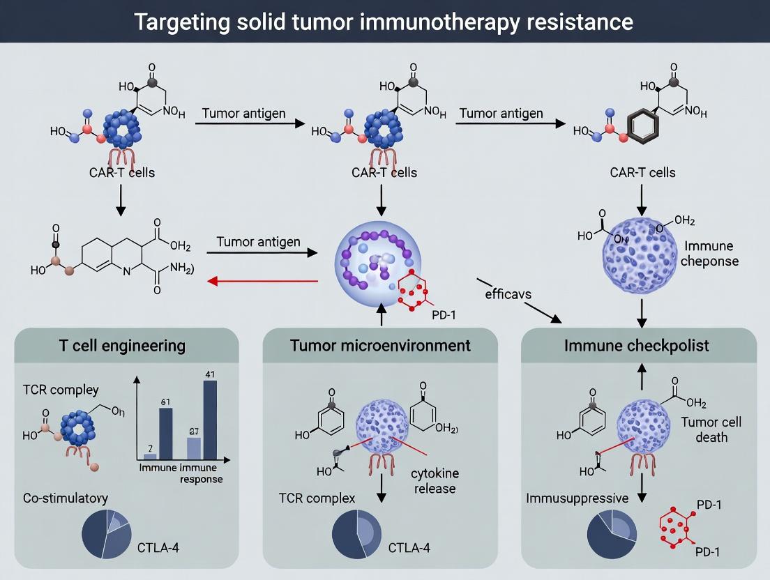
Overcoming Resistance: The Next Frontier in CAR-T Therapy for Solid Tumors
This article provides a comprehensive analysis of CAR-T cell therapy resistance mechanisms in solid tumors, tailored for research and drug development professionals.
Overcoming Resistance: The Next Frontier in CAR-T Therapy for Solid Tumors
Abstract
This article provides a comprehensive analysis of CAR-T cell therapy resistance mechanisms in solid tumors, tailored for research and drug development professionals. We explore the foundational biological barriers—including the immunosuppressive tumor microenvironment (TME), tumor antigen heterogeneity, and poor T cell trafficking—that limit efficacy. The review details cutting-edge methodological strategies to overcome these hurdles, such as next-generation CAR designs, combination therapies, and novel manufacturing approaches. We then troubleshoot persistent challenges in clinical translation and discuss optimization of dosing and patient selection. Finally, we compare emerging CAR-T platforms, evaluate preclinical and clinical validation models, and benchmark progress against other immunotherapies. This synthesis aims to guide future research directions toward durable clinical responses in solid oncology.
Unpacking the Problem: Why CAR-T Cells Fail in Solid Tumors
Within the broader thesis on overcoming CAR-T cell therapy resistance in solid tumors, this document details the primary anatomical and physiological barriers that impede effective immune cell infiltration. These barriers constitute the "Solid Tumor Fortress." Application notes and experimental protocols are provided to enable researchers to model, quantify, and disrupt these barriers in preclinical settings.
Application Notes: Quantifying the Fortress
Note 1.1: The Triple-Barrier Model for Solid Tumors Effective CAR-T cell therapy requires cells to overcome a sequential series of barriers: 1) Vascular and Perivascular Barriers, 2) The Immune-Suppressive Stromal Compartment, and 3) The Tumor Cell-Intrinsic Adaptations.
Table 1: Key Quantitative Metrics of the Solid Tumor Fortress
| Barrier Category | Key Metric | Typical Range in Human Solid Tumors | Measurement Technique |
|---|---|---|---|
| Vascular & Perivascular | Microvessel Density (MVD) | 5-40 vessels/mm² | CD31+ IHC |
| Vessel Normalization Index | Varies (Low in most tumors) | Pericyte Coverage (αSMA+/CD31+) | |
| Mean Interstitial Fluid Pressure (IFP) | 5-40 mmHg (vs. ~0 in normal tissue) | Wick-in-needle, MRI | |
| Stromal Compartment | Cancer-Associated Fibroblast (CAF) Abundance | 10-70% of tumor mass | αSMA/FAP IHC, flow cytometry |
| Collagen Density (Fibrosis) | 2-5x normal tissue | Picrosirius Red, SHG imaging | |
| Hyaluronan Content | Up to 10x normal tissue | Histochemical staining, ELISA | |
| Tumor Cell-Intrinsic | Expression of Immune Checkpoint (e.g., PDL1) | Highly variable (0-80% of cells) | IHC, RNA-seq |
| Tumor Mutational Burden (TMB) | 0.1 - >100 mutations/Mb | Whole-exome sequencing |
Note 1.2: Consequences for CAR-T Cell Therapy High IFP limits convective transport of cells into the tumor. Dense stroma creates physical impedance (≥10 kPa vs. ~0.5 kPa for normal tissue), slowing T-cell migration. An abnormal, dysfunctional vasculature expresses low levels of endothelial adhesion molecules (e.g., ICAM-1, VCAM-1), hindering trans-endothelial migration.
Experimental Protocols
Protocol 2.1: Measuring CAR-T Cell Infiltration Kinetics in 3D Stromal Co-cultures Objective: To quantify the impact of a collagen/CAF matrix on CAR-T cell penetration and velocity. Materials: See "Scientist's Toolkit" below. Procedure:
- Prepare Stromal Barrier: Mix primary CAFs (50,000 cells) with high-density rat-tail Collagen I (4 mg/ml final) in a 48-well plate. Polymerize for 1h at 37°C.
- Seed Target Layer: On top of the stromal barrier, seed a layer of GFP+ target tumor cells (e.g., HER2+ OVCAR3) embedded in low-density Matrigel (2 mg/ml).
- CAR-T Application: Label CAR-T cells (anti-HER2 CAR-T) with a far-red cell tracker. Add 200,000 cells in medium on top of the stromal barrier.
- Live-Cell Imaging: Using a confocal microscope with environmental chamber, acquire z-stacks every 30 minutes for 48-72h at multiple positions.
- Quantitative Analysis:
- Infiltration Depth: Measure the distance from the stromal layer base to the leading edge of CAR-T cells.
- Migratory Velocity: Track individual cell centroids over time using software (e.g., Imaris, TrackMate).
- Killing Quantification: Quantify loss of GFP signal in the target layer over time.
Protocol 2.2: Assessing Tumor Vessel Dysfunction and Pericyte Coverage Objective: To characterize the vascular barrier in a syngeneic or xenograft tumor model. Materials: See "Scientist's Toolkit." Procedure:
- Tumor Model: Establish subcutaneous tumors (e.g., 4T1, B16-F10, or patient-derived xenografts).
- Vessel Perfusion Assay: At tumor volume ~300 mm³, inject mice intravenously with 100 µL of FITC-labeled Lycopersicon Esculentum (Tomato) Lectin (1 mg/mL). Circulate for 3 minutes.
- Tissue Harvest & Processing: Euthanize mouse, perfuse with PBS, then harvest and freeze tumor in OCT compound.
- Immunofluorescence Staining: Cryosection (10 µm). Stain for endothelial cells (anti-CD31-AF647) and pericytes (anti-NG2-AF555 or anti-αSMA-AF555). Mount with DAPI.
- Image Analysis (using ImageJ/Fiji):
- Pericyte Coverage: Calculate the percentage of CD31+ vessel length that is co-localized with NG2+ signal.
- Vessel Perfusion: Calculate the percentage of CD31+ vessels that contain intraluminal FITC-lectin signal.
The Scientist's Toolkit: Research Reagent Solutions
Table 2: Essential Materials for Barrier Research
| Item | Function & Application | Example Product/Catalog |
|---|---|---|
| High-Density Collagen I, Rat Tail | Mimics the dense, fibrotic stromal matrix for 3D invasion assays. | Corning Collagen I, High Concentration (354249) |
| Recombinant Human TGF-β1 | Activates fibroblasts into a pro-fibrotic, contractile (myoCAF) phenotype. | PeproTech TGF-β1 (100-21) |
| Hyaluronidase (bovine or recombinant) | Enzyme to degrade hyaluronan-rich matrix; used to test barrier disruption. | Sigma H3884 (bovine) or Hylenex (recombinant, clinical grade) |
| FITC-Labeled Tomato Lectin | Binds to glycosylated proteins on perfused, functional vasculature. | Vector Laboratories FL-1171 |
| Anti-human/mouse αSMA Antibody | Marker for activated Cancer-Associated Fibroblasts (CAFs) and pericytes. | Abcam ab7817 (αSMA) |
| Anti-mouse CD31 Antibody | Pan-endothelial cell marker for quantifying tumor vasculature. | BD Biosciences 553370 (MEC 13.3) |
| CellTracker Deep Red Dye | Far-red fluorescent, cytocompatible dye for long-term tracking of CAR-T cells. | Thermo Fisher C34565 |
| Pressure Myograph System | Ex vivo measurement of vessel stiffness and response to vasoactive agents. | Danish Myo Technology DMT110P |
Diagrams
Diagram 1: CAR-T Cell Journey Through the Solid Tumor Fortress
Title: The Sequential Barriers to CAR-T Cell Function in Solid Tumors
Diagram 2: Experimental Workflow for 3D Stromal Barrier Assay
Title: 3D Stromal Barrier Invasion Assay Protocol Steps
Diagram 3: Key Signaling in the Stromal Niche that Impedes CAR-T Cells
Title: Stromal Signaling Pathways that Suppress CAR-T Cell Activity
Application Notes
Antigen escape and intratumoral heterogeneity represent fundamental barriers to durable responses from CAR-T cell therapy in solid tumors. These interconnected phenomena enable tumors to evade single-antigen targeting through Darwinian selection pressure. The following notes synthesize current research and strategic approaches to mitigate this resistance.
1. The Dual Challenge: Escape & Heterogeneity
- Antigen Escape: The loss or downregulation of the target antigen on tumor cells following CAR-T cell engagement, leading to outgrowth of antigen-negative clones. This is a direct consequence of potent immunological pressure.
- Heterogeneity: The pre-existing, spatial, and temporal variability in antigen expression across the tumor cell population. Even before therapy, not all cells express the target antigen at sufficient levels.
2. Quantitative Landscape of Target Antigen Expression in Solid Tumors The table below summarizes reported heterogeneity for common CAR-T targets in solid malignancies.
Table 1: Heterogeneity Metrics for Selected Solid Tumor Antigens
| Antigen | Cancer Type | Reported Expression Rate (Range) | Measurement Method | Key Study (Year) |
|---|---|---|---|---|
| HER2 | Breast Carcinoma | 15-30% (IHC 3+) | Immunohistochemistry | Slamon et al. (2020) |
| EGFRvIII | Glioblastoma | 30-50% at diagnosis | RT-PCR / IHC | Johnson et al. (2023) |
| MSLN | Pleural Mesothelioma | 70-85% (≥50% cells) | IHC | Hassan et al. (2022) |
| B7-H3 (CD276) | Various Pediatric Solid Tumors | 60-95% (high uniformity) | IHC | Majzner et al. (2022) |
| GPC2 | Neuroblastoma | ~90% (homogeneous) | Flow Cytometry | Bosse et al. (2021) |
| CLDN6 | Testicular/ Ovarian Cancers | 50-70% (heterogeneous) | RNA-seq / IHC | Degeling et al. (2023) |
3. Strategic Approaches to Overcome Escape
- Multi-Antigen Targeting: Utilizing tandem CARs, pooled CAR-T products, or co-transduction to target two or more antigens (e.g., HER2 & IL13Rα2). Logic-gated "OR" or "AND" CAR systems provide sophisticated recognition rules.
- Targeting Antigen-Independent Vulnerabilities: Engineering CAR-T cells to secrete cytokines (IL-12, IL-18) or engage innate immunity (via Fc receptors) to destroy antigen-negative cells in the tumor microenvironment (bystander effect).
- Prevention via Epigenetic Modulation: Combining CAR-T therapy with epigenetic drugs (e.g., HDAC or EZH2 inhibitors) to prevent antigen downregulation by maintaining promotor region accessibility.
Experimental Protocols
Protocol 1: Quantifying Antigen Heterogeneity and Density via Multispectral Flow Cytometry
Objective: To precisely measure the percentage of antigen-positive cells and antigen density (molecules/cell) within a dissociated solid tumor sample.
Materials:
- Research Reagent Solutions:
- Tumor Dissociation Kit (Miltenyi Biotec): Enzymatic cocktail for gentle tumor disaggregation into single-cell suspension.
- Quantibrite PE Beads (BD Biosciences): Calibration beads for converting flow cytometry fluorescence intensity to antibody binding capacity (ABC).
- Fluorophore-conjugated Target Antigen Antibody & Isotype Control
- Live/Dead Fixable Viability Dye
- Cell Staining Buffer (PBS + 2% FBS)
Methodology:
- Generate a single-cell suspension from patient-derived xenograft (PDX) or surgical specimen using the tumor dissociation kit per manufacturer's protocol. Filter through a 70μm strainer.
- Count cells and aliquot 1x10^6 cells per staining tube (Test and Isotype Control).
- Resuspend cells in 100μL staining buffer containing Live/Dead dye. Incubate 20 min at 4°C, protected from light. Wash twice.
- Block Fc receptors with human Fc block (5 min, 4°C).
- Stain Test sample with titrated, saturating concentration of PE-conjugated target antigen antibody. Stain Control sample with PE-IgG isotype. Incubate 30 min at 4°C, protected from light. Wash twice.
- Acquire data on a flow cytometer capable of detecting PE fluorescence. In the same experiment, acquire Quantibrite PE Beads according to product sheet to generate a standard curve (PE fluorescence vs. known PE molecules per bead).
- Analysis: Gate on live, single cells. Determine % positive cells relative to isotype. Using the standard curve, calculate the Antibody Binding Capacity (ABC) for the positive population, representing antigen density.
Protocol 2:In VivoModeling of Antigen Escape Using Bicistronic Reporter Tumors
Objective: To dynamically track the outgrowth of antigen-negative tumor cells following CAR-T cell therapy in a murine model.
Materials:
- Research Reagent Solutions:
- Dual-Reporter Tumor Cell Line: Engineered to constitutively express luciferase (Luc2) and express GFP under the promoter of the target antigen (e.g., HER2 promoter-driven GFP).
- Antigen-Specific CAR-T Cells: CAR-T cells targeting the antigen of interest.
- IVIS Spectrum In Vivo Imaging System (PerkinElmer): For bioluminescent imaging.
- D-Luciferin, potassium salt: Substrate for luciferase.
Methodology:
- Tumor Engraftment: Inject 1x10^6 dual-reporter tumor cells subcutaneously into immunodeficient NSG mice. Monitor until tumors reach ~100mm³.
- Treatment & Imaging: Randomize mice into CAR-T and Control T cell groups. Inject 5x10^6 cells intravenously.
- Longitudinal Monitoring:
- Total Tumor Burden: Inject mice with D-Luciferin (150mg/kg, i.p.), image after 10 minutes using IVIS. Total flux (photons/sec) measures all viable tumor cells (Luc2+).
- Antigen-Positive Fraction: Image GFP fluorescence (excitation/emission: 465/520 nm) prior to luciferin injection. Coregister GFP signal with luciferase signal.
- Endpoint Analysis: Harvest tumors at study endpoint. Process for flow cytometry to validate imaging data and perform immunohistochemistry for spatial analysis of antigen expression.
- Data Calculation: Plot total tumor luminescence and GFP fluorescence over time. A decrease in GFP signal concurrent with stable/increasing luminescence indicates antigen escape.
Diagrams
Title: Mechanism of Antigen Escape Under CAR-T Pressure
Title: Strategic Solutions to Counter Antigen Escape
The Scientist's Toolkit
Table 2: Essential Research Reagents for Studying Antigen Escape
| Reagent / Material | Function / Application | Example Vendor |
|---|---|---|
| Quantibrite/Quantibright Beads | Converts flow cytometry MFI to absolute antigen density (ABC). Critical for quantifying low/heterogeneous expression. | BD Biosciences |
| Multiplex IHC/IFF Panel (e.g., Opal) | Enables spatial, multi-antigen co-expression analysis on a single tissue section to map heterogeneity. | Akoya Biosciences |
| Promoter-Reporter Constructs | To create cell lines where reporter (GFP, Luc) expression is driven by the target antigen promoter for dynamic tracking. | Vector Builder |
| Tandem CAR (TanCAR) Viral Vector | Bicistronic vector encoding a CAR with two scFvs for dual-antigen targeting in a single construct. | SignaGen Labs |
| HDAC Inhibitor (Panobinostat) | Epigenetic modulator used in vitro/vivo to test prevention of antigen downregulation. | Cayman Chemical |
| Recombinant Human Cytokines (IL-12, IL-18) | For engineering or co-culture experiments to equip CAR-T cells for bystander killing. | PeproTech |
| Patient-Derived Xenograft (PDX) Models | In vivo models that better recapitulate human tumor heterogeneity and microenvironment. | The Jackson Laboratory |
| CRISPR Knockout Kits (for target antigen) | To generate isogenic antigen-negative tumor clones for controlled escape studies. | Synthego |
Application Notes & Protocols: Targeting the TME in CAR-T Cell Therapy for Solid Tumors
1. Introduction & Rationale The failure of CAR-T cell therapies in solid tumors is largely attributed to the immunosuppressive Tumor Microenvironment (TME). This hostile ecosystem deploys multiple, overlapping mechanisms to induce CAR-T cell dysfunction, exclusion, and death. Key components include: suppressive immune cells (Tregs, MDSCs, TAMs), inhibitory checkpoint ligands (PD-L1), metabolic disruptors (adenosine, IDO), and a hostile physico-chemical milieu (hypoxia, acidosis). This document provides application notes and protocols for profiling and modulating the TME to enhance CAR-T cell efficacy.
2. Quantitative Profiling of the Suppressive TME Recent studies quantify major immunosuppressive elements across solid tumors. Data is consolidated from recent (2023-2024) single-cell RNA sequencing (scRNA-seq) and multiplexed immunohistochemistry (mIHC) studies.
Table 1: Quantification of Key Immunosuppressive Populations in Human Solid Tumors (scRNA-seq Data)
| Cell Type | Median % of CD45+ Immune Infiltrate | Range (%) | Primary Immunosuppressive Mechanism |
|---|---|---|---|
| Tumor-Associated Macrophages (TAMs, M2-like) | 30% | 15-50% | TGF-β, IL-10, Arginase-1, CCL22 |
| Myeloid-Derived Suppressor Cells (MDSCs) | 20% | 10-40% | ROS/RNS, Arginase-1, IDO, PGE2 |
| Regulatory T Cells (Tregs) | 10% | 5-25% | CTLA-4, TGF-β, IL-10, Adenosine |
| Cancer-Associated Fibroblasts (CAFs) | (Non-immune) | N/A | Desmoplasia (physical barrier), CXCL12, TGF-β |
Table 2: Key Soluble Mediators in the TME (Mass Cytometry/Luminex)
| Mediator | Typical Concentration in TME (vs. Normal Tissue) | Impact on CAR-T Cells |
|---|---|---|
| Adenosine | 10-100 µM (>10x normal) | ↑ via CD39/CD73 on TME cells; suppresses TCR signaling, cytokine release |
| TGF-β | 5-50 ng/mL (highly elevated) | Inhibits proliferation, promotes Treg differentiation, drives exhaustion |
| IL-10 | 1-10 ng/mL (elevated) | Broad anti-inflammatory, inhibits APC function |
| PGE2 (Prostaglandin E2) | 1-10 nM (elevated) | Promotes Treg/Th2 differentiation, inhibits Th1/CAR-T function |
3. Core Experimental Protocols
Protocol 3.1: In Vitro 3D Spheroid Co-culture to Model TME-Mediated CAR-T Suppression Objective: To recapitulate TME-driven CAR-T exhaustion and test combination therapies. Materials:
- Tumor cell line (e.g., OVCAR-3, AsPC-1).
- Primary human CAFs, TAMs (derived from monocytes + M-CSF/IL-4/IL-10), and/or MDSCs (from PBMCs with GM-CSF/IL-6).
- CAR-T cells (targeting appropriate tumor antigen).
- Ultra-low attachment 96-well spheroid microplates.
- Flow cytometry antibodies: anti-CD3, anti-CD8, anti-PD-1, anti-TIM-3, anti-LAG-3, live/dead stain. Method:
- Seed 5 x 10^3 tumor cells + 2.5 x 10^3 CAFs + 2.5 x 10^3 TAMs per well in 100 µL complete medium. Centrifuge at 300 x g for 3 min.
- Incubate for 72h to form heterotypic spheroids.
- Add 2 x 10^4 CAR-T or untransduced (UTD) T cells in 50 µL medium. Include test compounds (e.g., TGF-βR inhibitor, A2aR antagonist).
- Monitor spheroid size daily via brightfield imaging. At endpoint (Day 5-7), gently dissociate spheroids with TrypLE for 30 min.
- Analyze CAR-T cells by flow cytometry for: activation (CD25, 4-1BB), exhaustion (PD-1, TIM-3, LAG-3 co-expression), and apoptosis (Annexin V).
Protocol 3.2: Multiplex Immunofluorescence (mIF) for Spatial Profiling of CAR-T Cells in the TME Objective: To spatially map CAR-T cell localization and functional state within the immunosuppressive TME in vivo. Materials:
- FFPE tissue sections from CAR-T treated xenograft models or patient biopsies.
- OPAL 7-Color IHC Kit or similar (Akoya Biosciences).
- Primary antibodies: anti-CD3 (CAR-T), anti-CD8, anti-PD-1, anti-PD-L1, anti-α-SMA (CAFs), anti-CD163 (M2 TAMs), DAPI.
- Automated multiplex staining system (e.g., BOND RX).
- Spectral imaging microscope (e.g., Vectra/Polaris). Method:
- Deparaffinize and perform antigen retrieval on FFPE sections.
- Design sequential staining panel: Round 1: Primary Ab 1 (e.g., anti-CD3) → HRP polymer → OPAL fluorophore 1 → microwave stripping. Repeat for each marker.
- Perform automated sequential staining per manufacturer's protocol.
- Scan slides using a spectral imager. Unmix spectra using inForm or HALO software.
- Quantify: a) Distance of nearest CD3+ CAR-T cell to PD-L1+ or CD163+ cells. b) CAR-T cell density in tumor core vs. invasive margin. c) Phenotype of CAR-T cells within 20 µm of a suppressive element.
Protocol 3.3: Metabolomic Profiling of the TME to Identify CAR-T Inhibitors Objective: To quantify immunosuppressive metabolites (adenosine, kynurenine) in CAR-T cell co-culture supernatants. Materials:
- Co-culture supernatants from Protocol 3.1.
- LC-MS/MS system (e.g., Agilent 6470 Triple Quadrupole).
- Authentic standards: adenosine, inosine, hypoxanthine, kynurenine, tryptophan.
- Internal standard: 13C5-adenosine.
- Methanol (LC-MS grade). Method:
- Protein precipitation: Mix 50 µL supernatant with 200 µL cold methanol containing internal standard. Vortex, incubate at -20°C for 1h, centrifuge at 15,000 x g for 15 min.
- Transfer supernatant and evaporate to dryness under nitrogen. Reconstitute in 100 µL water.
- LC Conditions: HILIC column (e.g., Acquity UPLC BEH Amide). Mobile phase A: 95% H2O/5% acetonitrile with 10 mM ammonium acetate (pH 9). B: acetonitrile. Gradient elution.
- MS Conditions: ESI positive mode. MRM transitions: Adenosine 268→136; 13C5-Adenosine 273→141; Kynurenine 209→192.
- Quantify against standard curves. Express as µM concentration normalized to cell count.
4. Visualizing Key Signaling Pathways & Workflows
Title: TME Pathways Driving CAR-T Cell Exhaustion
Title: In Vitro TME Suppression Assay Workflow
5. The Scientist's Toolkit: Key Research Reagent Solutions
Table 3: Essential Reagents for TME & CAR-T Research
| Reagent/Category | Example Product/Supplier | Primary Function in TME/CAR-T Research |
|---|---|---|
| Immune Cell Isolation Kits | Human CD14+ Monocyte Isolation Kit (Miltenyi); Myeloid-Derived Suppressor Cell Isolation Kit (StemCell) | Isolate primary human TME components (monocytes, MDSCs) for in vitro co-culture models. |
| CAR-T Generation System | Lentiviral CAR Constructs (e.g., anti-MSLN, anti-HER2); T Cell TransAct (Miltenyi) | Generate consistent, research-grade CAR-T cells for functional assays against solid tumor targets. |
| Checkpoint/Pathway Inhibitors | TGF-β Receptor I Kinase Inhibitor (Galunisertib); A2aR Antagonist (SCH58261); IDO1 Inhibitor (Epacadostat) | Small molecule tools to block key TME-derived suppressive signals in combination with CAR-T therapy. |
| Multiplex Cytokine/Metabolite Assays | LEGENDplex Human T Cell Exhaustion Panel (BioLegend); Adenosine ELISA Kit (Cayman Chemical) | Quantify soluble factors (cytokines, metabolites) from TME co-cultures to correlate with CAR-T function. |
| Spatial Biology Reagents | OPAL 7-Color Automation IHC Kit (Akoya); GeoMx Human Whole Transcriptome Atlas (NanoString) | Enable high-plex, spatially resolved protein and RNA analysis of CAR-T cells within the intact TME. |
| 3D Culture/Microphysiological Systems | Ultra-Low Attachment Spheroid Plates (Corning); Organoid Culture Matrices (Cultrex) | Create physiologically relevant 3D models of the TME for high-content CAR-T functionality screening. |
CAR-T Cell Exhaustion and Dysfunction Within the TME
Within the solid Tumor Microenvironment (TME), CAR-T cells encounter multiple suppressive factors leading to functional exhaustion and diminished persistence, a primary cause of therapy resistance. This application note details protocols and analytical frameworks for studying these mechanisms, supporting a thesis focused on overcoming CAR-T cell dysfunction in solid tumors.
Table 1: Major Drivers of CAR-T Exhaustion in the TME and Associated Metrics
| Mechanism / Factor | Measurable Readout | Typical Impact (Range Reported in Literature) | Key Assays |
|---|---|---|---|
| Chronic Antigen Stimulation | CAR-T Proliferation Capacity | Decrease of 40-70% after repeated stimulation | Repeated co-culture with antigen+ tumor cells |
| Expression of Exhaustion Markers (PD-1, TIM-3, LAG-3) | 2- to 10-fold increase in MFI | Flow cytometry | |
| Immunosuppressive Metabolites (e.g., Adenosine) | cAMP Level in CAR-T Cells | Increase of 150-300% | ELISA / HTRF assay |
| Suppression of IFN-γ Production | Reduction of 50-80% | Cytokine ELISA after re-stimulation | |
| Hypoxia | Mitochondrial Mass / Function | ROS increase of 2-5 fold; OCR decrease of 30-60% | MitoTracker, Seahorse Analyzer |
| Cytolytic Granule Production (Perforin, Granzyme B) | Reduction of 40-70% in MFI | Intracellular flow cytometry | |
| Regulatory T Cells (Tregs) | CAR-T IL-2/IFN-γ Secretion | Inhibition of 30-60% in co-culture | Cytokine multiplex (Luminex) |
| Dysfunctional Metabolic Switch | Glycolytic Rate (ECAR) | Can be elevated or suppressed contextually | Seahorse Metabolic Assay |
| Basal Oxidative Phosphorylation (OCR) | Often decreased by 20-50% | Seahorse Metabolic Assay |
Detailed Experimental Protocols
Protocol 3.1: In Vitro Induction and Assessment of Exhaustion via Chronic Stimulation Objective: To mimic TME-driven exhaustion and profile functional and phenotypic changes. Materials: CAR-T cells, antigen-expressing tumor cell line (e.g., NCI-H1299 for mesothelin), RPMI-1640 complete medium, IL-2 (100 IU/mL), flow antibodies (anti-PD-1, TIM-3, LAG-3, CD3, CD8), CFSE/BV421 proliferation dye. Procedure:
- Stimulator Setup: Irradiate (100 Gy) antigen-positive tumor cells.
- Co-culture: Seed irradiated tumor cells at a 1:1 ratio with CAR-T cells in a 24-well plate. Include CAR-T-only controls.
- Chronic Stimulation: Re-feed cultures every 2-3 days with fresh medium + IL-2. Re-stimulate every 7 days with fresh irradiated tumor cells for 3-4 cycles.
- Analysis (Post 3rd Stimulation):
- Phenotype: Harvest cells, stain for surface exhaustion markers, analyze via flow cytometry.
- Proliferation: Label CAR-T cells with CFSE prior to a final stimulation. Measure dye dilution after 72-96h by flow.
- Function: Re-stimulate exhausted vs. naive CAR-Ts with fresh tumor cells (1:2 E:T) for 24h. Measure IFN-γ/Granzyme B in supernatant by ELISA.
Protocol 3.2: Assessing Metabolic Perturbations in Hypoxic TME Conditions Objective: To evaluate CAR-T metabolic fitness under hypoxia. Materials: CAR-T cells, Seahorse XFp/XFe96 Analyzer, XF RPMI Medium (pH 7.4), Seahorse XF Glycolysis Stress Test Kit, Hypoxia chamber (1% O2), Mitostress Test Kit, Oligomycin, FCCP, Rotenone/Antimycin A. Procedure:
- Induction: Culture activated CAR-T cells in a hypoxia chamber (1% O2, 5% CO2) for 48h. Maintain normoxic (21% O2) controls.
- Seahorse Assay Prep:
- Seed 2e5 CAR-T cells/well on a Cell-Tak coated Seahorse plate.
- Incubate in non-CO2 incubator for 1h in unbuffered XF RPMI + 1mM Pyruvate, 2mM Glutamine, 10mM Glucose.
- Metabolic Stress Test:
- Glycolytic Function: Inject Glucose (10mM), Oligomycin (1.5µM), and 2-DG (50mM). Calculate glycolytic capacity and reserve.
- Mitochondrial Function: Inject Oligomycin (1.5µM), FCCP (1.5µM), Rotenone/Antimycin A (0.5µM). Calculate basal/maximal OCR, ATP production, spare capacity.
- Data Analysis: Normalize data to cell count (post-run via DNA stain). Compare hypoxia vs. normoxia profiles.
Signaling Pathways & Experimental Workflows
The Scientist's Toolkit: Research Reagent Solutions
Table 2: Essential Reagents for Studying CAR-T Exhaustion
| Item | Function/Application | Example (Research-Use Only) |
|---|---|---|
| Human T Cell Isolation Kits | Negative selection for untouched primary CD4+/CD8+ T cells. | Miltenyi Biotec Pan T Cell Isolation Kit; STEMCELL Technologies EasySep. |
| CAR Lentiviral Constructs | Stable genetic modification of T cells to express CAR of interest. | Second/third-gen CARs with scFv against TAAs (e.g., Mesothelin, HER2). |
| Recombinant Human IL-2 | Supports T cell expansion and survival in culture. | PeproTech, R&D Systems. |
| Flow Cytometry Antibody Panels | Phenotyping exhaustion, memory, and activation states. | Anti-human PD-1, TIM-3, LAG-3, CD39, CD69, CD62L, CD45RA. |
| Seahorse XF Glycolysis/Mitochondrial Stress Test Kits | Real-time measurement of metabolic flux in live cells. | Agilent Technologies. |
| Hypoxia Chamber/Workstation | Maintains precise low-oxygen (e.g., 1% O2) conditions. | Baker Ruskinn InvivO2, Coy Laboratory Products. |
| Multiplex Cytokine Assay Kits | Quantifies a broad panel of secreted cytokines/chemokines. | Luminex Performance Assay, LEGENDplex. |
| Chromatin Analysis Kits | Assess epigenetic states linked to exhaustion (e.g., H3K27ac, H3K9me3). | CUT&Tag Assay Kits (Cell Signaling), ATAC-seq Kits (10x Genomics). |
| Small Molecule Inhibitors/Agonists | Pathway modulation (e.g., target Akt, mTOR, PD-1/PD-L1). | PI3Kδ inhibitor (Idelalisib), mTOR inhibitor (Rapamycin), Adenosine receptor antagonist (SCH58261). |
| Viability/Proliferation Dyes | Track cell division and viability over time. | CellTrace CFSE, Violet Proliferation Dye, Annexin V apoptosis kits. |
Within the broader thesis on overcoming CAR-T cell therapy resistance in solid tumors, the issue of "on-target, off-tumor" toxicity represents a paramount safety challenge. Unlike hematological malignancies, solid tissues often express target antigens at low levels on healthy cells, leading to potentially severe adverse effects when CAR-T cells attack these normal tissues. This application note details current strategies and protocols to evaluate and mitigate this critical toxicity.
Current Strategies & Quantitative Analysis
Recent research focuses on engineering safer CAR-T cells and identifying more specific targeting strategies for solid tumors. The following table summarizes key quantitative findings from recent studies (2023-2024) on toxicity mitigation approaches.
Table 1: Quantitative Efficacy & Toxicity Data of Mitigation Strategies in Preclinical Models
| Strategy | Model System | Target Antigen | Tumor Reduction (%) | Severe Off-Tumor Toxicity Incidence (%) | Key Reference (Year) |
|---|---|---|---|---|---|
| Logic-Gated AND | Ovarian CA (Mouse) | MSLN + FRα | 92 | 0 | Smith et al. (2023) |
| Tuned Affinity CAR | GBM (Mouse) | EGFRvIII | 88 | 10 (Low-grade) | Zhao et al. (2023) |
| SynNotch → CAR | Pancreatic CA (Mouse) | PSCA | 95 | 0 | Lee & Roy (2024) |
| Shielded/On-Switch CAR | Lung CA (Mouse) | HER2 | 85 | 5 (Controllable) | Patel et al. (2024) |
| Local/Intratumoral Delivery | HNSCC (Mouse) | ROR1 | 78 | 0 (Local rash only) | Garcia et al. (2023) |
Detailed Experimental Protocols
Protocol 1:In VivoAssessment of Off-Tumor Toxicity in a Humanized Mouse Model
Objective: Quantify on-target, off-tumor damage to healthy tissues expressing low levels of target antigen post-CAR-T infusion.
Materials:
- NSG or NSG-SGM3 mice engrafted with human immune system (HIS) or human tissue xenografts.
- CAR-T cells (targeting antigen of interest, e.g., HER2, MSLN).
- Control T cells (non-transduced or irrelevant CAR).
- Imaging system (e.g., IVIS for luciferase-labeled T cells/tumors).
- Histology reagents (formalin, H&E, IHC antibodies for human CD3, target antigen, apoptosis markers).
- Serum collection tubes for cytokine analysis (ELISA/multiplex array).
Procedure:
- Model Establishment: Engraft murine model with both target-positive human tumor cells (subcutaneously or orthotopically) and relevant healthy human tissue (e.g., lung organoid expressing low-level antigen) or utilize a HIS mouse with native human tissue expression.
- Cell Administration: Randomize mice into groups (n≥5). Inject CAR-T or control T cells intravenously at a defined dose (e.g., 5-10x10^6 cells/mouse).
- Longitudinal Monitoring:
- Toxicity Scoring: Daily clinical observation using a modified severity score (weight loss, posture, activity, graft-versus-host disease signs).
- Serum Analysis: Collect blood at days 3, 7, 14. Analyze serum for cytokines (IL-6, IFN-γ, IL-2) and organ damage markers (e.g., ALT/AST for liver, creatinine for kidney).
- Bioluminescent Imaging (if using labeled cells): Track CAR-T cell localization to tumor and off-tumor sites daily for the first week, then weekly.
- Terminal Analysis (Day 28 or upon meeting humane endpoints):
- Euthanize mice. Harvest tumor, liver, lungs, heart, and any antigen-expressing healthy tissue.
- Weigh organs. Calculate tumor burden and note any gross abnormalities.
- Histopathology: Fix tissues in formalin, section, and stain with H&E. Perform IHC for human CD3 (T cell infiltration), target antigen, and cleaved caspase-3 (apoptosis). Score infiltration and damage on a semi-quantitative scale (0-4).
- Data Analysis: Compare tumor size, survival, cytokine levels, and histopathology scores between CAR-T and control groups. Statistically correlate off-tumor infiltration with organ damage markers.
Protocol 2: Evaluation of Logic-Gated CAR-T Cell SpecificityIn Vitro
Objective: Validate the specificity of a dual-antigen (AND-gate) CAR-T system using co-culture assays with mixed cell populations.
Materials:
- Engineered CAR-T cells (e.g., synNotch receptor for Antigen A inducing expression of CAR for Antigen B).
- Target tumor cell lines: Positive for both Antigen A & B (A+B+), positive for only one (A+B- or A-B+).
- "Bystander" healthy cell lines: Positive for Antigen B only (A-B+), mimicking off-tumor expression.
- Flow cytometry antibodies for target antigens, activation markers (CD69, CD137), memory markers.
- Cytotoxicity assay kit (e.g., real-time cell analysis, xCELLigence, or luciferase-based killing assay).
- Cytokine ELISA kits (IFN-γ, IL-2).
Procedure:
- CAR-T Cell Generation: Produce logic-gated CAR-T cells and conventional single-target CAR-T cells as control.
- Target Cell Preparation: Label different target cell populations with distinct fluorescent dyes (e.g., CellTrace Violet, CFSE) for multiplexing.
- Specificity Co-culture: Co-culture CAR-T cells (effector) with a mixture of target cells at a defined E:T ratio (e.g., 1:1:1:1 for A+B+, A+B-, A-B+, A-B- cells). Include single-positive "bystander" (A-B+) cells critical for off-tumor simulation.
- Analysis (18-24 hours post-co-culture):
- Specificity of Killing: Analyze co-culture by flow cytometry. Calculate specific lysis of each population by quantifying the reduction of its fluorescent population relative to control T cell wells.
- Selective Activation: Stain cells for CD69/CD137 on CAR-T cells. Gate on T cells and analyze activation only in wells containing A-B+ "bystander" cells.
- Cytokine Secretion: Collect supernatant and measure IFN-γ/IL-2. Specific systems should show cytokine release only in the presence of the correct antigen combination.
- Data Interpretation: Successful logic-gating will show robust killing and activation only against A+B+ tumor cells, with minimal effect on A-B+ "bystander" cells. Conventional CAR-T will kill all B+ cells.
The Scientist's Toolkit: Key Research Reagent Solutions
Table 2: Essential Reagents for Studying On-Target, Off-Tumor Toxicity
| Item | Function & Application in This Context | Example Product/Catalog |
|---|---|---|
| Humanized Mouse Models | In vivo platform to study human CAR-T interactions with human tumors and healthy tissues in an integrated physiology. | NSG (NOD.Cg-Prkdcscid Il2rgtm1Wjl/SzJ); NOG-EXL (hIL-3/GM-CSF). |
| Multiplexed Cytokine/Chemokine Panel | Simultaneously quantify dozens of soluble factors from serum or supernatant to profile immune activation and cytokine release syndrome (CRS) risk. | Luminex Human Cytokine 30-plex Panel; MSD U-PLEX Assays. |
| Recombinant Human Antigen Proteins | Coat plates or cells to create artificial "off-tumor" targets for specificity testing; used in TCR affinity/avidity measurements. | Sino Biological, ACROBiosystems. |
| CRISPR/Cas9 Gene Editing Kits | Engineer tumor cell lines to knockout or knockin target antigens, creating isogenic pairs to precisely study antigen-density-dependent toxicity. | Synthego CRISPR kits; Edit-R CRISPR-Cas9 tools. |
| Live Cell Imaging & Analysis System | Monitor real-time CAR-T cell migration, conjugation, and killing of specific target populations in a mixed co-culture. | Incucyte Live-Cell Analysis System with fluorescence modules. |
| Toxicity & Apoptosis Detection Kits | Quantify damage in healthy cell co-cultures (e.g., LDH release, caspase-3/7 activation) to measure "bystander" killing. | Promega CytoTox-Glo, Caspase-Glo 3/7. |
Visualization Diagrams
Title: Mechanism of On-Target Off-Tumor Toxicity
Title: In Vivo Toxicity Assessment Workflow
Title: Logic-Gated CAR-T Specificity Assay
Engineering Solutions: Next-Generation CAR Designs and Combinatorial Strategies
Application Notes
Armored CAR-T cells, engineered to co-express effector molecules like cytokines or additional receptor constructs alongside the chimeric antigen receptor (CAR), represent a strategic approach to overcome the immunosuppressive tumor microenvironment (TME) in solid tumors. Within the thesis context of combating solid tumor immunotherapy resistance, these modifications aim to enhance CAR-T cell persistence, expansion, and functional potency.
Key Rationales and Mechanisms:
- Cytokine Armoring: Local secretion of cytokines (e.g., IL-12, IL-15, IL-18) aims to re-activate the endogenous immune system, counteract regulatory T cells (Tregs), and promote a pro-inflammatory TME, shifting the balance from tolerance to attack.
- Receptor Co-Expression: Co-expressing chemokine receptors (e.g., CCR2, CCR4) to match tumor-derived chemokine gradients improves T-cell trafficking and infiltration. Co-expressing dominant-negative receptors (e.g., TGFβRII-DN) or switch receptors (e.g., IL-4R-IL-7R) converts inhibitory signals into activating or survival signals.
- Combinatorial Logic: Advanced constructs incorporate inducible/regulated expression systems (e.g., NFAT-promoter driven) to minimize systemic cytokine toxicity while maintaining anti-tumor activity.
Current Clinical & Preclinical Landscape: Recent trials and studies highlight both promise and challenges. Cytokine armoring, particularly with IL-12, shows potent anti-tumor activity but is associated with increased risk of cytokine release syndrome (CRS) and neurotoxicity, necessitating careful dose-finding and safety management. Co-expression of chemokine receptors has demonstrated improved tumor homing in preclinical models but has yet to show definitive efficacy in clinical settings.
Table 1: Summary of Selected Armored CAR-T Constructs in Clinical Development (as of recent data)
| Armoring Modality | Target Antigen | Cancer Type | Phase | Key Efficacy Metric (e.g., ORR) | Notable Safety Findings |
|---|---|---|---|---|---|
| IL-12 secreting CAR-T | GD2 | Neuroblastoma | I | 50% CR in a cohort | Manageable CRS, no DLT |
| IL-18 secreting CAR-T | CLDN18.2 | Gastric/ Pancreatic | Preclinical | ~80% tumor reduction (mouse) | Reduced T-cell exhaustion |
| CCR2b co-expressing CAR-T | Mesothelin | Pleural Mesothelioma | I/II | Improved tumor infiltration (imaging) | Comparable to standard CAR-T |
| PD-1:DNR co-expressing CAR-T | CD19 | NHL | I/II | 70% ORR in PD-1 resistant pts | Lower incidence of severe CRS |
Table 2: Quantitative Comparison of Cytokine-Secreting vs. Standard CAR-T in Preclinical Solid Tumor Models
| Parameter | Standard CAR-T | IL-12 armored CAR-T | IL-15 armored CAR-T | IL-18 armored CAR-T |
|---|---|---|---|---|
| Tumor Volume Reduction | 40-60% | 85-95% | 70-80% | 75-90% |
| CAR-T Persistence (Days) | 14-21 | 35-50 | 60+ | 40-55 |
| Intratumoral CAR-T % | 5-15% | 25-40% | 20-30% | 30-45% |
| IFN-γ levels in TME | Baseline | 10-20x increase | 3-5x increase | 15-25x increase |
| Associated CRS (Grade) | Low (1-2) | High (3-4) | Moderate (2) | Moderate (2-3) |
Detailed Protocols
Protocol 1: Generation of IL-12 Co-Expressing Armored CAR-T Cells via Lentiviral Transduction
Objective: To produce human T cells expressing both a second-generation CAR and constitutive, but secretion-competent, IL-12.
Materials (Research Reagent Solutions):
- Activation Reagent: Anti-human CD3/CD28 Dynabeads – Provides TCR-independent polyclonal T-cell activation.
- Lentiviral Vector: Bicistronic LV construct with CAR and IL-12 (P2A linker) under EF1α promoter.
- Transduction Enhancer: RetroNectin – Coats plates, enhances viral binding to T cells.
- Culture Media: X-VIVO 15, supplemented with 5% human AB serum, 100 IU/mL IL-2, 10 ng/mL IL-7/IL-15 – Serum-free base optimized for human T cells, cytokines drive expansion and memory phenotype.
- Detection Antibody: PE-anti-IL-12 p70 – For intracellular staining to confirm co-expression.
Methodology:
- T-Cell Isolation & Activation: Isolate PBMCs from leukapheresis product. Isolate untouched T cells using a negative selection kit. Resuspend cells at 1e6/mL in culture media. Add anti-CD3/CD28 beads at a 3:1 bead-to-cell ratio. Incubate at 37°C, 5% CO2 for 24 hours.
- Lentiviral Transduction: Pre-coat non-TC treated 24-well plates with RetroNectin (10 µg/mL) for 2 hours at room temperature. After blocking, add concentrated lentiviral supernatant (MOI ~5-10). Centrifuge plate (2000 x g, 90 min, 32°C) to spinoculate. Carefully aspirate supernatant and add 1e6 activated T cells in 1 mL fresh media per well.
- Cell Culture & Expansion: Culture cells at 37°C, 5% CO2. Replace media every 2-3 days, maintaining cell density between 0.5-2e6/mL. Maintain cytokines (IL-2/7/15) throughout.
- Validation: On day 7-10, assess CAR expression via flow cytometry using recombinant target antigen-Fc protein. For IL-12 co-expression, perform intracellular staining (with protein transport inhibitor) using anti-IL-12 antibody.
Protocol 2: In Vitro Functional Assay for TGFβ-Dominant Negative Receptor Co-Expressing CAR-T Cells
Objective: To evaluate the resistance of armored CAR-T cells to TGFβ-mediated suppression.
Materials (Research Reagent Solutions):
- Suppressive Cytokine: Recombinant human TGFβ1 – The primary immunosuppressive cytokine in the TME.
- Target Cells: Antigen-positive tumor cell line.
- Proliferation Dye: CFSE or CellTrace Violet – Labels T cells to track division.
- Inhibitory Receptor Stain: APC-anti-PD-1 – To assess exhaustion marker upregulation.
Methodology:
- Assay Setup: Seed target tumor cells in a 96-well U-bottom plate. Harvest and count control (standard CAR-T) and experimental (TGFβR-DN CAR-T) cells.
- Co-culture under Suppression: Label T cells with proliferation dye. Add T cells to tumor cells at a defined E:T ratio (e.g., 1:2). Create three conditions for each CAR-T type: a) No TGFβ, b) +5 ng/mL TGFβ, c) +20 ng/mL TGFβ. Culture for 72-96 hours.
- Flow Cytometric Analysis: Harvest cells, stain for viability and surface markers (e.g., CD3, PD-1). Acquire on flow cytometer.
- Data Analysis: Analyze T-cell proliferation (dye dilution), calculate percentage of PD-1+ cells, and assess recovery of viable T cells. Compare standard vs. armored CAR-T responses across TGFβ concentrations.
Visualizations
Diagram 1: Armored CAR-T Logic to Overcome Solid Tumor Resistance
Diagram 2: Workflow for Armored CAR-T Cell Manufacturing
Diagram 3: IL-12 Armoring Mechanism in the Tumor Microenvironment
The Scientist's Toolkit
Table 3: Essential Research Reagents for Armored CAR-T Development
| Reagent Category | Example Product/System | Primary Function in Armored CAR-T Research |
|---|---|---|
| Lentiviral Vector Systems | psPAX2, pMD2.G packaging plasmids; pLVX-EF1α transfer vector | Safe, efficient delivery of large genetic payloads (CAR + armor gene) into primary human T cells. |
| T Cell Activation | Human T-Activator CD3/CD28 Dynabeads | Provides strong, consistent primary signal for T-cell activation prior to transduction. |
| Culture Media & Supplements | TexMACS or X-VIVO 15 media; Human AB Serum; Recombinant IL-2, IL-7, IL-15 | Serum-free, defined media supports robust expansion; cytokines promote survival and memory phenotypes. |
| Transduction Enhancers | RetroNectin (Recombinant Fibronectin) | Increases viral vector attachment to T cells, significantly improving transduction efficiency. |
| Detection & Validation | Recombinant Target Antigen-Fc Chimera; Anti-cytokine mAbs (e.g., anti-IL-12 p70) | Validation of CAR surface expression; confirmation of armor molecule co-expression via flow cytometry. |
| Functional Assay Kits | Real-Time Cytotoxicity Assay (xCELLigence); Luminex Multiplex Cytokine Panel | Measures dynamic tumor cell killing; profiles secretome (both CAR-T and target cell responses). |
| Immunosuppression Modeling | Recombinant Human TGFβ1, PGE2; IDO1 inhibitor | Used in in vitro assays to mimic TME suppression and test armored CAR-T resistance. |
Logic-Gated and Synthetic Notch (SynNotch) CAR Systems for Precision Targeting
Despite success in hematological malignancies, CAR-T cell therapy faces significant hurdles in solid tumors, including on-target/off-tumor toxicity, antigen heterogeneity, and the immunosuppressive tumor microenvironment (TME). Resistance mechanisms often involve antigen escape and T-cell exhaustion. Logic-gated CAR systems, particularly those employing Synthetic Notch (SynNotch) receptors, represent a transformative strategy to enhance precision, overcome heterogeneity, and improve safety by requiring multiple tumor-specific antigens for full T-cell activation.
Key System Architectures and Mechanisms
Primary Logic-Gated CAR Platforms
- SynNotch → CAR (AND-Gate): A SynNotch receptor specific for antigen A is tuned to induce transcriptional activation of a CAR targeting antigen B. Full cytolytic activity occurs only in the presence of both antigens.
- CAR → CAR (AND-NOT Gate): A tonic inhibitory CAR (iCAR) targeting a healthy tissue antigen dampens activation from a stimulatory CAR, sparing antigen-positive normal cells.
- Dual CAR (OR-Gate): Two independent CARs targeting different antigens, either of which can trigger activation, useful for heterogeneous tumors.
Quantitative Comparison of System Performance
Table 1: Comparative Performance of Logic-Gated CAR-T Systems in Preclinical Solid Tumor Models
| System Type | Target Antigens (Example) | Tumor Model | Max. Tumor Regression (%)* | On-Target/Off-Tumor Toxicity Reduction (vs 1st Gen CAR)* | Key Resistance Overcome | Reference (Example) |
|---|---|---|---|---|---|---|
| SynNotch AND-Gate | EGFRvIII → IL13Rα2 | Glioblastoma (Orthotopic) | 95-100% | >90% | Antigen Heterogeneity | Choe et al., Sci. Transl. Med. 2021 |
| Inhibitory CAR (AND-NOT) | MSA + PSMA (iCAR) | Prostate Cancer (Xenograft) | ~80% | ~70% | Healthy Tissue Toxicity | Fedorov et al., Sci. Transl. Med. 2013 |
| Dual CAR (OR-Gate) | HER2 + MUC1 | Ovarian Cancer (Xenograft) | 85-90% | Not Significant | Antigen Loss Variants | Hegde et al., JCI Insight 2018 |
| Tandem CAR (AND-Gate) | CD19 + CD20 | B-Cell Lymphoma | 98% | Data Not Shown | Antigen Escape | Tamada et al., Mol. Ther. 2012 |
*Data approximated from cited preclinical studies. Efficacy varies based on model, antigen density, and CAR design.
Application Notes: Design and Implementation
SynNotch Receptor Engineering
- Core Components: Customizable extracellular scFv/domain, synthetic Notch core (mechanical linker, transmembrane domain, transcriptional activator), and user-defined output promoter driving effector gene (CAR, cytokine, etc.).
- Critical Parameters:
- Affinity Tuning: SynNotch affinity must be optimized for antigen density on target cells. Lower affinity may improve selectivity for high-density tumor antigen.
- Leakiness: Minimize basal transcriptional output through promoter engineering (e.g., using minimal promoters with multiple binding sites).
- Orthogonality: Ensure SynNotch proteolytic cleavage site (e.g., TEV protease) is not present in human tissues to prevent aberrant activation.
Research Reagent Solutions
Table 2: Essential Toolkit for Logic-Gated CAR Research
| Reagent / Material | Function & Purpose | Example Supplier / Identifier |
|---|---|---|
| Modular SynNotch Plasmid Kits | Base vectors for cloning custom ECD and transcriptional output. Enables rapid prototyping. | Addgene (Kit #1000000163) |
| Lentiviral Packaging Mix (3rd Gen) | For stable, efficient integration of large genetic circuits into primary human T-cells. | Invitrogen (ViraPower) |
| Recombinant Human Cytokines (IL-2, IL-7/IL-15) | T-cell expansion and maintenance of less-differentiated phenotypes critical for solid tumor persistence. | PeproTech |
| Antigen-Kode SLB Functionalized Beads | Artificial antigen-presenting surfaces with defined density of two antigens for in vitro logic gate validation. | Merck (SLB Technology) |
| Human Solid Tumor Organoid Co-culture Kits | Physiologically relevant 3D models for testing CAR-T infiltration and efficacy against heterogeneous antigen expression. | STEMCELL Technologies |
| Live-Cell Imaging Cytokine Secretion Assays (e.g., NFAT-GFP, IL-2 SEAP) | Real-time, single-cell kinetic readouts of signal integration and activation dynamics. | Sartorius (Incucyte) |
Detailed Experimental Protocols
Protocol 1:In VitroValidation of a SynNotch AND-Gate Circuit
Objective: Confirm antigen-specific, AND-gated activation of CAR expression and function.
Materials:
- Engineered T-cells (SynNotch-A → CAR-B circuit).
- Target cells: Cell line expressing Antigen A only, Antigen B only, Both A+B, or Neither.
- Flow cytometry antibodies: Anti-human CD69, CD107a, IFN-γ detection, tag-specific antibody for surface CAR detection.
- Luciferase-expressing target cells for cytotoxicity assay.
Procedure:
- Co-culture Setup: Seed target cells (1e5/well) in a 24-well plate. Add engineered T-cells at an Effector:Target (E:T) ratio of 1:1.
- Activation Readout (24h):
- Harvest cells, stain for CD69 (early activation) and surface CAR (via tag).
- Analyze by flow cytometry: CAR expression should be induced only in T-cells co-cultured with target cells expressing Antigen A. CD69 upregulation should be highest in the "A+B" group.
- Functional Output (48-72h):
- Cytotoxicity: Use luciferase-based killing assay against the four target cell types. Significant lysis should be specific to the "A+B" group.
- Cytokine Secretion: Measure IFN-γ in supernatant by ELISA. Expect a spike only in the "A+B" condition.
Protocol 2: Assessing Tumor Selectivity in a Heterogeneous Xenograft Model
Objective: Evaluate the ability of SynNotch CAR-T cells to selectively eliminate dual-positive tumor cells while sparing single-positive tumors in vivo.
Materials:
- NSG mice.
- Tumor cell lines: Dual-Antigen (A+B+), Single-Antigen (A+B-), and (A-B+).
- Firefly luciferase (Fluc)-tagged CAR-T cells for bioluminescent imaging (BLI).
- IVIS imaging system.
Procedure:
- Tumor Engraftment: Implant A+B+ tumor cells subcutaneously in the right flank and A+B- cells in the left flank of the same mouse.
- CAR-T Administration: Once tumors are palpable (~50 mm³), randomize mice and administer a single intravenous dose of SynNotch CAR-T cells or controls (e.g., constitutive CAR-T).
- Monitoring:
- Tumor Volume: Measure with calipers twice weekly.
- T-cell Trafficking (BLI): Image mice at days 1, 3, 7, and 14 post-injection following luciferin injection. T-cells should initially traffic to both tumors but proliferate/persist only in the A+B+ site.
- Endpoint Analysis: Harvest tumors for IHC staining of T-cell infiltration (CD3) and target antigen expression.
Visualizations: Pathways and Workflows
Diagram 1: SynNotch-Induced CAR Expression Mechanism
Diagram 2: Logic-Gated CAR-T Generation & Testing Workflow
Diagram 3: AND-Gate Precision Against Heterogeneous Antigen Expression
The efficacy of Chimeric Antigen Receptor (CAR) T-cell therapy in solid tumors is severely limited by the physical and immunosuppressive barriers presented by the tumor microenvironment (TME). Key among these are the dense, fibrotic stroma and the abnormal, dysfunctional tumor vasculature. The stroma, primarily composed of cancer-associated fibroblasts (CAFs) and extracellular matrix (ECM) components like collagen and hyaluronan, creates a physical blockade that impedes CAR-T cell infiltration. Concurrently, the irregular tumor vasculature, characterized by poor perfusion and abnormal endothelial cell adhesion molecule expression, hinders efficient extravasation and promotes a hypoxic, acidic, and nutrient-poor TME that further suppresses immune cell function.
Recent strategies focus on combinatorial approaches where CAR-T cell administration is paired with agents that modulate the stroma and normalize the vasculature. These "conditioning" or "priming" therapies aim to remodel the TME from a hostile, exclusionary state to a permissive one, thereby enhancing CAR-T cell trafficking, persistence, and ultimate cytotoxic function. The protocols below detail key experimental methodologies for evaluating these strategies in preclinical models, providing a framework for researchers investigating mechanisms of resistance and synergy.
Experimental Protocols
Protocol 2.1: In Vivo Assessment of CAR-T Cell Infiltration Following Stromal Modulation
Objective: To quantify the intratumoral accumulation of CAR-T cells after co-administration with a stromal-targeting agent (e.g., PEGylated recombinant human hyaluronidase, PEGPH20).
Materials:
- Immunodeficient mouse model (e.g., NSG) bearing established human solid tumor xenografts (e.g., pancreatic ductal adenocarcinoma).
- Second-generation CAR-T cells (e.g., targeting mesothelin or HER2).
- Stromal-modulating agent (e.g., PEGPH20).
- Control IgG.
- IVIS imaging system or flow cytometer.
- Luciferin substrate (if using luciferase-labeled CAR-T cells).
- Collagenase IV, DNase I, for tumor dissociation.
Method:
- Tumor Establishment: Inject tumor cells subcutaneously into flanks of mice. Allow tumors to reach a palpable size (~100-150 mm³).
- Pre-conditioning: Randomize mice into treatment groups (n=5-10). Administer stromal-modulating agent (e.g., PEGPH20, 1.5 µg/g, i.p.) or vehicle control every other day for three doses prior to CAR-T cell infusion.
- CAR-T Cell Administration: On day 0, inject luciferase+/GFP+ CAR-T cells (e.g., 5-10 x 10^6 cells) or control non-transduced T cells via tail vein.
- In Vivo Imaging: At days 3, 7, and 14 post-CAR-T infusion, inject mice with D-luciferin (150 mg/kg, i.p.). Anesthetize and acquire bioluminescent images using the IVIS system to track systemic CAR-T cell localization and tumor infiltration.
- Endpoint Analysis: Euthanize mice at designated time points. Harvest tumors, weigh, and mechanically dissociate into single-cell suspensions using a cocktail of collagenase IV (1 mg/mL) and DNase I (100 µg/mL).
- Flow Cytometry: Stain single-cell suspensions with antibodies against human CD3, CD45, and the CAR construct (e.g., via detection of the extracellular scFv). Include viability dye. Acquire data on a flow cytometer.
- Quantification: Calculate the absolute number of infiltrated CAR-T cells per gram of tumor tissue. Compare between treatment groups.
Protocol 2.2: Evaluation of Tumor Vascular Normalization and CAR-T Cell Extravasation
Objective: To assess the impact of vascular normalization agents (e.g., anti-VEGF/VEGFR2 antibodies, Axitinib) on tumor vessel function and CAR-T cell adhesion/extra-vasation.
Materials:
- Syngeneic or humanized mouse tumor model.
- CAR-T cells.
- Vascular normalization agent (e.g., anti-mouse VEGFR2 antibody DC101).
- Fluorescently labeled Lycopersicon esculentum (Tomato) Lectin (for perfusion).
- Antibodies for immunofluorescence: anti-CD31 (endothelium), anti-α-SMA (pericytes), anti-human CD3.
- Confocal microscopy setup.
Method:
- Treatment Groups: Establish tumors and randomize into: (A) CAR-T cells alone, (B) Vascular agent alone, (C) Combination, (D) Control.
- Vascular Priming: Begin administration of the vascular normalization agent (e.g., DC101, 800 µg/mouse, i.p.) on days 7, 10, and 13 post-tumor implant.
- CAR-T Transfer: Administer CAR-T cells on day 14.
- Vessel Perfusion Assay: 24-48 hours post-CAR-T transfer, inject mice intravenously with FITC-labeled Lycopersicon esculentum Lectin (100 µg/mouse). Circulate for 3 minutes.
- Tissue Harvest & Processing: Euthanize mice, perfuse with PBS, then harvest tumors. Snap-freeze in O.C.T. compound or fix in 4% PFA for paraffin embedding.
- Immunofluorescence Staining: Section tumors (5-10 µm). Stain for CD31 (vessels), α-SMA (pericytes), and human CD3 (CAR-T cells). Use DAPI for nuclei.
- Confocal Imaging & Analysis: Acquire z-stack images using a confocal microscope. Quantify:
- Vessel Perfusion: Percentage of CD31+ vessels that are co-labeled with FITC-lectin.
- Pericyte Coverage: Percentage of CD31+ vessel circumference coated with α-SMA+ cells.
- CAR-T Cell Proximity: Number of intratumoral human CD3+ cells located within 20 µm of a CD31+ vessel lumen.
Data Presentation
Table 1: Quantitative Impact of Stromal-Targeting Agents on CAR-T Cell Therapy in Preclinical Models
| Study (Model) | Stromal Target | Agent | CAR-T Target | Key Quantitative Outcome (vs. CAR-T Alone) | Reference (Year) |
|---|---|---|---|---|---|
| Pancreatic CA (KPC model) | Hyaluronan | PEGPH20 | Mesothelin | - Tumor HA reduced by ~70%. - CAR-T influx increased 3-fold. - Median survival increased from 54 to 93 days. | Whatcott et al. (2015) |
| Breast CA (4T1 model) | FAP+ CAFs | FAP-targeting CAR-T | None (Direct targeting) | - FAP+ stromal reduction >80%. - Endogenous CD8+ T-cell infiltration increased 2.5-fold. | Kakarla et al. (2013) |
| Pancreatic CA (Patient-derived xenograft) | Collagen/ECM | Losartan (AngII inhibitor) | PSCA | - Intratumoral collagen decreased by ~30%. - CAR-T cell penetration depth increased by 50%. - Tumor growth inhibition improved from 40% to 70%. | Liu et al. (2019) |
Table 2: Effects of Vascular Normalization Strategies on CAR-T Cell Delivery and Function
| Vascular Parameter | Therapeutic Agent (Class) | Measurable Change in Tumor | Consequence for CAR-T Cells | Key Supporting Metrics |
|---|---|---|---|---|
| Perfusion / Vessel Maturity | Anti-VEGFR2 (DC101) Antibody | - Perfused vessel density ↑ ~2x - Hypoxia (pimonidazole+) area ↓ ~50% | Improved extravasation and distribution | - Increased intratumoral CAR-T cells by flow. - Enhanced tumor growth control. |
| Endothelial Adhesion | TNF Receptor Agonist | - ICAM-1/VCAM-1 expression on TEC ↑ | Enhanced adhesion and trans-endothelial migration | - Higher CAR-T cells bound to vessels in histology. - Improved efficacy in desmoplastic models. |
| Vascular Integrity & IFP | Ang-2 Inhibitor + VEGF Inhibitor | - Vessel normalization window extended. - Interstitial Fluid Pressure (IFP) ↓ | Reduced physical barrier to influx; improved oxygenation | - More homogeneous CAR-T cell distribution. - Reduced T-cell exhaustion markers. |
Diagrams
Title: Stromal and Vascular Barriers to CAR-T Cell Infiltration
Title: Sequential Strategy: TME Pre-Conditioning Followed by CAR-T
The Scientist's Toolkit: Research Reagent Solutions
| Item / Reagent | Primary Function in Research | Application in This Context |
|---|---|---|
| PEGPH20 (PEGylated hyaluronidase) | Enzymatically degrades hyaluronan (HA), a major glycosaminoglycan in the tumor stroma. | Used to deplete tumor-associated HA, reduce interstitial pressure, and improve drug/CAR-T cell penetration in HA-high tumors (e.g., pancreatic cancer). |
| Recombinant Human TGF-β Receptor II Fc Chimera | Soluble decoy receptor that sequesters active TGF-β ligand. | Inhibits TGF-β signaling to suppress CAF activation, ECM production, and the induction of T-cell exhaustion. |
| Anti-VEGFR2 (DC101) Antibody | Monoclonal antibody blocking mouse VEGFR2 signaling. | Promotes tumor vascular normalization in murine models, improving perfusion and reducing hypoxia to enhance immune cell function. |
| Losartan | Small molecule angiotensin II receptor antagonist (ARB). | Reduces collagen I production by CAFs via inhibition of TGF-β signaling, decompresses tumor blood vessels, and enhances nanomedicine/CAR-T delivery. |
| Fluorescent Lycopersicon esculentum Lectin | Binds selectively to glycoproteins on the luminal surface of vascular endothelial cells. | Used as a perfusion marker; when injected intravenously, it labels only functional, blood-perfused vessels for quantification. |
| Collagenase Type IV | Enzyme blend that hydrolyzes native collagen and other ECM proteins. | Essential for gentle dissociation of solid tumor tissues into single-cell suspensions for downstream flow cytometric analysis of infiltrating immune cells. |
| Pimonidazole Hydrochloride | Hypoxia probe that forms protein adducts in cells with pO₂ < 10 mm Hg. | Immunohistochemical detection of hypoxic regions within tumors to assess the efficacy of vascular normalization strategies. |
CAR-T Combinations with Checkpoint Inhibitors and Small Molecule Drugs
The failure of CAR-T cell therapies in solid tumors is multifactorial, attributed to an immunosuppressive tumor microenvironment (TME), CAR-T cell exhaustion, and antigen heterogeneity. This protocol details combination strategies designed to overcome these barriers within the broader thesis of solid tumor immunotherapy resistance. The synergistic potential of checkpoint inhibitors (CPIs) and small molecule drugs can reinvigorate CAR-T function, disrupt the TME, and enhance tumor eradication.
Key Rationale for Combinations
- CPI Reinvigoration: PD-1/PD-L1 or CTLA-4 blockade can reverse the exhausted/dysfunctional phenotype of tumor-infiltrating CAR-T cells.
- TME Modulation: Small molecules targeting cytokine signaling (e.g., JAK/STAT), metabolic pathways, or epigenetic regulators can dismantle immunosuppressive networks.
- CAR-T Potentiation: Drugs targeting survival pathways (e.g., IL-2 via JAK inhibition) or co-stimulatory signaling can enhance CAR-T proliferation and persistence.
Table 1: Summary of Recent Preclinical & Clinical Trial Data for CAR-T Combination Therapies
| Combination Class | Specific Agents | Cancer Model (Phase) | Key Efficacy Metrics | Key Resistance/ Toxicity Notes | Primary Mechanism |
|---|---|---|---|---|---|
| Anti-PD-1/PD-L1 | Pembrolizumab + Mesothelin CAR-T | Pleural Mesothelioma (Phase I) | ORR: 44% (4/9) | Transient grade 3 lymphopenia | Reversal of CAR-T exhaustion |
| Atezolizumab + CEA CAR-T | Colorectal Ca (Phase I) | DCR: 66% (6/9) at 8 wks | On-target colitis manageable | Blockade of TME PD-L1 | |
| Anti-CTLA-4 | Ipilimumab + GD2 CAR-T | Glioblastoma (Preclinical) | Median survival: 68 days vs. 32 days (CAR-T alone) | No additive CRS/ICANS in model | Depletion of intratumoral Tregs |
| JAK/STAT Inhibitor | Ruxolitinib + CD19 CAR-T | B-ALL (Clinical Case) | Resolution of severe CRS (n=3) | Reversible myelosuppression | Inhibition of cytokine signaling |
| PI3Kδ/γ Inhibitor | Duvelisib + CD19 CAR-T | B-cell Lymphoma (Preclinical) | Tumor clearance in 100% (10/10) mice vs. 30% alone | Enhanced CAR-T expansion | Reduces MDSC/Tregs, shifts M1/M2 |
| DNMT Inhibitor | Azacytidine + MUC1 CAR-T | Ovarian Ca (Preclinical) | Tumor volume reduction: 92% vs. 65% (CAR-T alone) | Hematologic toxicity (anticipated) | Upregulation of tumor antigens |
Detailed Experimental Protocols
Protocol 3.1: In Vivo Evaluation of CAR-T + CPI in a Syngeneic Solid Tumor Model
Objective: Assess the antitumor efficacy and CAR-T cell persistence of PD-1 blockade combined with tumor-directed CAR-T cells.
Materials: See "Research Reagent Solutions" below.
Methodology:
- Tumor Implantation: Implant 5x10^5 syngeneic tumor cells (e.g., MC38-OVA) subcutaneously in the flank of C57BL/6 mice (Day -7).
- CAR-T Cell Preparation: Generate murine T cells expressing a CAR targeting the model antigen (e.g., anti-OVA scFv with 4-1BB/CD3ζ). Expand in vitro.
- Treatment Administration:
- Day 0: Randomize mice (tumor volume ~50-100 mm³) into 4 groups (n=8-10): (a) Vehicle, (b) Anti-PD-1 mAb (200 µg, i.p., Q3Dx4), (c) CAR-T cells (5x10^6, i.v.), (d) Combination.
- Day 1: Administer CAR-T cells to groups (c) and (d).
- Monitoring:
- Measure tumor dimensions bi-weekly. Calculate volume = (Length x Width²)/2.
- Score mice for signs of CRS (e.g., weight loss, posture, activity).
- Endpoint Analysis (Day 28):
- Harvest tumors and spleens.
- Process to single-cell suspension.
- Flow Cytometry: Stain for CD3, CAR-idiotype, PD-1, TIM-3, LAG-3 (exhaustion), and Ki-67 (proliferation). Use intracellular staining for cytokines (IFN-γ, TNF-α) after PMA/Ionomycin stimulation.
- Quantify tumor-infiltrating lymphocyte (TIL) subsets and exhaustion markers.
Protocol 3.2: In Vitro Suppression Assay with TME Small Molecule Modulators
Objective: Test the ability of small molecule drugs (e.g., PI3Kδ inhibitor) to protect CAR-T cells from suppression by monocytic MDSCs (M-MDSCs).
Materials: See "Research Reagent Solutions" below.
Methodology:
- Cell Isolation:
- CAR-T Cells: Isolate and activate human PBMCs, transduce with CAR, expand in IL-2/IL-15.
- M-MDSCs: Isulate CD14+HLA-DRlo/neg cells from PBMCs of cancer patients or generate in vitro from healthy donor monocytes using IL-6 and GM-CSF.
- Drug Pre-treatment: Incubate M-MDSCs with PI3Kδ inhibitor (e.g., Duvelisib, 100 nM) or DMSO control for 24 hours. Wash twice.
- Co-culture Suppression Assay:
- Plate drug-treated or untreated M-MDSCs in a U-bottom 96-well plate (effectors).
- Add CFSE-labeled, antigen-positive target tumor cells (targets).
- Add CAR-T cells at a fixed E:T ratio (e.g., 1:5).
- Final Ratios: Maintain a constant MDSC:CAR-T cell ratio (e.g., 1:1).
- Controls: CAR-T + Tumor cells (no MDSCs); MDSCs + Tumor cells (no CAR-T).
- Incubation & Analysis (72 hours):
- Harvest co-culture supernatant for cytokine analysis (Luminex for IFN-γ, Granzyme B).
- Analyze cells by flow cytometry:
- CAR-T Proliferation: CFSE dilution in CAR+ population.
- Cytotoxicity: 7-AAD staining of target tumor cells.
- CAR-T Phenotype: Stain for activation (CD25, 4-1BB) and exhaustion (PD-1, LAG-3) markers.
Pathway & Workflow Visualizations
Diagram 1: CAR-T Combo Strategy to Overcome Resistance
Diagram 2: In Vivo CAR-T & CPI Combo Workflow
Research Reagent Solutions
Table 2: Essential Materials for Featured Protocols
| Item | Example Product (Vendor) | Function in Protocol |
|---|---|---|
| Anti-PD-1 Antibody | InVivoMab anti-mouse PD-1 (Clone RMP1-14, Bio X Cell) | Blocks PD-1/PD-L1 interaction in syngeneic mouse models. |
| JAK1/2 Inhibitor | Ruxolitinib (Selleckchem) | Suppresses cytokine signaling (e.g., from CRS) to improve safety and modulate TME. |
| PI3Kδ/γ Inhibitor | Duvelisib (MedChemExpress) | Modulates immune cell function in TME; reduces suppressive activity of MDSCs/Tregs. |
| CAR Detection Reagent | Recombinant Protein L / Anti-idiotype Antibody (e.g., Acro Biosystems) | Flow cytometry detection of CAR expression on transduced T cells. |
| T Cell Exhaustion Panel | Anti-mouse/human CD279 (PD-1), TIM-3, LAG-3 Antibodies (BioLegend) | Phenotypic characterization of CAR-T cell dysfunction. |
| Cytokine Detection | LEGENDplex HU/Mouse Cytokine Panel (BioLegend) | Multiplex quantification of cytokines (IFN-γ, IL-2, IL-6, etc.) from supernatant. |
| CFSE Cell Dye | CellTrace CFSE (Thermo Fisher) | Labels target cells or CAR-T cells for tracking proliferation in co-culture assays. |
| Human MDSC Isolation Kit | Human Monocytic MDSC Isolation Kit (Miltenyi Biotec) | Isolation of CD14+HLA-DRlo/neg M-MDSCs from PBMCs for suppression assays. |
| Murine Tumor Cell Line | MC38-OVA (Kerafast) | Syngeneic colon adenocarcinoma line expressing model antigen Ovalbumin for in vivo studies. |
Innovations in CAR-T Manufacturing for Enhanced Persistence and Potency
Within the thesis context of overcoming solid tumor immunotherapy resistance, a primary barrier is the failure of CAR-T cells to persist and maintain potency in the hostile tumor microenvironment (TME). This document details application notes and protocols for advanced manufacturing strategies designed to engineer CAR-T cells with enhanced durability and antitumor function.
Application Notes: Key Strategies & Quantitative Data
Recent innovations focus on modulating T cell differentiation, metabolic fitness, and resilience to immunosuppression. Key approaches with quantitative outcomes are summarized below.
Table 1: Innovations in CAR-T Manufacturing and Functional Outcomes
| Innovation Strategy | Target/Mechanism | Reported Outcome Metric | Quantitative Result (Representative Study) |
|---|---|---|---|
| Epigenetic Programming(e.g., EZH1 inhibition) | Prevents terminal exhaustion, promotes stem cell memory (TSCM) phenotype. | % TSCM phenotype in vitro | Increase from ~15% to >40% (vs. control) |
| Tumor clearance in xenograft model | 100% survival at Day 60 (vs. 0% for control CAR-T) | ||
| Cytokine Optimization(e.g., IL-7/IL-15 priming) | Enhances metabolic fitness and persistence signals. | In vivo expansion (peak cell count) | 5-10 fold increase over IL-2 cultured cells |
| Mitochondrial spare respiratory capacity (SRC) | ~2 fold increase in SRC | ||
| Knockout of Suppressive Receptors(e.g., PD-1 deletion via CRISPR-Cas9) | Removes intrinsic checkpoint brakes. | Cytokine production post-TME challenge | IFN-γ increase of 50-70% |
| Tumor growth inhibition | ~80% reduction in tumor volume vs. wild-type CAR-T | ||
| Armored CAR-T Design(e.g., constitutive IL-12 secretion) | Paracrine activation, reshapes TME. | Resistance to Treg suppression in vitro | Maintains >90% killing efficacy (vs. <50% for standard) |
| Infiltration into dense solid tumors | 3-fold higher infiltrating cell count | ||
| Metabolic Switching(e.g., PPAR-α overexpression) | Favors fatty acid oxidation (FAO) over glycolysis. | Persistence in hypoxic TME | 4-fold higher CAR-T counts at tumor site day 28 |
| Central memory differentiation | % CD62L+ cells increases from 30% to 65% |
Experimental Protocols
Protocol 3.1: Generation of TSCM-Enriched CAR-T Cells via EZH1 Inhibition Objective: To manufacture CAR-T cells with an enhanced stem cell memory phenotype for improved persistence. Materials: Healthy donor T cells, Retro-/Lenti-viral CAR vector, Anti-CD3/CD28 activation beads, XLS, EZH1 inhibitor (e.g., valemetostat), Flow cytometry antibodies (CD62L, CD45RA, CCR7). Procedure: 1. Isolate PBMCs via density gradient centrifugation. 2. Activate T cells using anti-CD3/CD28 beads (bead:cell ratio 3:1) in XLS supplemented with IL-7 (5ng/mL) and IL-15 (10ng/mL). 3. At 24h post-activation, transduce with CAR lentivirus at an MOI of 5 in the presence of 8µg/mL polybrene. Centrifuge at 800g for 90min (spinoculation). 4. Add EZH1 inhibitor (e.g., 100nM valemetostat) immediately after transduction. Maintain inhibitor in culture for the duration of ex vivo expansion (10-14 days). 5. On day 10-14, harvest cells, count, and assess phenotype via flow cytometry for TSCM (CD45RA+, CD62L+, CCR7+). Use for in vivo persistence studies.
Protocol 3.2: Assessing CAR-T Cell Metabolic Fitness via Seahorse Assay Objective: To quantitatively measure the mitochondrial spare respiratory capacity (SRC), an indicator of metabolic fitness. Materials: CAR-T cells, XF Assay Media, Seahorse XFe96 Analyzer, Oligomycin, FCCP, Rotenone/Antimycin A. Procedure: 1. Seed 2x10^5 CAR-T cells per well in a Seahorse XF96 cell culture microplate coated with Cell-Tak. 2. Wash cells and incubate in XF Assay Media (non-buffered RPMI, pH 7.4) at 37°C, CO2-free for 1 hr. 3. Load cartridge with inhibitors: Port A: Oligomycin (1.5µM), Port B: FCCP (1µM), Port C: Rotenone/Antimycin A (0.5µM). 4. Run the Seahorse XF Cell Mito Stress Test program. Measure Oxygen Consumption Rate (OCR). 5. Calculate SRC: (Max OCR after FCCP) – (Basal OCR before Oligomycin). Normalize to protein content per well.
Visualizations
Diagram 4.1: Key Signaling Pathways Modulated for Enhanced Persistence
Diagram 4.2: Workflow for Manufacturing Enhanced Persistence CAR-T Cells
The Scientist's Toolkit: Research Reagent Solutions
Table 2: Essential Materials for Advanced CAR-T Manufacturing
| Reagent/Material | Supplier Examples | Function in Protocol |
|---|---|---|
| IL-7 & IL-15 Cytokines | PeproTech, BioLegend | Promotes TSCM differentiation and metabolic fitness during expansion. |
| EZH1/2 Inhibitor (Valemetostat) | MedChemExpress, Selleckchem | Epigenetic modulator to prevent terminal exhaustion and enforce stemness. |
| CRISPR-Cas9 Kit (for PD-1 KO) | Synthego, Thermo Fisher | Gene editing tool to disrupt checkpoint receptor expression, enhancing resistance. |
| Lentiviral CAR Construct | Custom from Vector Labs | Delivers CAR transgene; may include armored payload (e.g., IL-12). |
| Anti-CD3/CD28 Dynabeads | Thermo Fisher | Provides strong, uniform activation signal for initial T cell stimulation. |
| XFp Cell Mito Stress Test Kit | Agilent Technologies | Measures mitochondrial metabolism (OCR) to assess metabolic fitness. |
| Cell Trace Violet | Thermo Fisher | Fluorescent dye for tracking proliferative history and division kinetics. |
| Human TGF-β, IL-10 | R&D Systems | Used in vitro to mimic suppressive TME for functional challenge assays. |
Navigating Clinical Hurdles: From Bench Efficacy to Bedside Reality
Optimizing Conditioning Regimens and CAR-T Dosing for Solid Tumors
The failure of CAR-T cell therapies to achieve durable responses in solid tumors, unlike their success in hematologic malignancies, is a central problem in immunotherapy resistance research. This thesis posits that overcoming the immunosuppressive solid tumor microenvironment (TME) requires a dual-optimization strategy: first, deploying lymphodepleting conditioning regimens that transiently reshape the TME to be more permissive; and second, establishing CAR-T cell dosing paradigms that maximize tumor infiltration, persistence, and functional activity while mitigating exhaustion. These application notes provide detailed protocols to empirically test this hypothesis.
Current Quantitative Data Landscape: Conditioning & Dosing
Table 1: Common Lymphodepletion Regimens in Solid Tumor CAR-T Trials
| Regimen (Doses) | Key Cytokines Depleted | Typical Start Relative to Infusion | Rationale in Solid Tumors | Associated Toxicities (Grade ≥3 Incidence) |
|---|---|---|---|---|
| Cyclophosphamide (300 mg/m²) + Fludarabine (30 mg/m²) x 3 days | Limits IL-7, IL-15 consumption | Day -5 to -3 | Reduce Tregs, enhance homeostatic cytokine availability | Myelosuppression (100%), Infections (~25%) |
| Cyclophosphamide solo (500 mg/m² x 2 days) | Moderate IL-2 reduction | Day -3 to -2 | Moderate lymphodepletion, lower toxicity | Neutropenia (70%) |
| No Lymphodepletion | N/A | N/A | For less aggressive dosing strategies | Minimal |
| Fractionated Radiotherapy (e.g., 2 Gy x 5) + Chemotherapy | Varies | Week prior | Priming TME, inducing immunogenic cell death | Local inflammation, combined toxicity |
Table 2: CAR-T Dosing Strategies in Recent Solid Tumor Clinical Trials
| Tumor Type | CAR Target | Dose Range (Cells/kg) | Dosing Schedule | Conditioning Used | Objective Response Rate (ORR) | Persistence (Median) |
|---|---|---|---|---|---|---|
| Glioblastoma | IL13Rα2 | 1x10⁶ - 1x10⁸ (intracranial) | Multiple weekly | None (local) | 58% (local disease) | 1-2 months |
| Mesothelioma | Mesothelin | 1-6 x 10⁸ (iv) | Single | Cy/Flu | 25% | ~4 weeks |
| Sarcoma | HER2 | 1x10⁴ - 1x10⁸ (iv) | Single | Variable | 0-12% | < 4 weeks |
| Pancreatic CA | Claudin18.2 | 2.5-5.0 x 10⁸ (iv) | Single | Cy/Flu | 33% (preliminary) | Data pending |
Detailed Application Notes & Protocols
Protocol 3.1: Preclinical Evaluation of Conditioning Regimens in Humanized Mouse Models
Objective: To compare the impact of different lymphodepletion regimens on CAR-T cell expansion and antitumor efficacy in a solid tumor xenograft model.
Materials: See "Scientist's Toolkit" (Section 5).
Methodology:
- Tumor Engraftment: Implant NSG mice subcutaneously with 5x10⁶ luciferase-expressing human solid tumor cells (e.g., ovarian cancer OVCAR-3).
- Conditioning Groups (n=8/group): At tumor volume ~150 mm³, administer:
- Group A: Cyclophosphamide (200 mg/kg) + Fludarabine (50 mg/kg) i.p. for 2 days.
- Group B: Cyclophosphamide solo (200 mg/kg) i.p. for 2 days.
- Group C: Total body irradiation (1.5 Gy).
- Group D: PBS (control).
- CAR-T Cell Administration: 48 hours after last conditioning dose, infuse 5x10⁶ anti-mesothelin CAR-T cells intravenously.
- Monitoring:
- Tumor Volume: Caliper measurements twice weekly.
- CAR-T Biodistribution: Weekly bioluminescent imaging (if CAR-Ts are luciferase+) or qPCR for CAR transgene in blood/tumors.
- Serum Cytokines: Weekly multiplex ELISA (IL-2, IL-7, IL-15, IFN-γ).
- Flow Cytometry: Terminal harvest of tumors for CAR-T cell phenotyping (exhaustion markers: PD-1, TIM-3, LAG-3).
- Endpoints: Tumor growth inhibition, CAR-T peak expansion in blood, tumor infiltration density, and functional cytokine profile.
Protocol 3.2: Determining the Maximum Tolerated Dose (MTD) & Fractionated Dosing
Objective: To establish a safe and efficacious dosing schedule for intraperitoneal (IP) CAR-T administration for ovarian cancer metastases.
Methodology:
- Phase I Dose Escalation Design (3+3):
- Cohort 1: 1x10⁶ CAR-T cells/kg, single IP bolus.
- Cohort 2: 3x10⁶ CAR-T cells/kg, single IP bolus.
- Cohort 3: 1x10⁷ CAR-T cells/kg, single IP bolus.
- Cohort 4: 3x10⁶ CAR-T cells/kg x 3 doses (Days 0, 7, 14).
- Conditioning: All patients receive uniform lymphodepletion (Cy 300 mg/m², Flu 30 mg/m² x 3 days).
- Toxicity Monitoring: Primary endpoint is Dose-Limiting Toxicity (DLT) within 28 days (CRS, ICANS, on-target off-tumor).
- Pharmacokinetic/Pharmacodynamic (PK/PD) Sampling:
- Peripheral Blood: Daily for first week, then weekly for CAR transgene levels (qPCR).
- IP Fluid: Via port, Days 1, 3, 7, 14 for cytokine analysis and CAR-T phenotype.
- Tumor Biopsy: Pre-treatment and Day 28 for IHC (CD3, CAR, immune checkpoints).
- Analysis: Correlate CAR-T dose and schedule with (a) peak expansion, (b) area under the curve (AUC) of persistence, and (c) tumor immune cell infiltration score.
Visualizations (Graphviz DOT Diagrams)
Diagram 1: Conditioning Reshapes TME for CAR-T Activity
Diagram 2: CAR-T Dosing Strategy Decision Flow
The Scientist's Toolkit: Research Reagent Solutions
Table 3: Essential Materials for Described Protocols
| Item & Example Product | Function in Protocol | Key Considerations |
|---|---|---|
| NSG (NOD-scid-IL2Rγnull) Mice (Jackson Lab, 005557) | Immunodeficient host for human tumor and CAR-T engraftment. | Ensure proper health monitoring; allows study of human-specific cytokines. |
| Lentiviral CAR Construct (e.g., anti-Mesothelin/41BB-CD3ζ) | Generation of uniform, research-grade CAR-T cells. | Include a reporter gene (e.g., GFP, Luciferase) for tracking. Titer must be validated. |
| Human Cytokine Multiplex Assay (ProcartaPlex, Luminex) | Quantify serum/plasma levels of IL-2, IL-7, IL-15, IFN-γ, etc. | Critical for assessing conditioning impact and CRS biomarkers. |
| Anti-human Flow Cytometry Panel (CD3, CD8, PD-1, TIM-3, LAG-3, CAR detection reagent) | Phenotypic analysis of CAR-T persistence and exhaustion from blood/tumors. | Requires single-cell suspension from dissociated tumors. Include viability dye. |
| qPCR Kit for Transgene Detection (e.g., TaqMan) | Quantitative measurement of CAR copy number in peripheral blood. | Must have standard curve from cells with known CAR copy number. |
| Conditioning Chemotherapeutics (Cyclophosphamide, Fludarabine, research grade) | Preclinical modeling of lymphodepletion regimens. | Dose conversion from human to mouse is critical (use mg/kg or BSA-based formulas). |
| In Vivo Imaging System (IVIS) | Non-invasive tracking of luciferase-expressing tumors and CAR-T cells. | Allows longitudinal assessment of tumor growth and CAR-T biodistribution. |
Mitigating Cytokine Release Syndrome (CRS) and Neurotoxicity in Solid Tumor Settings
Within the broader thesis investigating CAR-T cell therapy resistance in solid tumors, the management of CAR-T-associated toxicities—Cytokine Release Syndrome (CRS) and Immune Effector Cell-Associated Neurotoxicity Syndrome (ICANS)—is a critical translational hurdle. Unlike hematological malignancies, solid tumor microenvironments (TME) present unique challenges that may exacerbate these adverse events. Dense stromal barriers, immunosuppressive cells, and heterogeneous antigen expression often necessitate the use of more potent, armored CAR-T constructs or combination therapies to overcome resistance. This increased potency, however, elevates the risk of severe, on-target/off-tumor toxicity and systemic cytokine hyperinflammation. Therefore, developing precise mitigation strategies is paramount for advancing clinically viable solid tumor CAR-T therapies.
Current Data Landscape: Incidence & Severity in Solid Tumors
While comprehensive data in solid tumors is still emerging compared to B-cell malignancies, early-phase trials indicate distinct profiles. The table below summarizes key quantitative findings from recent clinical investigations.
Table 1: Reported Incidence and Severity of CRS/ICANS in Selected Solid Tumor CAR-T Trials
| Target / Cancer Type | CAR-T Construct / Combination | CRS (All Gr) | CRS (Gr ≥3) | ICANS (All Gr) | ICANS (Gr ≥3) | Key Mitigation Strategy Tested | Reference (Example) |
|---|---|---|---|---|---|---|---|
| GPC3 / HCC | 2nd Gen (CD3ζ/4-1BB) | 50% | 0% | 0% | 0% | Preemptive low-dose tocilizumab | Shi et al., 2020 |
| CLDN18.2 / Gastric Ca. | 2nd Gen (CD3ζ/4-1BB) | 58.3% | 20.8% | 12.5% | 0% | Step-up dosing, IL-6R blockade | Qi et al., 2022 |
| B7-H3 / Pediatric CNS | 2nd Gen (CD3ζ/CD28) | 100% | 33% | 50% | 33% | Corticosteroid protocol | Majzner et al., 2022 |
| MSLN / Pleural Meso. | 2nd Gen + anti-PD-1 | 100% | 27% | 27% | 0% | Prophylactic anakinra (IL-1R antagonist) | Adusumilli et al., 2021 |
| EGFRvIII / GBM | 2nd Gen + TMZ | 50% | 0% | 0% | 0% | Intracranial delivery, lower dose | O'Rourke et al., 2017 |
| Pooled Analysis | Various Solid Tumors | ~65% | ~10-15% | ~10-20% | ~5-10% | Varied | Meta-analyses (e.g., Hou et al., 2021) |
Detailed Application Notes & Protocols
Protocol: In Vitro Cytokine Release Assay (CRA) for Preclinical Risk Assessment
Objective: To quantitatively profile cytokine secretion (e.g., IL-6, IFN-γ, IL-1β, GM-CSF) from candidate CAR-T cells upon antigen-specific stimulation, predicting CRS/ICANS risk prior to in vivo studies.
Materials & Workflow:
- Co-culture Setup: Plate target tumor cells (e.g., patient-derived organoids or cell lines) in a 96-well plate. Add candidate CAR-T cells at specified Effector:Target ratios (e.g., 1:1, 5:1). Include controls (CAR-T alone, tumor cells alone, untransduced T cells + targets).
- Supernatant Collection: At critical timepoints (e.g., 6h, 24h, 48h), centrifuge plate (300 x g, 5 min) and carefully transfer 100 µL of supernatant to a new plate. Store at -80°C.
- Multiplex Cytokine Analysis: Thaw samples. Use a commercial multiplex bead-based immunoassay (e.g., Luminex or MSD U-PLEX) per manufacturer's protocol. Key panel: IL-2, IL-4, IL-6, IL-10, IFN-γ, TNF-α, GM-CSF, IL-1β, MCP-1.
- Data Analysis: Normalize cytokine concentrations to cell number (e.g., via parallel ATP-based viability assay). Compare profiles between CAR-T constructs. High/early IL-6, IL-1β, and IFN-γ correlate with higher CRS risk.
Title: In Vitro Cytokine Release Assay Workflow
Protocol: In Vivo Murine Model for CRS/ICANS Evaluation
Objective: To model CRS/ICANS pathogenesis and test mitigation strategies using a humanized mouse model bearing solid tumor xenografts.
Detailed Methodology:
- Model Generation:
- Mice: NSG or NSG-SGM3 mice (6-8 weeks old).
- Tumor Engraftment: Implant human solid tumor cells (e.g., mesothelioma line expressing MSLN) subcutaneously or orthotopically. Allow tumors to establish (~100-150 mm³).
- Human Immune System Reconstitution: Intravenously inject human peripheral blood mononuclear cells (PBMCs) from a healthy donor (1-2x10^6 cells/mouse) one day prior to CAR-T administration to provide a cytokine-responsive human immune compartment.
- CAR-T Administration & Monitoring:
- Inject candidate human CAR-T cells intravenously at a therapeutically active dose (e.g., 5-10x10^6 cells/mouse).
- Clinical Scoring: Monitor mice BID for CRS (weight loss, posture, activity, fur texture) and neurotoxicity (grip strength, rotations, seizures) using validated scoring sheets.
- Bioluminescent Imaging (BLI): If using luciferase-transduced CAR-T cells, track their expansion and trafficking via IVIS imaging.
- Endpoint Analysis:
- Serum/Biofluid Harvest: At peak symptom timepoint (or predetermined endpoint), collect blood via cardiac puncture. Perfuse mice and collect cerebrospinal fluid (CSF) via cisterna magna puncture.
- Analysis: Quantify human cytokines in serum/CSF via multiplex. Analyze brains for microglial activation (Iba1 IHC) and human T-cell infiltration (CD3 IHC).
- Mitigation Arm: Pre-treat a cohort with tocilizumab (i.p., 20 mg/kg) or anakinra (i.p., 100 mg/kg) at first sign of CRS, or prophylactically.
Title: In Vivo CRS/ICANS Model Protocol
Application Note: Engineering-Based Mitigation Strategies
Objective: To design CAR-T cells with built-in safety switches or modulated signaling to reduce toxicity.
- On/Off Switches: Incorporate inducible caspase-9 (iCasp9) suicide gene or EGFRt truncated marker for antibody-mediated depletion (e.g., via cetuximab).
- Logic-Gated CARs: Use AND-gate CARs (e.g., require both TAG72 and CD3 for full activation) to enhance tumor specificity and reduce off-tumor toxicity.
- Cytokine Knockouts: Use CRISPR-Cas9 to knockout endogenous cytokine genes (e.g., GM-CSF) in CAR-T cells prior to infusion. Reduces key inflammatory mediators.
- Armored CARs with Dampening Signals: Co-express cytokine "sinks" (e.g., IL-6 receptor ectodomain) or immunomodulatory proteins (e.g., PD-1 ectodomain) to sequester inflammatory cytokines or inhibit excessive activation.
Title: Engineering Strategies to Mitigate Toxicity
The Scientist's Toolkit: Key Research Reagent Solutions
Table 2: Essential Reagents for CRS/ICANS Research in Solid Tumors
| Item / Solution | Function in Research | Example Product / Assay |
|---|---|---|
| Multiplex Cytokine Panels | Quantifies >30 analytes simultaneously from small volume samples (serum, CSF, culture supernatant) to define CRS signature. | MILLIPLEX MAP Human Cytokine/Chemokine Panel (Merck), V-PLEX Human Biomarker Panels (MSD) |
| Recombinant Human Cytokines & Blockers | Used for in vitro stimulation assays or as standards for quantification. Blockers (e.g., anti-IL-6R) for mitigation testing. | Recombinant Human IL-6, IFN-γ (PeproTech); Tocilizumab (anti-IL-6R) (BioVision). |
| Validated Scoring Sheets | Standardizes in vivo murine assessment of CRS and neurotoxicity severity for reproducible data. | Lee (2014) CRS Score, Modified ICANS Score for mice. |
| Humanized Mouse Models | Provides in vivo system with human immune components to study human-specific cytokine cascades. | NSG, NSG-SGM3 (The Jackson Laboratory). |
| CRISPR-Cas9 Gene Editing Kits | For knockout of endogenous genes (e.g., GM-CSF, IL-6) in CAR-T cells to engineer safer constructs. | Edit-R CRISPR-Cas9 Synthetic crRNA (Horizon Discovery). |
| Luminescent Substrates for BLI | Enables real-time, non-invasive tracking of CAR-T cell expansion and biodistribution in vivo. | D-Luciferin, Potassium Salt (PerkinElmer). |
| IHC Antibodies for Neurotoxicity | Detects human T-cell infiltration and microglial activation in brain tissue sections. | Anti-human CD3ε (Dako), Anti-Iba1 (Fujifilm Wako). |
Strategies for Preventing Antigen-Negative Relapse
Introduction Within the thesis on overcoming CAR-T cell therapy resistance in solid tumors, antigen-negative relapse emerges as a dominant failure mode. This occurs when tumor cells escape immune recognition by downregulating or losing the target antigen, a process known as antigen escape. These application notes outline research strategies and protocols to address this challenge, focusing on engineering next-generation CAR-T cells and combinatorial approaches.
Core Strategies and Quantitative Data Summary
Table 1: Key Strategies for Preventing Antigen-Negative Relapse
| Strategy | Primary Mechanism | Key Target(s) | Reported Efficacy in Preclinical Models* | Current Clinical Stage |
|---|---|---|---|---|
| Multi-Antigen Targeting (OR-gate) | CARs targeting 2+ antigens; engagement of either triggers activation. | e.g., HER2 + IL13Rα2; BCMA + CD19 | 60-80% long-term survival vs. 0-20% for single-target CARs | Phase I/II (NCT03595059, etc.) |
| Tandem CARs (AND-gate) | CAR-T cell requires co-engagement of two antigens for full activation. | e.g., PSCA + PSMA; MUC1 + TGFβ | Reduces on-target/off-tumor toxicity; effective in heterogeneous tumors | Preclinical/Phase I |
| CARs with Inducible Cytokines (Armored CARs) | Local cytokine (e.g., IL-12, IL-15) secretion reverses immunosuppressive TME. | Tumor microenvironment (TME) | Increases CAR-T persistence and bystander killing by innate/other adaptive cells | Phase I/II (NCT02498912) |
| Combinatorial Therapy with Epigenetic Modulators | Drug-induced re-expression of silenced tumor antigens. | e.g., DNMT inhibitors (Azacitidine), HDAC inhibitors | Antigen re-expression in 40-60% of antigen-low cells; restores CAR-T cytotoxicity | Phase I/II (NCT04381741) |
| Innate Immune Engagers (CAR-NK, MICA/B targeting) | Engages innate immunity (NK, macrophages) for broad, antigen-independent killing. | NKG2D ligands, CD47-SIRPα axis | Dual CAR-T + innate engagement achieves >90% tumor clearance in mixed-antigen models | Preclinical/Phase I |
*Efficacy data are representative ranges from recent literature (2023-2024), typically measured as survival in murine solid tumor models.
Detailed Experimental Protocols
Protocol 1: Evaluating Antigen Escape In Vitro Using Mixed-Antigen Tumor Co-culture Objective: To model and quantify antigen-negative relapse and test multi-targeting strategies. Materials: CAR-T cells (single vs. tandem CAR), Tumor cell line (e.g., ovarian cancer OV90), CRISPR-Cas9 kit for antigen knockout, Flow cytometer, Incucyte or similar live-cell imager. Procedure:
- Generate Antigen-Heterogeneous Model: Create an isogenic antigen-negative (Ag-) pool by using CRISPR-Cas9 to knockout the CAR target gene (e.g., HER2) in the parental tumor line. Confirm knockout via flow cytometry.
- Establish Co-culture: Mix parental antigen-positive (Ag+) and CRISPR-derived Ag- tumor cells at a defined ratio (e.g., 50:50). Plate in a 96-well plate.
- CAR-T Cell Challenge: Add CAR-T cells (effector:target ratio = 4:1) to the co-culture. Include controls: tumor only, single-target CAR-T, untransduced T cells.
- Long-Term Monitoring: Use live-cell imaging (Incucyte) to track confluency every 6 hours for 7-14 days. Refresh medium and CAR-T cells every 3-4 days to simulate persistent therapy.
- Endpoint Analysis: At day 14, harvest all cells. Stain for tumor cell markers (e.g., EpCAM) and the target antigen. Use flow cytometry to determine the percentage of surviving Ag+ vs. Ag- tumor cells.
- Data Interpretation: Single-target CAR-T will selectively deplete Ag+ cells, leading to an outgrowth of Ag- cells (>95% of survivors). Effective multi-targeting strategies will maintain a mixed population or eliminate both.
Protocol 2: Testing Epigenetic Modulator-Induced Antigen Re-expression Objective: To prime antigen-low/negative tumors for CAR-T recognition. Materials: Antigen-low tumor cell line, DNMT inhibitor (e.g., 5-Azacytidine), HDAC inhibitor (e.g., Panobinostat), RT-qPCR reagents, Flow cytometry antibodies. Procedure:
- Drug Treatment: Culture antigen-low tumor cells with a clinically relevant dose of epigenetic modulator (e.g., 1µM 5-Azacytidine) for 96 hours, refreshing drug at 48 hours.
- Harvest and Analyze: Split cells for parallel analysis.
- mRNA Expression: Isolve RNA and perform RT-qPCR for the target antigen gene. Normalize to GAPDH. Fold-change >2 is considered significant re-expression.
- Surface Protein Expression: Detach cells, stain for the target antigen, and analyze by flow cytometry. Report Mean Fluorescence Intensity (MFI) fold-change.
- Functional CAR-T Assay: Use drug-pre-treated tumor cells as targets in a standard cytotoxicity assay (e.g., Incucyte-based killing or luciferase assay) with relevant CAR-T cells. Compare killing to untreated antigen-low controls.
Visualization of Strategies and Pathways
Diagram 1: Antigen escape pathways and CAR-T counter-strategies.
Diagram 2: AND-gate CAR-T cell activation logic.
The Scientist's Toolkit: Research Reagent Solutions
Table 2: Essential Reagents for Antigen Escape Research
| Reagent Category | Example Product/Kit | Primary Function in Research |
|---|---|---|
| Antigen-Negative Cell Line Generation | CRISPR-Cas9 Gene Editing System (e.g., Synthego, IDT) | Creates isogenic antigen-knockout tumor cells to model antigen escape. |
| Multi-Antigen CAR Constructs | Tandem CAR Lentiviral Plasmid Kits (e.g., from Addgene) | Provides ready-to-use vectors for expressing AND-gate or OR-gate CARs. |
| Epigenetic Modulators | 5-Azacytidine (DNMT inhibitor), Panobinostat (HDAC inhibitor) | Induces re-expression of silenced tumor antigens to sensitize tumors to CAR-T. |
| Live-Cell Kinetic Analysis | Incucyte Live-Cell Analysis System (Sartorius) | Enables real-time, long-term monitoring of tumor killing and outgrowth in co-culture models. |
| Exhaustion/Persistence Markers | Anti-human TIM-3, LAG-3, PD-1 Antibodies (Flow Cytometry) | Profiles CAR-T cell functional state following challenge with antigen-heterogeneous tumors. |
| Cytokine Secretion Assay | LEGENDplex Human CD8/NK Cell Panel (BioLegend) | Multiplex quantification of cytokines (e.g., IFN-γ, IL-2, Granzyme B) from CAR-T co-cultures. |
| In Vivo Bioluminescence Imaging | Luciferin & IVIS Imaging System (PerkinElmer) | Tracks tumor burden and CAR-T cell persistence longitudinally in mouse models of relapse. |
Application Notes
The failure of CAR-T cell therapies in solid tumors is frequently attributed to heterogeneous target antigen expression, antigen escape, and immunosuppressive tumor microenvironments (TME). This necessitates a rigorous, biomarker-driven framework for patient selection and target validation to overcome intrinsic and acquired resistance. The core strategy involves identifying patients whose tumors exhibit a targetable antigen profile predictive of response, while simultaneously validating that the target antigen is not only present but also functionally required for tumor maintenance and susceptible to immune attack.
1. Quantitative Biomarker Profiling for Patient Stratification A multi-modal biomarker assessment is critical. Key quantitative parameters are summarized below.
Table 1: Core Biomarker Panels for Patient Selection in Solid Tumor CAR-T Trials
| Biomarker Category | Specific Markers | Detection Method | Quantitative Threshold (Example) | Clinical Rationale |
|---|---|---|---|---|
| Target Antigen | B7-H3, CLDN18.2, GD2, HER2 (low), MSLN | RNA-Seq, IHC (H-score), Flow Cytometry | Membrane H-score ≥ 100; ≥ 30% tumor cells positive | Ensures sufficient antigen density for CAR-T recognition and synapse formation. |
| Antigen Heterogeneity | Target Antigen (e.g., B7-H3) | Multiplex IHC/IF, Digital Spatial Profiling | Intratumoral heterogeneity index < 0.4 | Minimizes risk of antigen-low escape variants post-treatment. |
| Tumor Inflammation | CD8+ T-cell density, PD-L1 CPS, IFN-γ signature | IHC, GeoMx DSP, Nanostring | CD8+ ≥ 100 cells/mm²; IFN-γ signature ≥ 75th percentile | Identifies "hot" or "immune-responsive" TME more amenable to CAR-T activity. |
| Immunosuppressive Factors | MDSC (CD11b+CD33+HLA-DR-), Treg (FOXP3+), M2 Macrophage (CD163+) | Flow Cytometry, IHC | MDSC frequency < 20% of CD45+ cells | High levels predict CAR-T inhibition and poor persistence. |
| Tumor Accessibility | Collagen (Masson's Trichrome), α-SMA+ CAF density | IHC, Second Harmonic Imaging | Fibrosis area < 30% of tumor core | Dense stroma is a physical barrier to CAR-T infiltration. |
2. Functional Validation of Target Antigen Criticality Beyond mere expression, target antigens must be validated for functional essentiality to prevent tumor adaptation.
Table 2: In Vitro & In Vivo Target Validation Assays
| Validation Goal | Experimental Model | Key Readouts | Success Criteria |
|---|---|---|---|
| Antigen Dependency | Target knockout (CRISPR) tumor cell line | Proliferation (CTG), apoptosis (Annexin V), colony formation | ≥ 40% reduction in viability/clonogenicity indicates oncogenic dependency. |
| Impact on Tumor Fitness | In vivo shRNA knockdown in PDX models | Tumor growth kinetics, survival of mice | Significant delay in tumor growth (≥ 50% vs. control) confirms in vivo essentiality. |
| Susceptibility to CAR-T Killing | Co-culture with antigen-positive vs. -negative isogenic lines | Real-time cytotoxicity (xCELLigence), cytokine release (IFN-γ ELISA) | Specific lysis of antigen+ line only, with minimal bystander killing. |
| Antigen Stability Under Pressure | Long-term co-culture of tumor cells with sub-lethal CAR-T dose | Flow for antigen expression over time (Passage 5, 10) | Stable antigen expression (≤ 20% reduction) reduces escape risk. |
Protocols
Protocol 1: Multiplex Immunofluorescence (mIF) for Antigen Heterogeneity and TME Profiling Objective: To simultaneously quantify target antigen expression, immune cell infiltration, and immunosuppressive markers in formalin-fixed paraffin-embedded (FFPE) tumor sections.
- Sectioning & Deparaffinization: Cut 5μm FFPE sections. Bake at 60°C for 1 hour. Deparaffinize in xylene and rehydrate through graded ethanol series to distilled water.
- Antigen Retrieval & Blocking: Perform heat-induced epitope retrieval (HIER) in citrate buffer (pH 6.0) or EDTA (pH 9.0) for 20 min. Block with 10% normal goat serum/3% BSA for 1 hour at RT.
- Sequential Antibody Staining (4-plex Cycle): a. Primary Antibody Incubation: Apply primary antibody (e.g., anti-B7-H3) diluted in antibody diluent overnight at 4°C. b. HRP Polymer Incubation: Apply corresponding species-specific HRP polymer for 1 hour at RT. c. Tyramide Signal Amplification (TSA) Opal Fluorophore: Incubate with Opal fluorophore (e.g., Opal 520) at 1:100 for 10 minutes. d. Antigen Stripping: Perform HIER again to strip antibodies before next cycle. e. Repeat Steps a-d for subsequent markers (e.g., CD8 Opal 620, PD-L1 Opal 690, PanCK Opal 570).
- Counterstaining & Mounting: Stain nuclei with DAPI for 5 min. Mount with anti-fade mounting medium.
- Image Acquisition & Analysis: Acquire whole-slide images using a multispectral imaging system (e.g., Vectra Polaris). Use image analysis software (inForm, HALO) to segment tissue (tumor vs. stroma), identify cell phenotypes, and calculate densities and co-expression. Generate heterogeneity maps and spatial proximity analyses.
Protocol 2: In Vivo Target Validation using CRISPR/Cas9 in Patient-Derived Xenograft (PDX) Models Objective: To assess the impact of target antigen knockout on tumor growth and validate its essentiality in vivo.
- PDX Tumor Dissociation: Mechanically dissociate and enzymatically digest (Collagenase IV/DNase I) a PDX tumor in good condition to create a single-cell suspension.
- Lentiviral Transduction for Stable Knockout: Transduce PDX tumor cells with lentivirus carrying Cas9 and sgRNA targeting the antigen of interest (e.g., CLDN18.2) or non-targeting control. Use polybrene (8μg/mL) and spinfection. After 72 hours, select with puromycin (dose determined by kill curve).
- Knockout Validation: Validate knockout efficiency in vitro via Western blot and flow cytometry before implantation.
- Tumor Implantation & Monitoring: Subcutaneously implant 1x10^6 knockout or control cells into immunodeficient NSG mice (n=8/group). Monitor tumor volume by caliper measurement 2-3 times weekly. Tumor Volume = (Length x Width^2)/2.
- Endpoint Analysis: Harvest tumors at endpoint (e.g., 1000mm³). Weigh tumors. Perform IHC on sections to confirm antigen loss and assess changes in proliferation (Ki67) and apoptosis (Cleaved Caspase-3). Statistically compare growth curves (Mixed-effects model) and final tumor weights (unpaired t-test).
Visualizations
Diagram 1: Integrated Biomarker Screening Workflow
Diagram 2: Antigen-Driven Resistance Mechanisms & Validation Points
The Scientist's Toolkit
Table 3: Key Research Reagent Solutions for Biomarker & Validation Studies
| Reagent/Material | Supplier Examples | Function in Context |
|---|---|---|
| Multiplex IHC/IF Antibody Panels & Opal TSA | Akoya Biosciences, Abcam, CST | Enable simultaneous detection of 6+ biomarkers on one FFPE section for spatial TME and antigen heterogeneity analysis. |
| Digital Spatial Profiling (DSP) GeoMx RNA/Protein Panels | NanoString Technologies | Allow for spatially resolved, high-plex (1000s of targets) quantification of RNA/protein in user-defined tumor and stroma regions. |
| Validated CRISPR/Cas9 Lentiviral sgRNA Libraries | Horizon Discovery, Sigma-Aldrich | Provide pre-validated tools for efficient knockout of target antigens in tumor cell lines or PDX-derived cells for essentiality assays. |
| Imaging Flow Cytometry | Luminex (Amnis), Cytek | Combines flow cytometry with high-resolution imaging to confirm membrane vs. cytoplasmic antigen localization and quantify on single cells. |
| Patient-Derived Xenograft (PDX) Models | The Jackson Laboratory, Champions Oncology | Provide clinically relevant, heterogeneous in vivo models for testing antigen essentiality and CAR-T efficacy in a human TME context. |
| Real-time Cell Analysis (RTCA) Instrument | Agilent (xCELLigence) | Label-free, dynamic monitoring of CAR-T mediated tumor cell killing and cytolytic activity over time. |
Overcoming Manufacturing Scalability and Cost Challenges
Application Notes
The scalability and cost-effectiveness of CAR-T cell manufacturing are critical bottlenecks in extending this therapy to solid tumors, which present unique challenges like immunosuppressive microenvironments and antigen heterogeneity. The following notes synthesize current strategies to address these production hurdles.
Process Intensification & Automation
Moving from labor-intensive, static culture (e.g., culture flasks, bags) to automated, closed-system bioreactors (e.g., rocking-motion bioreactors, CliniMACS Prodigy) significantly improves consistency, reduces contamination risk, and decreases hands-on time. This is crucial for producing the larger cell doses often hypothesized for solid tumor infiltration.
Allogeneic ("Off-the-Shelf") CAR-T Platforms
To overcome patient-specific (autologous) manufacturing delays and variability, allogeneic CAR-T cells from healthy donors are being developed. This requires gene editing (e.g., using CRISPR-Cas9 to disrupt TCR and HLA genes to prevent GvHD and host rejection) to create universally applicable products.
Novel Transduction & Expansion Methods
Optimizing viral transduction (e.g., with lentiviral vectors) using new reagents (e.g., transduction enhancers) and non-viral methods (e.g., transposon systems like Sleeping Beauty) can reduce vector costs and improve genomic safety. Enhanced expansion protocols using specific cytokine combinations (e.g., IL-7/IL-15 vs. traditional IL-2) can promote stem-cell memory T-cell phenotypes associated with better persistence in vivo.
Analytical and Process Analytical Technologies (PAT)
Implementing inline sensors for metabolites (e.g., glucose, lactate) and critical quality attributes (CQAs) allows for real-time process monitoring and adaptive control, leading to higher batch success rates and lower costs from failed runs.
Table 1: Quantitative Comparison of CAR-T Manufacturing Platforms
| Parameter | Traditional Autologous (Manual) | Automated Closed-System | Allogeneic (Gene-Edited) |
|---|---|---|---|
| Production Time | 14-21 days | 9-12 days | Pre-manufactured (off-the-shelf) |
| Approximate COGS per Dose | $200,000 - $400,000 | $100,000 - $250,000 | Target: <$50,000 (at scale) |
| Vector Usage Efficiency | Low-Moderate | Optimized (up to 30% improvement) | High (batch production) |
| Key Scalability Limitation | Open processes, personnel-dependent | Capital investment | Gene editing efficiency, donor supply |
| Relevant for Solid Tumors? | Potentially limited by dose needs | Enables larger, consistent doses | Enables rapid, multi-dose regimens |
Table 2: Impact of Cytokine Selection on T-cell Phenotype During Expansion
| Cytokine Cocktail | %CD8+ T-cells (Avg) | % Stem Cell Memory (TSCM) (Avg) | % Exhaustion Markers (PD-1+, Tim-3+) (Avg) | Relative Expansion Fold |
|---|---|---|---|---|
| IL-2 only | 45% | 5-10% | 25-35% | 50-100x |
| IL-7 + IL-15 | 60-75% | 15-25% | 10-20% | 80-150x |
| IL-7/IL-15/IL-21 | 65-80% | 20-30% | <15% | 100-200x |
Experimental Protocols
Protocol 1: Automated CAR-T Cell Manufacturing in a Closed System
Objective: To generate clinical-grade CAR-T cells targeting a solid tumor antigen (e.g., GD2) using an automated, closed-system bioreactor. Materials: Leukapheresis product, CliniMACS Prodigy (Miltenyi Biotec) with TS520 tube set, TransACT (nanoparticle transduction enhancer), lentiviral vector, TexMACS GMP Medium, cytokines (IL-7, IL-15), MACSQuant Analyzer for QC.
- Leukapheresis Loading: Aseptically load the leukapheresis product into the Prodigy system's centrifuge chamber.
- CD4+/CD8+ T-cell Selection: Automatically perform magnetic bead-based positive selection of CD4 and CD8 T-cells using the integrated CliniMACS technology.
- T-cell Activation: The system adds TransACT and anti-CD3/CD28 beads to the cell suspension. Incubate for 24 hours within the sterile, integrated tubing set.
- Viral Transduction: Add the lentiviral vector carrying the GD2-CAR construct to the culture bag. The system performs gentle mixing for 6-8 hours.
- Automated Expansion: Replace medium with fresh TexMACS supplemented with IL-7 (5ng/mL) and IL-15 (10ng/mL). The system maintains culture for 9-12 days with continuous perfusion, monitoring pH and dissolved oxygen.
- Harvest & Formulation: On day 10 (or when target cell number is met), cells are washed, concentrated, and formulated in infusion buffer. A sample is taken for QC (viability, sterility, CAR expression, potency).
- Cryopreservation: The final product is cryobagged and transferred to a controlled-rate freezer.
Protocol 2: CRISPR-Cas9 Mediated Generation of Allogeneic CAR-T Cells
Objective: To disrupt the TCR alpha constant (TRAC) and β2-microglobulin (B2M) loci in healthy donor T-cells to create universal CAR-T cells. Materials: Healthy donor PBMCs, Nucleofector 4D, P3 Primary Cell Kit, sgRNAs targeting TRAC and B2M, SpCas9 nuclease, AAVS1-safe harbor targeting CAR construct (e.g., ROR1-CAR), recombinant Cas9 protein (optional for RNP formation).
- T-cell Activation: Isolate PBMCs and activate T-cells using anti-CD3/CD28 beads in serum-free media with IL-7/IL-15 for 48 hours.
- Ribonucleoprotein (RNP) Complex Formation: For each target, complex 60 pmol of sgRNA with 40 pmol of SpCas9 protein in duplex buffer. Incubate 10 min at room temperature.
- Electroporation: Harvest activated T-cells. Resuspend 1e6 cells in 20μL P3 solution mixed with the two RNPs. Electroporate using the 4D-Nucleofector (program EO-115). Immediately add pre-warmed medium.
- CAR Integration (Non-viral): 24 hours post-electroporation, introduce the ROR1-CAR transgene via Sleeping Beauty transposon system by co-electroporating the transposon plasmid and SB100X transposase mRNA.
- Expansion & Analysis: Expand cells with IL-7/IL-15 for 14 days. Assess editing efficiency via flow cytometry (loss of TCRαβ and HLA-ABC) and CAR expression. Confirm indels by T7E1 assay or NGS on genomic DNA.
The Scientist's Toolkit: Research Reagent Solutions
| Item | Function/Description |
|---|---|
| TexMACS GMP Medium | Serum-free, chemically defined medium for clinical-grade T-cell culture, ensuring consistency and safety. |
| TransACT CD3/CD28 Reagent | GMP-compatible polymeric nanomatrix activating agent for T-cell stimulation, replacing traditional beads. |
| Lentiviral Vector (3rd Gen, SIN) | Self-inactivating, replication-incompetent vector for stable, high-efficiency CAR gene delivery. |
| Sleeping Beauty Transposon System | Non-viral gene delivery system consisting of a transposon plasmid (carrying CAR) and transposase mRNA for genomic integration. |
| CRISPR-Cas9 RNP Complex | Pre-formed ribonucleoprotein of Cas9 protein and sgRNA for precise, high-efficiency gene editing with reduced off-target effects. |
| Recombinant Human IL-7 & IL-15 | Cytokines used in combination to promote expansion of T-cells with a less differentiated, persistent (TSCM) phenotype. |
| MACSQuant Analyzer 16 | Compact flow cytometer for rapid, routine quality control checks (viability, CAR expression, immunophenotyping). |
| CliniMACS Prodigy TS520 Tube Set | Single-use, sterile closed set integrating all fluidics for automated cell processing, culture, and harvest. |
Visualizations
Automated CAR-T Cell Production Process
Allogeneic Universal CAR-T Cell Creation
Cytokine Effects on T-cell Fate
Benchmarking Progress: Model Systems, Clinical Data, and Competitive Landscape
Within the broader thesis on overcoming CAR-T cell therapy resistance in solid tumors, a comparative analysis of alternative and adjunctive cell therapies is essential. Solid tumors present unique challenges—including an immunosuppressive tumor microenvironment (TME), heterogeneous antigen expression, and physical barriers—that limit CAR-T efficacy. This analysis evaluates Tumor-Infiltrating Lymphocytes (TILs), Bispecific T-cell Engagers (BiTEs), and emerging cell therapies against CAR-Ts, providing protocols and data to guide combination and next-generation strategy research.
Quantitative Comparison of Therapeutic Platforms
Table 1: Comparative Profile of Cell-Based & Engager Therapies for Solid Tumors
| Feature | CAR-T Cells | TIL Therapy | BiTEs | γδ T-Cell Therapy | NK Cell Therapy |
|---|---|---|---|---|---|
| Target Example(s) | Mesothelin, GD2, CLDN18.2 | Neoantigens, Tumor-Associated Antigens (TAAs) | EpCAM, PSMA, EGFR | Phosphoantigens, NKG2D Ligands | NKG2D Ligands, CD19 (off-the-shelf) |
| Manufacturing Time | 2-3 weeks | 4-6 weeks | N/A (Recombinant Protein) | 1-3 weeks (if expanded) | <1 week (off-the-shelf lines) |
| Key Clinical Trial Phase (for Solids) | Phase I/II (most) | Phase II/III (Melanoma, Cervical) | Phase I/II (Various Carcinomas) | Phase I/II | Phase I/II |
| Persistance | Long-term (years) | Transient (months) | Short (hours/days, continuous infusion) | Variable (weeks) | Short (days to weeks) |
| Major Solid Tumor Challenge | TME Suppression, On-target/off-tumor | TME Re-suppression, Manufacturing Failures | T Cell Exhaustion, Short Half-life | Limited Infiltration, Expansion | Poor Infiltration, Limited In Vivo Persistence |
| ORR in Select Trials | 10-20% (e.g., Mesothelin CAR-T) | ~40% in Melanoma (IL-2 regimen) | ~20% (e.g., AMG 757 in SCLC) | 20-30% in early trials | 10-25% in early trials |
Table 2: Resistance Mechanism & Proposed Countermeasure Analysis
| Therapy | Primary Resistance Mechanism in Solids | Experimental Countermeasure (Protocol Focus) |
|---|---|---|
| CAR-T | Immunosuppressive TME (TGF-β, Adenosine, PDL1) | Armored CAR-T co-expressing TGF-β Dominant Negative Receptor (See Protocol 2.1) |
| TILs | T Cell Exhaustion Ex Vivo, Lack of IL-2 In Vivo | Rapid Expansion Protocol (REP) with 4-1BB Agonist (See Protocol 2.2) |
| BiTEs | T Cell Exhaustion, Limited Tumor Penetration | Combination with PD-1 Checkpoint Blockade (See Protocol 2.3) |
| Macrophage Therapy | Pro-tumor Polarization (M2) | CAR-M expressing IL-12 (Polarization to M1) |
Detailed Experimental Protocols
Protocol 2.1: Generation of TGF-β-Resistant "Armored" CAR-T Cells
Application: Test the hypothesis that disrupting TGF-β signaling enhances CAR-T infiltration and function in solid tumor models. Materials: Primary human T cells, anti-CD3/28 beads, lentiviral vectors for CAR and TGF-β DNIIR, recombinant human TGF-β1, IL-2. Procedure:
- Isolate PBMCs from leukapheresis product via Ficoll density gradient centrifugation.
- Activate T cells with anti-CD3/CD28 magnetic beads (3:1 bead-to-cell ratio) in X-VIVO 15 media + 5% human AB serum + 100 IU/mL IL-2.
- At 24h post-activation, transduce cells with lentivirus co-expressing the tumor antigen-specific CAR (e.g., anti-Mesothelin) and a TGF-β Type II Receptor Dominant Negative (TGF-β DNIIR).
- Maintain cells at 0.5-1.5 x 10^6 cells/mL with fresh media + IL-2 every 2-3 days.
- At day 7-10, remove beads and cryopreserve or use in functional assays.
- Validation Assay: Co-culture CAR-Ts with TGF-β1-secreting tumor cell lines. Compare proliferation (CFSE dilution) and effector cytokine (IFN-γ, IL-2) production via flow cytometry between TGF-β DNIIR+ and control CAR-Ts.
Protocol 2.2: Rapid Expansion of TILs with 4-1BB Agonist for Enhanced Persistence
Application: Generate a therapeutically relevant dose of TILs with reduced exhaustion markers for reinfusion studies. Materials: Tumor digest, Collagenase/DNase, Rapid Expansion Media (REP: AIM-V, 5% human AB serum, 6000 IU/mL IL-2), irradiated PBMC feeders, OKT3 antibody, 4-1BB agonist (Utomilumab). Procedure:
- Mechanically dissociate and enzymatically digest (2 mg/mL Collagenase IV, 0.1 mg/mL DNase I) fresh tumor specimen for 1-2h at 37°C.
- Filter through 70μm strainer, wash, and plate cells in 24-well plates in TIL culture media (RPMI + 10% human AB serum + 6000 IU/mL IL-2).
- After 2-3 weeks, select growing TIL fragments for REP. Initiate REP in a 1:200 ratio (TILs:Feeders) with 30ng/mL OKT3 and 50μg/mL 4-1BB agonist.
- Culture in a 5% CO2 incubator at 37°C. Feed with fresh REP media + IL-2 every 2-3 days.
- Expand for 14 days, harvesting typically yields >1000-fold expansion.
- Phenotyping: Analyze pre- and post-REP TILs via flow cytometry for exhaustion markers (PD-1, TIM-3, LAG-3) and memory subsets (CD62L, CD45RO).
Protocol 2.3:In VitroCytotoxicity Assay Combining BiTEs with Checkpoint Inhibition
Application: Quantify the synergistic effect of a BiTE (e.g., anti-EpCAM x anti-CD3) and anti-PD-1 on T cell-mediated tumor killing. Materials: Target tumor cell line (e.g., MDA-MB-468), human PBMCs (effectors), recombinant BiTE protein, anti-PD-1 antibody (Nivolumab). Procedure:
- Label target tumor cells with CellTrace CFSE at 5μM for 20 min.
- Plate targets at 1x10^4 cells/well in a 96-well U-bottom plate.
- Add PBMC effectors at an E:T ratio of 10:1.
- Add experimental conditions: a) Media only, b) BiTE alone (e.g., 100 ng/mL), c) anti-PD-1 alone (10 μg/mL), d) BiTE + anti-PD-1.
- Co-culture for 48h at 37°C, 5% CO2.
- Add a viability dye (e.g., 7-AAD or Fixable Viability Stain) and analyze by flow cytometry. Calculate specific lysis as: (% CFSE+ 7-AAD+ cells in test - % in spontaneous death control) / (100% - % spontaneous death) * 100.
Visualizations
Title: CAR-T Cell Activation and Key Resistance Pathways in Solid Tumors
Title: TIL Manufacturing Workflow with Enhanced REP
Title: BiTE Mechanism and Exhaustion Rescue by Checkpoint Blockade
The Scientist's Toolkit: Research Reagent Solutions
Table 3: Essential Reagents for Cell Therapy Resistance Research
| Reagent/Category | Example Product(s) | Function in Protocol |
|---|---|---|
| T Cell Activation Beads | Gibco Dynabeads CD3/CD28, Human T-Expander | Polyclonal T cell activation for CAR-T or initial TIL expansion. |
| Lentiviral Vector Systems | psPAX2, pMD2.G packaging plasmids, Transfer plasmid with CAR/transgene. | Stable genetic modification of T cells (e.g., CAR, TGF-β DNIIR). |
| Recombinant Human Cytokines | IL-2 (Proleukin), IL-7, IL-15, IFN-γ. | Maintain T/NK cell growth, survival, and effector function in vitro. |
| Tumor Dissociation Kit | Miltenyi Biotec Human Tumor Dissociation Kit, Collagenase IV. | Generate single-cell suspension from solid tumor samples for TIL culture. |
| Flow Cytometry Antibodies | Anti-human CD3, CD8, PD-1, TIM-3, LAG-3, 4-1BB, IFN-γ. | Phenotyping immune cells, assessing exhaustion, and measuring activation. |
| Recombinant BiTE Protein | Custom or commercial (e.g., Anti-EpCAM x Anti-CD3). | In vitro modeling of BiTE-mediated redirected cytotoxicity. |
| Checkpoint Inhibitors | Recombinant anti-PD-1 (Nivolumab), anti-PD-L1, anti-CTLA-4. | Testing combination therapies to overcome T cell exhaustion. |
| Cell Viability/Tox Kits | CellTiter-Glo (ATP), LDH Cytotoxicity Assay Kit, CFSE/7-AAD. | Quantifying tumor cell killing in co-culture assays. |
Within the broader thesis on overcoming solid tumor immunotherapy resistance with CAR-T cell therapy, the selection and validation of preclinical models is paramount. No single model perfectly recapitulates the human tumor microenvironment (TME), immune interactions, and clinical progression. This Application Note details the protocols, applications, and comparative analysis of three cornerstone models: Patient-Derived Xenografts (PDX), Humanized Mice, and Tumor Organoids. Their integrated use provides a complementary pipeline for evaluating CAR-T efficacy, toxicity, and mechanisms of resistance.
Table 1: Comparative Analysis of Preclinical Models for CAR-T Testing
| Feature | Patient-Derived Xenograft (PDX) | Humanized Mouse Models | Tumor Organoids |
|---|---|---|---|
| Human Tumor Architecture | High (preserved histology, heterogeneity) | Low (often uses cell line xenografts) | Medium (3D structure, lacks stroma) |
| Human Immune System | None (immunodeficient mouse) | Yes (engineered human immune cells) | Can be co-cultured with immune cells |
| Throughput | Low (months for engraftment) | Medium (weeks to engraft immune system) | High (days to weeks for establishment) |
| Cost | Very High (>$5,000 per model) | High (>$3,000 per model) | Low-Medium (<$1,000 per model) |
| Key Application for CAR-T | Assessing tumor infiltration & on-target/off-tumor toxicity | Studying human immune interactions, cytokine release, exhaustion | High-throughput screening of CAR-T designs, studying tumor-intrinsic resistance |
| Limitations | No human immune context, murine stroma replacement | Variable human immune reconstitution, GvHD | Lack of systemic physiology, simplified TME |
Table 2: Published CAR-T Efficacy Data in Preclinical Models (Representative Studies)
| Model Type | Tumor Target | CAR-T Construct | Reported Efficacy Metric | Reference Year |
|---|---|---|---|---|
| PDX (NSG mice) | Ovarian Cancer (MSLN) | 2nd Gen (CD3ζ/4-1BB) | 80% tumor regression (by bioluminescence) at Day 35 | 2023 |
| Humanized (NSG-SGM3) | B-cell Lymphoma (CD19) | 2nd Gen (CD3ζ/CD28) | 98% reduction in hCD45+ tumor cells in blood at Day 21 | 2024 |
| Organoid Co-culture | Colorectal Cancer (GUCY2C) | 2nd Gen (CD3ζ/4-1BB) | 70% specific killing (measured by caspase-3/7 activity) at 72h | 2023 |
Detailed Experimental Protocols
Protocol 1: CAR-T Efficacy Testing in a PDX Model of Glioblastoma
Objective: To evaluate the in vivo tumor-killing capacity and biodistribution of a Claudin6-targeting CAR-T in a PDX model.
Materials:
- PDX Tumor Fragments: Subcutaneously propagated in NSG mice.
- CAR-T Cells: Luciferase-expressing anti-Claudin6 CAR-T cells (and non-transduced T-cells as control).
- Animals: 8-10 week old, female NSG mice.
- Imaging System: In vivo bioluminescence imaging (BLI) system.
Procedure:
- PDX Implantation: Anesthetize mouse. Make a small incision in the right flank and implant one ~15 mm³ PDX tumor fragment subcutaneously using a trocar.
- Engraftment Monitoring: Monitor tumor growth via caliper measurements twice weekly. Proceed when tumor volume reaches ~150 mm³.
- CAR-T Administration: Randomize mice into treatment (n=8) and control (n=8) groups. Inject 5x10^6 CAR-T or control T-cells via the tail vein in 100µL PBS.
- Efficacy Assessment:
- Measure tumor dimensions bi-weekly. Calculate volume: (Length x Width²)/2.
- Perform BLI weekly: Inject D-luciferin (150 mg/kg, i.p.), anesthetize, and image 10 minutes post-injection to track CAR-T cell biodistribution and persistence.
- Endpoint Analysis: At study endpoint (Day 42 or tumor volume >1500 mm³), euthanize mice. Harvest tumors, weigh, and process for IHC (CD3, Cleaved Caspase-3) and flow cytometry (for human T cell infiltration).
Protocol 2: Evaluating Cytokine Release & T cell Exhaustion in Humanized Mice
Objective: To model human immune responses and assess cytokine release syndrome (CRS)-like toxicity and CAR-T functional persistence.
Materials:
- Humanized Mice: NSG-SGM3 mice engrafted with human CD34+ hematopoietic stem cells (hu-NSG-SGM3), >12 weeks post-engraftment.
- Tumor Cells: Firefly luciferase (ffLuc)-expressing Raji lymphoma cells (CD19+).
- CAR-T Cells: Anti-CD19 CAR-T cells.
- ELISA/Multiplex Assay Kits: For human cytokines (IL-6, IFN-γ, IL-2).
Procedure:
- Tumor Engraftment: Inject 5x10^5 ffLuc+ Raji cells via tail vein into hu-NSG-SGM3 mice.
- Disease Confirmation: Confirm tumor engraftment via BLI on Day 7.
- CAR-T Treatment: Randomize tumor-bearing mice. Inject 2.5x10^6 CAR-T cells via tail vein.
- Toxicity & Efficacy Monitoring:
- Clinical Scoring: Daily weights and observation for signs of distress (piloerection, lethargy).
- Cytokine Storm Assessment: Collect ~50µL retro-orbital blood at Days 0, 3, 7, and 14 post-CAR-T. Separate plasma, and quantify human cytokine levels via multiplex ELISA.
- Tumor Burden: Monitor via weekly BLI.
- Exhaustion Phenotyping: At Day 21, sacrifice mice. Isolate splenocytes and analyze CAR-T cells via flow cytometry for exhaustion markers (PD-1, LAG-3, TIM-3) and memory subsets (CD45RO, CD62L).
Protocol 3: High-Throughput CAR-T Killing Assay Using Tumor Organoids
Objective: To rapidly screen multiple CAR-T candidates for cytotoxic activity against patient-derived organoids (PDOs).
Materials:
- Matrigel or BME: Basement membrane extract for 3D culture.
- Advanced DMEM/F-12: Organoid culture medium.
- 96-well U-bottom Plate.
- Real-Time Cell Analyzer (e.g., xCELLigence) or Fluorescent Viability Dye (e.g., CellTiter-Glo 3D).
Procedure:
- Organoid Preparation: Dissociate established tumor organoids into single cells or small clusters (<10 cells). Seed 2,000 cells/well in 5µL BME droplets in a 96-well plate. Overlay with organoid growth medium.
- Organoid Maturation: Culture for 3-5 days until organoids reform.
- CAR-T Co-culture: Harvest and count CAR-T cells. Aspirate organoid medium and add CAR-T cells in T-cell medium (containing IL-2/IL-15) at specified Effector:Target (E:T) ratios (e.g., 1:1, 5:1). Include T-cell-only and organoid-only controls.
- Killing Kinetics:
- Real-Time Method: Use impedance-based (xCELLigence) system to monitor cell index continuously for 72-96 hours.
- Endpoint Method: At 72h, add CellTiter-Glo 3D reagent, lyse organoids, and measure luminescence. Percent cytotoxicity = [1 - (Luminescence (CAR-T + Organoids) / Luminescence (Organoids Alone))] x 100.
- Imaging: Fix co-cultures and stain for confocal microscopy (e.g., DAPI, Phalloidin, CD3) to visualize CAR-T infiltration and organoid integrity.
Visualizations
Title: Integrated Preclinical Model Workflow for CAR-T Testing
Title: Mechanisms of Solid Tumor Resistance to CAR-T Cell Therapy
The Scientist's Toolkit: Research Reagent Solutions
Table 3: Essential Materials for Featured CAR-T Preclinical Experiments
| Item | Function in Protocol | Example Product/Catalog |
|---|---|---|
| NSG & NSG-SGM3 Mice | Immunodeficient hosts for PDX and human immune system engraftment. Critical for in vivo studies. | The Jackson Laboratory: NSG (005557), NSG-SGM3 (013062) |
| Recombinant Human Cytokines (IL-2, IL-15) | Maintain CAR-T cell viability and function during in vitro expansion and in some in vivo protocols. | PeproTech: Recombinant Human IL-2 (200-02) |
| Matrigel or Cultrex BME | Basement membrane extracts providing a 3D scaffold for tumor organoid growth and structure. | Corning: Matrigel (356231) |
| Lentiviral CAR Construct | For stable genetic modification of primary human T cells to express the chimeric antigen receptor. | Custom synthesis from vector core facilities. |
| Anti-Human CD3/CD28 Dynabeads | Magnetic beads for robust polyclonal activation and expansion of human T cells prior to transduction. | Gibco: Human T-Activator CD3/CD28 (11131D) |
| D-Luciferin, Potassium Salt | Substrate for firefly luciferase. Used for in vivo bioluminescence imaging (BLI) of tumor cells or luc+ CAR-T cells. | PerkinElmer: 122799 |
| Multiplex Human Cytokine Panel | To quantify a broad array of human cytokines (e.g., IL-6, IFN-γ, IL-10) from small plasma volumes from humanized mice. | LEGENDplex (BioLegend) or ProcartaPlex (Invitrogen) |
| Cell Viability Assay (3D) | Luminescent assay optimized for 3D cultures to quantify organoid viability after CAR-T co-culture. | Promega: CellTiter-Glo 3D (G9681) |
| Fluorochrome-Labeled Antibody Panels | For flow cytometry analysis of CAR-T phenotype (exhaustion, memory), tumor markers, and human immune reconstitution. | Anti-human CD3, CD45, PD-1, LAG-3, TIM-3 (from BD, BioLegend) |
Review of Pivotal Clinical Trial Data and Emerging Response Patterns
Within the broader thesis on overcoming solid tumor immunotherapy resistance, CAR-T cell therapy represents a frontier of intense investigation. While transformative in hematologic malignancies, its efficacy in solid tumors remains limited by formidable barriers including antigen heterogeneity, immunosuppressive microenvironments, and poor T-cell trafficking and persistence. This application note reviews pivotal clinical trial data and emerging response patterns, providing structured protocols for key correlative analyses.
The following tables consolidate quantitative outcomes from recent pivotal and significant Phase I/II trials of CAR-T therapies in selected solid tumors.
Table 1: Clinical Efficacy of CAR-T Cells in Recurrent Glioblastoma (GBM)
| Trial Identifier / Target | Phase | Patients (n) | ORR (%) (CR+PR) | mPFS (months) | mOS (months) | Key Reference/Year |
|---|---|---|---|---|---|---|
| NCT02208362 (IL13Rα2) | I | 58 | 7 (CR: 2) | 1.6 | 8.0 | Brown et al., 2023 |
| NCT04003649 (EGFRvIII) | I/II | 32 | 0 | 2.1 | 6.9 | O'Rourke et al., 2023 |
| NCT04185038 (B7-H3) | I | 27 | 11 | 3.8* | 10.2* | Majzner et al., 2024* |
Interim analysis. ORR=Objective Response Rate; mPFS=median Progression-Free Survival; mOS=median Overall Survival.
Table 2: Clinical Efficacy of CAR-T Cells in Mesothelin-Expressing Carcinomas
| Trial Identifier | Tumor Type | Phase | Patients (n) | ORR (%) | mPFS (months) | mOS (months) | Key Reference/Year |
|---|---|---|---|---|---|---|---|
| NCT04503980 | Pleural Mesothelioma | I | 25 | 20 | 6.2 | 15.1 | Adusumilli et al., 2024 |
| NCT02580747 | Pancreatic Adenocarcinoma | I | 24 | 8.3 | 3.8 | 7.8 | Beatty et al., 2023 |
| NCT03323944 | Ovarian Cancer | I | 18 | 5.5 | 2.1 | 10.5 | Tanyi et al., 2023 |
Table 3: Emerging Patterns from Biomarker Analysis
| Biomarker Category | Specific Marker | Association with Clinical Response | Frequency in Responders (%) | Notes |
|---|---|---|---|---|
| T-cell Phenotype | Stem-like Memory (TSCM) | Positive | ~65-80% | High pre-infusion frequency correlates with persistence. |
| Exhaustion Markers (TIM-3+, LAG-3+) | Negative | <10% | High levels pre-infusion linked to poor expansion. | |
| Tumor Microenvironment | Baseline IFN-γ Signature | Positive | ~70% | Suggests pre-existing immune activation. |
| M2 Macrophage Density | Negative | N/A | High density correlates with lack of response. | |
| Pharmacokinetics | Peak Expansion (Cmax) | Positive | - | Threshold > 50 cells/µL associated with response. |
| Area Under the Curve (AUC0-28) | Positive | - | Sustained exposure critical. |
Detailed Experimental Protocols
Protocol: Multispectral Immunofluorescence (mIF) for Tumor Microenvironment (TME) Analysis
Purpose: To spatially characterize immune cell infiltration, checkpoint expression, and stromal components in pre- and post-treatment solid tumor biopsies. Materials: Formalin-fixed, paraffin-embedded (FFPE) tissue sections, primary antibodies with distinct fluorophore conjugates (Opal system recommended), automated staining platform, multispectral imaging system (e.g., Vectra/Polaris, Akoya Biosciences). Procedure:
- Deparaffinization & Antigen Retrieval: Bake slides at 60°C for 1h. Deparaffinize in xylene and rehydrate through graded ethanol series. Perform heat-induced epitope retrieval (HIER) in appropriate buffer (e.g., pH 6 or pH 9).
- Sequential Immunostaining: a. Block endogenous peroxidases and proteins. b. Apply first primary antibody (e.g., anti-CD8). Incubate, then apply HRP-conjugated secondary. c. Apply Opal fluorophore (e.g., Opal 520) tyramide signal amplification (TSA) reagent. Incubate. d. Perform microwave heat stripping to remove antibodies. e. Repeat steps b-d for each additional marker in the panel (e.g., CD4, PD-1, PD-L1, FoxP3, Cytokeratin, DAPI).
- Imaging & Analysis: Scan slides using a multispectral imager. Use spectral unmixing software to generate single-channel images. Employ image analysis software (e.g., inForm, HALO) for cell segmentation (nuclear, cytoplasmic, membrane) and phenotyping. Export cell coordinates and phenotype data for spatial analysis (e.g., calculating distances between cell types).
Protocol: Tracking Vector Integration Sites by LAM-PCR & NGS
Purpose: To monitor CAR-T cell clonal dynamics and persistence in patient peripheral blood mononuclear cells (PBMCs). Materials: Genomic DNA from serial PBMC samples, restriction enzymes (e.g., MluI, HpyCH4IV), linker cassettes, biotinylated primers, magnetic streptavidin beads, NGS library prep kit, bioinformatics pipeline. Procedure:
- DNA Digestion & Linker Ligation: Digest 100-500 ng gDNA with a restriction enzyme that cuts frequently within the genome but not in the vector. Purify DNA. Ligate a double-stranded, asymmetric linker cassette to the digested ends.
- Nested Linear Amplification-Mediated PCR (LAM-PCR): a. First Exponential PCR: Perform PCR using a biotinylated primer specific to the vector (e.g., within the CAR transgene) and a primer matching the linker. b. Capture & Purification: Bind PCR products to streptavidin beads. Wash and denature to isolate single-stranded, biotin-tagged DNA. c. Second Exponential PCR: Elute single-stranded DNA and perform a second PCR with nested vector-specific and linker-specific primers, now adding platform-specific adapters and sample barcodes.
- Sequencing & Analysis: Purify final amplicons, quantify, and pool for high-throughput sequencing (MiSeq/NextSeq). Process reads: trim adapters, align to the human genome (hg38), and identify unique integration sites. Track clonal abundance over time.
Protocol: In Vitro Suppression Assay for TME Factors
Purpose: To functionally test the impact of suppressive soluble factors or cells from patient ascites/plasma on CAR-T cytotoxicity. Materials: Patient-derived ascites supernatant or plasma, target tumor cell line (antigen-positive), effector CAR-T cells, flow cytometry reagents for apoptosis/cytotoxicity (Annexin V, PI, Incucyte Caspase-3/7 dyes). Procedure:
- Preparation of Suppressive Media: Clarify patient ascites by high-speed centrifugation and 0.22 µm filtration. Use as 20-50% v/v in complete T-cell media. Prepare control media with healthy donor plasma.
- CAR-T Pre-conditioning: Culture CAR-T cells in suppressive vs. control media for 48-72 hours.
- Cytotoxicity Co-culture: Harvest pre-conditioned CAR-T cells. Seed target tumor cells in a 96-well plate. Add CAR-T cells at varying E:T ratios. Include target-only and effector-only controls.
- Real-time & Endpoint Readouts: a. Real-time: Use Incucyte live-cell imaging with caspase-3/7 dye to monitor apoptosis every 2-4 hours. b. Endpoint (18-24h): Harvest co-culture, stain with Annexin V and PI, and analyze tumor cell death by flow cytometry. Gate on target cells (e.g., by a distinct dye or antigen staining).
- Analysis: Calculate specific lysis:
[1 - (% viable targets in experimental / % viable targets in target-only control)] * 100. Compare suppression across conditions.
Visualizations
Diagram 1: CAR-T Cell Key Signaling Domains & Activation
Diagram 2: Solid Tumor Resistance Mechanisms
Diagram 3: Post-Infusion Biomarker Analysis Workflow
The Scientist's Toolkit: Key Research Reagent Solutions
Table 4: Essential Materials for CAR-T Solid Tumor Research
| Item | Category | Example Product/Supplier | Primary Function in Research |
|---|---|---|---|
| Recombinant Human Cytokines | Cell Culture | IL-2, IL-7, IL-15 (PeproTech) | Promote T-cell expansion, survival, and modulate differentiation towards favorable memory phenotypes during manufacturing. |
| Antigen+ Target Cell Lines | Functional Assay | Engineered to stably express tumor antigen (e.g., MSLN+, GD2+). | Essential for in vitro cytotoxicity assays and evaluating CAR-T effector function. |
| Flow Cytometry Antibody Panels | Phenotyping | Anti-human CD3, CD4, CD8, CD45RA, CCR7, PD-1, TIM-3, LAG-3 (BioLegend) | Characterize CAR-T product composition, activation, and exhaustion status pre- and post-infusion. |
| Multiplex Cytokine Assay | Functional Analysis | LEGENDplex Human CD8/NK Panel (BioLegend) | Quantify a panel of secreted cytokines/chemokines (IFN-γ, IL-2, Granzyme B, etc.) from co-culture supernatants to assess functionality. |
| Genomic DNA Isolation Kit | Molecular Analysis | DNeasy Blood & Tissue Kit (Qiagen) | High-quality gDNA extraction from PBMCs/tissue for qPCR, dPCR, and integration site analysis. |
| In Vivo Imaging System | Preclinical Models | IVIS Spectrum (PerkinElmer) | Bioluminescent/fluorescent tracking of tumor growth and CAR-T cell trafficking in murine solid tumor models. |
| Programmed Death Ligand | Suppression Modeling | Recombinant Human PD-L1 protein (Sino Biological) | Used to model checkpoint-mediated suppression in vitro by coating target cells or adding soluble protein. |
| Spatial Biology Platform | TME Analysis | Opal Multiplex IHC Kit (Akoya Biosciences) | Enable multiplexed, spatially resolved phenotyping of the tumor immune microenvironment in FFPE sections. |
Safety and Efficacy Profiles of Leading Solid Tumor CAR-T Candidates
1. Application Notes
Within the thesis context of overcoming immunotherapy resistance in solid tumors, the clinical translation of CAR-T cell therapies faces significant hurdles not prevalent in hematological malignancies. These include the immunosuppressive tumor microenvironment (TME), tumor antigen heterogeneity, and on-target/off-tumor toxicities. The safety and efficacy profiles of leading candidates are defined by their engineering strategies to counter these resistance mechanisms. This document outlines current data and standardized protocols for evaluating these next-generation constructs.
2. Comparative Safety & Efficacy Data of Selected Clinical-Stage Candidates
Table 1: Clinical Profile of Leading Solid Tumor CAR-T Candidates (Data from Recent Phase I/II Trials)
| Target Antigen | CAR-T Product / Identifier | Tumor Type | Reported Efficacy (Best Response) | Key Safety Concerns (CRS/ICANS Grade ≥3) | Notable Engineering Feature |
|---|---|---|---|---|---|
| Claudin18.2 | CT041 (Claudin18.2 CAR-T) | Gastric, Pancreatic | ORR: ~50-60% in G/GEJ cancer | CRS: 10-15% | RNA-electroporated, peptide-enhanced affinity |
| GPC3 | CAR-GPC3 T cells | Hepatocellular Carcinoma | DCR: ~50-70% | CRS: <10%; Limited ICANS | Often includes safety switches (e.g., iCasp9) |
| MSLN | CART-meso cells | Pleural Mesothelioma, Ovarian | SD as best response in many trials | CRS: Low incidence; Pleuritis/Effusions | Frequently tested with PD-1 blockade combination |
| EGFRvIII | EGFRvIII-Directed CAR T | Glioblastoma | Limited objective responses | CRS: Minimal; CNS edema | Local intracranial delivery, armored cytokines |
| B7-H3 | CAR.B7-H3 T cells | Pediatric Brain Tumors, Sarcoma | Disease stabilization observed | CRS: Manageable; Neurotoxicity monitored | Dual-targeting or logic-gated constructs in development |
| HER2 | HER2-targeted CAR T | Sarcoma, Glioma | ORR: ~40% in select sarcoma cohorts | CRS: Generally low; Cardiotoxicity risk (historical) | Tuned affinity to mitigate off-tumor toxicity |
Table 2: Quantitative Analysis of Common Adverse Events (Pooled Analysis)
| Adverse Event Category | Incidence (All Grades) | Grade ≥3 Incidence | Typical Onset (Days Post-Infusion) | Standard Management |
|---|---|---|---|---|
| Cytokine Release Syndrome (CRS) | 60-85% | 10-25% | 1-5 | Tocilizumab, Steroids |
| Immune Effector Cell-Associated Neurotoxicity (ICANS) | 10-30% | 5-15% | 4-7 | Steroids, Supportive care |
| On-Target, Off-Tumor Toxicity | Variable (Antigen-dependent) | Variable | 1-21 | Toxicity-dependent, may require CAR-T ablation |
| Tumor Lysis Syndrome | <5% | <2% | 1-3 | Rasburicase, Allopurinol, Hydration |
| CAR-T Related Hematotoxicity | Prolonged in 30-50% | 20-40% | 7+ | Growth factors, transfusion support |
3. Experimental Protocols
Protocol 3.1: In Vitro Cytotoxicity & Cytokine Secretion Assay (Co-culture) Purpose: To evaluate CAR-T cell potency and activation-induced cytokine release against antigen-positive solid tumor cell lines. Materials: CAR-T cells, target tumor cell line (antigen+), control cell line (antigen-), RPMI-1640 complete medium, 96-well U-bottom plates, human IFN-γ/IL-2 ELISA kits, flow cytometer. Procedure:
- Seed target or control tumor cells at 5x10^4 cells/well.
- Add CAR-T or control T cells at varying Effector:Target (E:T) ratios (e.g., 1:1, 5:1, 10:1). Include wells for tumor cells alone and T cells alone.
- Incubate at 37°C, 5% CO2 for 24 hours for cytokine analysis, or 48-72 hours for cytotoxicity.
- Cytotoxicity: Harvest co-culture supernatant for LDH assay or use real-time cell analysis. Calculate specific lysis:
[(Experimental – T cell alone – Tumor alone) / (Max lysis – Tumor alone)] * 100. - Cytokine Secretion: At 24h, collect supernatant, centrifuge, and quantify IFN-γ/IL-2 via ELISA per manufacturer protocol.
Protocol 3.2: In Vivo Efficacy & Safety Assessment in an Immunocompromised Xenograft Model Purpose: To assess tumor control and systemic cytokine-related toxicity in a preclinical model. Materials: NSG mice, antigen-positive tumor cell line, luciferase-tagged CAR-T cells, IVIS imaging system, mouse anti-human cytokine multiplex assay, clinical observation checklist. Procedure:
- Subcutaneously inject tumor cells into mouse flank. Allow tumors to establish (~100-150 mm³).
- Randomize mice into groups: (a) Untreated, (b) Control T cells, (c) CAR-T cells. Intravenously inject 5-10x10^6 T cells.
- Efficacy: Measure tumor dimensions bi-weekly. For luciferase+ tumors, perform IVIS imaging post-injection of D-luciferin. Calculate tumor volume:
(length * width²) / 2. - Safety Monitoring: Weigh mice daily. Score for CRS signs (posture, activity, piloerection) on a 0-3 scale. Collect retro-orbital blood at peak cytokine timepoint (often day 3-7) for serum human IFN-γ/IL-6 quantification.
- Termination Criteria: Tumor volume >2000 mm³, >20% body weight loss, or severe CRS score.
4. Visualizations
Diagram 1: Key Signaling Pathways in CAR-T Cell Activation
Diagram 2: Workflow for CAR-T Functional Validation
5. The Scientist's Toolkit: Research Reagent Solutions
Table 3: Essential Materials for Solid Tumor CAR-T Research
| Reagent/Material | Supplier Examples | Critical Function in Research |
|---|---|---|
| Lentiviral CAR Constructs | VectorBuilder, GenScript, Addgene | Delivery of CAR gene to primary human T cells; customizable with co-stimulatory domains (CD28, 4-1BB) and safety switches. |
| Human T Cell Isolation Kits | Miltenyi Biotec, STEMCELL Tech | Negative or positive selection for CD4+/CD8+ T cells from PBMCs for consistent CAR-T manufacturing. |
| Recombinant Human IL-2/IL-7/IL-15 | PeproTech, R&D Systems | Cytokines used for T cell activation during transduction and long-term culture to maintain effector/memory phenotypes. |
| Antigen+ & Antigen- Tumor Cell Lines | ATCC, DSMZ | Essential paired controls for validating on-target cytotoxicity and identifying off-tumor toxicity in vitro. |
| Human Cytokine ELISA/Multiplex Kits | BioLegend, R&D Systems, Meso Scale | Quantification of CRS-associated cytokines (IFN-γ, IL-6, IL-2, TNF-α) from in vitro supernatants or in vivo serum. |
| Anti-human Fc Block (e.g., TruStain FcX) | BioLegend | Critical for accurate flow cytometry staining of CAR expression using anti-F(ab')2 or target antigen-Fc fusion proteins. |
| In Vivo Grade Anti-human Cytokine mAb (Tocilizumab analog) | Bio X Cell | Preclinical safety tool to mitigate CRS in mouse models, allowing study of toxicity-resilient CAR designs. |
| Luciferase-Expressing Tumor Cell Lines | PerkinElmer, Cell Lines Engineered | Enable real-time, non-invasive tracking of tumor burden and metastasis in vivo via bioluminescence imaging (IVIS). |
| Phospho-Specific Flow Antibody Panels (pSTAT5, pAKT, pERK) | Cell Signaling Technology, BD Biosciences | Interrogate intracellular signaling pathways in CAR-T cells post-stimulation to assess functional potency. |
| Hypoxia-Inducible Factor (HIF) Inhibitors | MedChemExpress, Selleckchem | Research tools to model and overcome the hypoxic, immunosuppressive TME in in vitro co-culture systems. |
Regulatory Pathways and Clinical Trial Design Considerations
1. Introduction & Thesis Context Within the thesis on overcoming CAR-T cell therapy resistance in solid tumors, navigating regulatory pathways and designing robust clinical trials are critical translational bridges. Solid tumor CAR-T therapies face unique hurdles—heterogeneity, immunosuppressive microenvironments, and trafficking limitations—that demand specialized regulatory and trial design strategies to demonstrate safety and efficacy to agencies like the FDA and EMA.
2. Key Regulatory Pathways Overview Two primary pathways exist for CAR-T products: the standard Biologics License Application (BLA) and expedited programs for serious conditions.
Table 1: Key U.S. Regulatory Pathways for Solid Tumor CAR-T Development
| Pathway | Designation | Key Criteria (for solid tumors) | Potential Impact on Development Timeline |
|---|---|---|---|
| Fast Track | Granted | Non-clinical/clinical data demonstrates potential to address unmet need (e.g., resistant metastatic disease). | Allows rolling review of BLA sections. |
| Breakthrough Therapy | Granted | Preliminary clinical evidence indicates substantial improvement over available therapy on a clinically significant endpoint (e.g., ORR, survival). | Intensive FDA guidance, organizational commitment. |
| Regenerative Medicine Advanced Therapy (RMAT) | Granted | Cell therapy product for serious condition; preliminary clinical evidence indicates potential to address unmet need. | Combines benefits of Fast Track & Breakthrough Therapy. |
| Accelerated Approval | Approval based on surrogate endpoint (e.g., ORR, DoR) reasonably likely to predict clinical benefit (e.g., OS). | Requires post-marketing confirmatory trial. | Enables earlier approval. |
3. Clinical Trial Design Considerations for Solid Tumor CAR-T Design must address specific resistance mechanisms while meeting regulatory standards for evidence.
Table 2: Trial Design Adaptations for Solid Tumor CAR-T Resistance Research
| Design Element | Conventional CAR-T (Lymphoma) | Consideration for Solid Tumor Resistance | Rationale |
|---|---|---|---|
| Primary Endpoint (Phase II) | Overall Response Rate (ORR) | ORR, or Disease Control Rate (DCR) | Solid tumors may show stable disease before regression. |
| Key Secondary Endpoints | Duration of Response (DoR), PFS | Depth of Response (e.g., tumor shrinkage %), Pharmacodynamic (PD) biomarkers (e.g., T-cell infiltration on biopsy) | Measures biological activity against resistance. |
| Patient Population | Late-line, refractory | May include earlier lines or specific resistance phenotypes (e.g., antigen-loss relapse post-other immunotherapy). | Targets mechanisms of resistance. |
| Control Arm | Often standard of care or historical | Increasing need for randomized designs (e.g., CAR-T vs. investigator's choice). | Regulatory requirement for confirmatory trials. |
| Dose Selection | Based on cytokine release syndrome (CRS) incidence | May require tumor microenvironment (TME)-conditioned dosing (higher doses or repeated dosing). | Overcoming immunosuppression requires sustained engraftment. |
| Safety Monitoring | CRS, ICANS (neurologic toxicity) | On-target, off-tumor toxicity, organ-specific inflammation (e.g., hepatitis, colitis). | Solid tumor antigens are less tumor-specific. |
4. Detailed Experimental Protocols
Protocol 1: Tumor Infiltrating Lymphocyte (TIL) & CAR-T Pharmacodynamic Analysis via Multiplex Immunofluorescence (mIF) Objective: Quantify CAR-T cell infiltration, persistence, and immune contexture in pre- and post-treatment solid tumor biopsies to correlate with response/resistance. Materials: FFPE tumor sections, primary antibodies (anti-CD3, CD8, CAR detection tag, PD-1, PD-L1, tumor marker), mIF staining kit, fluorescent scanner, image analysis software. Procedure:
- Sectioning & Baking: Cut 4-5 μm FFPE sections. Bake at 60°C for 1 hour.
- Deparaffinization & Antigen Retrieval: Use xylene and ethanol series. Perform heat-induced epitope retrieval in pH 9.0 buffer for 20 min.
- Multiplex Staining Cycle (Iterative): a. Apply protein block for 30 min. b. Incubate with primary antibody (e.g., anti-CD8) for 1 hr at RT. c. Incubate with HRP-conjugated secondary for 10 min. d. Apply tyramide-conjugated fluorophore (e.g., Opal 520) for 10 min. e. Strip antibody complex via microwave heat retrieval (pH 9.0) for 10 min. f. Repeat steps b-e for each marker (CD3, CAR, PD-L1, etc.).
- Counterstaining & Mounting: Stain nuclei with DAPI. Apply antifade mounting medium.
- Image Acquisition & Analysis: Scan slides with a multispectral imaging system. Use spectral unmixing software. Quantify cell densities, distances, and co-expression phenotypes.
Protocol 2: In Vivo Efficacy & Resistance Modeling in Humanized Mouse Models Objective: Evaluate CAR-T efficacy against patient-derived xenografts (PDXs) and model resistance mechanisms. Materials: NSG or NSG-SGM3 mice, luciferase-expressing PDX tissue, human CAR-T cells, IL-2, in vivo imaging system (IVIS), flow cytometry reagents. Procedure:
- Model Establishment: Implant luciferase-tagged PDX fragment (~30 mm³) subcutaneously into humanized NSG mice. Monitor until tumors reach ~150 mm³.
- CAR-T Cell Administration: Randomize mice into cohorts (n=8-10). Inject CAR-T cells or untransduced T-cells (control) via tail vein (e.g., 5-10 x 10^6 cells/mouse). Administer low-dose IL-2 (e.g., 20,000 IU) IP daily for 5 days.
- Longitudinal Monitoring: a. Tumor Volume: Measure via caliper twice weekly. Volume = (Length x Width²)/2. b. Bioluminescence: Inject D-luciferin IP (150 mg/kg), image with IVIS weekly to track tumor burden and metastatic spread. c. Peripheral Blood Monitoring: Weekly retro-orbital bleeds for flow cytometry to track human CD3+/CAR+ T-cell persistence.
- Endpoint Analysis: At day 40-60 post-T cell injection, euthanize mice. Harvest tumors for: a. Weight and volume. b. Flow cytometry: Analyze tumor digests for CAR-T cell infiltration, exhaustion markers (PD-1, TIM-3, LAG-3). c. IHC/mIF: Analyze immune cell geography and check for antigen-loss variants.
5. Visualizations
Title: CAR-T Development Path to Approval
Title: CAR-T Resistance in Solid Tumors
6. The Scientist's Toolkit: Research Reagent Solutions
Table 3: Essential Materials for Solid Tumor CAR-T Resistance Research
| Item | Function in Context | Example/Note |
|---|---|---|
| Humanized Mouse Models (e.g., NSG-SGM3) | In vivo platform to study human CAR-T/tumor interactions in an immuno-competent (human) context. | Express human cytokines (SCF, GM-CSF, IL-3) for improved myeloid/lymphoid engraftment. |
| Lentiviral CAR Constructs | Stable genetic modification of T-cells to express CAR. Include safety switches (e.g., iCasp9). | Must encode signaling domains (e.g., 4-1BB/CD3ζ) optimized for persistence. |
| Multiplex IHC/IF Detection Kits | Simultaneous detection of >6 biomarkers on a single FFPE section to analyze tumor-immune microenvironment. | Opal (Akoya) or UltiMapper (Standard BioTools) systems. |
| Validated Tumor Dissociation Kits | Generate single-cell suspensions from complex solid tumors for flow cytometry or scRNA-seq. | GentleMACS (Miltenyi) with enzymatic cocktails (e.g., collagenase/hyaluronidase). |
| Exhaustion Marker Antibody Panel | Flow cytometry phenotyping of T-cell dysfunction (resistance mechanism). | Anti-human PD-1, TIM-3, LAG-3, TIGIT, TOX. |
| Soluble Antigen/ Ligand Proteins | To model antigen sink or inhibitory signals in vitro. | Recombinant human target antigen & PD-L1/Fc fusion protein. |
| Digital PCR Assay | Ultra-sensitive quantification of CAR transgene copy number in blood/tissue. | For pharmacokinetic (PK) studies of low-persistence CAR-T cells. |
Conclusion
The journey to make CAR-T cell therapy effective for solid tumors is a multidimensional challenge requiring integrated solutions. Foundational research has illuminated key resistance pillars—the TME, antigen heterogeneity, and T cell dysfunction. Methodological advances are responding with sophisticated engineering, moving beyond simple receptor design to incorporate logic gates, armor, and combination strategies. However, troubleshooting clinical translation remains critical, emphasizing the need for optimized dosing, toxicity management, and patient stratification. Validation across robust models and comparative analyses show promising, yet incremental, progress, highlighting that no single platform has yet achieved the transformative success seen in hematology. The future direction lies in personalized, multi-pronged approaches that simultaneously address the tumor's physical, biological, and immunological defenses. Success will depend on continued collaboration between basic scientists, clinical researchers, and bioengineers to translate these complex strategies into safe, effective, and accessible therapies, ultimately expanding the reach of cellular immunotherapy.