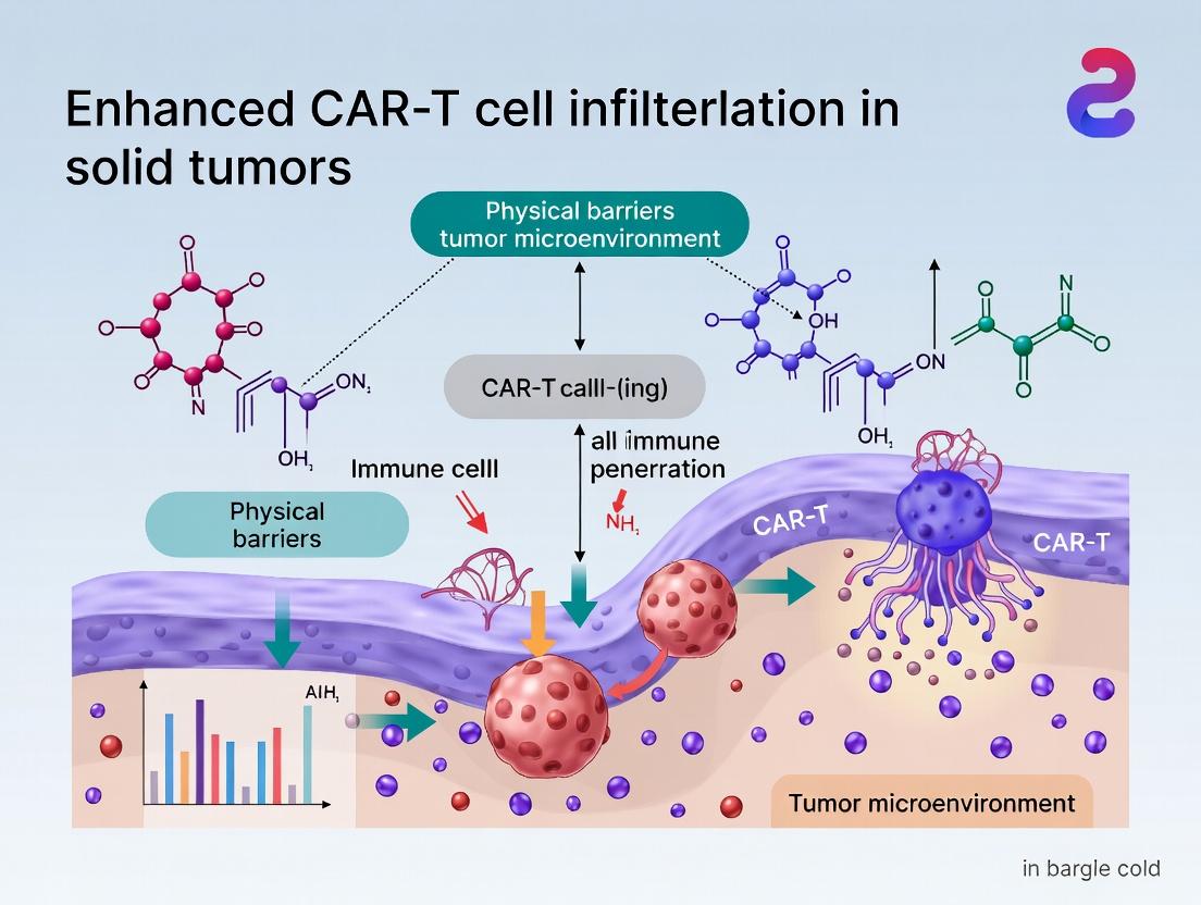
Overcoming the Fortress: Strategies to Enhance CAR-T Cell Infiltration Through Solid Tumor Physical Barriers
This article provides a comprehensive analysis for researchers, scientists, and drug development professionals on the critical challenge of CAR-T cell infiltration into solid tumors.
Overcoming the Fortress: Strategies to Enhance CAR-T Cell Infiltration Through Solid Tumor Physical Barriers
Abstract
This article provides a comprehensive analysis for researchers, scientists, and drug development professionals on the critical challenge of CAR-T cell infiltration into solid tumors. It explores the foundational biology of tumor microenvironments and physical barriers like dense extracellular matrix and aberrant vasculature. The review details cutting-edge methodological approaches, including engineering strategies to modify CAR-T cells and tumor stroma, troubleshooting common hurdles in preclinical models, and comparative validation of emerging techniques. The synthesis offers a roadmap for translating enhanced infiltration into improved clinical efficacy for solid tumor immunotherapy.
Understanding the Fortress: Deconstructing Solid Tumor Barriers to CAR-T Cell Infiltration
Troubleshooting Guides & FAQs
Q1: In our mouse xenograft model, infused CAR-T cells are detected in peripheral blood but fail to accumulate in the subcutaneous solid tumor. What are the primary barriers and how can we troubleshoot this?
A1: This indicates a failure in Tumor Infiltration, a major hurdle. The primary barriers are:
- Dysfunctional Tumor Vasculature: Abnormal blood vessels with irregular structure and poor perfusion impede T cell extravasation.
- Dense Extracellular Matrix (ECM): Hypersecretion of collagen, fibronectin, and hyaluronan by cancer-associated fibroblasts (CAFs) creates a physical barrier.
Troubleshooting Steps:
- Analyze Tumor Vasculature: Perform immunohistochemistry (IHC) for CD31 (endothelial cells) and α-SMA (pericytes) on tumor sections. Poor pericyte coverage (low α-SMA+ vessels) correlates with dysfunction.
- Modulate Vasculature: Pre-treat with low-dose antiangiogenic agents (e.g., bevacizumab, sunitinib) to "normalize" vasculature, improving perfusion and T-cell entry. See Protocol A.
- Assess ECM Density: Stain tumor sections with Masson's Trichrome (collagen) or Alcian Blue (hyaluronan). High density confirms an ECM barrier.
- Target ECM: Engineer CAR-T cells to express ECM-degrading enzymes (e.g., heparanase, hyaluronidase) or co-administer enzymatic disruptors. See Protocol B.
Q2: Our CAR-T cells infiltrate the tumor but show immediate functional exhaustion and poor persistence. What are the key suppressive factors in the Tumor Microenvironment (TME) and how can we counteract them?
A2: The solid TME is highly immunosuppressive. Key factors include:
- Immunosuppressive Cells: Regulatory T cells (Tregs), Tumor-Associated Macrophages (TAMs), and Myeloid-Derived Suppressor Cells (MDSCs).
- Inhibitory Ligands: PD-L1, often upregulated on tumor cells and myeloid cells upon IFN-γ exposure from CAR-T cells.
- Metabolic Competition: Low glucose, low oxygen (hypoxia), and high adenosine.
Troubleshooting Steps:
- Profile the TME: Use flow cytometry to quantify infiltrating MDSCs (CD11b+ Gr-1+), TAMs (CD11b+ F4/80+), and Tregs (CD4+ FoxP3+).
- Arm CAR-T Cells: Engineer "armored" CAR-T cells to secrete cytokines (e.g., IL-12, IL-18) to reprogram the TME or express dominant-negative receptors (e.g., dnTGF-βR) to resist suppression.
- Combine with Checkpoint Inhibition: Administer anti-PD-1/PD-L1 antibodies concurrently to block this dominant exhaustion pathway.
- Target Metabolism: Engineer CAR-T cells with hypoxia-inducible factors (HIFs) or adenosine-degrading enzymes (e.g., CD39/CD73 knockout). See Protocol C.
Q3: We observe "on-target, off-tumor" toxicity in preclinical models targeting a solid tumor antigen. How can we improve the safety profile of our CAR-T design?
A3: Target antigen heterogeneity in solid tumors makes safety critical.
Troubleshooting Steps:
- Implement Safety Switches: Incorporate inducible caspase-9 (iCasp9) or herpes simplex virus thymidine kinase (HSV-TK) suicide genes for controlled ablation of CAR-T cells upon adverse events.
- Use Logic-Gated CARs: Develop "AND-gate" CARs that require recognition of two tumor-specific antigens for full T-cell activation, increasing specificity.
- Tune Affinity: Reduce the affinity of the CAR's scFv to preferentially target cells with high antigen density (tumor) over low-density (healthy tissue).
- Employ Pre-clinical "De-risking" Assays: Thoroughly screen candidate antigens across human tissue arrays (e.g., from the Human Protein Atlas) and using organoid models of healthy tissues.
Detailed Experimental Protocols
Protocol A: Tumor Vasculature Normalization & Assessment Objective: To improve CAR-T cell infiltration by modulating abnormal tumor blood vessels. Method:
- Therapy: Administer sunitinib (20 mg/kg/day, oral gavage) or anti-VEGF-A antibody (e.g., B20-4.1.1, 5 mg/kg, i.p.) to tumor-bearing mice for 5-7 days prior to CAR-T cell infusion.
- Assessment: 24 hours after the last dose, inject 100 µL of FITC-labeled Lycopersicon esculentum lectin (1 mg/mL, i.v.) 10 minutes before sacrifice to label perfused vessels.
- Analysis: Harvest tumors, section, and co-stain for CD31. Image via confocal microscopy. Calculate:
- Vessel Perfusion: (FITC-Lectin+ area / CD31+ area) x 100%.
- Vessel Maturation: Co-stain CD31 with α-SMA; report % α-SMA+ vessels.
Protocol B: Engineering CAR-T Cells to Overcome ECM Barriers Objective: Generate CAR-T cells capable of degrading hyaluronan-rich ECM. Method:
- Vector Design: Clone the cDNA for human PH20 hyaluronidase (secreted form) into your CAR construct using a P2A or T2A self-cleaving peptide sequence, creating a bicistronic vector: [CAR] - [P2A] - [PH20].
- T Cell Transduction: Activate human T cells with CD3/CD28 beads. Transduce with the lentiviral vector at an MOI of 5-10 in the presence of 8 µg/mL polybrene by spinoculation (1000g, 90 min, 32°C).
- Validation:
- In vitro: Culture engineered CAR-T cells for 48h, collect supernatant. Assess hyaluronan degradation using a hyaluronic acid (HA) ELISA or a turbidimetric assay.
- In vivo: Use HA-specific probes (e.g., HA-binding protein, HABP) in IHC to visualize HA reduction in tumors treated with PH20-CAR-T vs. standard CAR-T.
Protocol C: Profiling Metabolic Stress in the TME Objective: Quantify key metabolic parameters that inhibit CAR-T function. Method:
- Tumor Harvest & Processing: Rapidly excise tumors from treated mice, place in ice-cold PBS. A portion is flash-frozen for metabolite analysis.
- Glucose & Lactate: Prepare tumor homogenates. Measure concentrations using commercial fluorometric/colorimetric assay kits (e.g., from BioVision). Normalize to total protein.
- Adenosine: Extract metabolites from frozen powder using cold 80% methanol. Quantify adenosine via LC-MS/MS.
- Hypoxia: 1 hour before sacrifice, inject pimonidazole HCl (60 mg/kg, i.p.). Detect hypoxic regions in fixed sections using an anti-pimonidazole antibody (IHC).
Table 1: Impact of TME Modulating Agents on CAR-T Efficacy in Preclinical Models
| Modulator Class | Example Agent | Target/Mechanism | Typical Dose (Mouse) | Outcome on CAR-T Infiltration* | Outcome on Tumor Growth* | Key Reference |
|---|---|---|---|---|---|---|
| Antiangiogenic | Sunitinib | VEGFR/PDGFR | 20-40 mg/kg/day, oral | Increase (1.5-3x) | Enhanced Inhibition | Smith et al., 2020 |
| ECM Degrader | PEGPH20 (Hyaluronidase) | Hyaluronan | 4.5 mg/kg, i.p., 2x/week | Increase (2-4x) | Enhanced Inhibition | Caruana et al., 2015 |
| Checkpoint Inhibitor | anti-PD-1 mAb | PD-1/PD-L1 axis | 200 µg, i.p., every 3-4 days | Variable | Synergistic Inhibition | Cherkassky et al., 2016 |
| Metabolic Modulator | CB-839 (Telaglenastat) | Glutaminase | 200 mg/kg, oral, BID | Improved T-cell function | Enhanced Inhibition | Leone et al., 2019 |
*Compared to CAR-T treatment alone.
Table 2: Common Solid Tumor Antigens & Associated Clinical Challenges
| Target Antigen | Key Cancers | Expression in Normal Tissue | Major Clinical Challenge | Mitigation Strategy in Development |
|---|---|---|---|---|
| Mesothelin | Mesothelioma, Pancreatic, Ovarian | Pleura, Pericardium, Peritoneum | On-target, off-tumor toxicity | Local/regional delivery, affinity-tuned CARs |
| HER2 | Breast, Gastric, Glioblastoma | Low levels on epithelial cells | Fatal toxicity from high-affinity CAR | Lower affinity scFv, dose-finding |
| PSMA | Prostate | Prostate, Salivary Gland | Target heterogeneity, antigen loss | Combinatorial targeting |
| GD2 | Neuroblastoma, Glioblastoma, Sarcoma | CNS neurons, peripheral nerves | Neurotoxicity | ScFv selection, co-stimulation domain choice |
| EGFRvIII | Glioblastoma | Not expressed | Heterogeneity, antigen loss | Target multiple antigens, include WT EGFR? |
Diagrams
The Scientist's Toolkit: Key Research Reagent Solutions
| Item | Function/Description | Example Product/Catalog # | |
|---|---|---|---|
| Recombinant Human Hyaluronidase (PH20) | Enzyme to digest hyaluronan for in vitro barrier assays or vector cloning. | Sigma-Aldrich, H3884 | |
| Anti-Human/Mouse PD-1 Antibody | For in vivo checkpoint blockade combination studies in mouse models. | Bio X Cell, clone RMP1-14 | |
| Pimonidazole HCl | Hypoxia marker for detecting low-oxygen regions in tumor sections. | Hypoxyprobe, HP1-1000 | |
| LIVE/DEAD Fixable Viability Dyes | Critical for flow cytometry to exclude dead cells during TME immune profiling. | Thermo Fisher Scientific | L34955 (Near-IR) |
| Recombinant TGF-β1 | To model TGF-β-mediated suppression in in vitro CAR-T functional assays. | PeproTech, 100-21 | |
| Lentiviral CAR Constructs | Backbone for stable CAR expression, often with fluorescent/selection markers. | Addgene (various), or custom from VectorBuilder | |
| CD3/CD28 T Cell Activator | Magnetic beads for robust, consistent human T cell activation pre-transduction. | Gibco Dynabeads, 11131D | |
| Extracellular Matrix (ECM) Proteins | For coating transwells to model infiltration barriers (Collagen I, IV, Fibronectin). | Corning, 354236 (Collagen I) |
Troubleshooting Guides & FAQs
This technical support center addresses common experimental challenges in research focused on overcoming physical barriers to CAR-T cell infiltration in solid tumors.
FAQ 1: Our CAR-T cells show poor migration through dense extracellular matrix (ECM) in 3D assays. What are the key factors to check?
- A: Poor migration often relates to ECM composition and CAR-T cell receptor/ligand interactions. First, quantify the major ECM components in your model.
- Troubleshooting Steps:
- Characterize the ECM: Use assays like Masson's Trichrome (collagen), Alcian Blue (glycosaminoglycans), or immunofluorescence for specific proteins (e.g., Collagen I, III, IV, Hyaluronan, Fibronectin).
- Check CAR-T Integrin Expression: Profile integrins (e.g., VLA-4, LFA-1) on your CAR-T cells via flow cytometry. Their engagement with ECM ligands (e.g., VCAM-1, ICAM-1) is crucial for adhesion and migration.
- Modulate ECM Density: Treat your 3D model with enzymatic degraders (e.g., collagenase, hyaluronidase) as a control. If migration improves, the physical density is a primary barrier.
- Evaluate Chemokine Mismatch: Ensure your CAR-T cells express receptors (e.g., CXCR3, CCR2) matching chemokines (e.g., CXCL9/10/11, CCL2) secreted by your tumor model.
- Troubleshooting Steps:
FAQ 2: We observe inconsistent CAR-T cell penetration in our patient-derived xenograft (PDX) or orthotopic mouse models. How can we standardize this measurement?
- A: Inconsistency often stems from variable tumor stroma and measurement techniques. Implement a multi-modal imaging and analysis protocol.
- Troubleshooting Steps:
- Use a Dual-Labeling System: Label CAR-T cells with a far-red dye (e.g., CellTracker Deep Red) and tumors with a vascular marker (e.g., anti-CD31). Perform multiplex immunohistochemistry (IHC) or immunofluorescence (IF).
- Adopt Quantitative Spatial Analysis: Use digital pathology or image analysis software (e.g., QuPath, HALO) to define tumor regions (e.g., invasive margin, tumor core) and calculate CAR-T cell density per mm² in each region.
- Correlate with Barrier Markers: Co-stain for ECM components (collagen, hyaluronan) and stromal cells (Cancer-Associated Fibroblasts, CAFs, marked by α-SMA). Correlate high-density areas with low CAR-T infiltration.
- Troubleshooting Steps:
FAQ 3: When engineering CAR-T cells to degrade ECM (e.g., express heparanase, hyaluronidase), how do we control off-target effects and maintain cell viability?
- A: Precise targeting and inducible systems are key to mitigating off-target effects.
- Troubleshooting Steps:
- Use a Tumor Microenvironment (TME)-Activated System: Engineer CAR-T cells to express ECM-modulating enzymes under the control of a TME-specific promoter (e.g., hypoxia-responsive element (HRE) or nuclear factor of activated T-cells (NFAT) promoter). This localizes enzyme production.
- Employ a Tet-On System: Use a doxycycline-inducible expression system for the enzyme. This allows you to control the timing and duration of expression after CAR-T cell infusion.
- Fuse Enzyme to a Targeting Domain: Create a fusion protein where the ECM enzyme is linked to a tumor antigen-specific scFv. This can help concentrate enzymatic activity at the tumor site.
- Monitor Vitality: Perform in vitro assays comparing proliferation and exhaustion markers (PD-1, LAG-3, TIM-3) between engineered and control CAR-T cells after long-term co-culture with tumor spheroids.
- Troubleshooting Steps:
Experimental Protocol: Quantifying CAR-T Cell Infiltration and Stromal Barriers in a 3D Collagen-Hyaluronan Matrix
Objective: To simulate and measure CAR-T cell migration through a defined, tunable ECM barrier.
Materials:
- High-density Type I Collagen solution (rat tail)
- High-molecular-weight Hyaluronic Acid (HA)
- Fluorescently labeled CAR-T cells (e.g., CFSE)
- Transwell inserts with 3.0 µm pores (for migration) or 96-well spheroid plates (for infiltration)
- Recombinant human chemokines (e.g., CXCL10, CCL2)
- Confocal or multiphoton microscope
Methodology:
- Prepare ECM Hydrogels: Mix collagen (2-4 mg/ml final concentration) with HA (0.5-2 mg/ml) on ice. Adjust pH to 7.4, then polymerize in a 37°C incubator for 1 hour in the bottom of a transwell or around a pre-formed tumor spheroid.
- Establish Chemokine Gradient: Add medium with chemokine to the bottom chamber. For spheroid assays, seed tumor cells expressing the chemokine.
- Seed CAR-T Cells: Add 1x10^5 labeled CAR-T cells to the top of the hydrogel (transwell) or at a defined distance from the spheroid.
- Image and Quantify: Acquire z-stack images at 0, 6, 12, 24, and 48 hours using confocal microscopy.
- Analysis: Use Imaris or FIJI software to track cell movement, calculate migration velocity, and measure infiltration depth into the spheroid core.
Table 1: Common Physical Barriers in the TME and Their Molecular Components
| Barrier Type | Key Molecular Components | Primary Producer Cells | Impact on CAR-T Infiltration |
|---|---|---|---|
| Dense ECM | Collagen I, III, IV; Hyaluronan; Fibronectin; Laminin | Cancer-Associated Fibroblasts (CAFs), Tumor Cells | Increases matrix stiffness, physically blocks cell motility. |
| Abnormal Vasculature | Poorly aligned endothelial cells (CD31+), Pericyte deficiency (NG2+) | Endothelial Cells, Pericytes | Limits CAR-T extravasation from blood; creates hypoxic regions. |
| High Interstitial Fluid Pressure (IFP) | Collagen, HA (reduce drainage), Leaky vessels | Tumor/Stromal Cells | Creates a pressure gradient opposing inward cellular migration. |
| Stromal Cell Sheath | α-SMA+ CAFs, FAP+ cells | Activated CAFs | Forms a contractile, cellular barrier around tumor nests. |
Table 2: Efficacy of ECM-Modulating Strategies in Preclinical Models
| Strategy | Target | Model Used | Outcome Metric | Typical Result (Range) |
|---|---|---|---|---|
| Pharmacological Degradation | Hyaluronan (PEGPH20) | Pancreatic PDX | CAR-T cells in tumor core | 2 to 5-fold increase |
| CAF Depletion/Reprogramming | FAP+ CAFs (αFAP-drug conjugate) | Lung Carcinoma | Tumor volume reduction | 40-60% reduction vs. control |
| CAR-T Expressing Heparanase | Heparan Sulfate Proteoglycans | Melanoma (mouse) | Infiltration depth | 150-200% increase vs. standard CAR-T |
| Vascular Normalization | VEGF/VEGFR (Axitinib, low dose) | Breast Cancer Orthotopic | Pericyte coverage (α-SMA+/CD31+) | Increase from ~20% to ~60% |
Pathway & Workflow Diagrams
Diagram 1: CAR-T Cell Confronts TME Barriers
Diagram 2: Troubleshooting Low CAR-T Infiltration
The Scientist's Toolkit: Research Reagent Solutions
| Reagent / Material | Primary Function | Example Use Case |
|---|---|---|
| Recombinant Human Hyaluronidase (PEGPH20-like) | Enzymatically degrades hyaluronan in the ECM. | Pre-treatment of solid tumor models in vitro/vivo to reduce barrier density before CAR-T administration. |
| 3D Bioprintable ECM Hydrogels | Provides a tunable, physiologically relevant matrix for migration assays. | Creating defined barriers with specific collagen/HA ratios to test engineered CAR-T cell motility. |
| Hypoxia-Responsive Element (HRE) Reporter Constructs | Reports on hypoxic conditions within the TME. | Identifying regions of high interstitial pressure and poor perfusion that are inaccessible to CAR-T cells. |
| Anti-FAP Antibody or FAP Inhibitor | Targets and depletes or inhibits Fibroblast Activation Protein (FAP)+ CAFs. | Disrupting the stromal cell shield around tumor cells to enhance CAR-T access. |
| Multiplex IHC/IF Panel (CD3, CD31, α-SMA, Collagen I) | Simultaneously visualizes immune cells, vasculature, CAFs, and ECM. | Quantifying spatial relationships between CAR-T infiltration and physical barriers in tumor sections. |
| Live-Cell Imaging Matrigel Invasion Chambers | Enables real-time tracking of cell movement through a basement membrane matrix. | Measuring the kinetic parameters of CAR-T cell invasion toward a chemokine gradient. |
Technical Support Center: Troubleshooting CAR-T Cell Infiltration in Solid Tumors
Common Issue: My CAR-T cells show poor infiltration and persistence in solid tumor models in vivo.
FAQs & Troubleshooting Guides
Q1: Our 3D spheroid invasion assays show minimal CAR-T cell penetration. What are the primary ECM components likely responsible, and how can we test this?
A: High collagen I and hyaluronan density are frequent culprits. Quantify your model's ECM.
Experimental Protocol: Quantification of Major ECM Components
- Collagen Quantification: Use the Sircol Soluble Collagen Assay. Homogenize tumor tissue or decellularized ECM in 0.5M acetic acid with pepsin (1mg/mL). React with Sirius Red dye, measure absorbance at 555nm. Compare to a standard curve.
- Hyaluronan Quantification: Use an ELISA-like assay (e.g., R&D Systems DuoSet). Digest tissue with papain (125µg/mL) at 60°C for 24h. Follow kit protocol for HA binding protein.
- Visualization: Stain spheroids or tissue sections with Picrosirius Red (collagen) and Hyaluronan Binding Protein (HABP).
Quantitative Data Table: Common Solid Tumor ECM Composition
| ECM Component | Typical Range in Fibrotic Tumors (e.g., Pancreatic, Breast) | Assay/Method | Key Implication for CAR-T Cells |
|---|---|---|---|
| Collagen I | 20-50 mg/g tissue (can be 5-10x higher than normal) | Sircol Assay, Masson's Trichrome | Increases matrix stiffness (>2 kPa), physically blocks migration. |
| Hyaluronan (HA) | 5-30 µg/mg protein | HABP ELISA, Staining | Creates osmotic pressure, hydration barrier; binds CD44 on T cells, causing anergy. |
| Fibronectin (EDA+) | High expression (qualitative) | Immunohistochemistry, Western Blot | Promotes integrin-mediated adhesion, can trap cells. |
| Elastin | Variable, often cross-linked | Elastin-specific ELISA (Fastin) | Contributes to matrix rigidity and recoil. |
Q2: We suspect high matrix stiffness is inhibiting motility. How do we measure this in vitro and modify our CAR-T cells to cope?
A: Use tunable stiffness hydrogels for testing and engineer CAR-T cells with matrix-remodeling enzymes.
Experimental Protocol: Testing CAR-T Motility on Tunable Stiffness Substrates
- Substrate Preparation: Prepare polyacrylamide gels of defined stiffness (0.5 kPa, 2 kPa, 8 kPa) coated with collagen I (100 µg/mL) or fibronectin (10 µg/mL).
- CAR-T Cell Seeding: Label CAR-T cells with CellTracker dye. Seed 5x10^4 cells onto the gel.
- Live-Cell Imaging: Image every 10 minutes for 12-24 hours using a confocal microscope with environmental control.
- Analysis: Use tracking software (e.g., ImageJ Manual Tracking) to calculate mean migration speed and persistence.
Q3: What are the most promising pre-conditioning strategies to degrade the tumor ECM in vivo prior to CAR-T infusion?
A: Pharmacological enzymatic targeting shows clinical promise. Critical: Timing and specificity are essential to avoid metastasis.
Experimental Protocol: Pre-treatment with ECM-Targeting Enzymes in a Mouse Model
- Animal Model: Use an orthotopic, fibrotic tumor model (e.g., PAN02 pancreatic, 4T1 breast).
- Pre-treatment: Administer PEGylated recombinant human hyaluronidase (PEGPH20) intraperitoneally at 4.5 µg/g, 24 and 48 hours before CAR-T cell infusion. Control: Vehicle alone.
- CAR-T Administration: Infuse 5-10x10^6 anti-mesothelin or anti-HER2 CAR-T cells intravenously.
- Assessment: Monitor tumor volume (calipers), CAR-T infiltration by IHC (CD3ε), and intratumoral pressure if possible. Monitor for off-target tissue damage (e.g., skin).
The Scientist's Toolkit: Key Research Reagent Solutions
| Reagent / Material | Function in ECM/CAR-T Research | Example Product / Clone |
|---|---|---|
| PEGPH20 (PEGylated hyaluronidase) | Degrades hyaluronan in the tumor stroma to reduce pressure and increase permeability. | Halozyme Therapeutics (clinical grade) |
| Collagenase Type I | Digests collagen for in vitro tumor dissociation or ECM disruption assays. | Worthington Biochemical |
| TGF-β Receptor I Kinase Inhibitor (e.g., Galunisertib) | Inhibits TGF-β signaling, a master regulator of cancer-associated fibroblast activation and ECM deposition. | LY2157299 (Selleckchem) |
| Anti-αvβ6 Integrin Antibody | Blocks integrin-mediated activation of latent TGF-β in the ECM. | Clone 6.3G9 (R&D Systems) |
| MMP-14 (MT1-MMP) Reporter | To engineer CAR-T cells that secrete MMP-14 for localized collagen I/III degradation. | Recombinant protein (Sino Biological) |
| Tunable Stiffness Hydrogel Kit | To create 3D environments of physiologically relevant stiffness for motility studies. | Bioink or Polyacrylamide Kit (Cellendes, Matrigen) |
| Pan-Collagen Probe (CNA35) | For real-time visualization of collagen architecture in live 3D cultures. | Fluorescently labeled CNA35 (e.g., Cytoskeleton Inc.) |
Visualization: Signaling Pathways & Experimental Workflows
Title: TGF-β Driven Fibrosis Barrier to CAR-T Cells
Title: Workflow for Testing ECM-Modifying Therapies
Technical Support Center: Enhancing CAR-T Cell Infiltration
Troubleshooting Guides & FAQs
Q1: Our CAR-T cells show poor extravasation and infiltration in our orthotopic solid tumor mouse model. What are the primary vascular-related checkpoints to investigate? A: Poor extravasation often stems from dysfunctional tumor vasculature. Key checkpoints to analyze include:
- Vessel Normalization Markers: Assess pericyte coverage (α-SMA, NG2) and basement membrane integrity (Collagen IV). Low coverage indicates abnormality.
- Adhesion Molecule Expression: Profile tumor endothelial cells for low ICAM-1, VCAM-1, and E-selectin. These are critical for CAR-T cell adhesion.
- Vascular Permeability: Measure leakage using Evans Blue or fluorescent dextran. Hyperpermeability can hinder directed migration.
- Hypoxia and Angiogenic Factors: Quantify HIF-1α and VEGF-A levels. High levels sustain abnormal vasculature.
Q2: When using a vascular normalization agent (e.g., anti-angiogenic therapy), we sometimes see reduced tumor perfusion. How can we optimize the dosing schedule to improve CAR-T cell delivery? A: This is a common "normalization window" issue. The goal is transient stabilization, not permanent pruning.
- Monitor the Window: Use dynamic contrast-enhanced MRI (DCE-MRI) or Doppler ultrasound to track tumor perfusion over time post-treatment.
- Protocol Optimization: Initiate CAR-T cell infusion during the peak of the normalization window, typically 2-6 days after starting a low, metronomic dose of an agent like axitinib or bevacizumab in murine models. Administering CAR-T cells simultaneously with or after high-dose anti-angiogenics can reduce perfusion.
- Biomarker-Guided Dosing: Table 1 summarizes key parameters to monitor for timing CAR-T administration.
Table 1: Parameters for Identifying the Vascular Normalization Window
| Parameter | Abnormal Vasculature | Normalized Vasculature (Target Window) | Excessive Pruning |
|---|---|---|---|
| Pericyte Coverage (Index) | Low (<50%) | Increased (50-80%) | High but on regressed vessels |
| Vessel Density | High, chaotic | Moderately Reduced | Severely Reduced |
| Hypoxia (% pIMO positive) | High (>60%) | Reduced (20-40%) | Variable, can increase |
| Tumor Perfusion | Heterogeneous, low | Improved, Homogeneous | Severely Diminished |
| Recommended for CAR-T Infusion? | No | Yes | No |
Q3: What are reliable in vitro assays to model and test CAR-T cell adhesion and transendothelial migration under tumor vasculature conditions? A: Use a Tumor Endothelial Cell (TEC) co-culture system.
- Primary Protocol: Static Adhesion Assay
- Isolate or culture primary Tumor-Associated Endothelial Cells (TECs) or use stimulated HUVECs (with TNF-α & VEGF to mimic tumor conditions).
- Seed TECs in a 96-well plate and culture to confluence.
- Fluorescently label CAR-T cells (e.g., with Calcein AM).
- Add CAR-T cells to the TEC monolayer (e.g., 5:1 effector:endothelial ratio) and centrifuge briefly (100g, 3 min) to initiate contact.
- Incubate for 30-60 min at 37°C.
- Gently wash wells 3x with warm medium to remove non-adherent cells.
- Measure fluorescence. Calculate % adhesion relative to input control wells.
- Primary Protocol: Transendothelial Migration (TEM) Assay
- Culture TECs on collagen-coated transwell inserts (3-5µm pore size) until a tight monolayer forms (2-3 days). Verify integrity (e.g., by FITC-dextran leakage).
- Add a chemoattractant to the lower chamber (e.g., CXCL10, CCL5 at 100 ng/mL, or tumor cell conditioned medium).
- Add fluorescently labeled CAR-T cells to the upper chamber.
- Incubate for 4-24 hours at 37°C.
- Collect cells from the lower chamber and count migrated cells by flow cytometry.
Q4: Which murine tumor models best recapitulate the dysfunctional vasculature seen in human solid tumors for testing combination (normalization + CAR-T) therapies? A: The choice depends on the tumor type. Syngeneic models allow study of full immune context.
- MC38 (Murine Colon Adenocarcinoma): Moderately vascularized, responsive to immunotherapies.
- B16-F10 (Melanoma): Highly aggressive, with irregular vasculature and hypoxic regions.
- 4T1 (Mammary Carcinoma): Highly metastatic, characterized by abnormal, leaky vessels.
- Pan02 (Pancreatic Ductal Adenocarcinoma): Desmoplastic and hypovascular, presenting a severe physical barrier.
- RT2 (Rip1-Tag2; Pancreatic Neuroendocrine): Stages well-defined angiogenesis, useful for studying normalization.
Q5: What are the key cytokines and signaling pathways to profile in the Tumor Microenvironment (TME) when assessing the impact of vasculature on CAR-T infiltration? A: Focus on pathways linking endothelium, chemokines, and immune cell function.
Diagram Title: Key TME Pathways Affected by Faulty Tumor Vasculature
The Scientist's Toolkit: Research Reagent Solutions
Table 2: Essential Materials for Studying Tumor Vasculature & CAR-T Infiltration
| Item / Reagent | Function / Application | Example Catalog # (Vendor Examples) |
|---|---|---|
| Recombinant Human/Murine VEGF-A | To mimic tumor-like endothelial stimulation in in vitro TEC models. | 293-VE (R&D Systems) |
| Anti-Mouse CD31 (PECAM-1) Antibody | Immunofluorescence staining for visualizing tumor blood vessel density and morphology. | 102501 (BioLegend) |
| Anti-αSMA (Alpha Smooth Muscle Actin) Antibody | Immunostaining for assessing pericyte coverage and vessel maturity. | 19245S (CST) |
| Recombinant Murine CXCL10/IP-10 | Chemoattractant for in vitro T cell transmigration assays; key chemokine for effector T cell recruitment. | 250-16 (PeproTech) |
| Axitinib (Small Molecule Inhibitor) | VEGFR TKI used in pre-clinical studies to induce a vascular "normalization window" in murine models. | S1005 (Selleckchem) |
| Fluorescein Griffonia Simplicifolia Lectin I (GSL I) | Intravenous injection for in vivo labeling of functional, perfused vasculature in mice. | FL-1101 (Vector Labs) |
| pimonidazole hydrochloride | Hypoxia probe for immunohistochemistry; binds to proteins in hypoxic (<1.3% O2) regions of tumors. | HP2-1000Kit (Hypoxyprobe) |
| Collagenase IV & DNAse I | Enzyme cocktail for digesting solid tumors to single-cell suspensions for flow cytometry analysis of infiltrated CAR-T cells. | LS004188, LS002139 (Worthington) |
| Anti-Human/Mouse ICAM-1 (CD54) Antibody | For blocking studies or flow cytometry to assess endothelial adhesion molecule expression. | 353107 (BioLegend) |
| Matrigel Growth Factor Reduced | For in vitro tube formation assays to test endothelial cell function or create 3D invasion models. | 356231 (Corning) |
Experimental Protocol: Integrated In Vivo Analysis of CAR-T Infiltration Post-Vascular Normalization Title: Timing CAR-T Cell Administration Within the Vascular Normalization Window. Objective: To evaluate the optimal schedule for combining anti-angiogenic therapy with CAR-T cell transfer to maximize infiltration. Materials: Murine tumor model (e.g., MC38), vascular normalization agent (e.g., Axitinib), fluorescently or luciferase-labeled CAR-T cells, IVIS imaging system, flow cytometer. Procedure:
- Tumor Implantation: Implant tumor cells subcutaneously or orthotopically into syngeneic mice.
- Normalization Therapy: When tumors reach ~100 mm³, initiate axitinib treatment (e.g., 25 mg/kg via oral gavage, daily).
- Window Monitoring: On days 0 (pre-treatment), 2, 4, 6, and 8 post-axitinib initiation:
- Inject fluorescent lectin (e.g., Griffonia Simplicifolia Lectin I, 100µg/mouse) IV 10 minutes before sacrifice to label perfused vessels.
- Harvest tumors. Part is snap-frozen for IHC (CD31, αSMA, Hypoxyprobe). Part is digested for flow cytometry to analyze endothelial activation markers (ICAM-1).
- CAR-T Cell Transfer: Administer CAR-T cells intravenously to different cohorts of mice on days 0 (concurrent), 2, 4, or 6 after starting axitinib. Include control cohorts (CAR-T only, axitinib only, untreated).
- Infiltration Quantification:
- At 24-48h post-CAR-T transfer: Digest tumors, stain for immune cell markers (CD3, CAR-specific tag), and quantify absolute numbers of infiltrated CAR-T cells by flow cytometry using counting beads.
- Spatial Analysis: Perform multiplex IHC/IF (e.g., CD3, CD31, collagen) on tumor sections to determine CAR-T cell proximity to blood vessels.
- Efficacy Correlates: Monitor tumor growth and survival. Correlate with infiltration data and vascular parameters (pericyte coverage, hypoxia reduction) from Step 3.
Diagram Title: Workflow for Timing CAR-T Delivery with Vascular Normalization
Troubleshooting Guide & FAQs
Common Experimental Issues & Solutions
Q1: During in vivo IFP measurement in our mouse xenograft model, we get inconsistent readings between tumors, even of similar size. What are the potential causes and solutions?
A: High variability is common. Key factors and fixes include:
- Cause: Probe placement. Measurements are highly sensitive to location within the tumor (core vs. edge).
- Solution: Standardize insertion depth and coordinate (e.g., always measure at the geometric center under ultrasound guidance). Use a stereotactic apparatus for consistency.
- Cause: Tumor necrosis. Placing the probe in a necrotic area yields artificially low pressure.
- Solution: Use imaging (e.g., ultrasound, MRI) to guide probe away from visibly necrotic zones. Correlate IFP with histology post-measurement.
- Cause: Fluid leakage or tissue compression around the needle.
- Solution: Use a side-ported needle and ensure it is properly calibrated and zeroed before each insertion. Allow pressure to stabilize for 3-5 minutes post-insertion before recording.
Q2: Our collagen-based 3D in vitro model shows poor CAR-T cell migration. Could IFP be a contributing factor even in a gel, and how can we modulate it?
A: Yes, compaction and matrix density can generate interstitial pressure. To troubleshoot:
- Check: Gel compaction and density. Highly compacted gels generate higher resistance.
- Solution: Systematically vary collagen concentration (e.g., 1.5 mg/mL vs. 3 mg/mL vs. 6 mg/mL) and measure pore size and stiffness. Incorporate hyaluronan to increase osmotic pressure.
- Experimental Modulator: Add osmotic agents like PEG (polyethylene glycol) to the medium to increase osmotic pressure externally, simulating high IFP conditions. Use collagenase or hyaluronidase to degrade the matrix and lower pressure.
Q3: When using vascular normalization agents (e.g., anti-VEGF) to lower IFP, we see improved small molecule delivery but no significant improvement in CAR-T cell infiltration. Why might this happen?
A: This highlights the multi-faceted nature of the barrier.
- Cause: Vascular normalization primarily improves perfusion and reduces hydrostatic IFP, but does not fully address the matrix and osmotic components (e.g., from hyaluronan and collagen).
- Solution: Combine vascular normalization with stromal depletion agents. Consider a sequential protocol: Anti-VEGF treatment first (Days 1-3), followed by a matrix-modifying enzyme (e.g., PEGPH20 - hyaluronidase) or an angiotensin system inhibitor (e.g., losartan) to further decompress vessels and loosen the matrix.
Detailed Experimental Protocol: Measuring IFPIn Vivo(Wick-in-Needle Technique)
Objective: To quantitatively measure interstitial fluid pressure within a solid tumor xenograft.
Materials:
- Anesthetized tumor-bearing mouse (e.g., subcutaneous MDA-MB-231 xenograft, ~500 mm³).
- Pressure monitoring system (e.g., Millar Mikro-Tip transducer connected to a pressure control unit and data acquisition software).
- Wick-in-needle setup: 23-gauge needle, side-ported, filled with sterile saline-soaked cotton thread.
- Stereotactic needle holder.
- Warming pad to maintain mouse body temperature.
- Ultrasound imaging system (optional, for guided placement).
Procedure:
- Calibrate and zero the pressure transducer according to manufacturer instructions in a saline bath at 37°C.
- Anesthetize the mouse and place it on a warming pad. Fix the tumor position using a custom holder.
- Under visual or ultrasound guidance, insert the wick-in-needle slowly into the central region of the tumor using the stereotactic apparatus. Avoid visible surface vessels.
- Allow the pressure reading to stabilize for 3-5 minutes post-insertion.
- Record the mean stable pressure over a 1-minute period. This is the IFP (in mmHg).
- Withdraw the needle slowly. Repeat measurement in a different region if required by protocol.
- Euthanize the mouse and excise the tumor. Document probe placement track via histology.
Key Controls: Measure IFP in contralateral normal subcutaneous tissue as a baseline control.
| Tumor Model (Mouse) | Baseline IFP (mmHg) | Modulating Agent/Intervention | Post-Intervention IFP (mmHg) | Change in CAR-T Infiltration (vs. Control) | Key Citation (Example) |
|---|---|---|---|---|---|
| MDA-MB-231 (Breast) | 15 - 25 | Anti-VEGF Antibody (Bevacizumab) | 8 - 12 | +20% (modest) | Salnikov et al., 2006 |
| U87-MG (Glioblastoma) | 20 - 30 | Losartan (Angiotensin Inhibitor) | 10 - 15 | +40% (significant) | Diop-Frimpong et al., 2011 |
| PAN02 (Pancreatic) | 35 - 50 | PEGPH20 (Hyaluronidase) | 15 - 25 | +60% (high) | Provenzano et al., 2012 |
| CT26 (Colon) | 10 - 20 | TGF-β Receptor Inhibitor | 5 - 10 | +30% (significant) | Mariathasan et al., 2018 |
| Normal Tissue | 0 - 3 | N/A | N/A | N/A | Reference Standard |
The Scientist's Toolkit: Key Research Reagent Solutions
| Item | Function in IFP/CAR-T Research | Example Product/Catalog # |
|---|---|---|
| Recombinant Human VEGF | To induce hyper-permeable vasculature and high IFP in in vitro vessel models. | PeproTech, 100-20 |
| PEGPH20 (Recombinant Hyaluronidase) | Enzymatically degrades hyaluronan in the tumor stroma, reducing matrix-based IFP. | Halozyme Therapeutics (for research) |
| Losartan Potassium | Angiotensin II receptor antagonist; reduces collagen production and vessel compression to lower IFP. | Sigma-Aldrich, L9656 |
| Collagenase Type I | Digests collagen I matrix in 3D cultures to modulate physical resistance and pressure. | Worthington Biochemical, LS004196 |
| Anti-VEGF Neutralizing Antibody | Promotes vascular normalization, reducing hydrostatic component of IFP. | Bio X Cell, BE0052 (B20-4.1.1) |
| Transwell Permeable Supports | Used in modified assays to study T-cell migration under pressure gradients. | Corning, 3422 |
| Millar Mikro-Tip Pressure Catheter | Gold-standard tool for direct in vivo IFP measurement via wick-in-needle technique. | Millar, SPR-1000 |
Diagrams
Diagram 1: IFP Formation & CAR-T Barrier Pathway
Diagram 2: IFP Modulation Experimental Workflow
Troubleshooting Guide & FAQ
FAQ: Common Issues in CAR-T Cell Infiltration Experiments
Q1: Our engineered CAR-T cells show robust activation in vitro but fail to accumulate at the tumor site in vivo. What could be the cause?
- A: This is a classic symptom of a chemokine-receptor mismatch. Your CAR-T cells may lack the appropriate receptor (e.g., CXCR3, CCR2, CCR5) to respond to the chemokines (e.g., CXCL9/10/11, CCL2, CCL5) secreted by your specific solid tumor model. Check the chemokine profile of your tumor via qPCR/ELISA and ensure your T cells express the corresponding homing receptors.
Q2: We observe CAR-T cells in the tumor vasculature but not extravasating into the tumor parenchyma. What is the likely failure point?
- A: This indicates a probable adhesion molecule deficiency. The multi-step extravasation process (tethering, rolling, adhesion, transmigration) is failing at the firm adhesion step. This often involves interactions between Integrins (e.g., LFA-1, VLA-4) on T cells and Ig-family Adhesion Molecules (e.g., ICAM-1, VCAM-1) on tumor endothelium. Check if the tumor endothelium is inflamed and expressing sufficient levels of these adhesion molecules.
Q3: Our data shows variable CAR-T infiltration across different patient-derived xenograft (PDX) models, even with the same cell product. How do we standardize our analysis?
- A: Infiltration efficiency is highly model-dependent due to variable tumor microenvironment (TME) biology. Standardization requires quantifying key parameters:
- TME Chemokine Secretion: Use a multiplex chemokine array.
- Endothelial Activation Status: Measure adhesion molecule expression (ICAM-1, VCAM-1) via flow cytometry of dissociated tumors (CD31+ fraction).
- Infiltration Metrics: Use immunohistochemistry (IHC) with automated image analysis to report cells/mm², not just relative percentages.
- A: Infiltration efficiency is highly model-dependent due to variable tumor microenvironment (TME) biology. Standardization requires quantifying key parameters:
Table 1: Quantitative Benchmarks for Key Infiltration Parameters
| Parameter | Typical Measurement Method | Low/Problematic Range | Desired/Functional Range | Notes |
|---|---|---|---|---|
| Tumor [Chemokine] | ELISA/Luminex (pg/mg protein) | < 50 pg/mg for key chemokines | > 200 pg/mg | Target depends on chemokine (e.g., CXCL10, CCL2). |
| % Tumor Endothelium ICAM-1+ | Flow Cytometry (CD31+ cells) | < 15% | > 60% | Indicator of endothelial inflammation. |
| Intratumoral CAR-T cell density | IHC (cells/mm²) | < 100 cells/mm² | > 500 cells/mm² | Varies by tumor type; internal controls are critical. |
| Circulating vs. Tumor CAR-T Ratio | qPCR (vector copies/µg DNA) | > 100:1 | < 10:1 | Assesses preferential tumor homing. |
Experimental Protocols
Protocol 1: Assessing Chemokine-Receptor Mismatch via Transwell Migration Assay
Objective: To functionally test the homing capacity of CAR-T cells toward tumor-derived chemotactic signals.
Materials:
- Recombinant human chemokines (e.g., CXCL10, CCL2, CCL5).
- 24-well plates with 5.0 µm pore transwell inserts.
- Serum-free RPMI-1640 medium.
- CAR-T cells and control T cells.
- Conditioned media from tumor cell lines or dissociated tumor explants.
Method:
- Add 600 µL of serum-free medium containing a specific chemokine (e.g., 100 ng/mL CXCL10) or tumor-conditioned medium to the lower chamber. Use medium alone as a negative control.
- Resuspend 1 x 10⁵ CAR-T cells in 100 µL of serum-free medium and seed them into the upper chamber of the transwell insert.
- Incubate the plate at 37°C, 5% CO₂ for 4 hours.
- Carefully remove the insert. Collect cells that have migrated to the lower chamber and count them using a hemocytometer or flow cytometer.
- Calculate the migration index: (# cells migrated to chemokine / # cells migrated to medium control).
Protocol 2: Evaluating Integrin-Mediated Adhesion Under Flow (Static Assay Proxy)
Objective: To quantify the adhesive capacity of CAR-T cells to key endothelial ligands.
Materials:
- 96-well plates coated with recombinant ICAM-1 or VCAM-1 (5 µg/mL overnight).
- CAR-T cells, activated with PMA/Ionomycin or specific chemokines.
- Blocking buffer (1% BSA in PBS).
- Adhesion buffer (HBSS with Ca²⁺/Mg²⁺ and 1% HSA).
- Fixative (4% paraformaldehyde).
Method:
- Block the ligand-coated plates with 1% BSA for 1 hour at 37°C.
- Activate CAR-T cells (to induce integrin high-affinity state) or leave unactivated as a control.
- Label CAR-T cells with calcein AM (2 µM) for 30 minutes at 37°C.
- Wash cells, resuspend in adhesion buffer, and add 1 x 10⁵ cells per well.
- Allow adhesion to proceed for 30 minutes at 37°C.
- Gently wash wells 3x with pre-warmed adhesion buffer to remove non-adherent cells.
- Fix cells with 4% PFA for 15 minutes.
- Measure fluorescence (Ex/Em ~495/515 nm). Express data as % Adhesion relative to total fluorescence input.
Visualizations
Title: Chemokine-Receptor Mismatch Impairs Homing
Title: Adhesion Cascade Failure Points
The Scientist's Toolkit: Key Research Reagent Solutions
| Item | Function & Application in Infiltration Research |
|---|---|
| Recombinant Chemokines | Used in migration assays to test specific receptor functionality (e.g., CXCL10 for CXCR3). Also for pre-conditioning T cells. |
| Integrin Activation Antibodies | Flow cytometry antibodies (e.g., mAb24 for LFA-1 high-affinity conformation) to measure activation state of adhesion molecules on CAR-T cells. |
| Ligand-Coated Plates | Plates pre-coated with ICAM-1-Fc or VCAM-1-Fc for static adhesion assays under controlled conditions. |
| Small Molecule Integrin Activators | Agents like MnCl₂ or TS1/18 antibody used as positive controls to induce maximal integrin affinity in adhesion assays. |
| Neutralizing/Antibodies | Blocking antibodies against chemokine receptors (e.g., α-CCR5) or integrins (e.g., α-LFA-1) to confirm pathway specificity in functional assays. |
| Multiplex Cytokine/Chemokine Array | Kit to quantitatively profile dozens of soluble factors from tumor-conditioned media or tumor lysates simultaneously. |
| Fluorescent Cell Linkers (e.g., CFSE, CTV) | Vital dyes for labeling CAR-T cells prior to co-culture or injection to enable clear tracking and quantification during migration/adhesion assays. |
Engineering the Breach: Cutting-Edge Strategies to Force and Facilitate CAR-T Entry
Technical Support Center
Troubleshooting Guides & FAQs
Q1: Our CAR-T cells expressing ectopic CXCR2 show poor surface expression despite confirmed mRNA levels. What could be the issue? A: This is often a post-translational or trafficking issue. Ensure the chemokine receptor is codon-optimized for human cells. Check for improper folding by performing a flow cytometry staining for the receptor on permeabilized vs. non-permeabilized cells. If it's retained intracellularly, consider adding a leader sequence from a well-expressed protein (e.g., CD8α) and verify the vector's promoter strength (use EF1α or PGK over CMV for more consistent expression in T cells). Include a positive control (e.g., GFP from an IRES or P2A element) to confirm transduction success.
Q2: In an in vitro Transwell migration assay towards a CXCL12 gradient, our CCR7-expressing CAR-T cells show minimal migration. How can we troubleshoot? A: Follow this systematic guide:
- Chemokine Gradient Integrity: Confirm the concentration gradient is stable. Use a higher concentration in the lower chamber (e.g., 200 ng/mL CXCL12) and ensure no convection currents.
- CAR-T Cell Viability & Activation: Ensure cells are >90% viable and tested in a minimally activated state (rest 24-48h post-activation/transduction). Over-activated cells have reduced motility.
- Receptor Functionality: Verify CCR7 surface expression via flow cytometry on the day of the assay. Pre-treat a subset of cells with Pertussis Toxin (100 ng/mL, 1 hour), which inhibits Gi-protein coupling; this should abolish migration, confirming a GPCR-mediated process.
- Assay Controls: Include parental (non-transduced) T cells as a negative control and untransduced T cells activated with IL-2/IL-7/IL-15 as a positive motility control.
Q3: We co-expressed a chemokine (e.g., CCL19) with our CAR via a P2A peptide linker, but we detect very low levels of secreted chemokine via ELISA. Why? A: P2A-mediated "self-cleavage" is not 100% efficient, leading to fusion proteins that may impair secretion. Troubleshoot by:
- Switching to a different linker (e.g., T2A or Furin/GSG linker).
- Placing the chemokine gene before the P2A sequence, as the upstream gene typically has higher expression.
- Using an internal ribosome entry site (IRES) instead, though this lowers chemokine expression relative to the CAR.
- Confirming secretion by intracellular flow cytometry or Western Blot of cell lysates and supernatant concentrates.
- Checking the chemokine's native signal peptide is present and functional.
Q4: In our murine solid tumor model, CAR-T cells engineered to express PSGL-1 and Sialyl-LewisX still fail to infiltrate the tumor core. What are potential reasons? A: Infiltration requires more than just tethering/rolling. Consider:
- Selectin Ligand Activity: The glycosylation (e.g., fucosylation by FUT7) of PSGL-1 is critical. Use a recombinant P- or E-selectin IgG chimera in a flow-based adhesion assay to confirm functional binding.
- Shear Stress: The in vivo vascular environment has shear forces absent in static assays.
- Downstream Integrin Activation: The "second signal" for firm adhesion (via integrins like LFA-1) may be absent. Co-express chemokine receptors matched to the tumor's chemokine profile (e.g., CXCR2 for CXCL1/2/5).
- Physical Barriers: The tumor core may have high interstitial fluid pressure or dense fibrotic stroma. Consider combining homing modifications with strategies to degrade extracellular matrix (e.g., heparanase).
Experimental Protocols
Protocol 1: Flow Cytometry-Based Adhesion Assay to Validate Selectin Ligand Function Purpose: To quantitatively assess the binding of engineered CAR-T cells to selectins under static conditions. Materials: Recombinant human P-selectin/Fc Chimera, Protein A/G-coated plates, Calcein-AM dye, HBSS buffer with 2mM Ca2+. Steps:
- Coat a 96-well plate with Protein A/G (10 µg/mL) overnight at 4°C. Block with 1% BSA for 1 hour.
- Bind P-selectin/Fc (5 µg/mL) to the plate for 2 hours at RT.
- Label CAR-T cells (modified and unmodified controls) with 5 µM Calcein-AM for 30 minutes at 37°C.
- Wash cells, resuspend in HBSS/Ca2+, and add 1x10^5 cells per well.
- Allow adhesion to proceed for 15 minutes at 37°C on an orbital shaker (50 rpm).
- Gently wash wells 3x with pre-warmed HBSS/Ca2+ to remove non-adherent cells.
- Measure fluorescence (Ex/Em ~494/517 nm) on a plate reader. Calculate % Adhesion = (Fluorescence post-wash / Fluorescence pre-wash) * 100.
Protocol 2: In Vitro 3D Migration Assay in Tumor Spheroid Co-Culture Purpose: To model CAR-T cell infiltration into a solid tumor mass. Materials: U-bottom low-attachment plates, tumor cell line (e.g., OVCAR-3, U87), collagen type I matrix, time-lapse fluorescent microscope. Steps:
- Generate tumor spheroids by seeding 5x10^3 cells per well in a U-bottom plate. Centrifuge at 300g for 3 min and culture for 72-96 hours.
- On day of assay, carefully mix spheroids with 1.5 mg/mL collagen type I solution on ice. Pipette 50 µL drops into a glass-bottom 24-well plate and incubate at 37°C for 30 min to polymerize.
- Label CAR-T cells (1x10^6 cells/mL) with a cytoplasmic dye (e.g., CellTracker Red CMTPX).
- Resuspend labeled CAR-T cells in complete media and gently layer over the polymerized collagen containing the spheroids.
- Immediately place plate in a live-cell imaging system. Acquire z-stack images (e.g., 50 µm depth, 5 µm intervals) every 20 minutes for 12-24 hours.
- Analysis: Use Imaris or similar software to track individual T cell movement. Key metrics: Migration speed, directionality, and penetration depth into the spheroid.
Research Reagent Solutions
| Reagent | Function in Experiment | Key Considerations |
|---|---|---|
| Lentiviral Vector (pLVX-EF1α) | Stable gene delivery of homing receptors (e.g., CXCR2, CCR7) into human T cells. | Use a 3rd generation system for safety. Pseudotype with VSV-G for broad tropism. |
| Recombinant Selectin/Fc Chimeras | Validate functional adhesion of engineered PSGL-1/SLeX ligands in static or flow assays. | Requires divalent cations (Ca2+/Mn2+) for binding. Protein A/G coating ensures correct orientation. |
| Pertussis Toxin (PTx) | Inhibits Gi-protein coupled receptor (GPCR) signaling. Serves as a negative control for chemokine receptor-mediated migration. | Use at 100-200 ng/mL for 1-2 hour pre-treatment. Confirms migration is GPCR-dependent. |
| Transwell Permeable Supports (5.0 µm) | Assess chemotactic migration of CAR-T cells toward a chemokine gradient in vitro. | Polycarbonate membrane, 5.0 µm pores for lymphocytes. Coat with fibronectin (10 µg/mL) for integrin-mediated migration studies. |
| Calcein-AM | Fluorescent, cell-permeant dye for labeling live cells for adhesion/migration assays. | Non-fluorescent until cleaved by intracellular esterases. Minimal impact on cell function. |
| Recombinant Human Chemokines (e.g., CXCL12, CCL19) | Establish a chemotactic gradient in migration assays or activate corresponding receptors in vivo. | Aliquot and store at -80°C to prevent degradation. Check species reactivity for in vivo models. |
Table 1: Comparative Migration Efficiency of CAR-T Cells Expressing Different Homing Receptors in Transwell Assay
| CAR-T Cell Construct | Chemokine in Lower Chamber (100 ng/mL) | % Migrated Cells (Mean ± SD) | Fold Change vs. Parental CAR-T | Reference |
|---|---|---|---|---|
| Parental (CAR only) | CXCL12 | 5.2 ± 1.1 | 1.0 | N/A |
| CAR + CXCR4 | CXCL12 | 21.8 ± 3.4 | 4.2 | (Jin et al., 2022) |
| CAR + CCR2b | CCL2 | 18.5 ± 2.9 | 3.6 | (Moon et al., 2021) |
| CAR + CCR7 | CCL19 | 15.3 ± 2.5 | 2.9 | (Müller et al., 2023) |
| CAR + CXCR2 | CXCL1 | 24.7 ± 4.1 | 4.8 | (Park et al., 2023) |
Table 2: In Vivo Tumor Infiltration Data from Murine Xenograft Models
| Study Modification | Tumor Model | Route of T cell Admin. | Tumor Infiltration (Cells/mm²) | Impact on Tumor Volume (% Reduction vs Control) |
|---|---|---|---|---|
| CAR (Control) | Subcutaneous Melanoma | Intravenous | 12 ± 4 | 25% |
| CAR + CXCR2 | Subcutaneous Melanoma | Intravenous | 85 ± 15 | 68% |
| CAR (Control) | Orthotopic Pancreatic | Intravenous | 8 ± 3 | No significant change |
| CAR + CCR2b + Heparanase | Orthotopic Pancreatic | Intravenous | 110 ± 22 | 60% |
| CAR + PSGL-1/SLeX | Subcutaneous Breast | Intravenous | 45 ± 9 | 40% |
Diagrams
Technical Support Center
Troubleshooting Guides & FAQs
Category 1: CAR Construct Design & Transduction
- Q1: Our heparanase (HPSE)-secreting CAR-T cells show poor CAR surface expression post-transduction. What could be the cause?
- A: This is often due to promoter interference or excessive genetic load impacting viral titer or transcript stability.
- Troubleshooting Steps:
- Verify Construct: Sequence the full lentiviral/retroviral construct to ensure no mutations in the CAR or secretion signal.
- Promoter Choice: Use a strong, ubiquitous promoter (e.g., EF-1α) for the CAR and a separate, internal promoter (e.g., PGK, SFFV) for the enzyme gene. Avoid identical tandem promoters.
- Titer Check: Re-titer your viral supernatant. Low functional titer can lead to low copy number and expression.
- Flow Control: Include a P2A or T2A ribosome-skipping peptide between the CAR and enzyme gene to ensure equimolar expression from a single transcript.
- Q2: We are not detecting the secreted enzyme (e.g., hyaluronidase) in our T-cell culture supernatant. How can we verify expression?
- A: First, confirm expression at multiple levels.
- Troubleshooting Steps:
- Intracellular Stain: Perform flow cytometry for intracellular enzyme (post-permeabilization) to confirm translation.
- mRNA Check: Use RT-qPCR with primers specific for the transgene to confirm transcript presence.
- Functional Assay: Use a substrate-based assay (e.g., ELISA for sulfated glycosaminoglycan fragments for HPSE; colorimetric assay for released N-acetylglucosamine for HYAL) on concentrated supernatant.
- Secretion Signal: Verify the correct secretion signal peptide (e.g., IL-2 or IgG signal peptide) is fused to the enzyme's N-terminus.
Category 2: Functional & Potency Assays
- Q3: Our enzyme-secreting CAR-T cells degrade ECM in vitro but show no improved migration in a 3D tumor spheroid model.
- A: The issue may lie in the spheroid model or the enzyme's activity profile.
- Troubleshooting Steps:
- Spheroid ECM Content: Characterize your spheroid's ECM composition. If it's primarily collagen I, HPSE/HYAL will have limited effect. Consider adding matrigel or exogenous hyaluronan.
- Enzyme Activity Timing: Enzyme secretion and ECM remodeling are time-dependent. Pre-treat spheroids with conditioned media from engineered T-cells for 24-48h before adding fresh CAR-T cells for migration assay.
- Control: Use a catalytically inactive enzyme mutant (e.g., HPSE-E225A, HYAL-D129A) as a negative control to confirm effects are activity-dependent.
- Q4: How do we quantify the specific degradation of hyaluronan (HA) by HYAL-secreting CAR-T cells in a co-culture?
- A: Use a combination of probes and biochemical assays.
- Experimental Protocol: HA Degradation Assay
- Label HA: Pre-label tumor cells or ECM with fluorescently-conjugated hyaluronic acid binding protein (HABP) or incorporate bio-orthogonal click chemistry tags (e.g., tetracycline) into HA.
- Co-culture: Establish co-culture of tumor cells/ECM with engineered CAR-T cells.
- Quantification: At endpoint (e.g., 72h), measure:
- Fluorescence Loss: Loss of HABP signal via microscopy or flow cytometry of disaggregated spheroids.
- Soluble Fragments: Use an HA ELISA kit to detect increased low-molecular-weight HA fragments in the supernatant.
Category 3: Safety & Exhaustion Profiles
- Q5: Could constitutive secretion of ECM-degrading enzymes induce premature T-cell exhaustion or activation-induced cell death (AICD)?
- A: Chronic signaling from enzyme production or exposure to remodeled microenvironment components can be a risk.
- Troubleshooting & Monitoring:
- Exhaustion Markers: Regularly profile cells by flow cytometry for PD-1, TIM-3, LAG-3, and intracellular TOX.
- Proliferation & Recall: Perform repetitive tumor challenge assays in vitro. Compare expansion and cytokine (IFN-γ, IL-2) release upon secondary/tertiary antigen exposure to non-secreting CAR-T cells.
- Inducible Systems: Consider switching to an inducible expression system (e.g., drug-induced or hypoxia-induced) for the enzyme to limit chronic signaling.
Experimental Protocols
Protocol 1: In Vitro ECM Barrier Migration Assay Purpose: To test the enhanced migratory capacity of enzyme-secreting CAR-T cells through a dense ECM. Materials: Transwell inserts (5.0µm pores), Matrigel (high concentration), rhHyaluronan, Type I Collagen, serum-free media, cytokine (IL-15, 10ng/mL) as chemoattractant. Method:
- ECM Coating: Mix Matrigel (2mg/mL final) with hyaluronan (1mg/mL final) and collagen I (1mg/mL final) on ice. Pipette 100µL into the top chamber of each transwell insert. Incubate at 37°C for 2h to polymerize.
- Cell Preparation: Harvest engineered CAR-T cells and control cells. Wash and resuspend in serum-free RPMI at 2.5 x 10^6 cells/mL.
- Migration: Add 100µL cell suspension to the top chamber. Add 600µL of serum-free media with IL-15 to the lower chamber.
- Incubation: Culture for 24-48h at 37°C.
- Quantification: Carefully swab cells from the top chamber. Fix and stain cells migrated to the bottom chamber with crystal violet. Count cells in 5 random fields per insert under 20x magnification.
Protocol 2: Validation of Enzymatic Activity in Co-culture Purpose: To directly measure heparanase activity from CAR-T cells co-cultured with target tumor cells. Materials: Target tumor cells, Heparan Sulfate (HS)-coated plates (commercially available), Heparanase ELISA kit (for human HPSE), cell culture lysis buffer. Method:
- Co-culture Setup: Plate tumor cells in a 24-well plate. After adherence, add engineered CAR-T cells at a 1:1 E:T ratio.
- Supernatant Collection: At 24h and 48h, collect supernatant, centrifuge to remove debris, and store at -80°C.
- Cell Lysate Collection: Lyse the remaining adherent and non-adherent cells with lysis buffer to measure intracellular enzyme.
- Analysis: Use the HPSE ELISA kit per manufacturer's instructions on both supernatant and lysate samples. Compare to a standard curve. Activity can be normalized to total cellular protein (via BCA assay).
Data Presentation
Table 1: Comparison of ECM-Degrading Enzymes for CAR-T Cell Engineering
| Enzyme | Primary ECM Target | Common Isoform Used | Reported Fold-Change in T-cell Infiltration (In Vivo Models) | Key Safety Considerations |
|---|---|---|---|---|
| Heparanase (HPSE) | Heparan Sulfate Proteoglycans | HPSE-1 (human, 65 kDa) | 2.5 - 4.1 fold (vs. std CAR-T) | Potential promotion of angiogenesis, metastasis via VEGF/HS release. |
| Hyaluronidase (HYAL) | Hyaluronan (HA) | PH20 (human, 53-65 kDa) | 3.0 - 5.5 fold (vs. std CAR-T) | Anaphylaxis risk (use human recombinant), potential disruption of normal tissue HA. |
| Chondroitinase ABC | Chondroitin Sulfate | Bacterial (ChABC, 120 kDa) | 1.8 - 3.0 fold (vs. std CAR-T) | High immunogenicity risk (bacterial protein); consider PEGylation. |
The Scientist's Toolkit: Research Reagent Solutions
| Reagent/Material | Function/Purpose | Example Vendor/ Catalog Consideration |
|---|---|---|
| Lentiviral Vector (2nd/3rd Gen) | Stable gene delivery for human T-cell engineering. | Addgene (pre-made CAR/backbones), System Biosciences |
| T-cell Activation Beads (anti-CD3/CD28) | Polyclonal T-cell activation and expansion prior to transduction. | Gibco Dynabeads, Miltenyi Biotec TransAct |
| Recombinant Human IL-2 & IL-7/IL-15 | Supports T-cell growth; IL-7/15 promotes central memory phenotype. | PeproTech, R&D Systems |
| 3D Tumor Spheroid Kit | Creates avascular tumor models with native ECM for infiltration assays. | Cultrex Spheroid BME, Corning Spheroid Microplates |
| Fluorescent HABP (Hyaluronic Acid Binding Protein) | To label and visualize HA in ECM for degradation assays. | MilliporeSigma (Biotinylated HABP) |
| Heparanase Activity Assay Kit | Fluorometric or colorimetric quantitation of HPSE enzymatic activity. | Biovision, Redox Bioscience |
| Matrigel (Growth Factor Reduced) | Basement membrane extract for in vitro ECM barrier models. | Corning Matrigel Matrix |
| Anti-human Exhaustion Marker Antibody Panel | Flow cytometry panel for profiling PD-1, TIM-3, LAG-3. | BioLegend, BD Biosciences |
Visualizations
Title: CAR-T Vector with ECM Enzyme Cassette
Title: Engineered CAR-T Cell Development Workflow
Title: Enzyme-Mediated Breakdown of Tumor ECM Barrier
Technical Support Center
Troubleshooting Guides & FAQs
FAQ Category 1: Priming Agent Selection & Validation
Q1: My priming agent (e.g., TGF-β inhibitor) fails to show consistent extracellular matrix (ECM) reduction in our 3D tumor spheroid model. What could be wrong?
- A: Inconsistent ECM modulation often stems from suboptimal spheroid maturity or incorrect priming agent concentration/timing.
- Troubleshooting Steps:
- Validate Spheroid Maturity: Ensure spheroids have developed a dense, collagen-rich ECM (typically by day 7-10). Confirm using confocal microscopy with stains like Picrosirius Red for collagen or immunofluorescence for Fibronectin.
- Titrate Priming Agent: Perform a dose-response assay (see Protocol 1). High doses can cause overt cytotoxicity, skewing results.
- Optimize Timing: Administer the priming agent for 48-72 hours before adding CAR-T cells. Shorter exposures may be insufficient for stromal remodeling.
Q2: How can I quantitatively measure the increase in CAR-T cell infiltration following stroma-targeting priming?
- A: Use a multi-modal approach combining flow cytometry and 3D imaging.
- Troubleshooting Steps:
- Flow Cytometry: Digest control and primed tumors/spheroids at a defined endpoint (e.g., 24h post-CAR-T addition). Stain for human CD3 (if using human CAR-Ts in immunodeficient models) and a live/dead marker. Calculate the absolute number of live CAR-T cells per mg of tumor tissue.
- 3D Confocal Imaging: Fix spheroids/tumor sections, stain nuclei (DAPI), tumor cells (e.g., cytokeratin), and CAR-T cells (e.g., CD3ε). Use Imaris or similar software to render 3D volumes and calculate the penetration depth (µm) of CAR-T cells from the periphery.
FAQ Category 2: Combination Therapy & Efficacy
Q3: Our priming agent improves CAR-T infiltration but does not enhance tumor killing in our in vivo model. Why?
- A: Improved physical infiltration may be offset by increased immunosuppressive signals in the tumor microenvironment (TME).
- Troubleshooting Steps:
- Analyze TME Post-Priming: Use multiplex IHC or RNA-seq on tumors harvested after priming but before CAR-T transfer. Look for upregulation of alternative checkpoints (e.g., LAG-3, TIM-3) or recruitment of immunosuppressive cells (MDSCs, Tregs).
- Consider Sequential Targeting: Implement a second priming agent targeting the identified resistance pathway (e.g., an anti-LAG-3 antibody) in a staggered schedule.
- Monitor CAR-T Function: Isolate CAR-T cells from the TME and perform an ex vivo re-stimulation assay to check for cytokine (IFN-γ, IL-2) production impairment.
Q4: What are the critical controls for in vivo studies combining a stromal priming agent with CAR-T cells?
- A: A comprehensive set of controls is mandatory to attribute effects correctly.
- Group 1: Vehicle control + Untransduced T cells.
- Group 2: Priming Agent + Untransduced T cells.
- Group 3: Vehicle control + CAR-T cells.
- Group 4: Priming Agent + CAR-T cells. (Experimental group)
- Key Metrics: Tumor volume (caliper), survival, endpoint tumor weight, and detailed TME analysis via IHC/flow cytometry from Groups 3 & 4.
- A: A comprehensive set of controls is mandatory to attribute effects correctly.
Table 1: Efficacy of Common Stromal Priming Agents in Preclinical Models
| Priming Agent Class | Example Compound | Target Pathway | Key Effect on Stroma | Typical % Reduction in Collagen Density (vs. Control)* | Reported Fold Increase in CAR-T Infiltration* |
|---|---|---|---|---|---|
| TGF-β Inhibitor | Galunisertib (LY2157299) | TGF-βR1 kinase | Reduces CAF activation, decreases ECM production | 40-60% | 2.5 - 4.0x |
| FAK Inhibitor | Defactinib (VS-6063) | Focal Adhesion Kinase (FAK) | Disrupts tumor-stroma adhesion, reduces fibrosis | 30-50% | 2.0 - 3.5x |
| Hedgehog Inhibitor | Vismodegib | Smoothened (SMO) | Modifies CAF phenotype, normalizes stroma | 20-40% | 1.8 - 3.0x |
| Angiotensin Inhibitor | Losartan | AT1 Receptor | Reduces collagen I and hyaluronan deposition | 50-70% | 3.0 - 5.0x |
| Enzyme (Hyaluronidase) | PEGPH20 | Hyaluronan (HA) | Degrades hyaluronan matrix | 60-80% (in HA-high tumors) | 4.0 - 6.0x |
*Representative ranges compiled from recent literature (2022-2024). Actual values are model and dosing regimen dependent.
Experimental Protocols
Protocol 1: Dose-Response Assay for Priming Agent on CAF-Mediated Collagen Contraction
- Objective: Determine the optimal non-cytotoxic concentration of a priming agent that inhibits cancer-associated fibroblast (CAF) activity.
- Materials: Primary human CAFs, collagen type I, 24-well plates, priming agent stock.
- Method:
- Mix CAFs (5 x 10^4 cells/mL) with neutralized collagen I (1.5 mg/mL) on ice.
- Plate 500 µL/well in a 24-well plate. Allow to polymerize at 37°C for 1h.
- Add medium containing priming agent in a serial dilution (e.g., 0.1, 1, 10 µM). Include vehicle and cytotoxic control (e.g., 1µM Staurosporine).
- Incubate for 72h. Carefully release the gels from the well edges.
- Image gels at 0h and 24h post-release. Measure gel area using ImageJ.
- Calculate % contraction:
[(Area_0h - Area_24h) / Area_0h] * 100. - Optimal Dose: The highest dose that significantly inhibits contraction without reducing CAF viability (by parallel MTT assay) by >20%.
Protocol 2: Evaluating CAR-T Cell Infiltration in Primed 3D Tumor Spheroids
- Objective: Quantify the enhanced penetration of fluorescently labeled CAR-T cells into primed tumor spheroids.
- Materials: Tumor cell line, priming agent, fluorescent dye (e.g., CellTracker), confocal microscope.
- Method:
- Generate tumor spheroids (e.g., 300-500 µm diameter) using U-bottom plates or hanging drop method.
- At day 7, treat spheroids with optimized priming agent dose or vehicle for 72h.
- Label CAR-T cells with a far-red fluorescent dye (e.g., CellTracker Deep Red, 1 µM, 20 min).
- Add labeled CAR-T cells to each spheroid at a 5:1 (Effector:Target) ratio.
- After 48h co-culture, fix spheroids with 4% PFA, stain nuclei with DAPI, and mount for imaging.
- Acquire Z-stacks (10-20 µm steps) through the entire spheroid using a confocal microscope.
- Analysis: Use 3D rendering software (e.g., Imaris) to calculate the distance of each CAR-T cell from the spheroid periphery. Compare the median penetration depth between primed and control groups.
Diagrams
Title: TGF-β Pathway and Inhibitor Mechanism
Title: Workflow for Testing Priming Agents
The Scientist's Toolkit: Research Reagent Solutions
Table 2: Essential Materials for Stroma Modulation & CAR-T Infiltration Studies
| Reagent/Material | Primary Function | Example Product/Catalog # (for informational purposes) |
|---|---|---|
| Recombinant Human TGF-β1 | Activate CAFs and induce a fibrotic phenotype in vitro to create a high-barrier stroma model. | PeproTech, 100-21 |
| TGF-β Receptor I Kinase Inhibitor | Prime the stroma by blocking canonical SMAD signaling in CAFs. | Galunisertib (LY2157299), Selleckchem, S2230 |
| Anti-human/mouse α-SMA Antibody | Marker for activated, contractile CAFs via immunofluorescence/IHC. | Abcam, ab5694 |
| Picrosirius Red Stain Kit | Histological stain to visualize and quantify collagen I/III fibers in fixed tissues/spheroids. | Abcam, ab150681 |
| CellTracker Deep Red Dye | Fluorescently label CAR-T cells for long-term tracking in live or fixed 3D infiltration assays. | Thermo Fisher, C34565 |
| Type I Rat Tail Collagen, High Concentration | Polymerize to create in vitro 3D matrices for CAF contraction and tumor spheroid embedding assays. | Corning, 354249 |
| LIVE/DEAD Viability/Cytotoxicity Kit | Distinguish live from dead cells in digested tumor samples for accurate flow cytometric quantification. | Thermo Fisher, L3224 |
| Ultra-Low Attachment (ULA) Round-Bottom Plates | Facilitate consistent formation of single tumor spheroids for barrier models. | Corning, 7007 |
Technical Support Center
Troubleshooting Guide & FAQs
Q1: In our mouse xenograft model, anti-angiogenic treatment (e.g., anti-VEGFR2) did not improve CAR-T cell infiltration as expected. What are the potential causes? A: This often indicates an incorrect dosing window or regimen. Vascular normalization is a transient state. Excessive or prolonged high-dose anti-angiogenic therapy leads to excessive pruning, re-increased hypoxia, and worsened barrier function.
- Troubleshooting Steps:
- Monitor the Normalization Window: Implement longitudinal, non-invasive imaging (e.g., Dynamic Contrast-Enhanced MRI or Photoacoustic Imaging) to measure key parameters before CAR-T infusion. The goal is to identify the transient "normalization window."
- Key Parameters to Track: Look for:
- Increased Pericyte Coverage: (via IHC for α-SMA/CD31).
- Reduced Vessel Diameter & Branching: (versus the chaotic, dilated pre-treatment vasculature).
- Improved Perfusion & Reduced Hypoxia: (via pimonidazole staining or hypoxia probes).
- Action: Time your CAR-T cell administration to coincide with the peak of this normalization window, typically after a lower, pulsed dose of the anti-angiogenic agent.
Q2: We are trying to enhance endothelial adhesion molecule (e.g., ICAM-1, VCAM-1) expression on tumor vessels to improve CAR-T cell rolling and adhesion. What are reliable pharmacological inducers, and how do we control for systemic inflammation? A: TNF-α and IL-1β are potent inducers but cause harmful systemic inflammation. Low-dose, tumor-localized approaches are preferred.
- Recommended Protocol: Targeted Cytokine Delivery
- Reagent: Use a tumor vasculature-targeting conjugate (e.g., anti-VEGFR2 or RGD-peptide fused to a low dose of TNF-α (e.g., 0.1-0.5 µg/mouse)).
- Control: Include groups for: a) Untargeted systemic TNF-α, b) Targeted conjugate without payload, c) Isotype control.
- Validation: 24h post-induction, harvest tumors and perform:
- Flow Cytometry: on CD45-/CD31+ endothelial cells for ICAM-1/VCAM-1 expression.
- IV Injection of Fluorescently-labeled CAR-T cells: Perform intravital microscopy to directly observe adhesion and rolling fractions in real-time.
- Systemic Inflammation Check: Measure serum IL-6 and body weight daily. Histology of liver and lungs for immune infiltration.
Q3: Our in vitro flow adhesion assay using a tumor endothelial cell monolayer and CAR-T cells under shear stress shows inconsistent results. What is a robust protocol? A: A standardized flow chamber assay is critical.
- Detailed Experimental Protocol:
- Endothelial Cell Preparation: Seed Human Umbilical Vein Endothelial Cells (HUVECs) or Tumor-Derived Endothelial Cells (TdECs) onto a fibronectin-coated µ-Slide I 0.4 Luer slide. Culture to confluency.
- Stimulation: Treat cells with your normalizing agent (e.g., 10 ng/mL Recombinant Human Angiopoietin-1, 1 µM Sunitinib) or adhesion inducer (e.g., 2 ng/mL TNF-α) for 6-24 hours.
- CAR-T Cell Preparation: Label CAR-T and non-transduced (NT) T cells with a fluorescent dye (e.g., Calcein AM).
- Flow Assay: Place slide on a vacuum-mounted stage. Connect to a programmable syringe pump. Perfuse cells at a defined wall shear stress (e.g., 0.5 - 2.0 dyn/cm², simulating post-capillary venules). Record 5-10 random fields via live fluorescence microscopy.
- Quantification: Analyze videos to count firmly adhered cells (stationary for >5 seconds) per field after 5 minutes of flow.
Q4: What are the key quantitative biomarkers to confirm vascular normalization in vivo, and what are typical target values? A: A combination of structural, functional, and molecular biomarkers is required. Below are target ranges observed in responsive murine models during the normalization window.
Table 1: Key Biomarkers for Assessing Vascular Normalization In Vivo
| Biomarker Category | Specific Measure | Normalization Trend | Typical Measurement Technique |
|---|---|---|---|
| Structural | Pericyte Coverage (α-SMA+ area / CD31+ area) | Increase to ~70-90% | Immunofluorescence (IF) / Confocal |
| Structural | Vessel Diameter | Decrease (towards ~10-20 µm) | CD31 IHC / IF |
| Structural | Vascular Density | Stable or Moderate Decrease | CD31 IHC |
| Functional | Tumor Perfusion | Increase | DCE-MRI, Lectin perfusion (IF) |
| Functional | Tumor Hypoxia | Decrease (pimonidazole+ area) | Pimonidazole IHC / IF |
| Functional | Intratumoral Pressure | Decrease | Micropressure catheter |
| Molecular | Vessel Maturation Score (e.g., Ang-1/Ang-2 Ratio) | Increase (>1) | qPCR from sorted ECs |
| Molecular | Adhesion Molecule (ICAM-1) Expression | Context-Dependent Increase | Flow Cytometry (CD31+ cells) |
Research Reagent Solutions
Table 2: Essential Toolkit for Vascular Normalization & Adhesion Studies
| Reagent / Material | Function & Application | Example (Vendor Cat. #) |
|---|---|---|
| Recombinant Human Angiopoietin-1 | Key Tie2 agonist; used in vitro and in vivo to promote vessel maturation and stabilization. | R&D Systems, 923-AN |
| Anti-VEGFR2 (DC101) Antibody | Murine-specific monoclonal antibody; the gold-standard for preclinical vascular normalization studies. | Bio X Cell, BE0060 |
| Sunitinib Malate | Small molecule RTK inhibitor (VEGFR, PDGFR); used at low metronomic doses to induce normalization. | Selleckchem, S1042 |
| Recombinant Mouse TNF-α | Potent inducer of endothelial adhesion molecules (ICAM-1, VCAM-1); used at very low, localized doses. | PeproTech, 315-01A |
| Fluorescein Lycopersicon Esculentum (Tomato) Lectin | Plant lectin that binds selectively to perfused vasculature; injected intravenously to label functional blood vessels. | Vector Laboratories, FL-1171 |
| Pimonidazole HCl | Hypoxia probe; forms adducts in live cells at pO₂ < 10 mm Hg; detected by antibody for IHC. | Hypoxyprobe, HP3-100Kit |
| µ-Slide I 0.4 Luer (Ibidi) | Parallel plate flow chamber slide for standardized, quantitative cell adhesion assays under shear stress. | Ibidi, 80176 |
| Anti-Human/Mouse ICAM-1 (CD54) Antibody | Critical for validating upregulation of adhesion molecules on tumor endothelium via flow cytometry or IHC. | BioLegend, 116102 (mouse) |
Pathway & Workflow Diagrams
Title: Vascular Normalization Workflow for CAR-T Cell Therapy
Title: TNF-α Induced Endothelial Adhesion Pathway
Troubleshooting & FAQs for Administration in Solid Tumor Research
This technical support center addresses common experimental challenges in local/regional delivery within the context of Enhancing CAR-T cell infiltration solid tumors physical barriers research. The following Q&A format provides specific guidance.
FAQ 1: During intratumoral (IT) injection in a murine model, we observe significant backflow and leakage along the needle tract, leading to inconsistent dosing. How can this be mitigated?
- Answer: Backflow is a common issue with viscous cell suspensions or high-pressure injections. Implement the following protocol adjustments:
- Needle Selection & Technique: Use a smaller gauge needle (e.g., 30-33G) with a sharp, bevelled edge. Employ a "step-wise" injection technique: insert the needle to the full depth of the tumor, withdraw slightly (e.g., 0.5 mm) to create a small cavity, then inject slowly (5-10 µL/min). Pause for 30-60 seconds before slowly withdrawing the needle.
- Matrix Agents: Suspend your CAR-T cells in a biodegradable, thermo-responsive hydrogel (e.g., Matrigel or a chitosan/β-glycerophosphate solution). This increases viscosity, retains cells at the injection site, and can provide a supportive matrix. Prepare a 50% (v/v) Matrigel-Cell suspension on ice, then load into a pre-chilled syringe for injection. The gel will polymerize at body temperature.
- Pressure Monitoring: Use a syringe pump for consistent, low-pressure delivery rather than manual injection.
- Sealant Application: After needle withdrawal, apply a drop of surgical glue (e.g., Vetbond) or a biodegradable sealant to the puncture site.
FAQ 2: For intraperitoneal (IP) delivery of CAR-T cells in ovarian cancer models, we see rapid clearance from the cavity and limited tumor contact. What strategies improve retention and tumor infiltration?
- Answer: IP clearance is predominantly mediated by the lymphatic system. Strategies to enhance retention include:
- Frequent, Fractionated Dosing: Instead of a single bolus, administer smaller cell doses every 48-72 hours over a 1-2 week period to maintain a therapeutic concentration.
- Co-administration with Cytokines: Inject CAR-T cells in a solution containing low-dose IL-2 or IL-15 (e.g., 10,000 IU). This promotes T cell survival and proliferation within the IP space. See Table 1 for a sample dosing schedule.
- Pre-conditioning the Cavity: 24-48 hours prior to CAR-T infusion, administer a non-therapeutic, inflammatory agent like IFN-γ (5,000 IU) or a low dose of a TLR agonist to create a pro-inflammatory, chemokine-rich environment that attracts and retains immune cells.
- Use of Spheroids or Microcarriers: Load CAR-T cells onto biodegradable collagen or alginate microcarriers. These act as temporary, local scaffolds, shielding cells from immediate clearance and releasing them gradually.
FAQ 3: How do we accurately quantify CAR-T cell infiltration and persistence after local delivery to correlate with tumor response?
- Answer: Employ a multi-modal validation approach:
- In Vivo Imaging: Use CAR-T cells transduced with a triple-modality reporter gene (e.g., Firefly Luciferase, GFP, and a truncated EGFR tag). Perform serial bioluminescence imaging (BLI) to track overall persistence. A sample workflow is provided in Diagram 1.
- Ex Vivo Analysis: At endpoint, harvest tumors and process into single-cell suspensions. Use flow cytometry to quantify absolute numbers of human CD3+ cells (for human CAR-T in mice) and cells expressing the CAR transgene (via the EGFRt tag or specific antibody). Compare to systemically delivered controls. See Table 2 for a typical panel.
- Spatial Context: Perform immunohistochemistry (IHC) on tumor sections using anti-human CD3ε antibodies. Use digital pathology tools to calculate infiltration density (cells/mm²) and determine the penetration depth from the injection site or peritoneal surface.
Table 1: Comparison of CAR-T Cell Persistence: IT vs. IP Delivery (Representative Murine Study Data)
| Metric | Intratumoral (Single Bolus) | Intraperitoneal (Fractionated, 3 doses) | Systemic IV Delivery |
|---|---|---|---|
| Peak Tumor Concentration (Day) | Day 2-3 | Day 7-10 (after last dose) | Day 14+ |
| % of Injected Dose in Tumor (at peak) | 60-80% | 15-30% | < 5% |
| Detection Window (by BLI) | 10-14 days | 21-28 days | 35+ days |
| Penetration Depth from Injection Site | 500-1000 µm | N/A (surface contact) | Limited to perivascular areas |
Table 2: Flow Cytometry Panel for Analyzing Infiltrated Human CAR-T Cells
| Marker | Fluorochrome | Purpose | Expected Result (Positive Population) |
|---|---|---|---|
| Viability | Zombie NIR | Exclude dead cells | Negative (Live) |
| Mouse CD45 | BV510 | Exclude host leukocytes | Negative |
| Human CD45 | APC-Cy7 | Identify human immune cells | Positive |
| Human CD3 | BV785 | Identify human T cells | Positive |
| CAR (e.g., EGFRt) | PE | Identify transduced CAR+ T cells | Positive |
| PD-1 | APC | Check exhaustion status | Variable |
| Ki-67 | FITC | Assess proliferation | Variable |
Experimental Protocols
Protocol 1: Standardized Intratumoral Injection in Subcutaneous Murine Tumors
- Objective: To deliver CAR-T cells locally with minimal leakage.
- Materials: See Scientist's Toolkit.
- Procedure:
- Anesthetize mouse and stabilize the tumor.
- Load chilled cell suspension (in optional Matrigel) into a 0.3 mL insulin syringe with a 30G needle.
- Insert needle along the longest tumor axis, near the center. Withdraw slightly.
- Inject volume not exceeding 20% of tumor volume (e.g., 50 µL for a 250 mm³ tumor) at a rate of 10 µL/min using a pump.
- Wait 60 seconds. Apply gentle pressure with sterile gauze during needle withdrawal.
- Apply a drop of tissue adhesive to seal the puncture.
Protocol 2: Fractionated Intraperitoneal Administration
- Objective: To enhance CAR-T cell retention in the peritoneal cavity.
- Procedure:
- Warm PBS and cell preparation to 37°C.
- Restrain mouse in a head-down position (approx. 30-degree angle).
- Using a 27-29G needle, inject a volume of 0.5-1.0 mL (containing cells ± cytokines) into the lower left quadrant of the abdomen.
- Administer subsequent doses every 72 hours for the desired cycle (e.g., 3 doses total).
- Monitor mice for signs of distress or ascites formation.
Visualizations
Diagram 1: Workflow for Tracking Locally Delivered CAR-T Cells
Diagram 2: Key Barriers & Strategies for Enhancing CAR-T Infiltration
The Scientist's Toolkit: Key Research Reagent Solutions
| Item | Function in Local/Regional Delivery | Example Product/Catalog |
|---|---|---|
| Ultra-Fine Insulin Syringes | Precise, low-volume injection with minimal trauma. 30-33G recommended for IT. | BD Ultra-Fine II, 0.3mL, 31G |
| Phenol Red-Free Matrigel | Thermo-responsive hydrogel for cell suspension; enhances retention at injection site. | Corning Matrigel Matrix, Phenol Red-Free |
| Recombinant Human IL-2 | Cytokine for co-injection to support CAR-T survival/proliferation in IP space. | PeproTech, 200-02 |
| Triple Reporter Lentivirus | For engineering CAR-T cells with Firefly Luc, GFP, and a surface tag (e.g., EGFRt) for tracking. | VectorBuilder Custom Construct |
| D-Luciferin, K+ Salt | Substrate for in vivo bioluminescence imaging (BLI) to track luciferase-expressing cells. | PerkinElmer, 122799 |
| Anti-human CD3ε (for IHC) | Primary antibody for immunohistochemical staining of infiltrated human CAR-T cells. | Agilent, A045229-2 |
| Tissue Adhesive | Surgical glue to seal injection site and prevent backflow. | 3M Vetbond Tissue Adhesive |
| LIVE/DEAD Fixable Stain | Vital dye for flow cytometry to exclude dead cells during analysis of tumor digests. | Thermo Fisher, L34966 |
Technical Support Center
Troubleshooting Guides & FAQs
Section 1: Low-Intensity Pulsed Ultrasound (LIPUS) for Enhanced T-Cell Infiltration
Q1: Our in vitro Transwell migration assay shows no improvement in CAR-T cell movement after applying LIPUS to the tumor cell layer. What could be wrong?
- A: First, verify your acoustic setup. Ensure the ultrasound transducer is correctly coupled to the plate/basolateral chamber using degassed ultrasound gel. Air bubbles severely attenuate energy transfer. Confirm the frequency (typically 1-3 MHz) and spatial average temporal average (SATA) intensity (often 50-500 mW/cm²) using a calibrated hydrophone. Secondly, check your tumor cell barrier. The assay requires a confluent, polarized monolayer (e.g., endothelial or cancer-associated fibroblast cells) expressing relevant adhesion molecules (ICAM-1, VCAM-1). Validate barrier integrity with TEER measurement before LIPUS application.
Q2: In our in vivo model, LIPUS application causes localized heating beyond the desired thermal index. How can we mitigate this?
- A: This indicates excessive mechanical index (MI) or continuous wave settings. Switch to a pulsed regimen (e.g., 20% duty cycle, 1 kHz pulse repetition frequency). Increase the cooling interval between sonication periods (e.g., 30 sec on, 60 sec off). Utilize a temperature probe for real-time feedback and adjust power accordingly. Ensure the sonication target is not near large, acoustically reflective surfaces (e.g., bone) that can create standing waves.
Experimental Protocol: Assessing LIPUS-Mediated CAR-T Infiltration in a 3D Spheroid Model
- Spheroid Formation: Seed tumor cells (e.g., U87-MG glioblastoma) in ultra-low attachment 96-well plates (500-1000 cells/well). Culture for 72-96 hours to form compact spheroids (~500 µm diameter).
- CAR-T Cell Preparation: Fluorescently label CAR-T cells with a cytoplasmic dye (e.g., CFSE, 5 µM for 20 min).
- LIPUS Setup: Position plate on a stage above an immersion-focused transducer (1.5 MHz). Couple with degassed PBS. Parameters: SATA Intensity = 150 mW/cm², Duty Cycle = 20%, PRF = 1 kHz, Total Duration = 5 minutes.
- Co-culture & Sonication: Add labeled CAR-T cells (5:1 E:T ratio) to spheroid wells. Immediately apply LIPUS.
- Imaging & Quantification: At 24h post-sonication, image spheroids using confocal z-stack microscopy. Quantify CAR-T cell infiltration depth (µm) and number in the spheroid core using image analysis software (e.g., Imaris, Fiji).
Section 2: Targeted Radiation Therapy (RT) for Vascular Normalization
Q3: We are trying to replicate the "vascular normalization window" post-radiation to improve CAR-T delivery, but our time-course studies are inconsistent. What is the critical timing?
- A: The window is narrow and model-dependent. Generally, for fractionated radiation (e.g., 3x8 Gy), peak vascular normalization (marked by increased pericyte coverage, reduced vessel diameter, and improved perfusion) occurs 4-7 days after the final fraction. You must empirically determine it by measuring:
- Day 0, 2, 4, 7, 10 post-RT: Vessel permeability (Evans Blue assay), perfusion (lectin staining), and immunohistochemistry for α-SMA (pericytes) and CD31 (endothelium). Administer CAR-T cells at each time point to correlate with infiltration.
- A: The window is narrow and model-dependent. Generally, for fractionated radiation (e.g., 3x8 Gy), peak vascular normalization (marked by increased pericyte coverage, reduced vessel diameter, and improved perfusion) occurs 4-7 days after the final fraction. You must empirically determine it by measuring:
Q4: Should we use single-dose or fractionated radiation to precondition our solid tumor model for CAR-T therapy?
- A: Fractionated regimens (e.g., 3x8 Gy, 5x5 Gy) are generally superior for promoting a pro-inflammatory tumor microenvironment and sustained vascular normalization. Single ablative doses (>12 Gy) may cause severe vascular damage and increase hypoxia. See the comparison table below.
Quantitative Data Summary: Radiation Parameters for Barrier Modulation
| Radiation Regimen | Typical Dose | Key Biological Effects | Optimal CAR-T Infusion Window | Potential Drawbacks |
|---|---|---|---|---|
| Single High Dose | 12-20 Gy | Rapid tumor cell killing, potent DNA damage release, acute inflammation. | 1-3 days post-RT | Can cause vascular collapse, increased fibrosis, sustained hypoxia. |
| Hypofractionated | 3x8 Gy, 5x5 Gy | Induces vascular normalization, reduces hypoxia, promotes sustained immune cell recruitment. | 4-7 days after last fraction | Requires precise timing; immunosuppressive cell influx possible later. |
| Stereotactic Body RT (SBRT) | 1-3 fractions of 8-20 Gy | Ablative yet spatially precise, can combine direct killing and abscopal effects. | Highly model-dependent; often 3-7 days. | Complex delivery; bystander effects on healthy tissue possible. |
Section 3: Photodynamic Therapy (PDT) & Photothermal Therapy (PTT) for Stromal Remodeling
Q5: Our PDT treatment with Verteporfin appears to cause complete vascular shutdown instead of mild stromal disruption, blocking CAR-T entry. How do we adjust parameters?
- A: You are likely using an excessive light fluence or drug dose. For stromal modulation, aim for a "sub-ablative" or "metronomic" PDT regimen.
- Reduce the photosensitizer dose (e.g., 0.25-0.5 mg/kg Verteporfin vs. 1-2 mg/kg for ablation).
- Lower the light fluence (e.g., 30-60 J/cm² at 690 nm) and use a lower fluence rate (e.g., 50-100 mW/cm²) to avoid rapid oxygen consumption and vessel occlusion.
- Administer light 15-60 minutes after photosensitizer injection for vascular-targeted effects, rather than 3-24 hours for cellular-targeted ablation.
- A: You are likely using an excessive light fluence or drug dose. For stromal modulation, aim for a "sub-ablative" or "metronomic" PDT regimen.
Q6: For in vivo PTT with gold nanorods, how do we accurately measure the local temperature increase at the tumor site?
- A: Non-invasive real-time thermometry is essential. Use an infrared thermal camera calibrated for the specific tissue surface. For deeper tumors, insert a fluoroptic temperature probe (e.g., Luxtron) directly into the tumor periphery under image guidance. Maintain surface temperature between 42-45°C for 5-10 minutes to induce hyperthermia-mediated stromal decompaction without causing widespread necrosis.
Experimental Protocol: Sub-ablative PDT for Modulating Tumor Extracellular Matrix (ECM)
- Photosensitizer Administration: Inject tumor-bearing mice intravenously with Visudyne (liposomal Verteporfin) at 0.5 mg/kg.
- Light Irradiation: At 30 minutes post-injection, anesthetize the mouse. Deliver 690 nm laser light to the tumor surface via a fiber optic. Parameters: Fluence Rate = 75 mW/cm², Total Fluence = 50 J/cm², Spot Size = Cover entire tumor + 1-2 mm margin.
- Tissue Analysis: Sacrifice mice 24-48 hours post-PDT.
- Histology: Stain tumor sections for Masson's Trichrome (collagen), Picrosirius Red (under polarized light for collagen organization), and Hyaluronan Binding Protein.
- Mechanical Properties: Use atomic force microscopy (AFM) on fresh tumor slices to quantify tissue stiffness (Young's modulus).
- CAR-T Administration: Infuse CAR-T cells intravenously 48 hours post-PDT, then monitor tumor growth and perform infiltration analysis.
The Scientist's Toolkit: Key Research Reagent Solutions
| Item | Function in Context |
|---|---|
| Calibrated Hydrophone (e.g., needle-type) | Measures acoustic pressure fields in vitro to verify ultrasound intensity and distribution. |
| Fluoroptic Temperature Probe | Provides accurate, MRI-compatible temperature monitoring during in vivo ultrasound or photothermal therapy. |
| α-SMA Antibody | Immunohistochemical marker for activated cancer-associated fibroblasts and pericytes; key for assessing vascular normalization. |
| Liposomal Visudyne (Verteporfin) | Clinically approved photosensitizer for PDT; used for vascular-targeted stromal modulation at sub-ablative doses. |
| PEGylated Gold Nanorods (Absorption ~800 nm) | Near-infrared absorbing agents for precise, localized photothermal therapy (PTT)-induced hyperthermia. |
| CFSE Cell Trace Dye | Fluorescent cytoplasmic dye for stable, long-term tracking of CAR-T cell migration and infiltration in vitro and in vivo. |
| Lectin (e.g., Lycopersicon esculentum) | Intravenous injection labels perfused blood vessels; critical for assessing functional vasculature post-radiation or PDT. |
| Atomic Force Microscopy (AFM) Cantilevers | Used to measure the mechanical stiffness (elastic modulus) of tumor tissue before/after physical barrier disruption. |
Pathway and Workflow Diagrams
Title: Physical Methods Enhance CAR-T Infiltration via Distinct Pathways
Title: Workflow for Optimizing Physical Preconditioning of Tumors
Navigating Pitfalls: Critical Considerations for Optimizing Infiltration Strategies
Technical Support & Troubleshooting Center
This center addresses common experimental challenges in research focused on modulating the tumor extracellular matrix (ECM) to improve CAR-T cell infiltration into solid tumors, with a specific emphasis on mitigating off-target toxicity.
FAQ & Troubleshooting Guide
Q1: Our in vivo model shows significant weight loss and signs of organ dysfunction after systemic administration of a matrix-degrading enzyme (e.g., hyaluronidase, collagenase) combined with CAR-T cells. What could be the cause and how can we troubleshoot?
A: This is a classic sign of off-target toxicity due to systemic matrix degradation. The enzyme is degrading baseline ECM in healthy tissues (e.g., skin, cartilage, blood vessel basement membranes).
- Troubleshooting Steps:
- Local vs. Systemic Delivery: Immediately switch from intravenous (IV) to intratumoral (IT) or tumor-adjacent administration to confine enzyme activity.
- Dose Titration: Perform a rigorous dose-response study. Start with enzyme doses 10-fold lower than your current protocol.
- Enzyme Engineering: Consider using engineered, tumor-activated pro-enzymes or versions with higher affinity for tumor-specific ECM variants.
- Biomarker Monitoring: Implement serum tests for biomarkers of tissue damage (e.g., ALT/AST for liver, Creatinine for kidney, COMP for cartilage) pre- and post-treatment.
Q2: We observe improved CAR-T tumor infiltration via imaging, but the treatment also increases tumor metastasis in our models. How do we address this?
A: Excessive or untargeted ECM degradation can disrupt physical barriers that normally contain tumors, facilitating cancer cell escape.
- Troubleshooting Steps:
- Temporal Control: Tightly couple the timing of enzyme administration with CAR-T infusion. Use short-lived enzymes or inhibit them pharmacologically after a defined window (e.g., 24-48 hours).
- Spatial Control: Utilize tumor-targeting antibody-enzyme conjugates or gene therapies that express the enzyme specifically within the tumor microenvironment (TME).
- Combinatorial Approach: Co-administer drugs that transiently inhibit cancer cell motility (e.g., non-muscle myosin II inhibitors) during the degradation window.
Q3: Our modified CAR-T cells, designed to express a matrix-degrading enzyme (e.g., heparanase, MMP), show poor persistence and expansion both in vitro and in vivo. What are potential solutions?
A: Ectopic expression of potent enzymes can be cytotoxic to the T cell itself or induce exhaustion through aberrant signaling.
- Troubleshooting Steps:
- Promoter Selection: Switch from a strong constitutive promoter (e.g., EF1α) to a T-cell activation-inducible promoter (e.g., NFAT-responsive promoter) to limit expression to the tumor site.
- Secretory Signal Optimization: Ensure the enzyme is efficiently secreted to minimize intracellular accumulation. Test different leader sequences.
- Enzyme Activity Modulation: Express a less active, engineered variant that requires tumor-specific factors for full activation.
- In Vitro Culture: Add specific ECM substrates to your expansion media to see if it improves fitness, suggesting substrate depletion is an issue.
Q4: How can we quantitatively measure off-target ECM degradation in healthy tissues?
A: Implement the following experimental protocol:
- Experimental Protocol: Quantitative Histology for Off-Target ECM Assessment
- Sample Collection: 24 hours post-enzyme treatment, harvest target tumor and healthy organs (e.g., skin, liver, lung).
- Fixation: Fix tissues in 4% PFA for 24 hours.
- Staining: Process and embed in paraffin. Section (5µm) and stain using:
- Picrosirius Red (for Collagen): Visualize under polarized light.
- Alcian Blue (for Glycosaminoglycans): Standard brightfield.
- Quantification: Use image analysis software (e.g., ImageJ, QuPath) to measure:
- Area Fraction: % of stained area per total tissue area.
- Intensity Mean Density: Measure of ECM component density.
- Comparison: Compare treated vs. untreated healthy tissues. A statistically significant decrease in area fraction or intensity indicates off-target degradation.
Q5: What are the key parameters to titrate when optimizing an enzyme + CAR-T combination therapy?
A: The critical variables are interdependent and must be optimized in a matrix.
| Parameter | Typical Range (Starting Point) | Primary Readout | Off-Target Toxicity Indicator |
|---|---|---|---|
| Enzyme Dose | 0.1 - 50 µg (IT); 0.01 - 5 U/g (IV) | Tumor Volume, CAR-T Infiltration (IHC) | Body Weight Loss (>20%), Serum Biomarkers |
| Dosing Schedule | 24h pre-CAR-T to +24h post-CAR-T | Tumor Penetration Depth | Metastatic Incidence (IVIS) |
| CAR-T Cell Dose | 1x10^6 - 1x10^7 cells (mouse) | Tumor Clearance, Persistence (Flow) | Cytokine Release Syndrome (CRS) markers |
| Enzyme-CAR-T Interval | -48h to +24h | Synergistic Efficacy | Independent efficacy of either agent alone |
Experimental Protocol: Evaluating CAR-T Infiltration After Focal Matrix Degradation
Title: Protocol for Spatial Analysis of CAR-T Cell Infiltration Following Localized Enzymatic Treatment.
Objective: To quantitatively assess the depth and distribution of CAR-T cells in a solid tumor after localized, controlled matrix degradation.
Materials:
- Subcutaneous tumor-bearing mouse model.
- Recombinant enzyme (e.g., PEGylated hyaluronidase PH20).
- Fluorescently labeled CAR-T cells (e.g., GFP+ or Luc2+).
- Optimal Cutting Temperature (OCT) compound.
- Cryostat.
- Antibodies for immunofluorescence (IF): anti-CD3, anti-GFP, DAPI.
Methodology:
- Treatment: On Day 0, administer enzyme (or vehicle) via intratumoral injection.
- CAR-T Administration: 24 hours post-enzyme, administer 1x10^7 GFP+ CAR-T cells intravenously.
- Tumor Harvest: At 72 hours post CAR-T infusion, harvest tumors.
- Processing: Embed tumor in OCT. Snap-freeze in liquid nitrogen-cooled isopentane. Store at -80°C.
- Sectioning: Cut 10-20 serial sections (8-10 µm thick) from the tumor center using a cryostat.
- Staining: Perform IF staining for GFP (CAR-Ts), DAPI (nuclei), and a tumor marker (e.g., Pan-Cytokeratin).
- Imaging: Acquire whole-slide images using a confocal or multiplex fluorescence microscope.
- Quantitative Analysis:
- Using image analysis software, define the tumor periphery and core.
- Measure the distance of each GFP+ cell from the nearest perfused blood vessel (CD31+).
- Calculate the Infiltration Index: (Number of CAR-T cells in tumor core >100µm from vessel) / (Total number of CAR-T cells in tumor section).
Visualizations
Diagram 1: ECM Degradation & Toxicity Risk Pathway
Diagram 2: Strategy for Mitigating Toxicity
The Scientist's Toolkit: Research Reagent Solutions
| Reagent/Material | Primary Function | Key Consideration for Toxicity Mitigation |
|---|---|---|
| PEGylated Recombinant Hyaluronidase (rHuPH20) | Degrades hyaluronan, a major ECM barrier. | PEGylation increases tumor retention and reduces systemic exposure. |
| MMP-14 (MT1-MMP) Selective Inhibitor (NSC405020) | Inhibits a key collagenolytic membrane protease. | Use to pharmacologically "turn off" degradation after a set window. |
| Tumor-Targeting Ab-Enzyme Conjugates | Directs enzyme activity to tumor antigens (e.g., FAP, TEM8). | Maximizes tumor-localized activity, spares healthy tissue. |
| NFAT-Responsive Promoter Plasmids | Drives gene expression only upon T-cell receptor activation. | Limits transgene (e.g., enzyme) expression to activated CAR-Ts in the TME. |
| Luciferase-expressing CAR-T Cells | Enables longitudinal in vivo tracking of CAR-T biodistribution. | Correlate CAR-T location with toxicity; ensure tumor-specific accumulation. |
| Picrosirius Red Stain Kit | Histologically stains collagen fibers (birefringent under polarized light). | Gold standard for quantifying collagen density in tumor vs. healthy tissues. |
| Protease-Activated Fluorescent Substrates (e.g., MMPSense) | In vivo imaging agent activated by specific enzyme activity. | Visualizes the spatial and temporal activity of your enzyme in real time. |
Technical Support Center
Welcome to the Persistence Troubleshooting Hub. This center is designed to assist researchers in diagnosing and resolving issues related to CAR-T cell dysfunction after successful infiltration into the solid tumor microenvironment (TME), a critical focus area within the broader thesis of Enhancing CAR-T cell infiltration solid tumors physical barriers research.
Troubleshooting Guide: Common Post-Infiltration Failure Modes
| Symptom | Potential Cause | Diagnostic Check | Proposed Intervention |
|---|---|---|---|
| Rapid loss of CAR-T cell numbers post-infiltration. | Activation-Induced Cell Death (AICD) or fratricide. | Check for persistent high expression of activation markers (e.g., CD25) and caspase activity in situ. | Engineer CARs with lower affinity scFv or incorporate dominant-negative caspase components. |
| Infiltrated CAR-T cells show an exhausted phenotype (high PD-1, TIM-3, LAG-3). | Chronic antigen exposure & immunosuppressive TME. | Perform multiplex IHC on tumor sections for exhaustion markers co-localized with CAR-T cells (via CAR-specific stain). | Arm CAR-T cells with a PD-1/CD28 switch receptor or administer checkpoint blockade combination therapy. |
| CAR-T cells are present but not proliferating. | Lack of costimulation, T cell anergy, or nutrient deprivation. | Assess phospho-STAT5 and Ki67 expression in tumor-infiltrating CAR-T cells. | Use a 4-1BB-containing CAR construct; engineer cells to express IL-7 receptor or IL-15. |
| CAR-T cells lose CAR surface expression. | TME-induced epigenetic silencing or promoter shutdown. | Perform RNAscope for CAR transcript on tumor sections alongside flow cytometry for CAR protein. | Utilize a constitutive, robust promoter (e.g., EF-1α) and consider epigenetic modulators ex vivo. |
| Functional impairment despite presence (low cytokine production). | Treg suppression or inhibitory soluble factors (TGF-β, adenosine). | Measure TGF-β levels in TME supernatant; FoxP3+ T cell proximity analysis. | Engineer TGF-β dominant-negative receptor or knock out adenosine A2A receptor (A2AR) in CAR-T cells. |
Frequently Asked Questions (FAQs)
Q1: Our CAR-T cells infiltrate the tumor model effectively but fail to control tumor growth beyond 7 days. What are the first parameters we should measure from the tumor-infiltrating lymphocytes (TILs)?
A: Immediately profile the phenotype and functional state of retrieved CAR-T cells. Key metrics are summarized in the table below. This data will pinpoint whether the issue is primarily one of Persistence (cell number), Phenotype (differentiation state), or Potency (functionality).
Table: Key Quantitative Metrics for Post-Infiltration CAR-T Analysis
| Metric | Method | Target Values (Typical Healthy Effectors) | Indication if Sub-Optimal |
|---|---|---|---|
| Absolute Cell Number | Flow cytometry counting beads / IHC quantification. | Stable or increasing over time. | Poor expansion or survival. |
| Proliferation Index | Ki67 staining, CFSE dilution. | >30% Ki67+ at peak. | Anergy, lack of costimulation. |
| Memory Phenotype | Flow for CD45RO, CD62L, CCR7. | High CD62L+ central memory subset. | Terminal differentiation. |
| Exhaustion Markers | Flow for PD-1, LAG-3, TIM-3. | <20% PD-1hi TIM-3hi population. | Chronic activation/suppression. |
| Cytokine Production | Intracellular staining (IFN-γ, TNF-α) after re-stimulation. | >50% produce IFN-γ. | Functional impairment. |
| CAR Expression Level | Flow with protein L or target antigen. | Stable, uniform MFI. | Promoter silencing, fratricide. |
Q2: We suspect T cell exhaustion is the main persistence barrier. What is a robust in vitro protocol to model and test exhaustion-resistant CAR constructs before moving to in vivo models?
A: Chronic Antigen Stimulation Assay Protocol:
- Setup: Plate irradiated antigen-positive tumor cells (or artificial antigen-presenting cells expressing the target) in a 24-well plate.
- Co-culture: Add CAR-T cells at a 1:1 effector-to-stimulator ratio.
- Chronic Stimulation: Refresh medium and replace irradiated stimulators every 2-3 days for a total culture period of 21-28 days. Maintain cell density between 0.5-1.5 x 10^6 cells/mL.
- Sampling: At days 7, 14, 21, and 28, sample cells for:
- Phenotype: Surface staining for exhaustion markers (PD-1, LAG-3, TIM-3).
- Function: Re-stimulate with fresh antigen-positive cells for 6 hours (with Brefeldin A added after 1 hour); perform intracellular staining for IFN-γ and IL-2.
- Proliferation: Use dye dilution (e.g., CellTrace Violet) at the start to track division history.
- Endpoint Analysis: Compare fold-expansion, maintenance of cytokine polyfunctionality, and the kinetics of exhaustion marker expression between your standard and novel exhaustion-resistant CAR-T cells.
Q3: What are the most promising engineering strategies to enhance CAR-T cell metabolism for persistence in the nutrient-poor TME?
A: Metabolic fitness is critical. Key strategies include:
- Enhancing Oxidative Metabolism: Overexpression of PGC1-α to promote mitochondrial biogenesis and fatty acid oxidation, which is more sustainable in low-glucose conditions.
- Amino Acid Support: Knockout of regulatory enzymes that deplete essential amino acids (e.g., knock out of arginase or indoleamine 2,3-dioxygenase (IDO) in the CAR-T cell itself) or engineering to express amino acid transporters.
- Combating Immunosuppressive Metabolites: Knockout of the adenosine A2A receptor (A2AR) to prevent cAMP-mediated suppression triggered by high adenosine in the TME.
The Scientist's Toolkit: Research Reagent Solutions
| Item | Function / Application |
|---|---|
| LIVE/DEAD Fixable Viability Dyes | Critical for accurately excluding dead cells in flow cytometry analysis of retrieved TILs, which often have high apoptosis. |
| CellTrace Proliferation Kits (CFSE, Violet) | To track division history and kinetics of CAR-T cells in vivo after retrieval from tumors. |
| Magnetic or FACS-based TIL Isolation Kits | For gentle, high-quality isolation of lymphocytes from dissociated solid tumor samples. |
| Foxp3 / Transcription Factor Staining Buffer Set | For reliable intracellular staining of transcription factors (T-bet, EOMES) and Foxp3 in TILs. |
| Recombinant Human IL-2, IL-7, IL-15 | Essential cytokines for ex vivo expansion and for conditioning cultures to promote memory phenotypes. |
| Pathway Inhibitors (e.g., IDO inhibitor, A2AR antagonist) | Used in in vitro co-culture assays to model specific TME suppressive mechanisms and test combination strategies. |
| Multiplex Immunohistochemistry (mIHC) Panels | Pre-designed antibody panels for spatial profiling of CAR-T cells (via anti-idiotype or tag antibody), exhaustion markers, and tumor cells in a single FFPE section. |
| Single-Cell RNA-Seq Kits (e.g., 10x Genomics) | For deep, unbiased profiling of the transcriptional state of tumor-infiltrating CAR-T cells versus those in circulation. |
Pathway & Workflow Visualizations
Diagram Title: TME Stressors Leading to CAR-T Dysfunction
Diagram Title: Workflow for Testing CAR-T Persistence In Vivo
Technical Support Center: Troubleshooting CAR-T Cell Infiltration in Solid Tumor Models
Frequently Asked Questions (FAQs)
Q1: Our CAR-T cells show robust cytotoxicity in vitro but fail to infiltrate orthotopic mouse tumors. What are the primary physical barriers we should investigate? A1: The primary physical barriers are the Tumor Microenvironment (TME) components that are often under-represented in subcutaneous models. Key culprits include:
- Abnormal Tumor Vasculature: Disorganized, leaky, and compressed blood vessels hinder CAR-T cell extravasation.
- Dense Extracellular Matrix (ECM): Overproduction of collagen, fibronectin, and hyaluronan by cancer-associated fibroblasts (CAFs) creates a physical blockade.
- High Interstitial Fluid Pressure (IFP): Resulting from leaky vasculature and ECM compression, IFP opposes the inward migration of cells.
- Immunosuppressive Stromal Cells: Tumor-associated macrophages (TAMs) and MDSCs can create non-permissive niches at the tumor periphery.
Q2: Our immunocompetent mouse model shows CAR-T cell expansion but rapid exhaustion. How can we model the human TME's immunosuppressive mechanisms more accurately? A2: Standard immunocompetent mice lack human-specific antigens and cytokine interactions. Consider these strategies:
- Use humanized mouse models (e.g., NSG-SGM3) engrafted with human immune cells and tumor cells to better mimic human-specific immunosuppressive checkpoints (e.g., PD-1/PD-L1).
- Incorporate genetically engineered mouse models (GEMMs) that drive spontaneous, heterogeneous tumors with intact native stroma.
- Co-implant CAFs or TAMs derived from human samples with your tumor cell line to create a more representative stromal compartment.
Q3: How do we quantitatively measure CAR-T cell infiltration depth and distribution in mouse tumors to compare model efficacy? A3: Use multiplex immunohistochemistry (IHC) or immunofluorescence (IF) coupled with quantitative image analysis.
- Protocol: Stain tumor sections for human CD3 (CAR-T cells), tumor-specific antigen (e.g., HER2), and markers for vasculature (CD31) and stroma (α-SMA). Use whole-slide imaging.
- Analysis: Calculate:
- Infiltration Score: (# CAR-T cells within tumor nests / total tumor area).
- Penetration Distance: Average distance of CAR-T cells from the nearest blood vessel or tumor margin.
- Spatial Co-localization: Proximity analysis between CAR-T cells and immunosuppressive cells (e.g., PD-L1+ cells).
Q4: What are the key discrepancies in ECM composition between common mouse xenografts and human solid tumors? A4: Human tumors often have more extensive and cross-linked ECM. Mouse xenografts, especially subcutaneous ones, may not fully recapitulate this.
| ECM Component | Typical Human Pancreatic Tumor | Standard Mouse Subcutaneous Xenograft (e.g., Panc-1) | Implication for CAR-T Migration |
|---|---|---|---|
| Collagen I Density | High (aligned, cross-linked) | Moderate (less organized) | High density forms a physical barrier. |
| Hyaluronan Levels | Very High | Low to Moderate | Creates a hydrogel barrier, increases IFP. |
| Fibronectin Isoforms | Contains EDA+ isoforms | Primarily plasma isoforms | EDA+ fibronectin promotes stromal activation. |
Experimental Protocols
Protocol 1: Evaluating CAR-T Cell Migration Through a 3D ECM Barrier In Vitro
- Objective: To test the ability of CAR-T cells to degrade/invade through a human-relevant ECM.
- Materials: Transwell inserts (8µm pore), Matrigel (high concentration), human recombinant collagen I, hyaluronan.
- Steps:
- Prepare a composite ECM gel in the transwell insert: Mix 60% Matrigel, 30% Collagen I (5mg/mL), 10% Hyaluronan (2mg/mL). Let polymerize for 1h at 37°C.
- Seed fluorescently labeled tumor cells in the lower chamber. Add serum-free medium to the upper chamber.
- Add 1x10^5 CAR-T cells to the top of the ECM barrier in the insert.
- Incubate for 24-48h. Disassemble the insert and fix cells that migrated to the lower chamber.
- Count migrated (fluorescent) CAR-T cells via flow cytometry or microscopy.
Protocol 2: Modulating the TME to Enhance Infiltration in a Syngeneic Model
- Objective: To combine CAR-T therapy with ECM-modulating agents.
- Model: C57BL/6 mouse with established MC38 tumors.
- Intervention Groups: (1) Control, (2) CAR-T alone, (3) PEGPH20 (hyaluronidase) alone, (4) CAR-T + PEGPH20.
- Steps:
- Treat mice in groups 3 & 4 with PEGPH20 (i.v., 30µg/mouse, 3x weekly for 2 weeks).
- On day 3, administer 5x10^6 anti-GUCY2C CAR-T cells (i.v.) to groups 2 & 4.
- Monitor tumor volume. Harvest tumors at endpoint.
- Analysis: Process tumors for IHC (CD3, α-SMA, Hyaluronan) and measure IFP using a wick-in-needle technique.
Diagrams
Title: TME Barriers & Strategies to Enhance CAR-T Infiltration
Title: Workflow for Testing CAR-T Infiltration in Different Mouse Models
The Scientist's Toolkit: Research Reagent Solutions
| Item | Function in Experiment | Key Consideration |
|---|---|---|
| NSG-SGM3 Mice | Provide human cytokine support (SCF, GM-CSF, IL-3) for engraftment of human immune cells and tumors, creating a more human-like TME. | Use Patient-Derived Xenografts (PDXs) instead of cell lines for superior stromal fidelity. |
| Recombinant Human Hyaluronan | To supplement ECM in in vitro migration assays or in vivo models, recreating the high-hyaluronan barrier of pancreatic/breast tumors. | Use high molecular weight (HMW) forms (e.g., 500-2000 kDa) for physiologically relevant barriers. |
| PEGPH20 (Pegvorhyaluronidase alfa) | An ECM-modifying agent that degrades hyaluronan. Used in vivo to test if reducing ECM density enhances CAR-T infiltration. | Timing is critical; administer before CAR-T to pre-condition the TME. |
| Anti-human/mouse CD3ε Antibody (for IHC) | The primary antibody for identifying and quantifying infiltrated human (or mouse) T cells in fixed tumor sections. | Validate for cross-reactivity in your specific mouse model (human vs. mouse T cells). |
| Wick-in-Needle Catheter | A direct method to measure Interstitial Fluid Pressure (IFP) in mouse tumors, a key physical barrier metric. | Requires specialized equipment and practice for consistent, in vivo measurement. |
Overcoming Antigen Heterogeneity and the Risk of Accelerated Immune Escape
Technical Support Center
Troubleshooting Guide & FAQs
Q1: Our multi-targeted CAR-T cells show poor expansion ex vivo when transduced with two CAR constructs. What could be the cause?
A: This is a common issue. The simultaneous expression of two full CAR constructs can lead to T cell exhaustion or fratricide during manufacturing.
- Check: Assess vector design. Use promoters of differing strengths (e.g., EF1α vs. PGK) to modulate expression levels and reduce metabolic burden.
- Solution: Implement a self-limiting system like inducible caspase-9 (iCasp9) as a safety switch, or switch to a tandem CAR design (single receptor targeting two antigens) to reduce genetic payload. Ensure your culture media contains sufficient IL-7 and IL-15 (10-20 ng/mL each) to support less-differentiated T cell subsets.
Q2: In vivo, our solid tumor model shows initial regression but rapid relapse with antigen-negative tumors. How can we address this immune escape?
A: This indicates selection pressure leading to antigen loss variants.
- Action: Profile the relapsed tumor via flow cytometry or IHC for the original target and a panel of common alternative antigens.
- Strategy: Design a "CAR pool" approach. Generate separate CAR-T products targeting different tumor-associated antigens (e.g., EGFRvIII, IL13Rα2, HER2). Administer them as a cocktail or sequentially. Consider integrating a chemokine receptor (e.g., CXCR2) to improve infiltration of subsequent T cell waves.
Q3: We observe strong CAR-T activity in vitro but minimal infiltration into our orthotopic solid tumor model. What are the primary troubleshooting steps?
A: Poor infiltration is often a physical barrier issue.
- Diagnostic Steps:
- Check CAR-T Chemokine Receptor Profile: Use a qPCR array to compare chemokine receptor expression (e.g., CCR2, CXCR1, CXCR2, CXCR3) on your CAR-Ts versus tumor chemokine secretion.
- Modulate Tumor Microenvironment (TME): Pre-condition with low-dose radiotherapy (2-4 Gy) or an oncolytic virus 24-48 hours before CAR-T infusion to upregulate chemokine (e.g., CXCL9/10/11) release.
- Engineer Homing: Re-transduce CAR-Ts with the matched chemokine receptor (e.g., CXCR2 for CXCL1/5/8-rich tumors).
Q4: How do we quantitatively distinguish between tumor escape due to antigen loss versus immunosuppressive TME factors?
A: A multi-parametric analysis is required post-relapse.
- Protocol:
- Harvest relapsed tumor and process into a single-cell suspension.
- Run Flow Cytometry Panel:
- Antigen Status: Stain for target antigen(s).
- Immune Cell Profiling: Stain for CD3, CD8, CD4, CAR detection tag (e.g., Myc-tag), PD-1, LAG-3.
- Suppressive Cells: Stain for CD11b, Gr-1, F4/80 (myeloid cells), FoxP3 (Tregs).
- Correlate: If CAR-T cells are present but exhausted (high PD-1/LAG-3) amidst suppressive cells, the TME is dominant. If CAR-Ts are absent and tumor is antigen-negative, antigen escape is likely.
Experimental Protocols Cited
Protocol 1: Generating a "CAR Pool" for Cocktail Administration Objective: To create separate, potent CAR-T cell products targeting distinct antigens to overcome heterogeneity. Materials: See Scientist's Toolkit below. Method:
- Isolate PBMCs from leukapheresis product via density gradient centrifugation (Ficoll-Paque).
- Activate CD3+ T cells using anti-CD3/CD28 Dynabeads (bead-to-cell ratio 3:1) in TexMACS medium with 5% human AB serum, IL-7 (10 ng/mL), and IL-15 (10 ng/mL).
- At 24h post-activation, transduce T cells with lentiviral vectors encoding CAR-A, CAR-B, or CAR-C at an MOI of 5-10 in the presence of 8 µg/mL polybrene. Spinfect at 800 x g for 90 min at 32°C.
- Expand cells separately for 10-14 days, maintaining cell density between 0.5-2 x 10^6 cells/mL, refreshing cytokines every 2-3 days.
- On day 14, harvest, count, and cryopreserve aliquots. Perform QC: Flow cytometry for CAR expression (% positive), sterility tests, and in vitro cytotoxicity assay against antigen-positive and negative cell lines.
- For in vivo studies, thaw and briefly rest CAR-T products. Administer intravenously as a 1:1:1 mixture or in a staggered sequence (e.g., CAR-A on day 0, CAR-B on day 3).
Protocol 2: Modulating the TME to Enhance CAR-T Infiltration Objective: To pre-condition the solid tumor to recruit systemically administered CAR-T cells. Method:
- Establish subcutaneous or orthotopic tumors in NSG mice (e.g., 5x10^5 patient-derived xenograft cells).
- At tumor volume ~150 mm³, pre-condition with:
- Focal Radiotherapy: A single 4 Gy dose to the tumor site using a small animal irradiator with proper shielding. OR
- Oncolytic Virus (OV): Intratumoral injection of 1x10^7 PFU of an OV (e.g., HSV-1 based) in 50 µL PBS.
- 48 hours later, administer CAR-T cells intravenously (e.g., 5x10^6 cells per mouse).
- Monitor tumor volume (caliper measurements 3x/week) and perform bioluminescent imaging (if using luciferase-tagged CAR-Ts) at days 1, 3, 7, and 14 post-infusion to quantify infiltration.
Table 1: Efficacy of Multi-Targeting Strategies Against Antigen-Heterogeneous Tumors In Vivo
| Strategy | Model (Tumor Cell Mix) | Initial Complete Response (CR) Rate | Relapse Rate at Day 60 | Dominant Escape Mechanism (Post-Analysis) |
|---|---|---|---|---|
| Single-Target CAR (Antigen A) | 50% A+, 50% A- | 40% | 100% | Antigen Loss (A- outgrowth) |
| Tandem CAR (A-B) | 33% A+B+, 33% A+B-, 33% A-B+ | 75% | 50% | Dual Antigen Loss (A-B- outgrowth) |
| CAR Pool Cocktail (A + B CAR-Ts) | 33% A+B+, 33% A+B-, 33% A-B+ | 92% | 25% | Immunosuppressive TME (PD-L1↑, MDSCs) |
| CAR Pool + TME Modulation (RT) | 33% A+B+, 33% A+B-, 33% A-B+ | 100% | 8% | None Detected (controlled) |
Table 2: Impact of Chemokine Receptor Engineering on CAR-T Tumor Infiltration
| CAR-T Construct | Tumor CXCL8 Secretion (pg/mL) | Engineered Receptor | Tumoral CAR-T Cells per mg tissue (Day 3) | Tumor Volume Reduction vs. Control at Day 21 |
|---|---|---|---|---|
| Anti-EGFRvIII CAR | 450 ± 120 | None | 150 ± 40 | 45% |
| Anti-EGFRvIII CAR | 450 ± 120 | CXCR2 | 1,850 ± 310 | 92% |
| Anti-EGFRvIII CAR | < 50 (Low) | CXCR2 | 220 ± 60 | 50% |
| Anti-EGFRvIII CAR + RT* | 1,200 ± 250 (Induced) | CXCR2 | 3,400 ± 520 | 98% |
*RT: 4 Gy radiotherapy at tumor site 48h pre-infusion.
Pathway & Workflow Diagrams
Diagram Title: Immune Escape Pathways and CAR-T Countermeasures
Diagram Title: Iterative Development Workflow for Heterogeneity
The Scientist's Toolkit: Key Research Reagent Solutions
| Item & Supplier Example | Function in Context of Overcoming Heterogeneity/Escape |
|---|---|
| Lentiviral CAR Constructs (VectorBuilder, Sigma) | Deliver genetic payload for single or tandem CARs. Critical for creating the "CAR pool" or engineering chemokine receptors (e.g., CXCR2). |
| Recombinant Human IL-7 & IL-15 (PeproTech) | Cytokines for culturing less-differentiated, stem-like memory T cells (TSCM), which improve persistence and reduce exhaustion. |
| Anti-Human Myc-Tag Antibody (Cell Signaling Tech) | Detect surface CAR expression when CAR construct includes a Myc-tag, enabling tracking of transduced vs. non-transduced cells. |
| Mouse Anti-Human CD279 (PD-1) Antibody (BioLegend) | Key marker for assessing T cell exhaustion state via flow cytometry in relapsed tumors. |
| Ficoll-Paque PLUS (Cytiva) | Density gradient medium for isolating viable PBMCs from whole blood or leukapheresis samples, the starting material for CAR-T generation. |
| CellTrace Violet (Invitrogen) | Fluorescent cell dye for in vitro proliferation assays, to compare expansion rates of different CAR-T constructs. |
| Luciferase-Expressing Tumor Cell Line (ATCC) | Enables bioluminescent tracking of tumor burden in vivo in real time, critical for measuring escape kinetics. |
| Oncolytic Virus (e.g., HSV-1 based, Teseract) | Agent for TME pre-conditioning; lyses tumor cells, releases antigens and DAMPs, and can be engineered to express chemokines (e.g., CCL5, CXCL11). |
Dosing and Scheduling Challenges for Combination Modalities
Troubleshooting Guides & FAQs
Q1: In our study combining CAR-T cells with a stroma-disrupting enzyme (e.g., PEGPH20), we see no improvement in tumor infiltration despite using published doses. What could be wrong? A: The most common issue is a scheduling mismatch. Administering both agents simultaneously can lead to the CAR-T cells being exposed to a hostile, fluid-altered microenvironment before the stromal barrier is adequately degraded.
- Troubleshooting Steps:
- Verify Enzymatic Activity: Confirm the enzyme has successfully degraded its target (e.g., hyaluronan) in the tumor stroma before CAR-T infusion. Use histology (HA stain) or non-invasive imaging probes at your planned timepoint.
- Adjust Schedule: Implement a staggered schedule. A typical protocol is to administer the stroma-modifying agent first, followed by CAR-T cells 24-72 hours later. This allows for barrier reduction and microenvironment normalization.
- Monitor Cytokine Release: The modifying agent may induce pro-inflammatory cytokines. Measure serum cytokines (IL-6, IFN-γ) prior to CAR-T infusion; elevated levels may cause CAR-T cell exhaustion or activation-induced cell death.
Q2: When combining CAR-T with an immune checkpoint inhibitor (ICI, e.g., anti-PD-1), we observe severe CRS. How can we manage this while maintaining efficacy? A: This indicates a pharmacodynamic overlap leading to over-activation. The dosing of the ICI may be too high or too close to the CAR-T peak expansion phase.
- Troubleshooting Steps:
- Implement a Delayed, Lower Dose Schedule: Do not administer the ICI concurrently with the CAR-T infusion. Initiate ICI therapy after observing the initial CAR-T expansion phase (e.g., day +7 to +14 post-CAR-T). Consider a lower-than-standard ICI dose for the first administration.
- Intensify Monitoring: Increase the frequency of cytokine level checks (daily during the critical window) and utilize predictive biomarkers like serum IL-6 kinetics.
- Preemptive Tocilizumab: Have a protocol for preemptive administration of tocilizumab (anti-IL-6R) at defined cytokine thresholds, even before severe CRS symptoms manifest.
Q3: We are testing a "prime-boost" schedule with chemokine receptor-engineered CAR-T (e.g., CCR2b+) and a chemokine agonist. The boost seems to cause T-cell trapping in peripheral organs. A: This is likely due to excessive chemokine signaling causing desensitization/internalization of the engineered receptor or off-target attraction.
- Troubleshooting Steps:
- Titrate the Agonist Dose: Reduce the dose of the chemokine agonist (the "boost") by log-scale increments. The goal is to create a gradient, not a maximal signal.
- Analyze Receptor Expression: Check CCR2b surface expression on circulating CAR-T cells pre- and post-boost via flow cytometry. A significant drop confirms receptor internalization.
- Change Injection Route: If using systemic (IV) agonist, consider switching to peritumoral or intratumoral injection if the tumor is accessible, to create a local gradient.
Q4: Our pharmacokinetic (PK) data for the small molecule adjuvant and CAR-T pharmacodynamic (PD) data are highly variable in mouse models. How can we standardize dosing? A: Variability often stems from uncontrolled tumor physiology impacting drug distribution and CAR-T engagement.
- Troubleshooting Steps:
- Stratify by Tumor Volume/Perfusion: Before dosing, stratify animal cohorts by tumor volume (e.g., small: <100mm³, large: >200mm³) and measure perfusion via contrast-enhanced ultrasound. Adjust the adjuvant dose or schedule based on strata.
- Use a Biomarker-Driven Schedule: Instead of fixed timing, administer the adjuvant when a specific biomarker hits a threshold (e.g., serum LDH level, or a drop in CAR-T proliferation markers).
Key Experimental Protocols
Protocol 1: Determining the Optimal Schedule for CAR-T + Stroma-Modifier Combination Objective: To empirically define the lag time between stroma-disrupting agent and CAR-T cell infusion for maximal infiltration. Method:
- Animal Groups: Implant mice with solid tumors (e.g., pancreatic carcinoma). Group into (n=5-6): Control, CAR-T only, Modifier only, and combination groups with different intervals (Modifier at day 0, CAR-T at day 1, 2, 3, 5).
- Administration: Administer stroma-modifier (e.g., PEGPH20, i.p., 1mg/kg). Administer CAR-T cells (e.g., 5x10^6 cells, i.v.) at designated intervals.
- Analysis: At peak CAR-T expansion (e.g., day 7 post-CAR-T):
- Harvest tumors, digest, and analyze CAR-T cell count via flow cytometry (anti-human CD3/EGFRt etc.).
- Perform IHC for stromal component (e.g., hyaluronan) and T-cell marker (CD3).
- Outcome Measure: The schedule yielding the highest intratumoral CAR-T cell count with minimal residual stroma is optimal.
Protocol 2: Cytokine Monitoring for CRS Prediction in CAR-T + ICI Combinations Objective: To establish a cytokine signature predictive of severe CRS to guide prophylactic intervention. Method:
- Dosing: Administer CAR-T cells (day 0). Administer anti-PD-1 antibody (e.g., 200μg, i.p.) at day +7.
- Serial Blood Collection: Collect peripheral blood (e.g., 50μL via submandibular vein) daily from day +5 to day +14.
- Analysis: Use a multiplex bead-based assay (e.g., Luminex) to quantify a panel of cytokines (IL-6, IFN-γ, IL-2, IL-10, IL-8, MCP-1).
- Correlation: Correlate cytokine levels (especially the rate of IL-6 increase from baseline) with clinical CRS scores. Establish a threshold for preemptive tocilizumab administration.
Data Presentation
Table 1: Comparison of Dosing Schedules for CAR-T Combination Therapies
| Combination Type | Proposed Optimal Schedule | Key Rationale | Critical Monitoring Parameter |
|---|---|---|---|
| CAR-T + Stroma-Disruptor | Disruptor: Day 0, 3; CAR-T: Day 2 | Allows for barrier degradation and vascular normalization. | Intratumoral HA levels (IHC), Tumor pressure. |
| CAR-T + Immune Checkpoint Inhibitor | CAR-T: Day 0; ICI: Day +7 to +10 | Allows initial CAR-T expansion before preventing exhaustion. | Serum IL-6, PD-1 expression on CAR-Ts. |
| CAR-T + Cytokine (IL-2) | CAR-T: Day 0; Low-dose IL-2: Day +4 onward | Supports persistence without inducing exhaustion or CRS. | Treg counts, CAR-T proliferation assays. |
| CAR-T + Small Molecule Adjuvant | Adjuvant: Daily starting Day -1; CAR-T: Day 0 | Preconditions tumor microenvironment for infiltration. | Target engagement assay in tumor, Adjuvant PK. |
Table 2: Common Toxicities and Mitigation Strategies in Combination Therapies
| Toxicity | Most Likely Cause | Dosing/Scheduling Mitigation | Pharmacological Intervention |
|---|---|---|---|
| Severe CRS/Neurotoxicity | Synergistic over-activation (e.g., CAR-T + ICI) | Delay ICI by 7-10 days; use lower initial ICI dose. | Preemptive Tocilizumab, Corticosteroids. |
| On-Target, Off-Tumor + CRS | Adjuvant (e.g., cytokine) drives CAR-T to healthy tissue. | Lower adjuvant dose, shorter duration, or local administration. | Supportive care, cytokine blockade. |
| Lack of Efficacy | Pharmacokinetic mismatch or antagonism. | Stagger schedules based on PK/PD of each agent (see Protocol 1). | Re-evaluate combination rationale. |
| CAR-T Exhaustion | Repeated antigen exposure + inhibitory signals. | Pulse adjuvant schedule rather than continuous. | Switch ICI class (e.g., anti-TIM3 vs anti-PD-1). |
Mandatory Visualizations
Diagram 1: Decision Flow for Combination Therapy Scheduling
Diagram 2: PK/PD Overlap in CAR-T + ICI Combination
The Scientist's Toolkit: Research Reagent Solutions
Table 3: Essential Reagents for Dosing & Scheduling Studies
| Reagent / Material | Function in Experiments | Example Catalog # / Provider |
|---|---|---|
| Luminex Multiplex Cytokine Assay | Simultaneous quantification of key serum cytokines (IL-6, IFN-γ, etc.) for PK/PD and toxicity correlation. | Mouse Cytokine 30-Plex Panel (Invitrogen) |
| Hyaluronan Binding Protein (HABP) | Histological staining to quantify stromal hyaluronan degradation after enzyme treatment. | biotinylated HABP (AMS Biotechnology) |
| Recombinant Human IL-2 (low dose) | To support CAR-T persistence in vivo in cytokine-adjuvant models; requires careful dosing. | Proleukin (Clinigen) / Research grade (PeproTech) |
| Anti-human CD3 epsilon Antibody | Flow cytometry detection of human CAR-T cells in mouse tumor digests for infiltration quantification. | Clone OKT3 (BioLegend) |
| Programmable Syringe Pumps (Alzet Osmotic) | For continuous, low-dose delivery of small molecule adjuvants in preclinical models. | Alzet Model 1007D (Durect) |
| In Vivo Imaging System (IVIS) | Non-invasive tracking of luciferase-expressing CAR-T cells to visualize kinetics and localization. | IVIS Spectrum (PerkinElmer) |
| Cytometry Time-of-Flight (CyTOF) | Deep immunophenotyping to study CAR-T cell exhaustion states post-combination therapy. | Helios Mass Cytometer (Standard BioTools) |
Technical Support Center
Troubleshooting Guides & FAQs
Category A: CAR-T Cell Expansion & Viability
Q1: My CAR-T cells show poor expansion rates after transduction. What could be the cause?
- A: This is often linked to T-cell exhaustion or suboptimal activation. Ensure your activation reagent (e.g., anti-CD3/CD28 beads) is fresh and at the correct bead-to-cell ratio. Monitor early activation markers (CD69, CD25) 24-48 hours post-activation. A high starting percentage of naïve or stem cell memory T (TSCM) cells is crucial. Consider adding cytokine support (e.g., IL-7/IL-15) from day 3 onwards instead of IL-2 alone to promote a less differentiated phenotype.
Q2: I'm observing high cell death during the viral transduction step. How can I improve survival?
- A: High mortality typically indicates transduction-associated stress. Key parameters to optimize are MOI (Multiplicity of Infection), centrifugation speed/duration (for spinoculation), and the presence of enhancers like Polybrene or RetroNectin. Ensure the transduction medium is pre-warmed. See Table 1 for a summary of critical parameters.
Category B: CAR Construct & Transduction Efficiency
Q3: My multi-gene construct (e.g., CAR + chemokine receptor + safety switch) shows inconsistent expression. How do I ensure co-expression?
- A: For lentiviral systems, use bicistronic vectors with 2A peptides (P2A, T2A) or internal ribosome entry sites (IRES). IRES leads to differential expression, while 2A peptides yield more equimolar expression but can cause "ribosome skipping." Validate with flow cytometry for each protein. For high-complexity constructs, consider separate viral transductions or transposon systems (e.g., Sleeping Beauty) with co-delivery of transposase mRNA.
Q4: Transduction efficiency is low for my large (>5kb) CAR construct. What are my options?
- A: Large constructs challenge viral packaging limits. Solutions include: 1) Optimizing the vector design by removing non-essential sequences, 2) Using split-CAR designs or truncated signaling domains, 3) Switching to a non-viral method like electroporation of mRNA for transient expression or CRISPR/Cas9 for targeted genomic integration.
Category C: Functionality & Potency Assays
Q5: My multi-functional CAR-T cells show strong in vitro cytotoxicity but fail in solid tumor in vivo models. What should I check?
- A: This aligns with the thesis on enhancing infiltration. First, confirm the functional expression of the added homing module (e.g., chemokine receptor). Perform a transwell migration assay toward the relevant tumor-secreted chemokine (e.g., CXCL12 for CXCR4). Second, assess the persistence of these engineered phenotypes in vivo. Is the transgene expression stable? Finally, check the tumor microenvironment (TME) for immunosuppressive factors (e.g., adenosine, TGF-β) that may inactivate your cells and necessitate the inclusion of additional counter-modules.
Q6: How do I effectively test the functionality of an integrated "safety switch" (e.g., caspase-9, EGFRt)?
- A: Establish a stringent, dose-responsive in vitro killing assay. Co-culture your CAR-T cells with untransduced effector cells (as targets) in the presence of increasing concentrations of the activating drug (e.g., AP1903 for iCasp9, Cetuximab for EGFRt). Measure target cell death (via flow cytometry) and the elimination of your CAR-T product itself over 24-48 hours.
Data Presentation Tables
Table 1: Optimization Parameters for Viral Transduction of CAR-T Cells
| Parameter | Typical Range | Effect of Low Value | Effect of High Value | Recommendation for Complex Constructs |
|---|---|---|---|---|
| MOI (TU/cell) | 3 - 10 | Low transduction efficiency | Increased cell toxicity & risk of multiple integrations | Start at MOI=5, titrate based on viability. |
| Spinoculation Speed | 800 - 1200 xg | Reduced vector-cell contact | Elevated cell death | Use 1000 xg for 90 min at 32°C. |
| Polybrene Concentration | 4 - 8 µg/mL | Poor enhancer effect | Cytotoxic | Test at 6 µg/mL; consider RetroNectin coating as alternative. |
| Cell Density at Transduction | 0.5 - 1.5 x10^6/mL | Suboptimal cell-cell contact for activation | Nutrient depletion, contact inhibition | Maintain at 1.0 x10^6/mL in fresh, cytokine-supplemented media. |
Table 2: Key Cytokines for CAR-T Cell Phenotype Modulation
| Cytokine | Primary Receptor | Effect on T-cell Phenotype | Impact on Solid Tumor Infiltration (Thesis Context) |
|---|---|---|---|
| IL-2 | CD25 (IL-2Rα) | Promotes rapid expansion & effector differentiation. | May limit persistence and promote exhaustion, hindering infiltration. |
| IL-7 | IL-7R (CD127) | Enhances survival, promotes memory-like (TSCM/TCM) formation. | Supports long-term persistence, critical for sustained infiltration efforts. |
| IL-15 | IL-15R (CD122/γc) | Promotes survival and memory CD8+ T cells without terminal differentiation. | Favors generation of infiltrative, less exhausted T cells. |
| IL-21 | IL-21R | Drives a less differentiated, "stem-like" state. | Can enhance metabolic fitness and adaptability within the TME. |
Experimental Protocols
Protocol 1: Transwell Migration Assay for Chemokine Receptor Functionality
Purpose: To validate the migratory capacity of CAR-T cells engineered with a specific chemokine receptor towards a tumor-secreted chemokine gradient.
Materials: Transwell plates (5.0 µm pore, 24-well), serum-free RPMI, recombinant human chemokine (e.g., CXCL12), flow cytometer.
Method:
- Resuspend your multi-functional CAR-T cells and a control (CAR-T without the chemokine receptor) in serum-free RPMI at 1x10^6 cells/mL.
- Add 600 µL of complete medium with or without the chemokine (e.g., 200 ng/mL CXCL12) to the lower chamber of the transwell plate.
- Seed 100 µL of cell suspension (1x10^5 cells) into the upper chamber insert.
- Incubate the plate for 4 hours at 37°C, 5% CO2.
- Carefully collect cells from the lower chamber and count them using flow cytometry (counting beads recommended) or an automated cell counter.
- Calculation: % Migration = (Number of cells migrated to lower chamber / Total number of cells input) x 100. Compare +Chemokine vs. -Chemokine for both cell types.
Protocol 2: Flow Cytometry Panel for Multi-Functional CAR-T Product Characterization
Purpose: To simultaneously assess the co-expression of the CAR, a homing module (e.g., chemokine receptor), and a memory/differentiation marker.
Staining Procedure:
- Harvest Cells: Collect 2-5x10^5 CAR-T cells per sample in FACS buffer (PBS + 2% FBS).
- Surface Staining:
- Prepare antibody cocktail in FACS buffer: anti-CAR detection reagent (e.g., protein L, target antigen-Fc), APC.
- Anti-chemokine receptor (e.g., anti-CXCR4), PE.
- Anti-CD45, PerCP-Cy5.5 (live cell gate).
- Anti-CD3, BV510.
- Viability Dye: Add a fixable viability dye (e.g., Zombie NIR) for 10 min at RT in the dark, wash.
- Add the surface antibody cocktail, incubate for 30 min at 4°C in the dark. Wash twice.
- Intracellular Staining (if needed for transcription factors):
- Fix and permeabilize cells using a Foxp3/Transcription Factor Staining Buffer Set.
- Stain with anti-Ki-67 (FITC) or anti-TOX (PE-Cy7) for proliferation/exhaustion markers.
- Acquisition: Acquire on a flow cytometer capable of detecting at least 6 colors. Use FSC-A/SSC-A for lymphocytes, single cells (FSC-A/FSC-H), viability dye- for live cells, then analyze CAR+, chemokine receptor+ populations within CD3+CD45+ live cells.
Mandatory Visualizations
The Scientist's Toolkit: Research Reagent Solutions
| Category | Item | Function in Context of Multi-Functional CAR-T Development |
|---|---|---|
| Cell Activation & Culture | Human T-Activator CD3/CD28 Dynabeads | Provides strong, consistent activation signal for initial T-cell expansion prior to transduction. Crucial for achieving high viability and transduction efficiency. |
| Gene Delivery | Lentiviral Concentrator (e.g., Lenti-X) | Enables high-titer viral stock production for large or complex multi-gene constructs, improving transduction rates. |
| RetroNectin (Recombinant Fibronectin) | Enhances viral transduction efficiency by co-localizing viral particles and cells, reducing the need for cytotoxic enhancers like Polybrene. | |
| Phenotype Modulation | Recombinant Human IL-7 & IL-15 | Cytokines used in culture to drive a less differentiated, more persistent TSCM/TCM phenotype, favorable for solid tumor infiltration. |
| Functional Assays | Recombinant Human Chemokines (e.g., CXCL12, CCL2) | Used in transwell migration assays to validate the functionality of engineered homing receptors on CAR-T cells. |
| CellTrace Violet Proliferation Dye | Tracks CAR-T cell division history in vitro and in vivo, correlating proliferation with phenotype and persistence. | |
| Analytical Tools | Flow Cytometry Antibody: Anti-2A Peptide | Enables direct detection of transgene expression from constructs using 2A peptides, essential for validating co-expression of multiple genes. |
| Live Cell Imaging Dye (e.g., Calcein AM) | Labels effector or target cells for real-time, quantitative measurement of cytotoxicity in co-culture assays. |
Measuring Success: Models, Metrics, and Comparative Analysis of Infiltration Efficacy
Technical Support Center
FAQs & Troubleshooting Guides
Q1: In our 3D tumor spheroid co-culture assay, CAR-T cells cluster at the periphery but fail to infiltrate the core. What are the primary causes and solutions? A: This indicates a penetration barrier, often due to high spheroid density or lack of chemotactic signals.
- Troubleshooting Steps:
- Characterize Spheroid Compactness: Measure the spheroid's diameter and calculate its volume. Use image analysis (e.g., from confocal microscopy) to quantify the percentage of CAR-T cells in the outer 50μm rim vs. the core.
- Modify Spheroid Formation: Reduce cell seeding density or shorten the pre-culture period before adding CAR-T cells to create a less compact, more penetrable spheroid.
- Engineer CAR-T Cells: Consider co-expressing chemokine receptors (e.g., CCR2, CCR4) that match the chemokine profile of your tumor cell line. Validate by measuring chemokine secretion (CXCL9, CCL2, etc.) via ELISA from spheroid supernatants.
- Key Metrics Table:
Parameter Target Range for Optimal Infiltration Measurement Tool Spheroid Diameter Pre-Co-culture 200-400 μm Brightfield Microscopy ECM Component (e.g., Collagen I) Concentration < 1.5 mg/mL in surrounding matrix ELISA/Staining CAR-T Cell : Target Cell Seeding Ratio Start at 1:5 to 1:10 Cell Counter Infiltration Depth Index >30% of CAR-T cells >50μm into spheroid at 48h Confocal Z-stack Analysis
Q2: When using patient-derived organoids (PDOs) to test CAR-T infiltration, we observe high batch-to-batch variability. How can we standardize the assay? A: Variability stems from differences in PDO cellular composition, ECM, and size.
- Standardization Protocol:
- Size Standardization: After PDO formation, use serial filtration through cell strainers (e.g., 100μm followed by 70μm) or a microfluidic sorting device to isolate PDOs within a specific diameter range (e.g., 150-250μm).
- ECM Normalization: Embed all PDOs for assay in the same commercially defined matrix (e.g., Cultrex Reduced Growth Factor BME, Type I Collagen at fixed concentration).
- Pre-characterization: Pre-screen each PDO batch via flow cytometry for standard tumor antigen expression (e.g., % EGFR+ or HER2+ cells) and by RNA-seq for key barrier genes (e.g., FN1, COL1A1, HAVCR2). Only use batches within a defined range.
- Workflow Diagram: Diagram Title: PDO Batch Standardization Workflow for Infiltration Assays
Q3: In humanized mouse models, our CAR-T cells show poor tumor trafficking. What in vivo imaging and analysis strategies confirm this, and how can we improve homing? A: Poor trafficking can be due to lack of human cytokine support or mismatched adhesion molecules.
- Diagnostic & Solution Protocol:
- In Vivo Imaging: Label CAR-T cells with a near-infrared dye (e.g., DiR) or via luciferase transduction prior to infusion. Use IVIS imaging at 6, 24, 48, and 72 hours post-infusion to track whole-body biodistribution. Quantify fluorescence/luminescence signal intensity specifically at the tumor site versus off-target organs (spleen, liver).
- Ex Vivo Validation: At endpoint, digest tumors and lymphoid organs. Use flow cytometry with anti-human CD3/CD8 antibodies to quantify absolute numbers of infiltrated human CAR-T cells. Calculate the tumor-to-spleen ratio.
- Improvement Strategy: Pre-condition the host with intravenous human cytokines (e.g., IL-2, IL-15) 24 hours before CAR-T transfer. Alternatively, use second-generation humanized mice engrafted with human hematopoietic stem cells and thymic tissue (e.g., NSG-SGM3 or NOG-EXL) that provide better human cytokine support.
- Quantitative Analysis Table:
Metric Indicative of Poor Trafficking Indicative of Good Trafficking Assay Peak Tumor Luminescence < 5x background at 48h post-infusion > 10x background at 48h In Vivo Imaging (IVIS) Tumor-infiltrating CAR-T Cells < 5% of total injected dose > 10% of total injected dose Flow Cytometry of Digested Tumor Tumor:Spleen CAR-T Ratio < 0.5 > 2.0 Flow Cytometry
Q4: Our tumor spheroids/organoids rapidly disintegrate upon co-culture with CAR-T cells, confounding infiltration quantification. How do we prevent this? A: Rapid disintegration suggests overwhelming, non-specific cytotoxicity or excessive mechanical agitation.
- Troubleshooting Guide:
- Cause 1: Overly Potent CAR-T Activation.
- Solution: Reduce the Effector:Target (E:T) ratio. Titrate from 1:20 down to 1:100. Use a lower multiplicity of infection (MOI) if CAR-Ts are lentivirally transduced.
- Cause 2: Lack of Structural ECM Support.
- Solution: Embed the spheroid/organoid in a 30-50μL droplet of defined, phenol-free extracellular matrix (e.g., Matrigel, Cultrex BME) before adding media and CAR-T cells. This provides physiological anchorage.
- Cause 3: Excessive Media Movement.
- Solution: Perform the assay in a 96-well round-bottom ultra-low attachment plate. After adding CAR-T cells, centrifuge the plate gently (300 x g for 2 minutes) to initiate contact, then do not move the plate for the first 24-48 hours of co-culture.
- Cause 1: Overly Potent CAR-T Activation.
Q5: What is the optimal method to quantitatively score CAR-T cell infiltration in 3D models? A: A multi-modal approach combining imaging and molecular analysis is optimal.
- Detailed Scoring Protocol:
- Confocal Microscopy Imaging: Stain fixed spheroids/organoids with DAPI (nuclei), anti-CD3 (CAR-T cells), and phalloidin (tumor actin). Acquire z-stacks (e.g., 10μm steps).
- Image Analysis: Use software (e.g., Imaris, FIJI) to:
- Create a 3D surface rendering of the tumor mass.
- Render CAR-T cells as distinct spots.
- Calculate: (a) Infiltration Depth: Mean distance of each CAR-T cell spot from the tumor surface. (b) Uniformity Index: Distribution of CAR-T cells across tumor quadrants.
- Molecular Correlate: After imaging, dissolve the matrix and lyse the entire co-culture. Quantify human CD3E mRNA via qPCR (using human-specific primers) normalized to a human tumor housekeeping gene (e.g., GAPDH). This provides a complementary, quantitative measure of total CAR-T cell presence.
Signaling Pathways in CAR-T Cell Infiltration Barriers
Diagram Title: Key Signaling Pathways Governing CAR-T Cell Infiltration
The Scientist's Toolkit: Research Reagent Solutions
| Reagent/Material | Function in Infiltration Studies | Example Product/Catalog |
|---|---|---|
| Ultra-Low Attachment (ULA) Plates | Enables formation of uniform, single tumor spheroids via forced aggregation. | Corning Costar Spheroid Microplates |
| Defined, Phenol-Free ECM | Provides a standardized, physiologically relevant 3D scaffold for embedding organoids/spheroids during co-culture. | Cultrex Reduced Growth Factor Basement Membrane Extract, Type I Collagen High Concentration |
| Live-Cell, Far-Red Nuclear Dye | Allows long-term, non-toxic tracking of CAR-T cell nuclei in 3D co-cultures via confocal microscopy. | SiR-DNA (Cytoskeleton, Inc.) |
| Human-Specific Cytokine Mix | Supports survival and function of human CAR-T cells in vivo in humanized mouse models. | Human IL-2 + IL-15 Recombinant Proteins |
| Cell Dissociation Reagent (Tumor-Tested) | Gently dissociates 3D co-cultures or solid tumors into single-cell suspensions for flow cytometric analysis without losing cell surface markers. | Tumor Dissociation Kit, gentleMACS |
| Human/Mouse Species-Specific qPCR Primer Assays | Pre-validated primers for quantifying human CAR-T cell (CD3E, CAR transgene) and mouse stromal contamination in xenograft samples. | TaqMan Gene Expression Assays |
| Recombinant Human Chemokines | Used in transwell or preconditioning experiments to test and enhance CAR-T cell migratory capacity. | Recombinant Human CXCL9, CCL2, CCL5 |
| ROCK Inhibitor (Y-27632) | Added during CAR-T cell thawing and initial culture to improve viability and recovery after cryopreservation. | Y-27632 dihydrochloride (ROCKi) |
Welcome to the Technical Support Center for CAR-T Cell Infiltration Metrics. This resource provides troubleshooting and methodological guidance for experiments focused on quantifying the spatial penetration of therapeutic cells into solid tumors.
Frequently Asked Questions & Troubleshooting Guides
Q1: Our in vivo tumor slice imaging shows inconsistent CAR-T cell counts between different tissue sections from the same tumor. How can we improve sampling consistency? A: Inconsistent sampling is a common issue in heterogeneous tumors.
- Troubleshooting Steps:
- Systematic Sectioning: Implement a standardized protocol for tumor embedding and sectioning. Orient the tumor identically (e.g., transverse from a defined pole) and take sequential sections at defined intervals (e.g., every 200 µm).
- Whole-Slide Imaging: Do not rely on a few high-power fields. Use whole-slide digital scanners to image entire tumor sections for analysis.
- 3D Reconstruction: If resources allow, serially section and image the entire tumor or a significant portion to computationally reconstruct a 3D model of infiltration. This is the gold standard for understanding distribution.
- Key Metric Calculation: Report the Coefficient of Variation (CV) of cell counts across multiple sections from the same tumor to quantify internal heterogeneity.
Q2: When quantifying "infiltration depth," what is the best way to define the tumor boundary, especially in tumors with an invasive front? A: Defining the tumor-stroma interface is critical and context-dependent.
- Troubleshooting Steps:
- Multi-channel Co-staining: Always stain for a pan-cytokeratin (tumor cells) and a collagen marker (e.g., Masson's Trichrome, collagen IV) alongside the CAR-T cell marker (e.g., CD3ε, CAR-specific tag).
- Algorithmic Definition: Use image analysis software (e.g., QuPath, HALO, ImageJ) to set the tumor boundary based on a defined threshold of the tumor cell or collagen signal.
- Manual Annotation + Validation: Have multiple blinded researchers annotate the boundary on a training set of images. Use the consensus to train a machine learning classifier within your analysis software for consistent, high-throughput application.
- Protocol: Immunofluorescence-based Boundary Definition.
- Stain FFPE tumor sections with antibodies against Cytokeratin, CD3, and Collagen IV.
- Image using a confocal microscope with sequential channel acquisition.
- In image analysis software, create a tissue detection classifier based on DAPI.
- Create a tumor region classifier based on the Cytokeratin signal intensity and morphology.
- The software-generated tumor mask edge is used as the reference boundary (y=0) for all depth measurements.
Q3: Our flow cytometry data from dissociated tumors shows high CAR-T cell percentages, but spatial imaging reveals they are only perivascular. Which metrics should I prioritize? A: This discrepancy highlights the superiority of spatial metrics over bulk quantification.
- Solution: Prioritize Spatial Distribution Metrics over total cellular yield. Implement the following quantitative imaging analyses:
- Minimum Distance Analysis: Calculate the shortest distance from each CAR-T cell to the nearest blood vessel (CD31+). Plot the frequency distribution.
- Zone Analysis: Divide the tumor into concentric zones from the periphery (e.g., 0-100µm, 100-200µm, >200µm from the tumor boundary) and report the percentage of CAR-T cells in each zone.
- Penetration Index: Calculate the ratio of CAR-T cells in the tumor core (>200µm from boundary or vasculature) to those in the peripheral/perivascular region.
Q4: What are the best practices for normalizing infiltration data across tumors of different sizes and shapes? A: Raw counts must be normalized to enable comparison.
- Standard Normalization Metrics:
- Density: CAR-T cells per mm² of tumor area.
- Relative Frequency: CAR-T cells as a percentage of total nucleated cells (from DAPI+) in the region of interest.
- Penetration Efficiency: (Number of CAR-T cells in tumor parenchyma) / (Total number of CAR-T cells associated with the tumor section) x 100%.
| Metric Category | Specific Metric | Definition & Measurement Method | Typical Output/Units | Relevance to Infiltration |
|---|---|---|---|---|
| Overall Abundance | Cellular Density | (Number of CAR-T cells in Region of Interest) / (Area of ROI) | Cells / mm² | Measures overall engraftment success. |
| Depth | Mean Infiltration Depth | Average distance of all CAR-T cells from the nearest defined tumor boundary. | Micrometers (µm) | Describes how far cells travel on average. |
| Depth | Maximum Infiltration Depth | Distance of the deepest-lying CAR-T cell from the tumor boundary. | Micrometers (µm) | Reveals the extreme reach of the most penetrant cells. |
| Distribution | Zone Distribution | Percentage of total tumor-associated CAR-T cells located in pre-defined zones (e.g., periphery vs. core). | Percentage (%) | Quantifies heterogeneity and preferential localization. |
| Distribution | Distance to Vasculature | Minimum distance from each CAR-T cell to the nearest CD31+ blood vessel lumen. | Micrometers (µm) | Assesses perivascular trapping vs. extravasation. |
| Penetration Quality | Tumor Penetration Index | (CAR-T cells in core) / (CAR-T cells in periphery) | Ratio (unitless) | A single value summarizing penetration success. |
| Spatial Pattern | Nearest Neighbor Distance | Mean distance between a CAR-T cell and its nearest neighboring CAR-T cell. | Micrometers (µm) | Indicates clustering vs. dispersed infiltration. |
Detailed Experimental Protocol: Multiplex IHC/IF for 3D Infiltration Analysis
Objective: To quantify the depth, distribution, and spatial relationship of CAR-T cells within the tumor microenvironment.
Materials:
- FFPE or frozen tumor tissue sections (5-10 µm thick).
- Primary Antibodies: Anti-human CD3 (CAR-T cells), Anti-Pan-cytokeratin (Tumor cells), Anti-CD31 (Blood vessels), Anti-Collagen I/IV (Stroma/Barrier).
- Fluorescent-conjugated secondary antibodies or Opal/TSA multiplex kit.
- DAPI nuclear stain.
- Confocal or multiphoton microscope with tiling capabilities.
- Image Analysis Software (e.g., QuPath, HALO, Imaris).
Method:
- Tissue Preparation & Staining: Perform multiplex immunofluorescence. For Opal kits, follow sequential antibody application, HRP revelation, and fluorophore tyramide signal amplification with microwave stripping between rounds.
- Image Acquisition: Acquire high-resolution z-stacks (if 3D) or multi-channel whole-slide scans at 20x magnification or higher. Ensure minimal bleed-through between channels.
- Image Pre-processing: Apply flat-field correction, de-noising algorithms, and channel alignment if necessary.
- Segmentation & Classification:
- Use DAPI to segment all nuclei.
- Train a classifier to identify Tumor Cell Regions based on cytokeratin signal.
- Identify CAR-T cells as CD3+ nuclei (or CAR+ if tag available) that are DAPI+.
- Identify Blood Vessels as CD31+ structures with a lumen.
- Spatial Analysis:
- Boundary Definition: The tumor parenchyma boundary is defined by the cytokeratin-positive region classifier.
- Distance Maps: Calculate the distance from every pixel in the tumor region to this boundary. Assign each CAR-T cell the distance value from its location.
- Extract Metrics: For each cell, export: X,Y coordinates, Distance to Boundary, Distance to Nearest Vessel, Nearest Neighbor Distance.
- Data Aggregation: Compile metrics from all cells across all analyzed tumor sections. Perform statistical analysis across experimental groups.
Experimental Workflow Diagram
CAR-T Cell Infiltration Limiting Factors & Pathways
The Scientist's Toolkit: Key Research Reagents & Materials
| Item | Function / Application |
|---|---|
| Opal Tyramide Signal Amplification (TSA) Kits | Enables multiplexing of >4 biomarkers on a single FFPE tissue section with high sensitivity, critical for mapping the tumor microenvironment. |
| Recombinant Chemokines (e.g., CXCL9, CXCL10, CCL5) | Used in in vitro migration assays (e.g., transwell) to assess the chemotactic potential of CAR-T cells towards tumor-secreted factors. |
| Collagenase/Hyaluronidase (Tumor Dissociation Kits) | For gentle tumor dissociation to recover live CAR-T cells for downstream flow cytometry, providing complementary data to imaging. |
| Anti-human CD3 (Clone OKT3) & Anti-mouse CD28 | For in vitro stimulation and expansion of human CAR-T cells prior to in vivo administration. |
| Recombinant TGF-β, IL-10, PGE2 | Used to treat CAR-T cells in vitro to model the immunosuppressive TME and test resistant CAR designs. |
| Matrigel / 3D Collagen I Matrices | For establishing 3D spheroid or organoid co-culture models to study infiltration and killing in a more physiologically relevant context. |
| LIVE/DEAD Fixable Viability Dyes | To gate on live cells during flow cytometry analysis of tumor digests, ensuring accurate quantification of viable CAR-T cells. |
| QuPath / HALO / Imaris Image Analysis Software | Essential platforms for high-throughput, quantitative analysis of multiplex immunohistochemistry/fluorescence images and 3D renderings. |
Technical Support Center
Frequently Asked Questions (FAQs) & Troubleshooting
General Imaging & CAR-T Cell Issues
Q1: What is the most sensitive method for tracking early CAR-T cell biodistribution to solid tumors?
- A: For early tracking (first 1-7 days), bioluminescence imaging (BLI) is typically the most sensitive due to its low background and high signal-to-noise ratio for detecting small numbers of cells. However, it is semi-quantitative and lacks anatomical detail. For quantitative, anatomically precise biodistribution data, PET imaging with a direct radiolabel (e.g., ^89Zr-oxine) is superior but has lower sensitivity than BLI.
Q2: Why is my PET signal from [89Zr]Zr-oxine-labeled CAR-T cells declining too rapidly in vivo?
- A: Rapid signal decline can indicate:
- CAR-T Cell Death: Excessive radiation dose or oxidative stress during the radiolabeling procedure. Ensure the specific activity is optimized (typically 50-100 µCi/10^6 cells) and incubation time is minimized (<30 mins).
- Radionuclide Leakage: ^89Zr-oxine can leak from cells upon death and bind to serum transferrin. Confirm cell viability post-labeling is >95%. Consider using alternative labels like [89Zr]Zr-p-isothiocyanatobenzyl-desferrioxamine ([89Zr]Zr-DFO-Bz-NCS) for more stable conjugation to surface proteins.
- Poor In Vivo Persistence: The CAR-T cells may be getting cleared due to host immune response or lack of engraftment.
- A: Rapid signal decline can indicate:
Q3: My bioluminescent signal from firefly luciferase (Fluc)-expressing CAR-T cells is weak or absent. What should I check?
- A: Follow this checklist:
- Substrate Administration: Verify D-luciferin was injected intraperitoneally (150 mg/kg) at least 10 minutes before imaging to allow for systemic distribution.
- Stable Expression: Confirm stable, high-level Fluc expression in your CAR-T cell line via in vitro assays before injection.
- Cell Number: Ensure an adequate number of cells were injected (often >1x10^6 for systemic delivery).
- Anesthesia: Isoflurane/O₂ is preferred over ketamine/xylazine, as the latter can reduce tissue perfusion and luciferin delivery.
- Imaging Settings: Check for saturation or incorrect exposure times (start with 1-60 sec auto-exposure).
- A: Follow this checklist:
Q4: How can MRI be used to track CAR-T cells in the context of solid tumor infiltration barriers?
- A: MRI tracks CAR-T cells indirectly by labeling them with superparamagnetic iron oxide (SPIO) nanoparticles. This is crucial for research on infiltration barriers as it provides high-resolution, anatomical context showing where cells are arrested (e.g., at the tumor stroma). However, it has low sensitivity (~10^3-10^4 cells/voxel) and signal voids can be confounded by native structures (bleeding, calcification). It is best combined with a complementary modality like BLI.
Q5: What controls are essential for interpreting in vivo CAR-T cell imaging data?
- A: Critical controls include:
- Unlabeled/Luc-negative CAR-T cells to assess background/non-specific signal.
- Free radiotracer (for PET) or free SPIO particles (for MRI) to distinguish cell-specific localization from passive agent accumulation.
- Non-targeting CAR-T cells (e.g., lacking the antigen-binding domain) to assess antigen-specific tumor homing.
- Sham-injected animals for baseline imaging signals.
- A: Critical controls include:
Troubleshooting Guide: Common Experimental Problems
| Problem | Possible Cause | Solution |
|---|---|---|
| Low PET labeling efficiency (<10%) | Incorrect ^89Zr-oxine preparation, low cell viability, serum in labeling media. | Prepare ^89Zr-oxine in pure ethanol/ PBS. Use serum-free, pre-warmed media. Use fresh, high-viability cells (>90%). |
| High background in BLI | Contamination (e.g., fur, bedding), incomplete substrate spread, animal positioning. | Shave/ depliate imaging area. Ensure consistent luciferin injection technique. Use black paper in imaging chamber. |
| MRI signal voids too diffuse | SPIO nanoparticle aggregation causing non-specific uptake or embolism. | Filter SPIO agents (0.22 µm) before incubation. Optimize labeling concentration (25-50 µg Fe/mL) and time (24-48 hrs). |
| No tumor signal despite blood signal | CAR-T cells failing to extravasate or infiltrate tumor parenchyma. | Thesis Context: This is a key observation. Use imaging to quantify the "traffic index" (Tumor Signal/Blood Pool Signal). Correlate with histology to confirm perivascular arrest. Consider imaging after modifying CAR-T cells or tumor stroma (e.g., with enzyme PEGPH20). |
| High signal in non-target organs (liver, spleen) | This is normal for effector immune cell clearance (liver, spleen) and can indicate activation-induced cell death. | Use it as a biodistribution baseline. Compare signal kinetics between targeting and non-targeting CAR-T cells. For liver, ensure BLI isn't saturated. |
Quantitative Comparison of Key Imaging Modalities Table 1: Technical Specifications for CAR-T Cell Tracking Modalities
| Modality | Probe/Reporter | Sensitivity (Cells) | Spatial Resolution | Quantitative? | Key Advantage | Key Limitation |
|---|---|---|---|---|---|---|
| PET | ^89Zr-oxine, ^18F-FHBG | 10^3-10^4 | 1-2 mm | Yes (Absolute) | Clinical translation, deep tissue, quantitative pharmacokinetics | Radiation exposure, low sensitivity vs. BLI, complex logistics |
| Bioluminescence (BLI) | Firefly Luciferase (Fluc) | 10^2-10^3 | 3-5 mm | Semi-Quantitative | Extremely sensitive, low cost, high throughput | 2D only, limited tissue penetration, not clinical |
| MRI | SPIO nanoparticles (e.g., Ferumoxytol) | 10^3-10^4 | 50-100 µm | No (Indirect) | Excellent anatomical context, clinical translation, no radiation | Very low sensitivity, indirect (off-target effects), qualitative |
Experimental Protocols
Protocol 1: Direct Radiolabeling of CAR-T Cells with [89Zr]Zr-oxine for PET Imaging Objective: To label CAR-T cells with Zirconium-89 for quantitative in vivo PET tracking over 1-2 weeks. Materials: [89Zr]Zr-oxine, CAR-T cells in log growth phase, serum-free RPMI-1640, 0.9% NaCl, 50 mL conical tubes, radiation safety equipment. Procedure:
- Prepare Cells: Harvest and wash CAR-T cells twice with serum-free, pre-warmed media. Count and resuspend at 10-20 x 10^6 cells/mL in serum-free media.
- Labeling: Add [89Zr]Zr-oxine (50-100 µCi per 10^6 cells) to the cell suspension. Incubate for 30 minutes at 37°C with gentle agitation every 10 mins.
- Wash: Wash cells 3x with 0.9% NaCl containing 1% human serum albumin to remove unincorporated radiotracer.
- QC: Measure radioactivity in a dose calibrator. Assess cell viability via trypan blue exclusion (>95% required). Determine labeling efficiency: (Cell-associated activity / Total activity added) x 100%.
- Injection: Resuspend in saline for intravenous injection into tumor-bearing mice. Image via PET/CT at desired time points (e.g., 2h, 24h, 72h, 168h).
Protocol 2: In Vivo Bioluminescence Imaging of Fluc-Expressing CAR-T Cells Objective: To monitor the biodistribution and proliferation of CAR-T cells in tumor-bearing mice. Materials: D-Luciferin potassium salt (15 mg/mL in PBS), isoflurane anesthesia system, in vivo imaging system (IVIS), Fluc-expressing CAR-T cells. Procedure:
- Preparation: Inject mice i.v. with CAR-T cells. Before imaging, shave the region of interest.
- Substrate Injection: Anesthetize mouse with 2% isoflurane. Inject D-luciferin i.p. at 150 mg/kg body weight (e.g., 10 µL/g of 15 mg/mL stock).
- Imaging: Place mouse in imaging chamber under continuous isoflurane (1-2%). Acquire images 10-15 minutes post-luciferin injection using auto-exposure settings or a standardized time series (1 sec to 5 min).
- Analysis: Use system software to draw regions of interest (ROIs) over tumor and control sites. Report data as Total Flux (photons/sec) or Radiance (p/s/cm²/sr).
The Scientist's Toolkit: Research Reagent Solutions Table 2: Essential Materials for CAR-T Cell Imaging Experiments
| Item | Function & Application |
|---|---|
| [89Zr]Zr-oxine | PET radiopharmaceutical for direct, passive diffusion-based labeling of live CAR-T cells. Enables long-term (weeks) tracking. |
| D-Luciferin, Potassium Salt | Substrate for firefly luciferase (Fluc). Upon injection, it is metabolized by engineered CAR-T cells to produce bioluminescent light. |
| Superparamagnetic Iron Oxide (SPIO) Nanoparticles (e.g., Ferumoxytol) | FDA-approved iron supplement used as an MRI contrast agent. Internalized by CAR-T cells, creating local magnetic field distortions detectable as "blooming" dark spots on T2*-weighted MRI. |
| Firefly Luciferase (Fluc) Lentivirus | For engineering stable, long-term expression of the bioluminescence reporter gene in CAR-T cells. |
| Human Serum Albumin (HSA) | Used in wash buffers during radiolabeling to reduce non-specific sticking of radionuclides and improve cell viability. |
| Matrigel (for subcutaneous tumors) | Basement membrane extract used to create solid tumors with relevant extracellular matrix components, modeling physical barriers to infiltration for imaging studies. |
Visualizations
PET Tracking of CAR-T Cells with 89Zr-oxine Workflow
CAR-T Cell Journey and Solid Tumor Infiltration Barriers
Choosing an Imaging Modality for CAR-T Cell Research
Technical Support Center: Troubleshooting & FAQs
This support center is designed to assist researchers working on overcoming physical barriers to CAR-T cell infiltration in solid tumors. It provides solutions for common experimental hurdles in stroma-remodeling, CAR-T engineering, and combination approaches.
Frequently Asked Questions (FAQ)
Q1: My TGFβRII-DNR engineered CAR-T cells show excellent in vitro cytotoxicity but fail to control tumor growth in vivo. What could be the cause?
- A: This is a common issue. The dominant-negative receptor (DNR) blocks inhibitory signaling within the T cell but does not deplete TGF-β from the tumor microenvironment (TME). The ligand remains active on other stromal components. Troubleshooting Steps: 1) Quantify active TGF-β levels in treated tumors via Luminex assay. Persistently high levels suggest the need for a TGF-β neutralizing antibody or trap molecule as a combination therapy. 2) Perform IHC for α-SMA and collagen on endpoint tumors. If stroma density is unchanged, consider adding an enzymatic stroma-depleting agent (e.g., PEGPH20 for HA) to your regimen.
Q2: I am using a recombinant hyaluronidase (PEGPH20) to degrade the extracellular matrix (ECM). Post-treatment, I observe increased tumor dispersal and metastasis in my murine model. How can I mitigate this?
- A: This risk is documented. Hyaluronan degradation can temporarily increase tumor plasticity. Protocol Adjustment: Implement a staggered dosing schedule where CAR-T cell infusion is timed to coincide with peak ECM degradation but before significant tumor re-modeling (typically 24-48h post-enzyme administration). Monitor via non-invasive imaging. Always include a "hyaluronidase only" control arm to dissect effects.
Q3: The chemokine receptor (e.g., CXCR2) I overexpressed on my CAR-T cells is downregulated after in vivo administration. How can I improve receptor persistence?
- A: Downregulation is often due to ligand-induced internalization. Solution: Co-express a chemokine receptor switch, where the native intracellular tail is replaced with a signaling domain from a non-chemokine receptor (e.g., part of the IL-2Rβ chain) to uncouple ligand binding from internalization. Validate surface persistence via flow cytometry after ex vivo stimulation with the cognate chemokine.
Q4: In my combination study (Stroma-modulator + CAR-T), the treatment group shows severe off-tumor toxicity. How do I determine the culprit?
- A: A systematic deconstruction is required. Experimental Framework:
- Run a dose titration of the stroma-modulating agent alone (e.g., FAP-targeted IL-2 variant). Identify the maximum tolerated dose (MTD).
- Administer CAR-T cells alone at the planned dose.
- Combine, starting with the MTD of the modulator and a sub-therapeutic dose of CAR-T cells. Gradually escalate CAR-T dose.
- Use single-cell RNA-seq on peripheral blood mononuclear cells (PBMCs) during toxicity to identify hyperactivated immune cell populations and their origin.
- A: A systematic deconstruction is required. Experimental Framework:
Experimental Protocol: Assessing CAR-T Infiltration via Multiplex IHC
- Objective: Quantitatively compare CAR-T cell tumor infiltration depth across treatment arms.
- Method:
- Sample Prep: Harvest tumors, flash-freeze in O.C.T. compound. Section at 10µm.
- Staining: Use multiplex immunofluorescence (e.g., Akoya Biosciences Opal kits).
- Stain 1: Anti-CD3ε (T-cell marker, Opal 520).
- Stain 2: Anti-human CD19 scFv (for human CAR detection, Opal 570).
- Stain 3: Anti-α-SMA (stromal marker, Opal 620).
- Stain 4: DAPI (nuclei).
- Imaging: Acquire whole-slide scans using a multispectral microscope.
- Analysis: Use image analysis software (e.g., QuPath, HALO). Train a classifier to identify tumor (DAPI+/α-SMA-), stroma (α-SMA+), and CAR-T cells (CD3+/CD19+). Calculate the Infiltration Score: (Number of CAR-T cells in tumor region / Total CAR-T cells) × (Mean distance of CAR-T cells from nearest vessel).
Table 1: Efficacy Metrics from Preclinical Studies (Syngeneic Mouse Models)
| Approach | Model (Tumor Type) | Tumor Growth Inhibition (%) | Median Survival Increase | CAR-T Infiltration (Cells/mm²) | Key Limitation Observed |
|---|---|---|---|---|---|
| 2nd Gen. CAR-T (CD28) | MC38 (Colon) | 40-50% | 10 days | 15 ± 5 | Poor penetration, exclusion at stroma border |
| Stroma-Remodeling (Anti-FAP-IL2v) | KP (Lung) | 60%* | 15 days* | N/A | Treg expansion, no tumor-specific killing |
| Engineered CAR-T (CXCR2+) | PAN02 (Pancreatic) | 65-70% | 18 days | 85 ± 20 | Receptor desensitization over time |
| Combination (PEGPH20 + CAR-T) | 4T1 (Breast) | >90% | >35 days | 210 ± 45 | Transient edema, potential for metastatic spread |
Stromal targeting alone. *Synergistic effect noted.
Table 2: Clinical Trial Snapshot: Selected Combination Strategies
| Trial Identifier (Phase) | Intervention 1 (Stroma-Target) | Intervention 2 (CAR-T) | Solid Tumor Indication | Primary Endpoint | Status (as of 2023) |
|---|---|---|---|---|---|
| NCT03932565 (I) | PEGPH20 (Hyaluronidase) | Mesothelin CAR-T | Pancreatic Ductal Adenocarcinoma | Safety, ORR | Completed; results pending |
| NCT04037241 (I/II) | Nivolumab (anti-PD-1) | CLDN18.2 CAR-T | Gastric, Pancreatic | MTD, PFS | Recruiting |
| NCT05199519 (I) | TGF-β Trap (SRK-181) | PSMA CAR-T | Castration-Resistant Prostate Cancer | Incidence of AEs | Active, not recruiting |
Signaling Pathways & Experimental Workflows
Title: TGF-β Inhibition Strategies in the TME
Title: Post-Treatment Tumor Microenvironment Analysis Workflow
The Scientist's Toolkit: Key Research Reagent Solutions
| Reagent / Material | Function in Research | Example (Supplier) |
|---|---|---|
| LIVE/DEAD Fixable Viability Dyes | Critical for excluding dead cells in flow cytometry and mass cytometry (CyTOF) post-digestion of fibrous solid tumors. | Thermo Fisher Scientific |
| Recombinant Human/Murine Hyaluronidase (PEGPH20) | Enzymatically degrades hyaluronan in the ECM to reduce physical barrier and interstitial fluid pressure. | Halozyme Therapeutics |
| TGF-β Type II Receptor Neutralizing Antibody | Used as a positive control or combination agent to block TGF-β signaling in in vitro and in vivo studies. | R&D Systems |
| Gelfoam Sponge | Used for in vivo tumor cell implantation to create a more structured, stroma-rich "pseudo-solid" tumor model for infiltration studies. | Pfizer |
| Opal Multiplex IHC Kits | Enable simultaneous detection of 6-7 biomarkers on a single FFPE section to co-localize CAR-T cells, stroma, and tumor cells. | Akoya Biosciences |
| CellTrace Violet / CFSE | Fluorescent cell proliferation dyes to track CAR-T division in vitro and persistence in vivo after transfer. | Thermo Fisher Scientific |
| Matrigel (High Concentration) | Used to mimic the basement membrane matrix for 3D spheroid or tumor-on-a-chip invasion assays. | Corning |
| Cytokine/Chemokine 30-Plex Luminex Panel | Quantifies a broad panel of soluble factors in tumor homogenate or serum to profile TME changes post-therapy. | ProcartaPlex, Thermo Fisher |
Technical Support Center: Troubleshooting CAR-T Cell Infiltration & Efficacy Assays
FAQs & Troubleshooting Guides
Q1: Our in vivo CAR-T therapy shows poor tumor regression despite high peripheral T-cell counts. What could be the issue?
A: This typically indicates a failure in tumor infiltration. Key troubleshooting steps:
- Verify Tumor Model: Ensure your solid tumor model (e.g., patient-derived xenograft, syngeneic) has an intact, non-leaky extracellular matrix (ECM) and stroma. Over-passaged cell lines can lose these physical barriers.
- Assess Intratumoral CAR-T Cells: Use intratumoral flow cytometry or immunohistochemistry (IHC) for human CD3 or your CAR-specific marker (e.g., via Protein L binding) at multiple time points (e.g., days 3, 7, 14 post-infusion). Low numbers confirm an infiltration barrier.
- Check Chemokine Receptor Mismatch: Perform RNA-seq or a cytokine array on tumor homogenates. If the tumor secretes CXCL9/10/11 but your CAR-T cells lack CXCR3, there is a chemotaxis mismatch. Consider engineering CAR-T cells to express the appropriate receptor.
Q2: How do we reliably quantify CAR-T cell infiltration depth and distribution in a solid tumor?
A: Use a multi-modal imaging approach.
- Primary Protocol: Multiplex Immunofluorescence (mIF)
- Tissue Preparation: Flash-freeze or OCT-embed tumor samples. Generate 5-10 µm serial sections.
- Staining Panel: Design antibodies for: Human CD3 (CAR-T cells), Pan-cytokeratin (tumor cells), α-SMA (cancer-associated fibroblasts), CD31 (endothelium), DAPI (nuclei).
- Quantification: Use automated image analysis software (e.g., HALO, QuPath). Key metrics:
- Infiltration Score: (% of tumor area occupied by CD3+ cells).
- Distribution Index: (Distance of furthest CD3+ cell from nearest blood vessel).
- Exclusion Zones: Areas of tumor parenchyma completely devoid of CD3+ cells.
Q3: Our correlative analysis shows infiltration, but no regression. What functional assays should we run on the isolated tumor-infiltrating CAR-T cells (TILs)?
A: Isolate TILs via density gradient from dissociated tumors and assess exhaustion/dysfunction.
- Key Exhaustion Marker Panel: Run flow cytometry for PD-1, TIM-3, LAG-3, and TOX. High co-expression indicates severe exhaustion.
- Functional Assay: Re-stimulation: Co-culture isolated TILs with fresh, cognate tumor cells at a 1:1 ratio for 24h. Measure:
- IFN-γ/Granzyme B ELISA in supernatant.
- Proliferation via CFSE dilution.
- Compare to peripheral blood CAR-T cells from the same host. A >50% reduction in TIL function suggests an immunosuppressive microenvironment.
Q4: What are the most promising candidate biomarkers for predicting infiltration efficacy in preclinical models that could translate clinically?
A: Current leading biomarkers fall into three categories. Data should be tracked longitudinally.
Table 1: Candidate Biomarkers for Predicting CAR-T Cell Infiltration & Efficacy
| Biomarker Category | Specific Marker | Sample Source | Measurement Technique | Correlation with Positive Outcome |
|---|---|---|---|---|
| Tumor Microenvironment (TME) Modulator | Hyaluronan (HA) Level | Tumor Biopsy | IHC / ELISA | Negative: High HA correlates with poor infiltration. |
| Collagen Density (Cross-linking) | Tumor Biopsy | Second Harmonic Generation (SHG) Imaging | Negative: High, aligned collagen correlates with exclusion. | |
| Chemotaxis Signal | CXCL9/CXCL10 Protein | Tumor Homogenate | Multiplex Cytokine Array | Positive: High levels attract CXCR3+ T cells. |
| CAR-T Cell Intrinsic | CD8+ Central Memory (TCM) Phenotype | CAR-T Product | Flow Cytometry (CD45RO+, CCR7+, CD62L+) | Positive: Higher TCM % correlates with improved tumor trafficking. |
| In Vivo Persistence (d14) | Peripheral Blood | qPCR for CAR transgene | Positive: Higher copy number correlates with eventual infiltration. |
Q5: We are testing a proteolytic enzyme (e.g., heparanase, collagenase) to degrade barriers. What is the critical control for off-target effects on CAR-T cells?
A: You must perform an in vitro CAR-T vitality and potency assay after direct exposure to the enzyme.
- Protocol:
- Incubate your CAR-T cells with the enzyme at the in vivo dose-equivalent concentration for 4-6 hours.
- Wash cells thoroughly.
- Perform a standard cytotoxicity assay against tumor cells and measure IFN-γ release.
- Compare to PBS-treated CAR-T cells. A reduction of >20% in cytotoxicity indicates direct toxicity, and the enzyme delivery method (e.g., tumor-targeted vector, pre-conditioning timing) must be re-evaluated.
Experimental Protocols
Protocol 1: Standardized Tumor Dissociation & Infiltrating Leukocyte Isolation for Flow Cytometry Purpose: To obtain a single-cell suspension from solid tumors for quantifying CAR-T infiltration and phenotype.
- Tissue Processing: Weigh and mince tumor (~100 mg) in 1 mL of cold RPMI using sterile scalpels.
- Enzymatic Digestion: Transfer to C-Tube with 2 mL of Tumor Dissociation Enzyme Cocktail (e.g., Miltenyi Biotec, Mouse or Human Tumor Kit). Process on a gentleMACS Dissociator using the predefined "37CmTDK_1" program.
- Filtration & Washing: Pass cell suspension through a 70 µm strainer. Quench with 10 mL FBS-containing medium. Centrifuge at 400 x g for 5 min.
- Density Gradient: Resuspend pellet in 5 mL PBS. Layer carefully over 5 mL of Lymphoprep in a 15 mL tube. Centrifuge at 800 x g for 20 min (brake OFF).
- Harvest: Collect the mononuclear cell layer at the interface. Wash twice with PBS + 2% FBS. Proceed to staining for flow cytometry.
Protocol 2: Intratumoral CAR-T Cell Quantification via qPCR (for Human CAR in Mouse Models) Purpose: Highly sensitive quantification of CAR-T cell burden within murine tumors.
- DNA Extraction: Isolate genomic DNA from ~25 mg of snap-frozen tumor tissue using a DNeasy Blood & Tissue Kit. Include a "no-tumor" control and a "CAR-T cell spike-in" standard curve sample.
- qPCR Reaction: Use primers/probes specific for a unique, non-coding sequence in the CAR vector (e.g., a truncated EGFR tag or a specific linker) to avoid amplification of endogenous human or mouse genes.
- Reaction Mix: 50 ng gDNA, 500 nM primers, 250 nM probe, 1x TaqMan Master Mix.
- Cycling: 95°C for 10 min, then 40 cycles of 95°C for 15s and 60°C for 1 min.
- Analysis: Generate a standard curve from known numbers of CAR-T cells spiked into control mouse tissue. Express results as "CAR gene copies per µg of tumor genomic DNA" or "Equivalent CAR-T cell number per mg tumor."
Visualizations
Diagram 1: Logical Flow from Infusion to Survival Outcome (92 chars)
Diagram 2: Experimental Correlation Analysis Workflow (96 chars)
The Scientist's Toolkit: Research Reagent Solutions
Table 2: Essential Reagents for CAR-T Infiltration & Efficacy Studies
| Category | Item / Reagent | Function & Application | Example Vendor/Product |
|---|---|---|---|
| Tumor Modeling | Matrigel (GFR, Phenol Red-free) | Provides a basement membrane matrix for subcutaneous tumors, adding a relevant physical barrier. | Corning Matrigel |
| Patient-Derived Xenograft (PDX) Models | Maintains the original tumor stroma, ECM composition, and heterogeneity critical for infiltration studies. | Jackson Laboratories, Champions Oncology | |
| Infiltration Analysis | Anti-Human CD3ε (Clone OKT3) AF647 | Primary antibody for flow cytometry detection of human T cells in mouse tissue. | BioLegend, #317322 |
| Recombinant Protein L | Binds to the κ light chain of many CAR scFvs without activating the cell; used for CAR-specific detection. | ACROBiosystems | |
| Multiplex IHC/Antibody Panel | Enables simultaneous spatial analysis of CAR-T cells, tumor, stroma, and vasculature. | Akoya Biosciences (OPAL), Cell Signaling Tech | |
| Barrier Modulation | PEGylated Recombinant Human Hyaluronidase (PEGPH20) | Enzyme to degrade hyaluronan in the TME; used to test barrier removal. | Halozyme (investigational) |
| TGF-β Receptor I Kinase Inhibitor (Galunisertib) | Small molecule to inhibit TGF-β signaling, reducing ECM production and T-cell suppression. | Selleckchem (LY2157299) | |
| T Cell Engineering | Lentiviral Vector pLV[Exp]-CMV>hCAR] | Enables stable expression of your CAR construct; critical for testing chemokine receptor co-expression. | VectorBuilder, Cyagen |
| Functional Assays | CellTrace CFSE Cell Proliferation Kit | Fluorescent dye to track CAR-T cell division ex vivo after tumor re-stimulation. | Thermo Fisher Scientific |
| Mouse IFN-γ ELISA Kit (High Sensitivity) | Quantify T-cell functional cytokine release from isolated tumor-infiltrating CAR-T cells. | BioLegend, #430804 | |
| Data Analysis | HALO Image Analysis Platform | AI-based software for quantitative analysis of multiplex IHC and spatial relationships. | Indica Labs |
Troubleshooting & FAQ Center
FAQs: Common Experimental Hurdles in CAR-T/Barrier Research
Q1: Our CAR-T cells show excellent cytotoxic activity in vitro, but fail to control tumor growth in our murine solid tumor model. What are the primary suspects? A: This typically points to poor infiltration due to physical and chemical barriers. Key troubleshooting steps:
- Assess Infiltration: Perform IHC/IF on tumor sections for CAR-T cell markers (e.g., anti-human CD3). Use a positive control (e.g., tumor-infiltrating lymphocytes in a responsive model).
- Check Chemokine Mismatch: Verify if your tumor expresses chemokines (e.g., CXCL12) that your CAR-T cells lack the corresponding receptors for (e.g., CXCR4). Consider engineering CAR-T cells to express the needed receptor.
- Evaluate Physical Barriers: Analyze ECM components (e.g., collagen, hyaluronan) in your tumor model via Masson's trichrome or Alcian blue staining. High density suggests a need for ECM-modifying agents.
Q2: When using an enzyme (e.g., PEGPH20) to degrade the ECM (e.g., hyaluronan) to enhance infiltration, how do we control for potential increases in metastasis or adverse tissue remodeling? A: This is a critical safety consideration.
- Experimental Control: Include a treatment arm with the enzyme alone (no CAR-T) to monitor for any increase in metastatic events via imaging (IVIS) or terminal lung/liver nodule counts.
- Pharmacokinetic Timing: Optimize the timing of CAR-T administration after enzyme treatment. A short window (24-48h) can maximize permeability while minimizing long-term structural compromise. Use serial imaging to track tumor integrity.
- Biomarker Monitoring: In serum, track fragments of degraded ECM (e.g., hyaluronan fragments) as a pharmacodynamic marker of enzyme activity.
Q3: In our trial of CAF-targeting CAR-T cells, we observe initial tumor stasis followed by rapid regrowth. What resistance mechanisms should we investigate? A: The tumor stroma is highly adaptive.
- Stromal Reprogramming: Analyze post-treatment tumor tissue for the emergence of new suppressor cell populations (e.g., M2 macrophages, regulatory T cells) via flow cytometry.
- CAF Antigen Escape: Check if residual CAFs have downregulated the target antigen (via IHC/RNAseq).
- Compensatory Barrier Pathways: Probe for upregulated expression of alternative ECM proteins (e.g., fibronectin, collagens from other sources) or immunosuppressive cytokines (e.g., TGF-β).
Experimental Protocols from Key Cited Trials
Protocol 1: Assessing CAR-T Cell Infiltration in Solid Tumor Biopsies
- Objective: Quantify the density and spatial distribution of adoptively transferred CAR-T cells within human solid tumor tissue post-treatment.
- Methodology:
- Sample Acquisition: Obtain fresh tumor core needle biopsies at baseline (pre-dose) and on-treatment (e.g., Day 14-21 post CAR-T infusion).
- Fixation & Sectioning: Immediate fixation in 10% Neutral Buffered Formalin for 24-48h, followed by paraffin embedding and sectioning (4-5 μm).
- Multiplex Immunofluorescence (mIF): Use an automated staining platform. A representative panel: Anti-human CD3 (Cytotoxic T cells), Anti-human CD8 (if applicable), Anti-human CAR idiotype antibody (if available), Anti-αSMA (CAFs), Anti-PanCK (tumor cells), DAPI.
- Image Acquisition & Analysis: Scan slides with a high-throughput slide scanner. Use digital pathology software to perform:
- Density Analysis: CAR+ cells per mm² of total tumor area.
- Spatial Analysis: Distance of nearest CAR+ cell to nearest tumor cell or CAF.
Protocol 2: In Vivo Evaluation of ECM-Degrading Enzyme + CAR-T Combination Therapy
- Objective: Determine the efficacy and pharmacodynamic effect of hyaluronidase pretreatment on CAR-T tumor infiltration in an orthotopic pancreatic cancer model.
- Methodology:
- Model Establishment: Implant luciferase-expressing human pancreatic cancer cells (e.g., Panc-1-Luc) into the pancreas of NSG mice.
- Randomization & Treatment:
- Group 1: Vehicle Control
- Group 2: CAR-T cells alone (i.v.)
- Group 3: PEGylated hyaluronidase (PEGPH20) alone (i.p.)
- Group 4: PEGPH20 (Day 0, 2, 4) → CAR-T cells (Day 5).
- Monitoring: Track tumor bioluminescence weekly. Monitor mouse weight and behavior for toxicity.
- Terminal Analysis (Day 28): Harvest tumors, weigh, and divide for:
- Flow Cytometry: Single-cell suspension analyzed for % human CD45+/CAR+ cells.
- Histology: H&E and Alcian blue staining to visualize hyaluronan content.
- Hydrodynamic Size Measurement: Use a particle size analyzer on tumor homogenate to assess interstitial fluid pressure and matrix porosity indirectly.
Summarized Clinical Trial Data
Table 1: Selected Early-Phase Trials Targeting Physical Barriers for CAR-T Therapy
| Trial Identifier / Name | Target / Mechanism | Cancer Type | Key Efficacy Data (Best Response) | Key Safety Data (Gr ≥3 CRS/ICANS) | Infiltration Biomarker Result |
|---|---|---|---|---|---|
| NCT03634345 | CAR-T (Mesothelin) + PEGPH20 (Hyaluronidase) | Pancreatic Adenocarcinoma | Disease Control Rate: 50% (3/6 SD) | CRS: 17% (1/6 Gr1) ICANS: 0% | 2.5-fold increase in CAR+ cells/mm² in on-treatment biopsies vs. historical CAR-T alone |
| NCT03932565 | CAR-T (FAP) targeting CAFs | Malignant Pleural Mesothelioma | 1/12 PR, 6/12 SD | CRS: 8% (1/12 Gr2) | Reduction in αSMA+ area by 40% in responder biopsy. |
| NCT03182803 | IL-8 Receptor-Engineered CAR-T (CXCR2+) | Various Solid Tumors (e.g., Ovarian, Pancreatic) | 2/15 PR | CRS: 20% (3/15 Gr2) | Enhanced trafficking to tumor site confirmed via PET imaging with 89Zr-labeled CAR-T cells. |
| NCT04976218 | CAR-T (CLDN6) + CLDN6-encoding RNA Vaccine (drives T cell expansion) | Ovarian, Testicular | 4/10 PR | CRS: 30% (3/10 Gr2) | RNA vaccine associated with increased proliferative (Ki67+) CAR-T cells in blood and tumor. |
Visualizations
The Scientist's Toolkit: Key Reagent Solutions
| Reagent / Material | Primary Function in Context |
|---|---|
| PEGylated Recombinant Hyaluronidase (e.g., PEGPH20) | Degrades hyaluronan-rich ECM in tumors to decrease interstitial pressure and improve macromolecule/Cell diffusion. |
| Recombinant Human Chemokines (e.g., CXCL12, CCL2) | Used in transwell migration assays to test and validate the chemotactic capability of engineered CAR-T cells in vitro. |
| Anti-Human CAR Idiotype Antibody | Crucial reagent for specific detection of CAR-T cells via flow cytometry or IHC in in vivo models and patient samples. |
| Luciferase-Expressing Tumor Cell Lines | Enable real-time, non-invasive tracking of tumor burden in animal models via bioluminescence imaging (BLI). |
| Collagenase/Hyaluronidase Tumor Dissociation Kits | Generate single-cell suspensions from dense solid tumors for downstream flow cytometric analysis of immune infiltration. |
| Multiplex IHC/IF Panel Antibodies | Allow simultaneous visualization of CAR-T cells (CD3, CAR), tumor cells (PanCK), stroma (αSMA), and ECM components on one slide. |
| 89Zr-DFO Chelator for Cell Radiolabeling | Enables tracking of CAR-T cell biodistribution and tumor accumulation over time in preclinical and clinical settings via PET imaging. |
Conclusion
Overcoming the physical barriers of solid tumors is paramount for unlocking the full potential of CAR-T cell therapy. This review synthesizes that progress requires a multi-faceted attack, combining intrinsically engineered CAR-T cells with extrinsic tumor microenvironment modulation. While strategies like ECM degradation, vascular normalization, and local delivery show promise, they introduce new complexities in safety, manufacturing, and validation. Future directions must focus on developing more predictive humanized models, identifying synergistic combination regimens with precise spatial-temporal control, and establishing robust clinical biomarkers of successful infiltration. The convergence of immunology, bioengineering, and materials science will be critical to transform solid tumors from impenetrable fortresses into vulnerable targets, ultimately bridging the gap between remarkable preclinical infiltration and durable clinical responses.