The Green Algae's Hidden Weapon
How a Bulgarian Microbe is Revolutionizing Cancer Fight
Article Navigation
The Tiny Powerhouse in Our Midst

In the relentless battle against cancer, scientists are increasingly turning to nature's most unassuming warriors. Among them, microalgae—ancient photosynthetic organisms—have emerged as biochemical treasure troves. Recent breakthroughs spotlight Coelastrella sp. BGV, a green microalga strain isolated from a humble Bulgarian water trough, as a prolific producer of exopolysaccharides (EPS). These complex sugars exhibit startling precision in annihilating cancer cells while sparing healthy tissues 1 4 .
With cancer claiming nearly 10 million lives annually, the hunt for therapies with fewer side effects has never been more urgent. Coelastrella's EPS offers a promising solution: natural compounds that selectively trigger cancer cell self-destruction.
Decoding Nature's Sugar Shield
What Are Exopolysaccharides?
Exopolysaccharides are sugar-based polymers secreted by microorganisms into their environment. Functionally, they act as:
- Protective barriers against environmental stressors (heat, salinity)
- Structural scaffolds for microbial communities
- Signaling molecules in intercellular communication 9
Why Microalgae?
Unlike plants or fungi, microalgae like Coelastrella offer:
- Rapid growth (doubling biomass in days)
- Sustainable production (growth in wastewater/CO₂-rich environments)
- Metabolic flexibility (tunable EPS profiles via light/nutrient tweaks) 8
Coelastrella sp. BGV stands out for its rich EPS yield with unusual anticancer properties—a discovery pioneered by Bulgarian researchers 1 4 .
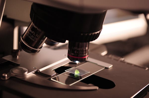
The Breakthrough Experiment: Isolating Cancer's Sugar Nemesis
Methodology: From Algae to Anticancer Agent
Researchers deployed a meticulously optimized protocol to extract and test Coelastrella's EPS 1 4 :
Cultivation
- Algae grown in Šetlik medium bubbled with 2–3% CO₂
- 20-day incubation under 132 μmol m⁻² s⁻¹ light
EPS Extraction
- Culture centrifuged to remove cells
- Supernatant mixed with ice-cold ethanol (1:2 ratio)
- EPS precipitated, pelleted (5,000 × g, 20 min), and lyophilized
Characterization
- Molecular weight profiling: High-performance size-exclusion chromatography
- Sugar analysis: HPLC-UV for monosaccharide identification
- Functional groups: FT-IR spectroscopy
Anticancer Testing
- Cell viability: MTT assays on cervical (HeLa), breast (MCF-7), and normal (BALB/3T3, HaCaT) cells
- Apoptosis detection: Annexin V-FITC staining and flow cytometry
- Migration inhibition: Wound-healing (scratch) assays
Results: Precision Strikes Against Cancer
Chemical Profile of Coelastrella EPS
| Property | Value |
|---|---|
| Molecular weight fractions | 115 kDa, 307 kDa, 724 kDa |
| Dominant sugars | Galactose, fucose |
| Uronic acid content | 4.5% (w/w) |
| Protein content | 7.14% (w/w) |
| Key functional groups | -OH, -COO, C-O-C (FT-IR confirmation) |
Anticancer Activity of EPS
| Cell Line | Cell Viability at 500 µg/mL EPS | Selectivity vs. Normal Cells |
|---|---|---|
| HeLa (cervical) | 22% | 3.5-fold higher toxicity |
| MCF-7 (breast) | 38% | 2.8-fold higher toxicity |
| BALB/3T3 | 78% | — |
| HaCaT | 83% | — |
Key Findings
Why This Matters
The EPS's selectivity—sparing non-cancerous cells—suggests a fundamentally different mechanism from conventional chemotherapy, which indiscriminately kills dividing cells.
Anticancer Activity Visualization
The Scientist's Toolkit: Key Reagents in EPS Anticancer Research
| Reagent/Method | Function | Experimental Role |
|---|---|---|
| Cold ethanol | Polysaccharide precipitant | Isolates EPS from culture broth |
| Annexin V-FITC | Binds phosphatidylserine exposed on apoptotic cells | Detects early apoptosis via flow cytometry |
| MTT reagent | Enzymatically reduced to purple formazan in live cells | Quantifies cell viability |
| FT-IR spectroscopy | Identifies functional groups (e.g., -OH, C-O-C) in molecules | Confirms EPS chemical signature |
| HPLC-UV | Separates and quantifies monosaccharides | Reveals sugar composition of EPS |
Beyond the Lab: Therapeutic Promise and Future Horizons
Coelastrella's EPS joins a growing arsenal of bioactive algal compounds. Previous studies show its fatty acids also combat tumors and pathogens 5 , while blue-light cultivation amplifies its antioxidant output by 60% 8 . Three frontiers loom large:
The Big Picture
As one researcher notes, "Microalgae are biochemical factories operating on solar power." Coelastrella epitomizes this—a wetland microbe that could redefine cancer therapy 4 .
Future Research Directions
- Clinical trial design
- Large-scale production
- Combination therapies
Sugar-Coated Hope
The story of Coelastrella sp. BGV underscores a profound truth: solutions to humanity's deadliest challenges may lie in nature's most overlooked corners. As EPS-based therapies advance toward clinical trials, these green microalgae illuminate a path to precise, sustainable, and accessible cancer treatment—where healing springs not from a lab, but from life itself.