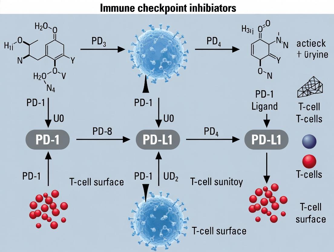
Unleashing the Attack: How Immune Checkpoint Inhibitors Reactivate Anti-Tumor T-Cell Responses
This article provides a comprehensive mechanistic and applied analysis of how immune checkpoint inhibitors (ICIs) overcome tumor-induced T-cell suppression to reactivate anti-cancer immunity.
Unleashing the Attack: How Immune Checkpoint Inhibitors Reactivate Anti-Tumor T-Cell Responses
Abstract
This article provides a comprehensive mechanistic and applied analysis of how immune checkpoint inhibitors (ICIs) overcome tumor-induced T-cell suppression to reactivate anti-cancer immunity. For researchers and drug developers, we detail the foundational biology of co-inhibitory pathways (e.g., PD-1/PD-L1, CTLA-4), explore current methodologies for assessing T-cell activation in vitro and in vivo, address key challenges in optimizing therapeutic response and overcoming resistance, and validate mechanisms through comparative analysis of different checkpoint targets. The synthesis offers a roadmap for next-generation immunotherapy development.
The Brakes on Immunity: Foundational Biology of T-Cell Checkpoints and Tumor Evasion
This whitepaper explicates the fundamental duality of immune checkpoint pathways, which are central to the thesis question: How do immune checkpoint inhibitors activate T-cells? Understanding the physiological purpose of these pathways is a prerequisite for comprehending their pathological co-option by tumors and the mechanistic basis of therapeutic intervention. Immune checkpoint inhibitors (ICIs) do not constitutively activate T-cells; rather, they block inhibitory signals, thereby shifting the equilibrium towards T-cell activation and restoring anti-tumor immunity.
Physiological Immune Checkpoint Function
Physiological immune checkpoints are inhibitory pathways crucial for maintaining self-tolerance (preventing autoimmunity) and modulating the duration and amplitude of immune responses to protect tissues from collateral damage during infection.
Canonical Pathways
- CTLA-4 (Cytotoxic T-Lymphocyte-Associated protein 4): An early checkpoint, primarily expressed upon T-cell activation. It outcompetes the co-stimulatory receptor CD28 for binding to B7-1/B7-2 (CD80/CD86) on antigen-presenting cells (APCs), transmitting an inhibitory signal that dampens the initial T-cell activation phase in lymphoid organs.
- PD-1 (Programmed Cell Death protein 1): An inducible checkpoint expressed on activated T-cells, B-cells, and myeloid cells. Its engagement by ligands PD-L1 or PD-L2 (expressed on APCs, stromal cells, and non-lymphoid tissues) inhibits kinase signaling, reducing T-cell effector functions, proliferation, and survival in peripheral tissues. This is critical for limiting immune activity in non-lymphoid organs.
Table 1: Core Physiological Immune Checkpoints
| Checkpoint | Primary Cellular Expression | Ligand(s) | Primary Physiological Role | Key Biochemical Effect |
|---|---|---|---|---|
| CTLA-4 | Activated T-cells (primarily CD4+), Tregs | B7-1 (CD80), B7-2 (CD86) | Attenuate initial T-cell activation in lymphoid organs; central tolerance | Outcompetes CD28 for B7, recruits phosphatases (e.g., SHP2) to dampen TCR/CD28 signaling. |
| PD-1 | Activated T-cells, B-cells, Myeloid cells | PD-L1, PD-L2 | Limit effector T-cell activity in peripheral tissues; maintain peripheral tolerance | Upon ligation, phosphorylated ITSM motif recruits SHP2, dephosphorylates key TCR/CD28 signaling molecules (e.g., ZAP70, PI3K). |
| LAG-3 | Activated T-cells, Tregs, NK cells | MHC Class II, FGL1, LSECtin | Modulate T-cell activation, proliferation, and homeostasis | Negatively regulates TCR signaling; exact mechanism is context-dependent. |
| TIM-3 | IFN-γ producing T-cells, Tregs, Myeloid | Galectin-9, CEACAM1, HMGB1 | Regulate Th1/Tc1 immunity and tolerance; promote macrophage activation. | Disrupts CD4+ T-cell co-stimulation; cross-linking induces T-cell death. |
Diagram Title: Physiological Checkpoint Function in Lymphoid vs. Peripheral Tissue
Pathological Hijacking by Tumors
Tumors exploit these regulatory mechanisms to create an immunosuppressive tumor microenvironment (TME), enabling immune evasion.
Mechanisms of Hijacking
- Upregulation of Checkpoint Ligands: Tumor cells and associated stromal/immune cells constitutively overexpress PD-L1, often driven by oncogenic signaling (e.g., PTEN loss, AKT activation) or inflammatory cytokines (IFN-γ) from infiltrating lymphocytes ("adaptive immune resistance").
- Induction of Checkpoint Receptors: Chronic antigen exposure and inflammatory cues in the TME lead to sustained high expression of PD-1, CTLA-4, TIM-3, LAG-3 on tumor-infiltrating lymphocytes (TILs), rendering them "exhausted" or dysfunctional.
- Recruitment of Regulatory Cells: Tumors secrete factors (e.g., CCL22, VEGF) that recruit Tregs, which express high levels of CTLA-4, further suppressing local anti-tumor immunity.
Quantitative Impact on Anti-Tumor Immunity
Table 2: Tumor Hijacking Strategies & Consequences for T-cells
| Hijacked Pathway | Tumor/Stromal Action | Effect on T-cell in TME | Associated T-cell Phenotype |
|---|---|---|---|
| PD-1/PD-L1 | Constitutive/inducible PD-L1 expression on tumor cells, myeloid cells. | Inhibited TCR signaling, reduced cytokine production (IFN-γ, TNF-α, IL-2), impaired proliferation, reduced cytotoxicity. | Exhaustion (TOX+), reduced polyfunctionality. |
| CTLA-4 | Increased B7 ligand expression on APCs; tumor-mediated Treg expansion. | Outcompetition of CD28 co-stimulation; direct suppression via Tregs. | Anergy, suppression of CD4+ T-cell help. |
| TIM-3/Gal-9 | Upregulated Galectin-9 on tumor endothelial and cells. | Induction of apoptosis in Th1/Tc1 cells; cross-linking reduces effector function. | Co-expression with PD-1 marks severe exhaustion. |
| Metabolic Checkpoints | Depletion of essential nutrients (glucose, arginine); accumulation of immunosuppressive metabolites (adenosine, kynurenine). | Impaired glycolytic flux, mTOR inhibition, impaired proliferation, altered differentiation. | Metabolic quiescence, functional impairment. |
Diagram Title: Tumor Microenvironment Hijacks Checkpoints to Induce T-cell Exhaustion
Experimental Protocols for Studying Checkpoints & ICI Action
Protocol: In Vitro T-cell Activation/Inhibition Assay
Purpose: To quantify the inhibitory effect of checkpoint engagement and its reversal by ICIs. Methodology:
- Isolate human CD4+ or CD8+ T-cells from PBMCs using magnetic-activated cell sorting (MACS).
- Coat plate with stimuli: Use a 96-well plate coated with anti-CD3 (OKT3, 1-5 µg/mL) and soluble anti-CD28 (1 µg/mL) for activation. For inhibition, also coat with recombinant PD-L1 Fc chimera (2-5 µg/mL) or add soluble anti-CTLA-4 blocking antibody (10 µg/mL) vs. isotype control.
- Culture T-cells: Seed purified T-cells (1e5 cells/well) in complete RPMI medium. Include conditions with PD-1/PD-L1 or CTLA-4/B7 blockade using therapeutic monoclonal antibodies (e.g., Nivolumab, Ipilimumab, 10 µg/mL).
- Assess readouts (72-96h):
- Proliferation: CFSE dilution or [3H]-thymidine incorporation.
- Cytokine Production: ELISA for IFN-γ and IL-2 in supernatant.
- Activation Markers: Flow cytometry for CD25, CD69.
- Signaling: Phospho-flow cytometry for p-AKT, p-S6, p-ERK.
Protocol: Ex Vivo Analysis of Tumor-Infiltrating Lymphocytes (TILs)
Purpose: To profile checkpoint expression and functional state of T-cells from the TME. Methodology:
- Tumor Digestion: Mechanically dissociate and enzymatically digest (Collagenase IV/DNase I) fresh human or murine tumor samples to create a single-cell suspension.
- Immune Cell Enrichment: Density gradient centrifugation (e.g., Ficoll-Paque) or negative selection kits to enrich lymphocytes.
- Multicolor Flow Cytometry Panel:
- Surface Staining: CD3, CD4, CD8, PD-1, CTLA-4, TIM-3, LAG-3, CD39.
- Intracellular Staining (after stimulation with PMA/Ionomycin + Brefeldin A): IFN-γ, TNF-α, IL-2, Granzyme B, TOX (exhaustion marker).
- Phenotypic Analysis: Identify exhausted (PD-1hiTIM-3+) vs. functional (PD-1lo/int) populations.
- Functional Validation: Sort specific TIL subsets and co-culture with autologous tumor cells or antigen-pulsed targets to measure cytotoxicity and cytokine production.
The Scientist's Toolkit: Key Research Reagent Solutions
Table 3: Essential Reagents for Immune Checkpoint Research
| Reagent Category | Specific Example(s) | Function & Application |
|---|---|---|
| Recombinant Proteins | Human/mouse PD-L1 Fc chimera, B7-1/B7-2 protein. | To engage checkpoint receptors in vitro for inhibition assays. Ligand-coated plates/cells. |
| Therapeutic & Blocking Antibodies | Anti-PD-1 (Nivolumab, Pembrolizumab), Anti-CTLA-4 (Ipilimumab), Anti-PD-L1 (Atezolizumab), Isotype controls. | Functional blockade of checkpoints in vitro and in vivo models. Positive controls. |
| Detection Antibodies (Flow Cytometry) | Anti-human/mouse CD3, CD4, CD8, PD-1 (clone EH12.2H7), CTLA-4 (clone BN13), TIM-3, LAG-3, CD69, CD25. | Phenotypic characterization of T-cell activation, exhaustion, and checkpoint expression. |
| Intracellular Cytokine Staining Kit | BD Cytofix/Cytoperm, FoxP3/Transcription Factor Staining Buffer Set. | Permeabilization and fixation for staining intracellular cytokines (IFN-γ, IL-2) and transcription factors (TOX, FoxP3). |
| T-cell Activation/Culture Systems | MACS CD4+/CD8+ T-cell Isolation Kits, Cell Activation Cocktail (PMA/Ionomycin), Human T-Activator CD3/CD28 Dynabeads. | Isolation and controlled stimulation of primary T-cells for functional assays. |
| Cell-based Reporter Assays | PD-1/PD-L1 Blockade Bioassay (NFAT Reporter Jurkat T-cells + PD-L1 aAPC/CHO cells). | Quantifying the functional potency of checkpoint-blocking therapeutics in a high-throughput format. |
| In Vivo Models | Syngeneic mouse tumor models (e.g., MC38, CT26), humanized PDX mice reconstituted with human immune system (HIS). | Evaluating ICI efficacy, pharmacodynamics, and biomarker discovery in an intact TME. |
Diagram Title: Experimental Workflow to Study ICI-Mediated T-cell Activation
The therapeutic success of ICIs is a direct application of the core concept elucidated here: they target pathways physiologically designed for negative regulation, which are pathologically co-opted by tumors. By blocking CTLA-4 or PD-1/PD-L1, ICIs disrupt this hijacking, shifting the signaling equilibrium within the TME and permitting the reactivation of pre-existing but suppressed tumor-specific T-cells. This restoration of functional anti-tumor immunity, rather than de novo activation, answers the central thesis question and underscores the importance of understanding fundamental immunobiology for rational drug development.
This whitepaper provides an in-depth technical analysis of the PD-1/PD-L1 and CTLA-4/CD80/CD86 signaling cascades, framed within the critical research thesis: How do immune checkpoint inhibitors activate T-cells? Understanding the precise molecular mechanisms of these pathways is fundamental for developing next-generation cancer immunotherapies and managing immune-related adverse events.
PD-1/PD-L1 Signaling Pathway
Molecular Mechanism
Programmed Death-1 (PD-1; CD279) is an inhibitory receptor primarily expressed on activated T-cells, B-cells, and myeloid cells. Its ligands, PD-L1 (B7-H1; CD274) and PD-L2 (B7-DC; CD273), are expressed on antigen-presenting cells (APCs) and various tumor microenvironments. Engagement of PD-1 with PD-L1/L2 initiates a signaling cascade that suppresses T-cell receptor (TCR)-mediated activation.
Core Signaling Events:
- Receptor Engagement: PD-L1 binding induces PD-1 clustering.
- SHP-1/SHP-2 Recruitment: The phosphorylated Immunoreceptor Tyrosine-based Switch Motif (ITSM) within the PD-1 cytoplasmic domain recruits phosphatases SHP-1 and SHP-2.
- Proximal TCR Signal Disruption: SHP-1/2 dephosphorylates key signaling molecules in the TCR cascade, including ZAP70, CD3ζ, and PKCθ.
- Metabolic Reprogramming: Downregulation of PI3K/AKT/mTOR pathway reduces glycolysis and amino acid metabolism.
- Transcriptional Modulation: Altered AP-1, NF-κB, and NFAT activity reduces effector cytokine production (IL-2, IFN-γ, TNF-α).
Table 1: Key Quantitative Parameters of PD-1/PD-L1 Interaction & Inhibition
| Parameter | Typical Value / Range | Experimental Context | Reference (Recent) |
|---|---|---|---|
| PD-1/PD-L1 Binding Affinity (K_D) | ~8 - 27 µM (SPR) | Recombinant extracellular domains | Yearley et al., 2023* |
| PD-1 Expression Density (Tumor-Infiltrating CD8+ T-cells) | 5,000 - 50,000 molecules/cell (CyTOF) | Human NSCLC biopsy | Zheng et al., 2022 |
| Serum Soluble PD-1 (sPD-1) in mRCC | Baseline: 10.2 ± 4.1 ng/mL | Predictive biomarker for Nivolumab response | Oh et al., 2023 |
| IC50 for Pembrolizumab (anti-PD-1) | 0.3 - 0.5 nM (Cell-based bioassay) | Blockade of PD-1/PD-L1 interaction | Keytruda FDA Label, 2024 |
| Tumor PD-L1 Positivity Cutoff (TPS) | ≥1%, ≥50% (IHC 22C3 pharmDx) | Companion diagnostic for NSCLC | NCCN Guidelines v.4.2024 |
Note: Values are representative from recent literature; specific conditions vary.
Detailed Experimental Protocol: Assessing PD-1/PD-L1 BlockadeIn Vitro
Protocol Title: Human T-cell Activation Assay Using PD-L1 Expressing Artificial Antigen Presenting Cells (aAPCs)
Objective: To quantify the functional reversal of T-cell suppression via PD-1/PD-L1 checkpoint blockade.
Materials:
- T-cells: Human CD4+ or CD8+ T-cells isolated from PBMCs (negative selection kit).
- aAPCs: K562 cells engineered to express: 1) HLA class I/II with a specific peptide, 2) CD80 (for costimulation), and 3) human PD-L1.
- Inhibitors: Clinical-grade anti-PD-1 (e.g., Nivolumab), anti-PD-L1 (e.g., Atezolizumab), or isotype control.
- Readouts: Flow cytometry for activation markers, ELISA/CBA for cytokines, CFSE/EdU for proliferation.
Procedure:
- T-cell Isolation & Labeling: Isolate naïve or memory T-cells. Label with CFSE (5µM, 10 min) if measuring proliferation.
- Co-culture Setup: Plate aAPCs in 96-well U-bottom plates at a 1:2 ratio (aAPC:T-cell). Typical density: 50,000 T-cells/well.
- Checkpoint Blockade: Add titrated doses of anti-PD-1/PD-L1 mAb (0.01 - 10 µg/mL) or controls at time zero.
- Incubation: Culture for 3-5 days in complete RPMI-1640 medium with IL-2 (10-20 IU/mL).
- Harvest & Analysis:
- Day 3: Harvest supernatant for IFN-γ/IL-2 quantification by ELISA.
- Day 5: Harvest cells for flow cytometry. Stain for: CD3, CD8/CD4, CD25 (activation), CD69 (early activation), and viability dye. Analyze CFSE dilution.
- Data Analysis: Calculate % of proliferated (CFSE-low) cells, MFI of activation markers, and cytokine concentration. Plot dose-response curves to determine EC50 of the therapeutic antibody.
CTLA-4/CD80/CD86 Signaling Pathway
Molecular Mechanism
Cytotoxic T-Lymphocyte-Associated protein 4 (CTLA-4; CD152) is a high-affinity inhibitory receptor constitutively expressed on Tregs and upregulated on conventional T-cells upon activation. It competes with the costimulatory receptor CD28 for binding to shared ligands CD80 (B7-1) and CD86 (B7-2) on APCs, but with ~20-fold higher affinity.
Core Signaling Events:
- Competitive Antagonism: CTLA-4 outcompetes CD28 for CD80/CD86 binding, directly removing CD28-mediated "Signal 2."
- Trans-endocytosis: CTLA-4 physically removes CD80/CD86 from the APC surface via a process of ligand stripping, degrading them in T-cell lysosomes.
- Phosphatase Recruitment: Similar to PD-1, its cytoplasmic tail recruits PP2A and SHP-2, dephosphorylating the TCR/CD28 signaling chain.
- Attenuation of Lipid Rafts: CTLA-4 disrupts the clustering of TCR and CD28 within the immunological synapse.
- Treg-Dependent Suppression: Intrinsic CTLA-4 signaling in Tregs enhances their suppressive function via modulation of lipid metabolism and cytokine production.
Table 2: Key Quantitative Parameters of CTLA-4/CD80/CD86 Interaction & Inhibition
| Parameter | Typical Value / Range | Experimental Context | Reference (Recent) |
|---|---|---|---|
| CTLA-4/CD80 Binding Affinity (K_D) | ~0.2 - 0.4 µM (SPR) | Recombinant proteins | Schildberg et al., 2022 |
| CTLA-4/CD86 Binding Affinity (K_D) | ~2.0 - 3.0 µM (SPR) | Recombinant proteins | Schildberg et al., 2022 |
| CD28/CD80 Binding Affinity (K_D) | ~4.0 µM (SPR) | Comparison to CTLA-4 | - |
| Serum sCTLA-4 in Melanoma | 4.8 ng/mL (Pre-ipi) vs 8.1 ng/mL (Post-ipi) | Correlation with Ipilimumab response | Gowen et al., 2023 |
| IC50 for Ipilimumab (anti-CTLA-4) | ~1.2 nM (Competitive Binding ELISA) | Blockade of CTLA-4/CD80 interaction | Yervoy FDA Label, 2023 |
| Frequency of CTLA-4+ Tregs in Tumor | 30-70% of intratumoral Tregs (CyTOF) | Human HNSCC & melanoma samples | Salerno et al., 2024 |
Detailed Experimental Protocol: Assessing CTLA-4 Ligand Competition and Trans-endocytosis
Protocol Title: Live-Cell Imaging and Flow Cytometry Assay for CTLA-4-Mediated CD80 Trans-endocytosis
Objective: To visualize and quantify the removal of CD80 from APC membranes by CTLA-4-expressing T-cells.
Materials:
- APCs: CHO cells or immature dendritic cells (iDCs) expressing GFP-tagged CD80.
- T-cells: Jurkat T-cells or primary human T-cells transduced with mCherry-tagged CTLA-4 and a control vector.
- Inhibitors: Ipilimumab (anti-CTLA-4), Fab fragments.
- Equipment: Confocal live-cell imaging system, flow cytometer.
Procedure:
- Cell Preparation: Generate APC line stably expressing GFP-CD80. Generate T-cell lines: a) mCherry-CTLA-4, b) mCherry-vector control.
- Live-Cell Imaging Setup: Co-culture T-cells and APCs on a poly-L-lysine coated imaging chamber at 37°C, 5% CO2. Use a 1:1 ratio. Add anti-CTLA-4 or control antibody (10 µg/mL) to designated wells.
- Image Acquisition: Begin time-lapse imaging immediately upon cell contact. Capture images for both GFP (CD80) and mCherry (CTLA-4) channels every 30 seconds for 60-90 minutes.
- Flow Cytometry Quantification: In parallel, set up identical co-cultures in tubes. After 60 minutes, gently disassociate cells, stain with an APC-conjugated anti-CD80 antibody that recognizes a different epitope than GFP.
- Gating Strategy: Gate on mCherry+ T-cells and analyze the GFP (total CD80) and APC (surface CD80) signals. Loss of APC signal on T-cells indicates internalization and degradation of CD80.
- Data Analysis:
- Imaging: Track fluorescence intensity of GFP-CD80 at the T-cell/APC contact site over time.
- Flow Cytometry: Calculate the ratio of surface (APC) to total (GFP) CD80 signal on mCherry+ T-cells. Compare CTLA-4+ vs control T-cells with and without antibody blockade.
Visualization of Signaling Cascades
Diagram 1: PD-1/PD-L1 inhibitory signaling and checkpoint blockade mechanism.
Diagram 2: CTLA-4 mechanism of action via competition and trans-endocytosis.
The Scientist's Toolkit: Essential Research Reagents & Solutions
Table 3: Key Research Reagent Solutions for Immune Checkpoint Studies
| Category | Reagent/Solution | Specific Example(s) | Function in Research |
|---|---|---|---|
| Recombinant Proteins | Human PD-1 Fc Chimera, PD-L1 His-tag | Sino Biological, R&D Systems | SPR/BLI binding assays, ELISA development, in vitro blocking studies. |
| Cell-Based Assays | Bioassay for PD-1 Inhibition: PD-1/NFAT Reporter Jurkat + PD-L1 aAPC | Promega (Checkpoint Cellular Assay), internal engineering. | High-throughput screening of therapeutic mAbs for functional potency (IC50). |
| Blocking Antibodies | Ultra-LEAF purified anti-human PD-1, CTLA-4, CD80, CD86 | BioLegend | Functional in vitro blockade in co-culture assays (low endotoxin, azide-free). |
| Flow Cytometry | Panel Design: CD3, CD4/8, PD-1, CTLA-4, CD69, CD25, Live/Dead | BD Biosciences, BioLegend, Thermo Fisher | Phenotyping immune cell subsets, assessing activation status in primary cells. |
| IHC/IF Reagents | Validated Clinical-Grade IHC Clones: PD-L1 (22C3, 28-8), CD8 (C8/144B) | Agilent/DAKO, Cell Signaling Tech | Spatial analysis of protein expression in tumor microenvironment (FFPE tissue). |
| Engineering Kits | Lentiviral vectors for human PD-L1, CTLA-4 (with GFP/mCherry tags) | VectorBuilder, Addgene | Generating stable cell lines (aAPCs or T-cells) for mechanistic studies. |
| Cytokine Detection | Multiplex Panels: Human Th1/Th2 Cytokine Panel 13-plex | LEGENDplex (BioLegend), MSD | Quantifying secreted cytokine profiles from activated T-cells post-blockade. |
| Animal Models | Syngeneic Tumor Models: MC38 (colorectal), B16-F10 (melanoma) | Charles River, JAX | In vivo efficacy testing of checkpoint inhibitors in immunocompetent mice. |
The clinical success of immune checkpoint inhibitors (ICIs) that block receptors such as PD-1 and CTLA-4 has revolutionized cancer immunotherapy. The central thesis of this research field is that ICIs function primarily by antagonizing inhibitory signaling in T-cells, thereby reversing functional suppression and restoring anti-tumor cytotoxicity. This whitepaper delves into the precise downstream molecular and cellular consequences of sustained checkpoint signaling that ICIs aim to overcome. Understanding the induction of T-cell exhaustion, anergy, and general dysfunction is fundamental to improving current therapies, predicting patient responses, and developing next-generation immunotherapies.
Core Signaling Pathways and Transcriptional Reprogramming
Chronic antigen stimulation, as occurs in tumors and persistent infections, leads to the sustained upregulation of checkpoint receptors. Ligation of these receptors by their ligands (e.g., PD-L1, B7-1/B7-2) initiates intracellular signaling cascades that actively suppress T-cell function.
Key Pathways from PD-1 and CTLA-4
PD-1 Signaling: Upon phosphorylation of its immunoreceptor tyrosine-based inhibitory motif (ITIM) and immunoreceptor tyrosine-based switch motif (ITSM), PD-1 recruits SHP-1 and SHP-2 phosphatases. These phosphatases dephosphorylate key signaling molecules proximal to the T-cell receptor (TCR).
CTLA-4 Signaling: CTLA-4 outcompetes CD28 for B7 ligands due to higher affinity, directly inhibiting costimulation. Intracellularly, it also recruits phosphatases (e.g., PP2A) and can disrupt TCR microcluster formation.
Diagram 1: Core Inhibitory Checkpoint Signaling Pathways
Table 1: Major Downstream Targets of Checkpoint Phosphatases
| Checkpoint Receptor | Recruited Phosphatase | Key Dephosphorylation Targets | Primary Signaling Pathway Disrupted |
|---|---|---|---|
| PD-1 | SHP-2 (primarily), SHP-1 | CD28, ZAP70, PKCθ, PI3K | TCR & CD28 signal transduction |
| CTLA-4 | PP2A | AKT, VAV1 | PI3K-AKT & TCR costimulation |
| TIM-3 | Unknown | BAT3 | Alternative NF-κB signaling |
| LAG-3 | Possibly ERK | Unknown | TCR signal duration |
Transcriptional Drivers of Dysfunction
The integrated signaling deficit leads to altered activity of key transcription factors (TFs). Reduced NFAT:AP-1 ratio promotes anergy and exhaustion profiles. TOX and NR4A are induced and sustain the exhausted phenotype.
Diagram 2: Transcriptional Network in T-Cell Exhaustion
Defining and Quantifying Dysfunctional States
Table 2: Comparative Features of T-Cell Dysfunctional States
| Feature | Exhausted T-Cell (Tex) | Anergic T-Cell | Functional Effector T-Cell |
|---|---|---|---|
| Proliferation | Severely impaired, lacks recall | Impaired upon restimulation | Robust, clonal expansion |
| Cytokine Profile | Low IL-2, IFNγ, TNFα (hierarchical loss) | Low IL-2 | High IL-2, IFNγ, TNFα, Granzyme B |
| Surface Markers | PD-1^hi^, TIM-3+, LAG-3+, CD39+, CXCR5-/+ | PD-1^int^, FR4+, CD73+ | PD-1^lo^, TIM-3-, KLRG1+ (short-lived) |
| Metabolism | Oxidative phosphorylation skewed, impaired glycolysis | Metabolic quiescence, impaired glycolysis | Aerobic glycolysis, OXPHOS |
| Epigenetic State | Stable, heritable chromatin modifications | Partially stable | Plastic, poised state |
| Reversibility by ICI | Partial (Progenitor Tex subset) | Yes (in some models) | N/A |
| Key Transcriptional Regulators | TOX, NR4A, Blimp-1 | EGR2/3, IKZF4, DGKα | T-bet, EOMES (for memory) |
Experimental Protocols for In Vitro and In Vivo Analysis
In Vitro Induction of Human T-Cell Exhaustion/Anergy
- Purpose: Generate a homogeneous population of dysfunctional T-cells for molecular profiling or drug screening.
- Materials: Human CD8+ T-cells isolated from PBMCs, anti-CD3/anti-CD28 coated beads or plates, recombinant human PD-L1 Fc or cell line expressing high PD-L1, IL-2.
- Protocol:
- Stimulation: Activate purified naïve CD8+ T-cells with plate-bound anti-CD3 (1-5 µg/mL) and soluble anti-CD28 (1 µg/mL) in RPMI-1640 + 10% FBS + 50 IU/mL IL-2.
- Chronic Stimulation & Checkpoint Engagement: At 24-48h post-activation, carefully harvest cells and transfer to a new plate pre-coated with PD-L1 Fc (5-10 µg/mL) and sub-saturating levels of anti-CD3 (0.1-0.5 µg/mL). Include controls (anti-CD3 only, isotype control).
- Maintenance: Re-feed cells with fresh media + IL-2 every 2-3 days. Re-plate onto freshly coated PD-L1/anti-CD3 plates every 5-7 days to maintain chronic signaling.
- Validation (Day 10-14): Re-stimulate cells with PMA/Ionomycin or anti-CD3/CD28 beads. Measure IFNγ and TNFα production by intracellular cytokine staining (ICS) and flow cytometry. Expect >70% reduction in cytokine-positive cells and high, sustained PD-1 expression in the PD-L1 treated group.
In Vivo Assessment of Reversal by Checkpoint Inhibitors
- Purpose: Evaluate the functional reinvigoration of exhausted T-cells following ICI treatment in a mouse tumor model.
- Materials: C57BL/6 mice, MC38 colon adenocarcinoma cells, anti-mouse PD-1 antibody (clone RMP1-14), anti-mouse CTLA-4 antibody (clone 9D9), flow cytometry antibodies (anti-CD8, PD-1, TIM-3, LAG-3, Ki-67, intracellular IFNγ/TNFα).
- Protocol:
- Tumor Engraftment: Inject 0.5x10^6^ MC38 cells subcutaneously into the right flank of mice.
- Treatment: When tumors reach ~50-100 mm³, randomize mice into groups (n=5-10). Administer anti-PD-1 (200 µg/mouse), anti-CTLA-4 (100 µg/mouse), combination, or isotype control via intraperitoneal injection every 3-4 days for 3 doses.
- Harvest & Analysis: Sacrifice mice 1-2 days after the final dose. Harvest tumors, digest to single-cell suspension, and enrich for lymphocytes using Percoll gradient.
- Ex Vivo Stimulation: Plate tumor-infiltrating lymphocytes (TILs) with PMA/Ionomycin + protein transport inhibitor for 5-6 hours.
- Flow Cytometry: Surface stain for CD8, PD-1, TIM-3, LAG-3. Fix, permeabilize, and intracellularly stain for Ki-67, IFNγ, and TNFα.
- Data Interpretation: Compare the frequency of CD8+ TILs expressing Ki-67 and producing multiple cytokines between treatment groups. Successful reinvigoration is indicated by a statistically significant increase in polyfunctional (IFNγ+TNFα+) CD8+ T-cells in ICI-treated groups.
Diagram 3: Workflow for Assessing T-Cell Reinvigoration by ICI
The Scientist's Toolkit: Key Research Reagent Solutions
Table 3: Essential Reagents for Studying T-Cell Dysfunction
| Reagent Category | Specific Example(s) | Function & Application |
|---|---|---|
| Recombinant Checkpoint Proteins | Human PD-L1 Fc Chimera, Mouse B7-1 Ig Fusion Protein | Coat plates to provide ligand-mediated inhibition in vitro assays. |
| Agonist/Antagonist Antibodies | Anti-human PD-1 (clone EH12.2H7), Anti-mouse CTLA-4 (clone 9D9), Anti-CD3 (OKT3, 145-2C11) | Activate or block signaling pathways in vitro and for in vivo therapeutic studies. |
| Multicolor Flow Cytometry Panels | Antibodies against: CD8, PD-1, TIM-3, LAG-3, CD39, CD62L, CD44, CXCR5, TCF-1 (by IC) | Phenotypic identification of exhausted (Tex), progenitor Tex, anergic, and functional T-cell subsets from tissues. |
| Intracellular Staining Kits | Foxp3/Transcription Factor Staining Buffer Set, BD Cytofix/Cytoperm | Permeabilize cells for staining of cytokines (IFNγ, TNFα), transcription factors (TOX, T-bet), and proliferation markers (Ki-67). |
| T-Cell Activation & Expansion Kits | Human T-Activator CD3/CD28 Dynabeads, CellXVivo Human T Cell Expansion Kit | Provide standardized, reproducible polyclonal T-cell activation for dysfunction induction experiments. |
| Metabolic Assay Kits | Seahorse XFp Cell Mito Stress Test Kit, Extracellular Acidification Rate (ECAR) Assays | Quantify real-time changes in oxidative phosphorylation (OCR) and glycolysis (ECAR) in dysfunctional vs. functional T-cells. |
| Epigenetic Analysis Kits | CUT&RUN Assay Kits, ATAC-Seq Kits | Map genome-wide changes in histone modifications (H3K27ac, H3K4me3) and chromatin accessibility in Tex cells. |
| In Vivo Tumor Models | MC38 (murine colon adenocarcinoma), B16-OVA (melanoma), CT26 (colon carcinoma) | Syngeneic models with defined immune contextures for studying ICI efficacy and T-cell reinvigoration. |
The efficacy of immune checkpoint inhibitors (ICIs) in reactivating tumor-infiltrating lymphocytes (TILs) is not solely determined by direct PD-1/CTLA-4 blockade. It is fundamentally constrained by the immunosuppressive tumor microenvironment (TME). This whitepaper details the cellular and molecular architecture of the TME, providing the essential context for understanding the variable clinical responses to ICIs. Successful T-cell activation requires overcoming this synergistic network of suppression.
Cellular Constituents of the Suppressive TME
The TME is populated by a consortium of myeloid and lymphoid cells that actively inhibit cytotoxic T-cell function.
Table 1: Key Immunosuppressive Cells in the TME
| Cell Type | Markers (Human) | Primary Immunosuppressive Mechanisms | Impact on ICI Response |
|---|---|---|---|
| Myeloid-Derived Suppressor Cells (MDSCs) | CD11b⁺, CD33⁺, HLA-DRlow/-, (PMN-MDSC: CD14⁻CD15⁺; M-MDSC: CD14⁺CD15⁻) | Arg1, iNOS, ROS/RNS production, cysteine depletion, T-cell sequestration. | Associated with primary and acquired resistance to anti-PD-1/CTLA-4. |
| Tumor-Associated Macrophages (TAMs), M2-like | CD68⁺, CD163⁺, CD206⁺, MerTK⁺ | TGF-β, IL-10, PGE2 secretion; expression of PD-L1; promotion of tissue remodeling & angiogenesis. | High TAM infiltration correlates with poor prognosis and ICI resistance. |
| Regulatory T-cells (Tregs) | CD4⁺, CD25⁺, FoxP3⁺ | CTLA-4-mediated APC suppression; consumption of IL-2; secretion of TGF-β/IL-10; expression of CD39/CD73. | Intratumoral Treg density inversely correlates with CD8⁺ T-cell activity post-ICI. |
| Cancer-Associated Fibroblasts (CAFs) | α-SMA⁺, FAP⁺, PDGFRβ⁺ | Deposition of dense ECM (creating a physical barrier); secretion of CXCL12; metabolic competition. | Creates a physical and chemical barrier limiting T-cell infiltration. |
Soluble Immunosuppressive Factors
These cells exert their function through a complex cocktail of soluble mediators.
Table 2: Key Soluble Immunosuppressive Factors in the TME
| Factor Category | Example Molecules | Primary Source in TME | Mechanism of T-cell Suppression |
|---|---|---|---|
| Anti-inflammatory Cytokines | TGF-β, IL-10 | TAMs, Tregs, CAFs | Inhibits T-cell proliferation & differentiation; drives Treg induction; blocks DC maturation. |
| Metabolic Enzymes | Arginase I (Arg1), Indoleamine 2,3-dioxygenase (IDO) | MDSCs, TAMs | Depletes L-arginine & tryptophan, essential for T-cell function; generates toxic metabolites (kynurenines). |
| Reactive Species | Reactive Oxygen/Nitrogen Species (ROS/RNS) | MDSCs | Induces T-cell receptor nitrosylation and apoptosis; inhibits IL-2 signaling. |
| Adenosine | Generated by CD39/CD73 ectoenzymes | Tregs, CAFs, some tumor cells | Binds A2A receptor on T-cells, suppressing cytokine production, proliferation, and cytotoxicity. |
| Prostaglandins | PGE2 | TAMs, tumor cells | Promotes differentiation of MDSCs & Tregs; inhibits DC maturation and T-cell activation. |
Signaling Pathways of Key Suppressive Mechanisms
The following diagrams detail critical pathways that must be overcome for effective ICI-mediated T-cell activation.
Experimental Protocols for TME Analysis
Protocol 1: Multicolor Flow Cytometry for TME Immune Profiling
- Objective: To simultaneously identify and quantify suppressive immune cells (MDSC subsets, TAMs, Tregs) from dissociated tumor tissue.
- Detailed Methodology:
- Tumor Dissociation: Process fresh tumor sample (≤1g) using a human/mouse tumor dissociation kit (e.g., Miltenyi Biotec) with a gentleMACS Octo Dissociator. Use RPMI-1640 medium.
- Single-Cell Suspension: Pass cells through a 70µm strainer, wash with PBS + 2% FBS. Perform RBC lysis.
- Viability Staining: Resuspend cells in PBS. Add a fixable viability dye (e.g., Zombie NIR) and incubate 15 min at RT in the dark. Wash.
- Surface Staining: Incubate cells with Fc receptor block (e.g., Human TruStain FcX) for 10 min. Add antibody cocktail for surface markers (see Toolkit) and incubate 30 min at 4°C in the dark. Wash.
- Intracellular Staining (for FoxP3): Fix and permeabilize cells using FoxP3/Transcription Factor Staining Buffer Set. Stain with anti-FoxP3 antibody for 30 min at 4°C. Wash.
- Acquisition: Resuspend in staining buffer and acquire data on a ≥3-laser flow cytometer (e.g., BD FACSymphony). Collect ≥100,000 live single-cell events.
- Analysis: Use FlowJo software. Gate: Single cells (FSC-A/FSC-H) → Live cells → Lineage (CD45⁺) → Subset analysis (see Table 1 for markers).
Protocol 2: Functional Assessment of T-cell Suppression (Co-culture Assay)
- Objective: To test the in vitro suppressive capacity of sorted TME cells on autologous or allogeneic T-cell proliferation/activation.
- Detailed Methodology:
- Effector Cell Isolation: Isolate naive CD4⁺ or CD8⁺ T-cells from peripheral blood using negative selection magnetic beads.
- Suppressor Cell Isolation: Sort target populations (e.g., CD33⁺HLA-DR⁻ MDSCs, CD4⁺CD25⁺ Tregs) from dissociated tumor single-cell suspension using FACS.
- CFSE Labeling: Label T-cells with 2.5µM CFSE in PBS for 10 min at 37°C. Quench with 5x volume of cold complete media.
- Co-culture Setup: Plate suppressor cells at varying ratios (e.g., 1:1 to 1:32 suppressor:T-cell) in a 96-well round-bottom plate. Add 1e5 CFSE-labeled T-cells per well. Include T-cells alone as a control.
- T-cell Stimulation: Stimulate all wells with soluble anti-CD3/CD28 antibodies (1µg/mL each) or Human T-Activator CD3/CD28 Dynabeads (1 bead:1 cell).
- Incubation: Culture for 3-5 days in complete RPMI-1640 medium with IL-2 (20 U/mL).
- Readout: Harvest cells, stain for activation markers (CD25, CD69), and analyze CFSE dilution via flow cytometry. Calculate % suppression:
[1 - (Prolif. in co-culture / Prolif. of T-cells alone)] * 100.
The Scientist's Toolkit: Key Research Reagent Solutions
Table 3: Essential Reagents for TME and ICI-Context Research
| Item | Example Product/Catalog # | Primary Function in TME Research |
|---|---|---|
| Tumor Dissociation Kit | Miltenyi Biotec, Human Tumor Dissociation Kit (130-095-929) | Enzymatic and mechanical dissociation of solid tumors into viable single-cell suspensions for downstream analysis. |
| Multicolor Flow Cytometry Antibody Panels | BioLegend, FoxP3 Buffer Set (422601); BD Biosciences, Anti-human CD39 (561717), CD73 (564489) | Surface and intracellular staining for comprehensive immune phenotyping of suppressive populations (MDSCs, Tregs, CAFs). |
| Cell Separation Magnets & Beads | STEMCELL Technologies, EasySep Human CD8⁺ T-cell Isolation Kit (17953) | Negative selection for high-purity isolation of specific lymphocyte subsets for functional co-culture assays. |
| Recombinant Cytokines & Factors | PeproTech, human TGF-β1 (100-21), IL-10 (200-10) | Used to polarize macrophages to M2 state in vitro or to mimic TME cytokine conditions in suppression assays. |
| Enzyme Activity Assays | Sigma-Aldrich, Arginase Activity Assay Kit (MAK112) | Quantification of functional enzymatic output from suppressive cells (e.g., Arg1 from MDSCs). |
| A2A Receptor Antagonist | Tocris, SCH58261 (2270) | Pharmacologic tool to block adenosine signaling in functional assays to test rescue of T-cell activity. |
| Phospho-Specific Antibodies for Signaling | CST, Phospho-STAT3 (Tyr705) (9145S) | Detection of activated signaling pathways (e.g., STAT3 in MDSCs) via western blot or intracellular flow cytometry. |
Within the broader thesis on how immune checkpoint inhibitors activate T-cells, research has expanded beyond the seminal pathways of PD-1 and CTLA-4. The next generation of investigation focuses on co-inhibitory receptors like LAG-3, TIGIT, and TIM-3. These pathways represent critical, non-redundant mechanisms of T-cell exhaustion in the tumor microenvironment. Their blockade, both as monotherapies and in rational combinations, is a central strategy to overcome resistance to existing immunotherapies and achieve more durable anti-tumor immunity. This whitepaper provides a technical guide to the biology, experimental interrogation, and therapeutic targeting of these emerging pathways.
Pathway Biology and Signaling Mechanisms
Lymphocyte Activation Gene-3 (LAG-3)
LAG-3 (CD223) is a type I transmembrane protein structurally homologous to CD4. Its primary ligand is MHC class II (MHC-II), but it also binds to Fibrinogen-like protein 1 (FGL1), LSECtin, and α-synuclein. Engagement inhibits T-cell activation and cytokine production. The exact signaling mechanism is less defined than for PD-1 but involves an inhibitory "KIEELE" motif in its cytoplasmic tail that may disrupt TCR signalosome assembly.
T-cell Immunoreceptor with Ig and ITIM domains (TIGIT)
TIGIT is a member of the CD28 family. It binds with high affinity to CD155 (PVR) and with lower affinity to CD112 (PVRL2, nectin-2), which are expressed on antigen-presenting cells and tumor cells. TIGIT exerts inhibition through two mechanisms: 1) direct intracellular signaling via an ITIM and an immunoglobulin tail tyrosine (ITT)-like motif that recruits phosphatases, and 2) cis competition with the costimulatory receptor CD226 (DNAM-1) for the same ligands.
T-cell Immunoglobulin and Mucin-domain containing-3 (TIM-3)
TIM-3 is a type I transmembrane protein with a diverse set of ligands, including galectin-9, phosphatidylserine (PtdSer), HMGB1, and CEACAM1. Its engagement typically leads to T-cell exhaustion and apoptosis. Signaling is complex and context-dependent, often involving the recruitment of the kinase Bat3 to a phosphorylated tyrosine in its cytoplasmic tail; ligand binding releases Bat3, enabling inhibition.
Table 1: Core Biology of Emerging Co-inhibitory Pathways
| Receptor | Primary Ligand(s) | Expression Profile | Key Signaling Motifs/Mechanisms | Functional Outcome on T-cells |
|---|---|---|---|---|
| LAG-3 | MHC-II, FGL1 | Activated CD4+/CD8+ T, Tregs, NKs | "KIEELE" motif; interferes with TCR signaling | Reduced activation, proliferation, cytokine secretion |
| TIGIT | CD155 (PVR), CD112 | Activated/CD8+ T, Tregs, NKs, Tfh | ITIM & ITT-like motif; competes with CD226 | Inhibits proliferation, cytokine production (IFN-γ, IL-10) |
| TIM-3 | Galectin-9, PtdSer, CEACAM1 | Exhausted CD8+ T, Th1, Tregs, NKs, DCs | Phospho-tyrosine/Bat3 interaction; galectin-9 induces apoptosis | Exhaustion, apoptosis, tolerance induction |
Quantitative Data on Expression and Therapeutic Impact
Recent clinical and preclinical studies highlight the significance of these pathways.
Table 2: Selected Clinical & Preclinical Data Summary
| Pathway | Key Context/Model | Quantitative Finding | Therapeutic Implication |
|---|---|---|---|
| LAG-3 | Melanoma (anti-PD-1 relapsed/refractory) | In RELATIVITY-047, relatlimab (α-LAG-3) + nivolumab vs. nivolumab alone: mPFS = 10.1 vs 4.6 mos (HR 0.75). | First FDA-approved LAG-3 combo demonstrates synergy with PD-1 blockade. |
| TIGIT | NSCLC (Phase III SKYSCRAPER-01) | Tiragolumab (α-TIGIT) + atezolizumab vs. atezolizumab: mPFS = 5.4 vs 3.6 mos (HR 0.74) in PD-L1-high. | Suggests benefit in high PD-L1 population; other trials ongoing. |
| TIM-3 | AML (Preclinical/Clinical) | TIM-3 is expressed on ~60% of leukemic stem cells (LSCs) and exhausted T-cells in AML. | Dual targeting of LSCs and restoring T-cell function. |
| Combined | T-cell Exhaustion Signature | Co-expression of 2+ checkpoints (e.g., PD-1+TIM-3+, PD-1+LAG-3+) defines severely exhausted T-cell subset with reduced proliferative capacity in vivo. | Rationale for dual or triple checkpoint blockade strategies. |
Detailed Experimental Protocols for Key Assays
Protocol: Multispectral Flow Cytometry for Co-inhibitory Receptor Profiling
Objective: To quantify co-expression patterns of LAG-3, TIGIT, and TIM-3 with PD-1 on tumor-infiltrating lymphocytes (TILs).
Materials: See "The Scientist's Toolkit" below. Method:
- Single-Cell Suspension: Process tumor tissue mechanically and enzymatically (e.g., using a murine Tumor Dissociation Kit) to create a single-cell suspension. Include a dead cell removal step.
- Surface Staining: Aliquot 2-5 x 10^6 cells per tube. Block Fc receptors with anti-CD16/32 or human Fc block for 10 min on ice.
- Antibody Cocktail: Add fluorochrome-conjugated antibodies against CD45, CD3, CD8, CD4, PD-1, LAG-3, TIGIT, and TIM-3. Include a live/dead viability dye. Vortex and incubate for 30 min in the dark at 4°C.
- Wash & Fix: Wash cells twice with FACS buffer. Fix cells in 1-2% paraformaldehyde (PFA) or a commercial fixative.
- Acquisition & Analysis: Acquire data on a spectral flow cytometer capable of detecting 12+ colors. Use compensation controls and fluorescence-minus-one (FMO) controls for accurate gating. Analyze using FlowJo or similar software. Gate sequentially on single cells -> live cells -> CD45+ leukocytes -> CD3+ T-cells -> CD4+/CD8+ subsets -> assess checkpoint co-expression.
Protocol:In VitroT-cell Functional Assay (Suppression/Reinvigoration)
Objective: To test the functional impact of LAG-3, TIGIT, or TIM-3 blockade on antigen-specific T-cell cytokine production and proliferation.
Materials: Human PBMCs or mouse splenocytes, antigenic peptide, recombinant ligand proteins (e.g., CD155-Fc, galectin-9), plate-bound anti-CD3/anti-CD28, blocking antibodies (α-LAG-3, α-TIGIT, α-TIM-3), CFSE. Method:
- T-cell Stimulation: Isolate CD8+ T-cells (e.g., using magnetic beads). Label with 5µM CFSE for proliferation tracking.
- Co-culture Setup: Plate α-CD3 (1 µg/mL) and α-CD28 (2 µg/mL) in a 96-well plate. Add T-cells (1 x 10^5/well). Include experimental conditions with:
- Recombinant ligand protein (e.g., 10 µg/mL CD155-Fc).
- Blocking antibodies (10 µg/mL each, alone or in combination).
- Isotype control antibodies.
- Incubation: Culture for 72-96 hours in complete RPMI medium.
- Readout: Harvest supernatant for multiplex cytokine analysis (IFN-γ, TNF-α, IL-2) by Luminex or ELISA. Analyze cells by flow cytometry to measure CFSE dilution (proliferation) and activation markers (CD25, CD69).
Pathway and Workflow Visualizations
Title: LAG-3 Inhibitory Signaling via MHC-II
Title: TIGIT-CD226 Competitive Binding and Signaling
Title: Experimental Workflow for Exhaustion Marker Profiling
The Scientist's Toolkit: Key Research Reagent Solutions
Table 3: Essential Reagents for Co-inhibitory Pathway Research
| Reagent Category | Specific Example(s) | Function & Application |
|---|---|---|
| Recombinant Proteins | Human/Mouse CD155-Fc, Galectin-9, LAG-3-Fc, TIM-3-Fc | Used to engage checkpoint receptors in in vitro suppression assays. Ligand blocking studies. |
| Blocking/Antagonistic Antibodies | Anti-human LAG-3 (Clone 17B4), Anti-human TIGIT (MBSA43), Anti-human TIM-3 (F38-2E2) | Functional studies to reverse T-cell exhaustion. Used in vitro and in vivo (murine analogs). |
| Flow Cytometry Antibodies | Fluorochrome-conjugated anti-human/mouse LAG-3, TIGIT, TIM-3, PD-1, CD3, CD8, CD4 | Phenotypic analysis of receptor expression on T-cell subsets. Critical for co-expression profiling. |
| Cell Isolation Kits | CD8+ T Cell Isolation Kit (Human/Mouse), Pan T Cell Isolation Kit | Isolation of pure T-cell populations for functional assays. Negative selection preserves cell activation status. |
| Functional Assay Kits | CFSE Cell Division Tracker, IFN-γ ELISpot/ELISA Kits, Luminex Cytokine Panels | Measure T-cell proliferation, cytokine secretion, and polyfunctionality upon checkpoint blockade. |
| In Vivo Models | Syngeneic mouse tumor models (MC38, CT26), Humanized PBMC or CD34+ NSG mice | Preclinical testing of therapeutic antibodies and combination efficacy in an intact immune system. |
Releasing the Brakes: Methodologies for ICI Action and Assessing T-Cell Reactivation
This analysis is framed within the broader thesis research question: How do immune checkpoint inhibitors activate T-cells? A critical component of the answer lies in understanding the precise mechanisms by which therapeutic monoclonal antibodies (mAbs) disrupt the immunosuppressive networks that restrain T-cell function. This whitepaper provides an in-depth technical guide on two principal strategies: blocking ligand-receptor interactions and depleting suppressive cell populations, both of which are fundamental to the efficacy of immune checkpoint inhibitors (ICIs) in oncology and autoimmunity.
Core Mechanisms of Action
Blocking Interactions (Receptor/Ligand Neutralization)
This mechanism involves mAbs that act as competitive antagonists, physically preventing the engagement of an inhibitory receptor on an effector cell (e.g., a T-cell) with its cognate ligand on an antigen-presenting cell (APC) or tumor cell.
- Target Examples: PD-1/PD-L1, CTLA-4/B7-1/B7-2, LAG-3/MHC-II.
- Outcome: The inhibitory signal is not transmitted, thereby preserving T-cell receptor (TCR) signaling, promoting T-cell proliferation, cytokine production, and cytotoxic activity.
Depleting Suppressive Cells (Cellular Cytotoxicity)
This mechanism leverages the Fc domain of the mAb to engage the immune system's cytotoxic effector functions to eliminate the cell expressing the target antigen.
- Primary Effector Mechanisms: Antibody-Dependent Cellular Cytotoxicity (ADCC), Antibody-Dependent Cellular Phagocytosis (ADCP), and Complement-Dependent Cytotoxicity (CDC).
- Target Examples: Depleting anti-CD25 mAbs (regulatory T-cells), anti-CD20 mAbs (B-cells), anti-CCR4 mAbs (Tregs in ATL).
- Outcome: Reduction in the number or frequency of immunosuppressive cells (e.g., Tregs, myeloid-derived suppressor cells (MDSCs)), thereby relieving direct suppression of effector T-cells and altering the tumor microenvironment (TME).
Table 1: Comparison of Key Monoclonal Antibody Mechanisms in Checkpoint Inhibition
| Mechanism | Primary Target(s) | Key Isotype(s) Used | Main Effector Process | Quantitative Impact (Example Metrics) |
|---|---|---|---|---|
| Blocking | PD-1, PD-L1, CTLA-4 | IgG4 (inert Fc), engineered IgG1 | Steric Hindrance | • ≥80% receptor occupancy on circulating T-cells1 • 2-10 fold increase in tumor-infiltrating lymphocyte (TIL) proliferation |
| Depleting | CD25 (IL-2Rα), CTLA-4* | IgG1, IgG1 with enhanced Fc | ADCC/ADCP by macrophages/NK cells | • >50% reduction in intratumoral Tregs within 24-72h2 • Increase in CD8+/Treg ratio from 1:1 to 10:1 in TME |
| Blocking & Depleting | CTLA-4 (Ipilimumab)* | Wild-type IgG1 | Blockade + Weak ADCC | • Dual mechanism hypothesized for clinical superiority vs. IgG4 anti-CTLA-4 |
Note: Ipilimumab (anti-CTLA-4 IgG1) exhibits both blocking and weak depleting activity, particularly in the TME, contributing to its distinct clinical profile. Sources: 1Pharmacodynamic assays; 2Flow cytometry of tumor digests.
Detailed Experimental Protocols
Protocol: In Vitro T-cell Activation Assay to Test Blocking mAbs
Objective: To quantify the functional rescue of T-cell activation by a PD-1/PD-L1 blocking mAb. Methodology:
- Human PBMC Isolation: Isolate peripheral blood mononuclear cells (PBMCs) from healthy donors via density gradient centrifugation (Ficoll-Paque).
- T-cell Stimulation: Coat a 96-well plate with anti-CD3 (OKT3, 1 µg/mL) and soluble anti-CD28 (1 µg/mL). Add PBMCs (2x10^5/well).
- Checkpoint Engagement: Recombinant PD-L1-Fc (2 µg/mL) is added to engage PD-1 on activated T-cells, suppressing activation.
- mAb Treatment: Add titrating concentrations (0.01-10 µg/mL) of anti-PD-1 or anti-PD-L1 blocking mAb. Include isotype control.
- Incubation: Culture for 72-96 hours at 37°C, 5% CO2.
- Readout:
- Proliferation: Add [3H]-thymidine for final 16-18h, measure incorporated radioactivity.
- Cytokine Secretion: Collect supernatant at 48h for IFN-γ ELISA.
- Activation Markers: Harvest cells at 24h for flow cytometry analysis of CD69 and CD25 expression on CD3+CD8+ cells.
Protocol: Ex Vivo ADCC Assay to Evaluate Depleting mAbs
Objective: To measure the potency of a depleting anti-CD25 mAb in eliminating regulatory T-cells (Tregs). Methodology:
- Target Cell Preparation: Isolate CD4+CD25+ Tregs from human PBMCs using magnetic bead separation. Label target Tregs with a fluorescent dye (e.g., CFSE, 5 µM).
- Effector Cell Preparation: Isolate Natural Killer (NK) cells from a different donor's PBMCs (e.g., CD56+ selection) to serve as effector cells.
- Coculture Setup: In a 96-well U-bottom plate, combine CFSE-labeled Tregs (targets) and NK cells (effectors) at an Effector:Target (E:T) ratio of 10:1.
- mAb Treatment: Add the anti-CD25 depleting mAb (IgG1) at relevant concentrations (e.g., 0.1-10 µg/mL). Include an isotype control and a no-antibody control.
- Incubation: Incubate for 4-6 hours at 37°C, 5% CO2.
- Viability Staining & Analysis: Add a viability dye (e.g., propidium iodide or 7-AAD). Analyze by flow cytometry. Calculate specific lysis:
% Specific Lysis = [(% Dead in Test - % Dead in Spontaneous) / (100 - % Dead in Spontaneous)] * 100.
Visualizations
Diagram 1: Blocking mAb prevents PD-1/PD-L1 interaction.
Diagram 2: Depleting mAb mediates ADCC to eliminate Tregs.
The Scientist's Toolkit: Key Research Reagent Solutions
Table 2: Essential Reagents for Investigating mAb Mechanisms
| Reagent Category | Specific Example(s) | Function in Experiment | Key Provider Examples |
|---|---|---|---|
| Recombinant | Human PD-L1-Fc, CTLA-4-Fc | To engage checkpoint receptors in in vitro blocking assays; used as a controlled suppressive signal. | R&D Systems, Sino Biological |
| Cell Isolation Kits | Human CD4+CD25+ Treg Isolation Kit; Human NK Cell Isolation Kit | To purify specific target (Treg) and effector (NK) cell populations for depletion/ADCC assays. | Miltenyi Biotec, STEMCELL Tech |
| Bioactive mAbs | Anti-human PD-1 (Nivolumab biosimilar), Anti-human CD25 (Daclizumab analog) | The primary therapeutic agents under study; used in functional assays. | Bio X Cell, InvivoGen |
| Isotype Controls | Human IgG4, κ; Human IgG1, κ (null variants) | Critical negative controls to confirm antigen-specific effects of test mAbs. | BioLegend, Invitrogen |
| Flow Cytometry | Anti-CD69-APC, Anti-IFN-γ-PE, 7-AAD Viability Stain | To quantify T-cell activation status, cytokine production, and target cell death. | BD Biosciences, BioLegend |
| Functional Assay Kits | IFN-γ ELISA Kit, LDH Cytotoxicity Assay Kit | To measure soluble cytokine release and specific cell lysis quantitatively. | Thermo Fisher, Promega |
This technical guide details the critical in vitro assays used to quantify the functional activation of T-cells following exposure to immune checkpoint inhibitors (ICIs). Within the broader thesis research on "How do immune checkpoint inhibitors activate T-cells," these assays provide the empirical foundation. They move beyond mere receptor occupancy to measure the downstream physiological consequences of checkpoint blockade—namely, enhanced proliferative capacity, increased effector cytokine secretion, and restored cytolytic function. These readouts are indispensable for elucidating the mechanistic potency of ICIs, comparing next-generation biologics, and identifying patient-derived T-cell responses in translational studies.
Core Assays: Methodologies and Data Interpretation
T-Cell Proliferation Assay
Purpose: To measure the expansion of T-cell populations following antigenic stimulation in the presence or absence of ICI. Detailed Protocol (CFSE Dilution):
- Isolate PBMCs from human blood via density gradient centrifugation (Ficoll-Paque).
- Label T-cells: Resuspend cells at 5-10x10⁶/mL in pre-warmed PBS containing 0.1-5 µM Carboxyfluorescein succinimidyl ester (CFSE). Incubate for 10 min at 37°C. Quench with 5 volumes of complete media (RPMI-1640 + 10% FBS).
- Stimulation & ICI Treatment: Co-culture CFSE-labeled T-cells with antigen-presenting cells (APCs) loaded with target antigen (e.g., peptide, tumor lysate). Include experimental groups with:
- Anti-PD-1/PD-L1 or anti-CTLA-4 antibody (5-10 µg/mL).
- Isotype control antibody.
- Positive control (e.g., anti-CD3/CD28 beads).
- Negative control (no stimulation).
- Culture: Incubate for 3-5 days in a humidified incubator at 37°C, 5% CO₂.
- Analysis: Harvest cells, stain with a viability dye and a CD3 antibody, and analyze by flow cytometry. The dilution of CFSE fluorescence in live CD3⁺ cells indicates proliferation generations.
Quantitative Data Summary: Table 1: Representative Proliferation Data Post-ICI Treatment
| Stimulation Condition | ICI Added | % Proliferated CD8⁺ T-cells | Proliferation Index |
|---|---|---|---|
| No Antigen | None | 2.1 ± 0.8 | 1.1 |
| Tumor Antigen | Isotype Control | 15.4 ± 3.2 | 1.9 |
| Tumor Antigen | Anti-PD-1 | 32.7 ± 5.1* | 3.4* |
| Anti-CD3/CD28 beads | None | 78.5 ± 6.3 | 8.2 |
Data is representative; p<0.01 vs. Isotype Control.
Cytokine Production Assay (IFN-γ, TNF-α)
Purpose: To quantify the secretion of effector cytokines, key mediators of anti-tumor immunity. Detailed Protocol (Enzyme-Linked Immunosorbent Spot - ELISpot):
- Plate Preparation: Coat a 96-well PVDF membrane plate with capture antibodies against human IFN-γ and TNF-α overnight at 4°C. Block with complete media for 2 hours.
- Cell Seeding & Stimulation: Seed T-cells/PMBCs (1-2x10⁵/well) with antigen-pulsed APCs. Add ICI or control antibodies as in 2.1.
- Incubation: Culture for 24-48 hours. Cytokines secreted by cells are captured locally on the membrane.
- Detection: Remove cells, add biotinylated detection antibody, followed by streptavidin-enzyme conjugate (e.g., Alkaline Phosphatase).
- Spot Development: Add precipitating substrate (e.g., BCIP/NBT). Each spot represents a single cytokine-secreting cell.
- Analysis: Enumerate spots using an automated ELISpot reader.
Quantitative Data Summary: Table 2: Cytokine-Secreting Cells Post-ICI (Spots per 10⁶ cells)
| Assay | Stimulation | Isotype Control | Anti-PD-1 | Fold Change |
|---|---|---|---|---|
| IFN-γ ELISpot | Tumor Antigen | 450 ± 120 | 1250 ± 280 | 2.8 |
| IFN-γ ELISpot | No Antigen | 20 ± 10 | 25 ± 12 | 1.3 |
| TNF-α ELISpot | Tumor Antigen | 320 ± 95 | 890 ± 210 | 2.8 |
Cytotoxicity Assay
Purpose: To directly measure the ability of ICI-treated T-cells to lyse target tumor cells. Detailed Protocol (Real-Time Cell Cytotoxicity - xCELLigence):
- Target Cell Seeding: Seed adherent tumor cells expressing the relevant antigen (e.g., HLA-A2 with target peptide) into an E-Plate. Monitor impedance (Cell Index) until stable growth log phase is reached.
- Effector Cell Preparation: Isolate and pre-activate T-cells with antigen ± ICI for 3-5 days.
- Co-Culture: Add effector T-cells to target wells at various Effector:Target (E:T) ratios (e.g., 20:1, 10:1, 5:1). Include target-only control wells.
- Real-Time Monitoring: Continuously monitor impedance for 24-72 hours. A decrease in Cell Index indicates target cell lysis and detachment.
- Data Calculation: Cytotoxicity % = [1 - (Cell IndexCo-culture / Cell IndexTargets alone)] x 100.
Quantitative Data Summary: Table 3: Cytotoxic Activity at 48 Hours (E:T = 10:1)
| T-cell Pre-treatment | Target Cell Lysis (%) | Max Killing Rate (ΔCI/hour) |
|---|---|---|
| No Antigen | 8.2 ± 3.1 | 0.05 |
| Antigen + Isotype Control | 35.5 ± 7.4 | 0.41 |
| Antigen + Anti-PD-1 | 68.8 ± 9.6* | 0.92* |
The Scientist's Toolkit: Key Reagent Solutions
Table 4: Essential Research Reagents for Post-ICI T-Cell Assays
| Reagent/Material | Function/Application | Example |
|---|---|---|
| Recombinant Human ICIs | Block PD-1, CTLA-4, LAG-3 etc., in functional assays. | Anti-PD-1 (Nivolumab biosimilar), Anti-CTLA-4 (Ipilimumab biosimilar). |
| CFSE or Cell Trace Violet | Fluorescent cell division tracker for proliferation assays. | Thermo Fisher CFSE Cell Division Tracker Kit. |
| ELISpot Kits (IFN-γ, TNF-α) | Pre-coated plates & matched antibodies for cytokine detection. | Mabtech Human IFN-γ/IL-2/TNF-α Fluorospot kit. |
| Flow Cytometry Antibody Panels | Surface (CD3, CD4, CD8, PD-1) & intracellular (IFN-γ, TNF-α) staining. | BioLegend TotalSeq antibodies for CITE-seq. |
| Real-Time Cytotoxicity System | Label-free, dynamic measurement of cell-mediated killing. | Agilent xCELLigence RTCA eSight. |
| Antigen-Presenting Cells | To present tumor antigen to T-cells. | Autologous dendritic cells, HLA-matched B-cells, or artificial APCs. |
| T-Cell Activation Beads | Positive control for maximum T-cell stimulation. | Gibco Dynabeads Human T-Activator CD3/CD28. |
Visualizing Pathways and Workflows
Title: ICI Mechanism and Downstream T-Cell Activation
Title: Integrated Workflow for Post-ICI T-Cell Assays
Immune checkpoint inhibitors (ICIs), targeting pathways like PD-1/PD-L1 and CTLA-4, function by reinvigorating tumor-infiltrating lymphocytes (TILs), particularly cytotoxic CD8+ T-cells. The core thesis of ICI mechanism centers on the disruption of inhibitory signaling that otherwise leads to T-cell exhaustion, apoptosis, or anergy. Validating this hypothesis and translating it into predictive models for patient response requires sophisticated biological systems that recapitulate the complexity of the tumor-immune microenvironment (TIME). This guide details the application of syngeneic, humanized, and organoid models as critical tools for dissecting ICI efficacy within this research framework.
Table 1: Key Characteristics of Preclinical ICI Efficacy Models
| Feature | Syngeneic Models | Humanized Immune System (HIS) Models | Patient-Derived Organoids (PDOs) & Immune-Organoid Co-cultures |
|---|---|---|---|
| Immune System | Fully immunocompetent, murine, syngeneic. | Functional human immune cells in immunodeficient host. | Can be derived from patient tumor epithelium; immune components added ex vivo. |
| Tumor Origin | Murine cancer cell lines (e.g., MC38, CT26). | Human tumor cell lines or patient-derived xenografts (PDX). | Patient-derived tumor epithelial cells. |
| Key Advantage | Intact, native murine TIME; cost-effective for in vivo efficacy screening. | Studies human-specific immune checkpoints and cell interactions. | Retains patient-specific genomic and phenotypic traits; suitable for high-throughput ex vivo drug testing. |
| Primary Limitation | Mouse-specific biology may not fully mirror human immunology. | Requires specialized hosts (e.g., NSG-SGM3); has inherent human vs. mouse stromal mismatch. | Lacks endogenous immune and stromal components unless co-cultured. |
| Primary Application | In vivo mechanistic studies of ICI in an intact system; biomarker discovery. | Preclinical evaluation of human-specific therapeutics in vivo. | Ex vivo patient-specific response profiling; modeling human TIME interactions. |
Detailed Methodologies & Protocols
In Vivo Efficacy Protocol: Syngeneic Model
- Objective: Evaluate anti-PD-1 therapy efficacy and correlate with T-cell activation markers.
- Materials: C57BL/6 mice, MC38 colon carcinoma cells, anti-mouse PD-1 antibody (clone RMP1-14), isotype control.
- Procedure:
- Inoculate mice subcutaneously with 0.5-1x10^6 MC38 cells.
- Monitor tumor growth via caliper measurements. Randomize mice into treatment cohorts when tumors reach ~50-100 mm³.
- Administer anti-PD-1 antibody (200 µg/dose, i.p.) or isotype control twice weekly for 3 weeks.
- Monitor tumor volume (TV = (length x width²)/2) and body weight bi-weekly.
- At endpoint, harvest tumors, process into single-cell suspensions.
- Flow Cytometry Analysis: Stain for CD45 (leukocytes), CD3 (T-cells), CD8, CD4, PD-1, Tim-3, Lag-3, and intracellular Ki-67 and IFN-γ (following ex vivo stimulation). Gating strategy: Live CD45+ → CD3+ → CD8+ or CD4+ → Analyze checkpoint and activation markers.
Ex Vivo Response Protocol: Immune-Organoid Co-culture
- Objective: Test patient-specific ICI response using autologous immune cell co-culture.
- Materials: Patient-derived organoids (PDOs) from dissociated tumor tissue, autologous peripheral blood mononuclear cells (PBMCs), recombinant human cytokines (IL-2, IL-15), anti-human PD-1/PD-L1 therapeutics, Basement Membrane Extract (e.g., Matrigel).
- Procedure:
- Embed PDOs in Matrigel dome and culture in appropriate defined medium.
- Isolve autologous PBMCs from patient blood via density gradient centrifugation.
- Co-culture Setup: Harvest PDOs, dissociate to small clusters/cells. Seed into 96-well plates. Add PBMCs at a pre-optimized tumor:immune cell ratio (e.g., 1:5 to 1:10).
- Add ICIs (e.g., 10 µg/mL nivolumab analogue) and low-dose cytokines.
- Assay Endpoints (72-96h):
- Viability: Measure tumor organoid killing via ATP-based luminescence (e.g., CellTiter-Glo 3D).
- Immune Phenotyping: Collect supernatant for cytokine ELISA (IFN-γ, Granzyme B). Harvest cells for flow cytometry to assess T-cell activation and exhaustion.
The Scientist's Toolkit: Key Reagent Solutions
Table 2: Essential Research Reagents for ICI Model Studies
| Reagent / Solution | Function & Application |
|---|---|
| Anti-mouse PD-1 (RMP1-14) | In vivo blockade of mouse PD-1 in syngeneic models. |
| Anti-human PD-1 (Nivolumab biosimilar) | For humanized mouse models or ex vivo human co-culture assays. |
| Recombinant IL-2 & IL-15 | Supports survival and activity of human T-cells and NK cells in ex vivo co-cultures. |
| Basement Membrane Extract (Matrigel) | 3D extracellular matrix for organoid growth and co-culture assays. |
| Cell Recovery Solution | For harvesting intact organoids from Matrigel without enzymatic degradation. |
| Cell Dissociation Reagents (e.g., TrypLE) | Gentle enzymatic dissociation of organoids for passaging or co-culture setup. |
| Multicolor Flow Cytometry Antibody Panels | For deep immunophenotyping of tumor-infiltrating lymphocytes (e.g., CD3/CD8/CD4/PD-1/Tim-3/Ki-67). |
| ATP-based Viability Assay (3D-optimized) | Quantifies viable cells in 3D organoid cultures and co-cultures. |
Visualizations of Pathways and Workflows
Diagram 1: ICI Blocks Inhibitory Signal to Reactivate T-cells
Diagram 2: Comparative Workflows for ICI Testing
Within the broader research thesis on How do immune checkpoint inhibitors activate T-cells, predictive biomarker development is paramount for identifying patients who will respond to therapy. Immune checkpoint inhibitors (ICIs), such as those targeting the PD-1/PD-L1 axis, function by releasing inhibitory signals on T-cells, allowing for tumor cell killing. Biomarkers like PD-L1 expression, Tumor Mutational Burden (TMB), and specific gene signatures aim to quantify the likelihood of this T-cell activation occurring in a given tumor microenvironment. This guide details the core techniques for assessing these biomarkers.
Assessing PD-L1 Expression
Core Techniques and Protocols
PD-L1 expression is primarily evaluated via immunohistochemistry (IHC) on formalin-fixed, paraffin-embedded (FFPE) tumor tissue.
Detailed Protocol: Companion Diagnostic IHC for PD-L1 (Ventana SP142 Assay)
- Sectioning: Cut FFPE tissue blocks at 3-5 µm thickness onto charged glass slides.
- Baking: Bake slides at 60°C for 20-60 minutes.
- Deparaffinization & Antigen Retrieval: Use the Ventana BenchMark ULTRA system with EZ Prep solution. Apply Cell Conditioning 1 (CC1, Tris-based EDTA buffer, pH 8.0) for 64 minutes at 95°–100°C.
- Primary Antibody Incubation: Apply the rabbit monoclonal anti-PD-L1 antibody (SP142) for 16 minutes at 36°C.
- Detection: Use the OptiView DAB IHC Detection Kit. Apply the OptiView HQ Linker for 8 minutes, followed by OptiView HRP Multimer for 8 minutes. Apply hydrogen peroxide and DAB chromogen for 8 minutes.
- Counterstaining & Mounting: Counterstain with Hematoxylin II for 12 minutes, followed by bluing reagent for 8 minutes. Rinse, dehydrate, and mount with a permanent medium.
Scoring: For the SP142 assay in triple-negative breast cancer, PD-L1 expression is scored on tumor-infiltrating immune cells (IC). The IC score is the percentage of tumor area occupied by PD-L1-positive immune cells. A positive result is defined as IC ≥ 1%.
Table 1: Comparison of Key FDA-Approved PD-L1 IHC Assays
| Assay Name (Platform) | Clone | Scoring Metric | Approved Use (Example) | Positivity Threshold (Example) |
|---|---|---|---|---|
| PD-L1 IHC 22C3 pharmDx (Agilent/Dako) | 22C3 | Tumor Proportion Score (TPS) | 1st-line NSCLC (pembrolizumab) | TPS ≥ 1% |
| PD-L1 IHC 28-8 pharmDx (Agilent/Dako) | 28-8 | Tumor Proportion Score (TPS) | 1st-line NSCLC (nivolumab + ipilimumab) | TPS ≥ 1% |
| Ventana PD-L1 (SP142) (Ventana) | SP142 | Immune Cell (IC) Score | TNBC (atezolizumab) | IC ≥ 1% |
| Ventana PD-L1 (SP263) (Ventana) | SP263 | Tumor Cell (TC) or Combined Positive Score (CPS) | Urothelial Carcinoma (durvalumab) | CPS ≥ 5% |
PD-1/PD-L1 Checkpoint Pathway Diagram
Diagram 1: PD-1/PD-L1 checkpoint blockade by ICIs.
Assessing Tumor Mutational Burden (TMB)
Core Techniques and Protocols
TMB is measured as the total number of somatic mutations per megabase (mut/Mb) of sequenced genome, typically via next-generation sequencing (NGS).
Detailed Protocol: TMB Calculation from Whole Exome Sequencing (WES)
- DNA Extraction & QC: Isolve high-quality DNA from matched tumor and normal (e.g., blood) samples. Quantity with fluorometry (Qubit) and assess integrity (TapeStation).
- Library Preparation & Sequencing: Prepare libraries using a comprehensive exome capture kit (e.g., IDT xGen Exome Research Panel). Sequence on an Illumina NovaSeq platform to achieve >100x median coverage for tumor and >60x for normal.
- Bioinformatics Pipeline:
- Alignment: Map reads to a human reference genome (GRCh38) using BWA-MEM.
- Variant Calling: Identify somatic single nucleotide variants (SNVs) and small insertions/deletions (indels) using callers like MuTect2 (GATK) and VarDict.
- Filtering: Remove known germline variants (using population databases like gnomAD), synonymous mutations, and variants in non-coding regions (unless using a panel).
- TMB Calculation: Count the total number of validated somatic, non-synonymous mutations. Divide by the size of the coding region targeted (in megabases). TMB (mut/Mb) = (Total qualifying mutations / Size of captured territory in Mb).
Note: For targeted NGS panels, a validated conversion factor to WES-equivalent TMB is required.
Table 2: TMB Classifications and Clinical Correlations
| TMB Level (mut/Mb) | Classification | Typical Clinical Implication (for ICI therapy) |
|---|---|---|
| < 5 | Low TMB | Lower predicted response rate to single-agent ICI |
| 5 - 10 | Intermediate TMB | Variable response |
| ≥ 10 (FDA threshold) | High TMB | FDA-approved pan-cancer indication for pembrolizumab; higher predicted response rate |
| ≥ 20 | Very High TMB | Often associated with hypermutated phenotypes (e.g., MSI-H, POLE mutations) |
Assessing Predictive Gene Signatures
Core Techniques and Protocols
Gene expression signatures (e.g., IFN-γ signature, T-cell-inflamed signature) are quantified using RNA sequencing (RNA-seq).
Detailed Protocol: RNA-seq for T-cell Inflamed Gene Expression Profile (GEP)
- RNA Extraction & QC: Isolve total RNA from FFPE sections using a kit designed for degraded RNA. Assess RNA Integrity Number (RIN) or DV200 score.
- Library Preparation: Use a stranded mRNA-seq library prep kit with ribosomal RNA depletion. For FFPE, include steps to repair fragmentation and remove crosslinks.
- Sequencing: Sequence on an Illumina platform to a depth of 20-50 million reads per sample.
- Bioinformatics & Signature Scoring:
- Alignment & Quantification: Align reads to GRCh38 with STAR and quantify gene counts using featureCounts.
- Normalization: Apply TPM (Transcripts Per Million) or similar normalization.
- Signature Calculation: Apply a pre-defined algorithm (e.g., from a published manuscript or commercial test). For an 18-gene T-cell-inflamed GEP:
- Log2-transform the normalized expression values of the 18 signature genes.
- Calculate a weighted sum of these values using pre-defined coefficients.
- The final score is a continuous variable; a higher score indicates a more inflamed, "hot" tumor microenvironment.
Key Gene Signature Example
Table 3: Components of a Representative T-cell-Inflamed Gene Signature
| Functional Category | Example Genes | Role in T-cell Activation/Inflammation |
|---|---|---|
| Chemokines | CXCL9, CXCL10, CXCL11 | Recruit effector T-cells to the tumor. |
| Cytotoxic Effectors | GZMA, GZMB, PRF1 | Mediate tumor cell killing by T-cells. |
| IFN-γ Responsive Genes | IDO1, STAT1, HLA-DRA | Induced by IFN-γ signaling, upregulate antigen presentation. |
| T-cell Markers | CD8A, PDCD1 (PD-1) | Direct indicators of T-cell presence and activity. |
Biomarker Integration Workflow Diagram
Diagram 2: Integrated workflow for predictive biomarker assessment.
The Scientist's Toolkit: Key Research Reagent Solutions
Table 4: Essential Materials for Biomarker Development Experiments
| Item | Function | Example Product/Catalog # |
|---|---|---|
| FFPE Tissue Sections | Standardized archival material for IHC, DNA, and RNA analysis. | Commercial tissue microarrays (TMAs) or in-house blocks. |
| Anti-PD-L1 IHC Antibody | Primary antibody for detecting PD-L1 protein expression. | Rabbit monoclonal anti-PD-L1 (Clone 28-8), Abcam cat# ab205921. |
| IHC Detection Kit | Chromogenic visualization of antibody-antigen complexes. | Agilent EnVision FLEX+ Detection System (cat# K8002). |
| Comprehensive Exome Capture Kit | Uniform enrichment of coding regions for WES-based TMB. | IDT xGen Exome Research Panel v2. |
| Stranded Total RNA Library Prep Kit | Library construction for RNA-seq from high- and low-quality RNA. | Illumina Stranded Total RNA Prep with Ribo-Zero Plus. |
| NGS-Compatible DNA/RNA Extraction Kits | Isolation of high-purity nucleic acids from FFPE. | Qiagen QIAamp DNA FFPE Tissue Kit (cat# 56404) and RNeasy FFPE Kit (cat# 73504). |
| TMB Reference Standards | Controlled samples for assay validation and benchmarking. | Seraseq FFPE TMB Reference Material (SeraCare). |
| Digital Slide Scanner | High-resolution scanning of IHC slides for quantitative image analysis. | Leica Aperio AT2 or Hamamatsu NanoZoomer S360. |
The therapeutic efficacy of immune checkpoint inhibitors (ICIs) is fundamentally rooted in their ability to reverse T-cell exhaustion, a state of dysfunction characterized by the sustained expression of inhibitory receptors such as PD-1, CTLA-4, LAG-3, and TIGIT. The core thesis of contemporary ICI research is to elucidate how blockade of these pathways reactivates intracellular signaling, leading to restored T-cell receptor (TCR) signaling, enhanced proliferation, cytokine production, and cytolytic function. Translating this mechanistic understanding from bench to bedside requires a disciplined, multi-phase approach integrating rigorous preclinical models with strategically designed clinical trials.
Preclinical Proof-of-Concept: Model Systems and Key Assays
Preclinical validation establishes the pharmacodynamic effect and anti-tumor activity of a novel ICI candidate.
2.1. In Vitro T-Cell Reactivation Assays
- Protocol: Human or mouse T-cells are isolated from peripheral blood or spleens and induced into an exhausted state via chronic antigen exposure (e.g., repeated stimulation with anti-CD3/CD28 beads). Cells are then treated with the novel ICI (monoclonal antibody or candidate molecule). Key readouts are taken 24-72 hours post-treatment.
- Key Readouts:
- Proliferation: Measured by CFSE or CellTrace Violet dye dilution via flow cytometry.
- Cytokine Production: IFN-γ, TNF-α, and IL-2 levels quantified by ELISA or intracellular cytokine staining (ICS).
- Signaling Pathway Analysis: Phospho-flow cytometry (p-S6, p-ERK, p-AKT) to assess proximal TCR signaling revival.
- Gene Expression: RNA-seq or Nanostring to profile exhaustion (TOX, PDCD1, HAVCR2) vs. effector (IFNG, GZMB) signatures.
2.2. In Vivo Syngeneic & Humanized Mouse Models
- Protocol:
- Syngeneic Model: Mouse tumor cells (e.g., MC38, CT26) are implanted subcutaneously into immunocompetent mice. When tumors reach ~100 mm³, mice are randomized and treated with the novel ICI (e.g., 10 mg/kg, twice weekly, i.p.). Control groups receive isotype antibody.
- Humanized Mouse Model: NSG or NOG mice are engrafted with human CD34+ hematopoietic stem cells or peripheral blood mononuclear cells (PBMCs). Human tumor xenografts are established. Mice are then treated with the human-specific ICI candidate.
- Key Endpoints: Tumor volume measurement (calipers) over time, survival analysis, and terminal immune profiling of tumor-infiltrating lymphocytes (TILs) by flow cytometry.
2.3. Quantitative Preclinical Data Summary Table 1: Exemplary Preclinical Data for a Novel Anti-PD-1 Candidate (Hypothetical Data)
| Model/Assay | Control Group (Mean ± SD) | Novel ICI Group (Mean ± SD) | P-value | Key Implication |
|---|---|---|---|---|
| In Vitro: IFN-γ (pg/mL) | 150 ± 25 | 950 ± 150 | <0.001 | Restores T-cell effector function |
| In Vitro: % Proliferating CD8+ T-cells | 15% ± 3% | 65% ± 8% | <0.001 | Reverses proliferative blockade |
| Syngeneic MC38: Tumor Volume Day 21 (mm³) | 1200 ± 250 | 300 ± 100 | <0.001 | Confers single-agent anti-tumor efficacy |
| Syngeneic MC38: % CD8+ TILs (of live cells) | 5% ± 1% | 20% ± 4% | <0.01 | Enhances T-cell tumor infiltration |
| PD-L1 Occupancy (Flow Cytometry) | 2% ± 1% | 85% ± 5% | <0.001 | Demonstrates target engagement |
2.4. The Scientist's Toolkit: Key Research Reagents Table 2: Essential Reagents for Preclinical ICI Development
| Reagent/Material | Function | Example/Supplier |
|---|---|---|
| Recombinant Anti-PD-1/CTLA-4/LAG-3 Antibodies | Tool compounds for in vitro/vivo proof-of-concept and comparison. | BioXCell, R&D Systems |
| Fluorochrome-Conjugated Antibodies for Exhaustion Markers | Immune profiling via flow cytometry (PD-1, TIM-3, LAG-3, TIGIT). | BD Biosciences, BioLegend |
| Mouse Syngeneic Tumor Cell Lines | Immunocompetent tumor models with defined ICI responsiveness. | ATCC (MC38, CT26) |
| Humanized Mouse Models (NSG, NOG) | In vivo testing of human-specific ICIs in a reconstituted human immune context. | The Jackson Laboratory |
| Phospho-Specific Antibodies (p-S6, p-ERK) | Detect reactivation of intracellular signaling pathways in T-cells. | Cell Signaling Technology |
| Multiplex Cytokine Assay Kits | Quantify secretome changes upon checkpoint blockade. | Meso Scale Discovery, Luminex |
Translational Pharmacology & Biomarker Strategy
Bridging preclinical findings to clinical trials requires defining Pharmacokinetics (PK)/Pharmacodynamics (PD) relationships and predictive biomarkers.
- Target Occupancy Assay: A critical PD assay using flow cytometry to measure the percentage of the checkpoint (e.g., PD-1) on circulating T-cells bound by the therapeutic antibody.
- Biomarker Development: Includes assessment of tumor PD-L1 expression by IHC, tumor mutational burden (TMB) by next-generation sequencing (NGS), and gene expression profiles of the tumor microenvironment.
Clinical Trial Design for Novel ICIs
Clinical development must test the hypothesis generated from preclinical T-cell activation research.
4.1. Phase I Trial Design
- Primary Objectives: Assess safety, tolerability, and determine the recommended Phase II dose (RP2D).
- Dose Escalation: Often uses a modified toxicity probability interval (mTPI) or Bayesian logistic regression model (BLRM) design.
- Key PK/PD Assessments: Serum concentration over time (PK), target occupancy on peripheral T-cells (PD), and cytokine release syndrome monitoring.
- Exploratory Biomarker Analysis: Pre- and post-treatment tumor biopsies for immune profiling and correlative studies.
4.2. Phase II/III Trial Design
- Population Selection: Enrichment strategies using biomarkers (e.g., PD-L1 high, TMB-high).
- Endpoints:
- Primary: Objective response rate (ORR) and progression-free survival (PFS) for Phase II; overall survival (OS) and/or PFS for Phase III.
- Secondary/Exploratory: Duration of response (DoR), health-related quality of life (HRQoL), and comprehensive biomarker analysis.
4.3. Quantitative Clinical Trial Considerations Table 3: Key Design Parameters in ICI Clinical Development
| Trial Phase | Primary Endpoint | Typical Sample Size | Key Biomarker Integration | Common Control Arm |
|---|---|---|---|---|
| Phase I (Dose Escalation) | Safety, MTD/RP2D | 20-60 patients | Peripheral PD (Target Occupancy) | N/A (within-patient) |
| Phase II (Signal Seeking) | ORR (RECIST v1.1) | 50-150 patients | Tumor PD-L1 IHC, TMB | Historical or non-randomized |
| Phase III (Pivotal) | OS and/or PFS | 300-800+ patients | Prospective biomarker stratification | Standard of Care (Chemo or existing ICI) |
Visualizing Key Concepts
Diagram 1: Translational roadmap for novel ICIs.
Diagram 2: ICI blocks PD-1/PD-L1 to revive TCR signaling.
Diagram 3: In vitro T-cell reactivation assay workflow.
Overcoming Resistance: Troubleshooting Failed ICI Response and Optimizing Combination Strategies
Within the broader thesis investigating How do immune checkpoint inhibitors activate T-cells, understanding resistance is paramount. While checkpoint inhibitors (ICIs) like anti-PD-1/PD-L1 and anti-CTLA-4 aim to reactivate tumor-infiltrating lymphocytes (TILs) by blocking co-inhibitory signals, a significant proportion of patients exhibit either primary (innate) resistance, showing no initial response, or acquired (adaptive) resistance, where tumors progress after an initial benefit. This technical guide delves into the principal biological mechanisms underlying these resistance phenotypes, providing a framework for their experimental identification.
Core Resistance Mechanisms
Lack of Antigenicity and Antigen Presentation
A fundamental prerequisite for T-cell activation is the recognition of tumor-specific antigens presented by MHC molecules. Deficiencies in this axis render tumors "invisible" to the immune system.
- Mechanisms: Mutations or epigenetic silencing in antigen processing machinery (APM) components (e.g., TAP1/2, β2-microglobulin). Low tumor mutational burden (TMB) resulting in few neoantigens.
- Experimental Evidence: Studies show non-responding tumors often have lower TMB and downregulated MHC-I expression. Quantitative analysis of responding vs. non-responding patient cohorts demonstrates a significant correlation.
Upregulation of Alternative Immune Checkpoints
Blockade of a single checkpoint pathway (e.g., PD-1) can lead to compensatory upregulation of alternative inhibitory receptors, maintaining T-cell suppression.
- Mechanisms: Expression of ligands for LAG-3, TIM-3, TIGIT, VISTA, or B7-H3 on tumor or myeloid cells engages corresponding receptors on T-cells, delivering co-inhibitory signals.
- Experimental Evidence: Single-cell RNA sequencing of TILs from patients with acquired resistance to anti-PD-1 therapy reveals increased co-expression of alternative checkpoints like LAG-3 and TIM-3 on exhausted T-cell clones.
Tumor Microenvironment (TME) Exclusion and Immunosuppression
The TME can create physical and chemical barriers that exclude T-cells or actively inhibit their function.
- Mechanisms:
- Exclusion: Aberrant tumor stroma (dense fibrosis, abnormal vasculature) physically impedes T-cell infiltration.
- Immunosuppression: Recruitment of regulatory T cells (Tregs), myeloid-derived suppressor cells (MDSCs), and tumor-associated macrophages (TAMs). Metabolic competition (e.g., via IDO, adenosine) or secretion of immunosuppressive cytokines (TGF-β, IL-10).
Intrinsic T-Cell Dysfunction
Even when T-cells infiltrate the tumor, they may be in a state of irreversible dysfunction or "exhaustion."
- Mechanisms: Epigenetic reprogramming of TILs towards a stable exhausted state, characterized by sustained expression of multiple inhibitory receptors and loss of effector functions (cytokine production, proliferation).
Table 1: Prevalence of Key Resistance Mechanisms in ICI-Treated Patients (Meta-Analysis Data)
| Resistance Mechanism | Associated Biomarker(s) | Estimated Prevalence in Non-Responders | Common Cancer Types |
|---|---|---|---|
| Impaired Antigen Presentation | Loss of MHC-I, Low TMB (<10 mut/Mb) | 25-40% | Colorectal (MSS), Prostate, Glioblastoma |
| Alternative Checkpoint Upregulation | High LAG-3/TIM-3/TIGIT expression on TILs | 30-50% (acquired resistance) | Melanoma, NSCLC |
| TME Exclusion | Low CD8+ T-cell infiltration, High fibroblast signature | 20-35% (primary resistance) | Pancreatic, Ovarian, Colorectal |
| T-cell Dysfunction/Exhaustion | High TOX, EOMES, Co-expression of >3 IRs | 15-30% | Various (chronic viral models) |
| Activation of Compensatory Oncogenic Pathways | Upregulation of VEGF, MAPK, WNT/β-catenin | 10-25% | Melanoma, RCC |
Table 2: Key Experimental Models for Studying ICI Resistance
| Model Type | Example System | Primary Use Case | Key Readouts |
|---|---|---|---|
| In Vivo Syngeneic | MC38 (responsive) vs. B16-F10 (resistant) tumors in C57BL/6 mice | Screening for primary resistance mechanisms; combination therapy testing | Tumor growth kinetics, TIL profiling by flow cytometry, cytokine analysis |
| Genetically Engineered Mouse Models (GEMMs) | KRAS/p53-driven lung adenocarcinoma models | Studying acquired resistance in autochthonous, immunocompetent settings | Sequential tumor biopsy analysis, single-cell omics |
| Patient-Derived Organoids (PDOs) Co-culture | PDOs + autologous immune cells (TILs/PBMCs) | Personalized profiling of antigen presentation and T-cell functionality | T-cell activation (CD137, IFN-γ), tumor cell killing |
| Ex Vivo TIL Cultures | Expanded TILs from resected tumors | Assessing reinvigoration capacity post-ICI and functional exhaustion | Proliferation assays, multiplex cytokine secretion, epigenetic profiling |
Key Experimental Protocols
Protocol: Multiplex Immunofluorescence (mIF) for Spatial Profiling of the TME
Objective: To simultaneously quantify immune cell infiltration, checkpoint expression, and spatial relationships (e.g., exclusion) in fixed tumor tissue. Methodology:
- Tissue Preparation: Cut 4-5 µm formalin-fixed, paraffin-embedded (FFPE) tumor sections.
- Antibody Panel Design: Select 5-7 antibodies conjugated to distinct fluorophores (e.g., Opal dyes). Example panel: CD8 (cytotoxic T cells), PD-1, LAG-3, CD68 (macrophages), Pan-CK (tumor), DAPI (nuclei).
- Staining: Perform sequential rounds of staining using tyramide signal amplification (TSA). For each round: (a) antigen retrieval, (b) primary antibody incubation, (c) HRP-conjugated secondary incubation, (d) TSA-fluorophore application, (e) microwave treatment to strip antibodies.
- Imaging & Analysis: Acquire whole-slide images using a multispectral microscope (e.g., Vectra/Polaris). Use spectral unmixing software. Analyze cell phenotypes and calculate spatial metrics (e.g., distance of CD8+ T cells to tumor margin, nearest-neighbor analyses).
Protocol: CRISPR Screening for Identification of Resistance GenesIn Vivo
Objective: To perform genome-wide loss-of-function screening in tumor cells to identify genes whose knockout confers ICI resistance. Methodology:
- Library Transduction: Infect a relatively immunogenic mouse tumor cell line (e.g., MC38) with a genome-wide lentiviral CRISPR-Cas9 sgRNA library (e.g., Brunello library, ~75,000 sgRNAs).
- Tumor Implantation & Treatment: Implant transduced cells subcutaneously into Cas9-expressing or wild-type syngeneic mice. Treat cohorts with isotype control or anti-PD-1 antibody.
- Harvest & Sequencing: Harvest tumors upon progression. Extract genomic DNA and amplify sgRNA regions via PCR.
- Bioinformatic Analysis: Sequence amplicons via NGS. Compare sgRNA abundance in treated vs. control tumors using specialized algorithms (MAGeCK, BAGEL). Genes with depleted sgRNAs in the treated group represent candidates whose loss promotes resistance.
Visualization Diagrams
Title: ICI Resistance Mechanism Pathways
Title: Integrated Pipeline for Resistance Mechanism Discovery
The Scientist's Toolkit: Key Research Reagent Solutions
Table 3: Essential Reagents for Investigating ICI Resistance Mechanisms
| Reagent Category | Specific Example(s) | Function in Research | Key Supplier(s) |
|---|---|---|---|
| Validated Antibody Panels for Flow/mIF | Anti-human/mouse CD8, PD-1, LAG-3, TIM-3, FoxP3, CD163, Cytokeratin | Phenotypic and spatial characterization of immune and tumor cells in the TME. Critical for identifying exclusion and checkpoint upregulation. | BioLegend, Cell Signaling Technology, Abcam |
| Recombinant Immune Checkpoint Proteins/Ligands | Human PD-L1 Fc, LAG-3 Fc, TIM-3 Fc, TIGIT Fc | Used in in vitro binding assays, T-cell suppression assays, and as calibrators for ligand-receptor interaction studies. | ACROBiosystems, Sino Biological |
| Functional T-cell Assay Kits | T-cell Exhaustion/Activation Panel (LEGENDplex), CFSE Cell Proliferation Kit, Real-Time Cytotoxicity Assay | Quantify cytokine secretion, measure proliferative capacity, and assess tumor-killing ability of TILs or engineered T-cells post-ICI exposure. | BioLegend, Thermo Fisher Scientific |
| Organoid/3D Culture Matrices | Matrigel Growth Factor Reduced, Synthetic PEG-based hydrogels | Support the growth of patient-derived tumor organoids for ex vivo co-culture experiments with autologous immune cells. | Corning, Cellendes |
| In Vivo Checkpoint Inhibitors (Syngeneic) | Ultra-LEAF anti-mouse PD-1, PD-L1, CTLA-4, LAG-3 antibodies | High-purity, low-endotoxin antibodies for mechanistic studies in mouse models of resistance and combination therapy testing. | Bio X Cell |
| CRISPR Screening Libraries & Systems | Mouse GeCKO v2, Brunello Human CRISPR Knockout Pooled Library, lentiCas9-Blast | Enable genome-wide loss-of-function screens to identify tumor-intrinsic genes mediating ICI resistance. | Addgene, Horizon Discovery |
The clinical success of immune checkpoint inhibitors (ICIs) in oncology is a direct outcome of foundational research into How do immune checkpoint inhibitors activate T-cells. By blocking inhibitory receptors such as CTLA-4, PD-1, or PD-L1, ICIs reinvigorate tumor-specific T-cell clones, promoting cytotoxic anti-tumor immunity. However, this broad activation lowers the threshold for self-tolerance, leading to a spectrum of immune-related adverse events (irAEs) that mimic autoimmune diseases. This whitepaper provides a technical guide to the mechanisms, management, and preclinical study of irAEs, contextualized within core T-cell activation research.
Mechanistic Pathways Linking T-cell Activation to irAEs
The pathophysiology of irAEs is multifaceted, stemming from the intended mechanism of action of ICIs.
Key Proposed Mechanisms:
- Enhanced Preexisting Autoreactivity: ICI-mediated disinhibition expands low-affinity self-reactive T-cell clones that escaped central tolerance.
- Epitope Spreading & Bystander Activation: Tumor cell death releases novel self-antigens, propagating immune responses to adjacent tissues.
- Cross-Reactive T-cells (Molecular Mimicry): Tumor neoantigens share homology with self-tissues, leading to cross-reactive cytotoxicity.
- Broad Immune Activation: Cytokine release (e.g., IL-6, IL-17, IFN-γ) creates a pro-inflammatory milieu affecting multiple organs.
- Altered Microbiome: Evidence suggests gut microbiome composition can influence both ICI efficacy and colonic irAE incidence.
Core Signaling Pathway: PD-1/PD-L1 Blockade & T-cell Activation
Diagram Title: Mechanism of PD-1/PD-L1 Blockade and T-cell Disinhibition
Live search data (2023-2024) from recent meta-analyses and clinical trials confirm the variable incidence of irAEs across different ICIs and combination therapies.
Table 1: Incidence of Selected Grade 3-5 irAEs by ICI Class (Monotherapy)
| ICI Target | Any Grade irAE (%) | Grade 3-5 irAE (%) | Most Common Organ Systems Affected (Top 3) |
|---|---|---|---|
| Anti-PD-1 (e.g., Nivolumab) | 66-74% | 14-20% | Dermatologic, Gastrointestinal, Hepatic |
| Anti-PD-L1 (e.g., Atezolizumab) | 63-70% | 12-18% | Dermatologic, Hepatic, Pulmonary |
| Anti-CTLA-4 (e.g., Ipilimumab) | 72-88% | 25-35% | Dermatologic, Gastrointestinal (Colitis), Endocrine |
| Anti-CTLA-4 + Anti-PD-1 | 89-96% | 55-60% | Gastrointestinal (Colitis), Hepatic, Endocrine |
Table 2: Typical Onset Timeline for Common irAEs
| irAE Category | Median Time to Onset (Weeks) | Notes |
|---|---|---|
| Dermatitis | 3-4 | Often earliest to appear. |
| Colitis | 6-7 | More common with CTLA-4 inhibition. |
| Hepatitis | 6-12 | Requires regular LFT monitoring. |
| Pneumonitis | 8-12 | Varies; can be later with PD-1/PD-L1. |
| Endocrinopathies | 10-24 (Variable) | Often irreversible (e.g., thyroiditis, hypophysitis). |
Experimental Protocols for Investigating irAEs
Protocol: Murine Model of ICI-Induced Colitis
This model is pivotal for studying gastrointestinal irAEs and testing mitigating strategies.
Objective: To recapitulate ICI-induced colitis and evaluate therapeutic interventions. Materials: Rag2-/- or Rag1-/- mice, naive CD4+ T cells (CD45RBhigh subset), anti-CTLA-4 and/or anti-PD-1 monoclonal antibodies. Procedure:
- Adoptive T-cell Transfer: Isolate naive CD4+ T cells (CD45RBhigh) from donor wild-type (WT) mice via fluorescence-activated cell sorting (FACS).
- Induction: Intraperitoneally inject 4x10^5 sorted CD45RBhigh T cells into Rag2-/- recipient mice (which lack T and B cells).
- ICI Treatment: One week post-transfer, begin treatment with anti-CTLA-4 (200 µg, IP, twice weekly) and/or anti-PD-1 (200 µg, IP, twice weekly) for 4-5 weeks. Control groups receive isotype antibody.
- Monitoring: Weigh mice twice weekly. Monitor for clinical signs (hunching, diarrhea, piloerection).
- Endpoint Analysis (Week 5-6): a. Histopathology: Harvest colon, Swiss-roll, fix in formalin, paraffin-embed, section, and stain with H&E. Score blindly for inflammation (0-4), crypt damage (0-4), and hyperplasia (0-3). b. Immune Profiling: Isolate lamina propria lymphocytes. Analyze by flow cytometry for T-cell subsets (CD4, CD8, Foxp3+ Tregs), activation markers (CD44, CD69), and cytokines (IFN-γ, IL-17A via intracellular staining). c. Cytokine Analysis: Measure serum or colon homogenate levels of IFN-γ, TNF-α, IL-6 by ELISA or Luminex.
Protocol: High-Parameter Spectral Flow Cytometry for irAE Biomarker Discovery
Objective: To deeply phenotype immune cell populations in peripheral blood or tissue pre- and post-ICI, identifying predictive biomarkers for irAEs. Materials: PBMCs or single-cell suspensions from tissue, antibody panel (≥30 markers), viability dye, spectral flow cytometer (e.g., Cytek Aurora). Procedure:
- Sample Preparation: Isolate PBMCs via density gradient centrifugation. Prepare single-cell suspensions from tissue (e.g., from biopsy using enzymatic digestion).
- Panel Design: Include markers for: T-cells (CD3, CD4, CD8, TCR α/β), activation/exhaustion (PD-1, CTLA-4, LAG-3, TIM-3, ICOS, CD38, HLA-DR), memory subsets (CD45RA, CCR7, CD62L), trafficking (CXCR3, CCR6), Tregs (CD25, Foxp3), innate cells (CD14, CD16, CD56, CD19).
- Staining: Stain cells with viability dye, surface antibody cocktail (30 mins, 4°C). For intracellular markers (Foxp3, cytokines), fix/permeabilize post-surface staining using a commercial kit.
- Acquisition: Acquire data on a spectral flow cytometer, aiming for >1 million events per sample where possible.
- Analysis: Use software (e.g., SpectroFlo, OMIQ). Perform unsupervised analysis (t-SNE, UMAP, PhenoGraph) to identify novel clusters. Correlate specific subsets (e.g., CCR6+CD4+ T-cells, CD16+ monocytes) with irAE onset, severity, or organ specificity.
The Scientist's Toolkit: Research Reagent Solutions
Table 3: Essential Reagents for irAE Research
| Item / Reagent | Function in irAE Research | Example/Supplier (Representative) |
|---|---|---|
| Immune Checkpoint Protein Recombinants | Coating for ELISA; ligands in in vitro suppression assays. | Human PD-L1 Fc Chimera (R&D Systems). |
| Blocking/Antagonistic Antibodies | In vivo induction of irAEs in models; in vitro functional studies. | InVivoMab anti-mouse PD-1 (CD279) (Bio X Cell). |
| Phospho-Specific Antibodies | Detect signaling changes downstream of TCR/checkpoint engagement. | Phospho-SHP2 (Tyr542) Rabbit mAb (CST). |
| Cytokine ELISA/Luminex Kits | Quantify inflammatory cytokines in serum or tissue culture. | LEGENDplex Human Th Cytokine Panel (BioLegend). |
| Foxp3/Transcription Factor Staining Kit | Identify and quantify regulatory T (Treg) populations. | Foxp3 / Transcription Factor Staining Buffer Set (eBioscience). |
| Collagenase/Dispase for Tissue Digestion | Generate single-cell suspensions from affected organs (colon, lung). | Collagenase IV, Liberase (Sigma, Roche). |
| Multicolor Flow Cytometry Panels | High-dimensional immune phenotyping of peripheral and infiltrating cells. | TotalSeq-C Antibodies for CITE-seq (BioLegend). |
| Organoid Co-culture Systems | Model tissue-specific immune cell attack ex vivo. | Human Colonic Organoids (STEMCELL Tech.) + PBMC co-culture. |
Workflow: Integrating Preclinical Models and Biomarker Discovery
Diagram Title: Integrated Workflow for irAE Mechanistic Research
Strategic Management & Future Directions
Managing irAEs requires a balance between preserving anti-tumor immunity and suppressing autoimmunity. Current guidelines are based on rapid corticosteroid initiation, with escalation to biologics (e.g., infliximab for colitis, mycophenolate for hepatitis) for steroid-refractory cases. Future research, grounded in the precise understanding of T-cell activation, focuses on:
- Predictive Biomarkers: Using the tools above to identify patients at high vs. low risk for irAEs.
- Preventive Strategies: Co-administration of prophylactic drugs (e.g., IL-6R antagonists) or selective microbiota transplants.
- Next-Generation ICIs: Engineering bispecific antibodies or conditionally active prodrugs that target tumors with greater spatial precision, sparing healthy tissue.
- Novel Therapeutic Targets: Inhibiting specific cytokines (IL-17, IL-6) or trafficking molecules (CCR4) involved in irAEs but not anti-tumor response.
Understanding irAEs is not merely a clinical challenge but a critical window into the fundamental mechanisms of immune homeostasis, directly extending from the core thesis of ICI-mediated T-cell activation.
This whiteparesents a technical examination of rationales for combining immune checkpoint inhibitors (ICIs) with other modalities, framed within the broader thesis question: How do immune checkpoint inhibitors activate T-cells? ICIs, primarily targeting PD-1/PD-L1 and CTLA-4, reinvigorate exhausted T-cells by blocking inhibitory signals. However, their efficacy is often limited by primary or adaptive resistance. Combination strategies aim to overcome these barriers by altering the tumor microenvironment (TME), increasing tumor immunogenicity, and promoting a more robust and sustained T-cell activation and infiltration.
ICI + Chemotherapy
Rationale: Chemotherapy can induce immunogenic cell death (ICD), releasing tumor-associated antigens (TAAs) and damage-associated molecular patterns (DAMPs) like ATP and HMGB1. This facilitates dendritic cell maturation and antigen presentation, priming naive T-cells. Concurrently, chemotherapy can selectively deplete immunosuppressive regulatory T-cells (Tregs) and myeloid-derived suppressor cells (MDSCs) in the TME, reducing barriers to ICI-mediated T-cell activation.
Key Quantitative Data:
Table 1: Select Clinical Trial Data for ICI + Chemotherapy Combinations
| Cancer Type | Regimen (ICI + Chemo) | Phase | Key Efficacy Metric | Result vs. Chemo Alone | Reference |
|---|---|---|---|---|---|
| Non-small cell lung cancer (NSCLC) | Pembrolizumab + Pemetrexed/Platinum | III | Median Overall Survival (OS) | 22.0 vs. 10.7 months (HR 0.56) | KEYNOTE-189 |
| Triple-negative breast cancer (TNBC) | Atezolizumab + Nab-paclitaxel | III | Progression-Free Survival (PFS) in PD-L1+ | 7.5 vs. 5.0 months (HR 0.62) | IMpassion130 |
| Gastric/GEJ adenocarcinoma | Nivolumab + XELOX/FOLFOX | III | OS & PFS | OS: 14.4 vs. 11.1 months (HR 0.71); PFS benefit | CheckMate 649 |
Experimental Protocol: Assessing ICD in vitro
- Cell Treatment: Plate tumor cells (e.g., murine colon carcinoma CT26). Treat with chemotherapeutic agents (e.g., oxaliplatin, doxorubicin) at sub-IC50 doses for 24 hours.
- DAMP Detection:
- Surface Calreticulin (CRT): Harvest cells, stain with anti-CRT antibody and a fluorescent secondary antibody, analyze via flow cytometry.
- ATP Release: Collect supernatant, measure ATP concentration using a luciferase-based bioluminescence assay.
- HMGB1 Release: Collect supernatant, quantify HMGB1 by ELISA.
- Phagocytosis Assay: Co-culture treated, CFSE-labeled tumor cells with bone marrow-derived dendritic cells (BMDCs). After 2 hours, stain BMDCs with anti-CD11c-APC and analyze phagocytosis (CFSE+CD11c+ cells) by flow cytometry.
- In Vivo Validation: Immunize mice with chemotherapy-treated, dying tumor cells. Challenge later with live tumor cells to assess protective immune memory.
ICI + Radiotherapy (RT)
Rationale: RT induces localized tumor cell death, releasing TAAs and DAMPs (similar to chemo). Crucially, it can also upregulate MHC-I expression on tumor cells and type I interferon (IFN) signaling, enhancing antigen presentation and T-cell recognition. The "abscopal effect"—regression of non-irradiated metastases—is theorized to result from systemic immune activation, which ICIs can potentiate by preventing T-cell exhaustion triggered by antigen surge.
Signaling Pathway Diagram:
Title: RT-Induced Immune Activation Potentiated by ICI
The Scientist's Toolkit: Key Research Reagents
Table 2: Essential Reagents for Studying ICI + RT Combinations
| Reagent / Material | Function / Application |
|---|---|
| Syngeneic Mouse Tumor Models (e.g., MC38, 4T1) | In vivo study of localized RT and systemic immune effects in immunocompetent hosts. |
| Small Animal Radiation Research Platform (SARRP) | Enables precise, image-guided focal irradiation of tumors in mice. |
| Anti-mouse PD-1/PD-L1/CTLA-4 Blocking Antibodies | For in vivo ICI treatment in combination models. |
| cGAS or STING Knockout Mice | To validate the critical role of the dsDNA sensing pathway in RT-immune synergy. |
| IFNAR1 Blocking Antibody | To inhibit type I IFN signaling and assess its contribution to the combination effect. |
| Multicolor Flow Cytometry Panels (CD45, CD3, CD8, CD4, FoxP3, PD-1, Tim-3, Lag-3, MHC-I) | For deep immunophenotyping of tumor-infiltrating lymphocytes (TILs) and tumor cells post-RT. |
ICI + Targeted Therapy
Rationale: Targeted agents (e.g., kinase inhibitors, anti-angiogenics) can modulate the TME to be more permissive to T-cell activity. For example, VEGF inhibitors normalize tumor vasculature, improving T-cell infiltration, and can reduce immunosuppressive cell populations. BRAF/MEK inhibitors can rapidly decrease immunosuppressive cytokine production and increase T-cell recognition markers on melanoma cells, creating a "window of opportunity" for ICI action.
Key Quantitative Data:
Table 3: Select Targeted Therapy + ICI Combination Data
| Target/Pathway | Cancer Type | Combination Example | Key Mechanistic Effect | Clinical Stage |
|---|---|---|---|---|
| VEGF/VEGFR | Renal Cell Carcinoma | Atezolizumab + Bevacizumab | Treg ↓, T-cell infiltration ↑, Vascular normalization | Approved (Phase III) |
| BRAF/MEK | Melanoma | Dabrafenib/Trametinib + Spartalizumab | MHC-I ↑, PD-L1 ↑, T-cell influx ↑ | Phase III |
| PARP | BRCA-mutated Ovarian | Olaparib + Durvalumab | Increased genomic instability, STING pathway activation, neoantigen load | Phase III |
ICI + Other Immunomodulators
Rationale: This strategy employs dual or sequential immunomodulation to target non-redundant inhibitory pathways or provide co-stimulation. Examples include combining ICIs with agonists of co-stimulatory receptors (e.g., OX40, GITR, 4-1BB) or inhibitors of other immunosuppressive elements (e.g., IDO1, TGF-β, adenosine pathway). The goal is to further lower the activation threshold for tumor-specific T-cells and counteract multiple layers of immune suppression.
Experimental Protocol: Evaluating T-cell Exhaustion Markers in vitro
- T-cell Activation & Exhaustion Induction: Isolate human PBMCs or mouse splenocytes. Activate CD8+ T-cells (isolated via magnetic beads) with anti-CD3/CD28 beads in the presence of IL-2 for 3 days. To induce exhaustion, maintain cells in high-dose IL-2 (1000 IU/mL) with repeated TCR stimulation for 7-10 days.
- Combination Treatment: Culture exhausted T-cells with tumor cells (or plate-bound ligands) in the presence of:
- Primary ICI (e.g., anti-PD-1).
- Secondary immunomodulator (e.g., anti-LAG-3, anti-TIGIT, TGF-β inhibitor).
- Combination of both.
- Functional Readouts:
- Proliferation: CFSE dilution or Ki67 staining by flow cytometry.
- Cytokine Production: Re-stimulate with PMA/ionomycin in the presence of brefeldin A, then intracellularly stain for IFN-γ and TNF-α.
- Degranulation: Surface stain for CD107a after re-stimulation.
- Exhaustion Marker Profiling: Surface stain for PD-1, TIM-3, LAG-3, TIGIT.
- Data Analysis: Compare the fold-change in functional markers and reduction in exhaustion markers across treatment groups.
Combination Immunomodulation Logic Diagram:
Title: Logic of Dual Immunomodulation for T-cell Re-invigoration
The rationales for combining ICIs with chemotherapy, radiotherapy, targeted therapy, and other immunomodulators are rooted in complementary mechanisms that collectively enhance the activation, infiltration, and effector function of tumor-specific T-cells. These strategies address the multifaceted nature of the immunosuppressive TME and tumor-induced T-cell exhaustion. The integration of quantitative preclinical models, detailed mechanistic experiments, and carefully designed clinical trials remains essential to identify the most effective, safe, and biomarker-driven combinations, ultimately answering the core thesis of how to optimally activate T-cells against cancer.
Within the context of advancing the thesis on how immune checkpoint inhibitors (ICIs) activate T-cells, optimizing dose and schedule is a critical development challenge. Traditional dose escalation based primarily on pharmacokinetic (PK) endpoints, such as maximum plasma concentration (Cmax) and area under the curve (AUC), is often insufficient for biologics like ICIs. Instead, pharmacodynamic (PD) endpoints, which measure the biological effect on the target (e.g., receptor occupancy, T-cell proliferation), are increasingly pivotal for defining optimal dosing regimens that maximize efficacy while minimizing toxicity. This guide details the comparative use of PK and PD endpoints in the clinical development of T-cell activating therapies.
Key Definitions and Relationships
Pharmacokinetics (PK): The study of what the body does to the drug (absorption, distribution, metabolism, excretion). Key parameters include Cmax, Tmax, AUC, clearance, and half-life (t1/2).
Pharmacodynamics (PD): The study of what the drug does to the body, specifically its biochemical and physiological effects. For ICIs, this involves measuring T-cell activation, proliferation, and cytokine release.
The relationship is described by the exposure-response model: PK → Exposure at Target Site → PD Effect → Clinical Outcome.
Quantitative Data Comparison: PK vs. PD Endpoints for ICI Development
Table 1: Comparative Analysis of Key Endpoint Types
| Endpoint Category | Specific Metric (Example) | Role in Dose Optimization | Typical Assay/Method | Advantages | Limitations for ICI |
|---|---|---|---|---|---|
| Pharmacokinetic (PK) | Serum Concentration (Cmin, Cmax) | Defines systemic exposure; establishes dosing interval to maintain trough levels. | ELISA, MSD-ECL | Quantitative, standardized, guides schedule. | Poor correlate of efficacy for saturable targets. |
| Pharmacokinetic (PK) | Area Under Curve (AUC) | Measures total drug exposure over time. | Serial sampling with ELISA/MSD-ECL | Robust integral of exposure. | May not reflect target engagement in tumor. |
| Pharmacodynamic (PD) | Target Receptor Occupancy (RO) | Measures % of target (e.g., PD-1) bound by drug on circulating or tumor-infiltrating lymphocytes. | Flow cytometry (competitive binding). | Direct measure of target engagement. | Circulating RO may not mirror tumor RO. |
| Pharmacodynamic (PD) | Peripheral T-cell Proliferation/Activation | Measures Ki-67+ or CD38+HLA-DR+ in CD8+ T-cells. | Multiparametric flow cytometry. | Functional proof of mechanism. | High inter-patient variability; requires baseline. |
| Pharmacodynamic (PD) | Cytokine Release (e.g., IFN-γ, IL-2) | Quantifies soluble immune activation markers. | MSD-ECL or Luminex. | Indicates downstream signaling. | Can be transient; may also reflect irAEs. |
| Pharmacodynamic (PD) | Tumor-based Biomarkers (pSTAT, GZMB) | Measures phospho-STAT in T-cells or granzyme B in tumor biopsies. | IHC, multiplex immunofluorescence. | Most relevant to site of action. | Invasive; heterogeneous; serial sampling difficult. |
Table 2: Example PK/PD Parameters from ICI Clinical Trials
| Drug (Target) | Recommended Phase 2 Dose (RP2D) Rationale | Key PK Parameter (Value) | Key PD Biomarker & Saturation Level | Schedule Chosen |
|---|---|---|---|---|
| Pembrolizumab (PD-1) | Saturation of PD-1 receptor occupancy. | t1/2 ~ 22 days | >95% PD-1 RO on circulating T-cells at 2 mg/kg Q3W. | Every 3 weeks |
| Nivolumab (PD-1) | Exposure covering near-maximal IL-2 release in vitro. | Cmin ~ 3 μg/mL at 3 mg/kg | Peripheral CD8+ Ki-67 response plateau at doses ≥ 0.3 mg/kg. | Every 2 weeks |
| Ipilimumab (CTLA-4) | Tolerability-driven; PK nonlinear. | Clearance decreases with dose. | Increased ICOS+ T-cells; no clear saturation. | Every 3 weeks for 4 doses |
Detailed Experimental Protocols for Key PD Assays
Protocol 1: Measuring PD-1 Receptor Occupancy on Circulating T-cells by Flow Cytometry
Objective: To quantify the percentage of PD-1 receptors on patient T-cells that are bound by the therapeutic anti-PD-1 antibody at various time points post-dose. Materials: Patient PBMCs, fluorescently labeled anti-human CD3, CD8, CD279 (PD-1) antibodies (clone EH12.1, which binds to a non-competitive epitope), isotype control, FACS buffer, fixative. Method:
- Sample Collection: Collect whole blood pre-dose and at multiple timepoints (e.g., 1h, 24h, Day 8, Day 21) post-infusion into heparin or EDTA tubes.
- PBMC Isolation: Isolate PBMCs via density gradient centrifugation (Ficoll-Paque). Wash cells twice.
- Staining for Total PD-1:
- Aliquot 1 million PBMCs into a "total PD-1" tube.
- Stain with surface antibodies (CD3, CD8, and the non-competitive anti-PD-1 clone) for 30 min at 4°C in the dark.
- Wash cells, fix with 2% PFA.
- Staining for Free PD-1 (Competitive Assay):
- Aliquot 1 million PBMCs into a "free PD-1" tube.
- First, add an excess of unlabeled therapeutic anti-PD-1 drug (to block any free receptor sites that were not occupied in vivo). Incubate 15 min.
- Without washing, add the fluorescent non-competitive anti-PD-1 antibody cocktail (CD3, CD8, EH12.1). The signal now reflects only PD-1 sites not occupied by the drug in vivo.
- Wash and fix.
- Flow Cytometry Acquisition: Acquire data on a ≥8-color flow cytometer. Gate on live, single CD3+CD8+ T-cells.
- Data Analysis: Calculate Median Fluorescence Intensity (MFI) of PD-1 staining in both tubes. % Receptor Occupancy = [1 - (MFIfree / MFItotal)] x 100.
Protocol 2: Assessing T-cell Activation via Intracellular Ki-67 Staining
Objective: To measure the proliferation of peripheral CD8+ T-cells as a functional PD marker of ICI activity. Materials: Patient PBMCs, surface antibodies (CD3, CD8, CD45RO), fixation/permeabilization buffer (Foxp3/Transcription Factor Staining Buffer Set), anti-human Ki-67 antibody, flow cytometer. Method:
- PBMC Preparation: Isolate PBMCs from patient blood collected pre-dose and at defined intervals (e.g., Day 15, Cycle 2).
- Surface Staining: Stain 1-2 million PBMCs with surface antibodies for 30 min at 4°C. Wash.
- Fixation and Permeabilization: Fix and permeabilize cells using the commercial buffer set per manufacturer's instructions.
- Intracellular Staining: Add anti-Ki-67 antibody (or isotype control) and incubate for 30-60 min at 4°C in the dark.
- Wash and Resuspend: Wash in permeabilization buffer, then resuspend in FACS buffer.
- Acquisition & Analysis: Acquire on flow cytometer. Gate on CD3+CD8+ T-cells. Report the percentage of Ki-67+ cells within this population. A ≥2-fold increase from baseline is considered a significant proliferative response.
Visualizing Key Concepts and Pathways
Diagram 1: PK/PD Relationship Driving ICI Dose Optimization
Diagram 2: PD Mechanism of ICI: Blocking the PD-1/PD-L1 Axis
The Scientist's Toolkit: Key Research Reagent Solutions
Table 3: Essential Reagents for ICI Pharmacodynamic Studies
| Reagent / Solution | Function in Experiment | Key Considerations & Examples |
|---|---|---|
| Recombinant Human ICIs & Ligands | Positive/Negative controls for binding and functional assays. | Use clinical-grade analog (e.g., nivolumab biosimilar) for competitive flow assays. |
| Fluorochrome-conjugated Antibodies (Flow Cytometry) | Multiplexed cell surface and intracellular staining for immune phenotyping. | Clones critical: Non-competitive anti-PD-1 (EH12.1), activation markers (CD25, CD69, ICOS), exhaustion markers (TIM-3, LAG-3). |
| Cytokine Multiplex Assay Kits (MSD/ Luminex) | Quantification of soluble PD biomarkers (IFN-γ, IL-2, IL-6, TNF-α) from serum or culture supernatant. | Meso Scale Discovery (MSD) offers high sensitivity with low sample volume. |
| Phospho-specific Antibodies (IHC/Flow) | Detection of signaling pathway activation (e.g., pSTAT1, pSTAT3, pAKT) in tumor or T-cell lysates. | Requires optimized fixation/permeabilization protocols for phospho-epitope preservation. |
| Multiplex Immunofluorescence Kits (e.g., Opal/ CODEX) | Spatial profiling of tumor immune infiltrate (CD8, PD-1, PD-L1, Ki-67) in FFPE tissue. | Enables correlation of PD markers with geography (invasive margin, tumor core). |
| PBMC Isolation Kits | Standardized isolation of viable lymphocytes from patient whole blood for functional assays. | Density gradient (Ficoll) or tube-based separator kits; preserve cell viability for functional assays. |
| T-cell Activation/Expansion Kits | Ex vivo stimulation of patient T-cells to assess functional competence post-ICI therapy. | Anti-CD3/CD28 beads, antigen-specific peptide pools; measure proliferation (CFSE) and cytokine production. |
| Digital PCR/RNA-seq Solutions | Gene expression profiling of immune activation signatures from limited samples (e.g., tumor biopsies). | NanoString PanCancer IO 360 Panel, RNA-seq for TCR repertoire clonality. |
Optimizing the dose and schedule for immune checkpoint inhibitors requires a paradigm shift from pure PK-driven models to integrated PK/PD approaches. For therapies targeting T-cell activation, PD endpoints such as sustained receptor occupancy and evidence of functional T-cell proliferation are more directly correlated with clinical efficacy than traditional PK metrics alone. The future of ICI development lies in leveraging sophisticated, often tumor-based, PD assays early in clinical trials to rationally identify biologically effective doses, which may be lower than the maximally tolerated dose, leading to safer and more effective treatment regimens.
1. Introduction within the Thesis Context
The dominant paradigm in immuno-oncology, centered on monoclonal antibodies (mAbs) blocking immune checkpoints like PD-1 and CTLA-4, has revolutionized cancer treatment. The core thesis of "How do immune checkpoint inhibitors (ICIs) activate T-cells?" examines the mechanisms of reversing T-cell exhaustion and restoring anti-tumor cytotoxicity. While foundational, this thesis is now being expanded and refined by second-generation protein engineering strategies. These novel approaches—bispecific antibodies, conditionally active agents, and optimized Fc engineering—aim to enhance the specificity, potency, and safety of immune activation beyond the limitations of first-generation ICIs.
2. Bispecific Antibodies: Redirecting and Bridging Immune Synapses
Bispecific antibodies (BsAbs) are engineered molecules that simultaneously bind two distinct antigens. In the context of T-cell activation, they physically bridge T-cells (via CD3ε engagement) and tumor cells (via a tumor-associated antigen, TAA), bypassing the need for MHC presentation and endogenous T-cell receptor specificity.
- Mechanism: This forced proximity induces immunological synapse formation, leading to potent, targeted T-cell activation and tumor cell lysis, independent of checkpoint signaling.
Diagram: Mechanism of a T-Cell Engager Bispecific Antibody
Key Research Reagent Solutions:
| Reagent/Kit | Function in BsAb Research |
|---|---|
| Recombinant Human CD3ε Protein (Biotinylated) | Used in SPR/BLI assays to measure binding kinetics of BsAb anti-CD3 arm. |
| TAA-Expressing Reporter Cell Line | Engineered tumor cell line stably expressing luciferase under a response element sensitive to T-cell killing (e.g., NFAT). |
| Human PBMCs (Peripheral Blood Mononuclear Cells) | Source of primary, unstimulated T-cells for in vitro cytotoxicity assays (e.g., Incucyte). |
| Cytotoxicity Detection Kit (LDH or Caspase-3/7) | Quantifies tumor cell lysis induced by BsAb-mediated T-cell activity. |
| Cytokine Multiplex Assay (e.g., Luminex) | Measures cytokine release (IFN-γ, TNF-α, IL-2, IL-6) to assess potency and potential CRS risk. |
Experimental Protocol: In Vitro T-Cell Activation and Cytotoxicity Assay
- Effector Cell Preparation: Isolate CD3+ T-cells from human PBMCs using a negative selection magnetic bead kit. Rest cells overnight in RPMI-1640 + 10% FBS.
- Target Cell Preparation: Culture TAA-expressing tumor cells (e.g., NCI-N87 for HER2) to logarithmic phase.
- Co-culture Setup: Seed target cells in a 96-well plate (e.g., 5,000 cells/well). Add effector T-cells at varying E:T ratios (e.g., 10:1, 5:1, 1:1). Add serial dilutions of the BsAb.
- Incubation: Incubate plate for 24-48 hours at 37°C, 5% CO₂.
- Viability Measurement: Quantify tumor cell viability using a real-time cell analysis system (e.g., Incucyte with caspase-3/7 green dye) or an endpoint assay (CellTiter-Glo luminescent assay).
- Activation Readout: Collect supernatant for cytokine analysis via ELISA or multiplex assay. Alternatively, stain T-cells with activation markers (CD69, CD25) for flow cytometry.
3. Conditionally Active Agents: Spatial and Temporal Control
These "smart" therapeutics are designed to be active only within the tumor microenvironment (TME), minimizing on-target, off-tumor toxicity. This directly addresses safety challenges in systemic immune activation.
- Probody Therapeutics (Protease-Activated): Antibodies masked by a peptide substrate that is cleaved by TME-specific proteases (e.g., matriptase, uPA).
- pH/Low-Oxygen Dependent Antibodies: Engineered to bind antigen only under acidic or hypoxic conditions prevalent in solid tumors.
Diagram: Protease-Activated Probody Therapeutic Mechanism
Quantitative Data: Selectivity of Conditionally Active Agents Table: Comparison of Tumor vs. Normal Tissue Activity for a Model pH-Dependent Anti-PD-L1 Antibody
| Condition | Binding Affinity (KD) to PD-L1 | In Vitro T-cell Activation (EC50) | Tumor Growth Inhibition (Mouse Model) | On-Target Skin Toxicity Score |
|---|---|---|---|---|
| Physiological pH (7.4) | > 100 nM | > 100 nM | Not Significant | 0 (Baseline) |
| Tumor pH (6.5-6.8) | 1.2 nM | 5.3 nM | 85% Reduction | 1 (Minimal) |
| Conventional mAb Control | 0.8 nM | 3.1 nM | 88% Reduction | 3 (Severe) |
4. Improved Fc Receptor Engineering: Modulating Innate Immune Engagement
Fc engineering of checkpoint inhibitors tailors their interaction with Fcγ receptors (FcγRs) on innate immune cells (macrophages, NK cells, dendritic cells). This can either deplete immunosuppressive cells or enhance antigen presentation, indirectly boosting T-cell activation.
- Depleting Fc (e.g., IgG1 with enhanced FcγRIIIa binding): Promotes antibody-dependent cellular cytotoxicity (ADCC) against regulatory T-cells (Tregs) or tumor cells within the TME.
- Non-depleting/Anti-inflammatory Fc (e.g., IgG2/IgG4 with silenced FcγR binding): Minimizes unintended depletion of immune cells, focusing action on pure checkpoint blockade.
- Selective Fc Engagers (e.g., Fc variants binding to activating but not inhibitory FcγRs): Designed to precisely stimulate dendritic cell maturation for better antigen cross-presentation to T-cells.
Diagram: Fc Engineering Outcomes for Anti-CTLA-4 Antibodies
Experimental Protocol: FcγR Binding and ADCC Potency Assay
- FcγR Binding Kinetics: Use surface plasmon resonance (SPR) with a Biacore system. Immobilize recombinant human FcγRs (FcγRI, IIa/b, IIIa variants) on a CM5 chip. Flow purified Fc-variant antibodies as analytes to determine binding kinetics (ka, kd, KD).
- Cell-Based ADCC Reporter Assay: Utilize engineered reporter cells (e.g., Jurkat cells expressing FcγRIIIa and an NFAT-response element driving luciferase). Co-culture these effector reporter cells with target cells (e.g., Tregs or tumor cells expressing the target antigen).
- Assay Execution: Add the Fc-engineered antibody. After 6-24 hours, measure luminescence, which is directly proportional to FcγR engagement and activation signaling.
- Primary Cell ADCC Assay: Isulate human NK cells from PBMCs. Label target cells (Tregs) with CFSE or a fluorescent dye. Co-culture with NK cells and antibody variants. After 4 hours, quantify specific lysis of labeled target cells via flow cytometry by counting viable (dye-negative) target cells.
5. Conclusion: Converging on Enhanced T-Cell Activation
These three engineering pillars are not mutually exclusive and are increasingly combined. A conditionally active, bispecific antibody with a tuned Fc region represents the cutting edge. They collectively refine the central thesis of T-cell activation by ICIs: from systemic blockade to targeted recruitment (BsAbs), from constant activity to context-dependent activation (Conditional Agents), and from a singular focus on T-cells to orchestrated engagement of the innate immune system (Fc Engineering). This multi-faceted engineering approach promises the next generation of immunotherapies with higher therapeutic indices.
Comparative Efficacy & Validation: Benchmarks, Biomarkers, and Clinical Correlates of ICI Success
The central thesis of modern immuno-oncology research is to decipher how immune checkpoint inhibitors (ICIs) overcome T-cell suppression to achieve durable anti-tumor immunity. Anti-PD-1, anti-PD-L1, and anti-CTLA-4 agents are the pillars of this field. While all aim to activate T-cells, they target distinct nodes within the complex inhibitory signaling network, leading to profound differences in mechanism, efficacy, and toxicity. This whitepaper provides a detailed comparative analysis, serving as a technical guide for researchers and drug developers.
Molecular Mechanisms and Signaling Pathways
The fundamental difference lies in the biological context and timing of the checkpoint interaction. CTLA-4 primarily regulates the amplitude of early T-cell activation in lymphoid organs, while PD-1 modulates effector T-cell activity in peripheral tissues and the tumor microenvironment (TME).
Pathway Diagram 1: CTLA-4 vs. PD-1/PD-L1 Inhibition in T-Cell Activation
Mechanistic Summary:
- Anti-CTLA-4: Blocks interaction between CTLA-4 on T-cells and B7-1/B7-2 on antigen-presenting cells (APCs). This enhances early T-cell priming and activation in lymph nodes and may increase the T-cell receptor (TCR) repertoire diversity, including de novo tumor-specific clones. It also depletes intratumoral Tregs via antibody-dependent cellular cytotoxicity (ADCC) in some contexts.
- Anti-PD-1: Blocks interaction between PD-1 on exhausted T-cells and its ligands (PD-L1/PD-L2), primarily on tumor and stromal cells. This reverses T-cell exhaustion and restores effector function within the TME.
- Anti-PD-L1: Blocks the PD-1/PD-L1 interaction from the ligand side. A theoretical advantage is preserving the PD-1/PD-L2 interaction, which may protect against certain immune-related adverse events (irAEs), though clinical significance is debated.
Quantitative Clinical & Pharmacological Data Comparison
Table 1: Core Pharmacologic & Clinical Action Differences
| Feature | Anti-CTLA-4 (e.g., Ipilimumab) | Anti-PD-1 (e.g., Nivolumab, Pembrolizumab) | Anti-PD-L1 (e.g., Atezolizumab, Durvalumab) |
|---|---|---|---|
| Primary Site of Action | Lymph node (priming phase) | Peripheral tissues & TME (effector phase) | Peripheral tissues & TME (effector phase) |
| Main Target Cell | Naïve/T-effector cells, Tregs | Exhausted T-cells, Tumor-Infiltrating Lymphocytes (TILs) | Tumor cells, Myeloid cells, Other PD-L1+ stromal cells |
| Typical Onset of Response | Often delayed (months) | Variable, can be rapid (weeks) | Variable, can be rapid (weeks) |
| Durability of Response | Very durable in responders | Highly durable in responders | Highly durable in responders |
| Key Resistance Mechanisms | Lack of pre-existing T-cells, Treg dominance, other checkpoints | JAK/STAT mutations, alternative checkpoints (LAG-3, TIM-3), TME exclusion | Upregulation of alternative immune suppressive pathways |
| Approved Dosing Schedule | Q3W or Q6W (weight-based or fixed dose) | Q2W, Q3W, Q4W, or Q6W (fixed dose) | Q2W, Q3W, or Q4W (fixed dose) |
| Half-life (Typical) | ~15 days | ~25-27 days (Nivo), ~22 days (Pembro) | ~17 days (Atezo), ~18 days (Durva) |
Table 2: Representative Efficacy & Safety Profiles in Selected Indications (Aggregated Data)
| Parameter | Anti-CTLA-4 (Ipilimumab 3mg/kg in Melanoma) | Anti-PD-1 (Pembrolizumab in 1L NSCLC, PD-L1≥50%) | Anti-PD-L1 (Atezolizumab in 1L mNSCLC, PD-L1-high) |
|---|---|---|---|
| Objective Response Rate (ORR) | ~10-15% (monotherapy) | ~39-45% | ~38% |
| Median Overall Survival (mOS) | ~10.1 months (vs. 6.4 mo for gp100) | ~26.3 months (vs. 13.4 mo for chemo) | ~20.2 months (vs. 13.1 mo for chemo) |
| Grade 3-5 irAE Incidence | ~20-30% | ~15-20% | ~10-15% |
| Most Common irAEs | Colitis, Dermatitis, Hypophysitis | Pneumonitis, Colitis, Hepatitis, Thyroiditis | Pneumonitis, Hepatitis, Thyroiditis, Rash |
Experimental Protocols for Key Mechanistic Studies
Protocol 1: Assessing T-Cell Proliferation and Cytokine Release In Vitro
Objective: To compare the functional impact of anti-PD-1, anti-PD-L1, and anti-CTLA-4 on human T-cell activation.
- Isolation: Isolate CD3+ T-cells from human PBMCs using negative selection magnetic beads.
- Stimulation: Coat a 96-well plate with anti-CD3 (OKT3, 1 µg/mL) and soluble anti-CD28 (1 µg/mL). Add T-cells (10^5/well).
- Checkpoint Blockade: Add clinical-grade blocking antibodies:
- Condition A: Anti-PD-1 (10 µg/mL, e.g., Nivolumab biosimilar)
- Condition B: Anti-PD-L1 (10 µg/mL, e.g., Atezolizumab biosimilar)
- Condition C: Anti-CTLA-4 (10 µg/mL, e.g., Ipilimumab biosimilar)
- Control: Isotype control antibody.
- Co-culture (Optional): For PD-1/PD-L1 assays, add PD-L1+ tumor cell lines (e.g., H1975) or CHO cells engineered to express human PD-L1 at a 1:1 ratio.
- Incubation: Culture for 72-96 hours at 37°C, 5% CO2.
- Readouts:
- Proliferation: Add EdU for the final 6 hours, then fix, stain, and analyze by flow cytometry.
- Cytokines: Collect supernatant at 48h. Quantify IFN-γ, IL-2, TNF-α using a multiplex Luminex assay.
- Activation Markers: Harvest cells at 24h and stain for CD25, CD69, and 4-1BB for flow cytometry.
Protocol 2: In Vivo Syngeneic Tumor Model for Combination Efficacy
Objective: To evaluate the synergistic mechanism of anti-CTLA-4 + anti-PD-1 therapy.
- Mice & Tumor Inoculation: Inject MC38 (colorectal) or B16-F10 (melanoma) cells (5x10^5) subcutaneously into the flank of C57BL/6 mice (n=10/group).
- Treatment Groups: Begin treatment when tumors reach ~50-100 mm³.
- Group 1: IgG isotype control (200 µg, i.p., Q3-4 days)
- Group 2: Anti-PD-1 (RMP1-14, 200 µg, i.p., Q3-4 days)
- Group 3: Anti-CTLA-4 (9H10, 200 µg, i.p., Q3-4 days)
- Group 4: Combination of both.
- Monitoring: Measure tumor volume (caliper) and mouse weight 2-3 times weekly.
- Endpoint Analysis (Day 21 or at humane endpoint):
- Harvest tumors and draining lymph nodes.
- Process to single-cell suspension.
- Flow Cytometry Panel: Stain for CD45, CD3, CD4, CD8, FoxP3 (Tregs), PD-1, TIM-3, LAG-3 (exhaustion), Ki-67 (proliferation), and intracellular IFN-γ after PMA/Ionomycin stimulation.
- Biodistribution: Use anti-mouse IgG secondary antibodies to detect bound therapeutic antibodies in different tissues via IHC or flow.
- Statistical Analysis: Compare tumor growth curves (mixed-model ANOVA) and immune cell frequencies (one-way ANOVA).
The Scientist's Toolkit: Key Research Reagent Solutions
Table 3: Essential Reagents for Checkpoint Inhibitor Research
| Reagent Category | Specific Examples | Function & Application |
|---|---|---|
| Recombinant Proteins | Human PD-1 Fc, PD-L1 His-tag, CTLA-4 Fc, B7-1 (CD80) | Surface plasmon resonance (SPR) binding assays, ELISA development, blocking studies. |
| Blocking Antibodies (In Vitro/In Vivo) | Human: Nivolumab (anti-PD-1), Ipilimumab (anti-CTLA-4) biosimilars. Mouse: RMP1-14 (anti-PD-1), 9H10 (anti-CTLA-4). | Functional assays in human cell systems and syngeneic mouse tumor models. |
| Engineered Cell Lines | Jurkat T-cells with NFAT-luciferase reporter + PD-1 overexpression; CHO cells expressing PD-L1. | High-throughput screening of antibody blocking efficacy and signal transduction studies. |
| Multiplex Cytokine Assays | Luminex panels for human IFN-γ, IL-2, IL-6, TNF-α, Granzyme B, etc. | Quantifying polyfunctional T-cell responses post-checkpoint blockade. |
| Flow Cytometry Panels | Antibodies against: pS6 (proliferation), pSTAT1/3/5 (signaling), TOX/TCF1 (exhaustion progenitors), CD39/CD103 (tissue residency). | Deep phenotyping of T-cell activation, differentiation, and functional states. |
| PBMCs & Immune Cell Kits | Cryopreserved human PBMCs from healthy & diseased donors, CD3/CD28 T Cell Activator beads. | Providing a physiologically relevant ex vivo system for functional testing. |
Visualization of Experimental Workflow for In Vivo Study
Within the thesis of "How do immune checkpoint inhibitors activate T-cells," this comparison clarifies that anti-CTLA-4 acts as a broad amplifier of the initial T-cell repertoire, while anti-PD-1/PD-L1 acts as a focused reinvigorator of exhausted effector cells. The complementary mechanisms underpin the superior efficacy of combination therapy, albeit with increased toxicity. Future research, guided by the protocols and tools outlined, is focused on predicting response/resistance through spatial omics of the TME, developing next-generation bispecific or conditional checkpoint inhibitors, and rationally combining them with other modalities to broaden the therapeutic index.
This whitepaper provides a technical guide for validating immune biomarkers in the context of immune checkpoint inhibitor (ICI) therapy. The core thesis investigates How do immune checkpoint inhibitors activate T-cells? A critical component of this research is establishing robust correlations between measurable immune parameters in the peripheral blood and/or tumor microenvironment (TME) and definitive clinical endpoints: Objective Response Rate (ORR), Progression-Free Survival (PFS), and Overall Survival (OS). Validating these biomarkers is essential for understanding therapeutic mechanisms, predicting patient response, and guiding combination therapy development.
Biomarkers are stratified by source and function. The following tables summarize key biomarkers and their reported correlations with clinical outcomes.
Table 1: Tumor Microenvironment (TME) Biomarkers
| Biomarker | Measurement Technique | Hypothesized Correlation with Positive Outcome (ORR/PFS/OS) | Example Clinical Evidence (Therapy Context) |
|---|---|---|---|
| PD-L1 Expression | IHC (TPS, CPS) | Positive | NSCLC (Pembrolizumab): ORR ~45% in TPS ≥50% vs. ~15% in TPS <50%. |
| Tumor Mutational Burden (TMB) | WES / NGS Panel | Positive | High TMB (≥10 mut/Mb) associated with improved PFS in multiple cancers. |
| CD8+ T-cell Density | IHC / Multiplex IF | Positive | High infiltrate in invasive margin correlates with longer OS in melanoma. |
| Immunoscore (CD3+/CD8+) | Digital Pathology IHC | Positive | High score correlates with prolonged recurrence-free survival in CRC. |
| T-cell Exhaustion Markers | RNA-seq (TOX, LAG3, TIM3) | Negative | High baseline expression correlates with resistance to ICI. |
| Treg Cell Density | IHC (FOXP3+) | Context-dependent (often negative) | High Treg infiltration often associated with immunosuppression. |
Table 2: Peripheral Blood Biomarkers
| Biomarker | Measurement Technique | Hypothesized Correlation with Positive Outcome | Example Clinical Evidence |
|---|---|---|---|
| Absolute Lymphocyte Count (ALC) | CBC / Differential | Positive (post-treatment increase) | ALC recovery at 2-8 weeks post-ICI linked to improved PFS/OS. |
| Neutrophil-to-Lymphocyte Ratio (NLR) | Calculated (CBC) | Negative (High NLR = Poor) | Baseline NLR >5-6 associated with significantly worse OS. |
| Circulating Tumor DNA (ctDNA) | NGS / PCR | Negative (Early clearance = Good) | ctDNA clearance on-treatment is a strong predictor of ORR and PFS. |
| PD-1 Receptor Occupancy | Flow Cytometry | Positive (Therapeutic target engagement) | High RO on CD8+ T-cells post-dose correlates with pharmacokinetics. |
| Ki67+ CD8+ T-cells | Flow Cytometry (PBMC) | Positive (Proliferative burst) | Early increase (Week 3) post-anti-PD-1 predicts response. |
| Soluble PD-L1 | ELISA / Luminex | Context-dependent | High baseline sPD-L1 may be negative prognostic. |
Experimental Protocols for Key Assays
Multiplex Immunofluorescence (mIF) for TME Profiling
Purpose: Simultaneous spatial quantification of immune cell phenotypes and functional states within the tumor. Detailed Protocol:
- Tissue Sectioning: Cut 4-5 µm formalin-fixed, paraffin-embedded (FFPE) tumor sections.
- Deparaffinization & Antigen Retrieval: Use xylene/ethanol series followed by heat-induced epitope retrieval (HIER) in citrate/EDTA buffer (pH 6.0 or 9.0).
- Sequential Staining Cycles: Employ tyramide signal amplification (TSA) or antibody elution-based multiplexing.
- Cycle 1: Apply primary antibody (e.g., anti-CD8), HRP-conjugated secondary, incubate with fluorescent TSA dye (e.g., Cy5), then apply microwave treatment to strip antibodies.
- Cycle 2-n: Repeat for subsequent markers (e.g., CD3, PD-1, PD-L1, FOXP3, Keratin, DAPI).
- Image Acquisition: Scan slides using a multispectral imaging system (e.g., Vectra, PhenoImager) at 20x magnification.
- Image Analysis: Use software (inForm, HALO, QuPath) for spectral unmixing, cell segmentation (based on DAPI), and phenotyping via marker co-expression.
- Data Output: Cell density (cells/mm²), spatial metrics (distance to tumor cells), and cellular interactions.
High-Parameter Flow Cytometry for Peripheral Immune Monitoring
Purpose: Deep immunophenotyping of PBMCs to track dynamic changes in T-cell activation and exhaustion. Detailed Protocol:
- PBMC Isolation: Collect blood in heparin or CPT tubes. Isolate PBMCs via Ficoll-Paque density gradient centrifugation. Cryopreserve in FBS + 10% DMSO or process fresh.
- Panel Design: Design a 14+ color panel with careful fluorophore-conjugate selection to minimize spillover.
- Key Markers: CD3, CD4, CD8, CD45RA, CCR7, PD-1, TIM-3, LAG-3, CTLA-4, Ki67, CD25, FOXP3 (intracellular), CD14, CD19 (dump channel).
- Staining:
- Surface Stain: Incubate cells with antibody cocktail for 30 min at 4°C in the dark. Wash.
- Viability Stain: Use live/dead fixable dye prior to surface staining.
- Fixation/Permeabilization: Use Foxp3/Transcription Factor Staining Buffer Set.
- Intracellular Stain: Incubate with antibodies against Ki67, FOXP3 for 30-60 min at 4°C. Wash.
- Acquisition: Run on a spectral or 3+ laser conventional flow cytometer (e.g., Aurora, Cytek). Collect ≥ 1x10^6 events per sample.
- Analysis: Use software (FlowJo, OMIQ) for manual gating or computational tools (Cytobank, t-SNE/UMAP) for unbiased clustering.
Circulating Tumor DNA (ctDNA) Analysis for Molecular Response
Purpose: Quantify tumor-derived DNA in plasma as a real-time measure of tumor burden. Detailed Protocol:
- Plasma Collection: Collect blood in Streck Cell-Free DNA or similar stabilizing tubes. Process within 6h: double centrifugation (1600xg, 3000xg) to isolate platelet-poor plasma.
- cfDNA Extraction: Use silica-membrane based kits (e.g., QIAamp Circulating Nucleic Acid Kit). Elute in low-volume buffer.
- Library Preparation & Sequencing:
- Tumor-Informed (PCR-based): For known tumor mutations (from tissue sequencing). Design patient-specific multiplex PCR assays (e.g., Signatera). Amplify and sequence.
- Tumor-Agnostic (Hybrid Capture): For panel-wide assessment (e.g., Guardant360). Perform hybrid capture targeting a gene panel, followed by NGS.
- Bioinformatic Analysis: Align sequences, call variants, filter germline and artifacts. For tumor-informed assays, quantify mutant molecules per mL plasma.
- Kinetic Modeling: Calculate ctDNA variant allele frequency (VAF) or mean tumor molecules/mL over time. Define "molecular response" as clearance of ctDNA below limit of detection.
Visualization of Pathways and Workflows
Diagram 1: ICI T-cell Activation & Biomarker Origin
Diagram 2: Multiplex Immunofluorescence Workflow
The Scientist's Toolkit: Research Reagent Solutions
Table 3: Essential Materials for Biomarker Validation Studies
| Item Category | Specific Product/Example | Function in Validation Pipeline |
|---|---|---|
| Tissue Staining | Opal TSA Multiplex Kits (Akoya) | Enable sequential, high-plex IHC/IF on a single FFPE section via fluorophore-conjugated tyramides. |
| Multispectral Imager | PhenoImager HT (Akoya) / Vectra Polaris | Automated whole-slide imaging and spectral unmixing for quantitative multiplex IF analysis. |
| Image Analysis Software | HALO (Indica Labs) / QuPath (Open Source) | AI-based digital pathology platforms for cell segmentation, phenotyping, and spatial analysis. |
| Flow Cytometry Antibodies | TruStain FcX (BioLegend) | Fc receptor blocking reagent to reduce non-specific antibody binding in flow cytometry. |
| Flow Cytometry Panel Design | Fluorochrome Brilliant Polymers (BD) / Spark dyes (BioLegend) | Bright, photostable dyes with minimal spillover for high-parameter panel design. |
| Viability Stain | Live/Dead Fixable Aqua Dead Cell Stain (Thermo) | Amine-reactive dye to identify and exclude dead cells during flow analysis. |
| Cytometry Analysis Software | FlowJo (BD) / OMIQ (Dotmatics) | Industry-standard software for manual gating and computational analysis of flow data. |
| ctDNA Collection Tubes | Streck Cell-Free DNA BCT | Blood collection tubes that stabilize nucleated cells to prevent genomic DNA contamination of plasma. |
| cfDNA Extraction | QIAamp Circulating Nucleic Acid Kit (Qiagen) | Silica-membrane based spin column for high-yield, high-quality cfDNA isolation from plasma. |
| NGS Library Prep | AVENIO ctDNA Analysis Kits (Roche) / Signatera (Natera) | Optimized kits for targeted sequencing or tumor-informed PCR-based ctDNA detection. |
| Statistical Analysis | R packages: survival, survminer, lme4 |
Open-source software for survival analysis, correlation statistics, and mixed-effects modeling of longitudinal biomarker data. |
This analysis is framed within the broader research thesis: How do immune checkpoint inhibitors activate T-cells? While the fundamental mechanism involves blocking inhibitory receptors (e.g., PD-1, CTLA-4) to restore anti-tumor T-cell function, the translation of this biology into clinical benefit across diverse malignancies reveals critical lessons. Approved immune checkpoint inhibitors (ICIs) have emerged from pivotal registrational trials whose designs, endpoints, and outcomes reflect the complex interplay between tumor biology, immune microenvironment, and therapeutic targeting.
Core T-cell Activation Pathways and ICI Targets
Immune checkpoint inhibitors function by interrupting key signaling pathways that suppress T-cell activation. The primary targets are the CTLA-4/CD80/CD86 and PD-1/PD-L1 axes.
CTLA-4 Pathway
CTLA-4 (Cytotoxic T-Lymphocyte-Associated protein 4) is expressed on T-cells and competes with the co-stimulatory receptor CD28 for binding to B7 ligands (CD80/CD86) on antigen-presenting cells (APCs). With higher affinity, CTLA-4 engagement delivers an inhibitory signal, dampening the early stages of T-cell activation, primarily in lymph nodes.
PD-1 Pathway
PD-1 (Programmed Death-1) is expressed on activated T-cells in peripheral tissues and tumors. Its ligands, PD-L1 and PD-L2, are often expressed on tumor cells and myeloid cells within the tumor microenvironment. PD-1 engagement inhibits T-cell receptor (TCR) signaling and effector functions, promoting T-cell exhaustion.
Diagram Title: Core Inhibitory Pathways Targeted by Approved ICIs
Comparative Analysis of Key Registrational Trials
Data sourced from recent FDA labels, NEJM, Lancet, and ESMO publications (2021-2024).
Table 1: Landmark Registrational Trials for First-in-Class or Paradigm-Shifting Approvals
| ICI (Target) | Trial Name | Tumor Type(s) | Phase | Key Design Feature | Primary Endpoint(s) Met | Key Result (Hazard Ratio [HR] & p-value) |
|---|---|---|---|---|---|---|
| Ipilimumab (CTLA-4) | CA184-024 | Unresectable Melanoma | III | Placebo-controlled | Overall Survival (OS) | OS: 10.1 vs 6.4 mo; HR 0.66, p<0.001 |
| Nivolumab (PD-1) | CheckMate 037 | Melanoma (post-CTLA-4) | III | vs Investigator's Choice Chemo | Objective Response Rate (ORR) | ORR: 31.7% vs 10.6%, p<0.001 |
| Pembrolizumab (PD-1) | KEYNOTE-006 | Advanced Melanoma | III | vs Ipilimumab | OS & Progression-Free Survival (PFS) | OS: HR 0.73, p=0.0005; PFS: HR 0.58, p<0.001 |
| Atezolizumab (PD-L1) | IMpower110 | PD-L1-high NSCLC (1L) | III | vs Platinum Chemotherapy | OS in TC3/IC3 population | OS: 20.2 vs 13.1 mo; HR 0.59, p=0.01 |
| Nivo + Ipi (CTLA-4+PD-1) | CheckMate 067 | Advanced Melanoma | III | Combo vs Monotherapy | PFS | PFS Combo vs Ipi: HR 0.55, p<0.001 |
| Dostarlimab (PD-1) | GARNET | dMMR Endometrial Cancer | II | Single-arm, basket | ORR & Duration of Response (DOR) | ORR: 45.5%; Complete Response: 15.6% |
Table 2: Key Biomarker and Subgroup Insights from Pivotal Trials
| Trial Name | ICI(s) | Pre-Specified Biomarker | Biomarker Assessment Method | Biomarker-Positive Result | Biomarker-Negative Result |
|---|---|---|---|---|---|
| KEYNOTE-042 | Pembrolizumab | PD-L1 TPS ≥1% | IHC 22C3 pharmDx | OS benefit vs chemo (TPS≥50%: HR 0.69; TPS≥20%: HR 0.77) | No significant OS benefit (TPS 1-49%) |
| CheckMate 227 | Nivo + Ipi | Tumor Mutational Burden (TMB) ≥10 mut/Mb | FoundationOne CDx | PFS benefit vs chemo (HR 0.58) | Limited benefit (HR ~0.8) |
| IMpassion130 | Atezolizumab + Nab-paclitaxel | PD-L1 on IC (VENTANA SP142) | IHC | PFS & OS benefit in IC+ (PFS HR 0.62; OS HR 0.78) | Minimal OS benefit (IC-: HR 0.93) |
| KEYNOTE-177 | Pembrolizumab | dMMR/MSI-H | PCR/NGS | PFS benefit vs chemo (HR 0.60, p=0.0002) | Not applicable (trial enrolled MSI-H only) |
Experimental Protocols for Core Mechanistic & Translational Studies
Understanding ICI action relies on key experimental models.
Protocol: In Vivo Assessment of ICI Efficacy in Syngeneic Mouse Models
Objective: Evaluate anti-tumor activity and immune correlates of ICIs.
- Tumor Inoculation: Inject 0.5-1x10^6 syngeneic tumor cells (e.g., MC38, CT26) subcutaneously into flank of immunocompetent mice (C57BL/6, BALB/c).
- Randomization & Dosing: Randomize mice into treatment groups (n=8-10) when tumors reach ~50-100 mm³. Administer:
- Anti-mouse PD-1/PD-L1/CTLA-4 antibody (e.g., 200 µg i.p.)
- Isotype control antibody.
- Dosing schedule: Q3-4 days for 3-4 doses.
- Tumor Monitoring: Measure tumor dimensions 2-3 times weekly with calipers. Volume = (length x width²)/2.
- Endpoint Analysis: At study endpoint (e.g., day 21 or tumor volume limit):
- Harvest tumors, weigh.
- Process for flow cytometry (see 4.2) and RNA sequencing.
- Isolate tumor-infiltrating lymphocytes (TILs) via collagenase/DNase digestion and density centrifugation.
Protocol: Multicolor Flow Cytometry for T-cell Phenotyping in TILs
Objective: Quantify and characterize immune cell subsets post-ICI treatment.
- Sample Preparation: Generate single-cell suspension from tumor (see 4.1, step 4). Include spleen/LN as systemic control.
- Viability Staining: Use LIVE/DEAD Fixable Aqua Dead Cell Stain.
- Surface Staining: Incubate cells with antibody cocktail in FACS buffer (PBS + 2% FBS) for 30 min at 4°C.
- Intracellular Staining (if needed): Fix/permeabilize (Foxp3/Transcription Factor Staining Buffer Set), stain for cytokines (IFN-γ, TNF-α) or transcription factors (FoxP3, T-bet).
- Acquisition & Analysis: Acquire on ≥13-color flow cytometer (e.g., Cytek Aurora). Analyze with FlowJo. Key T-cell panels:
- Effector/Exhausted: CD3+, CD8+, CD44hi, CD62Llo, PD-1+, TIM-3+, LAG-3+.
- Regulatory T-cells: CD3+, CD4+, CD25+, FoxP3+.
- Activation: CD69, CD137 (4-1BB), ICOS.
Diagram Title: Workflow for Preclinical ICI Efficacy & Immune Monitoring
The Scientist's Toolkit: Key Research Reagent Solutions
Table 3: Essential Reagents for ICI Mechanism Research
| Reagent / Material | Vendor Examples | Primary Function in ICI Research |
|---|---|---|
| Recombinant Anti-Human/Mouse PD-1, CTLA-4, PD-L1 Antibodies | Bio X Cell, Invivogen, R&D Systems | In vivo blockade of checkpoints in mouse models; in vitro functional assays. |
| Multicolor Flow Cytometry Antibody Panels | BioLegend, BD Biosciences, Thermo Fisher | High-dimensional phenotyping of T-cell subsets, activation, and exhaustion markers. |
| Phospho-Specific Antibodies (pAKT, pS6, pSTAT) | Cell Signaling Technology | Assessing downstream signaling changes in T-cells post-checkpoint blockade. |
| LIVE/DEAD Fixable Viability Dyes | Thermo Fisher | Distinguishing live from dead cells in flow cytometry to ensure accurate immune profiling. |
| Collagenase IV, DNase I | Worthington, Sigma-Aldrich | Enzymatic digestion of solid tumors for isolation of viable tumor-infiltrating lymphocytes (TILs). |
| Mouse Syngeneic Tumor Cell Lines (MC38, CT26, B16-F10) | ATCC, Charles River Labs | Preclinical tumor models with defined immunogenicity for ICI efficacy studies. |
| MSI/dMMR Status Assay Kits (PCR, IHC) | Promega, Roche, Agilent | Identifying microsatellite instability, a predictive biomarker for PD-1 blockade response. |
| PD-L1 IHC Assay Kits (22C3, SP142, SP263) | Agilent, Roche, Dako | Companion/complementary diagnostic tests for patient stratification in clinical trials. |
| Cytokine Multiplex Assays (Luminex/MSD) | R&D Systems, Meso Scale Discovery | Quantifying cytokine/chemokine profiles in serum or tumor supernatants post-ICI. |
| Single-Cell RNA-Seq Kits (10x Genomics) | 10x Genomics | Unbiased profiling of the tumor immune microenvironment at single-cell resolution. |
The comparative analysis of registrational trials reveals that successful ICI development hinges on:
- Targeting the Right Pathway in the Right Context: CTLA-4 inhibition benefits a subset, while PD-1/PD-L1 blockade shows broader efficacy, especially in tumors with high neoantigen load (e.g., melanoma, NSCLC, dMMR cancers).
- Biomarker-Driven Enrichment: PD-L1 expression (despite limitations), MSI-H/dMMR, and increasingly TMB are used to identify populations most likely to respond.
- Combination Rationale: Combining CTLA-4 and PD-1 blockade targets distinct phases (priming vs. effector) of the immune response, yielding superior efficacy at the cost of increased toxicity. These clinical lessons directly inform the core thesis on T-cell activation, confirming that reversing distinct inhibitory signals can restore anti-tumor immunity, but the magnitude of effect is dictated by the tumor's inherent immunogenicity and microenvironment.
The development of immune checkpoint inhibitors (ICIs) targeting the PD-1/PD-L1 axis represents a cornerstone in immunotherapy research. The core thesis of "How do immune checkpoint inhibitors activate T-cells" necessitates an understanding of the tumor-immune microenvironment (TIME). A critical component is the quantification of PD-L1 expression as a predictive biomarker for ICI response. However, significant discordance between different PD-L1 immunohistochemistry (IHC) assays, coupled with the dynamic and heterogeneous nature of PD-L1 expression, poses major challenges for accurate biomarker validation and patient selection. This whitepaper examines the technical roots of this discordance and explores the pursuit of more dynamic biomarkers aligned with the fundamental mechanisms of T-cell activation by ICIs.
Discordance Between PD-L1 IHC Assays: Quantitative Data
The discordance stems from differences in assay components, scoring algorithms, and tissue processing.
Table 1: Key Commercial PD-L1 IHC Assays and Their Specifications
| Assay Name (Clone) | Companion/Complementary Diagnostic For | Platform | Primary Antibody Clone | Scoring Algorithm (in NSCLC) | Thresholds for Positivity |
|---|---|---|---|---|---|
| 22C3 pharmDx | Pembrolizumab | Dako Autostainer Link 48 | Mouse anti-PD-L1, 22C3 | Tumor Proportion Score (TPS) | ≥1%, ≥50% |
| SP263 | Durvalumab | Ventana Benchmark | Rabbit anti-PD-L1, SP263 | Tumor Cell (TC) score | ≥25% (TC) |
| SP142 | Atezolizumab | Ventana Benchmark | Rabbit anti-PD-L1, SP142 | TC and Immune Cell (IC) score | TC ≥50% or IC ≥10% |
| 28-8 pharmDx | Nivolumab (+Ipilimumab) | Dako Autostainer Link 48 | Rabbit anti-PD-L1, 28-8 | TPS | ≥1%, ≥5%, ≥10% |
Table 2: Sources of Technical Discordance in PD-L1 IHC
| Source of Variance | Description | Impact on Concordance |
|---|---|---|
| Antibody Clone Epitope | Different clones bind to distinct, non-identical epitopes on the PD-L1 protein. | Major contributor; affects binding affinity and sensitivity. |
| IHC Platform & Protocol | Differences in automated stainers, antigen retrieval methods, and detection systems. | Affects staining intensity and background. |
| Scoring Algorithm | TPS vs. TC vs. IC scoring; manual vs. digital pathologist interpretation. | High inter-observer variability; direct cause of clinical threshold differences. |
| Tissue Pre-analytics | Fixation time (formalin), cold ischemia time, biopsy vs. resection specimen. | Affects antigen preservation, leading to pre-analytical variability. |
Experimental Protocols for Comparative Assay Studies
Protocol 1: Ring Study for Multi-Assay Comparison on Tissue Microarrays (TMAs)
- Objective: To evaluate the analytical concordance of multiple PD-L1 IHC assays.
- Materials: TMAs constructed from archival NSCLC FFPE blocks, sections at 4µm.
- Methods:
- Staining: Serial sections from each TMA block are stained with each assay (22C3, SP263, SP142, 28-8) according to the manufacturer's (FDA-approved) instructions on their respective platforms.
- Digitalization: All stained slides are digitally scanned at 40x magnification.
- Blinded Scoring: A minimum of 3 certified pathologists, blinded to the assay type, score each case using the assay-specific algorithm (TPS or TC/IC).
- Statistical Analysis: Calculate inter-assay concordance using Cohen's kappa coefficient (κ) for categorical data (positive/negative at specific cut-offs) and Pearson correlation for continuous scores (e.g., % staining).
Protocol 2: Multiplex Immunofluorescence (mIF) for Spatial Dynamic Biomarker Analysis
- Objective: To profile the dynamic TIME, including PD-L1 expression in context with T-cell activation and other checkpoints.
- Materials: FFPE tissue sections, Opal TSA-based mIF kit (Akoya Biosciences), antibody panel (e.g., CD8, CD4, FoxP3, PD-1, PD-L1, Ki67, Pan-CK).
- Methods:
- Panel Design & Validation: Optimize antibody species, concentration, and TSA fluorophore (Opal) order to prevent crosstalk.
- Sequential Staining: Perform sequential rounds of primary antibody incubation, HRP-conjugated secondary application, Opal fluorophore tyramide signal amplification, and microwave-mediated antibody stripping.
- Image Acquisition: Use a multispectral microscope (e.g., Vectra/Polaris) to capture the entire fluorescence spectrum per pixel.
- Spectral Unmixing & Analysis: Use inForm or HALO software to unmix spectra, segment tissue (tumor vs. stroma), and phenotype cells. Quantify cell densities, spatial relationships (e.g., distances between CD8+ T-cells and PD-L1+ tumor cells), and functional states.
Visualization of Pathways and Workflows
Title: IFN-γ Induced PD-L1 Upregulation Pathway
Title: PD-L1 IHC Assay Concordance Study Workflow
The Scientist's Toolkit: Key Research Reagent Solutions
Table 3: Essential Materials for PD-L1 and Dynamic Biomarker Research
| Item / Reagent | Function / Application | Key Consideration |
|---|---|---|
| FFPE Tissue Sections & TMAs | The substrate for IHC/mIF; TMAs enable high-throughput, comparative analysis. | Ensure consistent pre-analytical variables (fixation time). Critical for validation studies. |
| Validated PD-L1 IHC Clones (22C3, SP263, SP142, 28-8) | For comparative analytical studies and bridging to clinical trial data. | Must use FDA-cleared assays on specified platforms for companion diagnostic comparison. |
| Multiplex Immunofluorescence Kits (e.g., Opal, UltiMapper) | Enable simultaneous detection of 6+ biomarkers on one tissue section to phenotype the TIME. | Requires spectral unmixing software and optimization of antibody panels to prevent bleed-through. |
| Spatial Biology Platform (e.g., Phenocycler-FU, GeoMx) | For whole-slide, single-cell resolution spatial phenotyping or region-specific digital spatial profiling (DSP) of RNA/protein. | Allows correlation of spatial context with omics data. Ideal for discovering novel spatial biomarkers. |
| Digital Pathology & Image Analysis Software (e.g., HALO, QuPath) | For quantitative, reproducible scoring of IHC (TPS, IC) and complex mIF cell phenotyping/spatial analysis. | Reduces inter-observer variability. Algorithms must be rigorously trained and validated. |
| Recombinant Human IFN-γ | To stimulate PD-L1 upregulation in vitro (cell lines) or ex vivo models to study dynamic regulation. | Validates the functional link between T-cell activity and adaptive PD-L1 expression. |
This whitepaper provides an in-depth technical guide for validating novel immune checkpoint inhibitors (ICIs) and dual-targeting agents. The content is framed within the broader thesis question: How do immune checkpoint inhibitors activate T-cells? Understanding the precise molecular mechanisms of next-generation agents—which extend beyond PD-1/PD-L1 and CTLA-4 to targets like LAG-3, TIGIT, TIM-3, and novel co-stimulatory receptors—is critical for developing effective therapies that overcome resistance and enhance anti-tumor immunity. This document outlines current validation strategies, key experimental protocols, and essential research tools.
Part 1: Next-Generation Checkpoint Targets & Clinical Pipeline
The field is rapidly expanding beyond first-generation ICIs. Validation of these agents requires demonstration of target binding, pathway blockade, and functional T-cell activation.
Table 1: Selected Next-Generation Checkpoint Targets & Agents in Clinical Development (Phase II/III)
| Target (Mechanism) | Example Agent(s) | Developer(s) | Key Indication(s) in Trial | Primary Validation Readout |
|---|---|---|---|---|
| LAG-3 (Inhibitory) | Relatlimab, Fianlimab | BMS, Regeneron | Melanoma, NSCLC | Blockade of LAG-3/MHC-II interaction, increased CD8+ T-cell infiltration |
| TIGIT (Inhibitory) | Tiragolumab, Vibostolimab | Roche, Merck & Co. | NSCLC, ESCC | Inhibition of PVR/TIGIT interaction, restoration of CD226 signaling |
| TIM-3 (Inhibitory) | Sabatolimab, Cobolimab | Novartis, Tesaro | AML, NSCLC | Blockade of TIM-3/galectin-9, reversal of T-cell exhaustion markers |
| CD40 (Agonistic) | Selicrelumab, CDX-1140 | Roche, Celldex | Pancreatic Cancer, Melanoma | DC activation, increased antigen presentation, T-cell priming |
| GITR (Agonistic) | INCAGN01876, GWN323 | Agenus, Novartis | Solid Tumors | Enhanced effector T-cell function, reduced Treg suppression |
| PD-1/TIGIT Bispecific | BsAb (e.g., AZD2936) | AstraZeneca, others | NSCLC | Simultaneous blockade of PD-1 and TIGIT on T/NK cells |
Part 2: Core Experimental Protocols for Validation
Validation requires a multi-layered approach from in vitro biochemistry to complex in vivo models.
Protocol 2.1:In VitroT-Cell Activation Assay for Dual-Targeting Agents
Objective: To assess the ability of a novel PD-1/TIGIT bispecific antibody to enhance human T-cell activation compared to monospecific agents. Materials: Human PBMCs from healthy donors, anti-CD3/28 stimulation beads, target tumor cell line expressing PVR and PD-L1, test agents (bispecific, anti-PD-1, anti-TIGIT, isotype control), flow cytometry antibodies (CD8, CD4, IFN-γ, TNF-α, CD69). Methodology:
- Isolate PBMCs via density gradient centrifugation.
- Coat 96-well U-bottom plates with soluble anti-CD3 (1 µg/mL) and add soluble anti-CD28 (1 µg/mL).
- Add PBMCs (2x10^5/well) and titrated concentrations of test agents.
- Add irradiated tumor cells (at a 1:1 effector:tumor ratio).
- Culture for 72 hours at 37°C, 5% CO2.
- Add brefeldin A for the final 5 hours.
- Harvest cells, stain for surface markers, fix/permeabilize, and stain for intracellular cytokines.
- Acquire data on a flow cytometer and analyze frequency of CD8+ IFN-γ+ TNF-α+ T-cells. Validation: The bispecific agent should show superior enhancement of polyfunctional cytokine-positive T-cells compared to mono-therapy or combination.
Protocol 2.2:In VivoEfficacy in Humanized Mouse Model
Objective: To evaluate anti-tumor efficacy and immune correlates of a novel LAG-3 inhibitor. Materials: NOG-EXL or NSG-SGM3 mice, human CD34+ hematopoietic stem cells, LAG-3+ PD-1+ human T-cells, PD-L1+ MHC-II+ tumor cell line (e.g., A375 melanoma), test anti-LAG-3 mAb. Methodology:
- Engraft 6-week-old mice with human CD34+ cells. Validate human immune reconstitution (>25% hCD45+) at 12 weeks.
- Subcutaneously implant tumor cells (5x10^5) into humanized mice.
- Randomize mice into treatment groups (isotype, anti-PD-1, anti-LAG-3, combination) when tumors reach ~100 mm3.
- Administer agents intraperitoneally twice weekly for 3 weeks.
- Monitor tumor volume bi-weekly.
- At endpoint, harvest tumors, process to single-cell suspension, and analyze by high-parameter flow cytometry (hCD45, hCD3, hCD8, hPD-1, hLAG-3, hTIM-3, Ki-67, Granzyme B). Validation: Significant tumor growth inhibition in the anti-LAG-3 group should correlate with increased intratumoral CD8+ T-cells and decreased exhausted (PD-1+ LAG-3+ TIM-3+) T-cell population.
Part 3: Signaling Pathways & Logical Workflows
Diagram Title: Mechanism of Dual Checkpoint Blockade by Bispecific Antibodies
Diagram Title: Validation Workflow for Next-Generation ICIs
Part 4: The Scientist's Toolkit
Table 4: Key Research Reagent Solutions for ICI Validation
| Reagent / Material | Supplier Examples | Primary Function in Validation |
|---|---|---|
| Recombinant Human Checkpoint Proteins (PD-1, LAG-3, TIGIT, etc.) | Sino Biological, AcroBiosystems | Surface plasmon resonance (SPR) binding kinetics, ELISA development, competitor blocking assays. |
| Cell-Based Reporter Assays (NFAT/IL-2 Luciferase) | Promega, BPS Bioscience | High-throughput screening for agonist/antagonist activity on checkpoint pathways in engineered T-cell lines. |
| Multicolor Flow Cytometry Panels (Exhaustion, Activation) | BioLegend, BD Biosciences | Phenotypic profiling of T-cell subsets in vitro and ex vivo to assess functional modulation by agents. |
| PBMCs & Immune Cell Isolation Kits | STEMCELL Tech, Miltenyi Biotec | Source of primary human T-cells for functional assays. Negative selection kits preserve cell function. |
| Humanized Mouse Models (NOG, NSG variants) | Taconic, The Jackson Lab | In vivo platform for evaluating human-specific ICIs in context of a reconstituted human immune system. |
| Phospho-Specific Antibodies (pCD3ζ, pAKT, pSTAT) | Cell Signaling Tech | Western blot or flow cytometry to measure proximal and distal signaling changes upon checkpoint blockade. |
| Cytokine Multiplex Assays (Luminex/MSD) | R&D Systems, Meso Scale Discovery | Quantify secretome changes (IFN-γ, IL-2, TNF-α, Granzyme B) in co-culture supernatants. |
| 3D Tumor/Immune Co-culture Systems | Corning, Cultrex | More physiologically relevant in vitro models incorporating tumor spheroids and stromal/immune cells. |
Conclusion
Immune checkpoint inhibitors function by precisely disrupting the co-inhibitory signals that tumors exploit to suppress cytotoxic T-cell function. From foundational understanding of the PD-1 and CTLA-4 pathways to methodological advances in measuring reactivation, the field has matured significantly. However, overcoming therapeutic resistance through mechanistic troubleshooting and optimized combinations remains the central challenge. Comparative validation across agents and tumor types underscores that durable response requires a permissive immune context, which future strategies must engineer. The next frontier lies in developing highly selective multi-target agents, personalized combination regimens based on deep molecular profiling, and innovative approaches to modulate the broader tumor microenvironment, moving beyond checkpoint blockade alone to achieve curative outcomes in a wider patient population.