Unlocking Cancer Immunotherapy: How Circular RNAs Regulate Immune Checkpoint Molecules
This article provides a comprehensive review of the emerging role of circular RNAs (circRNAs) as critical regulators of immune checkpoint molecules in the tumor microenvironment.
Unlocking Cancer Immunotherapy: How Circular RNAs Regulate Immune Checkpoint Molecules
Abstract
This article provides a comprehensive review of the emerging role of circular RNAs (circRNAs) as critical regulators of immune checkpoint molecules in the tumor microenvironment. Targeted at researchers, scientists, and drug development professionals, we explore the foundational biology of circRNA-mediated immune modulation, detail cutting-edge methodologies for their study and therapeutic targeting, discuss common experimental challenges and optimization strategies, and critically evaluate validation techniques and comparative analyses with linear RNAs. By synthesizing the latest research, this article aims to illuminate the translational potential of circRNAs as novel biomarkers and therapeutic targets to overcome resistance in immune checkpoint blockade therapy.
Circular RNAs 101: Discovering Their Role in Immune Checkpoint Biology
This technical guide defines circular RNAs (circRNAs) within the context of their emerging role as regulators of immune checkpoint molecules in cancer and immunology. As a class of covalently closed, single-stranded RNA molecules, circRNAs are garnering significant interest for their potential to modulate pathways central to immune evasion, such as those involving PD-1, PD-L1, and CTLA-4. Understanding their unique biogenesis, stability, and characteristics is foundational for research aimed at developing novel RNA-based immunotherapies.
Biogenesis
Circular RNAs are primarily generated from precursor mRNA (pre-mRNA) through a process called "back-splicing," where a downstream 5' splice site is joined to an upstream 3' splice site, forming a covalently closed loop. This process contrasts with canonical linear splicing.
Key Mechanisms:
- Intron Pairing-Driven Circularization: Flanking introns contain complementary sequences (e.g., Alu repeats) that base-pair, bringing the splice sites into proximity. This is facilitated by RNA-binding proteins (RBPs) like QKI and FUS.
- RBP-Driven Circularization: Specific RBPs dimerize and bridge the flanking introns to promote back-splicing.
- Lariat-Driven Circularization (Exon Skipping): During alternative splicing, a lariat containing exons can undergo internal back-splicing.
Quantitative Data on Biogenesis Factors:
Table 1: Key RNA-Binding Proteins in circRNA Biogenesis
| RBP | Proposed Mechanism | Impact on circRNA Production | Associated Immune Checkpoint Study |
|---|---|---|---|
| QKI | Binds to intronic motifs, facilitates dimerization | Can increase specific circRNA yields by >5-fold | Upregulated in some cancers; may regulate PD-L1 pathways. |
| FUS | Binds to introns, promotes RNA pairing | Modulates subset of circRNAs; knockdown reduces levels by ~40-70% | Linked to DNA damage response intersecting with immune signaling. |
| MBL | Binds its own pre-mRNA, promotes circularization | Autoregulates its own circRNA (circMBL) production | Innate immune pathways. |
| ADAR1 | Edits Alu elements in introns | Generally suppresses circularization (~30-50% reduction upon knockdown) | Key suppressor of innate immune sensing of dsRNA; potential link to circRNA immunogenicity. |
Experimental Protocol: Detecting and Validating circRNA Biogenesis
Method: Divergent Primer PCR and RNase R Treatment
Objective: To specifically amplify and validate the circular RNA junction.
Procedure:
- RNA Extraction: Isolate total RNA using TRIzol reagent, ensuring minimal genomic DNA contamination (DNase I treatment recommended).
- RNase R Treatment: To degrade linear RNAs and enrich for circRNAs.
- Incubate 2-5 µg of total RNA with 20 units of RNase R (Epicentre) per µg RNA in 1x reaction buffer at 37°C for 15-30 minutes.
- Purify RNA using a standard column-based cleanup kit.
- cDNA Synthesis: Use random hexamers or gene-specific primers for reverse transcription. Avoid using oligo(dT) primers, as circRNAs lack poly-A tails.
- Divergent PCR: Design primers that are divergent (facing away from each other) on the genomic DNA but will be convergent across the back-splice junction on the mature circRNA.
- Perform PCR using a high-fidelity polymerase.
- Include a control with cDNA from non-RNase R-treated RNA and genomic DNA as template.
- Validation: Gel electrophoresis should show a product only in the RNase R-treated sample (and not from genomic DNA). Confirm by Sanger sequencing across the back-splice junction.
Stability
A defining characteristic of circRNAs is their exceptional stability, with half-lives often exceeding >48 hours, compared to <10 hours for their linear mRNA counterparts. This is primarily due to their resistance to exonucleases (e.g., XRN1, RNase R) conferred by the lack of free 5' and 3' ends.
Key Stability Factors:
- Exonuclease Resistance: Covalently closed structure prevents degradation.
- Secondary Structure & RBPs: Can form internal structures or be bound by proteins that further protect from endonucleolytic cleavage.
- Localization: Primarily cytoplasmic, where they can be sequestered in exosomes or stress granules.
Quantitative Data on circRNA Stability:
Table 2: Comparative Stability of circRNAs vs. Linear RNAs
| RNA Type | Average Half-life (Cell Culture) | Key Degradation Pathway | Experimental Manipulation for Measurement |
|---|---|---|---|
| circRNA | 18-48+ hours | Endonucleolytic cleavage, Argonaute2-mediated (some) | Actinomycin D transcription arrest + time-course qRT-PCR. |
| Linear mRNA | 4-9 hours | 5'→3' (XRN1) and 3'→5' exonucleolytic decay | Actinomycin D chase. |
| microRNA | Up to 120 hours | 3'-end tailing and trimming | Inhibit transcription. |
Key Characteristics
- Covalently Closed Loop: No 5' cap or 3' poly(A) tail.
- Sequence Overlap: Mostly consist of exonic sequences, sharing sequence with linear mRNA.
- Conservation: Many circRNAs are evolutionarily conserved, suggesting function.
- Cell/ Tissue-Specific Expression: Often expressed in a developmentally regulated or tissue-specific manner, more so than linear mRNAs.
- Abundance: Can constitute a significant fraction of the transcriptome in some cell types.
- Functional Mechanisms:
- miRNA Sponging: Act as competitive endogenous RNAs (ceRNAs), sequestering miRNAs.
- Protein Sponging/Decoys: Bind to and inhibit or modulate proteins (e.g., circ-FOXO3 binds to ID1 and E2F1).
- Protein Recruitment: Serve as scaffolds to facilitate protein complex assembly.
- Translation: Some can be translated into peptides/proteins via IRES or m6A-driven mechanisms.
- Regulation of Transcription: Nuclear circRNAs can interact with RNA Pol II or U1 snRNP to influence parent gene expression.
Context for Immune Checkpoint Regulation: These characteristics enable circRNAs to stably regulate networks governing immune checkpoint expression. For example, a circRNA sponging miR-214 could lead to increased PD-L1 expression if miR-214 normally suppresses PD-L1 mRNA.
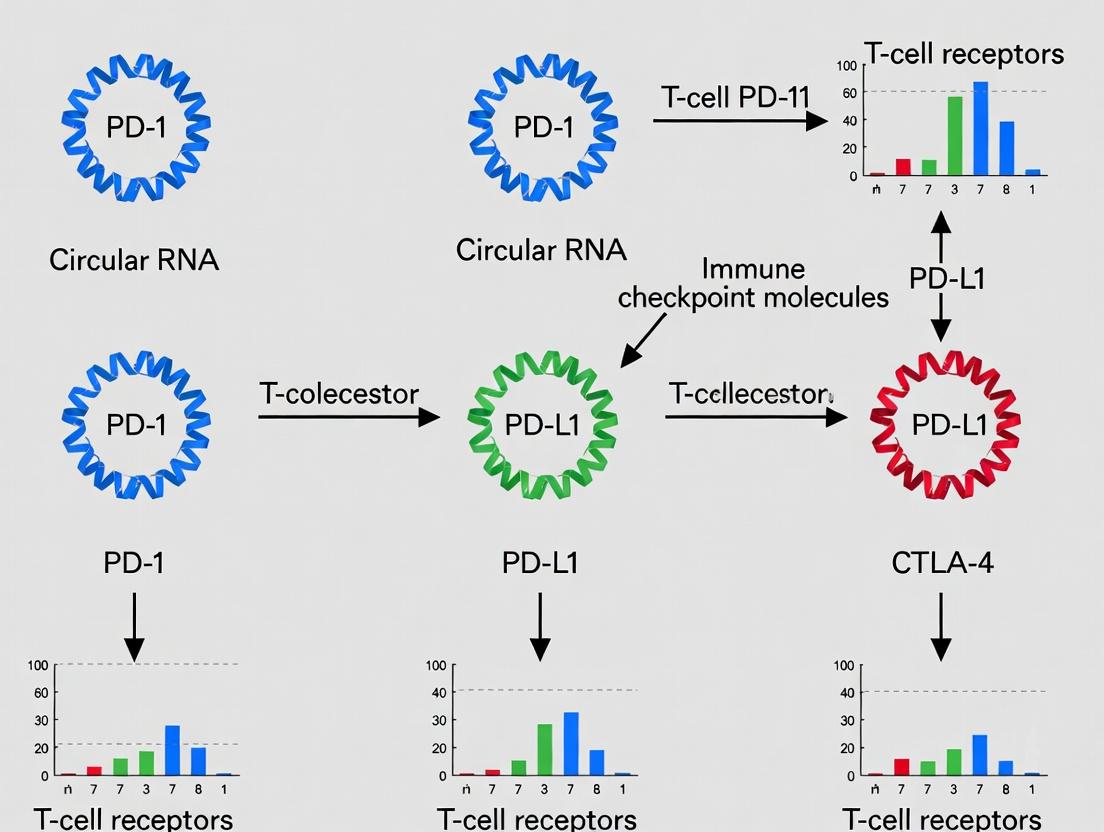
Visualization of circRNA Biogenesis, Function, and Immune Checkpoint Regulation
Diagram Title: circRNA Biogenesis Pathways & Immune Checkpoint Regulation
The Scientist's Toolkit: Key Research Reagent Solutions
Table 3: Essential Reagents and Tools for circRNA Research
| Reagent/Tool | Function/Application | Example/Notes |
|---|---|---|
| RNase R | Enzymatically degrades linear RNAs with free 3' ends, enriching for circRNAs in downstream assays. | Epicentre RNase R; critical for validation and sequencing library prep. |
| Divergent Primer Pairs | PCR primers designed to amplify the unique back-splice junction of a circRNA. | Must be validated with RNase R treatment and Sanger sequencing. |
| Back-splice Junction Probes | For Northern blot or in situ hybridization (FISH) specific detection of circRNAs. | Design probe spanning the back-splice junction; use LNA/Tyramide for FISH sensitivity. |
| circRNA-specific siRNA/shRNA | Knockdown of individual circRNAs without affecting linear host mRNA. | Target sequences exclusively at the back-splice junction. |
| CRISPR/Cas13 | Targeted degradation of specific circRNA sequences in cells. | dCas13 can be used for knockdown; requires careful gRNA design. |
| Polysome Profiling Kits | To investigate if a circRNA is associated with ribosomes and potentially translated. | Sucrose gradient centrifugation kits followed by circRNA detection in fractions. |
| circRNA Pull-down Kits | Isolate circRNA and its interacting molecules (miRNAs, RBPs) for identification. | Use biotinylated junction-specific probes and streptavidin beads. |
| circRNA Overexpression Vectors | Plasmid systems designed to express a specific circRNA from a minimal vector. | Often use engineered intronic complementary sequences or group I intron exons. |
| High-Depth RNA-seq & Bioinformatics Pipelines | For de novo discovery and quantification of circRNAs from total RNA-seq. | Tools: CIRI2, CIRCexplorer2, DCC, etc.; require >80M reads per sample. |
| Actinomycin D | Transcription inhibitor used in chase experiments to measure RNA half-life. | Standard concentration: 2 µg/mL; treat cells and harvest RNA over time course. |
Immune checkpoint molecules are critical regulators of immune homeostasis and T-cell activation. Their dysregulation is a hallmark of cancer and autoimmune diseases. This primer provides a technical overview of PD-1, PD-L1, CTLA-4, and emerging checkpoints, framed within the burgeoning research field exploring the regulatory roles of circular RNAs (circRNAs) on their expression and function. Understanding these interactions is pivotal for developing next-generation immunotherapies.
Core Immune Checkpoint Molecules: Mechanism & Structure
Programmed Cell Death Protein 1 (PD-1; CD279)
Function: An inhibitory receptor expressed on activated T cells, B cells, and myeloid cells. Engagement with its ligands leads to T-cell exhaustion, inhibition of proliferation, cytokine production, and cytotoxicity. Gene Location: Pdcd1 (human chromosome 2q37.3)
Programmed Death-Ligand 1 (PD-L1; CD274, B7-H1)
Function: The primary ligand for PD-1, expressed on antigen-presenting cells (APCs), tumor cells, and non-hematopoietic cells during inflammation. PD-L1 binding to PD-1 transmits an inhibitory signal. Gene Location: Cd274 (human chromosome 9p24.1)
Cytotoxic T-Lymphocyte-Associated Protein 4 (CTLA-4; CD152)
Function: A CD28 homolog that binds B7-1 (CD80) and B7-2 (CD86) with higher affinity, acting as a competitive inhibitor for CD28-mediated co-stimulation. Primarily regulates early T-cell activation in lymphoid organs. Gene Location: Ctla4 (human chromosome 2q33.2)
Table 1: Core Immune Checkpoint Molecules: Key Properties and Clinical Blockers
| Molecule | Type | Key Ligand(s) | Primary Cellular Expression | FDA-Approved Therapeutic Blockers (Examples) | Primary Signaling Effect |
|---|---|---|---|---|---|
| PD-1 | Transmembrane Receptor | PD-L1, PD-L2 | Activated T cells, B cells, Myeloid cells | Nivolumab, Pembrolizumab, Cemiplimab | Inhibits TCR/CD28 signaling via SHP-1/2 phosphatase recruitment |
| PD-L1 | Transmembrane Ligand | PD-1 | APCs, Tumor cells, Non-hematopoietic cells | Atezolizumab, Avelumab, Durvalumab | Engages PD-1 to transduce inhibitory signal into T cell |
| CTLA-4 | Transmembrane Receptor | CD80 (B7-1), CD86 (B7-2) | Activated T cells (esp. Tregs) | Ipilimumab, Tremelimumab* | Outcompetes CD28 for B7 ligands, dampens early activation |
*Tremelimumab is approved in specific combination regimens.
Beyond PD-1/PD-L1 & CTLA-4: Emerging Checkpoints
Table 2: Emerging Immune Checkpoint Targets Under Investigation
| Target | Expression | Ligand/Counterpart | Proposed Function & Therapeutic Rationale |
|---|---|---|---|
| LAG-3 (CD223) | Activated T cells, Tregs, NK cells | MHC Class II, FGL1, LSECtin | Promotes T-cell exhaustion; synergistic with PD-1. |
| TIM-3 | Th1 cells, Tc1 cells, Tregs, Myeloid cells | Galectin-9, CEACAM1, HMGB1, PtdSer | Diverse inhibitory roles; associated with anti-PD-1 resistance. |
| TIGIT | T cells, NK cells (esp. activated) | CD155 (PVR), CD112 (PVRL2) | Competes with stimulatory receptor CD226 for ligands. |
| VISTA | Myeloid cells, T cells | VSIG-3, PSGL-1? | Inhibits T-cell activation; potential target in hematologic malignancies. |
CircRNAs as Regulators of Immune Checkpoint Expression
Circular RNAs are covalently closed, single-stranded RNA molecules with emerging roles as miRNA sponges, protein decoys, and translational regulators. Their dysregulation in the tumor microenvironment can modulate immune checkpoint expression, presenting a novel layer of therapeutic intervention.
Proposed Mechanisms:
- miRNA Sponging: A circRNA containing complementary sequences can sequester miRNAs that normally target immune checkpoint mRNAs for degradation, leading to checkpoint upregulation. e.g., CircRNA_0000284 sponges miR-197-3p to upregulate PD-L1 in cervical cancer.
- Protein Binding/Sequestration: CircRNAs can bind to and modulate the activity of RNA-binding proteins (RBPs) involved in checkpoint mRNA splicing, stability, or translation.
- Direct Translation: Some circRNAs contain Internal Ribosome Entry Sites (IRES) and can be translated into checkpoint-related peptides.
Table 3: Examples of CircRNAs Regulating Immune Checkpoints in Cancer
| CircRNA | Cancer Type | Validated Target | Proposed Mechanism | Effect on Checkpoint |
|---|---|---|---|---|
| hsacirc0020394 | Colorectal | miR-138-5p | Sponges miR-138-5p, which targets PD-L1 mRNA | ↑ PD-L1 expression |
| circ-CPA4 | Non-small cell lung | let-7 miRNA | Acts as a let-7 sponge, derepressing PD-L1 translation | ↑ PD-L1 expression |
| circARSP91 | Hepatocellular | – | Binds to and stabilizes PD-L1 protein | ↑ PD-L1 protein levels |
| circFGFR1 | Non-small cell lung | miR-381-3p | Sponges miR-381-3p, which targets CTLA-4 mRNA | ↑ CTLA-4 expression |
Experimental Protocols for Investigating CircRNA-Checkpoint Axis
Protocol 1: Validating circRNA-miRNA-Checkpoint Axis
Objective: To confirm a specific circRNA sponges a miRNA to regulate checkpoint gene expression. Key Steps:
- Bioinformatic Prediction: Use tools like CircInteractome, StarBase, or custom scripts to predict miRNA response elements (MREs) on the circRNA and miRNA target sites on the 3'UTR of the checkpoint gene (e.g., PD-L1).
- Dual-Luciferase Reporter Assay:
- Clone the wild-type and mutant (MRE seed region mutated) sequence of the circRNA or the 3'UTR of PD-L1 downstream of a luciferase reporter gene (e.g., psiCHECK-2 vector).
- Co-transfect HEK293T cells with the reporter plasmid and either the miRNA mimic or inhibitor.
- Measure Firefly (control) and Renilla (experimental) luciferase activity 48h post-transfection. A decrease in Renilla signal with the mimic confirms direct targeting.
- Functional Rescue Experiment:
- In relevant cell lines (e.g., tumor cells), perform gain/loss-of-function: overexpress or knock down the circRNA.
- Quantify changes in miRNA level (qRT-PCR) and checkpoint protein level (Western blot/Flow cytometry).
- Rescue by co-transfecting the miRNA mimic (with circRNA OE) or inhibitor (with circRNA KD) to confirm the axis.
Protocol 2: Assessing circRNA-Protein Interaction for Checkpoint Regulation
Objective: To determine if a circRNA regulates checkpoint expression by interacting with an RNA-binding protein (RBP). Key Steps:
- RNA Pull-Down with Mass Spectrometry:
- Synthesize biotin-labeled sense (circRNA) and antisense control RNAs in vitro.
- Incubate lysates from target cells with the labeled RNAs, followed by streptavidin bead capture.
- Wash stringently, elute bound proteins, and analyze by SDS-PAGE and silver staining or mass spectrometry for identification of interacting RBPs.
- RIP-qPCR (RNA Immunoprecipitation):
- Crosslink cells, lyse, and immunoprecipitate the candidate RBP using a specific antibody.
- Reverse crosslinks, extract RNA, and perform qRT-PCR with divergent primers specific to the circRNA to confirm the endogenous interaction.
- RBP Functional Knockdown:
- Knock down the identified RBP (siRNA/shRNA).
- Assess changes in checkpoint mRNA stability (Actinomycin D assay) or protein expression.
Protocol 3:In VivoFunctional Study Using circRNA-Modified Tumor Models
Objective: To evaluate the impact of tumor cell-intrinsic circRNA on checkpoint levels and anti-tumor immunity in vivo. Key Steps:
- Generate Stable Cell Lines: Create tumor cell lines with stable knockdown (shRNA targeting the back-splice junction) or overexpression (circRNA expression vector) of the circRNA.
- Syngeneic Tumor Model: Implant these cells into immunocompetent mice (e.g., C57BL/6 for murine cells).
- Monitoring & Analysis:
- Monitor tumor growth and survival.
- Harvest tumors at endpoint, process into single-cell suspensions.
- Analyze by multicolor flow cytometry: quantify tumor-infiltrating lymphocytes (CD8+, CD4+, Tregs) and checkpoint expression (PD-1, LAG-3, TIM-3 on T cells; PD-L1 on tumor/ myeloid cells).
- Correlate findings with circRNA expression level in situ (RNA-FISH combined with immunofluorescence).
The Scientist's Toolkit: Key Research Reagent Solutions
Table 4: Essential Reagents for circRNA-Immune Checkpoint Research
| Reagent Category | Specific Example/Product | Function & Application |
|---|---|---|
| circRNA Detection | Divergent Primer Sets, RNase R | Specific amplification and enrichment of circRNA vs. linear mRNA. |
| Functional Modulation | siRNA/shRNA (Back-splice junction target), circRNA overexpression vectors (with flanking intronic sequences), CRISPR-Cas13 systems | Loss-of-function and gain-of-function studies for circRNAs. |
| miRNA Interaction | miRNA Mimics & Inhibitors, Dual-Luciferase Reporter Vectors (psiCHECK-2) | Validate miRNA sponging activity and target regulation. |
| Protein Interaction | Biotin RNA Labeling Kit, Streptavidin Magnetic Beads, RBP-specific Antibodies for RIP | Identify and confirm circRNA-protein interactions. |
| Immune Profiling | Fluorescently-conjugated Anti-Mouse/Human CD8, CD4, PD-1, PD-L1, CTLA-4, LAG-3, TIM-3 Antibodies, Fixable Viability Dyes | Multiparameter flow cytometry analysis of immune cell subsets and checkpoint expression. |
| In Vivo Models | Syngeneic Mouse Tumor Cell Lines (e.g., MC38, B16-F10), Immune-deficient Mice (e.g., NSG for humanized models) | Preclinical evaluation of the circRNA-checkpoint axis in an intact immune system. |
| Spatial Analysis | RNAscope Probes for circRNA, Multiplex Immunofluorescence Kits (e.g., Opal) | Co-localization analysis of circRNA and checkpoint protein in tumor tissue sections. |
Visualizations
Title: CircRNA Sponging Mechanism Upregulating Immune Checkpoint
Title: Experimental Workflow to Validate circRNA-miRNA-Checkpoint Axis
Title: Core PD-1/PD-L1 Inhibitory Signaling Pathway
This whitepaper, framed within the broader thesis on Circular RNAs regulation of immune checkpoint molecules, details the molecular mechanisms by which circular RNAs function as competitive endogenous RNAs to sequester microRNAs, thereby modulating the expression of immune checkpoint proteins. This regulatory axis presents a novel layer of post-transcriptional control in immune homeostasis and cancer immunotherapy.
Circular RNAs (circRNAs) are covalently closed, single-stranded RNA molecules formed by back-splicing of pre-mRNA. A primary validated function is their role as miRNA "sponges," competitively binding to microRNAs (miRNAs) to prevent them from repressing their target mRNAs. Key immune checkpoint molecules, such as PD-1, PD-L1, CTLA-4, and LAG-3, are frequently post-transcriptionally regulated by miRNAs. Therefore, circRNAs that sponge these miRNAs can directly or indirectly influence checkpoint expression, affecting T-cell exhaustion and tumor immune evasion.
Core Mechanism: The Sponging Process
Molecular Recognition and Binding
CircRNAs contain specific miRNA Response Elements (MREs) that are complementary to the seed region (nucleotides 2-8) of target miRNAs. Through Watson-Crick base pairing, the circRNA sequesters the miRNA, preventing its loading into the RNA-induced silencing complex (RISC). This de-represses the miRNA's native target mRNAs, which often include transcripts encoding checkpoint proteins or their upstream regulators.
Diagram: Core Sponging Mechanism
Key circRNA Examples in Checkpoint Regulation
The following table summarizes validated circRNAs, their sponged miRNAs, and the subsequent impact on checkpoint expression.
Table 1: Key circRNA-miRNA-Checkpoint Regulatory Axes
| circRNA ID | Sponged miRNA(s) | Derepressed Target(s) | Effect on Checkpoint Expression | Disease Context | Key Reference |
|---|---|---|---|---|---|
| circ_0020394 | miR-138-5p | PD-L1 | Upregulates PD-L1 | Colorectal Cancer | Zhang et al., 2021 |
| hsacirc0022318 | miR-543 | LASP1 → NF-κB | Upregulates PD-L1 | Glioblastoma | Chen et al., 2022 |
| circCCDC66 | miR-33a-5p, miR-93-5p | TBL1XR1 → β-catenin | Upregulates PD-L1 | Gastric Cancer | Wang et al., 2023 |
| circCPA4 | miR-760 | STAT3 | Upregulates PD-L1 | Lung Adenocarcinoma | Li et al., 2022 |
| circFGFR1 | miR-381-3p | CXCR4 | Upregulates PD-1 on T cells | Breast Cancer | Liu et al., 2023 |
| cIRS-7 (CDR1as) | miR-7 | EGFR/RAF1 → (Indirect) | Can influence PD-L1 | Multiple Cancers | Hansen et al., 2013 |
Experimental Protocols for Validation
A rigorous, multi-step approach is required to validate the circRNA→miRNA→Checkpoint axis.
Protocol 1: Identifying and Validating circRNA-miRNA Interaction
Aim: To confirm direct binding between a circRNA and a candidate miRNA. Steps:
- Bioinformatic Prediction: Use databases like CircInteractome, circBank, or StarBase to predict MREs within the circRNA for specific miRNAs.
- Dual-Luciferase Reporter Assay:
- Clone the wild-type (WT) circRNA sequence containing the predicted MRE into the 3'UTR of a firefly luciferase reporter plasmid (e.g., pmirGLO).
- Generate a mutant (MUT) reporter plasmid with point mutations in the miRNA seed binding site.
- Co-transfect HEK293T cells with: (i) the reporter plasmid (WT or MUT), and (ii) the miRNA mimic or a negative control (NC) mimic.
- Measure luminescence 48h post-transfection. Normalize Firefly to Renilla luciferase activity. Significant reduction in luminescence only in the WT + miRNA mimic group confirms specific interaction.
Protocol 2: Validating Functional Sponging and Checkpoint Outcome
Aim: To demonstrate that circRNA sponging alters miRNA activity and checkpoint protein levels. Steps:
- Gain/Loss-of-Function:
- Overexpression: Transfect cells with a circRNA overexpression vector (e.g., plasmid with backsplice junction flanked by intronic complementary sequences).
- Knockdown: Transfert cells with specific siRNAs targeting the circRNA's backsplice junction or use CRISPR/Cas13 systems.
- miRNA Activity Assay:
- Use a synthetic reporter plasmid containing tandem repeats of the miRNA target sequence upstream of luciferase.
- Co-transfect with circRNA-modulating vectors. Increased luminescence upon circRNA overexpression indicates functional miRNA sponging (de-repression).
- Downstream Checkpoint Analysis:
- qRT-PCR: Measure mRNA levels of the checkpoint gene (e.g., PD-L1). Use divergent primers for circRNA and convergent primers for linear mRNA.
- Western Blot / Flow Cytometry: Quantify checkpoint protein expression (e.g., membrane PD-L1) following circRNA modulation.
- Rescue Experiments: Co-transfect the miRNA mimic with the circRNA overexpression vector. Restoration of checkpoint repression confirms the axis.
Diagram: Core Experimental Validation Workflow
Key Signaling Pathways Involved
CircRNA sponging often influences checkpoint expression via canonical oncogenic or inflammatory pathways.
Diagram: Integrated Signaling Pathway via Sponging
The Scientist's Toolkit: Key Research Reagent Solutions
Table 2: Essential Materials and Reagents for circRNA-miRNA Sponging Research
| Reagent Category | Specific Item/Kit | Function & Application |
|---|---|---|
| circRNA Detection | Divergent Primer Sets | Specifically amplify the back-splice junction of circRNA via RT-qPCR. |
| RNase R Treatment | Digest linear RNA to enrich for circular RNA in northern blot or RNA-seq. | |
| circRNA Manipulation | Backsplice Vector (e.g., pLCDH-ciR) | Plasmid for stable circRNA overexpression via flanking intronic sequences. |
| CRISPR/Cas13 System (e.g., RfxCas13d) | Targeted knockdown of circRNA without affecting linear host mRNA. | |
| siRNAs Targeting Backsplice Junction | Sequence-specific knockdown of circRNA. | |
| miRNA Manipulation | miRNA Mimics & Inhibitors (antagomiRs) | Functionally increase or decrease specific mature miRNA activity in cells. |
| Interaction Validation | Dual-Luciferase Reporter Kit (e.g., Promega) | Quantify miRNA binding to cloned MRE sequences. |
| Biotinylated miRNA Pull-down Kit | Pull down circRNAs bound by a specific biotin-labeled miRNA. | |
| AGO2 RIP/CLIP Kit | Confirm circRNA association with the RISC complex via AGO2 immunoprecipitation. | |
| Downstream Analysis | Antibodies: Anti-PD-L1, Anti-PD-1, Anti-CTLA-4 | Detect checkpoint protein expression via Western Blot or Flow Cytometry. |
| Flow Cytometry Panels (Multicolor) | Simultaneously analyze immune cell populations and checkpoint surface expression. | |
| In Vivo Models | CircRNA-overexpressing Xenograft Mouse Models | Assess the impact of the circRNA axis on tumor growth and immune infiltration in vivo. |
| PDX (Patient-Derived Xenograft) Models | Study circRNA function in a more clinically relevant tumor microenvironment. |
The mechanistic understanding of circRNAs as natural miRNA sponges regulating immune checkpoint expression reveals a complex post-transcriptional network. Targeting specific oncogenic circRNAs (using antisense oligonucleotides or small molecules) or developing circRNA-based replacement therapies (for tumor-suppressive circRNAs) represents a promising frontier for next-generation combinatorial immunotherapies, with the potential to overcome resistance to current checkpoint blockade antibodies.
Circular RNAs (circRNAs) are a class of endogenous, covalently closed non-coding RNA molecules with critical regulatory functions. This whitepaper details the mechanisms by which circRNAs interact with proteins to modulate translation, a core pillar within the broader thesis on "Circular RNAs regulation of immune checkpoint molecules." Specifically, dysregulated circRNA-protein interactions can directly influence the expression of pivotal immune checkpoint proteins (e.g., PD-1, PD-L1, CTLA-4) in the tumor microenvironment, presenting novel therapeutic and diagnostic avenues in immuno-oncology.
Core Mechanisms of circRNA-Protein Interactions in Translational Control
circRNAs exert translational regulation primarily through sequestration of proteins, including RNA-binding proteins (RBPs) and translation initiation factors.
- RBP Sequestration ("Sponging"): circRNAs can act as molecular sinks for specific RBPs, preventing them from binding their target mRNAs and affecting the translation of those mRNAs.
- Direct Modulation of Translation Machinery: Certain circRNAs can directly bind to and regulate the activity of eukaryotic initiation factors (e.g., eIF4G, eIF3) or ribosomal subunits, thereby controlling the initiation phase of translation globally or for specific transcripts.
- Formation of Ternary Complexes: circRNAs can serve as scaffolds, bringing together proteins (e.g., enzymes and their substrates) to form functional complexes that influence the translational landscape.
Quantitative Data on circRNA-Protein Interactions in Immune Regulation
Table 1: Exemplary circRNAs Regulating Immune Checkpoints via Protein Interaction
| circRNA ID | Binding Partner(s) | Regulatory Target | Effect on Translation/Expression | Experimental System | Reference (Type) |
|---|---|---|---|---|---|
| circ-CPA4 | eIF4G, eIF3a | PD-L1 | Enhances PD-L1 translation | Non-small cell lung cancer cells | PMID: 35087400 |
| circ-0020394 | IGF2BP3 | PD-L1 | Stabilizes PD-L1 mRNA | Colorectal cancer cells | PMID: 34654796 |
| circ-ARSP91 | U2AF65 | ULBP1 (NK cell ligand) | Inhibits ULBP1 translation | Hepatocellular carcinoma | PMID: 30718861 |
| circ-FOXO3 | p21, CDK2 | Cell cycle proteins | Induces cell cycle arrest | Various cancer models | PMID: 27889213 |
Table 2: Common Experimental Techniques for Studying circRNA-Protein Interactions
| Technique | Key Measurable Output | Typical Throughput | Key Quantitative Metrics |
|---|---|---|---|
| RNA Immunoprecipitation (RIP) | Enrichment of circRNA bound to a specific protein | Medium | Fold-enrichment (qPCR); RPKM/Counts (Seq) |
| Crosslinking IP (CLIP) | Precise protein-binding sites on circRNA | Low | Number of binding peaks; mutation validation rate |
| Pull-down / MS | Identification of proteins bound to a specific circRNA | Low-High | Spectral counts; Peptide abundance |
| Fluorescent In Situ Hybridization & IP (FISH-IP) | Spatial co-localization of circRNA and protein | Low | Co-localization coefficient (e.g., Pearson's) |
Detailed Experimental Protocols
4.1. Protocol: Crosslinking RNA Immunoprecipitation (CLIP) for circRNA-Protein Complexes
- Objective: To identify direct binding sites of an RBP on a specific circRNA.
- Materials: UV crosslinker (254 nm), specific antibody for target RBP, Proteinase K, RNase inhibitors, magnetic beads, TRIzol.
- Steps:
- In Vivo Crosslinking: Expose cells to 254 nm UV light (400 mJ/cm²) to covalently link RBPs to bound RNA.
- Cell Lysis: Lyse cells in stringent RIPA buffer with RNase inhibitors.
- Partial RNase Digestion: Treat lysate with limited RNase to leave ~20-70 nucleotide fragments protected by the RBP.
- Immunoprecipitation: Incubate lysate with antibody-bound magnetic beads. Wash stringently.
- RNA Recovery: Treat beads with Proteinase K to digest proteins and release RNA. Extract RNA with TRIzol.
- Library Prep & Sequencing: Use circRNA-aware library preparation protocols (e.g., RNase R treatment, divergent primer design) followed by high-throughput sequencing.
- Validation: Validate binding sites via antisense oligonucleotide pull-down or luciferase reporter assays with mutated binding sites.
4.2. Protocol: circRNA Pull-down followed by Mass Spectrometry
- Objective: To identify proteins that interact directly with a specific, endogenous circRNA.
- Materials: Biotin-labeled, sequence-specific DNA probes complementary to the circRNA junction, streptavidin magnetic beads, mass spectrometer.
- Steps:
- Probe Design: Design and synthesize 3-5 overlapping biotinylated DNA probes targeting the unique back-splice junction of the circRNA.
- Cell Lysate Preparation: Prepare cell lysate under native conditions.
- Hybridization & Capture: Incubate lysate with probes overnight at 37°C. Add streptavidin beads to capture probe-circRNA-protein complexes.
- Washing: Wash beads extensively with increasing stringency buffers.
- Elution & Analysis: Elute bound proteins, digest with trypsin, and analyze peptides by LC-MS/MS. Compare to control (scrambled probe) samples.
Visualizations of Pathways and Workflows
Title: circRNA Scaffolds Initiation Factors to Enhance PD-L1 Translation
Title: CLIP-seq Workflow for circRNA-Protein Binding
The Scientist's Toolkit: Research Reagent Solutions
Table 3: Essential Reagents for circRNA-Protein Interaction Studies
| Reagent Category | Specific Item / Kit | Primary Function in Research |
|---|---|---|
| RNase Enzymes | RNase R | Degrades linear RNA to enrich for circRNAs in pull-down/sequencing samples. |
| Crosslinkers | Formaldehyde; AMT (4'-Aminomethyltrioxalen) | Formaldehyde for protein-protein & loose RNA-protein; AMT for stringent, RNA-protein specific crosslinking. |
| Beads & Kits | Streptavidin C1 Magnetic Beads; Magna RIP Kit | Beads for biotin-probe pull-downs; Kit for standardized RNA immunoprecipitation. |
| Probe Design | Biotinylated Junction-spanning Oligos | Specifically capture endogenous circRNAs via their unique back-splice junction. |
| Validation | Dual-Luciferase Reporter Assay (e.g., psiCHECK2) | Validate the functional impact of circRNA-protein interaction on translational regulation of a target (e.g., PD-L1 3'UTR). |
| circRNA Detection | Divergent Primer Sets; RNase H-based Assays | PCR primers to specifically amplify the back-splice junction; Enzymatic methods to confirm circularity. |
Circular RNAs (circRNAs) are stable, covalently closed non-coding RNA molecules formed by back-splicing. Within the tumor microenvironment (TME), circRNAs are differentially expressed and play crucial roles in modulating immune cell function, angiogenesis, fibroblast activation, and immune checkpoint molecule regulation. This whitepaper, framed within a thesis on circRNA regulation of immune checkpoint molecules, details the profiling landscape, key functional players, and methodologies for studying TME-associated circRNAs.
Profiling Studies: Technologies and Data
The comprehensive profiling of circRNAs in the TME relies on high-throughput sequencing and spatial transcriptomics. Key studies highlight distinct circRNA signatures between tumor parenchyma and stroma, and across immune cell subsets.
| Study Focus (Cancer Type) | Key Technology | Core Finding | Reference (Year) |
|---|---|---|---|
| Pan-cancer TME analysis | rRNA-depleted RNA-seq, CircExplorer2 | 1,048 circRNAs consistently upregulated in tumor stromal regions across 5 cancer types. | Liu et al. (2023) |
| Tumor-associated Macrophages (Glioblastoma) | Single-cell RNA-seq, CIRI2 | circHIPK3 enriched in M2 macrophages; knockdown reduced PD-L1 expression. | Zhang et al. (2024) |
| Cancer-associated Fibroblasts (Breast Cancer) | RNase R-treated RNA-seq | circFNDC3B secreted via exosomes, promotes CD8+ T cell exhaustion via IGF2BP2/PD-1 mRNA stabilization. | Wang et al. (2023) |
| T cell Dysfunction (Melanoma) | Nanostring nCounter, ARIC-Seq | circCPA4 positively correlates with PD-1 expression in tumor-infiltrating lymphocytes. | Chen & Gao (2024) |
Table 2: Quantified Dysregulated circRNAs in Major TME Components
| TME Component | Upregulated circRNAs (Avg. Fold Change) | Downregulated circRNAs (Avg. Fold Change) | Associated Immune Checkpoint |
|---|---|---|---|
| Myeloid-derived Suppressor Cells | circHPS5 (8.2x), circPICALM (5.7x) | circDYM (0.3x) | PD-L1, VISTA |
| Regulatory T Cells | circFoxp1 (12.5x) | circCd28 (0.2x) | CTLA-4 |
| Cancer-Associated Fibroblasts | circCOL5A1 (9.8x), circFNDC3B (15.3x) | circTIMP3 (0.1x) | Indirect via cytokine release |
| Endothelial Cells | circVEGFA (6.4x) | circSHPRH (0.4x) | B7-H3 |
Key Players: Functional Mechanisms and Pathways
Selected circRNAs have been validated as critical regulators of immune evasion.
circHPS5 (Host: HPS5): Sponges miR-122-5p in MDSCs, upregulating VISTA expression. Promotes T cell suppression. circFNDC3B (Host: FNDC3B): Packaged in CAF-derived exosomes, binds IGF2BP2 in T cells, stabilizing PD-1 mRNA. circFoxp1 (Host: FOXP1): Enhances Foxp1 protein stability in Tregs, driving CTLA-4 transcription.
Experimental Protocols
Protocol: circRNA Enrichment and Sequencing from TME Subpopulations
Objective: Isolate and sequence circRNAs from specific TME cell types (e.g., tumor-infiltrating lymphocytes, CAFs). Steps:
- Tissue Dissociation & Cell Sorting: Dissociate fresh tumor tissue into single-cell suspension using collagenase IV/DNase I. Sort target cells via FACS using surface markers (e.g., CD45+CD3+ for T cells, α-SMA+ for CAFs).
- RNA Extraction & Enrichment: Extract total RNA using TRIzol reagent. Treat 2-5 µg RNA with 3 U/µg RNase R (Epicentre) at 37°C for 15 min to degrade linear RNA. Purify using RNA Clean & Concentrator kit.
- Library Prep & Sequencing: Use rRNA-depletion kits (Ribo-Zero Gold). Prepare library with random hexamers (not oligo-dT) and fragment RNA. Perform paired-end 150 bp sequencing on Illumina NovaSeq.
- Bioinformatic Analysis: Align reads to reference genome using STAR. Identify back-splice junctions with tools like CIRI2, CIRCexplorer2, or find_circ. Quantify circRNA expression.
Protocol: Validating circRNA-Protein Interaction (RIP-qPCR)
Objective: Confirm direct binding of circRNA (e.g., circFNDC3B) to a protein (e.g., IGF2BP2) in T cells. Steps:
- Cell Crosslinking & Lysis: Crosslink 1x10^7 target cells with 0.3% formaldehyde for 10 min at RT. Quench with glycine. Lyse cells in RIPA buffer with RNase inhibitor.
- Immunoprecipitation: Pre-clear lysate with protein A/G beads. Incubate with 5 µg anti-IGF2BP2 antibody or IgG control overnight at 4°C. Add beads for 2 hours.
- RNA Isolation & Analysis: Wash beads stringently. Reverse crosslinks at 70°C for 45 min. Isolate co-precipitated RNA. Perform qPCR with divergent primers spanning the back-splice junction of circFNDC3B.
Visualization: Pathways and Workflows
Title: circFNDC3B in CAF-induced T Cell Exhaustion
Title: Workflow for TME-Specific circRNA Profiling
The Scientist's Toolkit: Research Reagent Solutions
Table 3: Essential Reagents for circRNA-TME Research
| Reagent / Kit | Vendor Examples | Primary Function in circRNA-TME Research |
|---|---|---|
| RNase R | Epicentre, Lucigen | Enzymatically degrades linear RNA to enrich for circular RNAs prior to sequencing or qPCR. |
| Ribonuclease R Treatment Kit | NORGEN Biotek | Complete kit for efficient circRNA enrichment. |
| rRNA Depletion Kits (Ribo-Zero Gold) | Illumina, Takara | Removes ribosomal RNA during NGS library prep to increase coverage of non-coding RNAs. |
| Divergent & Convergent Primer Sets | Custom Oligo Synthesis (IDT, Sigma) | Divergent primers amplify back-splice junctions for circRNA-specific validation. Convergent primers target linear mRNA. |
| Locked Nucleic Acid (LNA) GapmeRs | Qiagen, Exiqon | Antisense oligonucleotides for specific knockdown of circRNAs in vitro and in vivo. |
| Exosome Isolation Kits (from cell culture) | Invitrogen (Total Exosome Isolation), SBI | Isolate extracellular vesicles to study exosomal circRNA transfer in TME communication. |
| RNA Immunoprecipitation (RIP) Kits | Millipore (Magna RIP) | Validate direct interactions between circRNAs and RNA-binding proteins (e.g., IGF2BP2). |
| Spatial Transcriptomics Kits | 10x Genomics Visium | Map circRNA expression within the anatomical context of the tumor microenvironment. |
| Single-Cell RNA-seq Library Kits | 10x Genomics Chromium | Profile circRNA expression at single-cell resolution in complex TME populations. |
This whitepaper presents foundational case studies within the broader thesis that circular RNAs (circRNAs) are critical regulators of immune checkpoint molecules. By acting as microRNA sponges, protein decoys, or translational templates, circRNAs modulate key pathways in tumor immunology and inflammation, presenting novel therapeutic targets and biomarkers in immuno-oncology.
Core Case Studies and Quantitative Data
Case Study: circPD-L1 (hsacirc0089134)
Background: Identified in various cancers, circPD-L1 is derived from back-splicing of exons 2-4 of the PDCDL1G (PD-L1) gene. It functions as a competitive endogenous RNA (ceRNA) to regulate PD-L1 expression and tumor immune evasion.
Key Findings and Data:
Table 1: Quantitative Data Summary for circPD-L1 Studies
| Parameter | Value/Condition | Biological Context | Reference (Example) |
|---|---|---|---|
| Expression Fold-Change | Up to 8.5-fold increase | Melanoma vs. adjacent normal tissue | Zhang et al., 2021 |
| miRNA Sponged | miR-34a, miR-17-5p, miR-15a-5p | Releases inhibition of PD-L1 mRNA | |
| Correlation with PD-L1 Protein | R² = 0.78 | Positive correlation in NSCLC patient samples | |
| Impact on CD8+ T-cell Apoptosis | Increase of ~35% | Co-culture with circPD-L1-overexpressing tumor cells | |
| Half-life | >24 hours | Compared to ~4 hours for linear PD-L1 mRNA |
Experimental Protocol for circPD-L1 Functional Validation (Sponge Assay):
- Plasmid Construction: Clone the full-length circPD-L1 sequence into a circRNA expression vector (e.g., pLCDH-ciR) featuring flanking complementary intronic sequences to promote back-splicing.
- Cell Transfection: Transfect the circPD-L1 expression vector or a control empty vector into target cancer cell lines (e.g., A549, MDA-MB-231) using Lipofectamine 3000.
- Luciferase Reporter Assay:
- Co-transfect cells with: a) A firefly luciferase reporter plasmid containing the 3'UTR of PD-L1 mRNA (or other target gene like STAT3) which has binding sites for the relevant miRNA (e.g., miR-34a). b) A Renilla luciferase plasmid for normalization. c) Synthetic miR-34a mimic or a negative control mimic.
- After 48 hours, harvest cells and measure Firefly and Renilla luciferase activity using a dual-luciferase assay kit.
- Analysis: Compare the ratio of Firefly/Renilla luminescence. circPD-L1 overexpression should rescue luminescence (increase the ratio) in the presence of the miR-34a mimic, confirming miRNA sequestration.
Case Study: circ-CPA4 (hsacirc0006215)
Background: Derived from exons 2-5 of the CPA4 gene, circ-CPA4 is upregulated in non-small cell lung cancer (NSCLC) and glioblastoma, promoting tumor progression and immunosuppression via the let-7 miRNA sponge axis.
Key Findings and Data:
Table 2: Quantitative Data Summary for circ-CPA4 Studies
| Parameter | Value/Condition | Biological Context | Reference (Example) |
|---|---|---|---|
| Expression Fold-Change | Up to 6.2-fold increase | NSCLC vs. paired non-tumor tissues | Wang et al., 2022 |
| Primary miRNA Sponged | let-7g-5p, let-7i-5p | Derepresses downstream target MYC | |
| Correlation with Patient Survival | Hazard Ratio (HR) = 2.41 | Overall survival in glioma cohort | |
| Effect on M2 Macrophage Polarization | Increase of ~40% (CD206+ cells) | Conditioned media from circ-CPA4-high cells | |
| Tumor Volume Change (in vivo) | ~65% reduction | Upon circ-CPA4 knockdown in xenograft model |
Experimental Protocol for circ-CPA4 In Vivo Functional Study:
- Stable Cell Line Generation: Lentivirally transduce human cancer cells (e.g., U87MG glioblastoma cells) with shRNA specifically targeting the circ-CPA4 back-splice junction (circ-specific shRNA) or a non-targeting control shRNA. Select with puromycin for 2 weeks.
- Xenograft Model Establishment: Subcutaneously inject 5x10^6 stable knockdown or control cells into the flanks of immunodeficient (e.g., NOD/SCID) or humanized mice (n=8 per group).
- Tumor Monitoring: Measure tumor dimensions with calipers twice weekly. Calculate volume using the formula: Volume = (Length x Width^2) / 2.
- Endpoint Analysis: At day 30-35 post-injection, sacrifice mice, harvest tumors, and weigh them. A portion is snap-frozen for RNA/protein extraction (validating knockdown and analyzing MYC, PD-L1 levels), and another portion is fixed in 4% PFA for immunohistochemistry (IHC) staining of CD8, CD163, or PD-L1 to assess immune infiltration.
Visualizing Key Pathways and Workflows
Title: circPD-L1 sponges miR-34a to upregulate PD-L1 and inhibit T-cells.
Title: circ-CPA4 sponges let-7 to activate MYC and PD-L1.
Title: Core experimental workflow for circRNA immune function studies.
The Scientist's Toolkit: Research Reagent Solutions
Table 3: Essential Reagents for circRNA Immune Research
| Reagent Category | Specific Product/Kit Example | Function in Research |
|---|---|---|
| circRNA Enrichment | RNase R (Epicentre) | Digests linear RNA to enrich for circular RNAs, essential for validation and sequencing. |
| Detection & Quantification | Divergent Primer Sets, Bulge-Loop RT-qPCR Primers (Arraystar) | Specific amplification of the back-splice junction; gold standard for circRNA quantification. |
| Localization | Fluorescent In Situ Hybridization (FISH) Probes (BersinBio) | Visualizes subcellular distribution (cytoplasmic vs. nuclear) of circRNAs. |
| Functional Modulation | circRNA Overexpression Vectors (pLCDH-ciR, Addgene), circRNA-specific siRNAs/shRNAs | Enables gain- and loss-of-function studies; specificity for the junction is critical. |
| Interaction Pull-Down | Biotinylated circRNA Probes (for MS2-tagged RIP or CHART), Argonaute 2 Antibody (for RIP) | Identifies interacting miRNA or protein partners (e.g., miRNA sponge validation). |
| In Vivo Modeling | Lentiviral circ-shRNA Particles, Humanized Mouse Models (NSG-HLA) | Allows stable knockdown in vitro and assessment of immune function in a humanized context. |
| Immune Phenotyping | Flow Cytometry Antibody Panels (CD8, CD4, PD-1, PD-L1, CD163), LEGENDplex Assays | Quantifies immune cell populations, checkpoint expression, and cytokine secretion. |
From Bench to Bedside: Methods to Study and Target circRNA-Checkpoint Axes
Within the context of investigating circular RNAs (circRNAs) and their regulation of immune checkpoint molecules (e.g., PD-1, PD-L1, CTLA-4), precise detection and quantification are paramount. This whitepaper outlines advanced technical methodologies for circRNA research, integrating next-generation sequencing, enzymatic validation, and targeted quantification.
Core Methodologies
Advanced RNA-seq for circRNA Discovery
CircRNA-enriched RNA-seq is the first critical step for unbiased discovery.
Protocol: rRNA-depleted & Ribo-Zero Library Prep for circRNA-Seq
- Total RNA Extraction: Isolate RNA using guanidinium thiocyanate-phenol-chloroform extraction (e.g., TRIzol). Assess integrity (RIN > 8).
- rRNA Depletion: Treat 1-5 µg of total RNA with ribo-depletion kits (e.g., Illumina Ribo-Zero Gold) to remove ribosomal RNA.
- RNase R Treatment (Optional Pre-sequencing): To profoundly enrich for circular RNAs, incubate 1-2 µg of rRNA-depleted RNA with 3 U/µg RNase R (Epicentre) for 15 min at 37°C. Purify using RNA clean-up beads.
- Library Construction: Use strand-specific library prep kits (e.g., TruSeq Stranded Total RNA). Fragment RNA, synthesize cDNA, perform end repair, A-tailing, adapter ligation, and PCR amplification (12-15 cycles).
- Sequencing: Perform paired-end sequencing (2x150 bp) on an Illumina platform to a minimum depth of 100 million reads per sample.
Data Analysis Workflow: Reads are aligned to the reference genome using splice-aware aligners (STAR, BWA). circRNAs are identified by detecting back-splice junction (BSJ) reads using tools such as CIRI2, find_circ, or CIRCexplorer.
Table 1: Comparative Analysis of circRNA Detection Tools
| Tool | Algorithm Core | Key Strength | Reported Sensitivity | Best For |
|---|---|---|---|---|
| CIRI2 | SAM alignment parsing, seed matching | High accuracy, low false positive rate | ~85-90% | Comprehensive de novo detection |
| find_circ | Anchor alignment of unmapped reads | Robust for novel circRNAs | ~80-85% | Discovery in non-standard datasets |
| CIRCexplorer2 | Fusion alignment with TopHat2/CLEAR | Excellent integration with transcriptome | ~82-88% | Annotated circRNA analysis |
RNase R Treatment for circRNA Validation
RNase R digests linear RNAs with free 3' ends but not resistant circRNAs, providing essential biochemical validation.
Protocol: Standard RNase R Digestion Assay
- Sample Division: Split 2 µg of total RNA (DNAse I-treated) into two equal aliquots.
- Digestion Reaction:
- Test: 1 µg RNA + 1x Reaction Buffer + 20 U RNase R.
- Control: 1 µg RNA + 1x Reaction Buffer + No Enzyme.
- Incubation: Incubate for 15-20 minutes at 37°C.
- Enzyme Inactivation: Add 1 µl of Proteinase K, incubate at 55°C for 10 min.
- Purification: Clean up using silica-membrane columns. Elute in 30 µl RNase-free water.
- Analysis: Analyze by qPCR (see below) or agarose gel electrophoresis. Successful digestion is confirmed by the depletion of linear control genes (GAPDH, ACTB) in the +RNase R sample, while target circRNAs remain stable.
qPCR Design for circRNA Quantification
qPCR is the gold standard for targeted, sensitive quantification of circRNAs from immune checkpoint regulation studies.
Protocol: Divergent Primer Design and qPCR
- Primer Design Principle: Design "divergent" or "outward-facing" primers that span the unique back-splice junction (BSJ). Each primer must have its 3' end oriented away from the other, ensuring amplification only occurs from circular, not linear, cDNA.
- Specificity Check: Use BLAST against the transcriptome to ensure primers are unique to the BSJ. Avoid genomic DNA amplification by designing primers across exons or treating samples with DNase I.
- cDNA Synthesis: Use random hexamers or gene-specific primers for reverse transcription. Avoid oligo(dT) primers, as circRNAs lack poly-A tails.
- qPCR Reaction:
- Use a high-fidelity SYBR Green master mix.
- Standard 20 µl reaction: 10 µl master mix, 0.5 µM each primer, 2 µl cDNA template.
- Cycling: 95°C for 3 min; 40 cycles of 95°C for 10s, 60°C for 20s, 72°C for 30s; followed by a melt curve analysis.
- Normalization: Normalize circRNA levels to stable housekeeping circRNAs (e.g., circHIPK3) or the mean of multiple linear reference genes after confirming their stability post-RNase R treatment.
Table 2: Key qPCR Validation Parameters for circRNA
| Parameter | Target/Requirement | Purpose |
|---|---|---|
| Amplification Efficiency | 90-110% | Ensures accurate relative quantification |
| Melt Curve | Single sharp peak | Confirms primer specificity and single amplicon |
| No-Template Control (NTC) | No amplification | Rules out contamination |
| No-RT Control | Cq > 35 or no signal | Confirms absence of genomic DNA amplification |
| RNase R Resistance | ΔCq (Control - Treated) < 2 for circRNA | Biochemical validation of circularity |
Visualizations
Title: circRNA-Enriched RNA-seq Experimental Workflow
Title: RNase R Resistance Principle for circRNA Validation
Title: Divergent Primer Design Targeting circRNA Back-Splice Junction
The Scientist's Toolkit: Key Research Reagent Solutions
Table 3: Essential Materials for circRNA Research on Immune Checkpoints
| Item | Function | Example/Note |
|---|---|---|
| Ribo-depletion/Ribo-Zero Kits | Removes abundant ribosomal RNA to enrich for non-coding RNAs, including circRNAs. | Illumina Ribo-Zero Gold, NEBNext rRNA Depletion. |
| RNase R | Exoribonuclease used to degrade linear RNAs, validating and enriching circRNAs. | Epicentre RNase R (now Lucigen). Critical for functional assays. |
| Strand-Specific RNA Library Prep Kit | Preserves strand-of-origin information, crucial for accurate circRNA annotation. | Illumina TruSeq Stranded, SMARTer Stranded. |
| High-Fidelity Reverse Transcriptase | For efficient cDNA synthesis from often highly structured circRNAs. | SuperScript IV, PrimeScript RT. |
| BSJ-Specific qPCR Primers | Divergent primers spanning the back-splice junction for specific circRNA quantification. | Must be designed in-house using tools like Primer-BLAST. |
| circRNA Knockdown/Overexpression Tools | For functional validation of immune checkpoint regulation. | siRNA targeting BSJ, circRNA expression vectors (lenti/viral). |
| Immune Checkpoint Antibodies | For downstream protein-level validation (e.g., PD-L1 flow cytometry/WB). | Anti-PD-L1, anti-CTLA-4 for cell surface or intracellular staining. |
Within the burgeoning field of circular RNA (circRNA) biology, a key research axis investigates the regulation of immune checkpoint molecules (e.g., PD-1, PD-L1, CTLA-4) by specific circRNAs. Dysregulation of these checkpoints is a hallmark of cancer and autoimmune diseases, making this interplay a promising therapeutic target. Functional validation—definitively proving that a candidate circRNA directly influences checkpoint expression and function—is a critical step. This guide details three core strategies for this validation: CRISPR/Cas13-mediated knockdown, siRNA-mediated silencing, and overexpression, providing technical protocols and data interpretation frameworks tailored for immune checkpoint research.
Core Principles and Applications
Each validation strategy offers distinct advantages and is suited for different experimental phases.
- CRISPR/Cas13 Knockdown: Utilizes the RNA-targeting Cas13 nuclease (e.g., Cas13d/RfxCas13d) to specifically cleave and degrade mature circRNA transcripts. This method is highly specific due to programmable crRNAs and can discriminate circRNAs from their linear host mRNAs by targeting the unique back-splice junction (BSJ). It is ideal for in vitro and in vivo long-term, persistent loss-of-function studies.
- siRNA-Mediated Silencing: Employs synthetic small interfering RNAs to degrade target transcripts via the RNA-induced silencing complex (RISC). While traditional siRNAs often target exonic regions shared by both circRNA and mRNA, carefully designed siRNA pools or ASO-like siRNAs can achieve reasonable circRNA specificity. Best suited for rapid, transient knockdown in cell culture to assess acute phenotypic effects.
- Overexpression: Involves the delivery of an expression vector containing the circRNA sequence flanked by engineered introns with reverse complementary repeats or a plasmid with a permuted intron-exon structure to force back-splicing. This gain-of-function approach is essential to confirm if the circRNA alone is sufficient to drive immune checkpoint modulation.
Quantitative Comparison of Key Parameters
Table 1: Strategic Comparison for circRNA Functional Validation
| Parameter | CRISPR/Cas13 Knockdown | siRNA Silencing | Overexpression |
|---|---|---|---|
| Target | Mature circRNA (BSJ) | Shared exon or BSJ (if designed) | Ectopic circRNA production |
| Specificity | Very High | Moderate to High (BSJ-targeting) | High (for the specific circRNA) |
| Duration | Long-term/Persistent | Transient (3-7 days) | Stable or Transient |
| Primary Use | Definitive loss-of-function | Initial, rapid screening | Gain-of-function validation |
| Key Challenge | Efficient delivery of Cas13 + crRNA | Discriminating circRNA from linear RNA | Generating high-purity, authentic circRNA |
| Typical Efficiency (Knockdown/Expression) | 70-95% knockdown | 60-85% knockdown | 10-100 fold increase |
| Common Readout in Checkpoint Studies | Flow cytometry (PD-L1 surface), qPCR (checkpoint genes), Co-culture T-cell activation assays. | Same as CRISPR/Cas13, but on shorter timeline. | Same as loss-of-function, plus supernatant cytokine profiling. |
Detailed Experimental Protocols
Protocol: CRISPR/Cas13d-mediated circRNA Knockdown
Objective: To achieve specific, durable knockdown of a circRNA regulating PD-L1 in a cancer cell line.
Materials:
- Cells: A375 melanoma cells (constitutively high PD-L1).
- Cas13d System: LentiVector expressing RfxCas13d (pRfxCas13d) and crRNA expression plasmid (pU6-sgRNA). crRNA designed to target the unique BSJ of circRNA-XXXX.
- Controls: Non-targeting crRNA control vector.
- Transfection: Lipofectamine 3000 or lentiviral transduction particles.
- Validation: Divergent primer set for circRNA qPCR, convergent primers for linear mRNA, RNase R treatment, anti-PD-L1 antibody for flow cytometry.
Method:
- crRNA Design: Design 2-3 crRNAs targeting sequences spanning the BSJ of the target circRNA using established design tools (e.g., CHOPCHOP). Include a 28-nt spacer sequence.
- Cloning: Clone annealed oligos into the BsaI site of the pU6-sgRNA vector.
- Delivery:
- Transient: Co-transfect pRfxCas13d and pU6-sgRNA (target or control) into A375 cells using Lipofectamine 3000.
- Stable: Package lentiviral vectors for RfxCas13d and the crRNA. Transduce A375 cells sequentially, selecting with appropriate antibiotics (e.g., puromycin, blasticidin).
- Knockdown Validation (72 hrs post-transfection/selection):
- Isolate total RNA, treat with RNase R (3 U/µg, 37°C, 15 min) to degrade linear RNA.
- Perform RT-qPCR using divergent primers for circRNA and convergent primers for the linear host gene and GAPDH. Calculate ∆∆Ct.
- Phenotypic Assessment:
- Harvest cells, stain with anti-human PD-L1 APC antibody.
- Analyze PD-L1 surface expression via flow cytometry (compare MFI to control).
- Extract protein for Western blot analysis of PD-L1 and related signaling proteins (e.g., p-STAT3).
Protocol: siRNA-mediated circRNA Silencing
Objective: Rapid assessment of the effect of circRNA loss on PD-L1 transcript levels.
Materials:
- siRNAs: A pool of 3-4 siRNAs specifically designed to target the BSJ region of the circRNA. A scrambled siRNA pool as negative control.
- Transfection Reagent: RNAiMAX.
- Cells: A375 cells.
- Validation: Same as Step 4 in 3.1.
Method:
- Reverse Transfection: Seed A375 cells in a 24-well plate (1x10^5 cells/well) in antibiotic-free medium. Dilute siRNA (final concentration 20 nM) and RNAiMAX in Opti-MEM separately, combine, incubate 5 min, then add complex to cells.
- Incubation: Assay 48-72 hours post-transfection.
- Validation: Perform RNA isolation, RNase R treatment, and qPCR with divergent/convergent primers as in 3.1.
Protocol: circRNA Overexpression
Objective: To confirm sufficiency of circRNA in modulating PD-L1 expression.
Materials:
- Expression Vector: pLC5-ciR or similar circRNA mini-vector, where the circRNA sequence is flanked by engineered introns containing complementary Alu repeats.
- Control Vector: Empty vector or vector expressing a scrambled circRNA.
- Transfection: PEI or Lipofectamine 3000.
- Validation: Divergent primer qPCR, Sanger sequencing of BSJ PCR product.
Method:
- Cloning: Insert the full circRNA sequence, including the exon(s) forming the circle, into the multiple cloning site of pLC5-ciR.
- Transfection: Transfect A375 cells with the circRNA overexpression vector or control.
- Overexpression Validation (48 hrs post-transfection):
- Perform RT-qPCR with divergent primers.
- Confirm circularity: Perform PCR on cDNA (divergent primers) and gDNA (should be negative). Gel-purify and sequence the PCR product to confirm the exact BSJ.
- Phenotypic Assessment: Perform PD-L1 flow cytometry and Western blot as in 3.1.
Visualization of Experimental Workflows and Pathways
Figure 1: Strategy Selection & circRNA Immune Checkpoint Pathway
The Scientist's Toolkit: Research Reagent Solutions
Table 2: Essential Reagents for circRNA Functional Validation in Immune Checkpoint Research
| Reagent Category | Specific Example/Kit | Function in Experiment | Key Consideration |
|---|---|---|---|
| circRNA-Specific Detection | Divergent Primer Sets, RNase R (Epicentre) | Specifically reverse transcribe and amplify the back-splice junction of circRNA; degrade linear RNA for specificity confirmation. | Primer design is critical. Validate with RNase R + no-RT controls. |
| Cas13 Knockdown | pRfxCas13d-NLS (Addgene #138150), pU6-sgRNA cloning vector | Provides the RNA-guided nuclease and the expression scaffold for the targeting crRNA. | Optimize crRNA design (BSJ-centric). Monitor potential collateral activity. |
| siRNA Silencing | BSJ-targeting siRNA pools (e.g., Dharmacon, Qiagen) | Induce rapid, transient degradation of the target circRNA transcript via RISC. | Use pooled siRNAs for robustness. Include a non-targeting control with similar chemistry. |
| circRNA Overexpression | pLC5-ciR vector (Addgene #111165), permuted intron-exon (PIE) vectors | Plasmid system engineered to promote back-splicing and high-yield production of circular RNA in cells. | Sanger sequence the expressed circRNA's BSJ to confirm fidelity. |
| Immune Checkpoint Assay | Anti-human CD274 (PD-L1) APC antibody (BioLegend), Human IFN-γ ELISA Kit | Quantify surface protein expression of the target checkpoint; measure functional T-cell response in co-culture. | Use isotype controls for flow. For co-culture, use antigen-specific or PHA-stimulated T-cells. |
| Delivery | Lipofectamine 3000, RNAiMAX, Polyethylenimine (PEI), Lentiviral Packaging System (psPAX2, pMD2.G) | Enable efficient nucleic acid delivery into target cells for transient or stable expression. | Match transfection reagent to nucleic acid type (DNA vs. RNA). Titrate for optimal efficiency/toxicity. |
| Analysis Software | FlowJo, GraphPad Prism, CRISPR Design Tools (CHOPCHOP) | Analyze flow cytometry data; perform statistical analysis and create figures; design specific crRNAs/sgRNAs. |
Spatial Transcriptomics and Single-Cell Analysis for circRNA Localization
This technical guide details methodologies for spatially resolving circular RNA (circRNA) expression, a critical component of a broader thesis investigating circRNA-mediated regulation of immune checkpoint molecules (e.g., PD-1, PD-L1, CTLA-4) in the tumor microenvironment (TME). Understanding the spatial context of circRNA expression is paramount for elucidating their cell-type-specific roles in modulating immune responses and identifying novel therapeutic targets.
Core Methodologies & Protocols
Integrated Single-Cell and Spatial Transcriptomics Workflow
A synergistic approach combining single-cell RNA sequencing (scRNA-seq) with spatial transcriptomics (ST) is required to map circRNAs to specific cell types and anatomical locations.
Protocol: Paired scRNA-seq and Visium Spatial Transcriptomics
- Tissue Preparation: A fresh-frozen tissue sample (e.g., tumor biopsy) is cryosectioned.
- One section (10 µm) is placed on a Visium Spatial Gene Expression slide.
- Consecutive sections are collected for scRNA-seq library preparation via tissue dissociation.
- Spatial Transcriptomics (Visium):
- Fixation & Staining: The slide-mounted section is fixed in methanol and H&E-stained for histological annotation.
- Permeabilization: Tissue is permeabilized to release mRNA, which binds to spatially barcoded oligonucleotides on the slide.
- cDNA Synthesis & Library Prep: On-slide reverse transcription creates cDNA with spatial barcodes. A second strand is synthesized, and the cDNA is amplified and prepared for sequencing.
- Single-Cell RNA Sequencing:
- Dissociated cells from consecutive sections are processed through a platform (e.g., 10x Genomics Chromium).
- Libraries are constructed using a v3.1 gene expression kit with a key modification: RNase R treatment (see 2.2) prior to library prep to enrich for circRNAs.
- Computational Integration:
- scRNA-seq data is clustered to define cell types/states.
- Using tools like Seurat or Tangram, cell-type signatures are deconvoluted onto the Visium spots, creating a spatial cell-type map.
- CircRNAs identified in scRNA-seq are projected onto this spatial map.
circRNA-Specific Enrichment and Detection Protocol
Standard RNA-seq protocols favor linear RNAs. Reliable circRNA detection requires specific enrichment and bioinformatic pipelines.
Protocol: RNase R Treatment and Divergent Primer Design
- RNA Isolation & Enrichment:
- Extract total RNA using TRIzol with DNase I treatment.
- Divide RNA: one aliquot for standard ST/scRNA-seq, one for circRNA enrichment.
- Enrichment: Treat 2 µg of total RNA with RNase R (20 U/µg, 37°C for 30 min). RNase R degrades linear RNA but not circRNAs.
- Purify RNA using RNase Clean-up kits.
- Library Preparation & Sequencing:
- Use both treated and untreated RNA for stranded, ribosomal RNA-depleted library prep.
- Sequence on a platform providing 150bp paired-end reads (minimum 50M reads/sample for ST; 20K reads/cell for sc).
- Bioinformatic Identification:
- Map reads to reference genome using STAR or BWA.
- Identify back-splice junction (BSJ) reads using specialized tools (CIRCexplorer2, CIRI2, DCC).
- Filter candidates: require ≥2 unique BSJ reads and presence in RNase R-treated samples.
Table 1: Comparative Performance of circRNA Detection Tools
| Tool | Algorithm Principle | Sensitivity (Recall) | Precision | Key Feature for Spatial/SC |
|---|---|---|---|---|
| CIRCexplorer3 | Alignment-based, parses STAR output | ~85% | ~90% | Integrates with RNA-seq pipelines |
| CIRI2 | CIRC RNA Identifier using BWA-MEM | ~88% | ~92% | Good for repetitive regions |
| DCC | De novo circRNA identification | ~80% | ~95% | Excellent for discovery, high precision |
| CirComPara2 | Multi-tool consensus pipeline | >90% | >95% | High confidence via ensemble approach |
Table 2: Typical circRNA Detection Rates in Integrated Studies
| Sample Type | scRNA-seq (RNase R-treated) | Visium Spatial (Standard) | Key Limitation |
|---|---|---|---|
| Human Tumor (NSCLC) | 5,000 - 15,000 circRNAs (total) | 500 - 2,000 circRNAs (confidently mapped) | Low per-spot RNA capture in ST obscures low-abundance circRNAs. |
| Mouse Spleen | 8,000 - 20,000 circRNAs (total) | 1,000 - 3,000 circRNAs (confidently mapped) | High immune cell diversity increases cell-type-specific circRNA discovery. |
| Per Cell/Spot | Median: 10-50 circRNAs/cell | Median: 1-5 high-confidence circRNAs/spot | Spatial resolution limited by spot size (55 µm). |
Visualizations
Title: Integrated scRNA-seq and Spatial circRNA Analysis Workflow
Title: circPD-L1 Sponges miR-15/16 to Upregulate PD-L1
The Scientist's Toolkit: Key Research Reagent Solutions
Table 3: Essential Reagents for circRNA Spatial Localization Studies
| Item | Function in Experiment | Example Product/Cat. # |
|---|---|---|
| Visium Spatial Gene Expression Slide | Glass slide with ~5,000 barcoded spots for spatially resolved cDNA capture. | 10x Genomics (2000233) |
| Chromium Next GEM Chip K | Generates nanoliter-scale partitions for single-cell encapsulation and barcoding. | 10x Genomics (1000127) |
| RNase R (Epicentre) | Exoribonuclease that digests linear RNA but not circRNAs, enabling enrichment. | Lucigen (RNR07250) |
| RiboCop rRNA Depletion Kit | Removes ribosomal RNA to increase sequencing depth of non-coding and mRNA transcripts. | Lexogen (086.24) |
| Smart-seq HT Kit | For high-sensitivity full-length cDNA amplification from low-input or single cells. | Takara Bio (634437) |
| Divergent Primer Oligos | Custom primers spanning the back-splice junction for RT-qPCR validation of circRNAs. | IDT (Custom) |
| BaseScope Reagents | In situ hybridization (ISH) assay for visualizing specific circRNAs at single-molecule resolution in tissue. | ACD Bio (322900) |
| anti-PD-L1 Antibody | For immunohistochemistry (IHC) to correlate PD-L1 protein expression with circRNA spatial maps. | Cell Signaling (13684S) |
In Vitro and In Vivo Models for Testing circRNA Function in Immuno-oncology
Circular RNAs (circRNAs) are a novel class of endogenous, covalently closed non-coding RNA molecules implicated in the regulation of numerous biological processes, including cancer immunity. Within the broader thesis of Circular RNAs regulation of immune checkpoint molecules research, investigating their precise functions requires robust and reproducible experimental models. This guide details the current state of in vitro and in vivo models specifically tailored for dissecting circRNA roles in immuno-oncology, providing technical protocols and frameworks for their application.
CoreIn VitroModels and Methodologies
In vitro systems offer controlled environments for mechanistic studies of circRNA interactions with immune and tumor cells.
Co-culture Systems for Tumor-Immune Cell Interactions
These systems model the tumor microenvironment (TME) to study circRNA-mediated regulation of immune checkpoint expression (e.g., PD-1, PD-L1, CTLA-4).
Protocol: PBMC-Tumor Cell Co-culture for circRNA/PD-L1 Analysis
- Isolation of PBMCs: Isolate human Peripheral Blood Mononuclear Cells (PBMCs) from healthy donor buffy coats using Ficoll-Paque density gradient centrifugation.
- Tumor Cell Preparation: Culture adherent human cancer cells (e.g., A549, MDA-MB-231) to 80% confluency. Transfect cells with circRNA overexpression vector or siRNA/sponge for knockdown 48 hours prior to co-culture.
- Co-culture Setup: Harvest tumor cells by gentle trypsinization. Seed tumor cells (1x10^5 cells/well) in a 24-well plate. After adherence, add activated PBMCs (effector:target ratio of 5:1 or 10:1) in complete RPMI-1640 medium.
- Analysis:
- Flow Cytometry: After 24-48h co-culture, harvest cells and stain for surface PD-L1 on tumor cells (CD274 antibody) and PD-1 on T cells (CD279 antibody). Analyze using flow cytometry.
- Cytokine Profiling: Collect supernatant and measure IFN-γ, TNF-α, and Granzyme B levels via ELISA.
- Cell Viability: Assess tumor cell death using Annexin V/PI staining or real-time cell analysis (RTCA).
Primary Immune Cell Transfection/Transduction
Modulating circRNA levels in primary immune cells (T cells, NK cells, macrophages) is crucial for functional assays.
Protocol: Nucleofection of Primary Human T Cells
- T Cell Activation: Isolate naïve T cells from PBMCs using a negative selection kit. Activate cells with CD3/CD28 Dynabeads (1 bead per cell) in IL-2 (50 IU/mL) containing medium for 48 hours.
- Nucleofection Preparation: Use the Human T Cell Nucleofector Kit (Lonza). Per reaction, resuspend 1-2x10^6 T cells in 100 µL Nucleofector Solution supplemented with supplement.
- circRNA Modulation: Add 2-5 µg of purified in vitro-transcribed circRNA (for overexpression) or 2-5 µg of siRNA/LNA GapmeR (for knockdown). Transfer to a certified cuvette.
- Electroporation: Run the appropriate program on the Nucleofector device (e.g., EO-115).
- Recovery and Culture: Immediately add pre-warmed medium and transfer cells to a plate. Remove beads after 24h and continue culture. Assess transfection efficiency and phenotype after 48-72h.
Table 1: Key Quantitative Metrics from Recent In Vitro Studies (2023-2024)
| circRNA | Target Cell | Experimental Manipulation | Key Immune Metric Change | Reported Fold-Change/Effect Size | Primary Readout |
|---|---|---|---|---|---|
| circIGF2BP3 | A549 (NSCLC) | Knockdown | PD-L1 surface expression ↓ | 60% reduction | Flow Cytometry |
| circCPA4 | MCF-7 (Breast) | Overexpression | CD8+ T cell apoptosis ↑ | 2.5-fold increase | Annexin V assay |
| circARSP91 | Primary Macrophages | Overexpression | IL-10 secretion ↑, TNF-α ↓ | 3.1-fold ↑, 70% ↓ | Multiplex ELISA |
| circFAM53B | Primary CD8+ T cells | Knockdown | IFN-γ production ↓ | 55% reduction | Intracellular Cytokine Staining |
CoreIn VivoModels and Methodologies
In vivo models validate circRNA function within the complexity of a whole organism and intact immune system.
Syngeneic Mouse Models
Immunocompetent mice implanted with mouse tumor cells allow study of circRNA effects on endogenous anti-tumor immunity.
Protocol: circRNA Modulation in a Syngeneic MC38 Colon Carcinoma Model
- circRNA-Engineered Tumor Cell Generation: Stably overexpress or knockdown the mouse ortholog of the circRNA in MC38 cells via lentiviral transduction with puromycin selection.
- Tumor Inoculation: Subcutaneously inject 5x10^5 engineered MC38 cells into the right flank of 6-8 week old C57BL/6 mice (n=8-10 per group).
- Monitoring & Analysis:
- Tumor Growth: Measure tumor volume (0.5 x length x width^2) every 2-3 days.
- Flow Cytometric Analysis of TILs: At endpoint (day 21 or volume ~1500 mm³), harvest tumors, digest to single-cell suspension, and stain for CD45, CD3, CD4, CD8, PD-1, Tim-3, Lag-3.
- IHC/IF: Analyze tumor sections for CD8+ T cell infiltration and PD-L1 expression.
- RNA Extraction from Tumors: Use RNase R treatment followed by qRT-PCR to verify circRNA levels and immune gene signatures.
Humanized Mouse Models
These models, engrafted with human immune cells and patient-derived xenografts (PDX), are gold-standard for preclinical testing of circRNA-targeting therapies.
Protocol: NCG Mouse Humanization and PDX Challenge
- Human Immune System Reconstitution: Intravenously inject 1x10^5 human CD34+ hematopoietic stem cells into 3-week-old immunodeficient NOD/Shi-scid/IL-2Rγnull (NCG) mice.
- Engraftment Validation: At 12 weeks post-engraftment, assess human immune cell (hCD45+) reconstitution in peripheral blood via flow cytometry (>25% is acceptable).
- PDX Implantation: Implant a fragment of a circRNA-characterized PDX tumor subcutaneously into humanized mice.
- Therapeutic Intervention: Randomize mice into treatment groups. Administer circRNA-targeting antisense oligonucleotides (ASOs) or nanoparticle-encapsulated circRNA mimics systemically twice weekly.
- Comprehensive Endpoint Analysis: Monitor tumor growth. At endpoint, analyze tumors for human immune cell infiltration, exhaustion markers, and cytokine profiles. Correlate with circRNA levels.
Table 2: Key Quantitative Outcomes from Recent In Vivo Studies (2023-2024)
| Model Type | circRNA Target | Intervention | Efficacy Outcome | Immunophenotypic Change in TME |
|---|---|---|---|---|
| MC38 Syngeneic | circSnx12 | In vivo LNA GapmeR | Tumor growth inhibition: 65% vs control | CD8+/Treg ratio ↑ 3.2-fold; PD-1+ CD8+ T cells ↓ 40% |
| hCD34+ NSG | circBANP (human) | ASO via nanoparticle | PDX growth delay: 15 days vs scramble | Human CD45+ infiltration ↑ 2.8-fold; Granzyme B+ cells ↑ 4.1-fold |
| 4T1 Syngeneic | circPvt1 | AAV-mediated overexpression | Lung metastases ↓ 80% | MDSC (Gr1+CD11b+) infiltration ↓ 55% |
| Humanized NOG | circTRIM37 | circRNA mimic + anti-PD-1 | Synergistic effect: 92% tumor reduction | Exhausted (PD-1+Tim-3+) CD8+ T cells ↓ 70% |
The Scientist's Toolkit: Research Reagent Solutions
Table 3: Essential Reagents and Kits for circRNA Immuno-oncology Research
| Category | Item Name/Example | Function & Application |
|---|---|---|
| circRNA Detection | RNase R | Exonuclease that degrades linear RNA but not circRNA, essential for verifying circularity. |
| circRNA Modulation | Divergent Primer Pairs | Amplify back-splice junctions specifically for qPCR detection and validation. |
| LNA-modified GapmeRs | Antisense oligonucleotides for efficient and stable knockdown of nuclear circRNAs. | |
| In vitro Transcription Kit for circRNA (with permuted introns) | Generates high-purity synthetic circRNAs for overexpression studies in cells. | |
| Immune Phenotyping | Multi-color Flow Cytometry Panels (e.g., CD3/CD4/CD8/PD-1/Tim-3) | High-dimensional analysis of immune cell subsets and exhaustion states in co-culture or TME. |
| LEGENDplex Cytokine Assay Kits | Multiplex bead-based ELISA for quantifying multiple immune cytokines from supernatant/serum. | |
| In Vivo Delivery | In vivo-jetPEI or Lipid Nanoparticles (LNPs) | Non-viral delivery vehicles for systemic administration of circRNA modulators (ASOs, mimics). |
| Animal Models | Immunocompetent Syngeneic Mice (C57BL/6, BALB/c) | For studying circRNA in context of intact murine immune system. |
| Highly Immunodeficient Humanized Mice (e.g., NCG, NSG-SGM3) | For studying human circRNA in the context of a reconstituted human immune system. |
Visualized Pathways and Workflows
circRNA Regulation of PD-L1/PD-1 Checkpoint
Workflow for Testing circRNA Function in Immuno-Oncology
Circular RNAs (circRNAs) are covalently closed, single-stranded RNA molecules with significant stability due to their resistance to exonucleases. Within the broader thesis of circRNA regulation of immune checkpoint molecules (e.g., PD-1, PD-L1, CTLA-4), two primary therapeutic modalities emerge: vaccines and sponges. circRNA-based vaccines aim to elicit sustained and potent antigen-specific immune responses against tumors or pathogens. Conversely, circRNA sponges are designed to sequester endogenous microRNAs or RNA-binding proteins that regulate the expression of immune checkpoint proteins, thereby modulating the immune response in cancers or autoimmune diseases. This whitepaper provides a technical guide for designing and implementing these applications.
Core Design Principles for circRNA Therapeutics
2.1 Backbone and Vector Design The fundamental scaffold is a synthetic circRNA produced via a permuted intron-exon (PIE) or tRNA splicing mechanism in vitro. A standard vector backbone includes:
- Flanking RNA motifs: Hepatitis delta virus (HDV) ribozyme and T7 promoter for precise in vitro transcription (IVT) and circularization.
- Internal Ribosome Entry Site (IRES): For cap-independent translation in vaccine designs (e.g., from encephalomyocarditis virus (EMCV)).
- MicroRNA Response Elements (MREs): For sponge designs, tandem repeats of complementary sequences to the target miRNA seed region.
- Optimized ORF: Codon-optimized antigen sequence (vaccine) or non-coding scaffold (sponge).
2.2 Key Advantages Over Linear mRNA
| Property | Linear mRNA | Engineered circRNA | Implication for Therapy |
|---|---|---|---|
| Half-life | Hours to ~1 day | >48 hours, up to weeks | Sustained antigen/protein or sponge effect; lower dosing. |
| Immunogenicity | High (recognized by RIG-I, PKR) | Lower (can be engineered to evade PKR) | Reduced antiviral response, improved translation fidelity. |
| Production | Standard IVT + capping | IVT + enzymatic/ribozyme ligation | More complex purification to remove linear contaminants. |
Designing circRNA Vaccines for Cancer Immunotherapy
3.1 Antigen Selection and Design Target tumor-associated antigens (TAAs) or neoantigens. The circRNA encodes the full antigenic protein or epitope strings.
3.2 Experimental Protocol: In Vitro circRNA Vaccine Synthesis
- Step 1: Template Preparation. Clone antigen ORF, flanked by 5' and 3' homology arms and PIE sequences, into a plasmid downstream of a T7 promoter. Linearize plasmid via PCR.
- Step 2: In Vitro Transcription (IVT). Use T7 RNA polymerase, NTPs, and a cap analog (if linear control) in reaction buffer. Incubate 2-4 hours at 37°C.
- Step 3: Circularization. Treat linear RNA with T4 RNA ligase or use self-splicing ribozymes (e.g., HDV/AncV) co-transcriptionally. Purify via HPLC or affinity chromatography to isolate circular product.
- Step 4: Quality Control. Analyze via RNase R treatment (degrades linear RNA, circRNA resistant) followed by agarose gel electrophoresis. Confirm sequence by RNase H-mediated site-specific cleavage and sequencing.
- Step 5: Formulation & Delivery. Formulate circRNA in lipid nanoparticles (LNPs) targeting dendritic cells (e.g., using ionizable cationic lipids, PEG-lipids). Standard LNP formulation via microfluidic mixing.
3.3 Key Signaling Pathway for Vaccine Efficacy
Title: circRNA Vaccine Mechanism from Delivery to T-cell Activation
Designing circRNA Sponges to Regulate Immune Checkpoints
4.1 Sponge Target Selection Identify miRNAs that directly suppress immune checkpoint transcripts (e.g., miR-15a/16 target PD-L1; miR-138 targets PD-1). Sponge circRNA contains multiple, high-affinity binding sites (MREs) for the target miRNA.
4.2 Experimental Protocol: Validating Sponge Efficacy In Vitro
- Step 1: Sponge Construction. Synthesize a circRNA expression vector (e.g., pCD25-ciRS) containing ~8-10 tandem, bulged MRE sequences for the target miRNA. Include scrambled MRE controls.
- Step 2: Cell Transfection. Co-transfect circRNA sponge (or control) and a reporter plasmid (e.g., luciferase 3'UTR with target miRNA binding sites) into target immune cells (e.g., T-cells, tumor cell lines) using electroporation.
- Step 3: Functional Assays.
- qRT-PCR/Western Blot: Measure mRNA/protein levels of the immune checkpoint molecule (e.g., PD-1) 48-72h post-transfection.
- Dual-Luciferase Assay: Quantify luciferase activity to confirm miRNA sequestration.
- Flow Cytometry: Surface staining for checkpoint protein (e.g., PD-1, CTLA-4).
- Functional Co-culture: Co-culture sponge-transfected T-cells with tumor cells; measure IFN-γ release (ELISA) and tumor cell killing.
4.3 Mechanism of circRNA Sponge Action
Title: circRNA Sponge Mechanism for miRNA Sequestration
The Scientist's Toolkit: Essential Research Reagents & Materials
| Category | Reagent/Material | Function/Explanation | Example Vendor/Product |
|---|---|---|---|
| Template Generation | T7 High-Yield RNA Synthesis Kit | In vitro transcription to produce linear precursor RNA. | NEB HiScribe T7 Quick High Yield Kit |
| PIE vector (e.g., pCD25-ciRS) | Plasmid backbone for efficient circular RNA expression in cells. | Addgene #69999 | |
| Circularization | T4 RNA Ligase I | Enzymatically ligates 5' phosphate to 3' OH of linear RNA. | ThermoFisher Scientific |
| RNase R | Exoribonuclease to degrade linear RNA; enriches for circRNA. | Lucigen Rnase R | |
| Delivery | Lipid Nanoparticles (LNPs) | Formulation for efficient in vivo delivery of circRNA. | Precision NanoSystems NanoAssemblr |
| Electroporator (e.g., Neon) | For high-efficiency transfection of primary immune cells. | ThermoFisher Neon System | |
| Validation & QC | Northern Blot Kit | Gold-standard for distinguishing circRNA from linear isoforms. | Digoxigenin (DIG) Labeling Kit (Roche) |
| Dual-Luciferase Reporter Assay System | Quantifies miRNA activity and sponge efficacy. | Promega Dual-Luciferase Kit | |
| Functional Assays | Human IFN-γ ELISA Kit | Measures T-cell activation following vaccination or sponge treatment. | BioLegend ELISA Max |
| Flow Cytometry Antibodies | Antibodies for immune checkpoint proteins (anti-human PD-1, PD-L1, CTLA-4) | Quantifies surface protein expression changes. | BioLegend, BD Biosciences |
Table 1: Recent Preclinical Data on circRNA Therapeutics (2023-2024)
| Therapeutic Type | Target/Model | Key Metric | Result (circRNA vs. Linear Control) | Reference |
|---|---|---|---|---|
| Vaccine | SARS-CoV-2 Spike (RBD) | Antigen-specific IgG titer (Day 28) | ~8x higher | Chen et al., 2023, Mol Ther |
| Vaccine | OVA Melanoma Model | Tumor volume reduction (Day 21) | 85% vs. 45% | Zhang et al., 2023, Cell Rep Med |
| Vaccine | HPV E7 Cancer Model | Antigen-specific CD8+ T-cells (IFN-γ+) | 3.5-fold increase | Li et al., 2024, Nat Commun |
| Sponge | miR-21 in Glioblastoma | PDCD4 protein level (rescue) | 4.2-fold increase | Wang et al., 2023, Oncogene |
| Sponge | miR-138 in CAR-T Cells | PD-1 surface expression (↓) | 60% reduction | Zhao et al., 2024, Blood |
circRNA-based vaccines and sponges represent a potent and tunable platform within the immune checkpoint regulatory landscape. Vaccines offer durable antigen presentation, while sponges provide precise miRNA-mediated checkpoint de-repression. Critical future work includes optimizing in vivo delivery specificity, further minimizing unintended immunogenicity, and developing scalable GMP production processes. Integration with existing checkpoint blockade antibodies (e.g., anti-PD-1) may yield synergistic therapeutic outcomes.
This technical guide details the strategic pipeline for developing therapeutics against oncogenic circular RNAs (circRNAs), a critical frontier in disrupting their regulation of immune checkpoint molecules and restoring anti-tumor immunity.
Oncogenic circRNAs drive immune evasion in the tumor microenvironment by sponging miRNAs, translating immunomodulatory peptides, and interacting with RNA-binding proteins to upregulate immune checkpoint proteins like PD-1, PD-L1, CTLA-4, and LAG-3. Targeted disruption of these circRNAs presents a novel axis for combination immunotherapy.
Target Identification & Validation Pipeline
Table 1: Key Oncogenic circRNAs Regulating Checkpoints
| circRNA ID | Parental Gene | Validated Target Checkpoint(s) | Mechanism | Cancer Type(s) |
|---|---|---|---|---|
| circ_0000977 | PABPC1 | PD-L1 | Sponges miR-153, stabilizing PD-L1 mRNA | Pancreatic Adenocarcinoma |
| circFGFR1 | FGFR1 | PD-L1 | Binds to EGFR to stabilize β-catenin, inducing PD-L1 transcription | Non-Small Cell Lung Cancer |
| circCPA4 | CPA4 | PD-L1 | Sponges let-7 miRNA to upregulate MYC/STAT3, inducing PD-L1 | Gastric Cancer |
| circARSP91 | UBLCP1 | PD-1 | Encodes a peptide that binds and activates CDK1, promoting PD-1+ T-cell exhaustion | Hepatocellular Carcinoma |
| circIGHG | IGHG1 | CTLA-4 | Sponges miR-6891-3p, derepressing CTLA-4 | Colorectal Cancer |
Experimental Protocol 1: In Vitro Validation of circRNA-Checkpoint Axis
- Objective: Confirm direct regulation of a checkpoint molecule by a candidate oncogenic circRNA.
- Methodology:
- Cell Line & Culture: Use relevant human cancer cell lines (e.g., A549 for NSCLC). Culture in RPMI-1640 + 10% FBS.
- Knockdown: Transfect cells with 50 nM circRNA-specific siRNA (targeting back-splice junction) or scramble control using Lipofectamine RNAiMAX.
- Quantification (48h post-transfection):
- RNA: Extract total RNA (TRIzol). Treat with RNase R (3 U/μg, 37°C, 15 min) to degrade linear RNAs. Perform qRT-PCR for circRNA (divergent primers) and checkpoint mRNA (convergent primers). Use 18S rRNA for normalization.
- Protein: Lyse cells in RIPA buffer. Perform western blot for checkpoint protein (e.g., PD-L1) and loading control (β-actin).
- Functional Assay: Co-culture knockdown/control cancer cells with activated human peripheral blood mononuclear cells (PBMCs) at a 1:5 ratio for 48h. Measure T-cell apoptosis (Annexin V/PI flow cytometry) and IFN-γ secretion (ELISA).
Therapeutic Modalities: ASOs and Small Molecules
Antisense Oligonucleotides (ASOs)
- Design: Single-stranded 16-20 nt ASOs with 2'-O-methoxyethyl (MOE) or locked nucleic acid (LNA) modifications targeting the back-splice junction for specificity.
- Delivery: Lipid nanoparticles (LNPs) or GalNAc-conjugation for systemic, tumor-targeted delivery.
- Mechanism: RNase H1-dependent degradation of circRNA.
Experimental Protocol 2: Screening ASO Efficacy In Vivo
- Animal Model: Establish subcutaneous xenografts of human cancer cells in NSG mice (n=8/group).
- Dosing: Administer LNP-formulated circRNA-specific ASO (5 mg/kg) or scramble ASO control via tail vein injection, twice weekly for 3 weeks.
- Endpoints:
- Tumor volume measurement (calipers) every 3 days.
- Terminal harvest: Analyze tumor tissue for circRNA levels (qRT-PCR), PD-L1 protein (IHC), and infiltrating CD8+ T cells (flow cytometry).
Small Molecule Inhibitors
- Targets: Proteins essential for circRNA biogenesis (e.g., splicing factor QKI, METTL3/14 writer complex) or circRNA-protein interactions.
Table 2: Small Molecule Development Strategies
| Target | Molecule Example | Stage | Proposed Mechanism of Action |
|---|---|---|---|
| Splicing Factor QKI | QKI Stabilizer (e.g., 4EGI-1 analog) | Hit-to-Lead | Stabilizes QKI protein to promote host gene exon circularization of tumor-suppressive circRNAs. |
| METTL3 (m6A Writer) | STM2457 | Preclinical | Inhibits m6A deposition on precursor mRNAs, disrupting m6A-driven circRNA biogenesis of oncogenic circRNAs. |
| RNA Helicase DDX3X | RK-33 | Phase I/II | Inhibits DDX3X helicase activity, blocking its role in facilitating back-splicing of specific oncogenic circRNAs. |
Experimental Protocol 3: High-Throughput Screen for circRNA Biogenesis Inhibitors
- Reporter Assay: Generate a stable cell line with a luciferase gene flanked by introns containing complementary Alu repeats from an oncogenic circRNA's parent gene. Successful back-splicing produces a circular RNA encoding luciferase.
- Screening: Seed reporter cells in 384-well plates. Add compounds from a diverse library (10 μM final concentration). Incubate 48h.
- Readout: Measure luciferase activity (Bright-Glo reagent). Counter-screen against a constitutive linear luciferase control to exclude general transcription/translation inhibitors.
- Hit Validation: Confirm reduction of endogenous oncogenic circRNA (RNase R-qRT-PCR) in parental cancer cells treated with hit compounds.
Research Reagent Solutions Toolkit
Table 3: Essential Reagents for circRNA-Targeted Drug Development
| Reagent Category | Specific Product/Kit (Example) | Function in Research |
|---|---|---|
| circRNA Enrichment | RNase R (Epicentre) | Digests linear RNAs, enriching circular RNAs for detection and quantification. |
| circRNA Detection | Divergent Primer Sets (Custom Design, e.g., from IDT) | Amplify across the back-splice junction for specific circRNA detection via qPCR. |
| In Situ Hybridization | BaseScope Assay (ACD Bio) | Single-molecule visualization of circRNAs in formalin-fixed paraffin-embedded (FFPE) tissue sections. |
| circRNA Pull-Down | Biotinylated circRNA-specific Probe (e.g., from Exiqon) & Streptavidin Beads | Isolate circRNA and its interacting miRNA/RBP partners for mechanistic studies. |
| ASO Synthesis & Modification | LNA-/MOE-modified Oligos (e.g., from Bio-Synthesis Inc.) | High-affinity, nuclease-resistant antisense molecules for functional knockdown. |
| In Vivo Delivery | LNP Formulation Kit (e.g., from Precision NanoSystems) | Encapsulate ASOs or small RNAs for efficient systemic delivery to tumor tissue. |
| Functional Immune Assay | Human IFN-γ ELISA Kit (e.g., from BioLegend) | Quantify T-cell activation in co-culture experiments post-circRNA modulation. |
Visualizing Pathways and Workflows
Title: Drug Pipeline from circRNA ID to Clinical Outcome
Title: HTS for circRNA Biogenesis Inhibitors
Navigating the Complexities: Solving Key Challenges in circRNA-Immunity Research
Within the rapidly advancing field of circular RNA (circRNA) research, a critical bottleneck is their low natural abundance relative to their linear counterparts. This technical challenge is particularly acute in the context of our broader thesis: investigating the role of circRNAs in the regulation of immune checkpoint molecules (e.g., PD-1, PD-L1, CTLA-4) in the tumor microenvironment. Precise, sensitive, and artifact-free detection of circRNAs is paramount for elucidating their function as sponges, translators, or scaffolds in immune modulation. This whitepaper provides an in-depth technical guide to contemporary methods for circRNA enrichment and amplification, specifically tailored for applications in immune-oncology research and drug development.
Core Challenge: CircRNA Biogenesis and Detection
CircRNAs are generated through back-splicing, where a downstream 5' splice site joins an upstream 3' splice site. The resulting covalently closed loop lacks free termini, conferring resistance to RNase R but also complicating canonical cDNA synthesis. The primary obstacles in circRNA analysis from total RNA are:
- Overwhelming Linear RNA Background: Linear mRNAs and ribosomal RNAs constitute >99% of the transcriptome.
- Sequence Identity with Parental Genes: The junction is the only unique sequence, flanked by regions identical to linear mRNA.
- Low Copy Number: Many immune-modulatory circRNAs are expressed at a fraction of their linear isoforms.
Section 1: Enrichment Techniques
Enrichment is the critical first step to reduce complexity and increase the signal-to-noise ratio for downstream applications like RNA-seq or qPCR.
Ribosomal RNA (rRNA) Depletion
While not circRNA-specific, this is a necessary pre-enrichment step to increase the proportion of non-coding RNAs, including circRNAs, in the sequencing library.
Table 1: Common rRNA Depletion Methods
| Method | Principle | Efficiency | Suitability for circRNA |
|---|---|---|---|
| Ribo-Zero/RiboMinus | Biotinylated DNA probes hybridize to rRNA, followed by streptavidin bead removal. | >90% rRNA removal | High. Preserves full-length circRNAs. |
| RNase H-based | DNA oligos hybridize to rRNA; RNase H digests RNA-DNA hybrids. | 80-95% rRNA removal | Moderate. Risk of nonspecific digestion if oligos bind off-target. |
| 5' Phosphate-Dependent Exonuclease | Enzymatic digestion of RNAs with a 5' monophosphate (cleaved linear RNAs). | Enriches for RNAs with 5' cap or circular structure. | Very High. Directly enriches for circRNAs and other capped/closed RNAs. |
RNase R Treatment
The gold-standard enzymatic method for circRNA enrichment. RNase R is a 3'→5' exoribonuclease that degrades linear RNAs with free ends but stalls on highly structured RNA or circular RNAs.
Detailed Protocol: RNase R Enrichment
- Input: 1-5 µg of total RNA, already depleted of rRNA.
- Reaction Setup:
- RNA: 1-5 µg
- 10X RNase R Reaction Buffer: 5 µL
- RNase R Enzyme (20 U/µL): 10 U per µg RNA
- Recombinant RNasin Ribonuclease Inhibitor (40 U/µL): 20 U
- Nuclease-free water to 50 µL.
- Incubation: 37°C for 30 minutes.
- Enzyme Inactivation: 70°C for 10 minutes, then place on ice.
- Purification: Use a standard phenol-chloroform extraction or silica-membrane column (e.g., Qiagen RNeasy MinElute) to purify the enriched RNA. Elute in 15-20 µL nuclease-free water.
- QC: Analyze enrichment by running RNA on a Bioanalyzer (Agilent) or by qPCR for a known circRNA and its linear mRNA control.
Limitation: RNase R can inefficiently digest certain structured linear RNAs (e.g., dsRNA regions) and may partially degrade circRNAs with nicks or imperfections. Always include a no-enzyme control.
Poly(A)-/Ribosomal RNA- Depletion Combined
Since most circRNAs are non-polyadenylated, poly(A)+ RNA selection (common for mRNA-seq) will deplete them. Therefore, for circRNA studies, one must start with the poly(A)- fraction or perform rRNA depletion on total RNA.
Section 2: Amplification and Detection Techniques
Following enrichment, sensitive and specific amplification is required for quantification and sequencing.
Reverse Transcription (RT) for circRNAs
The choice of reverse transcriptase and primers is crucial.
- Priming Strategy:
- Random Hexamers: Preferred for unbiased amplification of all RNA species, including circRNAs. Ensures priming across the circRNA body.
- Divergent Primers: Primers designed to face away from each other, spanning the back-splice junction (BSJ). They can only prime and amplify from circular, not linear, templates. Essential for specific BSJ detection in qPCR.
- Enzyme: Use reverse transcriptases with high processivity and strand-displacement activity (e.g., SuperScript IV, PrimeScript RT) to read through the often-resistant circRNA structure.
Amplification for Sequencing (Library Prep)
Table 2: Library Preparation Kits for circRNA-Seq
| Kit Name | Core Technology | circRNA Compatibility | Key Feature |
|---|---|---|---|
| Illumina TruSeq Total RNA | rRNA depletion + random priming | High | Standardized, high-throughput. |
| NEB Next rRNA Depletion | RNase H-based depletion + random priming | High | Cost-effective, modular. |
| CircRNA-seq specific protocols | RNase R treatment + rRNA depletion + RNase H-mediated library prep | Very High | Maximizes circRNA reads; reduces linear background. |
Quantitative PCR (qPCR) for Validation
The definitive method for validating circRNA abundance and differential expression from immune cells (e.g., T-cells, macrophages). Detailed Protocol: Divergent Primer qPCR
- cDNA Synthesis: Use RNase R-treated RNA and random hexamers or specific divergent primers.
- Primer Design:
- Design divergent primers (outward-facing) that bind within the exons flanking the BSJ.
- Amplicon size should be 70-150 bp.
- Validate specificity using Sanger sequencing of the PCR product.
- qPCR Setup:
- SYBR Green Master Mix: 10 µL
- Forward Divergent Primer (10 µM): 0.8 µL
- Reverse Divergent Primer (10 µM): 0.8 µL
- cDNA template: 2 µL (diluted 1:5)
- Nuclease-free water to 20 µL.
- Cycling Conditions: Standard 2-step cycling (95°C denaturation, 60°C annealing/extension). Include a melt curve analysis to confirm a single specific product.
- Quantification: Use the ΔΔCt method, normalizing to a stable circRNA reference gene or spiked-in synthetic circRNA control.
Section 3: Application in Immune Checkpoint circRNA Research
To study circRNA-mediated regulation of PD-L1 in tumor cells, a typical workflow integrates these techniques:
- Isolate RNA from treated/untreated cells or tumor biopsies.
- Deplete ribosomal RNA.
- Enrich for circRNAs using RNase R.
- Construct sequencing libraries for genome-wide discovery of circRNAs correlated with PD-L1 expression.
- Validate candidate immune-related circRNAs using divergent primer qPCR.
- Use techniques like RIP-seq or CLIP-seq to confirm binding of circRNAs to immune-related miRNAs or proteins (e.g., miR-155, STAT3).
The Scientist's Toolkit: Research Reagent Solutions
Table 3: Essential Reagents for circRNA Research in Immune Oncology
| Item | Function & Rationale |
|---|---|
| RNase R (Epicentre/Lucigen) | Core enzyme for specific degradation of linear RNAs, enriching circular RNA populations. |
| RiboCop rRNA Depletion Kit (Lexogen) | Efficient removal of cytoplasmic and mitochondrial rRNA to increase sequencing depth for ncRNAs. |
| CircLigase II (Lucigen) | ATP-dependent ssDNA ligase; can be used to probe circRNA integrity or in detection assays. |
| SuperScript IV Reverse Transcriptase (Thermo Fisher) | High-temperature, processive RT for efficient cDNA synthesis from structured circRNAs. |
| SYBR Green qPCR Master Mix (e.g., Bio-Rad) | For sensitive and specific quantification of circRNAs via divergent primer assays. |
| Synthetic circRNA Spike-ins (e.g., from IDT) | Designed with unique sequences for absolute quantification and normalization control in qPCR/seq. |
| Arrested Cell-Free Protein Synthesis System | To test the protein-coding potential of immune-related circRNAs with IRES elements. |
| Locked Nucleic Acid (LNA) GapmeRs (Qiagen) | Antisense oligonucleotides for specific knockdown of nuclear-retained circRNAs regulating transcription. |
Visualizing the Workflow and Biology
Title: Core circRNA Enrichment and Sequencing Workflow
Title: circRNA Mechanisms in Immune Checkpoint Regulation
Circular RNAs (circRNAs) have emerged as pivotal regulators of gene expression and are now a central focus within the thesis of "Circular RNAs regulation of immune checkpoint molecules in cancer immunotherapy." Accurate discrimination of circRNAs from their linear mRNA isoforms is a foundational, yet challenging, step. This guide details common pitfalls and provides robust experimental solutions.
Common Pitfalls and Solutions Overview
| Pitfall Category | Specific Pitfall | Consequence | Recommended Solution |
|---|---|---|---|
| Experimental Design | Reliance on single-method validation. | High false-positive/negative rates. | Implement a multi-method orthogonal validation workflow. |
| RNA-seq Analysis | Using standard RNA-seq aligners (e.g., Bowtie2, BWA). | Fails to detect back-splice junctions (BSJs). | Use circRNA-specific tools (CIRCexplorer2, CIRI2, DCC). |
| RNase R Treatment | Incomplete or over-digestion. | Residual linear RNA or degraded circRNA. | Optimize RNase R concentration/U ratio; include proper controls. |
| Divergent Primer Design | Inefficient primer design for BSJ amplification. | No PCR product or false negatives. | Design primers flanking the BSJ with outward orientation. |
| Cellular Localization | Assuming uniform cytoplasmic localization. | Misinterpretation of function (e.g., miRNA sponge). | Perform subcellular fractionation followed by circRNA-specific assays. |
| Functional Validation | Using siRNA/shRNA for knockdown. | Ineffective due to lack of free ends. | Use RNase H-dependent antisense oligonucleotides (ASOs) targeting the BSJ. |
Key Experimental Protocols
1. RNase R Treatment for circRNA Enrichment
- Principle: RNase R is a 3'->5' exoribonuclease that degrades linear RNA but not lariat or circRNAs.
- Protocol:
- Purify total RNA (1-2 µg) using a column-based kit with DNase I treatment.
- Set up two reactions:
- Experimental: RNA + RNase R Buffer + RNase R (20 U/µg RNA).
- Control: RNA + RNase R Buffer + Nuclease-free Water.
- Incubate at 37°C for 15-30 minutes. Avoid longer incubation to prevent non-specific degradation.
- Purify RNA using RNA clean-up beads/columns.
- Analyze enrichment via qPCR for a known linear transcript (e.g., GAPDH exon) and a known circRNA.
2. Divergent RT-qPCR for Back-Splice Junction Detection
- Principle: PCR primers are designed to face away from each other, flanking the BSJ, ensuring amplification only from circular, not linear, templates.
- Protocol:
- Reverse Transcription: Use random hexamers or gene-specific primers and a reverse transcriptase with high processivity (e.g., SuperScript IV).
- qPCR Setup:
- Divergent Primer Pair: Targets the circRNA-specific BSJ.
- Convergent Primer Pair (Control): Targets a linear region from the same host gene, used post-RNase R treatment to confirm linear RNA depletion.
- GAPDH/POLR2A Linear Control: Validates RNA quality and RT efficiency.
- Run qPCR with a SYBR Green master mix. Calculate ∆Ct (circRNA Ct - linear control Ct) or use standard curves for absolute quantification.
3. Northern Blot for Direct circRNA Visualization
- Gold Standard for confirming size and circularity.
- Protocol:
- Separate 2-5 µg of total RNA (with/without RNase R) on a denaturing 1.5% agarose-formaldehyde gel or, for higher resolution, a 5% polyacrylamide-urea gel.
- Transfer to a positively charged nylon membrane.
- Generate a digoxigenin (DIG)- or ³²P-labeled DNA probe complementary to the BSJ region.
- Hybridize overnight at 42-68°C in appropriate buffer.
- Perform stringent washes. Detect via chemiluminescence (DIG) or autoradiography (³²P). A slower migrating band relative to linear RNA is indicative of circRNA.
Visualization of Core Workflows
Title: circRNA Discovery & Validation Pipeline
Title: Convergent vs. Divergent Primer Strategy
The Scientist's Toolkit: Essential Research Reagents
| Reagent / Material | Function & Rationale |
|---|---|
| RNase R (Epicentre) | Selective degradation of linear RNAs for circRNA enrichment. Critical for reducing background. |
| rRNA Depletion Kit (Ribo-Zero/G) | Removes ribosomal RNA without poly-A selection, preserving non-polyadenylated circRNAs for sequencing. |
| RNase H-based circRNA Library Prep Kit | Enzymatic method to deplete linear RNA and enrich for circular RNA prior to RNA-seq, increasing detection sensitivity. |
| BSJ-specific ASOs (GapmeRs) | Antisense oligonucleotides designed to recruit RNase H to the unique BSJ sequence for specific circRNA knockdown. |
| DIG Northern Starter Kit (Roche) | Non-radioactive labeling and detection for confirming circRNA size and identity via Northern blot. |
| Cytoplasmic/Nuclear Fractionation Kit (PARIS) | Isolates subcellular RNA fractions to determine circRNA localization (key for mechanistic studies). |
| CircRNA-specific qPCR Primer Design Software (circPrimer) | Assists in designing efficient outward-facing primers for BSJ detection. |
| Splice-Junction-Focused RNA-Seq Aligner (STAR) | Aligner that can be coupled with circRNA detection tools (CIRCexplorer2) for accurate BSJ identification. |
Quantitative Data Summary: circRNA Characteristics vs. Linear Isoforms
| Characteristic | Linear mRNA Isoform | Circular RNA (circRNA) | Typical Experimental Evidence |
|---|---|---|---|
| Structure | 5' Cap, 3' Poly-A tail, linear. | Covalently closed loop, no ends. | Resistance to RNase R; Northern blot migration. |
| Half-life | Generally hours (<10h average in mammalian cells). | Often >48 hours, highly stable. | Actinomycin D transcription arrest time-course. |
| Abundance | Can be highly expressed. | Usually <10% of linear counterpart, but exceptions exist. | RNA-seq reads spanning BSJ vs. linear splice junction. |
| Synthesis | Pol II transcription, canonical splicing. | Pol II transcription, backsplicing. | Inhibition by transcription/splicing inhibitors. |
| Conservation | Varies. | Often lower sequence conservation, but some are conserved. | Cross-species BSJ sequence comparison. |
1. Introduction: circRNA Therapeutics in Immune Checkpoint Regulation Circular RNAs (circRNAs) have emerged as promising therapeutic agents and targets due to their stability, specificity, and potent regulatory functions. Within the broader thesis of "Circular RNAs regulation of immune checkpoint molecules," delivering exogenous therapeutic circRNAs (e.g., sponge circRNAs to sequester miRNAs that suppress PD-1/PD-L1 expression, or circRNAs encoding immunomodulatory peptides) to specific immune cells is a critical translational challenge. Two leading delivery platforms, Lipid Nanoparticles (LNPs) and Viral Vectors, offer distinct advantages and limitations for this purpose. This technical guide provides an in-depth comparison and optimization strategies for these systems in the context of circRNA-based immunomodulation.
2. Platform Comparison: LNP vs. Viral Vector for circRNA Delivery
Table 1: Comparative Analysis of circRNA Delivery Platforms
| Parameter | Lipid Nanoparticles (LNPs) | Viral Vectors (AAV, Lentivirus) |
|---|---|---|
| Typical Payload Capacity | ~6-10 kb (ideal for large circRNAs) | AAV: ~4.7 kb; Lentivirus: ~8-10 kb |
| Delivery Efficiency (in vivo, immune cells) | High for hepatocytes; moderate for immune cells with targeted formulations | AAV: Varies by serotype; Lentivirus: High for dividing & non-dividing cells (e.g., T cells) |
| Immune Recognition | Lower immunogenicity; can be PEGylated to reduce clearance | AAV: Pre-existing immunity in population; LV: Lower pre-immunity but can trigger immune response |
| Therapeutic Duration | Transient expression (days to weeks), suitable for acute immunomodulation | Long-term, stable expression (months to years), ideal for chronic regulation |
| Manufacturing & Scalability | Highly scalable, synthetic process | Complex biological production, lower scalability |
| Key Safety Considerations | Dose-dependent reactogenicity (e.g., complement activation) | Insertional mutagenesis (LV risk), hepatotoxicity (AAV risk at high dose) |
| Primary Target Cell (for Immuno-oncology) | Easily re-targetable (e.g., to T cells, macrophages via antibody conjugation) | Limited by natural tropism; requires engineering of envelope/capsid |
| Best Suited For | Vaccines, transient immune stimulation, knockdown/blockade | Long-term gene replacement or sustained expression of immunomodulatory circRNA |
3. Optimizing Lipid Nanoparticles for circRNA Delivery
3.1 Core Formulation Components LNPs typically consist of four components: ionizable lipid, phospholipid, cholesterol, and PEG-lipid. For circRNA delivery, the ionizable lipid is critical for endosomal escape.
Table 2: Key LNP Components and Their Functions for circRNA Delivery
| Component | Example Molecules | Primary Function |
|---|---|---|
| Ionizable Cationic Lipid | DLin-MC3-DMA, SM-102, ALC-0315 | Binds negatively charged circRNA, forms core structure, enables endosomal escape via protonation. |
| Helper Phospholipid | DSPC, DOPE | Stabilizes LNP bilayer structure; DOPE can promote membrane fusion. |
| Cholesterol | Animal-derived or synthetic | Modulates membrane fluidity and stability, enhances in vivo efficacy. |
| PEGylated Lipid | DMG-PEG2000, ALC-0159 | Shields LNP surface, reduces aggregation and non-specific uptake, controls particle size. |
3.2 Experimental Protocol: Formulation of circRNA-LNPs via Microfluidics
- Materials: circRNA (purified via HPLC or gel extraction), ionizable lipid (e.g., SM-102), DSPC, cholesterol, DMG-PEG2000, ethanol, sodium acetate buffer (pH 4.0), microfluidic mixer (e.g., NanoAssemblr), PBS, dialysis cassettes.
- Method:
- Prepare the organic phase: Dissolve lipids (ionizable lipid:DSPC:cholesterol:DMG-PEG at molar ratio 50:10:38.5:1.5) in ethanol.
- Prepare the aqueous phase: Dilute circRNA (0.1 mg/mL target final concentration) in 25 mM sodium acetate buffer, pH 4.0.
- Using a microfluidic mixer, rapidly combine the organic and aqueous phases at a 3:1 flow rate ratio (aqueous:organic) with a total flow rate of 12 mL/min.
- Collect the formulated LNP mixture in a vessel containing PBS (1:4 dilution v/v) to allow for immediate dilution.
- Dialyze the suspension against PBS (pH 7.4) for 18 hours at 4°C using a dialysis cassette (MWCO 20 kDa) to remove ethanol and residual acetate buffer.
- Filter the final preparation through a 0.22 µm sterile filter. Characterize particle size (expected 70-100 nm) by DLS and encapsulation efficiency (>90%) by RiboGreen assay.
4. Optimizing Viral Vectors for circRNA Delivery
4.1 Vector Selection and Engineering Adeno-Associated Virus (AAV) and Lentivirus (LV) are the primary candidates. For circRNA expression, the vector must be engineered to accommodate the back-splice junction and promote circularization.
- AAV: Utilize a double-stranded AAV genome with the circRNA expression cassette flanked by ITRs. The limited cargo capacity requires minimal regulatory elements. Specific AAV serotypes (e.g., AAV6, AAV8, or engineered AAVs like AAV-DJ) can be selected for tropism towards antigen-presenting cells or hematopoietic stem cells.
- Lentivirus: Ideal for ex vivo transduction of T cells or NK cells for adoptive cell therapy. The circRNA sequence can be cloned into a self-inactivating (SIN) LV transfer plasmid downstream of a constitutive (EF1α) or inducible promoter.
4.2 Experimental Protocol: Production of Lentivirus Encoding circRNA
- Materials: HEK293T cells, LV transfer plasmid with circRNA expression cassette (e.g., pRRL-SFFV-circRNA), packaging plasmid (psPAX2), envelope plasmid (pMD2.G), PEI transfection reagent, DMEM medium, Ultracentrifuge, 0.45 µm filter.
- Method:
- Seed HEK293T cells in 15-cm dishes to reach 70-80% confluency at time of transfection.
- For each dish, prepare DNA mix: 20 µg transfer plasmid, 15 µg psPAX2, 10 µg pMD2.G in 1.5 mL serum-free DMEM.
- In a separate tube, dilute 90 µL PEI (1 mg/mL) in 1.5 mL serum-free DMEM. Incubate 5 min.
- Combine DNA and PEI dilutions, vortex, and incubate 20 min at RT.
- Add the DNA-PEI complex dropwise to the cells. Refresh medium after 6-8 hours.
- At 48 and 72 hours post-transfection, collect the virus-containing supernatant. Filter through a 0.45 µm PES filter.
- Concentrate the pooled supernatant by ultracentrifugation at 50,000 x g for 2 hours at 4°C. Resuspend the viral pellet in cold PBS overnight at 4°C. Aliquot and store at -80°C. Determine titer (TU/mL) via qPCR.
5. Critical Pathways: circRNA Mechanism in Immune Checkpoint Regulation
Diagram Title: circRNA Sponge Mechanism for Immune Checkpoint Regulation
6. The Scientist's Toolkit: Essential Reagents for circRNA Delivery Research
Table 3: Key Research Reagent Solutions
| Reagent/Material | Supplier Examples | Primary Function in circRNA Delivery Research |
|---|---|---|
| In Vitro-Transcribed circRNA Kit | Thermo Fisher, NEB, CellScript | High-yield production of pure, nuclease-resistant circRNA for encapsulation/transfection. |
| Ionizable Lipids (SM-102, ALC-0315) | Avanti Polar Lipids, MedChemExpress | Core component of LNPs for RNA complexation and endosomal escape. |
| Microfluidic Mixer (NanoAssemblr) | Precision NanoSystems | Enables reproducible, scalable formulation of monodisperse LNPs. |
| RiboGreen Assay Kit | Thermo Fisher | Quantifies both encapsulated and free circRNA to determine LNP encapsulation efficiency. |
| Lentiviral Packaging Plasmids (psPAX2, pMD2.G) | Addgene | Standard second/third-generation system for producing replication-incompetent lentivirus. |
| Polyethylenimine (PEI), Linear | Polysciences, Sigma | High-efficiency transfection reagent for plasmid DNA during viral vector production. |
| AAV Purification Kit | Takara Bio, Cell Biolabs | Purifies and concentrates AAV vectors from cell lysates or supernatants. |
| qPCR Titration Kit (Lenti/AAV) | Takara Bio, Applied Biological Materials | Accurately determines functional viral vector titer (transducing units/mL). |
| Primary Human Immune Cells (T cells, PBMCs) | STEMCELL Technologies | Target cells for in vitro and ex vivo functional validation of delivery systems. |
| Anti-PD-1/PD-L1 Antibodies (ELISA/Flow) | BioLegend, R&D Systems | Measures downstream changes in checkpoint protein expression post-circRNA delivery. |
Addressing Tumor Heterogeneity in circRNA Expression Analyses
This technical guide addresses a critical methodological challenge within the broader thesis research on Circular RNAs regulation of immune checkpoint molecules. Tumor heterogeneity—comprising intertumoral, intratumoral, and spatial variations—confounds the accurate profiling of circRNA expression, which is essential for understanding their role in modulating immune checkpoints like PD-1, PD-L1, and CTLA-4. Reliable deconvolution of circRNA signals from complex tumor tissues is a prerequisite for identifying therapeutic targets and biomarkers.
Quantifying Tumor Heterogeneity in circRNA Studies
The following tables summarize key quantitative data on circRNA heterogeneity and its impact on analysis.
Table 1: Sources of Heterogeneity Impacting circRNA Expression Profiles
| Heterogeneity Type | Description | Impact on circRNA Analysis | Typical Measurement (e.g., % Variation) |
|---|---|---|---|
| Intertumoral | Differences between patients or tumor subtypes. | Masks common regulatory patterns. | High (e.g., 40-60% of total variance) |
| Intratumoral | Regional differences within a single tumor (e.g., core vs. invasive front). | Leads to sampling bias in bulk RNA-seq. | Moderate-High (e.g., 20-40% variance) |
| Cellular | Differences between malignant, immune, stromal, and endothelial cells. | circRNA cannot be accurately assigned to cell-of-origin. | Critical (Cell-type specific expression) |
| Temporal | Changes during progression or therapy. | Complicates longitudinal biomarker studies. | Dynamic (Pre- vs post-treatment) |
Table 2: Comparison of circRNA Analysis Platforms for Heterogeneous Samples
| Platform/Method | Resolution | Suitability for Heterogeneous Samples | Key Limitation | Approximate Cost per Sample |
|---|---|---|---|---|
| Bulk RNA-seq (ribo-depleted) | Bulk tissue | Low - requires downstream deconvolution. | Averages signals across cell types. | $$$ |
| Single-Cell RNA-seq (full-length) | Single-cell | High - directly profiles heterogeneity. | Low capture efficiency for circRNAs. | $$$$ |
| Spatial Transcriptomics | Multi-cellular spot | Medium - preserves spatial context. | Limited resolution for single-cells. | $$$$ |
| RNase R-treated RNA-seq | Bulk tissue | Medium - enriches for circRNAs. | Does not resolve cellular composition. | $$ |
| Digital PCR (circRNA-specific) | Bulk or sorted cells | High - precise quantification in subsets. | Requires prior knowledge of target. | $ |
Experimental Protocols for Addressing Heterogeneity
Protocol: Single-Cell circRNA Sequencing (scCircRNA-seq)
Objective: To profile circRNA expression at single-cell resolution from a dissociated tumor suspension. Steps:
- Fresh Tumor Dissociation: Process fresh tumor tissue (≤1 hour post-resection) using a gentle enzymatic dissociation kit (e.g., Miltenyi Tumor Dissociation Kit) to maintain RNA integrity.
- Viable Cell Sorting: Use FACS to sort live (DAPI-) single cells into 384-well plates containing lysis buffer. Include index sorting to record surface protein markers (e.g., CD45 for immune cells, EpCAM for epithelia).
- CircRNA-Enriched Library Prep: Perform linear RNA depletion using RNase R (1 U/μg RNA, 37°C for 15 min) in each well. Follow with template-switching based full-length cDNA synthesis (Smart-seq2 protocol).
- PCR Amplification & Purification: Use divergent primers designed against back-splice junctions (BSJs) of interest, alongside poly-A primers for mRNA. Perform limited-cycle PCR. Clean up with bead-based purification.
- Library Construction & Sequencing: Fragment cDNA, add dual-indexed adapters, and sequence on a platform enabling long reads (e.g., Illumina NovaSeq, 150bp paired-end).
- Bioinformatic Analysis: Map reads to genome using STAR or BWA. Identify BSJs with CIRI2, DCC, or find_circ. Deduct background using matched negative control wells.
Protocol: Spatial Deconvolution of circRNA in FFPE Sections
Objective: To localize circRNA expression within the tumor microenvironment architecture. Steps:
- Sectioning & Deparaffinization: Cut 5μm sections from FFPE tumor blocks. Follow standard xylene and ethanol deparaffinization.
- Probe Design & Hybridization: Design padlock probes targeting the BSJ of the circRNA. Include a unique molecular identifier (UMI). Perform rolling circle amplification (RCA) on the tissue section to generate a localized fluorescent signal.
- Multiplexed Imaging: Use sequential hybridization and imaging cycles (e.g., using the GeoMx Digital Spatial Profiler or MERFISH platform) to profile multiple circRNAs and marker mRNAs (e.g., CD3, CD68, PanCK).
- Region of Interest (ROI) Selection: Select ROIs based on morphology or marker fluorescence (e.g., tumor nest, immune infiltrate, stroma).
- Data Analysis: For each ROI, count UMIs per circRNA. Normalize to housekeeping probes and area. Use reference mRNA profiles from single-cell data to deconvolute the cellular contribution to circRNA signal in each ROI.
Visualizations
Title: Integrated Workflow to Analyze circRNA in Tumors
Title: circRNA Modulates PD-L1/PD-1 Axis
The Scientist's Toolkit: Research Reagent Solutions
Table 3: Essential Reagents and Tools for circRNA Heterogeneity Studies
| Item | Function/Benefit | Example Product/Catalog # (if generic) |
|---|---|---|
| RNase R | Degrades linear RNA, enriching circular RNAs for sequencing or validation. | Epicentre RNase R (now Lucigen) |
| Divergent Primer Sets | PCR primers designed to span the back-splice junction (BSJ) for circRNA-specific amplification. | Custom-designed, e.g., IDT Oligos |
| RiboZero Gold Kit | Depletes ribosomal RNA from total RNA for circRNA-enriched sequencing libraries. | Illumina RiboZero Gold |
| Tumor Dissociation Kit | Gentle enzymatic mix for liberating single cells from solid tumors with high viability. | Miltenyi Biotec Tumor Dissociation Kit |
| Cell Surface Marker Antibody Panel | For FACS/index sorting to capture cell type metadata alongside scRNA-seq. | BioLegend TotalSeq antibodies |
| CircRNA-Specific FISH Probe Sets | Padlock or smFISH probes targeting the BSJ for spatial detection. | Advanced Cell Diagnostics (ACD) or custom Stellaris probes |
| Unique Molecular Identifier (UMI) | Barcodes added during reverse transcription to quantify absolute circRNA molecules and remove PCR duplicates. | Included in many scRNA-seq kits (e.g., 10x Genomics) |
| Spatial Barcoding Slides | Glass slides with arrayed oligonucleotides for capturing RNA from tissue sections in situ. | 10x Genomics Visium Slides |
| circRNA-specific Alignment Software | Accurately maps reads spanning BSJs. | STAR, BWA with CIRIquant, CircExplorer2 |
| Deconvolution Algorithm | Estimates cell type proportions from bulk circRNA data using single-cell reference. | CIBERSORTx, MuSiC, deconRNASeq |
Mitigating Off-Target Effects in Functional Knockdown Experiments
Within the burgeoning field of circular RNAs (circRNAs) and their regulation of immune checkpoint molecules (e.g., PD-1, PD-L1, CTLA-4), functional knockdown experiments are indispensable for establishing causality. Techniques such as RNA interference (RNAi) and CRISPR-Cas13 are routinely used to deplete specific circRNAs to study their subsequent impact on immune checkpoint expression and tumor immunology. However, the inherent risk of off-target effects—where the knockdown tool inadvertently affects non-target genes or circRNAs—can confound data interpretation and derail therapeutic development. This guide provides a technical framework for identifying, measuring, and mitigating these effects, ensuring robust conclusions in circRNA-immune checkpoint research.
Quantifying Off-Target Risk: Key Data
The following table summarizes common off-target profiles for leading knockdown technologies in the context of non-coding RNA research.
Table 1: Off-Target Risk Profiles of Knockdown Technologies
| Technology | Mechanism | Primary Source of Off-Target | Typical Off-Target Rate (Quantitative Range) | Key Validation Method |
|---|---|---|---|---|
| siRNA/shRNA (RNAi) | RISC-mediated mRNA cleavage/degradation | Seed region homology (nt 2-8 of guide strand) | 10-40% of genes with 7mer seed match | Transcriptome-wide RNA-seq |
| CRISPR-Cas13 (e.g., Cas13d) | RNA-guided RNase activity | Guide:target RNA mismatches, esp. in seed region | Variable; up to 20% transcriptomic changes reported | RNA-seq with careful controls |
| Antisense Oligos (ASOs) | RNase H1-mediated degradation or steric blockade | Partial homology, particularly in gapmer designs | Can be >10 non-targets per ASO | PolyA+ and Ribo-depleted RNA-seq |
| CRISPR-Cas9 (DNA level) | DNA cleavage, indels disrupting gene/circ locus | Guide:DNA mismatches, esp. PAM-distal region | Highly variable; predicted sites should be screened | Whole-genome sequencing or GUIDE-seq |
Experimental Protocols for Off-Target Assessment
Protocol A: Transcriptome-Wide RNA-seq for siRNA/shRNA Off-Targets
Objective: Identify unintended gene expression changes following siRNA or shRNA-mediated knockdown of a target circRNA.
- Design: Use at least two distinct siRNAs/shRNAs against the same circRNA-specific back-splice junction. Include a non-targeting control (NTC) and a mock transfection control.
- Transfection: Perform reverse transfection in relevant cell line (e.g., a melanoma line for PD-L1 studies) using standard lipid-based reagents. Harvest cells 48-72 hours post-transfection.
- RNA Extraction & Sequencing: Extract total RNA using TRIzol. Deplete ribosomal RNA (rRNA) to retain non-coding RNA species. Prepare stranded RNA-seq libraries. Sequence to a depth of 30-50 million paired-end reads per sample.
- Bioinformatics Analysis: Align reads to the reference genome/transcriptome. Quantify gene and circRNA expression. Identify differentially expressed genes (DEGs) between each knockdown and the NTC (FDR < 0.05). Filter for genes lacking perfect seed matches (nt 2-8) to the siRNA guide strand to identify seed-independent off-targets.
Protocol B: CIRCLE-seq for CRISPR-Cas13 Off-Targets
Objective: Empirically identify potential RNA off-targets for a given Cas13 guide RNA (gRNA).
- Library Construction: Generate a library representing the full transcriptome (including circRNAs) by fragmenting cellular RNA and ligating adapters.
- In Vitro Cleavage: Incubate the RNA library with purified Cas13 protein and the candidate gRNA. Include a no-guide control.
- Capture & Sequencing: Isolate cleaved RNA fragments, convert them to a sequencing library, and perform high-depth sequencing.
- Data Analysis: Map all cleavage sites to the transcriptome. Sites enriched in the +gRNA sample versus control are potential off-targets, even with mismatches. This list should be cross-referenced with RNA-seq data from cellular knockdowns.
Visualization of Strategies and Pathways
Diagram Title: Multi-Strategy Workflow for Off-Target Mitigation
Diagram Title: Off-Target mRNA Degradation Confounds CircRNA Phenotype
The Scientist's Toolkit: Research Reagent Solutions
Table 2: Essential Reagents for Mitigating Off-Target Effects
| Reagent Category | Specific Item/Kit | Function in Off-Target Mitigation |
|---|---|---|
| Control Oligos/Nucleic Acids | Non-targeting siRNA (Scramble) | Critical baseline for distinguishing on-target from off-target effects in RNAi. |
| Non-targeting gRNA (for Cas13) | Serves as a negative control for CRISPR-Cas13 systems. | |
| siRNA with 2'-O-methyl modifications at position 2 of guide strand | Reduces seed-mediated off-targeting by impeding RISC loading of passenger strand. | |
| Prediction & Design Tools | BLOCK-iT RNAi Designer (Thermo Fisher) or similar | Algorithms to design siRNAs with minimized seed region homology to other transcripts. |
| Cas13design (Zhang Lab) | Optimizes gRNA design for Cas13 systems to maximize on-target efficiency and predict off-targets. | |
| Detection & Validation Kits | Stranded Total RNA Prep with Ribo-Zero Plus (Illumina) | Prepares RNA-seq libraries with ribosomal RNA depletion, crucial for capturing circRNA and mRNA off-targets. |
| CIRCLE-seq Kit (commercialized protocols available) | Provides reagents for empirical, transcriptome-wide identification of Cas13 RNA off-target sites. | |
| PrimeFlow RNA Assay (Thermo Fisher) | Allows multiplexed, single-cell detection of target and suspected off-target mRNAs via FISH, validating cell-population heterogeneity. | |
| Rescue Experiment Tools | ORF Expression Clone for suspected off-target mRNA | Lacking the 3'UTR targeted by the siRNA, used to rescue phenotype and confirm off-target contribution. |
| CircRNA Expression Vector (with mutated binding sites) | Delivers back-splice junction competent plasmid to perform rescue of the true on-target circRNA function. |
Standardizing circRNA Nomenclature and Database Curation for Reproducibility
Circular RNAs (circRNAs) are a class of endogenous, covalently closed non-coding RNA molecules that have emerged as crucial regulators of gene expression. Within the burgeoning field of immuno-oncology, research into the regulation of immune checkpoint molecules (e.g., PD-1, PD-L1, CTLA-4) by circRNAs presents a paradigm-shifting opportunity for novel therapeutic discovery. However, the reproducibility of these findings is severely hampered by inconsistencies in circRNA naming conventions and fragmented, non-standardized database curation. This whitepaper provides an in-depth technical guide to establishing standardized practices, essential for advancing robust, reproducible science in this critical area.
Current Challenges: Inconsistency in Naming and Data Deposition
A live internet search (performed on April 11, 2024) reveals continued proliferation of naming styles and database entry methods. The lack of a unified system leads to:
- Ambiguity: The same circRNA may be listed under multiple names (e.g., "hsacirc0000064", "circCD44", "circRNA CD44").
- Irreproducibility: Inconsistent identifiers prevent accurate cross-referencing between publications and datasets.
- Data Siloing: Critical functional and clinical associations discovered in one study are not easily integrated into broader knowledge bases.
Proposed Standardized Nomenclature System
Based on consensus from recent literature and database initiatives, we propose the following naming convention for human circRNAs within immune checkpoint research:
Format: [Species Prefix]_circ_[Host Gene Symbol]_[Transcript ID]_[Exon(s) Circularized]_[Version]
- Species Prefix: Three-letter code (e.g.,
hsafor Homo sapiens). - Host Gene Symbol: Official gene symbol from HGNC.
- Transcript ID: The Ensembl or RefSeq transcript ID from which the circular junction is derived (e.g.,
ENST00000312345). - Exon(s) Circularized: The exon numbers involved in the back-splice junction (e.g.,
E2-E5). - Version: A numerical suffix (e.g.,
.v1) to account for potential updates or corrections.
Example: hsa_circ_CD44_ENST00000263398_E2-E5.v1
Table 1: Comparison of Legacy vs. Proposed Nomenclature
| Legacy/Common Name | Proposed Standard Name | Advantage of Proposed System |
|---|---|---|
| hsacirc0000064 | hsacircCD44ENST00000263398E2-E5.v1 | Unambiguous host gene and junction specificity |
| circPD-L1 | hsacircCD274ENST00000381577E3-E6.v1 | Distinguishes from linear CD274 mRNA |
| circRNA from BIRC3 | hsacircBIRC3ENST00000405234E4-E7.v1 | Provides precise genomic origin |
Standardized Database Curation Workflow
To ensure data integrity and utility, a standardized curation pipeline is mandatory for public repository submission.
Diagram 1: Standardized circRNA curation and submission workflow.
Detailed Experimental Protocol for circRNA Validation (Step 5 in Diagram 1)
A. RNase R Resistance Assay (Confirm Circular Nature)
- Isolate Total RNA: Extract RNA from cells/tissue of interest using TRIzol reagent.
- RNase R Treatment: Divide RNA (2 µg) into two aliquots.
- Treatment: Incubate one aliquot with 3 U/µg of RNase R (Epicentre) in 1x RNase R buffer at 37°C for 15 min.
- Control: Incubate the other aliquot in buffer only.
- Purification: Purify RNA using an RNA clean-up kit.
- Analysis: Perform RT-qPCR for the candidate circRNA and its linear host mRNA. Calculate fold-change (2^(-ΔΔCt)). A circRNA should be enriched (>2-fold) post-RNase R, while linear mRNA should be depleted.
B. Divergent Primer PCR & Sanger Sequencing (Confirm Back-Splice Junction)
- Primer Design: Design "divergent" primers that face away from each other, flanking the predicted back-splice junction. Design "convergent" primers for the linear transcript as a control.
- PCR: Perform RT-PCR on validated RNA using both primer sets.
- Gel Electrophoresis: Divergent primers should yield a product only from cDNA (not genomic DNA). Convergent primers work on both.
- Sequencing: Sanger sequence the divergent PCR product to confirm the exact back-splice junction sequence.
Quantitative Landscape of circRNA Databases (As of April 2024)
Table 2: Key Public circRNA Databases and Their Features
| Database | Species | Number of circRNA Entries (Approx.) | Key Feature Relevant to Immune Checkpoints | Standardization Level |
|---|---|---|---|---|
| circAtlas 3.0 | 6 vertebrates | 2,000,000+ | Tissue-specific expression, cancer associations | High (Uses circBase IDs) |
| circBank | Human, Mouse | 140,000+ | miRNA binding site predictions | Medium (Multiple naming styles) |
| CircFunBase | Multiple | 80,000+ | Functional annotations (RBP, miRNA) | Medium |
| circR2Cancer | Human | 3,000+ | Manually curated cancer-circRNA associations | High (Focus on disease) |
| circBase | Multiple | 180,000+ | Foundational repository, unified IDs | High (De facto standard) |
Table 3: Key Research Reagent Solutions for circRNA/Immune Checkpoint Research
| Item | Function in Research | Example Product/Catalog |
|---|---|---|
| RNase R | Digests linear RNA to enrich for and validate circular RNAs. | Epicentre RNase R (now Lucigen), 5 U/µL |
| Divergent PCR Primers | Amplify the unique back-splice junction of a circRNA for validation. | Custom-designed oligos from IDT or Thermo Fisher. |
| circRNA-specific siRNA/shRNA | Knockdown specific circRNAs without targeting the linear host mRNA. | Designed to target the back-splice junction; from Dharmacon or Sigma. |
| circRNA Overexpression Vector | Ectopically express full-length circRNA for functional gain-of-function studies. | pCD25-ciR or pLCDH-ciR minigene vectors. |
| Actinomycin D | Transcription inhibitor used in assays to compare the stability of circRNA vs. linear mRNA (circRNAs are typically more stable). | Sigma-Aldrich, A9415. |
| Anti-sense Oligonucleotides (ASOs) | Target and modulate (knockdown or block) specific circRNAs for therapeutic proof-of-concept. | GapmeRs (LNA-based) from Qiagen or Exiqon. |
Integrating Standardization into Immune Checkpoint Research Pathways
The ultimate goal is to map circRNA regulation onto immune checkpoint signaling networks reliably. The following pathway exemplifies a standardized reporting framework for a hypothetical discovery.
Diagram 2: Example circRNA (sponge) regulating PD-L1 checkpoint pathway.
Evidence and Evaluation: Validating circRNAs as Biomarkers and Comparing Therapeutic Modalities
This technical guide details the rigorous validation of circular RNAs (circRNAs) as predictive biomarkers within the broader thesis framework of Circular RNAs regulation of immune checkpoint molecules research. The central hypothesis posits that specific circRNAs directly modulate the expression of key immune checkpoint proteins (e.g., PD-1, PD-L1, CTLA-4). Consequently, validated circRNA biomarkers are not merely correlative but are mechanistically linked to checkpoint expression, influencing patient survival and response to immune checkpoint inhibitor (ICI) therapies. This guide outlines the multi-faceted validation strategy, integrating molecular biology, bioinformatics, and clinical biostatistics.
Core Validation Pillars & Experimental Protocols
Validation requires concurrent assessment across three interconnected pillars: molecular correlation, prognostic power, and predictive value.
Pillar 1: Correlation with Checkpoint Expression
Objective: Quantitatively establish a relationship between candidate circRNA abundance and immune checkpoint protein/mRNA levels in tumor tissues.
Protocol 1.1: Paired RNA/Protein Quantification from Tumor Lysates
- Tissue Processing: Snap-frozen tumor specimens are homogenized in a suitable lysis buffer (e.g., RIPA with RNase inhibitors). The lysate is split for parallel RNA and protein extraction.
- circRNA Quantification: Total RNA is treated with RNase R to degrade linear RNAs. cDNA is synthesized using random hexamers and circRNA-specific divergent primers. Quantitative PCR (qPCR) is performed with primers spanning the backsplice junction. Data are normalized to stable reference circRNAs or spiked-in synthetic circRNA controls.
- Checkpoint Protein Quantification: Protein lysates are analyzed via Quantitative Western Blot or ELISA. For Western Blot, use fluorescent-conjugated secondary antibodies and quantify signal using a Li-COR or similar imaging system. Normalize to β-actin or GAPDH.
- Data Analysis: Calculate Pearson or Spearman correlation coefficients (r) between normalized circRNA levels (ΔCq) and normalized protein levels.
Protocol 1.2: Multiplex Spatial Profiling (In Situ Validation)
- Assay: Perform RNAscope (Advanced Cell Diagnostics) for circRNA using a custom probe targeting the backsplice junction on formalin-fixed, paraffin-embedded (FFPE) tissue sections.
- Co-detection: Pair with multiplex immunofluorescence (mIF) for checkpoint proteins (e.g., PD-L1, CD8) using tyramide signal amplification (TSA) cycles.
- Imaging & Analysis: Use a multiplex fluorescence scanner (Vectra Polaris, Akoya Biosciences). Employ image analysis software (inForm, HALO, QuPath) to segment cells and quantify circRNA puncta intensity and protein expression within the same tumor region or single cell.
Pillar 2: Association with Patient Survival
Objective: Determine the prognostic significance of circRNA levels using clinical outcome data.
Protocol 2.1: Retrospective Cohort Kaplan-Meier Analysis
- Cohort: A well-annotated patient cohort with FFPE tumor blocks and long-term follow-up data (Overall Survival (OS) and Progression-Free Survival (PFS)).
- Stratification: Divide patients into "circRNA-high" and "circRNA-low" groups based on a predefined cut-off (e.g., median expression, X-tile determined optimum).
- Statistical Test: Generate Kaplan-Meier survival curves. Compare groups using the log-rank test. Report Hazard Ratio (HR) and 95% Confidence Interval (CI) from a univariate Cox proportional hazards model.
- Multivariate Analysis: Perform multivariate Cox regression including key clinicopathological variables (age, stage, tumor grade, etc.) to test if the circRNA is an independent prognostic factor.
Pillar 3: Predictive Value for Therapy Response
Objective: Assess if baseline circRNA levels predict objective clinical benefit from ICI therapy.
Protocol 3.1: Analysis from a Prospective or Retrospective ICI-Treated Cohort
- Cohort Definition: Patients with pre-treatment tumors who subsequently received anti-PD-1/PD-L1/CTLA-4 therapy. Response assessed per RECIST v1.1 criteria (Complete Response (CR), Partial Response (PR), Stable Disease (SD), Progressive Disease (PD)).
- Grouping: Define "Responders" (CR+PR) and "Non-Responders" (SD+PD).
- Statistical Tests:
- Compare circRNA levels between groups using Mann-Whitney U test.
- Evaluate predictive accuracy via Receiver Operating Characteristic (ROC) curve analysis, reporting the Area Under the Curve (AUC).
- Model the predictive power using logistic regression.
Data Presentation Tables
| Candidate circRNA | Checkpoint Target | Correlation Method | Cohort (n) | Correlation Coefficient (r) | p-value | Assay Used |
|---|---|---|---|---|---|---|
| hsacirc0003288 | PD-L1 Protein | Spearman | NSCLC (45) | 0.72 | 1.5e-07 | qPCR / Quant. WB |
| hsacirc0043278 | PD-1 mRNA | Pearson | Melanoma (60) | 0.68 | 4.2e-09 | RNA-seq |
| hsacirc0020397 | CTLA-4 Protein | Spearman | CRC (38) | -0.41 | 0.011 | RNAscope / mIF |
| circRNA | Cohort (Cancer Type) | Cut-off | Endpoint | High vs. Low Group Median OS (Months) | Hazard Ratio (HR) (95% CI) | p-value (log-rank) |
|---|---|---|---|---|---|---|
| hsacirc0003288 | NSCLC (n=120) | Median | OS | 28.1 vs 15.4 | 0.48 (0.31-0.75) | 0.0012 |
| hsacirc0043278 | Melanoma (n=95) | X-tile | PFS | Not Reached vs 9.8 | 0.36 (0.21-0.62) | 0.0003 |
Table 3: Predictive Value for ICI Response (Example Data)
| circRNA | Therapy | Cancer Type | Responders (n) | Non-Responders (n) | circRNA Level (Mean ΔCq) Responders vs NR (p-value) | AUC (95% CI) |
|---|---|---|---|---|---|---|
| hsacirc0003288 | Pembrolizumab (anti-PD-1) | NSCLC | 18 | 22 | 5.2 vs 8.7 (p=0.003) | 0.82 (0.69-0.95) |
| hsacirc0043278 | Nivolumab (anti-PD-1) | Melanoma | 25 | 20 | 6.1 vs 9.5 (p=0.001) | 0.78 (0.65-0.91) |
Signaling Pathways & Workflow Visualizations
Title: circRNA to Clinical Outcome Pathway
Title: Three-Pillar Biomarker Validation Workflow
The Scientist's Toolkit: Research Reagent Solutions
| Item | Function & Application in Validation |
|---|---|
| RNase R (Epicentre) | Exoribonuclease that degrades linear RNA but not circRNA, essential for enriching circRNA for qPCR or sequencing. |
| Divergent Primers | PCR primers designed to span the unique backsplice junction of a circRNA, ensuring specific amplification. |
| RNAscope Probe | Custom-designed, target-specific in situ hybridization probe for direct visualization of circRNA in FFPE tissue. |
| Multiplex IHC/IF Antibody Panels | Validated antibodies for checkpoint proteins (PD-L1, PD-1, CTLA-4) and immune cell markers (CD8, CD68) for spatial co-analysis. |
| Fluorescent Tyramide (TSA) Kits | Enable highly sensitive, multiplexed protein detection on a single tissue section when combined with RNAscope. |
| Synthetic circRNA Spike-in Controls | Synthetic, non-human circRNA sequences added to samples at lysis for absolute quantification and normalization. |
| Digital Image Analysis Software (HALO, QuPath) | AI-based platforms for quantitative analysis of multiplex RNA/protein signals from scanned tissue images. |
| NanoString GeoMx Digital Spatial Profiler | Enables high-plex, region-specific RNA and protein expression analysis from a single FFPE slide. |
This whitepaper serves as a technical guide for conducting clinical cohort studies to analyze circular RNA (circRNA) signatures in liquid biopsies. The content is framed within the overarching thesis research on "Circular RNAs regulation of immune checkpoint molecules," which posits that specific circRNAs act as critical post-transcriptional regulators of immune checkpoint genes (e.g., PD-1, PD-L1, CTLA-4) in the tumor microenvironment. Liquid biopsies—primarily blood plasma and serum—offer a minimally invasive window to capture these circRNA-based regulatory networks, providing diagnostic, prognostic, and predictive biomarkers for cancer immunotherapy.
Current Landscape and Quantitative Data
Recent studies have identified distinct circRNA signatures in liquid biopsies from cancer patients compared to healthy controls. These signatures correlate with immune checkpoint expression, treatment response, and patient survival.
Table 1: Key circRNA Biomarkers in Liquid Biopsies Related to Immune Checkpoint Regulation
| circRNA ID | Cancer Type | Sample Type | Regulation in Cancer | Putative Immune Checkpoint Target | Clinical Association (AUC / Hazard Ratio) | Key Reference (Year) |
|---|---|---|---|---|---|---|
| hsacirc0020394 | Colorectal Cancer | Plasma | Upregulated | PD-L1 | Diagnostic AUC: 0.89 | Zhang et al. (2021) |
| hsacirc0000977 | Pancreatic Cancer | Plasma | Downregulated | PD-1 | Prognostic HR: 2.45 | Li et al. (2022) |
| hsacirc0046523 | Non-Small Cell Lung Cancer | Serum | Upregulated | PD-L1 | Predictive of anti-PD-1 response (AUC: 0.78) | Wang et al. (2023) |
| circFGFR1 | Gastric Cancer | Plasma | Upregulated | PD-1/PD-L1 | Associated with immune cell infiltration | Fan et al. (2022) |
| hsacirc0000190 | Multiple Myeloma | Bone Marrow Plasma | Downregulated | LAG-3 | Diagnostic AUC: 0.92 | Pan et al. (2023) |
Table 2: Performance Metrics of circRNA Detection Platforms in Liquid Biopsies
| Platform/Technology | Required RNA Input | Detection Limit | Throughput | Cost per Sample | Best Suited For |
|---|---|---|---|---|---|
| RNA-seq (PolyA-depleted) | 1-10 ng | ~0.1-1 TPM | Low-Moderate | High | Discovery, Novel circRNA ID |
| qRT-PCR (Divergent Primers) | 5-50 ng | ~10 copies/μL | High | Low | Targeted Validation |
| NanoString nCounter | 5-100 ng | ~100 counts | High | Moderate-High | Multiplexed Profiling (up to 800 targets) |
| ddPCR | 1-20 ng | 1-5 copies/μL | Moderate | Moderate | Absolute Quantification, Rare Targets |
Detailed Experimental Protocols
Protocol: CircRNA Enrichment and Library Preparation from Plasma/Serum
Objective: To prepare sequencing libraries enriched for circRNAs from cell-free total RNA. Reagents: See The Scientist's Toolkit below.
Procedure:
- Blood Collection & Processing: Collect blood in EDTA or cfDNA tubes. Process within 2 hours: centrifuge at 1,600 x g for 10 min (4°C) to isolate plasma/serum. Transfer supernatant and re-centrifuge at 16,000 x g for 10 min to remove residual cells.
- RNA Extraction: Use a column-based or magnetic bead kit optimized for cell-free/circRNA. Add 1-2 volumes of lysis buffer with Proteinase K to plasma. Bind RNA, wash, and elute in 15-20 μL nuclease-free water. Critical: Treat samples with RNase R (see Toolkit) after purification for specific workflows.
- RNase R Treatment (Optional Enrichment):
- Combine 1-8 μL RNA, 2 μL 10x RNase R Reaction Buffer, 1 μL RNase R (20 U/μL), and nuclease-free water to 20 μL.
- Incubate at 37°C for 15-30 minutes.
- Purify RNA immediately using RNA Clean & Concentrator columns.
- Library Construction: Use a ribosomal RNA depletion kit (NOT poly-A selection). Fragment RNA (if required), synthesize first-strand cDNA, then second-strand cDNA. Ligate adaptors, perform PCR amplification (optimize cycles to avoid over-amplification). Validate library with Bioanalyzer.
- Sequencing: Perform paired-end 150 bp sequencing on an Illumina platform, aiming for 40-80 million reads per sample.
Protocol: Validation by Droplet Digital PCR (ddPCR)
Objective: Absolute quantification of a candidate circRNA in individual patient samples. Procedure:
- Design Primers/Probes: Design divergent primers spanning the back-splice junction (BSJ). A hydrolysis probe should target the BSJ sequence.
- Reverse Transcription: Use random hexamers and a circRNA-insensitive reverse transcriptase.
- ddPCR Reaction Setup: Prepare a 20 μL mix containing ddPCR Supermix, primers/probe, and cDNA. Generate droplets using a droplet generator.
- PCR Amplification: Run thermocycling: 95°C for 10 min, then 40 cycles of (94°C for 30s, 60°C for 60s), 98°C for 10 min (ramp rate: 2°C/s).
- Quantification: Read droplets on a droplet reader. Analyze with QuantaSoft software. Concentration is reported as copies/μL of input RNA.
Visualization: Pathways and Workflows
Title: Liquid Biopsy circRNA Analysis Workflow
Title: circRNA Sponge Mechanism Regulating Immune Checkpoint
The Scientist's Toolkit: Research Reagent Solutions
| Item | Function & Rationale |
|---|---|
| Cell-free DNA/RNA Tubes (e.g., Streck, PAXgene) | Preserves extracellular vesicles and prevents genomic DNA contamination during blood transport/storage. |
| cfRNA Extraction Kit (e.g., Qiagen Circulating Nucleic Acid, Norgen Plasma/Serum RNA) | Optimized for low-abundance, fragmented RNA in plasma/serum, maximizing yield of small RNAs and circRNAs. |
| RNase R (Epicentre) | 3'->5' exoribonuclease that degrades linear RNA but not circRNAs (lacking free ends). Used for in vitro circRNA enrichment. |
| Ribosomal RNA Depletion Kit (e.g., Illumina Ribo-Zero Plus) | Removes abundant rRNA, enriching for other RNA species including circRNAs. Essential for library prep from total RNA. |
| Divergent Primer Pairs | PCR primers designed to face away from each other, specifically amplifying the back-splice junction (BSJ) unique to circRNAs. |
| Random Hexamers | For reverse transcription. Preferred over oligo-dT for circRNAs, as most lack poly-A tails. |
| Droplet Digital PCR (ddPCR) Supermix | Enables absolute quantification of circRNA BSJs without a standard curve, offering high sensitivity for low-abundance targets. |
| CircRNA-Specific Databases (circBase, circAtlas, Circbank) | Reference databases for BSJ sequences, conservation, and miRNA binding predictions to guide primer design and functional analysis. |
The regulation of the PD-L1/PD-1 immune checkpoint axis is a complex process central to tumor immune evasion. While transcriptional and post-translational control of the linear PD-L1/CD274 mRNA is well-documented, emerging research highlights a critical and distinct layer of regulation exerted by circular RNAs (circRNAs). This analysis, framed within the broader thesis of circRNA regulation of immune checkpoint molecules, provides a technical comparison of circRNA and linear mRNA-mediated mechanisms controlling PD-L1/PD-1 expression and function, focusing on implications for cancer immunotherapy.
Table 1: Core Regulatory Mechanisms of circRNA vs. Linear mRNA in PD-L1/PD-1 Axis
| Feature | Linear PD-L1 mRNA | Regulatory circRNAs (e.g., circPD-L1, circRNA-002178) |
|---|---|---|
| Primary Function | Template for PD-L1 protein translation. | Act as miRNA sponges, protein decoys, or templates for translation. |
| Stability | Moderate; susceptible to exonuclease degradation (5' cap, 3' poly-A dependent). | High; resistant to RNase R due to covalently closed loop. |
| Regulatory Mode | Cis-regulation: Expression level directly correlates with protein output (subject to translational control). | Trans-regulation: Often sponges miRNAs that target PD-L1 mRNA (e.g., miR-34, miR-200), indirectly upregulating PD-L1. |
| Expression Context | Induced by oncogenic signals (IFN-γ, EGFR, PI3K/AKT). | Frequently upregulated in specific cancers (NSCLC, HCC) and associated with resistance to anti-PD-1 therapy. |
| Functional Output | Generates membrane-bound PD-L1 protein. | Can promote PD-L1 expression, but some circRNAs also encode novel protein isoforms (e.g., circPD-L1 encodes a functional protein). |
Key Experimental Protocols
3.1. Identifying and Validating PD-L1-Regulating circRNAs
- Method: RNA sequencing from anti-PD-1 resistant vs. sensitive tumor tissues or cell lines, followed by RNase R treatment to enrich circRNAs.
- Protocol:
- Total RNA Extraction: Use TRIzol reagent, treat with DNase I.
- RNase R Digestion: Incubate 2-5 µg total RNA with 3 U/µg RNase R (Epicentre) at 37°C for 15-30 min to degrade linear RNAs.
- Library Preparation & Sequencing: Use ribodepletion kits for rRNA removal. Construct libraries from RNase R-treated RNA for Illumina sequencing.
- Bioinformatic Analysis: Map reads to reference genome using tools like STAR or BWA-MEM. Identify back-splice junction reads with CIRI2 or find_circ.
- Validation: Design divergent primers spanning the back-splice junction for PCR; confirm resistance to RNase R via qRT-PCR and resistance to poly(A)- selection.
3.2. Functional Validation via miRNA Sponging
- Method: Luciferase Reporter Assay.
- Protocol:
- Vector Construction: Clone the wild-type circRNA sequence (or its fragment containing MREs) into the 3'UTR of a luciferase reporter plasmid (e.g., pmirGLO). Create a mutant control with deleted/ mutated miRNA response elements (MREs).
- Cell Transfection: Co-transfect HEK293T or relevant cancer cells with: a) Luciferase reporter plasmid, b) miRNA mimic or inhibitor, c) circRNA overexpression plasmid or siRNA.
- Assay: Harvest cells 48h post-transfection. Measure firefly and Renilla luciferase activity using a dual-luciferase assay kit. Normalize firefly to Renilla signal.
- Analysis: A decrease in luciferase activity with miRNA mimic indicates miRNA binding. circRNA overexpression should rescue this decrease if it acts as a sponge.
Signaling Pathways and Regulatory Networks
Diagram Title: circRNA and Linear mRNA Pathways Converge on PD-L1/PD-1 Axis
The Scientist's Toolkit: Key Research Reagent Solutions
Table 2: Essential Reagents for circRNA/PD-L1 Research
| Reagent/Category | Function & Application in PD-L1/PD-1 circRNA Studies |
|---|---|
| RNase R | Exoribonuclease that degrades linear RNA; essential for enriching and validating circRNAs in northern blot or RNA-seq. |
| Divergent & Convergent Primers | PCR primer sets designed outward (divergent) to amplify back-splice junctions or inward (convergent) for linear mRNA control. |
| CircRNA-Specific siRNA/shRNA | siRNA targeting the back-splice junction for specific circRNA knockdown without affecting linear mRNA. |
| Dual-Luciferase Reporter System (e.g., pmirGLO) | Validates direct interaction between circRNA and miRNA; circRNA sequence cloned into 3'UTR. |
| miRNA Mimics & Inhibitors | Functionally validate miRNA sponging activity of circRNAs in in vitro assays. |
| Actinomycin D | Transcription inhibitor; used in stability assays to compare half-lives of circRNA vs. linear PD-L1 mRNA. |
| Anti-PD-1/PD-L1 Antibodies (Therapeutic Grade) | Positive controls for functional immune checkpoint blockade assays comparing effects of circRNA modulation. |
| Ribo-Free RNase Inhibitor | Critical for all RNA work to prevent degradation during circRNA isolation and manipulation. |
| CircRNA Overexpression Vector | Plasmid with front and back fragments in reverse orientation, flanked by intronic complementary sequences, to drive circularization. |
Therapeutic Implications and Future Directions
The unique biogenesis, stability, and regulatory modes of PD-L1-related circRNAs present both challenges and opportunities. circRNAs like circPD-L1 represent promising biomarkers for predicting anti-PD-1 therapy resistance due to their stability and cancer-specific expression. Therapeutically, targeting oncogenic circRNAs via antisense oligonucleotides (ASOs) designed against the back-splice junction offers a strategy to specifically disrupt their function and potentially re-sensitize tumors to immunotherapy. Conversely, engineered circRNAs sponging immunosuppressive miRNAs could be developed as agonists to boost antitumor immunity. Future research must focus on in vivo delivery systems for circRNA-targeting agents and a more comprehensive atlas of circRNA networks regulating the tumor-immune synapse.
This whitepaper is framed within the broader thesis that Circular RNAs (circRNAs) are pivotal regulators of immune checkpoint molecules, representing a novel layer of immuno-oncology research. The dysregulation of specific circRNAs has been implicated in modulating the expression of PD-1, PD-L1, CTLA-4, and other checkpoints, thereby influencing tumor immune evasion. This document provides an in-depth technical comparison between the emerging strategy of targeting these regulatory circRNAs and the established use of traditional checkpoint inhibitor antibodies, evaluating their therapeutic efficacy, mechanisms, and translational potential.
Mechanistic Foundations: circRNA Regulation of Immune Checkpoints
CircRNAs can function as microRNA (miRNA) sponges, protein decoys, or templates for translation, directly influencing the pathways that control checkpoint protein expression. For instance, circRNA_0000285 has been reported to sponge miR-197-3p, leading to the upregulation of PD-L1 in cervical cancer cells.
Key Signaling Pathway
The following diagram illustrates a canonical pathway through which a circRNA can regulate PD-L1 expression.
Title: circRNA Sponging Mechanism Upregulating PD-L1
The following tables synthesize recent in vitro and in vivo data comparing the two modalities.
Table 1: In Vitro Functional Comparison
| Parameter | Traditional Anti-PD-1/PD-L1 mAb | circRNA-Targeting Therapy (e.g., siRNA/shRNA) |
|---|---|---|
| Target | PD-1/PD-L1 Protein (Extracellular) | Specific circRNA (Intracellular) |
| Primary Effect | Blocks ligand-receptor interaction | Reduces circRNA and downstream PD-L1 levels |
| T-cell Apoptosis Reduction | 40-60% (in co-culture assays) | 50-75% (in co-culture assays) |
| PD-L1 Downregulation | Minimal (blocks function only) | 60-85% (at mRNA/protein level) |
| Resistance Onset | Common (via alternative pathways) | Potentially delayed (upstream targeting) |
Table 2: In Vivo Efficacy in Murine Models (MC38, CT26)
| Metric | Anti-PD-1 Monotherapy | circRNA Knockdown | Combination |
|---|---|---|---|
| Tumor Growth Inhibition | 45-65% | 30-50% | 70-90% |
| Complete Response Rate | 10-20% | 5-15% | 30-40% |
| Median Survival Increase | 50-70% | 40-60% | 90-120% |
| Immune Infiltration (CD8+) | 2.5-4 fold | 1.8-3 fold | 4-7 fold |
Experimental Protocols for Key Investigations
Protocol: Validating circRNA-miRNA-PD-L1 Axis
Objective: To confirm that a candidate circRNA regulates PD-L1 via sponging a specific miRNA.
- CircRNA Pull-Down: Use biotinylated DNA probes complementary to the circRNA junction. Incubate with streptavidin beads and cell lysate. Elute and analyze bound miRNAs by qRT-PCR.
- Dual-Luciferase Reporter Assay: Clone wild-type and mutant PD-L1 3'UTR into a psiCHECK-2 vector. Co-transfect with miRNA mimic and circRNA overexpression plasmid into HEK293T cells. Measure Firefly/Renilla luciferase activity after 48h.
- Functional Rescue: In tumor cell lines, perform: a) circRNA knockdown (siRNA), b) co-knockdown of circRNA and miRNA inhibitor, c) circRNA knockdown plus PD-L1 overexpression. Assess PD-L1 protein by Western Blot and flow cytometry.
Protocol:In VivoEfficacy of circRNA Targeting
Objective: Evaluate tumor growth inhibition following systemic delivery of a circRNA-targeting agent.
- Mouse Model: Establish subcutaneous tumors in C57BL/6 mice (e.g., 5x10^5 B16-F10 cells).
- Therapeutic Agent: Prepare lipid nanoparticles (LNPs) encapsulating siRNA against the circRNA junction sequence.
- Dosing: Administer LNP-siRNA (2 mg/kg) via tail vein injection every 5 days for 4 cycles, starting at day 5 post-tumor inoculation. Include anti-PD-1 antibody (200 µg, i.p., twice weekly) as a comparator.
- Endpoint Analysis: Measure tumor volume bi-daily. Harvest tumors at endpoint for IHC (CD8, Granzyme B) and RNA-seq analysis. Use splenocytes for IFN-γ ELISpot.
Research Reagent Solutions Toolkit
Table 3: Essential Materials for circRNA-Immune Checkpoint Research
| Reagent/Material | Function & Application | Example Vendor/Product |
|---|---|---|
| RNase R | Digests linear RNA; enriches circRNAs for sequencing or PCR validation. | Epicentre, Lucigen |
| Biotinylated circRNA Probes | Specific pull-down of circRNAs for interaction studies (miRNA, RBPs). | IDT, Sigma-Aldrich |
| Divergent Primer Sets | Amplify back-splice junctions for circRNA-specific qRT-PCR. | Custom design (e.g., PrimerBank) |
| Lipid Nanoparticles (LNPs) | In vivo delivery vehicle for circRNA-targeting siRNAs/shRNAs. | Precision NanoSystems, BioNTech |
| Anti-PD-1/PD-L1 mAbs (Mouse) | Positive control for in vivo efficacy studies. | Bio X Cell (Clone RMP1-14, 10F.9G2) |
| Human Immune Cell Co-culture Kit | Standardized system for in vitro T-cell killing/tumor cell inhibition assays. | Promega (RealTime-Glo MT Cell Viability), Selleckchem |
| Single-Cell RNA-seq Kit | Profile immune cell populations and checkpoint expression in tumor microenvironment. | 10x Genomics Chromium |
Therapeutic Development Workflow
The following diagram outlines the parallel development pathways for both therapeutic classes.
Title: Parallel Development Paths for Checkpoint Therapies
The exploration of circular RNAs (circRNAs) as therapeutic agents represents a frontier in oncology, particularly within the broader thesis of regulating immune checkpoint molecules. Unlike linear mRNAs, circRNAs are covalently closed, single-stranded RNA molecules with high stability and potential for sustained, tunable protein expression. This makes them attractive candidates for modulating the expression of immunostimulatory cytokines (e.g., IL-12, IFN-α) or checkpoint blockers (e.g., anti-PD-1 nanobodies) within the tumor microenvironment. However, their unique biogenesis, persistence, and potential immunogenicity necessitate a rigorous, comparative assessment of toxicity profiles against established platforms like linear mRNA and viral vectors to ensure specificity and safety.
Comparative Toxicity Mechanisms: A Structural Perspective
The toxicity profiles of nucleic acid therapies are intrinsically linked to their structure and delivery. The following table summarizes the core mechanisms contributing to the toxicity profiles of each platform.
Table 1: Comparative Toxicity Mechanisms of Therapeutic RNA Platforms
| Platform | Primary Toxicity Concerns | Key Contributing Factors | Relationship to Immune Checkpoint Therapy |
|---|---|---|---|
| Viral Vectors (e.g., AAV) | Insertional mutagenesis (γ-retro), hepatotoxicity, capsid & pre-existing immunity, chronic transgene expression. | Viral genome integration, high tropism for liver, adaptive & humoral immune responses. | Long-term checkpoint modulation desirable, but pre-existing immunity can neutralize dose. Capsid immunity limits re-dosing. |
| Linear mRNA (LNP-delivered) | Innate immune activation (IVT RNA impurities, dsRNA), lipid nanoparticle (LNP)-related reactogenicity, hepatotropic delivery. | Recognition by TLR3/7/8, RIG-I, PKR; LNP components' ionizable lipid toxicity. | Acute inflammation may counteract immunotherapy efficacy. Transient expression requires repeated dosing, amplifying LNP toxicity. |
| circRNA (LNP-delivered) | 1. Innate Immune Activation: Potential from misfolded structures or impurities.2. Persistent Biological Activity: Extended protein expression may lead to cumulative off-target effects.3. LNP-Related Toxicity: Same carrier risks as linear mRNA.4. Unknown Long-Term Fate: Accumulation in tissues. | Purity of in vitro transcription (IVT), sequence/homology to endogenous RNAs, IRES activity heterogeneity, LNP biodistribution. | Sustained expression ideal for durable immune activation. Persistent, low-level expression may improve therapeutic index but requires careful control of immunogenicity. |
Key Experimental Protocols for Toxicity Assessment
A comprehensive safety assessment requires a multi-faceted experimental approach.
Protocol for Assessing Innate ImmunogenicityIn Vitro
- Objective: Quantify the innate immune response triggered by purified circRNA preparations compared to linear mRNA.
- Materials: HEK293T cells (TLR-deficient) and THP-1-derived macrophages or primary human PBMCs.
- Method:
- Transfection: Use a standardized reagent (e.g., lipofectamine 2000) to transfert cells with equimolar amounts of circRNA, linear mRNA (reference), and immunostimulatory RNA (positive control, e.g., poly(I:C)).
- Incubation: Harvest cell culture supernatant and lysate at 6, 24, and 48 hours post-transfection.
- Analysis:
- qPCR: Measure interferon-stimulated gene (ISG) expression (e.g., IFIT1, ISG15, CXCL10) from cell lysates.
- ELISA/MSD: Quantify secreted cytokines (IFN-α, IFN-β, IL-6, TNF-α) from supernatant.
- Sensor Cell Lines: Use reporter cells (e.g., HEK-Blue hTLR3,7,8) to identify specific receptor activation.
Protocol for Comparative Biodistribution and PersistenceIn Vivo
- Objective: Determine the tissue distribution, kinetics, and duration of expression of circRNA vs. linear mRNA.
- Materials: C57BL/6 mice, firefly luciferase (Fluc)-encoding circRNA/mRNA, identical LNP formulation, IVIS imaging system.
- Method:
- Dosing: Administer a single intravenous dose (0.1 mg/kg) of LNP-formulated Fluc-circRNA or Fluc-mRNA.
- Longitudinal Imaging: Inject D-luciferin substrate and image animals at 2h, 6h, 12h, 1, 2, 4, 7, 14, and 28 days post-dose.
- Terminal Analysis: At selected timepoints, harvest organs (liver, spleen, heart, lung, kidney). Perform ex vivo imaging, then homogenize tissues for:
- qRT-PCR: Quantify remaining RNA vector copies.
- LC-MS: Quantify luciferase protein levels.
- Histopathology: H&E staining for signs of cellular stress or inflammation.
Protocol for Evaluating Adaptive Immune Responses
- Objective: Assess the potential for anti-drug antibody (ADA) formation against the circRNA-encoded protein.
- Materials: BALB/c mice, LNP-formulated circRNA/mRNA encoding a human protein (e.g., anti-PD-1 nanobody).
- Method:
- Dosing Regimen: Administer 3 doses weekly via tail vein injection.
- Serum Collection: Collect serum pre-dose and 7 days after the final dose.
- ADA Assay: Use a bridging ELISA format. Coat plates with the purified encoded protein. Add diluted mouse serum, then detect with biotinylated protein and streptavidin-HRP. Compare signal to pre-dose serum controls.
Data Synthesis: Quantitative Toxicity Comparison
Recent studies enable a side-by-side comparison of key toxicity parameters.
Table 2: Quantitative Comparison of Toxicity Parameters Across Platforms
| Parameter | Viral Vector (AAV8) | Linear mRNA (LNP) | circRNA (LNP) | Measurement Technique |
|---|---|---|---|---|
| Peak Cytokine (IFN-α) | Low (~10-50 pg/mL)* | High (100-1000 pg/mL) | Low-Moderate (10-200 pg/mL) | ELISA of serum 6h post-IV dose. |
| Expression Half-life | Months to years | 6-48 hours | 3-14 days | In vivo bioluminescence imaging kinetics. |
| Hepatotoxicity (ALT Elevation) | Moderate-High (3-10x baseline) | High (5-15x baseline, dose-dependent) | Moderate (2-8x baseline) | Clinical chemistry analyzer. |
| Anti-vector Immunity | High (anti-capsid) | Low (anti-PEG) | Low (anti-PEG only) | Anti-capsid/PEG ELISA; neutralizing assays. |
| Dose-limiting Toxicity | Liver inflammation, thrombocytopenia | Cytokine release syndrome, complement activation | Prolonged pharmacology, potential tissue accumulation | Observed in preclinical repeat-dose studies. |
*Pre-existing immunity can drastically amplify responses. Data synthesized from recent preclinical publications (2023-2024).
The Scientist's Toolkit: Essential Reagent Solutions
Table 3: Key Research Reagents for circRNA Toxicity Profiling
| Reagent / Material | Function in Toxicity Assessment | Example Product / Note |
|---|---|---|
| RNase R | Digests linear RNA to enrich circRNA from in vitro transcription (IVT) reactions, reducing immunostimulatory contaminants. | Epicentre RNase R (Lucigen) |
| HPLC Purification System | Critical for purifying IVT circRNA to remove abortive transcripts, dsRNA, and nicked circRNA impurities that trigger innate immunity. | AKTA pure system with anion-exchange columns |
| Anti-dsRNA Antibody | Detects immunostimulatory dsRNA impurities in circRNA preps via dot-blot or ELISA; a key quality control metric. | J2 monoclonal antibody (SCICONS) |
| hTLR Reporter Cell Lines | Engineered cells to identify which specific Toll-like Receptor pathway is activated by a circRNA preparation. | HEK-Blue hTLR3/7/8 cells (InvivoGen) |
| Standardized LNP Formulation Kit | Enables direct comparison of RNA cargos by eliminating formulation variability as a confounding factor in toxicity studies. | PreciGenome LNP Kit (standardized lipid ratios) |
| NanoLuc Reporter | A small, bright luciferase ideal for in vivo biodistribution studies due to low immunogenicity and high sensitivity. | circRNA encoding NanoLuc (Promega) |
Visualizing Pathways and Workflows
Diagram 1: circRNA Immunogenicity Pathways
Diagram 2: Toxicity Profiling Workflow
CircRNA-based therapies present a distinctive safety profile characterized by the potential for prolonged pharmacological action with reduced acute immunogenicity compared to linear mRNA—a highly advantageous feature for immune checkpoint modulation. The primary safety considerations shift from acute cytokine storms to long-term exposure kinetics and the biological consequences of persistent, low-level protein expression. A rigorous, standardized assessment framework, as outlined herein, is paramount. Future work must establish clear correlations between circRNA design elements (IRES, sequence optimization, purification grade) and toxicity endpoints to fully realize the safe clinical application of circRNAs in immuno-oncology.
Integration with Multi-Omics Data for Systems-Level Validation
The investigation of circular RNAs (circRNAs) as regulators of immune checkpoint molecules (e.g., PD-1, PD-L1, CTLA-4) presents a complex systems biology challenge. Individual molecular profiling techniques provide limited snapshots. True validation of circRNA mechanisms requires a systems-level approach, integrating disparate omics datasets to construct predictive models and confirm functional impact on immune regulation. This guide details the technical framework for such integration, moving from candidate discovery to mechanistic validation.
Foundational Multi-Omics Technologies and Data Types
A systems-level validation pipeline relies on the coordinated generation and interpretation of data from multiple layers.
Table 1: Core Multi-Omics Data Types for circRNA-Immune Checkpoint Research
| Omics Layer | Key Technologies | Primary Output | Relevance to circRNA/Immune Checkpoint |
|---|---|---|---|
| Transcriptomics | rRNA-depleted RNA-seq, Single-cell RNA-seq (scRNA-seq) | Circular & linear RNA expression profiles, cell type specificity | Identifies circRNAs co-expressed with checkpoint genes across immune cell subsets. |
| Epigenomics | ATAC-seq, ChIP-seq (H3K27ac, etc.) | Chromatin accessibility, histone modification landscapes | Reveals if circRNA expression or host genes are under epigenetic control in T cells, dendritic cells, etc. |
| Proteomics | LC-MS/MS, RBD-seq (RNA-binding protein pull-down) | Protein abundance, post-translational modifications, circRNA-interacting proteins | Validates if circRNAs modulate checkpoint protein levels or bind to regulatory proteins (e.g., transcription factors). |
| Functional Genomics | CRISPRa/i screens, siRNA knockdown | Gene essentiality and phenotypic scores | Establishes causal links between circRNA loss/gain and immune checkpoint expression or function. |
Experimental Workflow for Integrated Validation
Diagram 1: Multi-Omics Validation Workflow
Protocol: Coordinated Sample Generation for Multi-Omics
- Cell Model: Use primary human CD8+ T cells or a relevant immune cell line (e.g., Jurkat, THP-1) with and without activation (α-CD3/CD28, PMA/Ionomycin).
- circRNA Perturbation: Perform stable lentiviral transduction for overexpression (vector with front- and back-spliced junctions) or CRISPR/Cas13d-mediated knockdown of the candidate circRNA. Include non-targeting controls.
- Sample Splitting: Harvest 5x10^6 cells per replicate. Aliquot cells for:
- RNA-seq: 1x10^6 cells in TRIzol. Use RNase R treatment to enrich for circRNAs prior to library prep.
- ATAC-seq: 5x10^4 cells, process immediately using transposase (Tn5) according to Omni-ATAC protocol.
- Proteomics: 2x10^6 cells pelleted and flash-frozen for LC-MS/MS.
- Functional Assay: Remaining cells for flow cytometry (checkpoint protein surface staining) or co-culture with tumor cells.
Protocol: Bioinformatics Integration Pipeline
- Individual Omics Processing:
- RNA-seq: Map to genome with STAR/BWA, quantify circular and linear transcripts using CIRCexplorer2, CIRIquant, or Salmon.
- ATAC-seq: Align reads, call peaks (MACS2), perform motif analysis (HOMER) to infer transcription factor activity.
- Proteomics: Process raw MS files (MaxQuant, FragPipe), normalize label-free quantitation (LFQ) intensities.
- Multi-Omics Integration:
- Use Multi-Omics Factor Analysis (MOFA+) or mixOmics R packages.
- Input matrices: circRNA counts, linear RNA counts, ATAC-seq peak intensities, protein LFQ intensities.
- Identify latent factors that capture shared variance across all data types, linking circRNA abundance to specific epigenetic and proteomic changes.
Key Signaling Pathways & Molecular Interactions
A common hypothesis is that circRNAs sponge miRNAs or bind proteins to regulate checkpoint expression.
Diagram 2: circRNA Sponge Mechanism in T Cell
The Scientist's Toolkit: Research Reagent Solutions
Table 2: Essential Reagents for Multi-Omics Validation Experiments
| Reagent / Material | Supplier Examples | Function in Validation Pipeline |
|---|---|---|
| RNase R | Epicentre, Lucigen | Digests linear RNA to enrich circular RNAs prior to RNA-seq library prep, ensuring accurate circRNA quantification. |
| Tn5 Transposase | Illumina (Nextera), DIY kits | Enzymatically fragments and tags genomic DNA for ATAC-seq, mapping open chromatin regions in immune cells. |
| CRISPR/Cas13d Kit | GenScript, Synthego | Enables targeted knockdown of specific circRNAs without altering the linear host gene mRNA, for functional causality tests. |
| Isoform-Specific circRNA FISH Probes | Advanced Cell Diagnostics (ACD) | Visualize subcellular localization of circRNAs (e.g., nuclear vs. cytoplasmic) via BaseScope technology. |
| Photoactivatable Ribonucleoside-Enhanced Crosslinking (PAR-CLIP) Kit | Sigma-Aldrich, Thermo Fisher | Identifies direct binding sites between circRNAs and RNA-binding proteins (RBPs) at nucleotide resolution. |
| CITE-seq Antibodies | BioLegend, 10x Genomics | Allows simultaneous measurement of surface protein (e.g., PD-1) and transcriptome in single cells during scRNA-seq. |
| Multi-Omics Integration Software (MOFA+) | GitHub (BioCore), Python/R | Statistical tool to decompose multiple omics data matrices into shared and specific factors of variation. |
Data Integration & Interpretation Table
Table 3: Example Integrated Data Output from a Hypothetical circRNA (circICM1) Study
| Omics Layer | Measurement | Control T Cell | circICM1-KD T Cell | Integrated Interpretation |
|---|---|---|---|---|
| Circular Transcriptomics | circICM1 backsplice junctions (RPKM) | 15.2 ± 1.8 | 2.1 ± 0.5 | Successful knockdown (>85% efficiency). |
| Linear Transcriptomics | Host Gene mRNA (FPKM) | 120.5 ± 10.2 | 118.7 ± 12.4 | Knockdown is circRNA-specific. |
| Linear Transcriptomics | PDCD1 (PD-1) mRNA (FPKM) | 45.6 ± 4.3 | 18.2 ± 2.1 | PD-1 transcription is reduced upon circICM1 loss. |
| Epigenomics (ATAC-seq) | Chromatin accessibility at PDCD1 enhancer (reads) | 250 | 85 | circICM1 may regulate PD-1 via chromatin remodeling. |
| Proteomics (LC-MS/MS) | PD-1 Protein (LFQ Intensity) | 1,000,000 | 450,000 | mRNA change translates to protein, confirming functional impact. |
| Functional Phenotype | % PD-1+ cells (Flow Cytometry) | 32% | 15% | Systems-level change validated at cellular level. |
Systems-level validation through multi-omics integration moves circRNA research from correlation to causation. By layering transcriptomic, epigenomic, and proteomic data upon precise genetic perturbations, researchers can construct testable, mechanistic models of how specific circRNAs regulate immune checkpoint networks. This rigorous framework is essential for de-risking circRNAs as potential biomarkers or therapeutic targets in immuno-oncology.
Conclusion
Circular RNAs represent a formidable and intricate layer of regulation over immune checkpoint molecules, offering unprecedented insights into tumor immune evasion and resistance mechanisms. From foundational discovery through methodological application, researchers are now equipped to interrogate these molecules with increasing precision, despite persistent technical challenges. The comparative and validation frameworks highlight the unique advantages of circRNAs—such as stability and specificity—over linear RNAs, positioning them as promising next-generation biomarkers and therapeutic targets. Future directions must focus on large-scale clinical validation, the development of sophisticated delivery systems, and combinatorial strategies that integrate circRNA modulation with existing immunotherapies. Ultimately, harnessing the regulatory power of circRNAs could pave the way for more personalized and effective cancer immunotherapies, transforming our approach to overcoming checkpoint blockade resistance.