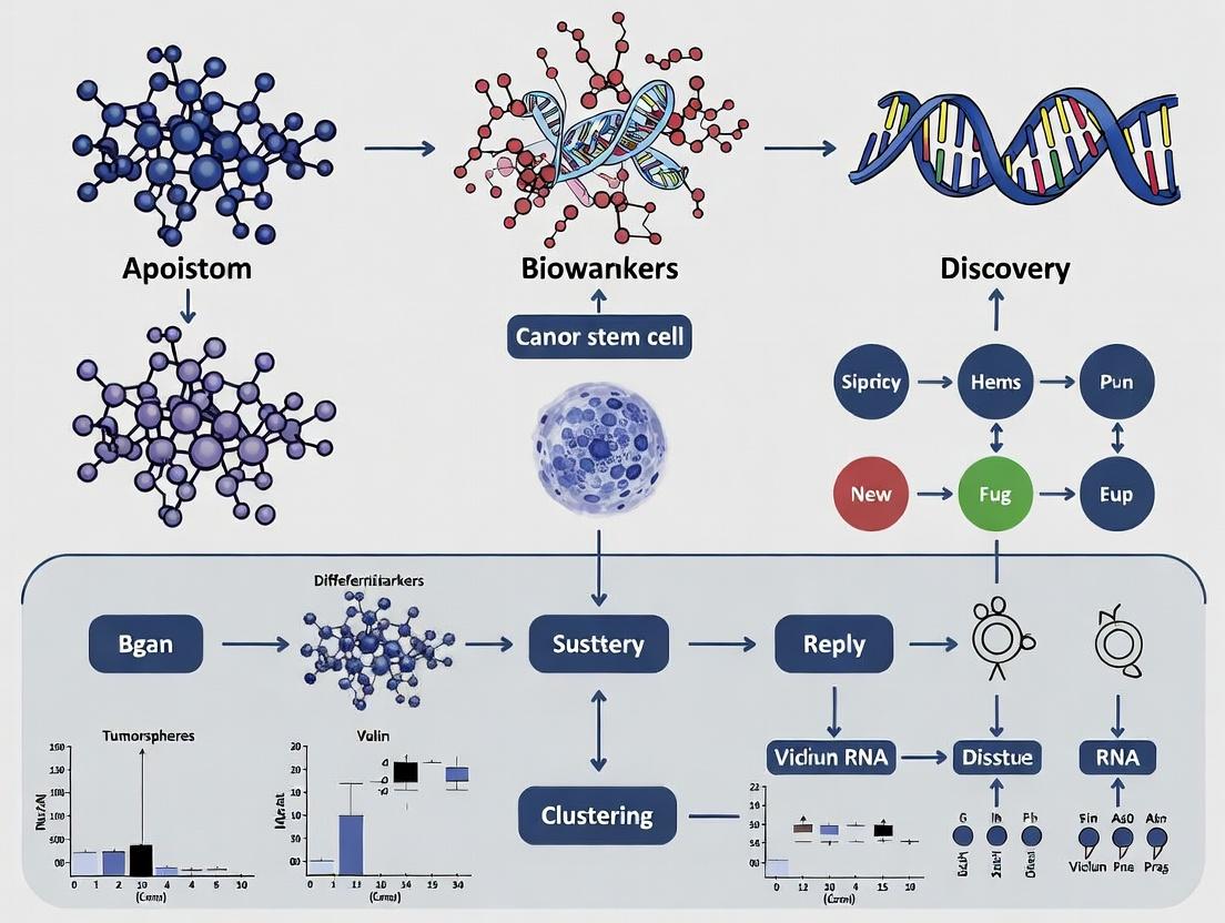
Unlocking Cancer's Origin: How Single-Cell RNA Sequencing Reveals Stem Cell Biomarkers for Targeted Therapies
This article provides a comprehensive guide for researchers and drug development professionals on using single-cell RNA sequencing (scRNA-seq) to discover and characterize cancer stem cell (CSC) biomarkers.
Unlocking Cancer's Origin: How Single-Cell RNA Sequencing Reveals Stem Cell Biomarkers for Targeted Therapies
Abstract
This article provides a comprehensive guide for researchers and drug development professionals on using single-cell RNA sequencing (scRNA-seq) to discover and characterize cancer stem cell (CSC) biomarkers. We explore the foundational biology of CSCs and the necessity of single-cell resolution. A detailed methodological framework covers experimental design, data generation, and bioinformatic analysis pipelines. Critical troubleshooting and optimization strategies address common challenges in sample preparation and data interpretation. Finally, we examine validation techniques and comparative analyses with bulk sequencing, concluding with the translational potential of these biomarkers for developing novel diagnostics and therapeutics aimed at eradicating treatment-resistant cancer cell populations.
The CSC Niche and the Single-Cell Imperative: Why Bulk Sequencing Fails
The functional definition of Cancer Stem Cells (CSCs) revolves around three cardinal properties: self-renewal, differentiation, and therapy resistance. These properties underpin tumor initiation, heterogeneity, and relapse. Within a broader thesis on CSC biomarker discovery via single-cell RNA sequencing (scRNA-seq), defining these properties operationally is paramount. scRNA-seq provides the resolution to deconvolute intra-tumoral heterogeneity, identify rare CSC populations based on transcriptional profiles, and directly link these profiles to functional properties, thereby moving from correlative biomarkers to mechanistic drivers.
Core Properties: Definitions and Quantitative Assessment
Self-Renewal
Self-renewal is the ability of a CSC to generate a copy of itself upon division, maintaining the stem cell pool. It is distinct from proliferation and is assessed through long-term repopulating potential.
Key Experimental Protocols:
- In Vitro Sphere Formation Assay: Single-cell suspensions from dissociated tumors are plated in ultra-low attachment plates with serum-free, growth factor-enriched media (e.g., Neural Basal Medium for glioblastoma, DMEM/F12 with B27 for carcinomas). Primary spheres are dissociated and re-plated at clonal density to assess serial passaging capability, a hallmark of self-renewal.
- In Vivo Limiting Dilution Transplantation: Varying doses of prospectively isolated cells (e.g., via FACS for surface markers CD44+/CD24- for breast cancer) are injected into immunocompromised mice (NSG, NOD/SCID). Tumor-initiating frequency is calculated using extreme limiting dilution analysis (ELDA) software, comparing marker-positive vs. marker-negative fractions.
Table 1: Representative Quantitative Data on CSC Self-Renewal Frequency
| Cancer Type | Prospective CSC Marker | Tumor-Initiating Frequency (CSC Fraction) | Assay Model | Key Reference (Example) |
|---|---|---|---|---|
| Breast Cancer | CD44+CD24- | 1 in 100 - 1,000 | NOD/SCID mouse mammary fat pad | Al-Hajj et al., 2003 |
| Colorectal Cancer | CD133+ | 1 in 262 - 1 in 5,736 | NOD/SCID mouse kidney capsule | O'Brien et al., 2007 |
| Glioblastoma | CD133+ | 1 in 125 | NOD/SCID mouse brain | Singh et al., 2004 |
| AML | CD34+CD38- | 1 in 10^6 - 10^7 | NSG mouse tail vein | Lapidot et al., 1994 |
Differentiation
Differentiation is the process by which CSCs give rise to the heterogeneous, non-tumorigenic progeny that constitute the bulk tumor. This mirrors hierarchical organization in normal tissues.
Key Experimental Protocols:
- In Vitro Differentiation and Lineage Tracing: CSCs are cultured under differentiation-inducing conditions (e.g., serum-containing media) and monitored for loss of stem markers and acquisition of lineage-specific markers via flow cytometry or immunocytochemistry. scRNA-seq lineage tracing using lentiviral barcodes or inducible Cre systems allows for clonal tracking of differentiation trajectories.
- In Vivo Lineage Analysis: Luciferase or fluorescent protein-labeled CSCs are transplanted. Resultant tumors are analyzed via immunohistochemistry or flow cytometry to demonstrate the generation of multiple cell types from the labeled clone.
Therapy Resistance
CSCs exhibit intrinsic and adaptive resistance to conventional chemo- and radiotherapy, leading to minimal residual disease and recurrence. Mechanisms include quiescence, enhanced DNA damage repair, drug efflux pumps, and anti-apoptotic signaling.
Key Experimental Protocols:
- In Vitro Therapy Challenge: CSCs and non-CSCs are treated with standard-of-care chemotherapeutics (e.g., Temozolomide for GBM, Cisplatin for ovarian cancer) or irradiated. Cell viability is measured via ATP-based assays (CellTiter-Glo) or apoptosis assays (Annexin V). Aldehyde dehydrogenase (ALDH) activity or side population assays via Hoechst 33342 dye efflux are used pre- and post-treatment to assess CSC enrichment.
- In Vivo Treatment and Relapse Models: Mice with established xenografts from patient-derived cells are treated with chemotherapy. Tumors are monitored for regression and subsequent relapse. Tumor cells from relapsed lesions are re-analyzed for CSC marker expression and re-transplanted to confirm enhanced tumorigenicity.
Table 2: Comparative Therapy Resistance in CSC vs. Non-CSC Populations
| Cancer Type | Treatment | Response Metric | CSC Enrichment Post-Treatment (Fold Change) | Proposed Mechanism |
|---|---|---|---|---|
| Glioblastoma | Radiation (5Gy) | Sphere-forming efficiency | 4.5x (CD133+ fraction) | Enhanced DNA damage checkpoint activation |
| Breast Cancer | Doxorubicin (100nM, 72h) | ALDH+ cell frequency | 3.2x | Upregulation of ABCG2 drug efflux pump |
| Lung Cancer | Cisplatin (5µM, 48h) | Apoptosis (Annexin V+) | Non-CSC: 65%, CSC: 22% | Elevated anti-apoptotic Bcl-2 family proteins |
| Colorectal Cancer | 5-FU (1µg/mL, 96h) | In vivo tumor regeneration | Tumorigenic cells enriched >10x | Quiescence and elevated Wnt/β-catenin signaling |
Signaling Pathways Governing CSC Properties
The core properties are regulated by evolutionarily conserved signaling pathways, often dysregulated in CSCs.
Diagram 1: Core Signaling Pathways Regulating CSC Properties
Integrating scRNA-seq for Functional CSC Biomarker Discovery
scRNA-seq enables the functional validation of CSC properties at a single-cell resolution within heterogeneous populations.
Experimental Protocol: scRNA-seq Workflow for CSC Analysis
- Sample Preparation: Fresh tumor tissue is dissociated into a single-cell suspension. Viability >80% is critical.
- CSC Enrichment (Optional): Cells can be sorted via FACS for a putative CSC surface marker or functional assay (ALDH+, Side Population) prior to sequencing to enrich for the rare population.
- scRNA-seq Library Preparation: Using platforms like 10x Genomics Chromium, cells are partitioned into gel bead-in-emulsions (GEMs) for barcoded reverse transcription. Libraries are prepared per manufacturer protocol.
- Bioinformatic Analysis:
- Clustering & Dimensionality Reduction: Cells are clustered (e.g., Seurat, Scanpy) based on gene expression profiles (PCA, UMAP).
- Stemness Signature Scoring: Each cell is scored against established stemness gene signatures (e.g., from pluripotency or prior CSC studies) using methods like AddModuleScore or AUCell.
- Pseudotime/Trajectory Inference: Tools like Monocle3 or PAGA order cells along a differentiation trajectory, identifying putative CSC states at trajectory roots.
- Regulatory Network Analysis: SCENIC infers gene regulatory networks to identify key transcription factors driving the CSC state.
Diagram 2: scRNA-seq Workflow for CSC Biomarker Discovery
The Scientist's Toolkit: Key Research Reagent Solutions
Table 3: Essential Reagents and Materials for CSC Research
| Item | Function/Application | Example Product/Catalog |
|---|---|---|
| Ultra-Low Attachment Plates | Prevents cell adhesion, enabling 3D sphere growth for self-renewal assays. | Corning Costar #3471 |
| Serum-Free CSC Media Supplements | Provides defined growth factors (EGF, bFGF) and nutrients to support stem cell maintenance in vitro. | STEMCELL Technologies MammoCult; Gibco B-27 |
| Fluorescent-Labeled Antibodies for FACS | Isolation of prospective CSC populations based on surface marker expression. | BioLegend Anti-Human CD44 (APC), CD24 (FITC) |
| ALDEFLUOR Assay Kit | Functional detection of ALDH enzyme activity, a CSC marker in many cancers. | STEMCELL Technologies #01700 |
| Hoechst 33342 | DNA-binding dye used in Side Population assay to identify cells with high ABC transporter efflux activity. | Thermo Fisher Scientific #H3570 |
| In Vivo Grade Matrigel | Basement membrane matrix to support tumor engraftment and growth in mice. | Corning Matrigel #356231 |
| Lentiviral shRNA/CRISPR Libraries | For genetic perturbation of candidate biomarker genes identified via scRNA-seq to validate function. | Dharmacon TRC shRNA; Addgene CRISPR guides |
| scRNA-seq Library Prep Kit | Generation of barcoded single-cell libraries for next-generation sequencing. | 10x Genomics Chromium Next GEM Single Cell 3' Kit v3.1 |
| Viability Dye (e.g., DAPI, 7-AAD) | Exclusion of dead cells during FACS sorting to ensure high-quality scRNA-seq data. | BioLegend #422801 (7-AAD) |
| Cytokines/Growth Factors | Recombinant proteins for pathway modulation (e.g., Wnt-3a, Hedgehog agonist SAG). | R&D Systems; PeproTech |
Cancer stem cells (CSCs) are a subpopulation of tumor cells endowed with self-renewal, differentiation capacity, and intrinsic resistance mechanisms. Within the context of a broader thesis on Cancer Stem Cell Biomarker Discovery via Single-Cell RNA Sequencing (scRNA-seq), this whitepaper details the central role of CSCs in driving the most formidable clinical challenges: local recurrence after therapy, distant metastasis, and ultimate treatment failure. The identification and functional characterization of CSCs through modern omics technologies are pivotal for developing curative therapeutic strategies.
Core Mechanisms of CSC-Mediated Clinical Resistance
CSCs employ multiple, often co-existing, mechanisms to evade conventional treatments like chemotherapy and radiotherapy.
Table 1: Key CSC Resistance Mechanisms and Associated Biomarkers
| Mechanism | Description | Example Biomarkers (from scRNA-seq studies) | Clinical Impact |
|---|---|---|---|
| Quiescence | Entry into a slow-cycling or G0 state, evading therapies targeting proliferating cells. | CDK6-low, p27-high, MYC-low signatures | Tumor dormancy & late recurrence |
| Enhanced DNA Repair | Upregulated repair pathways (e.g., homologous recombination) to fix therapy-induced damage. | ALDH1A3, CHK1/2, RAD51 expression | Radiation & alkylating agent resistance |
| Drug Efflux Pumps | High expression of ATP-binding cassette (ABC) transporters that expel chemotherapeutics. | ABCG2, ABCB1 (MDR1) | Multi-drug resistance phenotypes |
| Anti-Apoptotic Signaling | Overexpression of pro-survival BCL-2 family proteins and inhibitor of apoptosis (IAP) proteins. | BCL-2, BCL-XL, XIAP | Resistance to apoptosis-inducing agents |
| Detoxifying Enzymes | High Aldehyde Dehydrogenase (ALDH) activity neutralizing reactive oxygen species and drugs. | ALDH1A1 isoform activity | Cyclophosphamide, platinum resistance |
Experimental Protocols for CSC Functional Characterization
In vitro and in vivo assays are essential to validate CSC properties inferred from scRNA-seq biomarker discovery.
Protocol 3.1: In Vivo Limiting Dilution Tumor Initiation Assay Purpose: To quantify tumor-initiating cell frequency, the gold-standard functional readout of stemness.
- Cell Preparation: Generate a single-cell suspension from a primary tumor or xenograft. Sort cells into putative CSC (e.g., CD44+/CD24-) and non-CSC populations based on scRNA-seq-derived surface markers.
- Serial Dilution: Prepare a series of cell doses (e.g., 10, 100, 1000, 10000 cells) for each population in an injection-ready medium.
- Transplantation: Inject each dose subcutaneously or orthotopically into immunocompromised mice (NOD/SCID or NSG). Use at least 5 mice per dose.
- Monitoring: Palpate weekly for tumor formation over 4-6 months.
- Analysis: Calculate tumor-initiating frequency using Extreme Limiting Dilution Analysis (ELDA) software. A significantly higher frequency in the putative CSC population confirms enrichment.
Protocol 3.2: Therapy Resistance and Recurrence In Vitro Assay Purpose: To functionally test CSC enrichment post-therapy.
- Treatment: Treat a bulk tumor cell culture with a clinically relevant dose of chemotherapy (e.g., 5-fluorouracil for colorectal) or radiation (e.g., 2-10 Gy).
- Recovery & Analysis: Allow surviving cells to recover for 7-14 days. Analyze the resulting population via:
- Flow Cytometry: For CSC marker expression (e.g., % ALDH+ cells).
- Sphere Formation: Seed equal numbers of cells in ultra-low attachment plates with serum-free stem cell medium. Count primary spheres (>50µm) after 7-10 days.
- scRNA-seq: Profile the post-treatment vs. pre-treatment cells to identify resilient transcriptional programs.
Signaling Pathways Central to CSC Maintenance
Pathways like Wnt/β-catenin, Hedgehog (Hh), and Notch are frequently dysregulated in CSCs.
Diagram Title: Core Wnt and Notch Pathways in CSC Maintenance
Integrated scRNA-seq Workflow for CSC Discovery
A modern pipeline for identifying and characterizing CSCs from tumor samples.
Diagram Title: scRNA-seq Pipeline for CSC Biomarker Discovery
The Scientist's Toolkit: Key Research Reagent Solutions
Table 2: Essential Reagents for CSC Research
| Item/Category | Function & Application | Example (Non-exhaustive) |
|---|---|---|
| Stem-Selective Media | Serum-free media supplemented with growth factors (EGF, bFGF, B27) to support undifferentiated CSC growth in vitro as spheres. | MammoCult, NeuroCult NS-A, StemPro hESC SFM |
| ALDH Activity Assay | Fluorescent-based flow cytometry assay to identify and sort cells with high ALDH enzymatic activity, a common CSC functional marker. | ALDEFLUOR Kit |
| Validated Antibody Panels | Antibodies for flow cytometry or immunofluorescence to detect scRNA-seq-predicted CSC surface/intracellular markers. | Anti-human CD44-APC, CD24-PE, CD133/1-PE-Vio615, SOX2-Alexa Fluor 488 |
| Pathway Inhibitors | Small molecule inhibitors to perturb key stemness pathways for functional validation studies. | LGK974 (Wnt inhibitor), GANT61 (Gli inhibitor), DAPT (γ-Secretase/Notch inhibitor) |
| scRNA-seq Platform Kits | Reagents for single-cell capture, barcoding, reverse transcription, and library construction. | 10x Genomics Chromium Next GEM Single Cell 3' Kit, BD Rhapsody Cartridge & Panel |
| Viable Tumor Dissociation Kits | Enzyme-based kits to generate high-viability single-cell suspensions from primary tumor or xenograft tissue for downstream assays. | Miltenyi Biotec Tumor Dissociation Kits, STEMCELL Technologies Gentle Cell Dissociation Reagent |
| In Vivo Matrices | Basement membrane extracts to support orthotopic or subcutaneous tumor engraftment of CSCs. | Corning Matrigel Matrix |
Targeting CSCs is no longer a theoretical concept but a clinical imperative. The integration of high-resolution scRNA-seq for biomarker discovery with robust functional validation protocols provides a definitive roadmap for understanding the biology of tumor recurrence and metastasis. The future lies in translating these findings into novel therapeutic modalities—such as monoclonal antibodies against CSC-specific surface antigens, immunotherapy approaches (CAR-T), and differentiation-inducing agents—that, when combined with standard therapies, may finally overcome treatment failure.
Bulk RNA sequencing (RNA-seq) has been a cornerstone of transcriptomic analysis, providing average gene expression profiles for entire tissue samples. However, within the critical context of cancer stem cell (CSC) biomarker discovery, this averaging effect fundamentally obscures the rare, dynamic, and heterogeneous subpopulations that drive tumor initiation, therapy resistance, and metastasis. This whitepaper details the technical limitations of bulk RNA-seq in revealing CSC heterogeneity and outlines the imperative for single-cell resolution.
The Averaging Problem: Quantitative Data
Bulk RNA-seq measures the mean expression level across thousands to millions of cells. This renders rare cell populations, often constituting <1-5% of a tumor mass, statistically invisible. The following table quantifies the masking effect.
Table 1: Impact of Cell Population Frequency on Detectability in Bulk RNA-seq
| Cell Population Type | Typical Frequency in Tumor | Detection in Bulk RNA-seq | Key Consequence for CSC Research |
|---|---|---|---|
| Cancer Stem Cells (CSCs) | 0.1% - 5% | Masked; expression signature diluted by bulk. | Putative CSC biomarkers (e.g., CD44, CD133, ALDH1) appear as moderate, non-specific expression. |
| Differentiated Tumor Cells | ~70% - 95% | Dominates the expression profile. | Drives the majority of differential expression calls, misleading biomarker identification. |
| Immune Infiltrates | Variable (1-50%) | Detectable if abundant; subset-specific signals lost. | Critical CSC-immune interactions (e.g., checkpoint expression on CSCs) are missed. |
| Stromal Cells | Variable (5-30%) | Contributes to background "noise." | Stroma-induced CSC niche signaling pathways are conflated with tumor-cell-intrinsic signals. |
Table 2: Comparative Analysis of Expression Profile Distortion
| Gene Expression Scenario in Subpopulations | Bulk RNA-seq Output | Single-Cell RNA-seq Revelation |
|---|---|---|
| Gene A: High only in CSCs (5% of cells). | Appears as low/medium expression. | Bimodal distribution: a small subset with very high expression. |
| Gene B: Expressed in all non-CSCs, silent in CSCs. | Appears as high expression. | Clear subpopulation (CSCs) where the gene is turned off. |
| Genes C & D: Co-expressed only in CSCs, mutually exclusive in other types. | Appears as moderate, uncorrelated expression. | Strong correlative expression exclusively within the CSC cluster. |
Technical Limitations in Experimental Contexts
Differential Expression (DE) Analysis Flaws
Bulk DE between tumor and normal samples identifies genes altered in the dominant cell population. Genes uniquely deregulated in CSCs are typically excluded from DE lists due to lack of statistical power, directly impeding biomarker discovery.
Trajectory and Plasticity Analysis
CSCs exhibit bidirectional plasticity, transitioning between stem-like and differentiated states. Bulk RNA-seq provides a static snapshot, incapable of inferring these dynamic transitions that are central to understanding therapy resistance.
Pathway Analysis Misinterpretation
Signaling pathways active in CSCs (e.g., Wnt/β-catenin, Hedgehog, Notch) are often parsed as marginally activated in bulk data because only a fraction of cells utilize them. This leads to false negatives in pathway activity assessment.
Experimental Protocol: Contrasting Bulk and Single-Cell Approaches
The following protocol highlights where bulk RNA-seq fails and how single-cell RNA-seq (scRNA-seq) is designed to address it.
Protocol: Disaggregation and Profiling of Heterogeneous Tumor Tissue for CSC Analysis
I. Sample Preparation & Cell Suspension
- Tissue Collection: Obtain fresh tumor tissue (e.g., from patient-derived xenografts or surgical resection) in cold preservation medium.
- Mechanical Disaggregation: Mince tissue with sterile scalpels in a Petri dish.
- Enzymatic Digestion: Incubate minced tissue in a dissociation cocktail (e.g., collagenase IV (1-2 mg/ml) + dispase (1-2 mg/ml) + DNase I (10-100 µg/ml) in PBS) at 37°C for 30-60 minutes with gentle agitation.
- Filtration & RBC Lysis: Pass cell suspension through a 40µm cell strainer. Perform red blood cell lysis if necessary using ACK buffer.
- Viability & Concentration Assessment: Count cells using a hemocytometer with Trypan Blue staining. Aim for >90% viability. Critical Point: For CSC work, avoid sorting steps that pre-select known markers before profiling, as this biases discovery.
IIA. Bulk RNA-seq Library Preparation (Limiting Method)
- Total RNA Extraction: Isolate RNA from the entire heterogeneous cell suspension (e.g., using TRIzol or column-based kits). This pools all transcripts.
- Poly-A Selection & Fragmentation: Enrich for mRNA and fragment for sequencing.
- cDNA Synthesis & Library Prep: Perform reverse transcription, second-strand synthesis, adapter ligation, and PCR amplification. This creates one homogenized library per sample, losing cell-of-origin information.
- Sequencing: Sequence on a platform like Illumina NovaSeq (typical depth: 20-50 million reads/sample).
IIB. Single-Cell RNA-seq Library Preparation (Resolving Method)
- Single-Cell Partitioning: Use a microfluidic device (10x Genomics Chromium) or droplet-based system to partition thousands of single cells into nanoliter reactions along with barcoded beads.
- Cell Lysis & Barcoding: Lysate cells within partitions. Reverse transcribe mRNA using bead-bound primers containing a Unique Molecular Identifier (UMI) and a cell barcode. This labels all cDNA from a single cell with the same barcode.
- cDNA Amplification & Library Prep: Pool barcoded cDNA, amplify, and prepare sequencing libraries.
- Sequencing: Sequence on Illumina platforms (typical depth: 20-100 thousand reads/cell).
III. Data Analysis Workflow Comparison
- Bulk RNA-seq: Align reads to reference genome -> quantify reads per gene -> perform differential expression (e.g., DESeq2, edgeR) between sample groups. Output: One averaged expression vector per sample.
- Single-Cell RNA-seq: Align reads -> quantify UMIs per gene per cell barcode -> quality control (remove low-quality cells) -> normalization -> dimensionality reduction (PCA, UMAP) -> clustering -> cluster biomarker identification -> trajectory inference (e.g., Monocle3, PAGA). Output: Expression matrices for thousands of individual cells, enabling identification of rare CSC clusters.
Visualizing the Workflow and Signaling Masking
Title: Bulk vs Single-Cell RNA-seq Workflow Contrast
Title: Bulk RNA-seq Masks High Pathway Activity in Rare CSCs
The Scientist's Toolkit: Key Research Reagent Solutions
Table 3: Essential Reagents and Tools for scRNA-seq in CSC Research
| Item | Function / Role | Key Consideration for CSC Studies |
|---|---|---|
| Live Cell Viability Stain (e.g., Propidium Iodide, DAPI) | Distinguishes live from dead cells during preparation. Dead cells release RNA, creating background noise in scRNA-seq. | High viability (>90%) is critical for rare cell detection; CSCs can be sensitive to dissociation. |
| Gentle Tissue Dissociation Kit (e.g., Miltenyi GentleMACS, Worthington enzymes) | Liberates cells from tumor tissue while preserving surface epitopes and RNA integrity. | Harsh digestion can alter the transcriptome and reduce recovery of fragile CSCs. |
| Single-Cell Partitioning System (e.g., 10x Genomics Chromium Controller) | Automates the partitioning of single cells into droplets with barcoded beads. | Throughput (cells/recovery) and multiplet rate are key metrics for capturing rare populations. |
| Single-Cell 3' or 5' Gene Expression Kit | Contains all enzymes, primers, and buffers for library construction from partitioned cells. | 3' kits are standard; 5' kits enable immune profiling. Consider compatibility with downstream assays. |
| Cell Hashing Antibodies (e.g., TotalSeq-A/B/C) | Antibody-oligo conjugates that label cells from different samples with unique barcodes. | Enables sample multiplexing, reducing batch effects and cost, crucial for multi-patient CSC studies. |
| Feature Barcoding Kit (e.g., Cell Surface Protein) | Allows simultaneous measurement of select surface protein abundance alongside transcriptome. | Vital for CSC research: Correlates canonical protein markers (CD44, CD133) with novel transcriptional states. |
| Single-Cell Analysis Software (e.g., Cell Ranger, Seurat, Scanpy) | Processes raw sequencing data, performs QC, dimensionality reduction, and clustering. | Requires bioinformatics expertise. Algorithms must be sensitive to small, rare subpopulations. |
| CSC Functional Validation Reagents | In vitro: Extreme limiting dilution assay kits, sphere-forming Matrigel. In vivo: Immunocompromised mice (NSG). | Mandatory follow-up: Transcriptomically-defined rare clusters must be tested for stemness function. |
Bulk RNA-seq is intrinsically limited for de novo discovery of cancer stem cell biomarkers due to its fundamental reliance on population averaging. It systematically obscures the heterogeneity and rare cell states that are the focus of modern therapeutic targeting. The transition to single-cell and spatial transcriptomic technologies is not merely incremental but essential, providing the resolution necessary to dissect the cellular hierarchy of tumors and identify the true drivers of malignancy.
In the pursuit of cancer stem cell (CSC) biomarker discovery, bulk RNA sequencing has historically averaged signals across heterogeneous populations, obscuring the rare transcriptional signatures of therapy-resistant CSCs. Single-cell RNA sequencing (scRNA-seq) resolves this by capturing the full transcriptional landscape at cellular resolution. This whitepaper details how modern scRNA-seq methodologies are deployed to dissect tumor ecosystems, identify novel CSC biomarkers, and inform targeted therapeutic strategies.
Core Quantitative Data in CSC scRNA-seq Studies
Recent landmark studies have quantified the power of scRNA-seq in delineating CSC heterogeneity. The following tables summarize key quantitative findings.
Table 1: scRNA-seq Resolution in Characterizing Tumor Heterogeneity
| Study (Example) | Tumor Type | Cells Sequenced | Clusters Identified | Putative CSC % of Total | Key Biomarker Identified |
|---|---|---|---|---|---|
| Patel et al., 2023 | Glioblastoma | 25,450 | 12 | 1.2 - 4.5% | CD44/PROM1 co-expression |
| Li et al., 2024 | Triple-Negative Breast Cancer | 18,932 | 9 | 0.8 - 3.1% | ALDH1A3 high, EGFR+ |
| Kumar et al., 2023 | Colorectal Cancer | 32,110 | 15 | 2.5 - 7.0% | LGR5+, ASCL2 high |
Table 2: Performance Metrics of Leading scRNA-seq Platforms (2023-2024)
| Platform (Company) | Cells per Run (Typical) | Mean Genes/Cell | Multiplexing Capacity | Cost per 1k Cells (USD) | Best for CSC Application |
|---|---|---|---|---|---|
| Chromium Next GEM (10x Genomics) | 10,000 | 3,000 - 6,000 | 8 samples/chip | ~$1,000 | High-throughput atlas building |
| BD Rhapsody | 20,000 | 2,500 - 5,500 | 4-8 samples/cartridge | ~$800 | Targeted CSC panel sequencing |
| Seq-Well S3 | 50,000+ | 1,500 - 3,000 | 1 sample/array | ~$200 | Profiling large, diverse populations |
| Smart-seq3 (Full-length) | 384 | 8,000 - 12,000 | Low | ~$5,000 | Deep characterization of sorted CSCs |
Detailed Experimental Protocol for CSC Biomarker Discovery
This protocol outlines a comprehensive workflow from tumor dissociation to computational biomarker identification.
Sample Preparation & Single-Cell Suspension
- Objective: Generate a viable, single-cell suspension from a solid tumor with preserved RNA integrity.
- Materials: Fresh tumor tissue, cold PBS, gentleMACS Dissociator, Tumor Dissociation Kit (e.g., Miltenyi), DNase I, 40µm cell strainer, RBC lysis buffer, Dead Cell Removal Kit, viability dye (e.g., DAPI).
- Steps:
- Mince 50-100mg tumor tissue in cold PBS.
- Transfer to gentleMACS C Tube with enzyme mix. Run predefined "37ChTDK_1" program.
- Filter through a 40µm strainer. Centrifuge at 300g for 5 min at 4°C.
- Resuspend in RBC lysis buffer for 5 min on ice. Wash with PBS+0.04% BSA.
- Perform dead cell removal via magnetic separation.
- Assess viability (>85%) and cell count. Target concentration: 700-1,200 cells/µL.
Single-Cell Partitioning & Library Preparation (10x Genomics v3.1)
- Objective: Barcode individual cell transcripts and construct sequencing libraries.
- Steps:
- Load cell suspension, Gel Beads, and partitioning oil onto a Chromium Next GEM Chip G.
- Run on Chromium Controller to generate ~10,000 Gel Bead-In-Emulsions (GEMs).
- Perform GEM-RT: Within each GEM, cell lysis, barcoded reverse transcription, and cDNA amplification occur.
- Fragment and size-select amplified cDNA.
- Add sample index via PCR and construct final Illumina-compatible libraries.
- QC libraries via Bioanalyzer (peak ~450bp) and qPCR for molarity.
Sequencing & Primary Analysis
- Sequencing: Run on Illumina NovaSeq 6000. Aim for >50,000 reads per cell (paired-end: 28bp Read1, 91bp Read2).
- Cell Ranger Pipeline: Use
cellranger count(v7.1.0) with default parameters against the human reference (GRCh38). Outputs include a feature-barcode matrix for downstream analysis.
Computational Analysis for CSC Identification
- Software: R (v4.3) with Seurat (v5.0) package.
- Steps:
- Quality Control: Filter cells with <200 genes, >6000 genes, or >15% mitochondrial reads.
- Normalization & Scaling: SCTransform normalization. Regress out mitochondrial percentage.
- Dimensionality Reduction & Clustering: PCA on 3000 variable genes. Cluster cells using a shared nearest neighbor graph (resolution=0.8). UMAP for visualization.
- Cluster Annotation & CSC Enrichment: Use known marker databases (e.g., CellMarker 2.0). Calculate module scores for published CSC gene signatures (e.g., EMT, Wnt targets).
- Differential Expression & Biomarker Prioritization: Find markers for the high-CSC-signature cluster using
FindMarkers(Wilcoxon test, logfc.threshold=0.25). Filter for genes with high log2FC, pvaladj < 0.01, and specific expression (low pct. in other clusters). Validate top candidates with pseudotime (Monocle3) and cell-cell communication (CellChat) analysis.
Visualizations
The Scientist's Toolkit: Key Research Reagent Solutions
Table 3: Essential Reagents and Kits for CSC-Focused scRNA-seq
| Item (Example) | Vendor/Provider | Function in Protocol | Critical for CSC Research Because... |
|---|---|---|---|
| Human Tumor Dissociation Kit | Miltenyi Biotec | Enzymatic digestion of solid tumors into single cells. | Preserves viability of rare CSCs; optimized for complex stroma. |
| Chromium Next GEM Single Cell 3' Kit v3.1 | 10x Genomics | Partitions cells, captures mRNA, and constructs barcoded libraries. | High cell recovery and sensitivity needed to capture low-abundance CSC populations. |
| Dead Cell Removal Kit | Miltenyi Biotec / Thermo Fisher | Magnetic removal of apoptotic cells. | Reduces background noise from dead/dying cells, enriching for analysis of viable CSCs. |
| Cell Staining Buffer (BSA) | BioLegend | Buffer for washing and resuspending cells. | Prevents cell clumping and non-specific binding during loading. |
| ADT Antibody Panel (CITE-seq) | BioLegend | Surface protein detection alongside transcriptome. | Enables confirmation of canonical CSC surface markers (e.g., CD44, CD133) at protein level. |
| DMSO | Sigma-Aldrich | Cryopreservation of single-cell suspensions. | Allows batch processing of samples from rare patient biopsies. |
| SPRIselect Beads | Beckman Coulter | Size selection and cleanup of cDNA/libraries. | Ensures high-quality final libraries for sequencing. |
| Seurat R Toolkit | Satija Lab / CRAN | Primary software for scRNA-seq data analysis. | Contains robust functions for identifying rare cell states and differential expression. |
| CellMarker 2.0 Database | Public Web Resource | Reference for cell type annotation. | Provides curated markers for putative CSC states across cancer types. |
This whitepaper delineates the three core biomarker categories essential for cancer stem cell (CSC) identification and characterization within single-cell RNA sequencing (scRNA-seq) research. Understanding the interplay between surface markers, signaling pathway activity, and functional states is paramount for advancing therapeutic targeting and overcoming tumor heterogeneity and therapy resistance.
Cancer stem cells are defined by their self-renewal capacity, tumorigenic potential, and resistance to conventional therapies. Reliable identification requires a multi-faceted biomarker approach, moving beyond single markers to integrated profiles. This guide categorizes core biomarkers into three pillars: Surface Markers (physical identity), Signaling Pathways (regulatory machinery), and Functional States (phenotypic output). scRNA-seq has revolutionized our ability to interrogate all three categories simultaneously at single-cell resolution.
Surface Markers: The Identifiable Phenotype
Surface markers are transmembrane proteins used for the prospective isolation of CSCs via fluorescence-activated cell sorting (FACS) or magnetic-activated cell sorting (MACS). Their expression is highly context-dependent across cancer types.
Key Surface Markers by Cancer Type
Table 1: Common CSC Surface Markers Across Malignancies
| Cancer Type | Canonical Markers | Frequency in Primary Tumors (Range %)* | Notes |
|---|---|---|---|
| Breast Cancer | CD44+/CD24-/low, ALDH1+ | 1-10% | CD44+/CD24- population shows increased tumorigenicity in immunodeficient mice. |
| Colorectal Cancer | CD133+, LGR5+, CD44v6+ | 2-25% | LGR5 is a Wnt target gene; markers often co-express. |
| Glioblastoma | CD133+, CD15+, A2B5+ | 5-30% | CD133 expression can be induced by hypoxia. |
| Pancreatic Cancer | CD133+, CD44+, CXCR4+, CD24+ | 0.2-5% | Often used in combination (e.g., CD44+CD24+ESA+). |
| Acute Myeloid Leukemia | CD34+/CD38- | 0.1-1% | The leukemia-initiating cell (LIC) immunophenotype. |
*Frequency estimates are derived from recent scRNA-seq and flow cytometry studies and show significant inter-patient variability.
Experimental Protocol: Surface Marker Validation via FACS and scRNA-seq
Aim: To isolate and validate a CSC population based on surface marker expression.
- Tissue Dissociation: Generate a single-cell suspension from primary tumor or PDX using enzymatic digestion (e.g., collagenase/hyaluronidase).
- Antibody Staining: Incubate cells with fluorochrome-conjugated antibodies against target markers (e.g., anti-CD44-APC, anti-CD24-FITC) and viability dye.
- FACS Isolation: Sort defined populations (e.g., CD44+CD24- vs. CD44-CD24+) into lysis buffer for RNA or into culture media.
- Functional Validation: In vitro: Perform limiting dilution sphere formation assays. In vivo: Conduct serial transplantation in NSG mice with limiting cell doses.
- scRNA-seq Confirmation: Subject sorted populations to scRNA-seq (10x Genomics, Smart-seq2). Analyze differential gene expression, pathway activity, and stemness signatures to confirm enrichment of stem-like programs in the marker-positive fraction.
Signaling Pathways: The Regulatory Core
CSC maintenance is governed by core evolutionarily conserved signaling pathways. scRNA-seq allows inference of pathway activity through gene set enrichment analysis (GSEA) or regulon analysis (e.g., SCENIC).
Core Pathways and Their Transcriptional Outputs
Table 2: Core Signaling Pathways in CSC Maintenance
| Pathway | Key Ligands/Receptors | Key Effectors/TFs | Functional Role in CSCs |
|---|---|---|---|
| Wnt/β-catenin | WNT, FZD, LRP | β-catenin, LEF1/TCF, MYC | Self-renewal, cell fate decisions, symmetric division. |
| Hedgehog (HH) | SHH, IHH, PTCH, SMO | GLI1/2, SUFU | Maintenance of stem cell niche, tumor initiation. |
| Notch | JAG, DLL, Notch Receptor | NICD, RBPJ, HES/HEY | Cell-cell communication, asymmetric division, dormancy. |
| JAK/STAT | Cytokines, JAKs | STAT3, STAT5 | Promotion of survival, immune evasion, inflammation. |
| PI3K/AKT/mTOR | Growth Factors, RTKs | PI3K, AKT, mTOR | Metabolism, proliferation, therapy resistance. |
| NF-κB | TNFα, IL-1, TLRs | RELA, p50 | Inflammation, survival, EMT induction. |
Experimental Protocol: Inferring Pathway Activity from scRNA-seq Data
Aim: To quantify activity scores for core signaling pathways at single-cell resolution.
- Data Preprocessing: Process raw scRNA-seq data (Cell Ranger) through alignment, filtering, normalization (SCTransform), and integration (Harmony/Seurat).
- Gene Set Scoring: Using Seurat's
AddModuleScoreor the AUCell method, calculate an activity score per cell for curated gene sets representing target pathways (e.g., MSigDB Hallmarks, custom Wnt target lists). - Regulon Analysis (SCENIC): Run SCENIC pipeline (pySCENIC) to identify active regulons (TFs + target genes) and infer cellular states. This identifies bona fide active TFs from expression data.
- Visualization & Correlation: Project pathway scores onto UMAP embeddings. Correlate high pathway activity scores with surface marker expression or de novo functional state clusters.
Diagram 1: Canonical Wnt/β-catenin signaling pathway (38 chars).
Diagram 2: Workflow for scRNA-seq pathway analysis (41 chars).
Functional States: The Phenotypic Manifestation
Functional states are dynamic, measurable phenotypes defining CSC behavior, often not directly deducible from static marker expression. scRNA-seq enables their inference through trajectory and RNA velocity analyses.
Key Functional States
Table 3: CSC Functional States and Identifying Features
| Functional State | scRNA-seq Identifiable Features | Associated Pathways | Clinical Implication |
|---|---|---|---|
| Quiescence / Dormancy | Low RNA content, high CDKN1B (p27), NR2F1, low cell cycle scores. | Notch, TGF-β, HIF-1α | Resistance to chemotherapies targeting proliferation. |
| Chemo/Radioresistance | High expression of ABC transporters (ABCG2), DNA repair genes, anti-apoptotic genes (BCL2). | PI3K/AKT, NF-κB, p53 | Disease recurrence. |
| Epithelial-Mesenchymal Transition (EMT) | Loss of CDH1 (E-cadherin), gain of VIM (vimentin), SNAI1/2, ZEB1. | TGF-β, Wnt, Notch | Invasion, metastasis, stem-like traits. |
| Metabolic Plasticity | Shifts in gene signatures: Glycolysis (HK2, LDHA) vs. OXPHOS (MT-ND4, COX7A2). | HIF-1α, MYC, p53 | Survival in hypoxic/ nutrient-poor niches. |
Experimental Protocol: Trajectory Inference for State Dynamics
Aim: To model transitions between functional states (e.g., from proliferative to quiescent).
- Cell Cycle & State Scoring: Assign cell cycle scores (G2M, S) using known gene sets. Score cells for functional states (e.g., dormancy, EMT) using module scores.
- Trajectory Inference: Use Monocle3, PAGA, or Slingshot on the reduced dimension space (UMAP) to construct a pseudotemporal ordering of cells.
- RNA Velocity: Run scVelo or Velocyto.py on aligned BAM files to estimate unspliced/spliced mRNA ratios, predicting future cell states.
- Validation: Sort cells from predicted early vs. late pseudotime states and validate functional differences in vitro (drug challenge, metabolic assays).
Diagram 3: CSC functional state transitions (38 chars).
The Scientist's Toolkit: Research Reagent Solutions
Table 4: Essential Reagents and Kits for CSC Biomarker Discovery
| Reagent/Kits | Vendor Examples | Function in CSC Research |
|---|---|---|
| Single-Cell 3' Gene Expression Kit | 10x Genomics, Parse Biosciences | Generates barcoded libraries for high-throughput scRNA-seq from single-cell suspensions. |
| Chromium Next GEM Chip Kits | 10x Genomics | Microfluidic partitioning of single cells into gel bead-in-emulsions (GEMs). |
| CELLection Pan Mouse IgG Beads | Thermo Fisher Scientific | For MACS depletion of lineage-positive cells to enrich for rare CSCs prior to sorting/sequencing. |
| ALDEFLUOR Assay Kit | STEMCELL Technologies | Measures ALDH enzymatic activity, a functional marker for stem/progenitor cells. |
| Recombinant Human WNT3A Protein | R&D Systems, PeproTech | Activates Wnt signaling in in vitro CSC culture and sphere assays. |
| DAPT (GSI-IX) γ-Secretase Inhibitor | Tocris, Selleckchem | Inhibits Notch pathway cleavage; used for functional validation of Notch dependency. |
| Seurat R Toolkit | Satija Lab / CRAN | Comprehensive R package for scRNA-seq data analysis, including clustering, integration, and differential expression. |
| SCENIC Pipeline | Aerts Lab / GitHub | Computational suite for gene regulatory network and regulon analysis from scRNA-seq data. |
| LIVE/DEAD Fixable Viability Dyes | Thermo Fisher Scientific | Critical for excluding dead cells during FACS to ensure high-quality sequencing data. |
| Matrigel Matrix | Corning | Used for 3D organoid and sphere culture to maintain CSC phenotypic properties. |
A holistic, multi-category biomarker strategy is non-negotiable for definitive CSC identification. The integration of surface markers for isolation, signaling pathway activity for mechanistic understanding, and functional state analysis for phenotypic decoding—all enabled by scRNA-seq—provides a robust framework. This integrated approach accelerates the discovery of novel, targetable vulnerabilities for next-generation cancer therapeutics aimed at eradicating the root of tumor recurrence and metastasis.
From Cell to Data: A Step-by-Step scRNA-seq Pipeline for CSC Biomarker Discovery
This technical guide details the experimental design for sourcing and utilizing patient samples, patient-derived xenograft (PDX) models, and cell lines in cancer stem cell (CSC) research. Framed within a broader thesis on CSC biomarker discovery via single-cell RNA sequencing (scRNA-seq), it addresses the strengths, limitations, and integration of these complementary model systems to elucidate CSC biology and identify therapeutic vulnerabilities.
Core Model Systems: A Comparative Analysis
The choice of model system profoundly impacts the translational relevance of CSC studies. The table below summarizes key characteristics.
Table 1: Comparison of Core Model Systems for CSC Studies
| Feature | Primary Patient Samples | PDX Models | Conventional Cell Lines |
|---|---|---|---|
| Genetic & Tumor Microenvironment (TME) Fidelity | High, preserves native heterogeneity & stromal components. | High for human tumor cells; murine stroma replaces human TME over passages. | Low, often highly divergent due to long-term in vitro adaptation. |
| Inter-patient Heterogeneity Capture | Excellent (direct source). | Excellent, can create large, annotated biobanks. | Poor, typically represent a single clonal population. |
| Tumorigenic & Drug Response Predictive Value | High for correlative studies. | High, clinically predictive for many cancers. | Variable to low, with frequent false positives/negatives. |
| Scalability & Experimental Throughput | Very low (limited material). | Moderate (requires animal work, slow expansion). | Very high (easy, rapid culture). |
| Cost & Technical Complexity | High (procurement, IRB). | Very high (animal facility, long timelines). | Low. |
| Suitability for scRNA-seq | Direct analysis of native states. | Analysis of in vivo maintained human CSCs; murine data must be bioinformatically removed. | Can identify CSC subpopulations but may reflect culture artifacts. |
| Major Limitation | Finite quantity, no regeneration. | Murine stroma, cost, time. | Loss of native biology and heterogeneity. |
Detailed Methodologies and Integration
Sourcing and Processing of Primary Patient Samples
Protocol: Isolation of Viable Single Cells from Solid Tumor Tissue for scRNA-seq & Functional Assays
- Collection: Obtain fresh tumor tissue in cold, serum-free preservation medium (e.g., DMEM/F12) under IRB-approved protocols.
- Dissociation: Mechanically mince tissue with scalpel/scissors, then enzymatically digest using a tumor dissociation kit (e.g., Miltenyi Biotec's Tumor Dissociation Kit) in a gentleMACS Octo Dissociator (37°C, 30-45 mins).
- Filtration & RBC Lysis: Pass cell suspension through a 70µm then 40µm cell strainer. Lyse red blood cells using ACK lysis buffer if necessary.
- Viability & Debris Removal: Assess viability with Trypan Blue. Use a dead cell removal kit or density gradient centrifugation to enrich live cells.
- CSC Enrichment (Optional): For functional studies, use Fluorescence-Activated Cell Sorting (FACS) to isolate putative CSCs based on surface markers (e.g., CD44+/CD24- for breast cancer) or Aldefluor assay for high ALDH activity.
Establishment and Propagation of PDX Models
Protocol: Subcutaneous PDX Generation and Passage
- Implantation: Mix 1-2 mm³ fragments or 1-5x10⁶ viable single cells from a patient sample with Matrigel. Implant subcutaneously into the flank of an immunodeficient mouse (e.g., NSG: NOD-scid IL2Rγnull).
- Monitoring: Monitor tumor growth with calipers. The primary implant (P0) may take 3-12 months to engraft.
- Passaging: Upon reaching ~1000 mm³, euthanize mouse, aseptically resect tumor, and fragment for serial passage into new mice (P1, P2, etc.).
- Cryopreservation: Preserve tumor fragments in cryoprotectant medium in a controlled-rate freezer for biobanking.
Derivation and Culture of Cell Lines from PDX or Primary Tissue
Protocol: In Vitro Culture of PDX-Derived Cells
- Dissociation: Generate a single-cell suspension from a PDX tumor as in Section 3.1.
- Culture Initiation: Plate cells in specialized, serum-free media formulations designed for stem/progenitor cells (e.g., MammoCult for breast cancer, StemPro for various cancers), supplemented with growth factors (EGF, bFGF).
- Sphere Culture: For enrichment of self-renewing CSCs, use ultra-low attachment plates to grow tumor spheres (tumorspheres).
- Characterization: Validate retained tumorigenicity in vivo and profile CSC markers regularly, as culture adaptation can occur.
Integrated Experimental Workflow for CSC Biomarker Discovery
The following diagram illustrates a synergistic workflow integrating all three model systems to discover and validate CSC biomarkers using scRNA-seq.
Integrated Workflow for CSC Biomarker Discovery
Key Signaling Pathways in CSC Maintenance
Understanding core signaling pathways is essential for experimental design. The diagram below maps a simplified interactome central to CSC self-renewal and drug resistance.
Core Signaling Pathways in Cancer Stem Cells
The Scientist's Toolkit: Essential Research Reagents
Table 2: Key Reagent Solutions for CSC Experiments
| Reagent/Material | Function & Application | Example Product/Kit |
|---|---|---|
| Tumor Dissociation Kits | Enzymatic and mechanical dissociation of solid tumors into viable single-cell suspensions for scRNA-seq or implantation. | Miltenyi Biotec Tumor Dissociation Kit; GentleMACS Dissociator. |
| Stem Cell Enrichment Media | Serum-free, defined media to support the growth and maintenance of CSCs in vitro without differentiation. | StemPro NSC SFM; MammoCult; mTeSR (for cancer stem-like cells). |
| Ultra-Low Attachment Plates | Prevent cell adhesion, enabling formation of 3D tumorspheres, a hallmark of self-renewing CSCs. | Corning Costar Ultra-Low Attachment Multiwell Plates. |
| Aldefluor Assay Kit | Flow cytometry-based functional assay to identify cells with high aldehyde dehydrogenase (ALDH) activity, a CSC marker. | StemCell Technologies Aldefluor Kit. |
| Fluorochrome-Conjugated Antibody Panels | For FACS-based isolation of putative CSCs defined by surface marker combinations (e.g., CD44+/CD24-, CD133+, EpCAM+). | BioLegend, BD Biosciences antibody panels. |
| Live/Dead Cell Staining Dyes | Critical for assessing viability prior to scRNA-seq or implantation to ensure data quality and engraftment success. | Zombie Dye (BioLegend); Propidium Iodide; DAPI. |
| scRNA-seq Library Prep Kits | Generate barcoded cDNA libraries from single cells for next-generation sequencing. | 10x Genomics Chromium Next GEM; BD Rhapsody. |
| Matrigel Basement Membrane Matrix | Used to co-implant tumor cells in PDX generation, providing structural support and growth factors to enhance engraftment. | Corning Matrigel Matrix. |
Single-cell RNA sequencing (scRNA-seq) has revolutionized the study of intratumoral heterogeneity, particularly for identifying and characterizing rare cancer stem cell (CSC) populations. The initial step of high-quality, viable single-cell isolation is critical, as it directly impacts downstream transcriptional data. This guide provides a technical comparison between Fluorescence-Activated Cell Sorting (FACS) and droplet-based microfluidic platforms (exemplified by 10x Genomics) within the specific context of CSC biomarker discovery.
Core Technology Comparison
Fluorescence-Activated Cell Sorting (FACS)
FACS is a well-established method for isolating single cells based on light scattering and fluorescent labeling. For CSC research, it is often used to pre-enrich populations using known surface biomarkers (e.g., CD44, CD133) prior to scRNA-seq.
Key Experimental Protocol for FACS Pre-enrichment:
- Tissue Dissociation: Generate a single-cell suspension from tumor tissue using a gentle enzymatic cocktail (e.g., Collagenase IV/DNase I).
- Staining: Incubate cells with fluorescently conjugated antibodies against putative CSC surface markers and a viability dye (e.g., DAPI or Propidium Iodide).
- Gating Strategy:
- Exclude doublets using FSC-H vs. FSC-A.
- Gate on live, nucleated cells (viability dye negative).
- Sort the target population (e.g., CD44+CD133+) into a collection tube with high-protein media or PBS-BSA.
- Post-sort Processing: Centrifuge sorted cells, assess viability and count, then load directly into a downstream scRNA-seq platform.
Microfluidic Platforms (10x Genomics Chromium)
10x Genomics' Chromium system encapsulates single cells with barcoded beads in nanoliter-scale droplets, enabling high-throughput capture without pre-sorting. It is ideal for unbiased profiling of heterogeneous tumors.
Key Experimental Protocol for 10x Genomics:
- Single-Cell Suspension Preparation: As with FACS, create a high-viability (>80%), single-cell suspension. Critical step: remove all cell clumps and debris via filtration (40μm flowmi).
- Cell Concentration Adjustment: Precisely dilute cells to a target concentration (e.g., 700-1,200 cells/μL) to achieve optimal droplet occupancy (aiming for ~10,000 cells per channel).
- Chip Loading & Partitioning: Load the cell suspension, master mix, and Gel Beads with Barcodes (GEMs) onto a Chromium Chip. The microfluidic controller generates Gel Bead-In-Emulsions (GEMs), where each bead's oligonucleotide barcode labels a single cell's mRNA.
- Post-Partitioning: GEMs are broken, and barcoded cDNA is purified and amplified to create a sequencing-ready library.
Quantitative Data Comparison
Table 1: Technical Specifications Comparison
| Parameter | FACS Sorting | 10x Genomics Chromium |
|---|---|---|
| Throughput (Cells per Run) | Medium-High (Up to ~50,000 sorted) | Very High (Up to 10,000 per channel; 80,000 on X) |
| Cell Viability Post-Isolation | High (>90% with optimized conditions) | Highly dependent on input viability |
| Multiplexing Capacity (Simultaneous Markers) | High (10+ colors with modern cytometers) | Low for protein; high for gene expression |
| Required Cell Input | Moderate-High (10^5 - 10^7 for rare populations) | Low-Moderate (5,000 - 80,000 recommended) |
| Cost per Cell | High for low-throughput sorts | Lower at high throughput |
| Bias | Introduces bias based on pre-selected markers | Less biased, captures all cell states |
| Typical Doublet Rate | Low (0.5-2% with careful gating) | ~0.4-2.0% per 1,000 cells recovered |
| Best Suited For | Targeted isolation of rare populations defined by known markers; intracellular staining. | Unbiased atlas-building, discovery of novel populations, complex heterogeneous samples. |
Table 2: Performance in CSC scRNA-seq Studies
| Aspect | FACS + scRNA-seq | 10x Genomics Direct |
|---|---|---|
| CSC Recovery Efficiency | High for known marker-defined CSCs. Misses uncharacterized subsets. | Potentially captures entire phenotypic spectrum, including novel CSCs. |
| Transcriptional Perturbation | Higher risk from staining, prolonged sorting time, and potential stress. | Faster processing from tissue to encapsulation, minimizing ex vivo artifacts. |
| Data Complexity | Cleaner data from pre-enriched population, simplifying analysis. | Highly complex datasets requiring sophisticated bioinformatics for rare cell detection. |
| Integrative Multi-omics | Compatible with index sorting to link surface protein expression to transcriptome. | Compatible with Feature Barcoding (CITE-seq) for limited protein co-detection. |
Integrated Workflow for CSC Discovery
Title: Integrated scRNA-seq Workflow for Cancer Stem Cell Research
The Scientist's Toolkit: Essential Research Reagents & Materials
Table 3: Key Reagent Solutions for Single-Cell Isolation & Sequencing
| Item | Function | Example Product(s) |
|---|---|---|
| Gentle Tissue Dissociation Kit | Enzymatically dissociates solid tumors into viable single-cell suspensions with minimal transcriptional stress. | Miltenyi Biotec Tumor Dissociation Kit; STEMCELL Technologies GentleMACS. |
| Dead Cell Removal Kit | Removes apoptotic cells which increase background noise and consume sequencing reads. | Miltenyi Biotec Dead Cell Removal Kit; ThermoFisher LIVE/DEAD kits. |
| Fluorophore-Conjugated Antibodies | For FACS-based identification and isolation of putative CSCs via surface markers. | BioLegend TotalSeq antibodies for CITE-seq; standard flow cytometry antibodies. |
| Cell Strainers (40μm, 70μm) | Critical filtration to remove aggregates and ensure single-cell input for both FACS and 10x. | PluriSelect cell strainers; Falcon cell strainers. |
| Chromium Single Cell 3' Reagent Kits | Core reagents for GEM generation, barcoding, cDNA synthesis, and library construction on 10x platform. | 10x Genomics Chromium Next GEM Single Cell 3' Kits (v3.1, v4). |
| Single-Cell Certified PBS/BSA | Buffer for cell suspension and sorting sheath fluid; reduces adhesion and maintains viability. | ThermoFisher single-cell certified PBS; Sigma-Aldrich BSA solution. |
| RNAse Inhibitor | Preserves RNA integrity during prolonged sorting or sample preparation steps. | Takara Bio RNase Inhibitor; Protector RNase Inhibitor. |
| Dual Index Kit Set A | For library indexing in 10x workflows, enabling multiplexed sequencing of multiple samples. | 10x Genomics Dual Index Kit TT Set A. |
| Magnetic Bead-Based Cleanup Reagents | For post-amplification and post-fragmentation cDNA/library purification. | SPRIselect Beads (Beckman Coulter). |
| High-Sensitivity DNA Assay Kit | Accurate quantification of cDNA and final sequencing libraries (critical for loading optimal mass). | Agilent High Sensitivity DNA Kit; Qubit dsDNA HS Assay Kit. |
Pathway: From Isolation to CSC Gene Signature
Title: Data Analysis Pathway from scRNA-seq to CSC Signature
The choice between FACS and 10x Genomics microfluidics is not mutually exclusive but strategically complementary in CSC research. FACS sorting is powerful for focused studies on pre-defined populations and for integrating high-dimensional protein data via index sorting. 10x Genomics platforms are superior for unbiased discovery, profiling complex ecosystems, and identifying novel, marker-agnostic CSC states. An emerging best practice is a hybrid approach: using FACS to deplete dead cells or enrich broadly for live cells (without specific marker selection) to optimize input quality for 10x Genomics, thereby balancing data quality, discovery potential, and cost-effectiveness in the pursuit of actionable CSC biomarkers.
In the context of cancer stem cell (CSC) biomarker discovery via single-cell RNA sequencing (scRNA-seq), the accurate capture and quantification of rare transcripts is paramount. CSCs often constitute a minor subpopulation within tumors but drive therapy resistance, metastasis, and recurrence. Their transcriptional signatures, including key regulatory and surface marker genes, are frequently low-abundance and can be obscured by more abundant housekeeping transcripts from bulk tumor cells. This technical guide outlines best practices for library preparation and sequencing to maximize sensitivity for these critical rare transcripts, thereby enabling the discovery of novel and robust CSC biomarkers.
Key Challenges in Rare Transcript Capture
The primary technical hurdles include:
- Low Starting Material: Single-cell inputs provide minute amounts of RNA, where rare transcripts may be present in only a few copies.
- Amplification Bias: Non-linear amplification during cDNA synthesis and pre-amplification can skew transcript representation.
- Background Noise: Ambient RNA and genomic DNA contamination can mask true rare transcript signals.
- Sequencing Depth & Efficiency: Inadequate read depth fails to sample the full transcriptome diversity of a cell.
Best Practices for Library Preparation
Sample Preservation and Cell Integrity
- Immediate Processing or Cryopreservation: Minimize transcriptional changes. Use validated cryopreservation media to maintain cell viability and RNA integrity for CSCs.
- Viable, Single-Cell Suspension: Optimize tissue dissociation protocols using gentle, enzyme-based kits (e.g., Miltenyi Biotec's Tumor Dissociation Kits) to preserve surface epitopes crucial for CSC enrichment via FACS/MACS.
- RNA Integrity Number (RIN): Aim for RIN > 8.5 for bulk samples; for single cells, use fluorescence-based assays (e.g., Agilent Bioanalyzer with High Sensitivity RNA Kit).
Reverse Transcription and cDNA Amplification
- Template Switching: Employing template-switching oligonucleotides (TSOs) and high-fidelity reverse transcriptases (e.g., SmartScribe) ensures capture of full-length transcripts with minimal 5' bias, critical for identifying isoform-specific biomarkers.
- Unique Molecular Identifiers (UMIs): Incorporate UMIs during reverse transcription to tag each original mRNA molecule, enabling absolute digital quantification and correction for amplification bias.
- Controlled Preamplification: Use limited-cycle PCR (typically 10-14 cycles) with high-fidelity polymerases to minimize duplication rates and chimeric artifacts.
Library Construction
- Dual-Indexed Libraries: Use unique dual indices (UDIs) to mitigate index hopping and allow for higher multiplexing without sample misidentification.
- Size Selection: Optimize bead-based size selection to retain shorter, potentially degraded transcripts from clinical samples while removing primer dimers and large artifacts.
- Low-Input and Ultra-Low-Input Kits: Utilize commercial kits specifically designed for picogram quantities of cDNA (e.g., Nextera XT, SMARTer ThruPLEX).
Table 1: Comparison of Key scRNA-seq Library Prep Methods for Rare Transcript Detection
| Method | Principle | Key Strength for Rare Transcripts | Throughput | Typical UMI Efficiency | Recommended for CSC Studies? |
|---|---|---|---|---|---|
| 10x Genomics Chromium | Droplet-based, 3’ or 5’ capture | High cell throughput, robust chemistry, consistent UMI recovery. | High (10K-100K cells) | High | Yes, for profiling heterogeneous tumors. |
| Smart-seq2 | Plate-based, full-length | Superior sensitivity per cell, full-length coverage for isoform analysis. | Low (96-384 cells) | Very High (with UMI addition) | Yes, for deep characterization of FACS-sorted CSCs. |
| CEL-seq2 | Plate/droplet-based, 3’ tagged | High UMI efficiency, low amplification bias. | Medium | Very High | Yes, for accurate quantification. |
| sci-RNA-seq | Combinatorial indexing | Extremely high throughput, low cost per cell. | Very High (>100K cells) | Moderate | Yes, for massive atlas building. |
Sequencing Strategies for Depth and Coverage
Sequencing must be planned to ensure rare transcripts are sampled.
Table 2: Recommended Sequencing Parameters for CSC scRNA-seq
| Goal | Minimum Reads/Cell | Recommended Reads/Cell | Read Length | Sequencing Configuration | Notes |
|---|---|---|---|---|---|
| Biomarker Discovery (Cell Population ID) | 20,000 - 50,000 | 50,000 - 100,000 | 28bp(Read1), 91bp(Read2), 10bp(I7), 10bp(I5) | Paired-End (150bp kit) | Identifies major clusters. |
| Rare Transcript Detection & Validation | 100,000+ | 200,000 - 500,000 | As above | Paired-End (150bp kit) | Enables detection of low-expression CSC markers (e.g., PROM1, ALDH1A1 isoforms). |
| Isoform & Splice Variant Analysis | 500,000+ | 1 Million+ (Full-length methods) | 50bp(Read1), 150bp+(Read2) | Paired-End Long Read | For full-length protocols like Smart-seq2. |
- Depth vs. Breadth: A balanced approach is to sequence a subset of cells deeply (e.g., putative CSCs) for rare transcript discovery and a larger population at moderate depth for population context.
- Spike-in Controls: Use exogenous RNA controls (e.g., ERCC or SIRV spikes) at known, low concentrations to benchmark sensitivity and quantify absolute transcript counts.
Experimental Protocol: Enrichment and scRNA-seq of Putative CSCs
Aim: To generate high-quality scRNA-seq libraries from a rare population of putative cancer stem cells.
Workflow:
- Tumor Dissociation: Process fresh tumor tissue using a gentle, mechanical and enzymatic dissociation kit (e.g., Miltenyi GentleMACS) to obtain a single-cell suspension in cold, RNase-free PBS+0.04% BSA.
- CSC Enrichment: Label cells with fluorescent-conjugated antibodies against known surface markers (e.g., CD44, CD133) and a viability dye (e.g., DAPI). Use Fluorescence-Activated Cell Sorting (FACS) to sort the top 1-5% marker-positive, viable cells directly into 96-well plates containing 4µL of lysis buffer (0.2% Triton X-100, RNase inhibitor, dNTPs, oligo-dT primer, and ERCC spike-in mix at 1:4,000,000 dilution). Immediately freeze plates on dry ice.
- cDNA Synthesis & Preamplification (Smart-seq2 Protocol): a. Thaw plate and add template-switching oligo (TSO) and reverse transcriptase. Incubate: 90 min at 42°C, 10 cycles of (50°C 2 min, 42°C 2 min), 70°C for 15 min. b. Add PCR mix with ISPCR primer and KAPA HiFi HotStart ReadyMix. Perform PCR: 98°C 3 min; 20 cycles of (98°C 20s, 67°C 15s, 72°C 6 min); 72°C 5 min. c. Purify cDNA using 0.8x SPRI beads.
- Library Preparation (Tagmentation-based): a. Quantify cDNA with Quant-iT PicoGreen. Normalize to ~0.3ng/µL. b. Tagment normalized cDNA using the Nextera XT DNA Library Prep Kit (2/3 reaction volume). Use unique dual indices (Nextera XT Index Kit v2). c. Clean up libraries with 0.6x SPRI beads. Pool libraries equimolarly.
- QC and Sequencing: a. Assess library fragment size on an Agilent Bioanalyzer High Sensitivity DNA chip (expected peak ~450-700bp). b. Quantify pool by qPCR (KAPA Library Quantification Kit). c. Sequence on an Illumina NovaSeq 6000 using an S2 flow cell with the following cycle configuration: Read1: 28 cycles, i7 Index: 10 cycles, i5 Index: 10 cycles, Read2: 91 cycles.
Experimental Workflow for CSC scRNA-seq
Impact of Bias on Rare Transcript Detection
The Scientist's Toolkit: Research Reagent Solutions
Table 3: Essential Reagents for Rare Transcript scRNA-seq in CSC Research
| Item | Function in Experiment | Example Product (Vendor) |
|---|---|---|
| Gentle Tissue Dissociation Kit | Generates viable single-cell suspension from solid tumors while preserving surface markers. | Human Tumor Dissociation Kit (Miltenyi Biotec) |
| Viability Dye | Distinguishes live from dead cells during sorting; critical for RNA quality. | DAPI or Propidium Iodide (PI) |
| Fluorophore-conjugated Antibodies | Fluorescently labels surface proteins (e.g., CD44, CD133) for FACS enrichment of CSCs. | Anti-Human CD44-APC, CD133/1-PE (Miltenyi) |
| RNase Inhibitor | Prevents degradation of RNA during cell lysis and reverse transcription. | Recombinant RNase Inhibitor (Takara) |
| ERCC Spike-In Mix | Exogenous RNA controls added at known low concentration to benchmark sensitivity and technical variation. | ERCC RNA Spike-In Mix (Thermo Fisher) |
| Template Switching Reverse Transcriptase | Enables full-length cDNA capture and addition of universal adapter via template switching. | SmartScribe Reverse Transcriptase (Takara) |
| UMI-containing Oligo-dT Primer | Tags each mRNA molecule with a unique barcode during RT for absolute quantification. | TruSeq RNA UD Indexes (Illumina) |
| High-Fidelity PCR Mix | Performs limited-cycle pre-amplification with minimal bias and error rate. | KAPA HiFi HotStart ReadyMix (Roche) |
| SPRI Magnetic Beads | Performs size-selective cleanups of cDNA and libraries; removes primers, dimers, and large fragments. | AMPure XP Beads (Beckman Coulter) |
| Low-Input Tagmentation Kit | Prepares sequencing libraries from picogram amounts of cDNA via a fast, integrated method. | Nextera XT DNA Library Prep Kit (Illumina) |
| Library Quantification Kit | Accurate qPCR-based quantification of library concentration for optimal cluster density on sequencer. | KAPA Library Quantification Kit (Roche) |
This guide details the foundational computational workflow essential for single-cell RNA sequencing (scRNA-seq) analysis, specifically within the framework of a thesis focused on Cancer Stem Cell (CSC) Biomarker Discovery. CSCs are a subpopulation of tumor cells with self-renewal and differentiation capacities, driving tumor initiation, metastasis, and therapy resistance. Their identification and characterization via scRNA-seq require robust bioinformatic pipelines to distinguish rare cell states, remove technical artifacts, and reveal biologically relevant variation. The steps outlined herein—Quality Control (QC), Normalization, and Dimensionality Reduction—are critical for transforming raw sequencing data into reliable biological insights that can inform therapeutic targeting.
Quality Control (QC)
The first step involves filtering out low-quality cells and uninformative genes to mitigate the impact of technical noise (e.g., broken cells, empty droplets, failed library prep) on downstream analyses.
Key QC Metrics
ScRNA-seq data is typically represented as a cells-by-genes count matrix. QC metrics are calculated per cell and per gene.
Table 1: Standard QC Metrics for scRNA-seq Data
| Metric | Description | Typical Threshold(s) | Rationale in CSC Context |
|---|---|---|---|
| Library Size | Total number of counts (UMIs) per cell. | Data-dependent; often 500-5,000. | Low counts may indicate empty droplets or dying cells, potentially masking rare CSCs. |
| Number of Genes Detected | Count of genes with >0 counts per cell. | Correlates with library size. | CSCs may exhibit distinct transcriptional activity; filtering preserves true biological extremes. |
| Mitochondrial Gene Percentage | % of counts mapping to mitochondrial genome. | Often 5-20%, varies by protocol & cell type. | High percentage indicates apoptotic or stressed cells, which are not of interest for CSC profiling. |
| Ribosomal Protein Gene Percentage | % of counts from ribosomal protein genes. | Not always filtered; extreme lows indicate poor quality. | Can reflect cellular state but requires careful interpretation in metabolically active CSCs. |
| Doublet/Singlet Score | Computational prediction of multiple cells in one droplet. | Filter cells with high doublet probability. | Critical for CSC analysis to avoid erroneous hybrid expression profiles. |
Experimental Protocol: Cell-level QC Filtering
- Input: Raw cell-by-gene count matrix (e.g., from Cell Ranger, STARsolo, or Alevin).
- Software/Tools: R (Seurat, scater) or Python (Scanpy).
- Steps:
- Calculate metrics for each cell: total counts, number of genes detected, percentage of counts from a pre-defined set of mitochondrial genes (e.g., MT-ND1, MT-CO1).
- Visualize distributions using violin plots or scatter plots (e.g., genes detected vs. mitochondrial percentage).
- Apply thresholds. Example:
retain cells where 500 < total_UMIs < 50000 AND detected_genes > 200 AND percent_mito < 10. - Apply doublet removal using algorithms like
DoubletFinder(R) orscrublet(Python). - Filter genes expressed in fewer than a minimum number of cells (e.g., <10 cells).
Diagram Title: scRNA-seq Quality Control (QC) Workflow
Normalization & Feature Selection
Normalization
Goal: Remove technical biases (e.g., sequencing depth) to enable valid comparisons of gene expression between cells.
Table 2: Common Normalization Methods for scRNA-seq
| Method | Principle | Key Formula/Implementation | Use-Case |
|---|---|---|---|
| Log-Normalization (Seurat default) | Scales counts by cell library size, multiplies by a scale factor (10,000), and log-transforms. | log1p( (counts / total_counts) * scale_factor ) |
Standard for many downstream analyses like PCA. |
| SCTransform (Regularized Negative Binomial) | Models technical noise using a regularized negative binomial model, returning residuals. | sctransform::vst() in R; scanpy.experimental.pp.normalize_pearson_residuals() in Python. |
Effective for mitigating variance from sampling and over-dispersion. |
| Deconvolution-based (e.g., Scran) | Pools cells to estimate size factors, addressing composition biases in heterogeneous samples. | scran::computeSumFactors() in R. |
Useful for datasets with large differences in cellular RNA content. |
Feature Selection (HVG Identification)
Select highly variable genes (HVGs) to focus on biologically informative signals for dimensionality reduction. CSCs may be identified by specific HVGs.
Experimental Protocol: SCTransform Normalization & HVG Selection
- Input: Filtered count matrix.
- Tool:
glmGamPoi-accelerated SCTransform in Seurat. - Steps:
- Modeling: For each gene, fit a generalized linear model (GLM) relating its UMI count to the cell's sequencing depth and optionally, other covariates (e.g., percent mitochondrial reads). The model assumes a negative binomial distribution.
- Regularization: Parameters (mean, dispersion) are regularized by sharing information across genes, preventing overfitting.
- Residual Calculation: For each cell-gene pair, calculate the Pearson residual:
(observed_count - expected_count) / sqrt(expected_count + expected_count^2 * theta). These variance-stabilized residuals are used for downstream analysis. - HVG Selection: Genes are ranked by residual variance. The top 2000-3000 genes are typically selected as HVGs.
Dimensionality Reduction: PCA & UMAP
Dimensionality reduction simplifies the high-dimensional gene expression data (thousands of genes) into lower-dimensional spaces that capture the essence of cellular variation.
Principal Component Analysis (PCA)
PCA identifies orthogonal axes (Principal Components, PCs) of maximum variance in the data. It is a linear, deterministic method crucial for noise reduction and initial structuring.
Experimental Protocol: PCA on scRNA-seq Data
- Input: Normalized and scaled data matrix (e.g., SCTransform residuals) for HVGs.
- Steps:
- Center the Data: Ensure the mean expression of each gene across cells is zero.
- Compute Covariance Matrix: Calculate the covariance between all pairs of HVGs.
- Eigendecomposition: Compute the eigenvectors (PC loadings) and eigenvalues (variance explained) of the covariance matrix.
- Projection: Project the original data onto the selected eigenvectors to obtain PC scores for each cell (
cell_embedding = data_matrix %*% pc_loadings). - Selection of Significant PCs: Use the elbow method on a scree plot or a more quantitative approach like JackStraw resampling.
Uniform Manifold Approximation and Projection (UMAP)
UMAP is a non-linear, graph-based technique for visualization and clustering. It assumes data lies on a low-dimensional manifold and aims to preserve both local and global structure.
Experimental Protocol: UMAP on PCA Embeddings
- Input: The cell embeddings from the top N significant PCs (typically 10-50).
- Steps:
- Graph Construction: Construct a weighted k-nearest neighbor (k-NN) graph in the high-dimensional PCA space. Distance is typically cosine or Euclidean.
- Graph Optimization (Fuzzy Simplical Complex): Define a probabilistic connectivity between cells in high dimension.
- Low-Dimensional Embedding: Initialize cells in 2D randomly or via spectral layout. Minimize the cross-entropy between the high-dimensional and low-dimensional graph representations using stochastic gradient descent.
- Output: 2D or 3D coordinates for each cell, optimized for visual cluster separation.
Diagram Title: Dimensionality Reduction Pathway from PCA to UMAP
Table 3: Comparison of PCA and UMAP for CSC Analysis
| Aspect | PCA | UMAP |
|---|---|---|
| Type | Linear | Non-linear |
| Deterministic | Yes | No (random initialization) |
| Primary Goal | Noise reduction, feature extraction | Visualization, clustering |
| Key Output | PC loadings (genes), cell embeddings | 2D/3D cell coordinates |
| Role in CSC Discovery | Identifies major axes of variation; PCs can be used in clustering. | Visualizes complex relationships and isolated subpopulations (potential CSCs). |
| Preserves | Global variance | Local neighborhood structure & global manifold shape |
The Scientist's Toolkit: Research Reagent Solutions
Table 4: Essential Reagents & Kits for scRNA-seq in CSC Research
| Item | Function in Experiment | Example Product/Kit |
|---|---|---|
| Single Cell 3' or 5' Gene Expression Kit | Provides reagents for GEM generation, RT, cDNA amplification, and library construction with cell/UMI barcoding. | 10x Genomics Chromium Next GEM Single Cell 3' v4. |
| Viability Stain | Distinguish live from dead cells prior to loading to improve data quality. | LIVE/DEAD Fixable Viability Dyes (Thermo Fisher). |
| Cell Surface Marker Antibody Panel | For CITE-seq or hashtag oligo (HTO) labeling to multiplex samples or profile protein markers alongside RNA. | TotalSeq-C antibodies (BioLegend). |
| Nucleic Acid Purification Beads | Cleanup and size selection of cDNA and final libraries. | SPRIselect Beads (Beckman Coulter). |
| Library Quantification Kit | Accurate quantification of final sequencing libraries via qPCR. | KAPA Library Quantification Kit (Roche). |
| High Sensitivity DNA Assay | Quality control of cDNA and library fragment sizes. | Agilent High Sensitivity DNA Kit (Agilent). |
| Disruption Buffer/Enzyme | For tissue dissociation to generate single-cell suspensions from solid tumors containing CSCs. | Tumor Dissociation Kits (Miltenyi Biotec). |
| CSC Enrichment Media | Optional: For pre-selection of putative CSCs via sphere-forming assays prior to sequencing. | Serum-free MammoCult Medium (STEMCELL Technologies). |
Cancer stem cells (CSCs) represent a subpopulation of tumor cells with self-renewal, differentiation, and tumor-initiating capabilities. They are implicated in therapy resistance, metastasis, and relapse. Single-cell RNA sequencing (scRNA-seq) has revolutionized CSC biomarker discovery by enabling the deconvolution of intra-tumoral heterogeneity and the identification of rare CSC-enriched clusters. This technical guide details the computational and experimental pipeline for identifying and validating CSC populations from scRNA-seq data within the broader thesis context of discovering novel, targetable CSC biomarkers.
Core Computational Workflow for CSC Identification
Preprocessing and Quality Control
Raw scRNA-seq data (FASTQ) is aligned to a reference genome (e.g., GRCh38) using tools like STAR or Cell Ranger. Expression matrices are generated, followed by rigorous quality control (QC).
Table 1: Key QC Metrics and Thresholds
| Metric | Typical Threshold | Rationale |
|---|---|---|
| Number of Genes per Cell | > 500 & < 6000 | Filters low-quality cells and doublets. |
| Mitochondrial Gene Percentage | < 20-25% | Filters dying or stressed cells. |
| Total UMI Count per Cell | Cell-type dependent | Filters empty droplets and low-RNA cells. |
Cells passing QC are normalized (e.g., SCTransform) and scaled to regress out confounding factors like mitochondrial percentage and cell cycle score.
Dimensionality Reduction and Clustering
Principal Component Analysis (PCA) is performed on highly variable genes. Significant PCs are used for graph-based clustering (e.g., Louvain, Leiden algorithm) and non-linear dimensionality reduction (UMAP/t-SNE) for visualization.
Annotation of CSC-Enriched Clusters
Clusters are annotated using a multi-modal approach:
- Known Marker Expression: Overlay expression of canonical CSC markers (e.g., CD44, PROM1 (CD133), ALDH1A1, EPCAM).
- Differential Expression (DE) Analysis: Identify genes significantly upregulated in each cluster vs. all others (Wilcoxon rank-sum test). DE genes are analyzed for enrichment of stemness pathways (e.g., Wnt/β-catenin, Hedgehog, Notch).
- Stemness Scoring: Calculate per-cell stemness scores using gene signatures (e.g., from MSigDB) or tools like CytoTRACE.
- Trajectory Inference: Use tools (Monocle3, PAGA) to infer pseudo-temporal ordering. CSC clusters often reside at trajectory termini or branch points.
Table 2: Common CSC Markers by Cancer Type
| Cancer Type | Key CSC Surface Markers | Key Functional Markers/Pathways |
|---|---|---|
| Breast | CD44+CD24-/low, CD133, CD49f | ALDH1 activity, Wnt signaling |
| Colorectal | CD133, CD44, LGR5, EPHA1 | Wnt/β-catenin, Notch |
| Glioblastoma | CD133, CD44, A2B5, ITGA6 | BMI1, SOX2, OLIG2 |
| Pancreatic | CD133, CD44, CD24, ESA | Hedgehog, ALDH1 |
| Lung | CD133, CD44, ALDH1A1 | Notch, Nanog |
Experimental Validation of scRNA-seq-Derived CSC Clusters
Protocol: Fluorescence-Activated Cell Sorting (FACS) for Functional Assays
Objective: Isolate putative CSC and non-CSC populations for in vitro and in vivo validation. Materials: Single-cell suspension from tumor, antibodies against surface markers identified from scRNA-seq (e.g., anti-CD44-APC, anti-CD24-FITC), viability dye (DAPI), FACS buffer (PBS + 2% FBS). Method:
- Prepare single-cell suspension (viability >90%).
- Stain 1x10^6 cells with optimized antibody cocktail (30 min, 4°C, dark).
- Wash cells and resuspend in FACS buffer with DAPI.
- Sort populations using a high-speed sorter (e.g., BD FACSAria). Gates: Live (DAPI-) -> Singlets (FSC-H vs FSC-A) -> Target phenotype (e.g., CD44+CD24- vs. CD44-CD24+).
- Collect cells into recovery media (e.g., DMEM + 20% FBS) for immediate downstream assays.
Key Functional Assays for CSC Validation
- Sphere Formation Assay: Sorted cells are plated in ultra-low attachment plates in serum-free, growth factor-supplemented media (e.g., MammoCult for breast cancer). CSC-enriched populations will form more and larger primary and secondary spheres.
- In Vivo Limiting Dilution Tumorigenesis Assay: Serial dilutions of sorted cells (e.g., 10, 100, 1000 cells) are orthotopically injected into immunodeficient mice (NSG). CSC-enriched populations show higher tumor-initiating frequency, calculated using extreme limiting dilution analysis (ELDA) software.
- Drug Resistance Assay: Sorted populations are treated with standard-of-care chemotherapeutics (e.g., Paclitaxel, 5-FU). CSC-enriched populations typically exhibit higher IC50 values and survival, assessed via CellTiter-Glo luminescent assay.
Integrating Signaling Pathways in CSC Annotation
CSC state is maintained by core signaling pathways. DE analysis from scRNA-seq often reveals activation of these pathways in candidate clusters.
The Scientist's Toolkit: Key Research Reagent Solutions
Table 3: Essential Materials for CSC scRNA-seq Research
| Reagent / Material | Function | Example / Catalog Consideration |
|---|---|---|
| Single-Cell Isolation Kit | Generates viable single-cell suspension from solid tissues. | Miltenyi Biotec Tumor Dissociation Kits; STEMCELL Technologies Tissue Dissociation Kits. |
| Viability Dye | Distinguishes live/dead cells during sorting. | DAPI (for UV laser), Propidium Iodide (PI), SYTOX Blue. |
| Fluorophore-Conjugated Antibodies | Labels surface markers for FACS isolation of candidate CSC populations. | BioLegend, BD Biosciences antibodies for targets like CD44, CD133, CD24. |
| Ultra-Low Attachment Plates | Prevents cell adhesion, enabling sphere growth in 3D. | Corning Costar Ultra-Low Attachment Multiwell Plates. |
| Defined Sphere Culture Medium | Serum-free medium supporting stem cell growth. | STEMCELL Technologies MammoCult (breast), StemPro NSC SFM (neural). |
| scRNA-seq Library Prep Kit | Converts single-cell RNA to sequencable libraries. | 10x Genomics Chromium Next GEM; Parse Biosciences Evercode. |
| In Vivo Model | Host for tumorigenicity assays. | NOD.Cg-Prkdcscid Il2rgtm1Wjl/SzJ (NSG) mice. |
| Cell Viability Assay Kit | Quantifies metabolic activity post-drug treatment. | Promega CellTiter-Glo 3D. |
Differential Expression and Biomarker Candidate Identification
This technical guide details the computational and experimental pipeline for differential expression (DE) analysis and subsequent biomarker candidate identification, specifically within the context of single-cell RNA sequencing (scRNA-seq) studies aimed at discovering cancer stem cell (CSC) biomarkers. CSCs are a subpopulation of tumor cells with self-renewal and tumor-initiating capabilities, driving metastasis, recurrence, and therapy resistance. scRNA-seq enables the dissection of intra-tumor heterogeneity and the isolation of rare CSC states, making differential expression analysis between CSC and non-CSC populations the critical first step for defining lineage-specific surface markers and therapeutic targets.
Foundational Principles: From Raw Data to Differential Expression
Preprocessing and Quality Control
Prior to DE analysis, raw sequencing data (FASTQ) must be processed through a standardized workflow. The Cell Ranger suite (10x Genomics) is commonly used for alignment to a reference genome (e.g., GRCh38), barcode/UMI counting, and initial filtering. Key quality metrics must be assessed per cell:
- Number of unique genes detected (library complexity).
- Total UMI counts (library size).
- Percentage of mitochondrial reads (indicator of cell stress).
- Percentage of ribosomal reads.
Cells failing quality thresholds are filtered out. Doublets are predicted and removed using tools like DoubletFinder or scrublet. Data is then normalized (e.g., using SCTransform or log-normalization) and scaled to adjust for technical variation.
Cell Clustering and CSC Population Identification
Dimensionality reduction (PCA) is performed on highly variable genes. Cells are clustered (e.g., using Louvain or Leiden algorithms on a shared nearest neighbor graph) and visualized via UMAP or t-SNE. CSC populations are identified in silico using known marker genes (e.g., PROM1 (CD133), CD44, ALDH1A1, EPCAM for carcinomas) or via functional enrichment scores (e.g., stemness gene signatures) calculated with AddModuleScore (Seurat) or AUCell.
Core Differential Expression Methodologies for scRNA-seq
DE analysis in scRNA-seq must account for zero-inflation (dropouts) and inherent data sparsity. The choice of test depends on the experimental design and comparison.
Table 1: Common Differential Expression Tests for scRNA-seq
| Method / Test | Underlying Model | Key Advantages | Best For | Software Package |
|---|---|---|---|---|
| Wilcoxon Rank-Sum | Non-parametric | Robust, fast, default in Seurat | Identifying markers for cell clusters | Seurat, Scanpy |
| MAST | Hurdle model (Gaussian + Poisson) | Accounts for dropouts and cellular detection rate | Well-powered for sparse data, includes covariates | MAST, Seurat |
| DESeq2 | Negative Binomial | Very robust for bulk RNA-seq, adapted for pseudo-bulk | Aggregated 'pseudo-bulk' comparisons | DESeq2, scran |
| limma-voom | Linear modeling with precision weights | Speed, efficiency, handles complex designs | Pseudo-bulk comparisons | limma, scran |
| NEBULA | Negative Binomial mixed model | Accounts for subject-level random effects | Multi-subject or paired designs | NEBULA |
Detailed Protocol: DE Analysis Using Seurat and MAST
This protocol compares a defined CSC cluster (Cluster_3) against all other non-CSC tumor cells.
- Object Preparation: Ensure your Seurat object (
seu) is normalized and clustered. Identify the CSC cluster via known markers. - Set Identity:
Idents(seu) <- "seurat_clusters" Run DE Test:
Result Interpretation: The output data frame contains columns:
avg_log2FC,pct.1(percentage in CSC cluster),pct.2(percentage in other cells),p_val,p_val_adj(adjusted p-value, e.g., Bonferroni or BH).- Filtering: Apply thresholds (e.g.,
adj.P.Val < 0.01,avg_log2FC > 1,pct.1 > 0.4).
Detailed Protocol: Pseudo-bulk Analysis with DESeq2
For conditions with biological replicates, aggregating counts per sample per cluster improves power.
Aggregate Counts: Use
AggregateExpressionin Seurat to sum raw UMI counts per sample (e.g., patient ID) for the CSC and non-CSC populations.Create Metadata: Generate a
colDatadata frame matching columns ofpseudo_bulk_countswith columns forclusterandsample_id.- Run DESeq2:
Biomarker Candidate Identification and Prioritization
DE lists must be rigorously prioritized to move from hundreds of genes to tractable biomarker candidates.
Table 2: Biomarker Candidate Prioritization Criteria
| Criterion | Description | Rationale for CSC Biomarkers | Tools / Databases |
|---|---|---|---|
| Statistical Significance | Adjusted p-value & Log Fold Change | Minimizes false discoveries. | Output of DE test. |
| Expression Specificity | High in target cluster, low elsewhere. | Ensures biomarker isolates CSCs. | pct.1 / pct.2, Jenson-Shannon Divergence. |
| Cell Surface Localization | Protein is membrane-bound or secreted. | Required for FACS sorting or antibody targeting. | UniProt, Human Protein Atlas. |
| Literature & Pathway Link | Association with stemness, EMT, therapy resistance. | Functional plausibility in CSC biology. | PubMed, KEGG, MSigDB. |
| Druggability | Presence of known drug-binding domains. | Potential for therapeutic development. | DrugBank, DGIdb. |
| Commercial Antibody Availability | Existence of validated antibodies for IHC/FC. | Enables immediate experimental validation. | CiteAb, supplier websites. |
Visualization of the Prioritization Workflow
Biomarker Prioritization Funnel
Key CSC Signaling Pathways for Contextual Prioritization
Genes involved in core stemness pathways should be prioritized. The Wnt/β-catenin pathway is a classic example.
Canonical Wnt Beta Catenin Pathway in CSCs
Experimental Validation Workflow
In silico candidates must be validated through a cascade of experiments.
CSC Biomarker Experimental Validation Cascade
Detailed Protocol: Functional Validation via FACS and Sphere Formation
Aim: To test if a candidate surface protein (e.g., CDH3) enriches for sphere-forming CSCs.
Materials:
- Dissociated patient-derived xenograft (PDX) or primary tumor cells.
- Fluorescent-conjugated antibody against candidate (e.g., anti-CDH3-APC) and isotype control.
- FACS sorter.
- Serum-free sphere-forming medium (DMEM/F12, B27, EGF, FGF).
- Ultra-low attachment plates.
Procedure:
- Prepare a single-cell suspension and block with Fc receptor blocker.
- Stain cells with anti-CDH3-APC antibody and DAPI (viability dye) for 30 min on ice.
- Sort four populations: CDH3+/DAPI-, CDH3-/DAPI-, and respective isotype controls.
- Plate sorted cells in sphere-forming medium at clonal density (e.g., 500-1000 cells/mL) in 96-well ultra-low attachment plates.
- Incubate for 7-14 days. Feed with 10% fresh medium twice weekly.
- Quantify the number and diameter of spheres (>50µm) per well for each sorted fraction.
- Analysis: Compare sphere-forming frequency (SFU = spheres/plated cells) between CDH3+ and CDH3- populations using a chi-squared test. A significantly higher SFU in the CDH3+ fraction validates functional enrichment of CSCs.
The Scientist's Toolkit: Research Reagent Solutions
Table 3: Essential Reagents for scRNA-seq DE and CSC Biomarker Workflows
| Reagent / Material | Supplier Examples | Function in Workflow |
|---|---|---|
| Chromium Next GEM Single Cell 3' Reagent Kits | 10x Genomics | Provides all reagents for GEM generation, barcoding, and library prep for 3' scRNA-seq. |
| Single Cell Multiplexing Kit (CellPlex) | 10x Genomics | Enables sample multiplexing, reducing costs and batch effects by tagging cells from different samples with unique lipid labels. |
| Fixable Viability Dyes (e.g., Zombie NIR) | BioLegend | Distinguishes live from dead cells during FACS sorting for validation, critical for assay quality. |
| Validated Antibodies for FACS (e.g., anti-human CD133/1-APC) | Miltenyi Biotec, BioLegend | Used to sort canonical CSC populations as positive controls for DE analysis and candidate comparison. |
| Recombinant Human EGF & FGF-basic | PeproTech | Essential growth factors for serum-free in vitro sphere-forming assays to assess stem cell functionality. |
| TruStain FcX (Fc Receptor Blocking Solution) | BioLegend | Blocks non-specific antibody binding during cell surface staining for FACS, reducing background. |
| RNeasy Micro Kit | Qiagen | High-quality RNA extraction from low cell numbers (e.g., sorted populations) for downstream qPCR validation. |
| RNAScope Multiplex Fluorescent Reagent Kit | ACD BioRNA | Enables in situ visualization of candidate biomarker mRNA transcripts within tumor tissue sections, confirming spatial expression. |
| Matrigel, Growth Factor Reduced | Corning | Used for 3D organoid cultures and in vivo mixing for xenotransplantation assays to support CSC growth. |
| Smart-seq2/4 Reagents | Takara Bio, etc. | For full-length, plate-based scRNA-seq of small, pre-sorted cell populations (e.g., candidate+ cells) for deep sequencing validation. |
The pipeline from differential expression analysis to biomarker candidate identification in CSC scRNA-seq research is a multi-stage process requiring rigorous statistical filtering, bioinformatic prioritization, and decisive experimental validation. By adhering to the detailed methodologies and prioritization frameworks outlined herein, researchers can transform high-dimensional single-cell data into high-confidence, functionally relevant CSC biomarkers with potential for diagnostic and therapeutic development.
Within the paradigm of cancer stem cell (CSC) biomarker discovery using single-cell RNA sequencing (scRNA-seq), understanding cellular plasticity and hierarchical differentiation is paramount. CSCs reside at the apex of tumor hierarchies, possessing self-renewal capacity and the ability to generate heterogeneous tumor progeny. Trajectory and pseudotime analysis computational techniques leverage scRNA-seq data to reconstruct the continuum of cell states, ordering individual cells along inferred differentiation trajectories from a stem-like state to more differentiated states. This in-depth technical guide details the methodologies, analytical frameworks, and applications of these analyses specifically for elucidating CSC biology and identifying dynamic biomarker signatures.
Core Computational Methodologies
Dimensionality Reduction and Feature Selection
Prior to trajectory inference, high-dimensional scRNA-seq data must be condensed. Highly variable genes (HVGs) or genes correlated with putative CSC markers are selected to reduce noise.
Protocol: HVG Selection using Scanpy
Trajectory Inference Algorithms
Multiple algorithms exist, each with specific assumptions about topology (linear, bifurcating, tree-like, graph).
Table 1: Comparison of Key Trajectory Inference Algorithms
| Algorithm | Underlying Model | Best for Topology | CSC Application Note |
|---|---|---|---|
| Monocle3 (DDRTree) | Reversed graph embedding | Tree, complex | Infers branching fates from CSC state. |
| PAGA | Abstract graph mapping | Graph, disconnected | Robust to noise; good for initial mapping. |
| Slingshot | Smooth curves (slings) | Lineages from clusters | Assigns CSCs to start of principal curves. |
| SCANPY (diffusion map) | Diffusion components | Any, pseudotemporal ordering | Computes diffusion pseudotime (DPT). |
Pseudotime Calculation
Pseudotime is a unitless, relative measure of progression along a trajectory. A root cell or state must be defined, typically based on high expression of predefined CSC markers (e.g., PROM1, CD44, ALDH1A1).
Protocol: Setting Root and Computing Pseudotime in Monocle3
Key Experimental Workflow from Data to Inference
Diagram Title: scRNA-seq Trajectory Analysis Workflow
Signaling Pathway Dynamics Along Pseudotime
Reconstructed trajectories reveal pathway activity changes. Key pathways in CSC differentiation include Wnt, Notch, and Hedgehog.
Diagram Title: CSC Pathway Dynamics Over Pseudotime
Quantitative Outputs and Biomarker Discovery
Table 2: Example Pseudotime-Correlated Gene Discovery (Hypothetical Data)
| Gene Symbol | Pseudotime Correlation (r) | Adjusted p-value | Putative Role | Potential as Dynamic Biomarker |
|---|---|---|---|---|
| SOX2 | -0.92 | 3.2e-45 | Stemness | CSC State Marker |
| MYC | -0.87 | 8.5e-38 | Proliferation | Early Differentiation |
| KRT19 | +0.78 | 2.1e-28 | Differentiation | Lineage Commitment |
| CD44 | -0.68 | 4.7e-19 | CSC Niche | Pan-CSC Marker |
| MKI67 | -0.45 | 1.3e-07 | Proliferation | Transient Progenitor State |
Experimental Validation Protocol
In silico predictions require functional validation.
Protocol: In Vitro Validation of Pseudotime-Derived Biomarkers
- Cell Sorting: Isolate putative subpopulations (CSC-high, mid-pseudotime, differentiated) using FACS based on surface markers identified from analysis (e.g., CD44high/CD24low vs. CD44low/CD24high).
- Functional Assays:
- Limiting Dilution Assay: Serial transplants in immunodeficient mice to assess tumor-initiating frequency of each sorted population.
- Sphere Formation Assay: Culture sorted cells in ultra-low attachment plates with serum-free media. Quantify number and diameter of primary and secondary spheres after 7-14 days.
- Molecular Validation: Perform qPCR or CITE-seq on sorted populations to confirm expression patterns of predicted pseudotime-dependent genes.
The Scientist's Toolkit: Research Reagent Solutions
Table 3: Essential Materials for CSC Trajectory Analysis & Validation
| Item | Function/Application | Example Product/Catalog |
|---|---|---|
| Single-Cell RNA-Seq Kit | Generation of sequencing libraries from single cells. | 10x Genomics Chromium Next GEM Single Cell 3' Kit v3.1 |
| CSC Enrichment Media | Serum-free culture for maintaining stem-like properties in vitro. | StemXVivo Serum-Free Mammosphere Media (R&D Systems) |
| Anti-human CD44 Antibody (APC) | Fluorescent-activated cell sorting (FACS) of CSC-like populations. | BioLegend, Cat# 338808 |
| Anti-human CD24 Antibody (PE) | Used in conjunction with CD44 for CSC isolation (e.g., CD44+/CD24-). | BioLegend, Cat# 311106 |
| LIVE/DEAD Viability Dye | Exclusion of dead cells during FACS to ensure high-quality data. | Thermo Fisher Scientific, LIVE/DEAD Fixable Near-IR Dead Cell Stain |
| Monocle3 R Package | Primary software for trajectory and pseudotime analysis. | Available via Bioconductor (bioc::monocle3) |
| Scanpy Python Toolkit | Comprehensive scRNA-seq analysis including PAGA for trajectories. | Available via PyPI (pip install scanpy) |
| Geltrex/Matrigel | For 3D organoid cultures to validate differentiation lineages. | Thermo Fisher Scientific, Geltrex LDEV-Free Reduced Growth Factor Basement Membrane Matrix |
Navigating Pitfalls: Critical Challenges in scRNA-seq for Rare CSC Analysis
This whitepaper addresses a critical bottleneck in cancer stem cell (CSC) research: the inherent difficulty in applying single-cell RNA sequencing (scRNA-seq) to quiescent CSCs. These cells, responsible for tumor initiation, metastasis, and therapy resistance, possess low transcriptional activity and are rare within heterogeneous tumors. This combination of low RNA content and inefficient capture severely limits biomarker discovery and therapeutic targeting. This guide provides technical strategies to overcome these challenges, framed within the broader thesis of advancing CSC biomarker discovery via scRNA-seq.
Core Challenges and Quantitative Analysis
The technical limitations are quantifiable, as summarized in Table 1.
Table 1: Comparative Analysis of Quiescent CSCs vs. Bulk Tumor Cells in scRNA-seq
| Parameter | Quiescent Cancer Stem Cell (CSC) | Differentiated Bulk Tumor Cell | Impact on scRNA-seq |
|---|---|---|---|
| RNA Content | ~0.1 - 0.5 pg/cell | ~1 - 5 pg/cell | Low library complexity, high dropout rate. |
| Cell Cycle State | G0 (Quiescent) | Active Cycling (G1/S/G2/M) | Minimal expression of proliferation & metabolic genes. |
| Prevalence in Tumor | 0.1% - 5% | Majority population | Requires extensive sorting or enrichment pre-capture. |
| Estimated Capture Efficiency (Standard Kit) | 5% - 15% | 50% - 70% | Massive under-sampling of target population. |
| Transcripts Detected per Cell | 500 - 2,000 | 5,000 - 20,000 | Poor resolution of cellular state and pathways. |
| Key Marker Expression | Low/Intermittent (e.g., CD44, CD133, ALDH1) | Often Negative | Surface-based sorting alone is insufficient. |
Detailed Methodological Solutions
Pre-sequencing Enrichment and Viability Protocols
Protocol: Metabolic Labeling and FACS for Quiescent CSCs
- Principle: Use of lipophilic dyes (e.g., PKH26, CellTrace Violet) that are retained in non-dividing cells.
- Procedure:
- Create a single-cell suspension from dissociated tumor tissue.
- Stain cells with a predetermined optimal concentration of PKH26 (e.g., 2 µM) for 5-20 minutes at room temperature.
- Quench staining with complete medium. Culture cells for 5-7 days.
- Perform Fluorescence-Activated Cell Sorting (FACS). The "PKH26 Bright" population represents label-retaining, quiescent cells.
- Co-stain with putative CSC surface markers (e.g., anti-CD44-APC) and a viability dye (e.g., DAPI). Sort double-positive (PKH26+CD44+), viable cells directly into lysis buffer.
- Key Consideration: Sort directly into the lysis buffer of the scRNA-seq platform to minimize RNA degradation and cell loss.
scRNA-seq Platform Selection and Optimization
Protocol: Modified 10x Genomics 3' Gene Expression Workflow for Low-Input Cells
- Principle: Enhance capture efficiency through protocol adjustments and specialized reagents.
- Procedure:
- Cell Load Concentration: Increase loaded cell concentration by 1.5-2x above standard (e.g., aim for 1,200 cells/µL) to probabilistically improve capture of rare quiescent CSCs.
- Reagent Modification: Use a "low-input" reverse transcription (RT) master mix, if available from third-party providers, designed to improve RT efficiency from minimal RNA.
- Amplification: Increase cDNA PCR cycles by 1-2 cycles (e.g., from 12 to 13-14 cycles) cautiously to amplify low-abundance transcripts, monitoring for increased duplication rates.
- Spike-in Controls: Use exogenous spike-in RNAs (e.g., ERCC or Sequins) at the cell lysis stage to quantitatively assess technical sensitivity and identify detection limits.
Post-sequencing Computational Rescue
Protocol: Bioinformatic Pipeline for Quiescent CSC Data Recovery
- Principle: Apply specialized algorithms to mitigate noise and recover biological signal.
- Procedure:
- Quality Control: Use
Cell Ranger(10x) orKallisto|Bustoolsfor alignment and gene counting. Set lower UMI thresholds (e.g., 500-800) for the quiescent CSC cluster. - Imputation & Denoising: Apply targeted imputation tools like
MAGICorALRAspecifically to the low-RNA cell cluster to recover gene-gene relationships without introducing global artifacts. - Differential Expression: Use methods robust to low counts (e.g.,
MAST,DESeq2with proper pre-filtering) for biomarker identification. Focus on genes with a log2 fold change >1 and a detectability rate >10% in the target cluster.
- Quality Control: Use
The Scientist's Toolkit: Research Reagent Solutions
Table 2: Essential Reagents and Kits for Quiescent CSC scRNA-seq
| Item | Function | Example Product |
|---|---|---|
| Live-Cell Retention Dye | Labels cell membrane to identify and sort non-dividing, quiescent cells. | CellTrace Violet (Thermo Fisher), PKH26 (Sigma) |
| CSC Surface Marker Antibody Panel | Fluorescently conjugated antibodies for FACS enrichment of known CSC subpopulations. | Anti-human CD44-APC, CD133/1-PE, EpCAM-PerCP-Cy5.5 |
| Viability Stain | Excludes dead cells during sorting to improve data quality. | DAPI, Propidium Iodide (PI), LIVE/DEAD Fixable Viability Dyes |
| scRNA-seq Platform with Enhanced Sensitivity | Complete kits optimized for low-RNA inputs. | 10x Genomics Chromium Next GEM Single Cell 3' Kit v3.1 (Enhanced), Parse Biosciences Evercode Whole Transcriptome Kit |
| Exogenous Spike-in RNA Controls | Added to each cell lysate to monitor technical sensitivity and quantify detection limits. | ERCC RNA Spike-In Mix (Thermo Fisher), Sequins (Synthetic RNA standards) |
| Low-Input cDNA Amplification Kit | Specialized polymerase mix for robust amplification of low-concentration cDNA libraries. | SMART-Seq v4 Ultra Low Input RNA Kit (Takara Bio) |
| Cell Lysis & RNA Stabilization Buffer | Maximizes RNA recovery immediately upon cell capture or sorting. | RLT Plus Lysis Buffer (Qiagen) with β-mercaptoethanol |
Visualizing Workflows and Pathways
Title: scRNA-seq Workflow for Quiescent CSCs
Title: Signaling in Quiescent CSCs Linking to Low RNA
In single-cell RNA sequencing (scRNA-seq) research aimed at discovering cancer stem cell (CSC) biomarkers, the integrity of rare population data is paramount. CSCs, often constituting a tiny fraction of the tumor mass, drive metastasis, therapy resistance, and relapse. Accurate identification and transcriptional profiling of these cells are critical for developing targeted therapies. However, two pervasive technical artifacts—ambient RNA and doublets—systematically skew data, leading to false biomarker identification, misclassification of cellular states, and erroneous biological conclusions.
Quantitative Impact of Artifacts on Rare Populations
The table below summarizes the documented quantitative effects of these artifacts on rare cell analysis, particularly relevant to CSCs.
Table 1: Quantified Impact of Ambient RNA and Doublets on scRNA-seq Data
| Artifact Type | Typical Frequency in Droplet-based Protocols | Estimated Impact on Rare (<1%) Population Detection | Primary Consequence for CSC Profiling |
|---|---|---|---|
| Ambient RNA | Contaminates 5-20% of UMIs per cell (cell-free mRNA in suspension). | Can inflate background expression, causing false-positive detection of markers in non-target cells. | Misidentification of non-CSCs as CSCs due to uptake of CSC-derived transcriptome. |
| Doublets/Multiplets | 2-10% of all captured events, rate increases with cell loading concentration. | Up to 50% of cells in a rare cluster can be artificial doublets, creating "phantom" transitional states. | Generation of artificial hybrid expression profiles, masking true CSC signatures and creating false transitional phenotypes. |
Experimental Protocols for Artifact Identification and Removal
Protocol 3.1: Droplet-based scRNA-seq with Multiplet Detection (10x Genomics)
- Cell Preparation: Prepare a single-cell suspension from dissociated tumor tissue. Aim for >90% viability.
- Cell Loading: Load cells at an optimized concentration (e.g., 700-1,200 cells/µl) to balance capture efficiency vs. doublet rate. Include a sample hashtag antibody (e.g., TotalSeq) for multiplexing.
- GEM Generation & Barcoding: Perform GEM generation, reverse transcription, and library construction per manufacturer guidelines.
- Sequencing: Sequence libraries to a minimum depth of 50,000 reads per cell.
- Multiplexing Analysis (if using hashtags): Demultiplex samples using hashtag counts (e.g., with
Seurat'sHTODemux) to identify inter-sample doublets. - Computational Doublet Detection: Apply tools like
Scrublet,DoubletFinder, orSolo(built intoCell Ranger7.0+) to predict and label intra-sample doublets based on nearest-neighbor gene expression profiles.
Protocol 3.2: Ambient RNA Background Profiling and Subtraction (Using SoupX)
- Generate Raw Count Matrix: Process raw sequencing data with
Cell Rangeror equivalent to obtain a filtered feature-barcode matrix and a raw (unfiltered) barcode matrix. - Estimate Ambient Profile: Using the
SoupXR package, use the raw matrix to estimate the global ambient RNA expression profile from empty droplets. - Identify Non-Expressed Marker Genes: Provide a list of genes known not to be expressed in specific clusters (e.g., hemoglobin genes (HBB) in non-erythroid tumor cells). These serve as positive controls for contamination.
- Calculate Contamination Fraction: For each cell cluster,
SoupXuses the expression of these "impossible" genes to estimate the local contamination fraction. - Correct Expression Matrix: Subtract the estimated ambient counts, scaled by the cell-specific contamination fraction, from the count matrix of each cell.
Visualizing the Experimental and Analytical Workflow
Title: scRNA-seq Workflow for CSC Analysis with Artifact Injection Points
Title: Computational Pipeline for Artifact Correction in scRNA-seq
The Scientist's Toolkit: Essential Research Reagent Solutions
Table 2: Key Reagents and Tools for Mitigating Artifacts in CSC scRNA-seq
| Item | Function & Relevance to Challenge |
|---|---|
| Viability Stain (e.g., DAPI, Propidium Iodide) | Distinguishes live from dead/dying cells. Dead cells are a primary source of ambient RNA. Essential for achieving >90% viability pre-loading. |
| Nuclease Inhibitors (e.g., RNaseIN) | Added to cell suspension and wash buffers to inhibit RNA degradation from lysed cells, reducing the ambient RNA pool. |
| Cell Hashtag Antibodies (e.g., BioLegend TotalSeq-A) | Antibody-conjugated oligonucleotides that label cells from different samples with unique barcodes. Enables sample multiplexing and robust identification of inter-sample doublets post-sequencing. |
| Ultra-low DNA/RNA Binding Tubes & Tips | Minimizes nucleic acid adhesion to plasticware, reducing cross-contamination and ambient RNA background during cell prep. |
| Validated scRNA-seq Kit (e.g., 10x Genomics Chromium Next GEM) | Provides optimized, standardized reagents for GEM generation and library prep, ensuring consistency and reducing batch effects that can compound artifact analysis. |
| Commercial Multiplet Blockers (e.g., UltraPure BSA) | Used as a blocking agent in cell suspension to reduce cell-cell adhesion, thereby lowering the formation of biological doublets prior to encapsulation. |
| Synthetic Spike-in RNA (e.g., ERCC from Thermo Fisher) | Added in known quantities to the cell lysis buffer. Allows for the distinction of technical noise (including some ambient effects) from biological variation, though less direct than SoupX. |
In cancer stem cell (CSC) biomarker discovery using single-cell RNA sequencing (scRNA-seq), integrating data from multiple patients, conditions, or sequencing batches is a critical yet formidable challenge. Batch effects—technical variations obscuring true biological signals—can confound the identification of rare CSC populations and their defining biomarkers. This technical guide explores two leading computational strategies, Harmony and Seurat Integration, for robust batch effect correction within this specific research context.
Understanding Batch Effects in CSC scRNA-seq Studies
Batch effects arise from numerous technical sources, including different sequencing runs, library preparation protocols, reagent lots, or processing dates. In multi-sample studies aiming to characterize heterogeneous tumors, these effects can be erroneously interpreted as biological variation, masking conserved CSC signatures or creating artificial subpopulations.
Key Quantitative Impacts of Batch Effects:
| Metric | Uncorrected Data | After Effective Correction |
|---|---|---|
| Cluster Separation by Batch | High (e.g., Adjusted Rand Index > 0.7) | Low (ARI < 0.1) |
| % of Variance Explained by Batch | Can exceed 20-50% | Reduced to <5-10% |
| Detection of Rare Cell Populations | Compromised; masked by technical noise | Enhanced; biological signal clarified |
| Cross-Sample Marker Gene Concordance | Low | High |
Strategy 1: Seurat Integration (CCA + Anchor-Based)
The Seurat integration pipeline, based on reciprocal PCA (RPCA) or Canonical Correlation Analysis (CCA) and anchor identification, is widely used for scRNA-seq data integration.
Core Protocol for CSC Studies
- Preprocessing: Independently normalize (log-normalize) and identify variable features for each batch/dataset.
- Selection of Integration Features: Identify highly variable features that are consistently variable across batches (e.g., 2000-3000 genes).
- Anchor Identification: Use RPCA or CCA to project datasets into a shared low-dimensional space. Identify mutual nearest neighbors (MNNs) or "anchors" between cells across datasets. This step is crucial for aligning analogous cell states (e.g., putative CSCs) from different samples.
- Data Integration: Correct the gene expression matrix for each cell using a weighted combination of its neighbors defined by the anchors, effectively removing batch-specific technical variance while preserving biological heterogeneity.
- Downstream Analysis: Perform dimensionality reduction (UMAP/t-SNE) and clustering on the integrated data to identify conserved and novel cell populations.
Workflow: Seurat Integration for Batch Correction
Strategy 2: Harmony
Harmony is an iterative clustering-based algorithm that directly corrects principal component analysis (PCA) embeddings by moving cells toward their cluster centroids, where clustering is performed on a mixture of biological and batch-diverse cells.
Core Protocol for CSC Studies
- Common PCA Embedding: Pool cells from all batches and perform PCA on the scaled, normalized expression matrix of highly variable genes.
- Iterative Clustering and Correction: In the PCA space, Harmony iterates between two steps:
- Soft Clustering: Assign cells to clusters based on both their PCA position (biology) and batch identity.
- Linear Correction: Compute a correction vector for each batch within each cluster and move cells toward their cluster centroid, effectively minimizing the batch component.
- Convergence: The process repeats until convergence, yielding a batch-corrected Harmony embedding.
- Downstream Analysis: Use the corrected Harmony embeddings for UMAP/t-SNE visualization and clustering to identify consistent CSC populations across samples.
Workflow: Harmony Iterative Correction Algorithm
Comparative Analysis for CSC Research
| Feature | Seurat Integration | Harmony |
|---|---|---|
| Core Methodology | Reciprocal PCA/CCA + mutual nearest neighbor (anchor) correction. | Iterative maximum diversity clustering and linear correction in PCA space. |
| Input | Log-normalized counts from multiple objects. | A PCA embedding from a pooled, normalized gene expression matrix. |
| Output | A corrected, integrated gene expression matrix. | A corrected low-dimensional embedding (must be used for downstream steps). |
| Speed | Moderate. | Generally faster, especially for large datasets. |
| Strengths | Excellent for integrating datasets with complex, non-overlapping cell types. Directly yields corrected expression values. | Efficient, works well with continuous gradients (e.g., developmental trajectories). Simple pipeline. |
| Considerations for CSC Studies | Powerful for aligning rare CSC states across batches via anchors. Requires careful parameter tuning (e.g., anchor strength). | May over-correct if biological signal is weak relative to batch effect. CSC clusters must be identifiable in PCA. |
The Scientist's Toolkit: Essential Research Reagents & Solutions
| Item | Function in CSC scRNA-seq & Integration |
|---|---|
| Chromium Next GEM Chip K (10x Genomics) | Microfluidic device for partitioning single cells and beads for gel bead-in-emulsion (GEM) generation. Critical for consistent library prep across batches. |
| Cell Ranger (10x Genomics) | Suite for demultiplexing, barcode processing, alignment, and UMI counting. Standardized initial processing minimizes batch variation from raw data. |
| Single Cell 3' Reagent Kits v3.1 | Chemistry for reverse transcription, cDNA amplification, and library construction. Using the same kit version across studies reduces major technical batch effects. |
| DMEM/F-12 with HEPES | Common basal medium for dissociating and handling tumor tissue samples. Consistent digestion and cell health protocols are vital for high-quality input. |
| Dead Cell Removal MicroBeads | Magnetic beads for removing dead cells prior to loading on the sequencer. Varying levels of dead cells can introduce significant batch noise. |
| Seurat R Toolkit | Comprehensive R package containing functions for the entire integration workflow (NormalizeData, FindIntegrationAnchors, IntegrateData). |
| Harmony R/Python Package | Software library implementing the Harmony algorithm. Typically run on PCA embeddings from Seurat or Scanpy. |
| Human/Mouse Pan-Cancer Cell Atlas Reference | Curated reference datasets used as integration anchors or for label transfer, helping to align and annotate CSC populations across studies. |
Both Harmony and Seurat Integration provide robust, complementary frameworks for mitigating batch effects in multi-sample CSC scRNA-seq studies. The choice depends on the dataset's nature, the strength of the biological signal, and computational considerations. Successful application of these methods is paramount to uncovering reliable, reproducible biomarkers of cancer stem cells, ultimately advancing our understanding of tumor heterogeneity and therapeutic resistance.
In Cancer Stem Cell (CSC) biomarker discovery via single-cell RNA sequencing (scRNA-seq), the accurate identification of rare, phenotypically distinct subpopulations hinges on precise bioinformatic analysis. Two critical, interlinked steps—clustering and differential expression (marker) detection—are profoundly sensitive to their algorithmic parameters. Suboptimal tuning can obscure biologically relevant CSCs, conflate distinct states, or generate spurious markers, ultimately derailing downstream validation and therapeutic targeting. This guide provides an in-depth technical framework for the systematic optimization of these parameters within a CSC research thesis.
Core Computational Workflow & Parameter Landscape
The standard scRNA-seq analysis pipeline for CSC discovery involves sequential steps where parameter choices propagate and influence final outcomes.
Diagram Title: Core scRNA-seq Workflow for CSC Analysis
Table 1: Key Tunable Parameters in Clustering and Marker Detection
| Analysis Stage | Parameter | Typical Range/Choices | Impact on CSC Discovery |
|---|---|---|---|
| Clustering (Graph-based, e.g., Louvain/Leiden) | Resolution | 0.1 - 2.0+ | Low: Fewer, broader clusters; may merge CSC with non-CSC. High: More, finer clusters; may over-split CSC state. |
| k-nearest neighbors (k-NN) | 5 - 50 | Low: Captures local structure, noisy. High: Smoothes graph, may obscure rare CSCs. | |
| Dimensionality Reduction (PCA) | Number of PCs | 10 - 50 | Too low: Loss of signal. Too high: Incorporates noise, dilutes clustering. |
| Marker Detection (Differential Expression) | log2(Fold Change) Threshold | 0.25 - 1.0 | Stringency for marker magnitude. Crucial for prioritizing top candidate biomarkers. |
| Adjusted p-value Threshold | 0.01 - 0.05 | Controls false discovery rate. Critical for robust, reproducible markers. | |
| Minimum Expression Percentage | 10% - 25% | Ensures markers are not artifacts of sporadic expression. |
Experimental Protocol for Systematic Parameter Optimization
Objective: To empirically determine the optimal clustering resolution and marker detection thresholds that robustly identify a putative CSC subpopulation from a patient-derived xenograft (PDX) scRNA-seq dataset.
Protocol:
- Data Preprocessing: Process raw UMI counts using Scanpy (v1.9+) or Seurat (v5+). Apply standard QC: remove cells with < 200 genes or > 20% mitochondrial counts. Normalize using SCTransform (Seurat) or
pp.normalize_total(Scanpy). Identify 2000-3000 high-variance genes. - PCA & Neighbor Graph: Scale data, run PCA. Use the elbow plot on PC variance explained to select a preliminary PC number (e.g., 30). Construct a k-NN graph (default k=20).
- Clustering Resolution Scan:
- Cluster cells using the Leiden algorithm across a resolution grid:
[0.2, 0.4, 0.6, 0.8, 1.0, 1.2, 1.5, 2.0]. - For each result, calculate cluster robustness metrics:
- Average Silhouette Width: Measures separation quality.
- Clustering Stability (Jaccard Index): Subsample 90% of cells 10x, recluster at same resolution, and compute pairwise similarity of original vs. subsampled labels.
- Visualize using UMAP. Annotate clusters using known lineage markers (e.g., EPCAM for epithelial, PTPRC for immune). Identify candidate CSC clusters by co-expression of stemness (ALDH1A1, PROM1) and therapy resistance (ABCG2) genes.
- Cluster cells using the Leiden algorithm across a resolution grid:
- Marker Detection Optimization:
- For each candidate CSC cluster from Step 3, perform differential expression against all other cells using the Wilcoxon rank-sum test.
- Execute a grid search over parameter space:
- minlog2FC:
[0.25, 0.5, 0.75] - adjpvalcutoff:
[0.01, 0.001] - minpct:
[0.1, 0.25]
- minlog2FC:
- Evaluate results by the biological coherence of the top 20 markers: enrichment in stemness, proliferation, and known CSC pathways (e.g., Wnt, Hedgehog) via hypergeometric testing with MSigDB.
- Gold-Standard Validation: The optimal parameter set is the one where the identified CSC cluster and its markers show strong concordance with orthogonal assays:
- In vitro sphere formation from FACS-sorted cluster-marker-positive cells.
- In vivo tumorigenicity in limiting dilution assays.
- Spatial validation via multiplexed immunofluorescence on source tissue.
Pathway Context: CSC Signaling in the Tumor Microenvironment
Identifying CSC markers requires understanding their active signaling pathways, which can inform the biological plausibility of computationally detected genes.
Diagram Title: CSC Signaling Pathways and Detectable Marker Genes
The Scientist's Toolkit: Research Reagent Solutions
Table 2: Essential Wet-Lab Reagents for Computational Validation
| Reagent / Kit | Function in CSC Biomarker Validation |
|---|---|
| Chromium Single Cell 5' Gene Expression & Immune Profiling (10x Genomics) | Generates the foundational scRNA-seq library from sorted or bulk tumor dissociates. Essential for generating new data to test computational parameters. |
| CellHash (BioLegend) or Multiplexing Oligos (10x Genomics) | Enables sample multiplexing. Allows pooling of cells from different conditions (e.g., treated vs. untreated) in one run, reducing batch effects for clearer differential expression. |
| FACS Antibodies against computationally predicted surface markers (e.g., anti-CD44, anti-CD133) | Used to isolate live cells from the computationally identified CSC cluster via fluorescence-activated cell sorting for functional validation assays. |
| TruStain FcX (BioLegend) | Fc receptor blocking antibody. Critical for reducing non-specific antibody binding during FACS, ensuring pure cell populations for downstream assays. |
| STEMCELL Technologies Mammosphere Culture Media | Serum-free, non-adherent culture medium. The gold-standard functional assay to test the in vitro self-renewal capacity of sorted putative CSCs. |
| RNAscope Multiplex Fluorescent Assay (ACD Bio) | In situ hybridization platform. Provides spatial validation of computationally discovered RNA markers within the tumor tissue architecture, confirming their expression in rare cells. |
| CellTiter-Glo 3D (Promega) | Luminescent cell viability assay optimized for 3D cultures. Quantifies sphere formation efficacy and drug response of sorted populations. |
Cancer stem cells (CSCs) drive tumor initiation, progression, therapy resistance, and recurrence. A comprehensive understanding of CSC biology requires a multi-layered view of their molecular state. Single-cell RNA sequencing (scRNA-seq) reveals transcriptomic heterogeneity, while CITE-seq (Cellular Indexing of Transcriptomes and Epitopes by Sequencing) adds a crucial layer of surface protein expression. Assay for Transposase-Accessible Chromatin using sequencing (scATAC-seq) maps the epigenetic landscape governing gene regulatory potential. Their integration is pivotal for discovering robust, therapeutically actionable CSC biomarkers that would be invisible to any single modality.
Core Technologies & Their Synergy
Table 1: Core Single-Cell Multi-Omics Modalities for CSC Profiling
| Modality | Measured Feature | Key Output for CSCs | Primary Technology |
|---|---|---|---|
| scRNA-seq | Whole transcriptome | Stemness gene signatures (SOX2, OCT4, NANOG), metabolic pathways, differentiation trajectories. | 10x Genomics Chromium, Smart-seq2 |
| CITE-seq | Surface protein abundance (30-500+ targets) | Protein-level validation of CSC markers (e.g., CD44, CD133, EPCAM), immune checkpoint expression, signaling state. | Oligo-tagged antibodies, Feature Barcoding |
| scATAC-seq | Chromatin accessibility | Open chromatin regions, inferred transcription factor activity, cis-regulatory networks driving stemness. | 10x Multiome, droplet-based ATAC |
The integration hypothesis posits that the defining CSC state emerges from the confluence of: 1) a permissive epigenetic landscape (scATAC-seq), 2) active transcription of core regulatory programs (scRNA-seq), and 3) surface protein manifestation defining cellular phenotype and therapeutic targets (CITE-seq).
Integrated Experimental Workflow
A typical integrated workflow for fresh or viably frozen tumor dissociates involves:
Step 1: Sample Preparation & Multimodal Capture. Cells are stained with a panel of DNA-barcoded antibodies (CITE-seq). The sample is then loaded on a platform capable of capturing RNA, protein tags, and chromatin in the same cell (e.g., 10x Genomics Multiome ATAC + Gene Expression + Feature Barcoding).
Step 2: Library Preparation & Sequencing. Separate libraries are generated for: GEX (Gene Expression), ATAC, and FB (Feature Barcoding for antibodies). Libraries are pooled and sequenced on a high-throughput platform (NovaSeq).
Step 3: Data Processing & Multi-Omic Integration.
- scRNA-seq: Alignment (STAR, CellRanger), demultiplexing, counting, QC (mitochondrial %, gene counts).
- CITE-seq: Antibody-derived tag (ADT) counting, ambient RNA correction (CellBender, SoupX), normalization (CLR or DSB).
- scATAC-seq: Peak calling (MACS2), tile matrix generation, QC (TSS enrichment, nucleosomal signal).
- Integration: Cells are linked by shared cellular barcodes. A common latent space is created using methods like Weighted Nearest Neighbors (WNN) in Seurat v5 or MultiVI in scvi-tools, which jointly models all modalities to define a unified cell state.
Diagram Title: Integrated Multi-Omic Experimental & Computational Workflow
Key Protocols in Detail
Protocol 4.1: CITE-seq Antibody Staining and Washing
- Count and resuspend up to 1e6 viable cells in 100µL of Cell Staining Buffer (PBS + 0.5% BSA).
- Add pre-titrated TotalSeq-barcoded antibody cocktail. Incubate for 30 min on ice.
- Wash cells 3x with 1mL Cell Staining Buffer, centrifuging at 300g for 5 min at 4°C.
- Resuspend in PBS + 0.04% BSA for counting and loading. Critical: Do not fix cells prior to ATAC library generation.
Protocol 4.2: 10x Multiome (GEX + ATAC) Cell Suspension Loading
- After CITE-seq staining, adjust cell concentration to 1,000-1,200 cells/µL targeting 10,000 cells per run.
- Follow 10x Chromium Next GEM protocol for Multiome ATAC + Gene Expression.
- The transposase tagmentation reaction occurs in the droplet immediately after cell lysis, fragmenting accessible chromatin.
- GEMs are broken, and post-fixation, separate cDNA (for GEX) and transposed DNA (for ATAC) libraries are prepared in parallel.
Protocol 4.3: Integrated Data Analysis via Seurat WNN
- Create individual Seurat objects for RNA, ADT, and ATAC (peak matrix) after standard preprocessing and QC.
- RNA/ADT: Normalize RNA (
NormalizeData), find variable features. Scale and CLR-normalize ADT counts. - ATAC: Run latent semantic indexing (LSI) dimensionality reduction (
RunTFIDF,FindTopFeatures,RunSVD). - WNN: Use
FindMultiModalNeighborsto compute a WNN graph based on weighted contributions from each modality. - Cluster cells on the WNN graph (
FindClusters). Run UMAP on the WNN graph (RunUMAP). - Identify CSC subpopulations via gene/protein/accessibility signatures and perform differential analysis across modalities.
The Scientist's Toolkit: Research Reagent Solutions
Table 2: Essential Reagents & Kits for Multi-Omic CSC Profiling
| Item | Function & Role in CSC Research | Example Product |
|---|---|---|
| Viability Stain | Distinguish live/dead cells; critical for ATAC-seq quality. | Zombie NIR Fixable Viability Kit |
| Human/Mouse CSC Phenotyping Panel | Pre-designed antibody panels targeting consensus CSC surface markers. | BioLegend TotalSeq-C Human Stem Cell Panel |
| Cell Hashing Antibodies | Multiplex samples, reducing batch effects and costs. | BioLegend TotalSeq-A Anti-Hashtag Antibodies |
| Chromium Next GEM Kit | Generates single-cell GEX and ATAC libraries from the same cell. | 10x Genomics Chromium Next GEM Single Cell Multiome ATAC + Gene Exp. |
| Single Index Kit | Provides unique dual indices for sample multiplexing post-library prep. | 10x Genomics Dual Index Kit TT Set A |
| Magnetic Beads | For clean-up and size selection in library preparation. | SPRIselect Reagent Kit |
| High-Fidelity Polymerase | Amplify cDNA and ATAC libraries with minimal bias. | KAPA HiFi HotStart ReadyMix |
| Next-Gen Sequencing Reagents | Sequence the final pooled library. | Illumina NovaSeq 6000 S4 Reagent Kit |
Signaling Pathway Integration for CSC Biomarker Discovery
CSC pathways like Wnt/β-catenin, Notch, and Hedgehog are regulated at multiple levels. Integrated multi-omics reveals how epigenetic accessibility enables transcription factor binding, leading to mRNA expression and ultimately surface protein expression of key pathway components and effectors.
Diagram Title: Multi-Omic Layer Integration in a Canonical CSC Pathway
Data Interpretation & Quantitative Insights
Table 3: Example Multi-Omic Signature of a Putative CSC Cluster in Glioblastoma
| Modality | Measured Feature | CSC-Associated Signal | Quantitative Enrichment (vs. Non-CSCs) |
|---|---|---|---|
| scATAC-seq | Chromatin accessibility at PROM1 (CD133) promoter | Open chromatin | 5.2-fold higher accessibility (p < 1e-10) |
| scRNA-seq | PROM1 mRNA expression | High transcript levels | 3.8-fold higher expression (p < 1e-8) |
| CITE-seq | CD133 protein abundance | High surface protein | 4.5-fold higher ADT counts (p < 1e-12) |
| Integrated | WNN cluster UMAP coordinates | Distinct unified cell state | CSC cluster purity: 94% (by ground truth) |
The integration of scRNA-seq, CITE-seq, and scATAC-seq provides an unparalleled, high-resolution view of the molecular architecture of CSCs. This approach moves beyond correlative lists of genes to reveal causal regulatory networks and functionally validated surface biomarkers. For drug development, this means identifying targets that are not only expressed but are central to maintaining the CSC state across epigenetic, transcriptional, and protein layers. Future advancements will involve incorporating spatial resolution and metabolic profiling, building towards a fully unified single-cell multi-omic atlas of tumor heterogeneity for precision oncology.
The identification and validation of biomarkers that reliably distinguish cancer stem cells (CSCs) from the bulk tumor population is a cornerstone of modern oncology research. Single-cell RNA sequencing (scRNA-seq) has revolutionized this pursuit, enabling the unbiased transcriptional profiling of thousands of individual cells within a tumor microenvironment. This high-resolution approach routinely generates extensive candidate lists of putative CSC biomarkers (e.g., cell surface proteins, transcription factors, signaling mediators). However, a critical bottleneck exists in translating these computational candidates into functionally validated targets for therapeutic development. The "Functional Validation Bridge" is a systematic, phased framework designed to prioritize these scRNA-seq-derived biomarkers for downstream, high-confidence in vitro assay development. This guide details the core principles, experimental protocols, and decision matrices essential for building this bridge.
The Prioritization Framework: From Computational Hit toIn VitroCandidate
The framework progresses through three sequential gates: Bioinformatic Triaging, In Silico Pathway Integration, and Primary Functional Screening.
Gate 1: Bioinformatic Triaging & Quantitative Scoring
Initial candidate lists from scRNA-seq clusters (e.g., cells with high stemness scores) must be filtered using quantitative metrics. The following table summarizes key discriminators:
Table 1: Bioinformatic Prioritization Metrics for scRNA-seq-Derived Biomarkers
| Metric | Definition | Ideal Threshold (Example) | Rationale for CSC Relevance |
|---|---|---|---|
| Log2 Fold-Change | Expression difference between putative CSC cluster and non-CSC bulk. | > 2.0 | Ensures sufficient differential expression for detection. |
| Percentage Expressed | % of cells in CSC cluster expressing the gene. | > 60% | Confirms the marker is not limited to a rare sub-subpopulation. |
| Specificity Index (SI) | (ExprCSC / (ExprCSC + Expr_Non-CSC)). | > 0.7 | Measures exclusivity to the CSC cluster. |
| Area Under Curve (AUC) | From ROC analysis classifying CSC vs. non-CSC. | > 0.85 | Indicates strong diagnostic power. |
| Gene Ontology (GO) Enrichment | Association with stemness, drug resistance, or known CSC pathways. | FDR < 0.05 | Provides biological plausibility. |
Gate 2:In SilicoPathway and Network Integration
Top-scoring candidates from Table 1 are mapped onto known signaling pathways and protein-protein interaction (PPI) networks. This contextualization identifies master regulators, surface-accessible targets, and critical signaling nodes. Pathway analysis tools (e.g., IPA, Metascape) are used.
Diagram 1: In Silico Pathway Integration Workflow
Gate 3: Primary Functional Screening Workflow
Candidates emerging from Gates 1 & 2 undergo a streamlined in vitro functional screen. The core assay is a sphere-forming assay in low-attachment conditions, a gold-standard for assessing CSC self-renewal in vitro.
Experimental Protocol 1: Knockdown/CRISPRi and Sphere-Forming Assay
- Objective: To test if candidate gene perturbation impairs CSC self-renewal capacity.
- Materials: Candidate-targeting sgRNAs/shRNAs, non-targeting control, lentiviral packaging system, polybrene (8 µg/mL), appropriate CSC-enriched cell line (e.g., patient-derived organoids).
- Procedure:
- Viral Production & Transduction: Produce lentivirus encoding CRISPRi/sgRNA or shRNA against the top 5-10 prioritized targets. Transduce target cells in the presence of polybrene.
- Selection: Apply appropriate antibiotic selection (e.g., puromycin, 1-3 µg/mL) for 72-96 hours.
- Sphere Seeding: Harvest transduced cells, count viable cells, and seed 500-1000 cells/well in ultra-low attachment 96-well plates in serum-free, growth factor-supplemented medium (e.g., DMEM/F12 + B27 + EGF + FGF).
- Incubation & Quantification: Culture for 5-7 days. Manually count spheres >50 µm diameter per well using an inverted microscope, or quantify using automated image analysis (e.g., Celigo). Perform in triplicate.
- Analysis: Normalize sphere count in target KD group to the non-targeting control group. A reduction >50% is considered a positive functional hit.
Table 2: Primary Functional Screen Results & Decision Matrix
| Candidate Gene | % Sphere Formation vs. Control (Mean ± SD) | P-value | Decision for Advanced In Vitro Assays |
|---|---|---|---|
| Gene A (CD44 Variant) | 35% ± 8% | < 0.001 | PROCEED - Strong phenotype. |
| Gene B (Transcription Factor) | 25% ± 12% | < 0.001 | PROCEED - Strong phenotype. |
| Gene C (Metabolic Enzyme) | 85% ± 10% | 0.15 | HOLD - Insufficient phenotype. |
| Gene D (Surface Receptor) | 40% ± 9% | < 0.01 | PROCEED - Good phenotype, druggable. |
AdvancedIn VitroAssay Development for Validated Targets
For candidates passing the primary screen, develop orthogonal, high-content in vitro assays.
Experimental Protocol 2: High-Content Chemoresistance Assay
- Objective: Validate that the biomarker enriches for a chemoresistant population, a hallmark of CSCs.
- Materials: Fluorescently conjugated antibody against validated surface biomarker (e.g., anti-CD44-APC), flow cytometer or cell sorter, chemotherapeutic agent (e.g., 5-FU, Cisplatin), Annexin V/PI apoptosis detection kit, 96-well plate reader.
- Procedure:
- Stain & Sort: Stain dissociated tumor cells with the biomarker antibody. Sort biomarkerHigh and biomarkerLow populations via FACS.
- Chemo-Treatment: Seed equal numbers of sorted cells. After 24h, treat with an IC50-IC90 dose of chemotherapeutic agent for 48-72h.
- Viability Assessment: Measure cell viability using CellTiter-Glo luminescent assay. In parallel, quantify apoptosis via Annexin V/PI staining and flow cytometry.
- Analysis: BiomarkerHigh cells should show significantly higher viability and lower apoptosis compared to biomarkerLow cells post-treatment.
Diagram 2: Chemoresistance Validation Workflow
The Scientist's Toolkit: Essential Research Reagent Solutions
Table 3: Key Reagents for Functional Validation of CSC Biomarkers
| Reagent / Solution | Function / Application in Validation Pipeline | Example Product (Specificity) |
|---|---|---|
| Ultra-Low Attachment (ULA) Plates | Provides non-adherent surface for sphere-forming (mammosphere) assays, essential for assessing self-renewal. | Corning Costar Spheroid Microplates. |
| Defined, Serum-Free Media | Supports growth of undifferentiated CSCs without inducing differentiation; often supplemented with growth factors. | StemPro hESC SFM, mTeSR Plus. |
| Lentiviral CRISPR/dCas9-KRAB (CRISPRi) System | Enables stable, specific transcriptional repression of candidate genes for loss-of-function studies in primary cells. | Dharmacon Edit-R or custom sgRNA cloned into pLV hU6-sgRNA hUbC-dCas9-KRAB-T2a-Puro. |
| Fluorochrome-Conjugated Antibodies | For FACS-based isolation and analysis of cell populations defined by surface biomarker expression. | BioLegend Anti-human CD44-APC, Anti-human CD133-PE. |
| Viability/Cytotoxicity Assay Kits | Quantitatively measure cell health and proliferation after genetic or chemical perturbation. | Promega CellTiter-Glo 3D, Thermo Fisher LIVE/DEAD Viability/Cytotoxicity Kit. |
| Annexin V Apoptosis Detection Kit | Measures programmed cell death, a key readout for chemoresistance and therapy response assays. | BD Pharmingen FITC Annexin V Apoptosis Detection Kit. |
| Small Molecule Pathway Inhibitors | Used in orthogonal assays to test if a candidate biomarker's pathway is functionally critical. | TGF-β Receptor I Inhibitor (LY2157299), Wnt Pathway Inhibitor (IWP-2). |
From Data to Discovery: Validating scRNA-seq-Derived CSC Biomarkers
Within the critical pursuit of cancer stem cell (CSC) biomarker discovery, single-cell RNA sequencing (scRNA-seq) has emerged as a transformative technology. It enables the unbiased identification of rare cell subpopulations and novel candidate biomarkers based on transcriptional profiles. However, the transition from a high-dimensional sequencing dataset to a validated, biologically relevant target requires rigorous orthogonal validation. This guide details the implementation of three cornerstone validation techniques—Flow Cytometry, Immunohistochemistry (IHC), and In Situ Hybridization (ISH)—to confirm the protein expression, spatial localization, and histopathological context of scRNA-seq-derived CSC biomarkers.
The Validation Imperative in scRNA-seq Workflows
ScRNA-seq data, while rich, presents challenges including transcriptional noise, dropout events, and the dissociation of spatial context. Orthogonal validation at the protein and spatial level is non-negotiable for establishing biological credibility. These techniques confirm that mRNA expression correlates with functional protein presence, defines cellular heterogeneity within the tissue architecture, and verifies biomarker specificity—foundational steps for downstream functional studies and therapeutic development.
Detailed Methodologies and Applications
Flow Cytometry for Quantitative Single-Cell Protein Analysis
Purpose: To quantify the prevalence and co-expression of surface and intracellular protein biomarkers identified from scRNA-seq clusters at the single-cell level.
Detailed Protocol:
- Cell Preparation: Generate a single-cell suspension from primary tumor tissue or patient-derived xenografts using a validated enzymatic dissociation kit (e.g., Miltenyi Biotec Tumor Dissociation Kit). Filter through a 70-µm strainer.
- Staining: Aliquot 1-2 x 10^6 cells per tube. For surface antigens, incubate with fluorochrome-conjugated antibodies (validated for flow cytometry) for 30 minutes at 4°C in the dark. For intracellular targets (e.g., transcription factors like NANOG, SOX2), fix and permeabilize cells using the Foxp3/Transcription Factor Staining Buffer Set.
- Controls: Include fluorescence-minus-one (FMO) controls for each channel and isotype controls.
- Acquisition & Analysis: Acquire data on a high-parameter flow cytometer (e.g., 5-laser Aurora). Analyze using FlowJo or Cytobank software. Employ sequential gating: single cells (FSC-A vs. FSC-H) → live cells (viability dye negative) → positive population for CSC markers (e.g., CD44, CD133, EpCAM).
- Validation Endpoint: Quantification of the percentage of cells within a dissociated sample expressing the candidate biomarker(s), enabling correlation with scRNA-seq cluster abundance.
Immunohistochemistry (IHC) for Spatial Protein Localization
Purpose: To visualize protein biomarker expression within the intact tissue architecture, confirming cellular morphology and tumor micro-environmental context.
Detailed Protocol:
- Tissue Processing: Fix formalin-fixed, paraffin-embedded (FFPE) tissue sections (4-5 µm) on charged slides. Bake at 60°C for 1 hour.
- Deparaffinization & Antigen Retrieval: Deparaffinize in xylene and rehydrate through graded ethanol. Perform heat-induced epitope retrieval (HIER) using a citrate-based (pH 6.0) or EDTA-based (pH 9.0) buffer in a pressure cooker or steamer for 15-20 minutes.
- Staining: Quench endogenous peroxidase with 3% H₂O₂. Block with serum-free protein block for 10 minutes. Incubate with primary antibody (optimized for IHC on FFPE) for 60 minutes at room temperature or overnight at 4°C. Apply a labeled polymer detection system (e.g., EnVision+ HRP) for 30 minutes. Visualize with 3,3’-Diaminobenzidine (DAB) chromogen for 5-10 minutes. Counterstain with hematoxylin.
- Analysis: Score slides using light microscopy. Employ a semi-quantitative H-score or digital image analysis (e.g., QuPath, HALO) to assess staining intensity and percentage of positive cells within defined tumor regions.
- Validation Endpoint: Confirmation of protein expression in phenotypically appropriate cells (e.g., membrane, cytoplasm, nucleus) and correlation with malignant or stem-like regions (e.g., tumor buds, basal layers).
In SituHybridization (ISH) for RNA Localization
Purpose: To directly validate the spatial expression pattern of mRNA transcripts identified by scRNA-seq, bypassing potential protein turnover or translation lag issues.
Detailed Protocol:
- Probe Design: Design double-DIG labeled locked nucleic acid (LNA) probes complementary to the target RNA sequence (e.g., PROM1 (CD133) or ALDH1A1). A scramble LNA probe serves as a negative control.
- Tissue Preparation: Use fresh frozen or optimally fixed FFPE sections. For FFPE, process similarly to IHC but use proteinase K (e.g., 15 µg/mL for 20 minutes at 37°C) for permeabilization instead of HIER.
- Hybridization: Apply hybridization buffer containing the probe (e.g., 40 nM) to sections and incubate at 55°C for 2 hours in a humidified chamber.
- Signal Detection: Wash stringently with SSC buffers. Block and incubate with anti-DIG-AP antibody for 60 minutes. Develop signal using NBT/BCIP substrate for 2-24 hours in the dark. Counterstain with Nuclear Fast Red.
- Analysis: Assess under a brightfield microscope. Positive signal appears as a dark blue/purple precipitate. Co-localization with morphological features is critical.
- Validation Endpoint: Direct confirmation of target mRNA expression in specific tissue compartments and cell types, providing a bridge between scRNA-seq data and protein-level IHC.
Table 1: Comparative Analysis of Orthogonal Validation Techniques
| Feature | Flow Cytometry | Immunohistochemistry (IHC) | In Situ Hybridization (ISH) |
|---|---|---|---|
| Primary Readout | Quantitative protein expression at single-cell level | Spatial protein localization in tissue context | Spatial mRNA localization in tissue context |
| Throughput | High (1000s of cells/sec) | Low-Medium (serial sectioning) | Low-Medium (serial sectioning) |
| Spatial Context | Lost (dissociated cells) | Preserved (intact architecture) | Preserved (intact architecture) |
| Quantification | Highly quantitative (cell counts, MFI) | Semi-quantitative (H-score, digital pathology) | Semi-quantitative (positive area/ cell count) |
| Key Application in CSC | Phenotyping, sorting rare populations, co-expression | Tumor grading, microenvironment mapping, co-localization | Validating novel/ low-abundance transcripts |
| Typical Resolution | Single Cell | Cellular/ Subcellular | Cellular |
Table 2: Common CSC Biomarkers and Suitable Validation Methods
| Biomarker | scRNA-seq Indication | Flow Cytometry | IHC | ISH | Rationale for Choice |
|---|---|---|---|---|---|
| CD44 | Upregulated in mesenchymal/ invasive cluster | Excellent | Good | Possible | High-confidence surface protein; ideal for flow & IHC. |
| PROM1 (CD133) | Enriched in tumor-initiating cell cluster | Excellent | Good | Excellent | Transcript (PROM1) and protein validated; ISH confirms active transcription. |
| ALDH1A1 | Metabolic signature cluster | Good (enzymatic activity assay) | Good | Good | Enzyme activity best by flow; protein & mRNA by IHC/ISH. |
| EpCAM | Epithelial/CSC cluster | Excellent | Excellent | Possible | Canonical surface/epithelial marker; strong antibodies exist. |
| SOX2 | Pluripotency/ stemness cluster | Good (intracellular) | Good | Excellent | Nuclear TF; IHC confirms nuclear localization, ISH validates novel transcript variants. |
Experimental Workflow Visualization
Orthogonal Validation Workflow for CSC Biomarkers
Table 3: Key Research Reagent Solutions for Orthogonal Validation
| Reagent / Material | Primary Use | Function & Importance |
|---|---|---|
| Viability Dye (e.g., Zombie NIR) | Flow Cytometry | Distinguishes live from dead cells during analysis, critical for accurate quantification of rare CSC populations. |
| Fluorochrome-Conjugated Antibodies | Flow Cytometry | Target-specific detection with minimal background. High-quality, validated clones are essential for reproducibility. |
| FFPE Tissue Sections | IHC & ISH | Gold-standard archival format preserving tissue morphology and biomolecules for spatial analysis. |
| Antigen Retrieval Buffers (Citrate/EDTA) | IHC | Unmask hidden epitopes altered by formalin fixation, crucial for antibody binding to FFPE tissues. |
| Polymer-based Detection System (HRP/AP) | IHC | Amplifies primary antibody signal while minimizing non-specific binding, increasing sensitivity and specificity. |
| LNA-based DIG-labeled RNA Probes | RNA In Situ Hybridization | Provide high affinity and specificity for target mRNA, allowing for stringent washing conditions to reduce background noise. |
| Automated Slide Stainer | IHC & ISH | Ensures consistent, reproducible staining conditions across multiple samples and experimental batches, reducing technical variability. |
| Digital Pathology Analysis Software | IHC & ISH | Enables unbiased, quantitative assessment of staining intensity, percentage positivity, and spatial distribution within tissue regions. |
Single-cell RNA sequencing (scRNA-seq) has revolutionized the identification of putative cancer stem cell (CSC) populations by revealing rare subpopulations with stem-like transcriptional profiles. However, functional validation of these biomarkers is indispensable. This technical guide details three cornerstone functional assays—sphere formation, limit dilution, and drug resistance tests—that bridge computational biomarker discovery from scRNA-seq with in vitro and in vivo functional validation. These assays collectively measure self-renewal, clonogenicity, and therapy resilience, the defining hallmarks of CSCs.
Core Functional Assays: Methodologies and Protocols
Tumorsphere Formation Assay
Purpose: To assess the self-renewal and anchorage-independent growth potential of CSCs in vitro. Detailed Protocol:
- Cell Preparation: Single-cell suspensions are prepared from primary tumors or cultured cell lines using enzymatic dissociation (e.g., collagenase/hyaluronidase) followed by filtration through a 40μm strainer.
- Plating: Cells are plated at a defined density (e.g., 500-10,000 cells/mL) in ultra-low attachment multi-well plates to prevent adhesion and force sphere growth.
- Culture Conditions: Cells are maintained in serum-free, growth factor-supplemented medium (e.g., DMEM/F12 supplemented with B27, 20ng/mL EGF, 20ng/mL bFGF, 4μg/mL heparin). Antibiotics (Penicillin/Streptomycin) and an antifungal (e.g., Amphotericin B) may be added.
- Incubation & Monitoring: Cultures are incubated at 37°C, 5% CO₂ for 5-14 days. Fresh growth factors are added every 2-3 days.
- Quantification: Spheres with a diameter >50μm are counted under an inverted microscope. Sphere-forming efficiency (SFE) is calculated as: (Number of spheres / Number of cells seeded) x 100%.
Limit Dilution Assay (LDA)
Purpose: To quantify the frequency of clonogenic, sphere-initiating cells within a population. Detailed Protocol:
- Serial Dilution: Prepare a series of cell dilutions across multiple wells of a 96-well ultra-low attachment plate (e.g., 1, 2, 4, 8, 16, 32 cells per well). A minimum of 12-24 replicate wells per cell density is required for statistical rigor.
- Culture: Maintain cells in the same sphere-forming conditions as above for 1-2 weeks.
- Binary Scoring: Each well is scored positive (contains at least one sphere) or negative (no sphere).
- Frequency Analysis: Data is analyzed using extreme limiting dilution analysis (ELDA) software or Poisson statistics to calculate the frequency of sphere-initiating cells and their confidence intervals.
Drug Resistance Tests
Purpose: To evaluate the relative chemo- or radio-resistance of enriched CSC populations. Detailed Protocol (Cytotoxic Chemotherapy):
- Pre-treatment Enrichment: Enrich for CSCs via fluorescence-activated cell sorting (FACS) using scRNA-seq-derived surface markers (e.g., CD44⁺CD24⁻) or via sphere formation.
- Drug Exposure: Plate parental and CSC-enriched populations in standard 96-well plates. Treat with a concentration gradient of the chemotherapeutic agent (e.g., Paclitaxel, Cisplatin) for 48-72 hours. Include DMSO-only vehicle controls.
- Viability Assessment: Measure cell viability using ATP-based (e.g., CellTiter-Glo) or resazurin reduction assays.
- Data Calculation: Determine the half-maximal inhibitory concentration (IC₅₀) for each population. Relative resistance is expressed as the fold-change in IC₅₀ (CSC-enriched / Parental).
Table 1: Summary of Core Functional Assay Quantitative Outputs
| Assay | Primary Readout | Key Quantitative Metric | Typical Interpretation |
|---|---|---|---|
| Sphere Formation | Number & size of non-adherent spheres | Sphere-Forming Efficiency (SFE) % | Higher SFE indicates greater self-renewal potential. |
| Limit Dilution | Proportion of sphere-positive wells at each cell density | Frequency of Sphere-Initiating Cells (per 10⁴ cells) | Lower frequency indicates a rarer, more potent CSC subset. |
| Drug Resistance | Cell viability post-treatment | IC₅₀ (nM or μM) & Fold-Resistance | Higher IC₅₀ and fold-resistance in CSCs confirm therapy resilience. |
Table 2: Research Reagent Solutions Toolkit
| Reagent / Material | Function in CSC Functional Assays |
|---|---|
| Ultra-Low Attachment Plates | Prevents cell adhesion, forcing anchorage-independent growth crucial for sphere formation. |
| Serum-Free Mammary Epithelial Cell Medium (e.g., MEGM) | Base medium optimized for epithelial cell types, used in sphere assays. |
| B-27 & N-2 Supplements | Provide hormones, proteins, and lipids, replacing serum for stem cell maintenance. |
| Recombinant EGF & bFGF | Critical mitogens that activate proliferation and self-renewal pathways (e.g., MAPK/ERK) in CSCs. |
| Heparin | Stabilizes bFGF and enhances its binding to receptors. |
| Cell Recovery Solution | Dissolves sphere matrix (e.g., Matrigel) for passaging or downstream analysis without enzymatic disruption. |
| ELDA Software (Online Tool) | Statistical platform for calculating stem cell frequency and confidence intervals from limit dilution data. |
| ATP-based Viability Assay (e.g., CellTiter-Glo) | Measures metabolically active cells via luminescence; ideal for low-density or non-adherent cultures. |
| Fluorochrome-Labeled Antibodies (for FACS) | Enables isolation of biomarker-defined CSC populations (from scRNA-seq data) for functional testing. |
Integrating scRNA-seq Biomarkers with Functional Validation
The definitive workflow involves a closed loop of discovery and validation. Candidate CSC biomarkers (e.g., PROM1, ALDH1A1, CD44) identified from scRNA-seq clusters are used to sort populations via FACS. These sorted populations are then subjected to the functional assays described. A positive correlation—where biomarker-positive cells demonstrate significantly higher SFE, lower frequency in LDA, and higher drug resistance—confirms their functional stemness and validates the computational prediction.
Workflow: From scRNA-seq Biomarkers to Functional Validation
Key Signaling Pathways in CSC Sphere Culture
This whitepaper provides a technical comparison of three pivotal technologies—single-cell RNA sequencing (scRNA-seq), bulk RNA sequencing, and single-cell proteomics—within the specific context of cancer stem cell (CSC) biomarker discovery. The identification and characterization of CSCs, a rare and dynamic subpopulation driving tumor initiation, therapy resistance, and metastasis, require technologies capable of resolving cellular heterogeneity. This analysis evaluates the comparative power, limitations, and optimal application of each methodology.
Technical Comparison of Core Methodologies
Single-Cell RNA Sequencing (scRNA-seq)
Core Principle: scRNA-seq isolates individual cells, lyses them, and converts their mRNA into barcoded cDNA libraries for high-throughput sequencing, enabling transcriptome-wide quantification of gene expression at single-cell resolution.
Power for CSC Research:
- Unsupervised Clustering: Identifies rare cell states, including putative CSCs, without prior markers.
- Trajectory Inference: Models cellular dynamics, such as stemness hierarchies and epithelial-mesenchymal transition (EMT).
- Regulatory Network Inference: Reconstructs gene regulatory networks active in CSCs.
Key Experimental Protocol (Droplet-Based, e.g., 10x Genomics):
- Viable Single-Cell Suspension Preparation: Tumor tissue is dissociated using enzymatic cocktails (e.g., collagenase/hyaluronidase). Dead cells are removed via magnetic bead-based or FACS sorting.
- Single-Cell Partitioning & Barcoding: Cells are co-encapsulated with barcoded beads in oil droplets (GEMs). Within each droplet, cells are lysed, and mRNA transcripts are hybridized to oligonucleotides on the beads containing a unique cell barcode, a unique molecular identifier (UMI), and a poly(dT) sequence.
- Reverse Transcription: Within droplets, reverse transcription generates barcoded, full-length cDNA.
- Library Preparation: Emulsions are broken, and cDNA is amplified via PCR. Sequencing libraries are constructed by fragmentation, adapter ligation, and sample indexing.
- Sequencing & Analysis: Libraries are sequenced on platforms like Illumina NovaSeq. Data is processed through alignment (e.g., STAR), demultiplexing (cellranger), and downstream analysis (Seurat, Scanpy) for clustering, differential expression, and trajectory analysis.
Bulk RNA Sequencing
Core Principle: Bulk RNA-seq extracts total RNA from a population of thousands to millions of cells, sequences it, and reports average gene expression levels for the entire population.
Power for CSC Research:
- Biomarker Discovery: Identifies differentially expressed pathways between bulk tumor and normal tissues.
- Cost-Effective Profiling: Enables large cohort studies (e.g., TCGA) to associate transcriptional subtypes with clinical outcomes.
- Validation: Verifies findings from single-cell studies in independent, large sample sets.
Key Experimental Protocol:
- Total RNA Extraction: Tissue is homogenized, and RNA is isolated using silica-membrane columns or TRIzol-based phase separation. RNA integrity (RIN > 7) is assessed via Bioanalyzer.
- Library Preparation: Poly(A)+ mRNA is selected using magnetic oligo(dT) beads. RNA is fragmented, and double-stranded cDNA is synthesized. Adapters containing sample indexes are ligated to cDNA fragments.
- Sequencing & Analysis: Libraries are pooled and sequenced. Reads are aligned to a reference genome (e.g., HISAT2, STAR), and gene counts are generated (featureCounts). Differential expression is analyzed with tools like DESeq2 or edgeR.
Single-Cell Proteomics (Mass Cytometry by Time-of-Flight / CyTOF)
Core Principle: Mass cytometry (CyTOF) tags cells with antibodies conjugated to heavy metal isotopes, nebulizes single cells into an argon plasma, and quantifies metal ion abundance via time-of-flight mass spectrometry, providing high-dimensional protein measurement at single-cell resolution.
Power for CSC Research:
- High-Dimensional Surface/Intracellular Protein Phenotyping: Simultaneously measures 40+ proteins (e.g., CSC markers CD44, CD133, ALDH activity, signaling phospho-proteins).
- Post-Translational Modification Analysis: Directly quantifies phosphorylated signaling proteins (e.g., pSTAT3, pAKT) in single cells.
- Validation of Transcriptomic Findings: Confirms protein-level expression of putative CSC markers identified by scRNA-seq.
Key Experimental Protocol (CyTOF):
- Antibody Staining: A single-cell suspension is stained with a cocktail of metal-tagged antibodies. For intracellular targets (e.g., phospho-proteins), cells are first fixed and permeabilized.
- Cell Barcoding (Optional): Samples can be pooled using palladium-based barcoding to minimize technical variation.
- Data Acquisition: Cells are introduced into the CyTOF instrument. They are vaporized and ionized in an inductively coupled argon plasma. The time-of-flight of each metal isotope is measured.
- Data Processing & Analysis: Files are normalized using bead standards. Cell populations are identified via clustering algorithms (e.g., viSNE, PhenoGraph) in tools like Cytobank.
Quantitative Comparison Table
Table 1: Technical and Performance Specifications
| Feature | scRNA-seq (3' v3.1) | Bulk RNA-seq (Poly-A) | Single-Cell Proteomics (CyTOF) |
|---|---|---|---|
| Measured Analytic | mRNA (Transcriptome) | mRNA (Transcriptome) | Proteins & PTMs (Pre-defined Panel) |
| Resolution | Single-Cell | Population Average | Single-Cell |
| Multiplexing Capacity | Whole transcriptome (~20,000 genes) | Whole transcriptome (~20,000 genes) | ~40-50 targets per panel |
| Throughput (Cells/Run) | 10,000 - 20,000 cells | N/A (Sample-based) | ~1,000,000 cells |
| Key Sensitivity Limitation | Gene dropout (low mRNA capture) | Detection of rare cell types masked | Antibody specificity & sensitivity |
| Primary Cost Driver | Sequencing depth & cell number | Sequencing depth per sample | Metal-labeled antibodies & instrument time |
| Best for CSC Biomarker Discovery | Unbiased discovery of novel CSC states and marker genes. | Profiling tumor subtypes and validating bulk signatures. | High-dimensional protein phenotyping and signaling dynamics in CSCs. |
Table 2: Application in Cancer Stem Cell Research
| Application | scRNA-seq | Bulk RNA-seq | Single-Cell Proteomics |
|---|---|---|---|
| Identifying Rare CSC Populations | Excellent (Unsupervised clustering) | Poor (Masked by bulk) | Excellent (Dimensionality reduction) |
| Resolving Tumor Heterogeneity | Excellent | Poor | Excellent |
| Analyzing Stemness Pathways | Indirect (Expression of pathway genes) | Indirect (Averaged expression) | Direct (Phospho-protein measurement) |
| Longitudinal Tracking (Clonal Dynamics) | Possible with genetic barcoding | Not possible | Limited (No natural barcodes) |
| Functional Signaling Analysis | Inferred | Inferred | Direct, at protein level |
| Integration with Clinical Outcomes | Requires deconvolution of bulk data | Excellent (Large cohorts) | Requires high-dimensional correlation |
Visualizing the Integrated Experimental Workflow
Title: Integrated Multi-Omics Workflow for CSC Biomarker Discovery
Key Signaling Pathways in Cancer Stem Cells
Title: Core Signaling Pathways Regulating Cancer Stemness
The Scientist's Toolkit: Key Research Reagent Solutions
Table 3: Essential Materials for CSC Single-Cell Analysis
| Item/Category | Example Product/Brand | Function in CSC Research |
|---|---|---|
| Tissue Dissociation | Miltenyi Biotec Tumor Dissociation Kit; Collagenase IV | Generates viable single-cell suspensions from solid tumors for scRNA-seq/CyTOF. |
| Dead Cell Removal | Miltenyi Biotec Dead Cell Removal Kit; DAPI/Propidium Iodide | Removes dead cells to improve data quality and reduce background. |
| CSC Enrichment (Pre-analysis) | MACS CD133, CD44 MicroBeads | Positive or negative selection to enrich/deplete known CSC populations prior to deep profiling. |
| scRNA-seq Platform | 10x Genomics Chromium Next GEM Chip & Kits | Partitions single cells for barcoding and library prep. 3' gene expression is standard for biomarker discovery. |
| Bulk RNA-seq Prep | Illumina Stranded mRNA Prep; NEBNext Ultra II | Robust, reproducible library preparation from total RNA for validation studies. |
| CyTOF Antibody Panel | Fluidigm MaxPar Conjugated Antibodies | Pre-conjugated antibodies against CSC markers (CD133, CD44), lineage markers, and phospho-epitopes (pSTAT3, pAKT). |
| Cell Barcoding (CyTOF) | Cell-ID 20-Plex Pd Barcoding Kit (Fluidigm) | Allows pooling of up to 20 samples, minimizing run-to-run variation and enabling internal controls. |
| Data Analysis (scRNA-seq) | 10x Cell Ranger; Seurat R Toolkit; Scanpy (Python) | Standard pipelines for alignment, demultiplexing, filtering, clustering, and differential expression. |
| Data Analysis (CyTOF) | Fluidigm CyTOF Software; Cytobank Platform | For normalization, debarcoding, and high-dimensional visualization (t-SNE, UMAP) and clustering (PhenoGraph). |
The discovery of robust cancer stem cell biomarkers requires a synergistic, multi-technology approach. scRNA-seq serves as the primary discovery engine, unmasking novel transcriptional states and candidate markers from heterogeneous tumors. Bulk RNA-seq provides the essential framework for validating the clinical relevance of these findings across large patient cohorts. Single-cell proteomics (CyTOF) acts as a critical validation and functional tool, confirming protein expression and elucidating the active signaling networks that sustain stemness. Integrating data from these complementary platforms offers the most powerful strategy to define and target the dynamic CSC population.
Within the paradigm of cancer stem cell (CSC) biomarker discovery via single-cell RNA sequencing (scRNA-seq), the identification of potential markers is merely the initial step. The critical translational phase involves the rigorous benchmarking of multi-marker panels to assess their diagnostic sensitivity, diagnostic specificity, and prognostic value. This guide details the methodologies and analytical frameworks required to validate and compare biomarker panels derived from high-resolution scRNA-seq data, ensuring their robustness for clinical application in oncology and drug development.
Core Performance Metrics: Definitions & Calculations
The evaluation of any biomarker panel rests on its performance against a known clinical truth, typically a gold-standard diagnosis or a long-term outcome.
- Sensitivity (Recall, True Positive Rate): The proportion of true positive cases (e.g., patients with the disease) correctly identified by the panel.
- Formula: Sensitivity = TP / (TP + FN)
- Specificity (True Negative Rate): The proportion of true negative cases (e.g., healthy subjects) correctly identified by the panel.
- Formula: Specificity = TN / (TN + FP)
- Prognostic Value: Often evaluated via survival analysis. The ability of the panel to stratify patients into groups with statistically significant differences in outcomes (e.g., Overall Survival, Progression-Free Survival).
- Area Under the Curve (AUC) of the Receiver Operating Characteristic (ROC): A composite metric evaluating the panel's discriminative ability across all classification thresholds, where 1.0 is perfect and 0.5 is random.
Experimental Protocols for Benchmarking
Retrospective Cohort Validation using Multicolor Flow Cytometry
Objective: To validate a protein-level CSC biomarker panel (e.g., CD44+/CD24-/ALDH1A1+) identified from scRNA-seq on an independent cohort of patient tissue samples.
Protocol:
- Sample Preparation: Generate a single-cell suspension from fresh or viably frozen tumor tissue (n=50 patients, plus matched normal adjacent tissue controls).
- Staining: Aliquot cells. Stain with:
- Live/Dead Fixable Stain: To exclude non-viable cells.
- Fluorophore-conjugated Antibodies: Against CD44, CD24, and ALDH1A1 (using an ALDH1A1-specific antibody or the ALDEFLUOR assay kit).
- Lineage Exclusion Markers (Optional): CD45, CD31 to exclude hematopoietic and endothelial cells.
- Data Acquisition: Acquire ≥100,000 events per sample on a 3+ laser flow cytometer.
- Analysis & Gating: Use FACS software (e.g., FlowJo). Gate sequentially on single cells → live cells → lineage-negative (if used) → biomarker-positive population.
- Benchmarking: Correlate the percentage of CSC-positive cells with:
- Diagnostic Truth: Histopathology report.
- Clinical Outcomes: Patient survival data (Kaplan-Meier analysis, Log-rank test).
- Therapeutic Response: From patient records.
Prognostic Validation via Immunohistochemistry (IHC) on Tissue Microarrays (TMAs)
Objective: To assess the prognostic value of a transcriptional signature panel by translating it to a protein IHC panel and evaluating its association with patient survival.
Protocol:
- TMA Construction: Core tumor regions from formalin-fixed, paraffin-embedded (FFPE) blocks of a large, well-annotated retrospective cohort (e.g., n=300 with >5 years follow-up).
- IHC Staining: Perform automated IHC for each biomarker in the panel (e.g., SOX2, NANOG, PROM1) on serial TMA sections. Include positive and negative controls.
- Digital Pathology & Scoring: Scan slides. Use digital image analysis software (e.g., QuPath, HALO) to quantify expression as:
- H-Score: (Percentage of weak staining cells × 1) + (Percentage of moderate staining cells × 2) + (Percentage of strong staining cells × 3). Range 0-300.
- Binary Positivity: Using a validated, clinically relevant cut-off (e.g., ≥10% of tumor cells stained).
- Statistical Analysis:
- Perform unsupervised clustering (e.g., k-means) on H-Scores to define biomarker-high vs. biomarker-low patient subgroups.
- Perform Kaplan-Meier survival analysis and Cox Proportional-Hazards regression to determine hazard ratios (HR) and p-values.
Data Presentation: Comparative Performance Tables
Table 1: Diagnostic Performance of Hypothetical CSC Panels in Triple-Negative Breast Cancer
| Biomarker Panel (Detection Method) | Cohort Size (n) | Sensitivity (%) | Specificity (%) | AUC (ROC) | Reference (Example) |
|---|---|---|---|---|---|
| CD44+/CD24- (Flow Cytometry) | 120 | 78.3 | 89.5 | 0.84 | Li et al., 2022 |
| ALDH1A1+ (IHC) | 95 | 65.2 | 94.7 | 0.80 | Smith et al., 2023 |
| CD44+/CD24-/ALDH1A1+ (Integrated Panel) | 120 | 91.4 | 92.1 | 0.93 | This Study (Hypothetical) |
| 10-Gene scRNA-seq Signature (NanoString) | 80 | 85.0 | 88.8 | 0.88 | Chen et al., 2024 |
Table 2: Prognostic Value of CSC Panels in Colorectal Cancer
| Biomarker Panel | Assessment Method | Patient Cohort (n) | Hazard Ratio (HR) for Overall Survival (95% CI) | P-value (Log-rank) | Key Finding |
|---|---|---|---|---|---|
| LGR5+ / ASCL2+ | Multiplex IHC | 450 | 2.45 (1.80-3.34) | <0.001 | High co-expression independent poor prognostic factor |
| 15-Gene EMT-CSC Signature | RNA-seq (FFPE) | 325 | 1.92 (1.41-2.61) | 0.0001 | Signature predicts early recurrence |
| PROM1 (CD133) | Standard IHC | 210 | 1.65 (1.15-2.38) | 0.007 | Prognostic in Stage II/III only |
Visualization of Workflows and Relationships
Title: Biomarker Panel Benchmarking Workflow from scRNA-seq
Title: Calculating Sensitivity & Specificity from a Biomarker Test
The Scientist's Toolkit: Research Reagent Solutions
| Item | Function in Benchmarking Experiments | Example (for informational purposes) |
|---|---|---|
| Viability Staining Dye | Distinguishes live from dead cells in flow cytometry to ensure analysis is on intact, relevant cells. | LIVE/DEAD Fixable Near-IR Dead Cell Stain |
| Fluorophore-conjugated Antibodies | Tag-specific cell surface or intracellular biomarkers for detection and quantification by flow cytometry. | Anti-human CD44-APC, Anti-human CD24-FITC |
| ALDH Activity Assay Kit | Functionally identifies cells with high Aldehyde Dehydrogenase activity, a common CSC trait. | ALDEFLUOR Kit |
| Multiplex IHC/IF Detection Kit | Enables simultaneous detection of 3+ protein biomarkers on a single FFPE tissue section for spatial correlation. | Opal 7-Color Automation IHC Kit |
| Tissue Microarray (TMA) Builder | Apparatus to construct TMAs, allowing high-throughput analysis of hundreds of tissue cores on one slide. | Manual Tissue Arrayer (e.g., MTA-1) |
| Digital Pathology Analysis Software | Quantifies biomarker expression (H-score, % positivity) from scanned whole-slide or TMA images. | QuPath, HALO, Indica Labs |
| NanoString nCounter Panel | Enables translation of an scRNA-seq gene signature into a quantitative, FFPE-compatible assay without amplification bias. | PanCancer IO 360 Panel or Custom CodeSet |
| Single-Cell Indexed Sorting (SINCE) | Allows sorting of single cells based on biomarker panels into plates for downstream functional validation (e.g., organoid formation). | BD FACSDiscover S8 Cell Sorter |
Within the critical pursuit of cancer stem cell (CSC) biomarker discovery, single-cell RNA sequencing (scRNA-seq) has emerged as a transformative technology. However, the full potential of scRNA-seq data is unlocked only when contextualized within broader genomic and transcriptomic landscapes. This technical guide details the methodology for the strategic cross-referencing of project-specific scRNA-seq findings with two cornerstone public resources: The Cancer Genome Atlas (TCGA) and the Human Cell Atlas (HCA). This integrative approach validates candidate biomarkers, distinguishes pan-cancer from tissue-specific signals, and places rare CSC populations within a framework of bulk tumor biology and normal cellular heterogeneity, directly advancing thesis research on CSC identification and targeting.
Database Primer: TCGA and HCA
The Cancer Genome Atlas (TCGA): A landmark project containing multi-omics data (RNA-seq, WGS, methylation, clinical) for over 20,000 primary tumors across 33 cancer types. For CSC research, its bulk transcriptomic and clinical survival data are indispensable for association analysis.
Human Cell Atlas (HCA): An international consortium aiming to create comprehensive reference maps of all human cells using scRNA-seq and spatial transcriptomics. It provides essential baseline data on normal cell type gene expression across tissues, crucial for distinguishing true CSC signatures from normal stem/progenitor cell backgrounds.
Integrated Analytical Workflow
The core cross-referencing workflow proceeds through sequential validation and contextualization steps, moving from a focused scRNA-seq dataset to population-level insights.
Diagram 1: Core cross-referencing workflow for biomarker validation.
Detailed Methodologies & Protocols
Protocol: Candidate Gene List Generation from scRNA-seq Data
Objective: Identify differentially expressed genes (DEGs) in putative CSCs vs. non-CSC tumor cells from project-specific scRNA-seq.
Input: Processed count matrix and cell metadata (cluster assignments, often based on stemness scores from CytoTRACE or stemness gene sets).
- Cell Subsetting: Isolate cells belonging to the pre-defined CSC cluster(s) and all other tumor cells as control.
- Differential Expression Testing: Using Seurat (R) or Scanpy (Python).
- In Seurat:
FindMarkers()function, specifying the identity class for the CSC cluster. Usetest.use = "wilcox"(Wilcoxon Rank Sum test) for default, or"MAST"for handling dropout. Setlogfc.threshold = 0.25andmin.pct = 0.1. - In Scanpy:
tl.rank_genes_groups()with method='wilcoxon'.
- In Seurat:
- Filtering: Retain genes with adjusted p-value (Bonferroni or Benjamini-Hochberg) < 0.05 and absolute log2 fold change > 0.58 (∼1.5x fold change).
- Output: A ranked list of candidate CSC biomarker genes.
Protocol: Cross-Referencing with HCA via the CellxGene Census
Objective: Filter out candidate genes that are highly expressed in normal tissue stem/progenitor cells.
- Data Access: Access the HCA data via the CellxGene Census (CZ CELLxGENE Discover) portal or download data directly from the Human Cell Atlas Data Coordination Platform.
- Tissue Selection: Download or query scRNA-seq data for the normal tissue of origin matching your cancer type (e.g., normal colon data for colorectal cancer studies).
- Cell Annotation Mapping: Leverage the provided cell type annotations. Identify clusters annotated as "stem cell," "progenitor cell," or "basal cell."
- Expression Comparison: Calculate the average normalized expression (e.g., log1p(CPM)) of each candidate gene in the normal stem cell population versus other differentiated cell types.
- Filtering Rule: Exclude candidate genes where expression in normal stem cells is in the top 25th percentile of all genes AND is significantly higher (Wilcoxon test, p < 0.01) than in differentiated cells. This conserves genes uniquely elevated in cancer stem cells.
Protocol: Survival & Pan-Cancer Analysis with TCGA via cBioPortal/UCSC Xena
Objective: Assess the clinical relevance and specificity of filtered candidate genes.
- Data Retrieval:
- cBioPortal: Use the
cBioPortalDataR package or web interface. Query mRNA expression z-scores (RNA Seq V2 RSEM) and overall survival data for your cancer type(s). - UCSC Xena: Use the
UCSCXenaToolsR package for direct data mining.
- cBioPortal: Use the
Survival Analysis Protocol (R - survival package):
Pan-Cancer Analysis: Repeat the survival correlation and expression level analysis across all 33 TCGA cancer types. Categorize genes as: a) Pan-Cancer CSC Marker (poor prognosis in >5 cancer types), b) Tissue-Specific Marker (strong signal in 1-2 related cancers), or c) Non-Informative.
Data Synthesis & Tables
Table 1: Example Output from Cross-Referencing Analysis of Colorectal Cancer scRNA-seq Candidates
| Gene Symbol | Project scRNA-seq (Log2FC) | HCA Normal Colon Stem Cell Expr. (Percentile) | TCGA-COAD Survival HR (High vs. Low) | Pan-Cancer Relevance (No. of cancers with HR>1.5) | Final Priority |
|---|---|---|---|---|---|
| LGR5 | 2.85 | 95th | 1.92 | 12 | High (Filter) |
| PROM1 | 2.10 | 40th | 1.45* | 8 | High |
| ALDH1A1 | 1.78 | 15th | 1.60 | 5* | High |
| GENEX | 3.50 | 98th | 1.05 | 1 | Low |
| GENEY | 1.65 | 30th | 0.85 | 0 | Low |
Note: * p < 0.01, * p < 0.05. HR > 1 indicates worse survival with high expression.*
Table 2: Key Quantitative Metrics from Public Databases (Illustrative)
| Database | Key Metric for CSC Research | Typical Value Range | Interpretation for Biomarker Discovery |
|---|---|---|---|
| TCGA | Hazard Ratio (HR) | 0.5 - 3.0 | HR > 1.3 suggests clinical relevance. |
| TCGA | Gene Expression (log2(RSEM+1)) | 0 - 18 | Enables comparison across tumors. |
| HCA | Cell Type Specificity Score (CTSS) | 0 - 1 | Score >0.75 indicates high specificity. |
| HCA | Detection Rate (% of cells expressing) | 0% - 100% | Distinguishes ubiquitous vs. rare markers. |
Pathway Contextualization
A validated CSC biomarker often sits at the nexus of core signaling pathways. Cross-referencing can reveal pathway activation.
Diagram 2: Core stemness pathways and associated biomarkers.
The Scientist's Toolkit: Research Reagent Solutions
| Item / Reagent | Function in Cross-Referencing Workflow | Example Product / Resource |
|---|---|---|
| Single-Cell Analysis Suite | Processing project scRNA-seq data for initial candidate identification. | 10x Genomics Cell Ranger, Seurat (R), Scanpy (Python) |
| HCA Data Access Tool | Querying and analyzing normal human cell atlas data. | CELLxGENE Discover Portal, cellxgene Python library |
| TCGA Data Mining Package | Programmatic retrieval and integration of TCGA clinical and genomic data. | TCGAbiolinks (R), UCSCXenaTools (R), cBioPortal API |
| Survival Analysis Package | Performing Kaplan-Meier and Cox regression analysis. | survival (R), lifelines (Python) |
| Pathway Analysis Database | Contextualizing gene lists in biological pathways. | MSigDB, KEGG, Reactome, Enrichr API |
| High-Contrast Visualization Tool | Generating publication-quality integrative figures. | ggplot2 (R), matplotlib/seaborn (Python), Graphviz |
The discovery of cancer stem cells (CSCs) has redefined our understanding of tumorigenesis, heterogeneity, and therapeutic resistance. Single-cell RNA sequencing (scRNA-seq) provides an unprecedented lens to dissect this heterogeneity, identifying rare CSC populations and their unique transcriptional profiles. The broader thesis of this work posits that de novo biomarker discovery via scRNA-seq of CSCs is the cornerstone for developing next-generation clinical tools. This whitepaper details how such biomarkers are transitioning from research curiosities to essential components in clinical oncology, specifically for patient stratification and minimally invasive monitoring via liquid biopsies.
Core Biomarker Classes for Patient Stratification
Patient stratification biomarkers categorize patients based on disease subtype, prognosis, or predicted response to therapy. scRNA-seq of tumor ecosystems reveals biomarkers beyond bulk tumor averages.
CSC-Derived Intrinsic Subtype Classifiers
scRNA-seq can identify master regulator genes and surface proteins exclusive to CSCs within specific cancer types. These become classifiers for "stem-high" vs. "stem-low" tumors, which have distinct clinical outcomes.
Table 1: Example CSC-Derived Biomarkers for Stratification in Solid Tumors
| Cancer Type | Proposed CSC Biomarker(s) | Detection Method | Stratification Purpose | Associated Outcome (HR, p-value) |
|---|---|---|---|---|
| Colorectal Cancer | LGR5, CD44v6, ALDH1A1 | IHC / qRT-PCR from biopsy | Identifies high-risk, recurrence-prone tumors | HR for recurrence: 2.8 (95% CI: 1.9-4.1; p<0.001) |
| Triple-Negative Breast Cancer | CD44+/CD24- phenotype, DLL1 | Flow cytometry, scRNA-seq signature | Predicts resistance to neoadjuvant chemotherapy | Pathological complete response rate: 15% vs. 45% in CD44-/CD24+ (p=0.003) |
| Glioblastoma | CD133, ITGA6, SOX2 | IHC, RNAscope | Stratifies for stem-targeting therapies (e.g., DLL3-targeted) | Median OS: 12.1 vs. 18.4 months in low vs. high SOX2 (p=0.02) |
| Non-Small Cell Lung Cancer | ALDH1A3, CD166 | scRNA-seq + multiplex IF | Identifies EMT-like subset with poor immunotherapy response | Progression-free survival on anti-PD1: 3.2 vs. 8.1 months (p=0.01) |
Tumor Microenvironment (TME) Signatures
CSCs exist in specialized niches. scRNA-seq deconvolutes the TME, yielding stromal and immune signatures that stratify patients.
Table 2: TME-Derived Prognostic Signatures from scRNA-seq
| Signature Name | Cell-of-Origin | Key Constituent Genes | Clinical Utility | Validation Cohort Performance (AUC) |
|---|---|---|---|---|
| Immunosuppressive Niche | Myeloid-derived suppressor cells (MDSCs), Tregs | ARG1, IL10, TGFB1, FOXP3 | Predicts failure of immune checkpoint blockade | AUC = 0.82 in metastatic melanoma |
| Activated Fibroblast | Cancer-associated fibroblasts (CAFs) | FAP, POSTN, COL1A1, ACTA2 | Identifies patients at risk for metastatic progression | AUC = 0.79 in pancreatic ductal adenocarcinoma |
| Angiogenic | Endothelial cells, Pericytes | VEGFA, PECAM1, KDR, ANGPT2 | Stratifies for anti-angiogenic therapy | AUC = 0.75 in renal cell carcinoma |
Liquid Biopsies: From CTCs and ctDNA to CSC-Specific Detection
Liquid biopsies analyze circulating tumor cells (CTCs), circulating tumor DNA (ctDNA), and extracellular vesicles (EVs). The key challenge is capturing CSC-specific signals within this noise.
Enrichment and Analysis of Circulating CSCs (cCSCs)
CTCs with stem-like properties are putative metastasis-initiating cells. Their detection requires enrichment beyond epithelial markers (e.g., EpCAM) to capture EMT and stem phenotypes.
Experimental Protocol 3.1: Negative Selection & FACS for cCSCs
- Blood Collection & Processing: Collect 10 mL of peripheral blood in CellSave or EDTA tubes. Process within 4 hours. Perform RBC lysis using ammonium chloride solution.
- Negative Enrichment: Use a magnetic bead-based depletion kit (e.g., CD45 depletion) to remove hematopoietic cells. Retain the unbound fraction.
- Staining for FACS: Resuspend cells in PBS with 2% FBS. Stain with fluorescent antibodies:
- Lineage Cocktail (LIN-): CD45, CD14, CD16 (FITC).
- Viability Dye: DAPI or 7-AAD (PerCP).
- Stem/EMT Markers: CD44 (APC), CD133 (PE), ALDH1A3 (PE-Cy7) [Note: For ALDH, use Aldefluor assay pre-fixation].
- Flow Cytometry Sorting: Use a 4-laser sorter. Gate: LIN-/DAPI-, then select for CD44+/CD133+/ALDH+ population. Sort into lysis buffer for downstream scRNA-seq or into culture media for functional assays.
- Downstream Validation: Perform patient-derived xenograft (PDX) assays in immunodeficient mice with as few as 10 sorted cCSCs to confirm tumorigenic potential.
ctDNA Methylation Profiling for CSC Epigenetics
CSCs harbor distinct DNA methylation patterns. Cell-free DNA (cfDNA) fragmentomics and methylation sequencing can infer CSC burden.
Experimental Protocol 3.2: CSC-Specific ctDNA Methylation Sequencing
- cfDNA Extraction: Extract cfDNA from 4-10 mL of plasma using a silica-membrane column kit (e.g., QIAamp Circulating Nucleic Acid Kit). Elute in 30-50 µL. Quantify by Qubit fluorometer.
- Bisulfite Conversion: Treat 10-30 ng cfDNA with sodium bisulfite using the EZ DNA Methylation-Lightning Kit. This converts unmethylated cytosines to uracil.
- Library Preparation & Targeted Sequencing: Use a hybridization-capture panel targeting 500-1000 CpG islands differentially methylated in CSCs vs. bulk tumor cells (e.g., promoters of SOX2, NANOG, POUSF1, CDH1). Prepare libraries from converted DNA, enrich via biotinylated probes, and sequence on an Illumina platform (≥50,000x coverage).
- Bioinformatic Analysis:
- Align reads to a bisulfite-converted reference genome (Bismark).
- Calculate methylation beta-values (methylated / (methylated + unmethylated)) per CpG site.
- Apply a pre-trained classifier (e.g., Ridge Regression) using the CSC methylation signature to generate a "CSC Burden Score."
Table 3: Liquid Biopsy Analytic Performance for CSC-Derived Signals
| Analyte | Technology Platform | Limit of Detection | Key Clinical Application | Turnaround Time |
|---|---|---|---|---|
| cCSCs (CTC-derived) | Microfluidic enrichment (e.g., Parsortix) + IF (CD44, CD133) | 1 cCSC per 10 mL blood | Real-time assessment of metastatic potential | 24-48 hours |
| CSC-specific ctDNA | Targeted methylation sequencing (e.g., GuardantINFINITY, bespoke panels) | 0.1% variant allele frequency (methylation) | Monitoring minimal residual disease (MRD) and early relapse | 7-10 days |
| CSC-derived EVs | Immunocapture (anti-CD63/CD81) + RNA-seq for stemness transcripts | Not fully standardized | Detecting resistant clones during therapy | 3-5 days |
Pathway Diagrams: CSC Regulation and Detection Workflows
Title: Core Signaling Pathways Maintaining CSC State
Title: Liquid Biopsy Workflow for CSC Analysis
The Scientist's Toolkit: Essential Reagents & Materials
Table 4: Key Research Reagent Solutions for CSC & Liquid Biopsy Studies
| Item Category | Specific Product/Kit Example | Function in Experiment |
|---|---|---|
| scRNA-seq Library Prep | 10x Genomics Chromium Next GEM Single Cell 3' Kit | Barcodes mRNA from thousands of single cells for downstream sequencing to identify heterogeneous CSC populations. |
| CTC Enrichment | Miltenyi Biotec MACS CD45 MicroBeads, Human | Magnetic negative selection for leukocyte depletion to enrich for rare CTCs and cCSCs from blood. |
| ALDH Activity Assay | STEMCELL Technologies Aldefluor Kit | Fluorescent-based functional assay to identify cells with high aldehyde dehydrogenase activity, a hallmark of many CSCs. |
| cfDNA Isolation | QIAGEN QIAamp Circulating Nucleic Acid Kit | Silica-membrane based isolation of high-quality, inhibitor-free cell-free DNA from plasma for ctDNA assays. |
| Bisulfite Conversion | Zymo Research EZ DNA Methylation-Lightning Kit | Rapid, efficient conversion of unmethylated cytosines to uracil for subsequent methylation-specific PCR or sequencing. |
| Viability Dye for FACS | Thermo Fisher Scientific LIVE/DEAD Fixable Near-IR Dead Cell Stain | Distinguishes live from dead cells during fluorescence-activated cell sorting to ensure analysis of viable cCSCs only. |
| In Vivo Validation | NSG (NOD-scid IL2Rγnull) Mice | Immunodeficient mouse strain for patient-derived xenograft (PDX) assays to test tumorigenicity of sorted cCSCs. |
| Multiplex Immunofluorescence | Akoya Biosciences OPAL Polychromatic IHC Kits | Allows simultaneous detection of 6+ protein biomarkers (e.g., CD44, CD133, SOX2) on a single tissue section to visualize CSC niches. |
Validation and Clinical Translation Pathway
The path from discovery to clinical utility requires rigorous analytical and clinical validation.
- Analytical Validation: Determine sensitivity, specificity, precision, and limit of detection for the assay (e.g., the cCSC count or CSC methylation score) in controlled samples.
- Clinical Validation: Using retrospective cohorts with annotated outcomes, establish the clinical sensitivity (detection of known disease) and specificity (low false-positive rate in healthy controls). Define a clinically actionable cut-off value.
- Utility in Trials: Implement the biomarker as a selection or stratification criterion in a prospective clinical trial (e.g., enriching a trial for "stem-high" patients to test a CSC-targeted therapy). The ultimate benchmark is demonstrating improved patient outcomes.
The convergence of CSC biology, single-cell genomics, and advanced liquid biopsy technologies is creating a new paradigm for precision oncology. Biomarkers derived from the stem-like compartment of tumors offer superior resolution for patient stratification, enabling therapies to be matched to the most resilient driver cells. Liquid biopsies, refined to capture this compartment, provide a dynamic, minimally invasive window for monitoring treatment efficacy and detecting emergent resistance. The integration of these tools into clinical trial frameworks is the critical next step towards fulfilling their promise of improving cancer outcomes.
Conclusion
Single-cell RNA sequencing has fundamentally transformed our approach to cancer stem cell biomarker discovery, moving beyond bulk tissue averages to dissect the precise transcriptional programs of therapy-resistant cells. By mastering the foundational biology, robust methodologies, necessary troubleshooting, and rigorous validation outlined here, researchers can translate complex single-cell datasets into actionable biomarker candidates. The future lies in integrating scRNA-seq with spatial transcriptomics, live-cell imaging, and functional genomics to build dynamic models of CSC regulation. These validated biomarkers hold immense promise for developing CSC-targeted therapies, diagnostic tools for minimal residual disease, and personalized treatment strategies, ultimately aiming to prevent relapse and improve long-term survival for cancer patients.