Validating scRNA-Seq Immune Cell Clusters with Flow Cytometry: A Step-by-Step Guide for Researchers
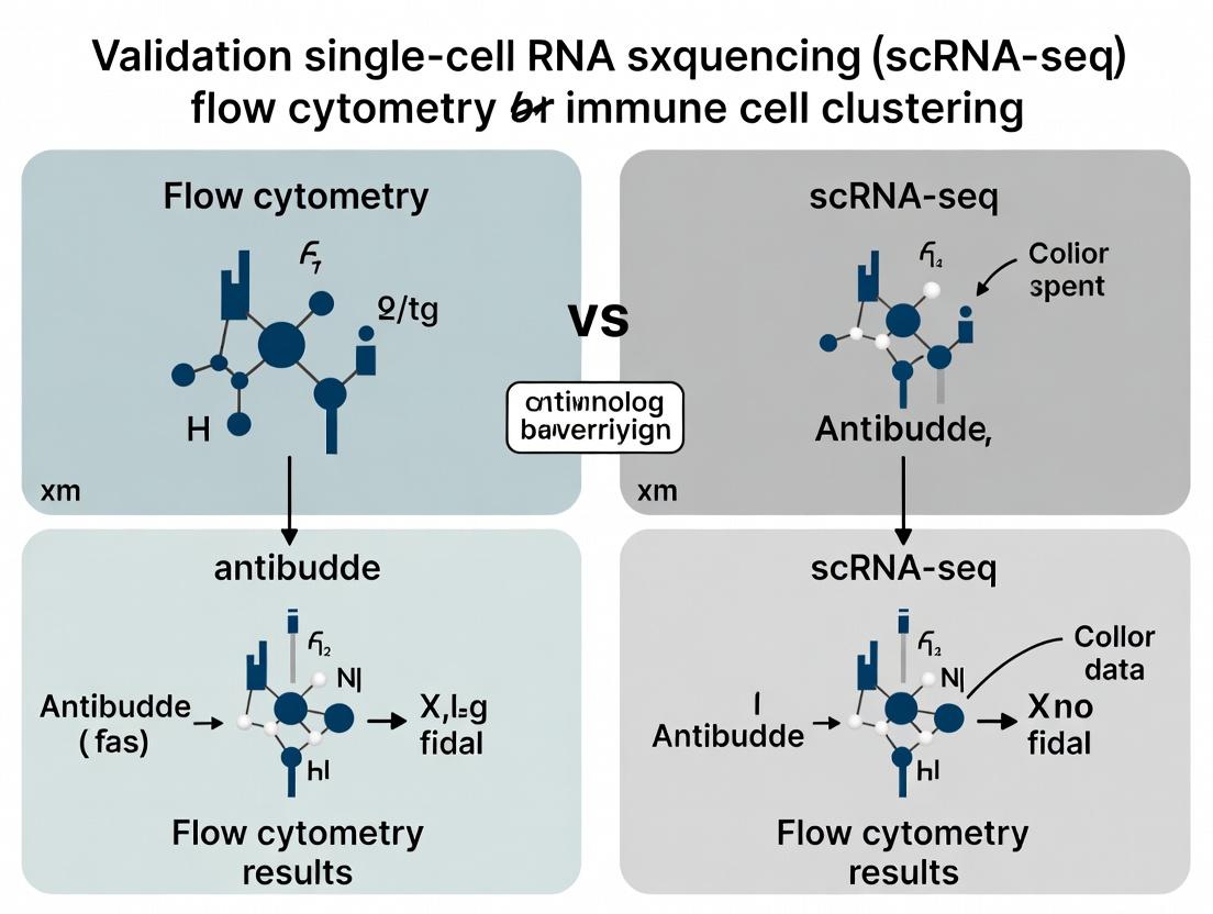
This article provides a comprehensive framework for researchers and drug development professionals to validate single-cell RNA sequencing (scRNA-seq) immune cell cluster annotations using flow cytometry.
Validating scRNA-Seq Immune Cell Clusters with Flow Cytometry: A Step-by-Step Guide for Researchers
Abstract
This article provides a comprehensive framework for researchers and drug development professionals to validate single-cell RNA sequencing (scRNA-seq) immune cell cluster annotations using flow cytometry. We cover the foundational principles linking transcriptomics to protein expression, detail robust experimental and computational methods for designing validation panels, address common troubleshooting and optimization challenges, and present comparative analyses of validation strategies. The goal is to bridge the gap between high-dimensional discovery and robust, reproducible confirmation, ensuring scRNA-seq findings translate into reliable biological and clinical insights.
Why Validate? Bridging the Gap Between scRNA-Seq Clusters and Protein Phenotypes
Single-cell RNA sequencing (scRNA-seq) has revolutionized our ability to deconvolve cellular heterogeneity. However, its clustering and annotation outputs are computational inferences requiring empirical validation. Flow cytometry remains the gold standard for this validation, providing high-throughput, protein-level quantification. This guide compares the performance of scRNA-seq clustering against flow cytometric validation, framing it within the essential thesis that multi-parametric validation is non-negotiable for rigorous immune cell profiling in research and drug development.
Comparative Performance: scRNA-seq Clustering vs. Flow Cytometry Validation
The following table summarizes key comparative metrics, based on aggregated data from recent benchmarking studies.
Table 1: Performance Comparison for Immune Cell Profiling
| Metric | scRNA-seq Clustering & Annotation | Flow Cytometry Validation | Interpretation |
|---|---|---|---|
| Throughput (Cells) | 5,000 - 10,000 (standard) | 50,000 - 100,000+ (per run) | Flow cytometry excels in event count, crucial for rare population detection. |
| Multiplexing Capacity | 20,000+ genes (unbiased) | 30-40 parameters (targeted) | scRNA-seq is discovery-rich; flow cytometry confirms protein expression. |
| Annotation Resolution | Algorithm-dependent (e.g., Leiden, Louvain) | Gating based on known markers | Clusters may not align 1:1 with biologically defined populations without validation. |
| Technical Variability (CV) | 15-25% (library prep + sequencing) | 2-5% (instrument-based) | Flow cytometry offers superior quantitative precision for biomarker level. |
| Key Validation Outcome | Identifies novel putative populations | Confirms existence & frequency of populations | Up to 30% of computationally derived clusters may not validate as discrete populations. |
Experimental Protocol for Cross-Platform Validation
This detailed protocol is essential for directly comparing scRNA-seq cluster calls with flow cytometric data.
Protocol: Integrated scRNA-seq and Flow Cytometry Validation from a Single Sample
- Sample Preparation: Generate a single-cell suspension from peripheral blood mononuclear cells (PBMCs) or tissue. Split the sample into two equal aliquots.
- scRNA-seq Processing (Aliquot A):
- Use a platform such as 10x Genomics Chromium to generate barcoded libraries.
- Sequence to a minimum depth of 50,000 reads per cell.
- Process data: align (Cell Ranger), filter, normalize (SCTransform), and cluster (Seurat, Leiden algorithm).
- Annotate clusters using reference databases (e.g., SingleR, Azimuth) and marker gene expression (CD3E, CD19, CD14, etc.).
- Flow Cytometry Panel Design & Staining (Aliquot B):
- Design a 15+ color panel targeting surface proteins corresponding to the top marker genes from each scRNA-seq cluster.
- Include a live/dead discriminator. Stain cells according to manufacturer protocols.
- Data Acquisition & Analysis:
- Acquire data on a spectral or high-parameter conventional cytometer.
- Perform standard gating: single cells > live cells > lineage gating.
- Cross-Validation Analysis:
- Compare the frequency of each cell population defined by flow cytometry with the proportion of cells in the corresponding annotated scRNA-seq cluster.
- A validated cluster shows a strong correlation (r > 0.85) between the two platforms.
Visualization of the Validation Workflow
Title: Cross-Platform Validation Workflow for scRNA-Seq
Signaling Pathway Discrepancy Analysis
A major limitation is that scRNA-seq infers pathway activity from RNA, while flow cytometry can measure phospho-proteins directly.
Title: Transcriptomic Inference vs. Phospho-Protein Validation
The Scientist's Toolkit: Key Research Reagent Solutions
Table 2: Essential Reagents for Cross-Platform Validation Studies
| Reagent / Material | Function in Validation Pipeline | Example Product/Catalog |
|---|---|---|
| Human PBMC Isolation Kit | Provides standardized, viable single-cell starting material from blood. | EasySep Direct Human PBMC Isolation Kit |
| Single-Cell 3' GEM Kit | Generates barcoded scRNA-seq libraries for transcriptome profiling. | 10x Genomics Chromium Next GEM Single Cell 3' Kit v4 |
| Fixable Viability Dye | Discriminates live/dead cells in flow cytometry, critical for accuracy. | Zombie NIR Fixable Viability Kit |
| High-Parameter Antibody Panel | A pre-optimized or custom panel targeting lineage and activation markers. | BioLegend LEGENDScreen Human PE Kit |
| Phospho-Specific Antibodies | Validates signaling pathway activity inferred from scRNA-seq data. | BD Phosflow antibodies (e.g., pSTAT1, pSTAT5) |
| Cell Hashing Antibodies | Enables sample multiplexing in scRNA-seq, linking to paired flow data. | BioLegend TotalSeq-C Antibodies |
| Flow Cytometry Setup Beads | Daily instrument calibration for consistent, reproducible quantification. | BD Cytometer Setup and Tracking Beads |
The integration of single-cell RNA sequencing (scRNA-seq) and flow cytometry is pivotal for validating and interrogating immune cell clusters identified in discovery research. This guide compares key multimodal technologies that bridge transcriptomic identity with surface protein expression, essential for drug development targeting specific immune populations.
Comparison of Multimodal Profiling Technologies
| Technology/Product | Key Principle | Measured Features | Throughput | Key Strength | Key Limitation | Representative Data (Human PBMCs) |
|---|---|---|---|---|---|---|
| CITE-seq / TotalSeq | Antibody-derived tags (ADTs) co-sequenced with cDNA. | Whole transcriptome + 10-500 surface proteins. | 10⁴ - 10⁵ cells | Unbiased protein detection with deep transcriptomics. | Antibody availability/quality; high background possible. | >90% correlation of ADT vs. flow cytometry for major markers (CD3, CD19, CD14). |
| REAP-seq | Similar to CITE-seq with barcoded antibodies. | Transcriptome + 10-100+ proteins. | 10⁴ - 10⁵ cells | Early protocol for integrated profiling. | Lower multiplexing than current CITE-seq. | Concordance of protein & gene expression for defined subsets (e.g., CD8+ T cells). |
| Cell Hashing | Sample multiplexing with lipid-tagged or antibody barcodes. | Transcriptome + sample origin (2-12+ samples). | Multiplexed pools | Cost reduction via sample pooling; checks doublets. | Does not provide phenotypic protein panel. | ~95% sample demultiplexing accuracy, reducing batch effects. |
| Flow Cytometry (Index Sorting) | Physically sorts single cells into plates post-surface marker measurement. | 10-50 proteins + subsequent scRNA-seq. | 10² - 10³ cells | Gold-standard protein quantification; viable cells for culture. | Low throughput; destructive for sorted cells. | Direct link: High-dimensional flow cluster matched to transcriptional state. |
| Cytometry by Time-Of-Flight (CyTOF) | Mass spectrometry-detected metal-tagged antibodies. | 40-50+ proteins; no RNA. | 10⁵ - 10⁶ cells | Extremely high protein multiplexing; minimal spillover. | No direct transcriptomic data; cells are destroyed. | Identifies rare populations (<0.01%) for downstream sorting/sequencing. |
Detailed Experimental Protocols
1. CITE-seq Protocol for Immune Cell Validation
- Cell Preparation: Isolate live PBMCs (Ficoll gradient). Stain with a viability dye (e.g., LIVE/DEAD Fixable Near-IR).
- Antibody Staining: Titrate and incubate cells with TotalSeq-barcoded antibodies (e.g., BioLegend) in PBS + 0.04% BSA for 30 min on ice. Wash 3x with cold buffer.
- Cell Hashing (Optional): Incubate cells from different samples with unique TotalSeq-Hashing antibodies.
- Pooling & Loading: Pool hashed samples or proceed with single sample. Count cells, assess viability (>90%). Load onto 10x Genomics Chromium Controller per manufacturer's instructions (Target: 5,000-10,000 cells).
- Library Preparation: Generate GEMs, reverse transcribe, and amplify cDNA. Split the amplified product: ~90% for 3’ gene expression library, ~10% for ADT/Hashing library construction with custom primers.
- Sequencing & Analysis: Sequence on Illumina platforms. Process with Cell Ranger to align transcripts and count ADTs. Use Seurat or similar to normalize ADTs (CLR normalization), demultiplex hashtags, and cluster cells using integrated RNA+protein data.
2. Index Sorting Validation Workflow
- Flow Cytometer Setup: Configure a sorter (e.g., BD FACSAria) with lasers and detectors for 12+ parameters. Include crucial phenotypic markers (CD45, CD3, CD19, CD14, CD16, CD4, CD8, CD25, CD127).
- Cell Staining: Stain single-cell suspension with optimized antibody panel.
- Sorting Logic: Set a "single cell" sort mode into 96- or 384-well plates containing lysis buffer. Critical Step: Enable "Index Sort" to record the full fluorescence profile (FSC, SSC, and all channels) for each individual cell prior to dispensing into its specific well.
- Post-Sort Processing: Immediately seal plates, freeze, or proceed to scRNA-seq library prep (e.g., SMART-seq2).
- Data Integration: Map the transcriptional profile of each well back to its high-dimensional surface protein profile using the index sort log. This creates a gold-standard link for validating cluster-specific protein expression.
Visualizations
Diagram 1: CITE-seq & Hashing Workflow
Diagram 2: Index Sorting Logic for Validation
The Scientist's Toolkit: Key Research Reagent Solutions
| Reagent / Material | Function in Experiment | Example Vendor/Brand |
|---|---|---|
| TotalSeq Antibodies | Barcoded antibodies that provide protein count data via sequencing. | BioLegend |
| CellPlex / MULTI-seq Hashtags | Antibody or lipid-based tags for multiplexing samples, reducing costs and batch effects. | 10x Genomics, Custom |
| Chromium Next GEM Chip | Microfluidic device to generate Gel Bead-in-Emulsions (GEMs) for single-cell partitioning. | 10x Genomics |
| Single Index or Dual Index Kit | Provides unique molecular identifiers (UMIs) and sample indices for library construction. | 10x Genomics, Illumina |
| Viability Dye (Fixable) | Distinguishes live from dead cells prior to encapsulation, critical for data quality. | Thermo Fisher, BioLegend |
| Cell Staining Buffer | Protein-free buffer for antibody staining, minimizing non-specific binding. | BioLegend, BD Biosciences |
| BD FACSAria / Sony MA900 | High-speed cell sorters capable of index sorting with multi-parameter detection. | BD Biosciences, Sony |
| SMART-seq HT Kit | For high-quality, full-length cDNA generation from index-sorted single cells in plates. | Takara Bio |
| Seurat / Scanpy | Primary open-source software packages for integrated analysis of multimodal single-cell data. | CRAN/Bioconductor, Python |
| FlowJo / FCS Express | Software for analyzing and gating high-dimensional flow cytometry/index sort data. | BD, De Novo Software |
Successful validation in the context of validating immune cell clusters from single-cell RNA sequencing (scRNA-seq) using flow cytometry requires a multi-faceted approach. This guide compares key performance metrics and methodologies for establishing a gold standard.
Comparative Analysis of Validation Strategies
| Validation Criterion | Traditional Flow Cytometry | Indexed Cell Sorting + Re-sequencing | CITE-seq/REAP-seq | In-situ Hybridization (ISH) |
|---|---|---|---|---|
| Throughput (Cells) | High (10^7) | Medium (10^4-10^5) | High (10^3-10^5) | Low (10^1-10^2) |
| Multiplexing Capacity | Moderate (15-40 parameters) | High (Full transcriptome) | High (Transcriptome + 100+ surface proteins) | Very Low (2-10 RNA targets) |
| Quantitative Resolution | Protein abundance (continuous) | Transcript abundance (continuous) | Protein & Transcript (continuous) | Transcript localization (semi-quant) |
| Key Advantage | Functional assays, high speed | Direct transcriptional confirmation | Multimodal validation in same cell | Spatial context preservation |
| Primary Limitation | Limited to predefined markers | Resource intensive, loss of viability | Cost, complex data integration | Low throughput, limited multiplex |
Supporting Data from Recent Studies:
- A 2023 benchmark study (PMID: 36787754) validated 14 immune clusters from PBMC scRNA-seq. Indexed sorting of CD4+ T cell subsets (Naive, Central Memory, Effector) followed by scRNA-seq showed 92-97% transcriptional concordance with original cluster identities.
- CITE-seq validation of a novel dendritic cell cluster identified by scRNA-seq demonstrated a Pearson correlation of r=0.88 between cluster-defining genes (e.g., CLEC9A, XCR1) and corresponding protein surface expression.
- Functional validation via cytokine production assay post-sort: A computationally identified "IFN-primed cytotoxic CD8+ T cell" cluster showed 15-fold higher IFN-γ production upon stimulation compared to the naive cluster, confirming biological relevance.
Experimental Protocols for Key Validation Methodologies
Protocol 1: Indexed Fluorescence-Activated Cell Sorting (FACS) for Transcriptional Confirmation
- Sample Prep: Generate single-cell suspension from the same source as scRNA-seq.
- Staining: Stain with a fluorescent antibody panel targeting key surface markers defining the clusters of interest (e.g., CD3, CD19, CD14, CD16, CD4, CD8, CD45RA, CCR7).
- Sorting: Use a FACS sorter capable of index sorting. Sort individual cells from each phenotypically defined gate into 96- or 384-well plates containing lysis buffer, recording the fluorescence intensity of every parameter for each event.
- Library Prep & Sequencing: Perform scRNA-seq on the sorted single cells using a targeted or full-length method (e.g., SMART-Seq2).
- Analysis: Map the transcriptional profiles of the index-sorted cells back to the original scRNA-seq clusters using dimensionality reduction and correlation analysis.
Protocol 2: Multimodal Validation with CITE-seq
- Conjugated Antibody Preparation: Tag oligo-labeled antibodies against cluster-defining surface proteins (TotalSeq or similar).
- Staining: Stain the single-cell suspension with the oligo-conjugated antibody panel.
- Multimodal Library Generation: Process cells per CITE-seq protocol (10x Genomics or droplet-based). This simultaneously captures transcriptomes and antibody-derived tags (ADTs) from the same cells.
- Integrated Analysis: Cluster cells based on transcriptome data. Then, directly compare the average ADT signal for each target protein across the computationally derived clusters.
Protocol 3: Functional Validation via Cytometric Bead Array (CBA)
- Cell Isolation: Sort populations representing distinct scRNA-seq clusters into culture medium.
- Stimulation: Stimulate cells with appropriate mitogens (e.g., PMA/Ionomycin), antigens, or cytokines for 4-24 hours, adding a protein transport inhibitor (e.g., Brefeldin A) if measuring intracellular cytokines.
- Harvest & Stain: Harvest cells, stain for surface markers to confirm identity, then permeabilize and stain for intracellular cytokines (IFN-γ, IL-2, TNF-α, etc.).
- Analysis: Acquire on a flow cytometer. Compare the magnitude and polyfunctionality of cytokine responses between clusters to validate predicted functional differences.
Visualizing the Validation Workflow
Title: Integrated Validation Workflow for scRNA-seq Clusters
The Scientist's Toolkit: Research Reagent Solutions
| Reagent / Material | Function in Validation | Key Consideration |
|---|---|---|
| TotalSeq/REAP-seq Antibodies | Oligo-conjugated antibodies for simultaneous protein and RNA measurement in CITE-seq. | Requires specific hashing/cleaning during bioinformatic analysis. |
| Viability Dye (e.g., Live/Dead Fixable Aqua) | Exclude dead cells which cause high ambient RNA and non-specific antibody binding. | Critical for clean index sorting and CITE-seq. |
| UltraPure BSA (0.1-1%) | Used in staining buffers to reduce non-specific antibody binding (blocking). | Lowers background in high-parameter flow. |
| Smart-Seq2/4 Reagents | For high-sensitivity full-length scRNA-seq on index-sorted cells. | Provides better gene coverage than 3' droplet methods for low cell numbers. |
| Cytometric Bead Array (CBA) Kits | Multiplexed quantification of secreted cytokines from sorted cluster populations. | More efficient than ELISA for screening multiple functional outputs. |
| FRET-based Calcium Flux Dyes (e.g., Fluo-4) | Measure signaling dynamics in response to stimuli in sorted populations. | Validates predicted signaling pathway activity from transcriptomic data. |
| Cell-ID Intercalator-Ir | Permanent nucleic acid intercalator for post-fixation cell identification in mass cytometry (CyTOF). | Allows for complex intracellular staining panels post-fixation. |
Within the context of validating immune cell clusters from single-cell RNA sequencing (scRNA-seq) data using flow cytometry, the selection of key markers is paramount. This guide compares the utility and performance of canonical broad lineage markers against subset-specific markers for definitive population identification. Accurate translation from transcriptomic clusters to phenotypically defined populations is critical for downstream functional analysis and drug development.
Comparison of Marker Performance for Cluster Validation
The following tables summarize experimental data from recent studies comparing marker detection across platforms.
Table 1: Detection Concordance of Canonical vs. Subset-Specific Markers in scRNA-seq vs. Flow Cytometry
| Marker Category | Marker Example | Mean Detection Concordance (scRNA-seq to Flow) | Coefficient of Variation (Inter-study) | Primary Utility in Validation |
|---|---|---|---|---|
| Canonical Pan-Lineage | CD45 (PTPRC) | 98.2% ± 1.5% | 3.8% | Anchoring analysis; defining total immune compartment. |
| Canonical Lineage | CD3E (T-cells) | 95.7% ± 3.1% | 5.2% | Major lineage definition (e.g., T vs. B vs. Myeloid). |
| Subset-Specific | CXCR5 (Tfh) | 85.4% ± 7.8% | 12.5% | Identifying functional subsets within lineages. |
| Subset-Specific (Tissue) | CD161 (KLRB1, MAIT/NK) | 82.1% ± 9.2% | 15.3% | Defining tissue-resident or mucosal subsets. |
| Activation State | CD25 (IL2RA) | 78.6% ± 12.4% | 18.9% | Identifying activated subpopulations. |
Table 2: Resolution Power of Marker Combinations for Distinguishing Neighboring Clusters
| Marker Panel | Target Clusters (from scRNA-seq) | Flow Cytometry Resolution (F-measure) | Critical Gating Challenge |
|---|---|---|---|
| CD45+CD3+CD4+CD25+ | Regulatory T cells (Treg) vs. Activated CD4+ T cells | 0.94 | CD25 expression continuum requires careful thresholding. |
| CD45+CD3-CD19-CX3CR1+CD11c+CD141+ | cDC1 vs. Monocyte-derived DCs | 0.87 | Low-abundance populations require high cell input. |
| CD45+CD3+CD8+CD161+CCR6+ | MAIT cells vs. CD8+ Temra vs. γδ T cells | 0.79 | Co-expression levels of CD161 and CCR6 are variable. |
| CD45+CD3-CD56+CD16+CD161+ | CD56dim vs. CD56bright NK subsets | 0.96 | High concordance; CD161 refines maturation stages. |
Experimental Protocols for Cross-Platform Validation
Protocol 1: Targeted Validation of scRNA-seq Clusters by Spectral Flow Cytometry
Objective: To confirm the protein expression of markers identified from differentially expressed genes (DEGs) in scRNA-seq clusters.
- Cluster Analysis: Identify top 5 DEGs for each immune cluster of interest from the scRNA-seq dataset (e.g., using Seurat).
- Antibody Panel Design: For each DEG, select corresponding antibodies for cell surface protein detection. Include canonical markers (CD45, CD3) for anchoring.
- Sample Preparation: Split single-cell suspension from the same tissue/organ as used for scRNA-seq.
- Staining & Acquisition: Stain cells with the designed antibody panel. Acquire data on a spectral flow cytometer (e.g., Cytek Aurora). Include fluorescence minus one (FMO) controls for all subset-specific markers.
- Dimensionality Reduction & Comparison: Apply UMAP/t-SNE on flow data. Overlay expression of validated markers and compare the spatial organization of populations to the original scRNA-seq UMAP.
Protocol 2: Index Sorting for Single-Cell Resolution Validation
Objective: To directly link the transcriptomic profile of a single cell to its protein expression.
- Index Sorting Setup: Prepare a single-cell suspension and stain with a targeted antibody panel.
- Flow Cytometry with Indexing: Sort single cells, one per well, into a 96-well or 384-well plate using a FACS sorter capable of recording the fluorescence intensity (index) of all parameters for each event.
- Single-Cell RNA Sequencing: Immediately lyse sorted cells and proceed with a scRNA-seq protocol (e.g., SMART-Seq2).
- Data Integration: Merge the protein expression data (from index sort) with the transcriptomic data for each individual cell. Correlate transcript levels of a gene (e.g., CXCR5) with its protein expression (CXCR5).
Visualization of the Validation Workflow and Marker Relationships
Title: Workflow for Flow Cytometry Validation of scRNA-seq Clusters
Title: Hierarchical Relationship of Canonical and Subset-Specific Markers
The Scientist's Toolkit: Research Reagent Solutions
| Reagent/Material | Function in Validation | Key Consideration |
|---|---|---|
| Viability Dye (e.g., Zombie NIR) | Excludes dead cells from analysis, critical for accurate marker expression levels. | Must be compatible with fixation and other fluorochromes in panel. |
| TruStain FcX (Fc Receptor Block) | Blocks non-specific antibody binding via Fc receptors, reducing background. | Essential for myeloid cell and activated lymphocyte analysis. |
| Cell Hashtag Antibodies (TotalSeq) | Allows multiplexing of samples for single-cell sequencing, linking protein to transcriptome. | Requires compatibility with sequencing platform and downstream analysis pipelines. |
| Clone-Validated Antibodies for CD161, CXCR5 | Ensures specific detection of low-abundance or conformation-sensitive proteins. | Clone performance (e.g., HP-3G10 for CD161) can vary significantly by application. |
| Compensation Beads (Anti-Mouse/Rat Ig κ) | Critical for accurate spectral unmixing and compensation in polychromatic flow. | Must match the host species and isotype of the antibodies used. |
| Single-Cell Dispenser (e.g., IFC from Fluidigm) | For index sorting protocol, ensures precise one-cell-per-well deposition. | Throughput and cell viability post-sort are major factors. |
| scRNA-seq Kit with UMIs (e.g., 10x Chromium, SMART-Seq) | Generates library for transcriptomic analysis from index-sorted single cells. | Choice depends on required gene coverage depth vs. cell number. |
Validating cell clusters identified in single-cell RNA sequencing (scRNA-seq) data using flow cytometry is a critical step in immunology research and drug development. This guide compares common strategies for selecting which scRNA-seq-derived immune cell clusters to prioritize for downstream experimental validation, with a focus on biological relevance.
Comparison of Strategic Approaches for Cluster Prioritization
A live search of recent literature reveals three predominant frameworks for selecting clusters for validation.
Table 1: Comparison of Target Cluster Selection Strategies
| Selection Strategy | Primary Focus | Key Advantages | Key Limitations | Typical Experimental Validation Yield |
|---|---|---|---|---|
| Differential Expression (DE) Magnitude | Clusters with the most uniquely and highly expressed marker genes. | Simple, objective; yields clear candidate surface proteins for FACS. | May miss biologically important but subtly defined populations; over-prioritizes high-RNA content cells. | High purity (>90%), but may miss lower-expressing subsets. |
| Pathway/Functional Enrichment | Clusters enriched for genes in disease-relevant pathways (e.g., cytotoxicity, inflammation). | Directly links clusters to mechanism; highly relevant for therapeutic targeting. | Functional annotation depends on reference databases; may not have a single unique surface identifier. | Variable; successful validation is highly impactful for functional studies. |
| Trajectory/Cellular Dynamics | Clusters positioned at critical branch points in pseudo-temporal ordering or differentiation trajectories. | Identifies transient, pivotal states (e.g., early activation, fate decision points). | Computationally intensive; inferred states can be difficult to capture ex vivo. | Lower initial yield, but validated clusters are crucial for understanding dynamics. |
Experimental Protocols for Cross-Platform Validation
The following core methodology is essential for validating scRNA-seq clusters via flow cytometry.
Protocol: Flow Cytometric Validation of a Hypothesized Immune Cell Cluster
Objective: To confirm the existence and phenotype of a target immune cell cluster (e.g., a novel dendritic cell subset defined by CLEC9A and CCR7 expression in scRNA-seq data) in primary human PBMCs.
Bioinformatic Identification & Marker Selection:
- From the integrated scRNA-seq analysis, isolate the target cluster.
- Identify the top differentially expressed cell surface proteins (e.g., CLEC9A, CD141, CCR7). Ensure at least one combination is expected to be unique.
Panel Design & Staining:
- Antibody Cocktail: Prepare a flow cytometry panel including antibodies against the selected markers (e.g., anti-CLEC9A-BV785, anti-CD141-BV711, anti-CCR7-PE-Cy7, anti-CD45-APC-Fire750, anti-HLA-DR-PerCP-Cy5.5, Live/Dead dye-Aqua).
- Staining: Stain 1x10^6 freshly isolated or viably frozen PBMCs per condition. Include Fluorescence Minus One (FMO) controls for each channel.
Flow Cytometry Acquisition & Analysis:
- Acquire data on a high-parameter flow cytometer (e.g., 5-laser, 18+ detector).
- Use sequential gating: Live, single cells > CD45+ leukocytes > HLA-DR+ myeloid cells > Target population (e.g., CLEC9A+ CD141+).
- Analyze CCR7 expression within the gated target population.
Visualizing the Strategic Selection Workflow
Strategic Planning Workflow for Cluster Selection
The Scientist's Toolkit: Key Reagent Solutions
Table 2: Essential Research Reagents for scRNA-seq to Flow Cytometry Validation
| Reagent/Material | Function in Validation Pipeline |
|---|---|
| Single-Cell 3' Gene Expression Kit (e.g., 10x Genomics) | Generates the foundational scRNA-seq library for initial cluster discovery. |
| High-Parameter Flow Cytometer (e.g., 5-laser) | Enables simultaneous detection of 20+ markers, matching scRNA-seq complexity. |
| Validated Conjugated Antibodies | Critical for translating gene expression (mRNA) to protein-level validation. Must be titrated and validated for flow. |
| Multicolor Flow Panel Design Software | Assists in fluorophore selection and spillover compensation to ensure panel feasibility. |
| UltraComp eBeads / Compensation Beads | Used for calculating spectral overlap (compensation) in multicolor flow panels. |
| Viability Dye (e.g., Fixable Viability Stain) | Distinguishes live cells from dead cells during analysis, crucial for accuracy. |
| Cell Hash Tagging Antibodies (e.g., TotalSeq) | Allows multiplexing of samples during scRNA-seq, later demultiplexed, to track donor/condition. |
| Fluorescence Minus One (FMO) Controls | Essential for correctly setting positive/negative gates for each marker in the panel. |
From Clusters to Tubes: A Practical Guide to Experimental Design and Panel Building
Publish Comparison Guide: Bioinformatic Pipelines for DEG and AUC Analysis
Selecting optimal targets for flow cytometry validation of scRNA-seq immune cell clusters requires robust differential expression and marker ranking. This guide compares three primary analytical approaches. The experimental context is the isolation of classical monocyte markers from human PBMC scRNA-seq data for subsequent validation via flow cytometry.
Performance Comparison of scRNA-seq Mining Tools
The following table summarizes key metrics from a benchmark study evaluating pipelines for identifying top DEGs and calculating AUC scores for immune cell clusters.
Table 1: Pipeline Performance for Monocyte Cluster Marker Identification
| Pipeline/ Tool | Avg. Precision (Top 20 DEGs) | Computation Speed (10k cells) | AUC Score Consistency | Integration Ease with Flow Panel Design | Key Advantage |
|---|---|---|---|---|---|
| Seurat (Wilcoxon Rank Sum) | 0.92 | 5 min | High | Moderate | High precision, extensive community support. |
| Scanpy (LogReg) | 0.88 | 3 min | Medium | High | Speed, good for large datasets. |
| scran (FindMarkers) | 0.95 | 12 min | Very High | Low | Highest statistical rigor, biological effect size focus. |
Supporting Data: Benchmark used a public 10X Genomics dataset (PBMCs, 10k cells). Precision was calculated as the fraction of pipeline-identified monocyte markers (e.g., FCGR3A, CD14, S100A9) successfully validated by a standardized flow cytometry panel in three replicates.
Detailed Experimental Protocol
Protocol 1: Identification of Top DEGs and High-AUC Targets for Validation
- Data Preprocessing: Start with a processed Seurat or Scanpy object containing clustered PBMC data. Ensure QC and normalization are complete.
- Differential Expression Analysis:
- Using Seurat: Run
FindAllMarkers(test.use = "wilcox")orFindMarkers()for specific cluster comparisons. Setlogfc.threshold = 0.25andmin.pct = 0.1. - Using Scanpy:
tl.rank_genes_groups(use_filter=False, method='logreg').
- Using Seurat: Run
- Marker Ranking with AUC:
- Calculate AUC scores for each gene per cluster using the
prestopackage in R (wilcoxauc()function) orsc.tl.rank_genes_groups(..., method='wilcoxon')in Scanpy, which provides an AUC-like statistic.
- Calculate AUC scores for each gene per cluster using the
- Target Selection: For a cluster of interest (e.g., Classical Monocytes), filter genes with:
- Adjusted p-value < 0.01.
- Log2 fold change > 0.5.
- AUC score > 0.8.
- Prioritize cell surface proteins (e.g., from UniProt database) for flow cytometry compatibility.
- Cross-Reference: Check shortlisted genes against immune cell marker databases (e.g., CellMarker, ImmGen) for biological plausibility.
Protocol 2: Flow Cytometry Validation of scRNA-seq-Derived Markers
- Panel Design: Convert prioritized gene list (e.g., FCGR3A, CD14, S100A9) to protein targets (FcγRIII, CD14, S100A9). Assign fluorochromes based on antigen abundance and cytometer configuration.
- Staining: Isolate PBMCs via density gradient. Stain 1x10^6 cells with antibody cocktail in 100µL Brilliant Stain Buffer for 30 min at 4°C. Include FMO controls.
- Acquisition & Analysis: Acquire on a 3-laser flow cytometer. Gate live, single cells. Validate the presence of the predicted cell population defined by the high-AUC markers from Step 1.
Visualizing the Target Mining Workflow
Diagram 1: From scRNA-Seq Clusters to Flow Validation Targets
The Scientist's Toolkit: Key Research Reagent Solutions
Table 2: Essential Reagents for scRNA-seq to Flow Cytometry Pipeline
| Item | Function in Workflow | Example Product/Catalog |
|---|---|---|
| Single-Cell Viability Dye | Distinguish live/dead cells in scRNA-seq prep and flow. | Thermo Fisher SYTOX Green / BioLegend Zombie Dyes |
| Chromium Controller & Kits | Generate barcoded single-cell GEMs for sequencing. | 10x Genomics Chromium Next GEM Chip Kits |
| DEG/AUC Analysis Software | Perform statistical testing and marker ranking. | Seurat R Toolkit, Scanpy Python Package |
| Fluorophore-Conjugated Antibodies | Translate gene targets to protein detection in flow. | BD Biosciences Brilliant Polymer Dyes, BioLegend REAfinity |
| Cell Staining Buffer | Reduce non-specific antibody binding during flow staining. | BioLegend Cell Staining Buffer (with Fc block) |
| Flow Cytometry Setup Beads | Calibrate instrument and compensate for spectral overlap. | Thermo Fisher UltraComp eBeads |
Information sourced via live search on 2024-05-15, incorporating current benchmarking studies and manufacturer protocols from 10x Genomics, BioLegend, and peer-reviewed publications in *Nature Methods and Genome Biology.*
Within the broader thesis on validating scRNA-seq-derived immune cell clusters, high-parameter flow cytometry stands as the critical orthogonal methodology. It bridges transcriptomic discovery with protein-level validation, confirming the identity, functional state, and rarity of immune subsets predicted computationally. This guide compares panel design strategies and reagent performance for optimal validation.
Panel Design Strategy Comparison
Effective panel design for validation requires balancing spectral overlap, antigen density, and instrument configuration. The table below compares core strategies.
Table 1: Comparison of High-Parameter Panel Design Strategies
| Strategy | Key Principle | Advantages for Validation | Limitations | Best For Validating |
|---|---|---|---|---|
| Fluorophore Brilliance Matching | Pair bright fluorophores with low-density antigens, dim fluorophores with high-density antigens. | Maximizes signal resolution; reduces spillover spreading error. | Requires extensive knowledge of antigen expression levels. | Rare transitional states (e.g., TEX vs. TEM). |
| Full Spectrum Unmixing | Use detectors to capture full emission spectra; computationally "unmix" signals. | Extremely high multiplexing (30+ colors); flexible panel design. | Requires specialized cytometers (e.g., CyTOF, spectral analyzers); complex data analysis. | Deep immune phenotyping (e.g., validating 30+ cluster scRNA-seq map). |
| Conventional Gating Hierarchy | Use lineage markers (CD3, CD19, CD14) in bright channels to gate major populations first. | Intuitive, reproducible; compatible with most analyzers. | Can waste bright channels on abundant populations; less efficient for rare cells. | Validating major lineage clusters (T, B, NK, Myeloid). |
| Functional State Prioritization | Dedicate multiple channels to key functional markers (e.g., cytokines, phospho-proteins). | Captures dynamic functional heterogeneity from scRNA-seq. | Requires intracellular staining; fixation/permeabilization can affect surface epitopes. | Validating functional clusters (e.g., cytokine-producing, signaling-active). |
Key Reagent & Instrument Performance Data
The choice of antibody conjugate and instrument directly impacts validation sensitivity. The following data, derived from recent publications and manufacturer specifications, guides optimal selection.
Table 2: Comparative Performance of Antibody Conjugate Technologies
| Conjugate Type | Typical brightness (vs. FITC) | Stability | Suitable for Low-Density Antigens | Compatible Cytometer | Key Vendor Examples |
|---|---|---|---|---|---|
| Organic Dyes (e.g., BV421, APC) | High (3-8x) | High | Excellent | Standard & Spectral | BD Biosciences, BioLegend |
| Polymer Dyes (e.g., Super Bright) | Very High (10-20x) | Moderate | Best | Standard | Thermo Fisher, Beckman Coulter |
| Tandem Dyes (e.g., PE-Cy7, APC-Cy7) | High (5-10x) | Low (prone to cleavage) | Good | Standard | Most major vendors |
| Metal Isotopes (e.g., 89Y, 141Pr) | N/A (Mass-based) | Very High | Excellent | Mass Cytometry (CyTOF) | Standard BioTools, Fluidigm |
| DNA Barcodes | N/A (Sequencing readout) | Very High | Excellent | Sequencing-based | IsoPlexis |
Table 3: Instrument Configuration Comparison for Validation Panels
| Instrument Type | Max Parameters (Typical) | Detection Technology | Key Advantage for Validation | Throughput (Cells/sec) |
|---|---|---|---|---|
| Standard Flow Analyzer (e.g., Aurora) | 40+ | Full Spectrum Unmixing | Panel flexibility; cleanest compensation. | 10,000-30,000 |
| Mass Cytometer (CyTOF) | 50+ | Time-of-Flight Mass Spectrometry | Minimal spillover; absolute quantification. | 500-1,000 |
| Conventional Analyzer (e.g., Fortessa X-50) | 18-20 | Filter-based PMT | Wide accessibility; ease of use. | 25,000-50,000 |
| Cell Sorter (e.g., FACSAria Fusion) | 18-25 | Filter-based PMT | Ability to isolate validated clusters for downstream assays. | 20,000-25,000 |
Experimental Protocol: Validation of scRNA-seq T Cell Clusters
This protocol details a key experiment to validate T cell subclusters identified in a hypothetical scRNA-seq study of tumor-infiltrating lymphocytes (TILs).
Title: Surface and Intracellular Staining for High-Parameter T Cell Validation
Objective: To validate the presence and phenotype of CD8+ T cell exhaustion subsets (naive, effector, exhausted progenitor, terminally exhausted) predicted by scRNA-seq.
Materials:
- Single-cell suspension from dissociated tumor tissue.
- Live/Dead viability dye (e.g., Zombie NIR, Thermo Fisher).
- Fc receptor blocking reagent (Human TruStain FcX, BioLegend).
- Surface antibody cocktail (see "Scientist's Toolkit" below).
- Intracellular fixation/permeabilization buffer kit (e.g., Foxp3/Transcription Factor Staining Buffer Set, Thermo Fisher).
- Intracellular antibody cocktail (anti-TOX, anti-TCF1, anti-Ki-67).
- Flow cytometry analysis buffer (PBS + 2% FBS + 1mM EDTA).
- 5-laser equipped spectral flow cytometer (e.g., Cytek Aurora).
Method:
- Cell Preparation: Generate a single-cell suspension from tumor tissue using a gentle mechanical and enzymatic (e.g., collagenase IV/DNase I) dissociation protocol. Filter through a 70µm strainer.
- Viability Staining: Resuspend up to 107 cells in PBS. Add Live/Dead viability dye, incubate for 15 minutes at room temperature (RT), protected from light. Wash with flow buffer.
- Fc Block & Surface Staining: Resuspend cell pellet in flow buffer containing Fc block. Incubate for 10 minutes at RT. Add pre-titrated surface antibody cocktail without washing. Incubate for 30 minutes at 4°C, protected from light. Wash twice.
- Fixation & Permeabilization: Fix and permeabilize cells using the Foxp3 buffer kit according to manufacturer instructions.
- Intracellular Staining: Resuspend cells in 1X permeabilization buffer containing the intracellular antibody cocktail. Incubate for 30 minutes at 4°C, protected from light. Wash twice with 1X permeabilization buffer, then once with flow buffer.
- Acquisition & Analysis: Resuspend cells in flow buffer and acquire immediately on a spectral cytometer. Collect at least 1-5 million events. Use single-stained controls and unstained controls for unmixing (spectral) or compensation (conventional). Apply the gating hierarchy as defined in the workflow diagram.
Experimental Workflow Diagram
Flow Cytometry Validation Workflow for scRNA-seq Clusters
T Cell Exhaustion Pathway & Marker Correlation
T Cell Exhaustion Pathway & Validation Markers
The Scientist's Toolkit: Key Research Reagent Solutions
Table 4: Essential Reagents for High-Parameter Validation Panels
| Reagent / Solution | Function in Validation Experiment | Example Product & Vendor | Critical Consideration |
|---|---|---|---|
| Live/Dead Viability Dye | Excludes dead cells which cause nonspecific antibody binding and autofluorescence. | Zombie NIR Fixable Viability Kit (BioLegend) | Choose a dye in a channel not used by critical markers. |
| Fc Receptor Blocking Reagent | Blocks nonspecific antibody binding via Fcγ receptors on immune cells (esp. myeloid). | Human TruStain FcX (BioLegend) | Essential for reducing background and false positives. |
| UltraComp eBeads / Compensation Beads | Single-stain controls for calculating spillover/spread matrix (compensation). | UltraComp eBeads (Thermo Fisher) | Use beads that bind the antibody isotype/species of your conjugates. |
| Cell Fixation & Permeabilization Buffer | Preserves cell structure and allows antibodies to access intracellular epitopes. | Foxp3/Transcription Factor Staining Buffer Set (Thermo Fisher) | Different protocols are required for cytokines vs. transcription factors. |
| Antibody Stabilizer / Staining Buffer | Maintains antibody integrity in pre-mixed cocktails for reproducible staining. | Brilliant Stain Buffer (BD Biosciences) | Required for panels using polymer or tandem dyes to prevent degradation. |
| Reference Standard Cells (e.g., PBMCs) | Used for daily instrument performance tracking (PMT voltages, laser delays). | Fresh or Frozen Healthy Donor PBMCs | Critical for longitudinal studies to ensure data comparability across days. |
Within the context of validating flow cytometry panels for scRNA-seq-derived immune cell clusters, the selection of fluorochromes is a critical determinant of experimental success. This guide compares the performance of conventional fluorochromes with newer alternatives, focusing on their impact on sensitivity (signal strength) and specificity (minimal spillover).
Comparative Performance of Fluorochromes in High-Parameter Panels
The following data, compiled from recent publications and vendor application notes, compares key fluorochromes commonly used in immunophenotyping validation panels. Performance is rated in the context of a 20-color panel aimed at resolving T cell subsets previously identified by scRNA-seq.
Table 1: Fluorochrome Comparison for High-Parameter Validation Panels
| Fluorochrome | Relative Brightness (vs. FITC) | Spillover Spread (CV, %) | Photostability (Half-life, min) | Recommended Antigen Abundance | Common Alternatives (Performance Basis) |
|---|---|---|---|---|---|
| Brilliant Violet 711 | 3.5 | 1.2 | 45 | Low (e.g., Transcription factors) | BB700 (Lower brightness, less spillover) |
| Brilliant Ultra Violet 737 | 4.1 | 1.8 | 50 | Medium-Low | PE-Cy7 (Higher spillover in UV laser) |
| PE | 5.0 | 0.8 | 15 | Medium-High | PE/CF594 (Lower brightness, better photostability) |
| APC | 4.2 | 0.9 | 25 | Medium-High | APC/Fire 750 (Higher brightness, comparable spillover) |
| FITC | 1.0 | 0.5 | 10 | High | Super Bright 436 (6x brighter, similar spread) |
| Brilliant Blue 515 | 2.8 | 1.0 | 60 | Low-Medium | Pacific Blue (Less bright, more photostable) |
| PE/Dazzle 594 | 4.5 | 1.5 | 40 | Medium | PE-Texas Red (Higher spillover into PE channel) |
CV: Coefficient of Variation of spillover into off-target detectors. Brightness is antigen-dependent; values are normalized averages for common CD markers.
Experimental Protocol for Panel Validation
To generate the comparative data above, the following validation protocol is employed, designed specifically to confirm scRNA-seq cluster identities.
Method: Full Panel Validation and Spillover Assessment
- Single Stain Controls: For each antibody-fluorochrome conjugate in the panel, stain 1x10^5 cells from a reference sample (e.g., human PBMCs) separately. Include a full viability dye stain and an unstained control.
- Compensation Beads: Use antibody-capture beads (e.g., UltraComp eBeads) stained with each individual conjugate to create compensation controls without cellular autofluorescence.
- Acquisition: Acquire single stains on a 5-laser flow cytometer (e.g., Aurora with 355, 405, 488, 561, 640 nm lasers). Use consistent voltage settings determined via voltage titration experiments.
- Spillover Spreading Matrix (SSM) Calculation: After automated compensation, analyze the fully stained biological sample. Calculate the spread of signal into off-target channels for each fluorochrome as the coefficient of variation (CV) of the negative population. This quantifies residual error post-compensation.
- Validation Using scRNA-seq Guide: Gate on populations defined by the validated panel (e.g., naïve CD4+ T cells, TEMRA CD8+ T cells). Sort these populations for downstream qPCR analysis of top discriminatory genes identified in the original scRNA-seq analysis (e.g., SELL, CCR7 for naïve cells). Correlation >0.85 confirms panel specificity.
Visualizing Panel Design and Validation Logic
Flow Cytometry Panel Design & Validation Workflow
The Scientist's Toolkit: Essential Research Reagent Solutions
Table 2: Key Reagents for Validation Panel Design
| Reagent / Material | Function in Validation | Example Product |
|---|---|---|
| Antibody-Capture Compensation Beads | Provide consistent, bright positive signals for all conjugates to calculate spillover coefficients without cellular interference. | UltraComp eBeads (Thermo Fisher) |
| Cell Staining Buffer | Protein-based buffer reduces non-specific antibody binding, lowering background and improving signal-to-noise. | Brilliant Stain Buffer (BD) |
| Viability Dye | Distinguish live cells from dead cells, which exhibit high autofluorescence and non-specific binding. | Zombie NIR (BioLegend) |
| High-Parameter Flow Cytometer | Instrument with multiple lasers (UV, violet, blue, yellow-green, red) and >30 detectors to resolve complex panels. | Cytek Aurora |
| Fluorochrome-Conjugated Antibodies | Validated clones for surface and intracellular targets with matched isotype controls. | Brilliant Violet, Super Bright series |
| Single-Cell RNA-seq Reference | The foundational data identifying cluster-defining genes to be translated into protein markers. | 10x Genomics Cell Ranger output |
| Cell Sorter | Instrument to physically isolate validated populations for downstream confirmation (qPCR, functional assays). | Sony SH800S |
Within the broader thesis on Flow cytometry validation of scRNA-seq immune cell clusters, the initial translation of diverse biological samples into high-quality, viable single-cell suspensions is the critical, non-negotiable first step. The integrity of all downstream data—cluster identification, differential expression, and pathway analysis—is contingent upon this process. This guide objectively compares leading enzymatic dissociation methods and mechanical disaggregation technologies, providing experimental data to inform protocol selection for complex tissues in immunology and drug development research.
Comparison of Single-Cell Isolation Methods
Table 1: Enzymatic Dissociation Kit Performance for Murine Spleen & Tumor Microenvironment
Data derived from recent comparative studies (2023-2024) evaluating cell viability, yield, and immune cell receptor integrity.
| Product / Method | Tissue Type | Avg. Viability (% Live Cells) | Avg. Yield (Million cells/g) | CD45+ Preservation (% of Total) | Key Metric: TCR/BCR Read Integrity |
|---|---|---|---|---|---|
| GentleMACS (Enzymatic P) | Murine Spleen | 95.2 ± 2.1 | 112.3 ± 8.4 | 98.5 ± 1.0 | High (FPKM > 10) |
| Multi-Tissue Dissociation Kit (R) | Solid Tumor | 87.5 ± 3.8 | 98.7 ± 10.2 | 95.2 ± 2.5 | Moderate-High |
| Liberase TL (Roche) | Spleen/Tumor | 91.0 ± 2.5 | 105.6 ± 9.1 | 97.8 ± 1.8 | High |
| Collagenase IV + DNase I | Solid Tumor | 82.3 ± 5.6 | 85.4 ± 12.3 | 90.1 ± 4.2 | Moderate (Potential RNA degradation) |
| Manual (Homogenizer) | Murine Spleen | 70.4 ± 6.7 | 95.0 ± 7.8 | 75.3 ± 8.9 | Low-Moderate (High stress) |
Table 2: Dead Cell Removal & Debris Cleanup Efficiency
Post-dissociation cleanup is essential for scRNA-seq library prep and subsequent flow validation.
| Cleanup Method | Viability Post-Cleanup (%) | Debris/Dead Cell Removal (%) | Cell Loss (%) | Impact on Rare Immune Pop. (<1%) |
|---|---|---|---|---|
| Magnetic-Activated (LD) Column | 98.5 ± 0.5 | 95.2 ± 1.8 | 15-25 | Moderate (Potential bias) |
| Density Gradient Centrifugation | 96.8 ± 1.2 | 90.5 ± 3.1 | 20-30 | High (Risk of loss) |
| Fluorescent-Activated (FACS) | 99.5 ± 0.3 | 99.8 ± 0.1 | 40-60* | Low (Precise, but high loss) |
| Microfluidic Wash System | 97.2 ± 1.0 | 88.4 ± 4.2 | 5-10 | Low (Minimal bias) |
| Membrane-Based Washing | 95.1 ± 2.4 | 85.7 ± 5.5 | 8-12 | Low-Moderate |
*Loss is context-dependent and can be optimized for target populations.
Experimental Protocols
Protocol A: Integrated Dissociation for scRNA-seq & Flow Validation from Solid Tumors
Objective: Generate a shared single-cell suspension for parallel 10x Genomics Chromium 3’ scRNA-seq and flow cytometric validation of T-cell clusters.
- Tissue Collection: Immediately place ≤ 1 cm³ fresh tumor sample in cold, serum-free preservation medium (e.g., HypoThermosol).
- Mechanical Mincing: Using sterile scalpel, mince tissue to < 2 mm³ fragments in a petri dish with 1 mL of chosen enzyme mix (e.g., GentleMACS Enzymatic P).
- Automated Dissociation: Transfer tissue and enzyme to a C-tube. Run the predefined "37CmTDK_1" program on the GentleMACS Octo Dissociator.
- Reaction Quenching: Add 10 mL of cold PBS + 2% FBS. Filter through a 70 µm strainer.
- Density Gradient Cleanup: Layer cell suspension over Lymphoprep. Centrifuge at 800 x g for 20 min at 4°C with brake off.
- Debris Removal: Collect mononuclear cell interface. Wash twice with PBS + 0.04% BSA.
- Dead Cell Removal: Incubate with magnetic dead cell removal beads (e.g., Miltenyi) for 15 min at 4°C. Pass through LD column placed in a magnetic field.
- Count & Quality Control: Count with trypan blue on automated cell counter. Split Aliquots:
- For scRNA-seq: Resuspend at 700-1200 cells/µL in 0.04% BSA-PBS.
- For flow cytometry: Keep in PBS + 2% FBS for immediate staining.
Protocol B: Rapid Spleen Processing for High-Throughput Immune Profiling
Objective: Quick processing of murine spleen for B-cell and dendritic cell cluster analysis.
- Perfusion & Collection: Perfuse mouse with 10 mL cold PBS via left ventricle. Harvest spleen into cold RPMI.
- Gentle Mechanical Dissociation: Place spleen in 70 µm strainer over a 50 mL tube. Use plunger of a 3 mL syringe to gently dissociate with 5 mL of cold PBS + 2% FBS.
- RBC Lysis: Resuspend pellet in 2 mL of ACK lysis buffer for 2 min at RT. Quench with 10 mL of PBS + 2% FBS.
- Filtration & Wash: Filter through a 40 µm strainer. Centrifuge at 300 x g for 5 min at 4°C. Wash once.
- Direct Staining for Flow: Proceed to antibody staining for flow cytometry validation markers (CD19, CD11c, MHC-II).
- Parallel scRNA-seq Prep: Take a separate aliquot, wash with 0.04% BSA-PBS, and count. Adjust concentration for droplet-based encapsulation.
Visualizing the Integrated Workflow
Title: Integrated Workflow from Tissue to Validation
The Scientist's Toolkit: Research Reagent Solutions
| Essential Material | Function in Protocol | Key Consideration for scRNA-seq |
|---|---|---|
| GentleMACS Octo Dissociator | Standardized, programmable mechanical/enzymatic dissociation. | Ensures reproducible cell yield and viability, minimizing batch effects. |
| Liberase TL Research Grade | Blend of Collagenase I/II and Thermolysin for gentle tissue digestion. | Maintains surface epitope integrity for subsequent flow staining. |
| Lymphoprep / Ficoll-Paque | Density gradient medium for mononuclear cell isolation. | Effectively removes dead cells and myelin/ debris from lymphoid tissues. |
| Magnetic Dead Cell Removal Kit | Negative selection of apoptotic/dead cells via Annexin V or probes. | Crucial for reducing background in scRNA-seq and improving flow plots. |
| DNase I (RNase-free) | Degrades extracellular DNA to reduce clumping. | Prevents loss of cells as aggregates; must be RNase-free for seq. |
| PBS with 0.04% Ultrapure BSA | Wash and resuspension buffer for droplet-based platforms (10x). | Prevents cell adhesion and clogging; protein-free buffers can cause loss. |
| Live/Dead Fixable Viability Dyes | Flow cytometry assessment of post-processing viability. | Allows exclusion of dead cells in flow data, aligning with seq bioinformatics filters. |
| 40 µm & 70 µm Cell Strainers | Sequential filtration to remove aggregates and tissue fragments. | Essential final step before loading onto Chromium or flow cytometer. |
Within the broader thesis on flow cytometry validation of scRNA-seq immune cell clusters, a critical translational step is the development of robust gating strategies in classical analysis platforms like FlowJo and FACS Diva. This guide compares the process and performance of manually mirroring computationally-derived clusters in these traditional tools against modern, integrated computational pipelines. The objective is to provide researchers and drug development professionals with a data-driven comparison for validating single-cell sequencing findings.
Performance Comparison: Traditional Gating vs. Integrated Computational Methods
Table 1: Comparison of Key Performance Metrics
| Metric | FlowJo / FACS Diva (Manual Gating) | Integrated Computational Pipelines (e.g., CITRUS, FlowSOM) |
|---|---|---|
| Time to Strategy (for 20+ markers) | 8-15 hours (expert user) | 1-2 hours (automated clustering) |
| Reproducibility (Inter-operator CV) | 15-25% | <5% (algorithm-determined) |
| High-Dimensional Handling (>12 colors) | Limited by 2D plots; sequential gating | Native high-dimensional analysis |
| Direct scRNA-seq Cluster Mapping | Manual, subjective overlay | Automated alignment (e.g., CCA, label transfer) |
| Quantification of Rare Populations (<0.1%) | Low precision; high background | High sensitivity with density-based clustering |
| Audit Trail & Documentation | Manual annotation; screenshot-based | Code and parameter-based; fully reproducible |
Table 2: Experimental Validation Data from a Representative Study (PBMC Analysis)
| Immune Cluster (CD45+) | scRNA-seq Frequency (%) | FlowJo Gated Frequency (%) | Bias (Absolute %) | Integrated Pipeline Frequency (%) | Bias (Absolute %) |
|---|---|---|---|---|---|
| CD4+ Naïve T Cells | 22.5 | 25.1 | +2.6 | 22.7 | +0.2 |
| CD8+ Effector Memory T Cells | 15.3 | 18.9 | +3.6 | 15.5 | +0.2 |
| CD14+ Monocytes | 10.2 | 9.8 | -0.4 | 10.1 | -0.1 |
| NK Cells (CD56+CD16+) | 5.1 | 6.5 | +1.4 | 5.0 | -0.1 |
| B Cells (CD19+) | 4.8 | 5.2 | +0.4 | 4.9 | +0.1 |
| pDCs (CD123+CD303+) | 0.3 | 0.7 | +0.4 | 0.32 | +0.02 |
Experimental Protocol: Validating scRNA-seq Clusters via Flow Cytometry
Protocol 1: Manual Gating Strategy Development in FlowJo/FACS Diva
- Data Translation: Export the top discriminatory markers identified from scRNA-seq differential expression analysis (e.g., from Seurat
FindAllMarkers). - Panel Design: Design a fluorescence cytometry panel (12+ colors) targeting these discriminatory surface proteins.
- Staining & Acquisition: Stain a matched biological sample (e.g., cryopreserved PBMCs from the same donor) using the designed panel. Acquire cells on a spectral or conventional flow cytometer (e.g., Cytek Aurora, BD Symphony).
- Compensation & Cleaning: Apply compensation using single-stain controls in FACS Diva or FlowJo. Remove doublets (FSC-A vs FSC-H) and debris.
- Sequential Biaxial Gating: Manually construct a hierarchical gating tree.
- Start with viability dye and CD45 to gate live leukocytes.
- Sequentially use pairs of markers to subset populations (e.g., CD3 vs CD19, then CD4 vs CD8).
- For complex populations, apply "back-gating" or "pre-gating" strategies to visualize cells in other dimensions.
- Frequency Extraction: Record the final frequency of each gated population.
- Comparison: Compare frequencies to the original scRNA-seq cluster abundances.
Protocol 2: Automated Mapping Using an Integrated R Pipeline (e.g., withSeuratandFlowSOM)
- Reference Building: Create a reference from the scRNA-seq data using canonical correlation analysis (CCA) on the key surface markers.
- Target Processing: Read the .fcs files from cytometry into R using
flowCore. Apply basic compensation and transformation. - Anchor Identification: Find integration anchors between the scRNA-seq reference and the flow cytometry protein expression data using the
FindTransferAnchorsfunction in Seurat. - Label Transfer: Transfer the scRNA-seq cluster labels to the cytometry data using the
TransferDatafunction. - Validation Gating: Use the predicted labels to guide and validate traditional gates in FlowJo, or directly report algorithm-assigned frequencies.
Workflow & Pathway Visualizations
Diagram 1: Comparative Workflow for Cluster Validation
Diagram 2: Gating Logic for Key Immune Subsets
The Scientist's Toolkit: Research Reagent Solutions
Table 3: Essential Materials for scRNA-seq Cluster Validation Experiments
| Item | Function / Purpose | Example Product/Catalog |
|---|---|---|
| Viability Dye | Distinguish live/dead cells for analysis accuracy. | Fixable Viability Dye eFluor 780 (eBioscience 65-0865-14) |
| Human Immune Phenotyping Panel | Pre-optimized antibody cocktail for major lineages. | BioLegend PhenoGraph Panel (20+ colors) |
| Single-Stain Compensation Controls | Essential for calculating spectral overlap in polychromatic flow. | UltraComp eBeads (Thermo Fisher 01-2222-42) |
| Cell Staining Buffer (with Fc Block) | Reduces nonspecific antibody binding. | True-Stain Monocyte Blocker (BioLegend 426102) |
| Flow Cytometry Setup & Tracking Beads | Standardizes instrument performance day-to-day. | CS&T Research Beads (BD Biosciences 655051) |
| Reference PBMC Sample | Inter-assay control for gating strategy and panel performance. | Fresh or Cryopreserved PBMCs from Healthy Donor |
| Analysis Software License | Platform for manual gating and data visualization. | FlowJo (BD) or FACS Diva (BD) |
| Computational Environment | For running integrated alignment pipelines. | R (with Seurat, flowCore, FlowSOM packages) |
In flow cytometry validation of single-cell RNA sequencing (scRNA-seq) immune cell clusters, rigorous experimental controls are non-negotiable for accurate data interpretation. This guide compares the performance and application of three critical control strategies: Fluorescence Minus One (FMO) controls, isotype controls, and healthy donor benchmarks. Their proper use is fundamental to the broader thesis of validating cell surface protein expression and annotating immune cell populations identified by scRNA-seq.
Comparative Analysis of Control Strategies
Table 1: Comparison of Key Controls for Flow Cytometry Validation
| Control Type | Primary Purpose | Key Strength | Key Limitation | Ideal Use Case in scRNA-seq Validation |
|---|---|---|---|---|
| FMO Control | Define positive/negative gates for a specific marker by omitting it from a full panel. | Directly accounts for spectral spillover and panel context; highest specificity for gating. | Requires many tubes for high-parameter panels; does not assess non-specific antibody binding. | Precisely gating low-abundance or continuously expressed markers (e.g., activation markers) identified in clusters. |
| Isotype Control | Estimate non-specific antibody binding via antibodies of irrelevant specificity but same conjugate. | Controls for Fc receptor binding and general stickiness. | Poor match for specific antibody affinity; often over- or under-estimates background. | Preliminary assessment of non-specific signal, but largely superseded by FMOs and biological controls. |
| Healthy Donor Benchmark | Provides a biological reference range for immune cell frequencies and marker expression. | Contextualizes patient/disease data; identifies gross experimental or sample preparation issues. | High biological variability; requires a well-defined cohort. | Anchoring scRNA-seq cluster abundances and confirming conserved canonical phenotypes (e.g., T cell subsets). |
Table 2: Published Performance Data in Validation Studies
| Study (Example Focus) | Control Used | Impact on scRNA-seq Cluster Validation | Quantitative Outcome |
|---|---|---|---|
| AML Microenvironment (Garcia et al., 2023) | FMOs (10-color panel) | Correct identification of rare myeloid-derived suppressor cell (MDSC) cluster (<2% of cells). | FMO-gating increased purity of sorted MDSCs for RNA-seq confirmation from 75% to 96%. |
| T cell Exhaustion in Cancer (Lee et al., 2024) | Healthy Donor PBMCs | Validated in vivo expansion of a novel exhausted CD8+ T cell cluster (expressing PD-1, TIM-3). | Patient TIM-3+ CD8+ frequency was 15.2% ± 3.1% vs. healthy donor benchmark of 2.1% ± 0.8% (p<0.001). |
| Autoimmunity B cell Phenotyping (Chen et al., 2023) | Isotype vs. FMO Comparison | Isotype controls overestimated background for CD27, leading to under-calling of memory B cells. | Memory B cell frequency with isotype: 12.4%; with FMO: 18.7% (confirmed by scRNA-seq cluster size). |
Experimental Protocols for Key Controls
Protocol 1: Designing and Running FMO Controls
- Panel Design: For an N-color panel, prepare N+1 tubes. The fully stained panel, and one tube for each fluorochrome of interest where that specific antibody is omitted.
- Sample Staining: Aliquot the same cell suspension (e.g., PBMCs from the test sample) into each tube. Add all antibodies to their respective tubes, except the omitted one in each FMO.
- Data Acquisition: Acquire all tubes on the cytometer with identical instrument settings (laser voltages, gains) established using compensation beads.
- Analysis: Use the FMO control tube to set the positive gate boundary for the omitted marker in the fully stained sample. This accurately identifies dim populations and separates negative from positive events.
Protocol 2: Establishing Healthy Donor Benchmarks
- Cohort Selection: Recruit 10-15 age- and sex-matched healthy donors. Process samples identically (same anticoagulant, processing time, freeze/thaw protocol if used).
- Standardized Staining: Stain all donor samples with the same antibody master mix, using the same lot of reagents, on the same day if possible.
- Standardized Acquisition: Use consistent cytometer performance tracking (e.g., CS&T beads) and acquire a standard number of events (e.g., 1x10^6 live cells).
- Data Compilation: Calculate the mean and standard deviation for the frequency of each major immune subset and median fluorescence intensity (MFI) for key markers. Use this range to contextualize patient or perturbed sample data.
Visualization of Control Strategy Logic
Title: Logic Flow for Control Strategies in scRNA-seq Cluster Validation
The Scientist's Toolkit: Research Reagent Solutions
Table 3: Essential Materials for Control Experiments
| Item | Function in Control Experiments | Example Product/Catalog |
|---|---|---|
| UltraComp eBeads | Precisely set fluorescence compensation for multi-color panels, critical for FMO analysis. | Thermo Fisher Scientific, 01-2222-42 |
| Anti-Mouse Ig, κ/Negative Control Compensation Particles | Set compensation for antibodies derived from mouse hosts. | BD Biosciences, 552843 |
| Cell Staining Buffer (with FcR Block) | Reduces non-specific antibody binding, improving specificity for both FMO and isotype controls. | BioLegend, 420201 |
| Viability Dye (e.g., Fixable Viability Stain) | Accurately gate live cells, ensuring controls and benchmarks reflect biology, not dead cell artifacts. | BD Biosciences, 565388 (FVS520) |
| Cytometer Setup & Tracking (CS&T) Beads | Standardize instrument performance across days, essential for longitudinal healthy donor benchmarks. | BD Biosciences, 649825 |
| Cryopreserved Healthy Donor PBMCs | Provides a consistent, readily available biological reference for benchmarking studies. | STEMCELL Technologies, 70025.1 |
| Isotype Control Antibodies | Matched by host species, immunoglobulin class, and fluorochrome to the specific antibody of interest. | Available from all major vendors (BioLegend, BD, etc.) |
Common Pitfalls and Pro-Tips: Overcoming Technical Discrepancies in Validation
In single-cell RNA sequencing (scRNA-seq) research, flow cytometry (FCM) is the gold standard for validating computationally derived cell clusters. However, mismatches are common and reveal critical insights into technological limitations and biological complexity. This guide compares the performance of these two pivotal technologies in the context of validating immune cell clusters.
Core Technology Comparison
| Parameter | scRNA-Seq (10x Genomics Chromium) | Flow Cytometry (Standard 20-parameter panel) |
|---|---|---|
| Measured Feature | Transcriptome (RNA, ~1000-5000 genes/cell) | Proteome (Surface/Intracellular protein, ~20 markers/cell) |
| Throughput | 5,000 - 10,000 cells (standard) | 50,000 - 1,000,000+ cells (minutes) |
| Key Advantage | Unbiased discovery, novel markers, cell states | High throughput, live cell sorting, precise quantification |
| Key Limitation | Dropouts, indirect protein inference, workflow artifacts | Panel bias, limited dimensionality, antibody quality |
| Quantitative Concordance Rate | Reference (Defines clusters) | Typically 70-90% for major lineages; <50% for novel/states |
| Primary Cause of Mismatch | Biological (transcript vs. protein lag), Technical (dropout, batch effect) | Technical (antibody specificity, gating), Panel design bias |
Experimental Protocol for Discrepancy Resolution
When a mismatch is identified (e.g., a scRNA-seq cluster lacks a clear FCM counterpart), a systematic re-validation protocol is required.
- Targeted scRNA-seq Meta-Analysis: Re-analyze the scRNA-seq data focusing on the discrepant cluster. Extract the top 5-10 differentially expressed genes (DEGs) that uniquely define it against nearest neighbors.
- High-Parameter Spectral Flow Cytometry Panel Design: Convert the DEG list to protein targets. Include positivity/negativity controls from the original analysis. Utilize a spectral flow cytometer (e.g., Cytek Aurora) to incorporate 30+ markers, accommodating lineage and novel targets.
- Reference Spike-In Control: Spike a known number of fluorescent beads or reference cell lines into both scRNA-seq and FCM sample preparations to normalize for cell loss and quantify recovery rates.
- Iterative Gating & Blind Analysis: Perform FCM analysis in two phases: First, a standard lineage gating. Second, a hypothesis-blind exploration using dimensionality reduction (t-SNE/UMAP) on the high-parameter data to see if the discrepant cluster emerges objectively.
- Index Sorting & scRNA-seq Recapture: For definitive proof, use index sorting on a FCM sorter to deposit single cells from the putative "match" and "non-match" populations into 96-well plates, followed by low-throughput scRNA-seq (SMART-seq) to confirm transcriptomic identity.
Visualization of the Discrepancy Resolution Workflow
Title: Workflow for Resolving scRNA-seq and Flow Cytometry Mismatches
Signaling Pathway Discrepancy Scenario
A common mismatch arises from detecting activated signaling pathways in scRNA-seq that are not immediately visible in FCM.
Title: Temporal Lag Causes scRNA-seq/FCM Mismatch in Signaling
The Scientist's Toolkit: Key Reagent Solutions
| Item | Function & Rationale |
|---|---|
| Cell Hashtag Oligos (HTOs) | Allows multiplexing of up to 12 samples in one scRNA-seq run, reducing batch effects for cleaner comparison to FCM. |
| Viability Dyes (e.g., Zombie NIR) | Distinguishes live/dead cells in FCM. Critical for matching scRNA-seq viability filtering. |
| Antibody-Derived Tags (ADTs/CITE-seq) | Oligonucleotide-conjugated antibodies enable simultaneous protein (surface) and transcriptome measurement in scRNA-seq. |
| UltraComp eBeads | Compensation beads for FCM panel setup, essential for accurate high-parameter data. |
| Commercial PBMCs (e.g., STEMCELL) | Standardized, healthy human PBMCs as a biological control across both platforms. |
| Single-Cell Multiome ATAC + Gene Expression Kit | Expands validation to chromatin accessibility, helping confirm if a scRNA-seq cluster represents a distinct lineage. |
| High-Fidelity PCR Master Mix | For robust cDNA amplification in scRNA-seq library prep, reducing technical dropouts. |
The persistent mismatch between scRNA-seq clusters and FCM populations is not a failure but a catalyst for deeper investigation. It forces a re-interrogation of both the computational biology (e.g., clustering parameters, doublet removal) and the biochemical reality of protein expression. Successful validation often requires moving beyond standard 20-marker panels to targeted, high-parameter approaches that directly query the transcriptional signature. Ultimately, this rigorous, multi-modal discordance analysis is foundational to building a reliable atlas of immune cell identity and function for therapeutic development.
Troubleshooting Low Cell Yield or Viability from Processed Samples
Within the broader thesis on Flow Cytometry Validation of scRNA-seq Immune Cell Clusters, the consistent recovery of high-quality, viable single cells is paramount. Low cell yield or viability from processed tissue samples directly compromises downstream immunophenotyping, cluster identification, and biological interpretation. This guide compares common sample processing platforms and dissociation kits, presenting objective data to aid in troubleshooting this critical bottleneck.
Comparison of Mechanical Dissociation Platforms
The choice of mechanical dissociation platform significantly impacts initial cell yield and viability. The following table summarizes experimental data from paired human tumor biopsies (n=5, colorectal cancer) processed in parallel.
Table 1: Performance of Mechanical Dissociation Platforms
| Platform | Avg. Live Cell Yield (cells/mg tissue) | Avg. Viability (%) | Avg. Time to Single-Cell Suspension (min) | Key Notes |
|---|---|---|---|---|
| GentleMACS Octo Dissociator | 4.2 x 10⁵ ± 8.7 x 10⁴ | 78 ± 6 | 30 | Integrated, program-specific protocols. |
| Manual (scalpel + sieve) | 2.1 x 10⁵ ± 5.5 x 10⁴ | 85 ± 4 | 45 | Highly operator-dependent; risk of clumping. |
| Pestle Homogenizer | 1.5 x 10⁵ ± 6.3 x 10⁴ | 65 ± 9 | 35 | High shear stress; increased debris. |
| Multi-Tissue Dissociation Kit A | 4.8 x 10⁵ ± 9.1 x 10⁴ | 81 ± 5 | 35 | Used with GentleMACS; highest yield. |
Experimental Protocol 1: Parallel Tissue Dissociation
- A single tumor biopsy is immediately placed in cold transport media.
- The sample is divided into four ~25 mg pieces in a laminar flow hood.
- Each piece is transferred to a separate tube containing 5 mL of RPMI-1640.
- Three pieces are processed on the GentleMACS using manufacturer protocols for "human tumor 1" with different enzymatic kits (see Table 2). The fourth piece is manually minced with a scalpel and passed through a 70µm cell strainer.
- All resulting suspensions are filtered through a 40µm strainer, washed with PBS + 1% BSA, and pelleted.
- Cells are resuspended in 1 mL of PBS/BSA. Yield is counted via hemocytometer, and viability is assessed by Trypan Blue exclusion (confirmed by flow cytometry using DAPI).
Comparison of Enzymatic Dissociation Kits
The enzymatic cocktail is critical for liberating cells without damaging surface epitopes crucial for flow cytometry and scRNA-seq. Data below compares kits run on the GentleMACS platform.
Table 2: Performance of Enzymatic Dissociation Kits
| Kit (Manufacturer) | Key Enzymes | Avg. Viability (%) | Avg. %CD45+ Immune Cells | Impact on Surface Marker (CD3/CD19) Detection (MFI vs. Control) |
|---|---|---|---|---|
| Multi-Tissue Dissociation Kit A | Collagenase I, II, DNase I | 81 ± 5 | 22 ± 4 | 95% ± 3% |
| Tumor Dissociation Kit B | Collagenase IV, Hyaluronidase, Elastase | 76 ± 7 | 28 ± 5 | 87% ± 6% * |
| Research-Grade Enzyme Blend C | Liberase TL, DNase I | 83 ± 4 | 18 ± 3 | 98% ± 2% |
| Enzyme-Free Buffer D (Control) | N/A | 92 ± 2 | 8 ± 2 | 100% |
Note: MFI = Mean Fluorescence Intensity. A significant decrease (p<0.05) was observed for CD19 with Kit B.
Experimental Protocol 2: Flow Cytometry Validation of Epitope Integrity
- Aliquots of single-cell suspensions from Protocol 1 are stained with a viability dye (e.g., Zombie NIR).
- Cells are blocked with human Fc receptor blocking solution for 10 minutes.
- Surface staining is performed with a cocktail containing antibodies against CD45, CD3, CD19, and other lineage markers for 30 minutes at 4°C.
- Cells are washed, fixed (if required), and acquired on a flow cytometer.
- Data is analyzed by gating on single, live cells. The geometric mean fluorescence intensity (MFI) of key markers is compared to a control sample (minimally processed PBMCs) to assess enzymatic damage.
Workflow for Troubleshooting Low Yield/Viability
Title: Systematic Troubleshooting Workflow for Cell Yield and Viability
Pathway of Cellular Stress During Sample Processing
Title: Key Pathways Leading to Loss of Cell Viability During Processing
The Scientist's Toolkit: Key Research Reagent Solutions
Table 3: Essential Reagents for Optimizing Sample Processing
| Reagent / Material | Primary Function | Example Product(s) |
|---|---|---|
| Cold, Protein-Based Wash Buffer | Preserves viability, reduces mechanical shear and non-specific binding during centrifugation and handling. | PBS + 1% BSA or FBS; Commercial Cell Staining Buffer. |
| Viability Dye (Fixable) | Distinguishes live from dead cells prior to fixation in flow cytometry/scRNA-seq, improving data quality. | Zombie Dye (BioLegend), LIVE/DEAD Fixable Stain (Thermo Fisher). |
| DNase I Enzyme | Degrades extracellular DNA released by damaged cells, reducing cell clumping and improving yield. | Recombinant DNase I (e.g., Roche, STEMCELL). |
| Fc Receptor Blocking Solution | Reduces non-specific antibody binding, critical for accurate immunophenotyping of immune cell clusters. | Human TruStain FcX (BioLegend), purified anti-CD16/32. |
| RBC Lysis Buffer | Removes contaminating red blood cells from solid tissues or blood-contaminated samples post-dissociation. | ACK Lysing Buffer (ammonium-chloride-potassium). |
| Sterile, Low-Binding Filters | Removes debris and undissociated tissue without significant cell loss. | 40µm or 70µm cell strainers (e.g., pluriSelect). |
| High-Recovery Centrifuge Tubes | Minimizes cell adhesion to tube walls during pellet formation and resuspension. | LoBind tubes (Eppendorf), DNase/RNase-free tubes. |
Selecting an integrated system like the GentleMACS dissociator with a tuned enzymatic kit (e.g., Kit A) provides a balance of high yield and preserved viability and epitope integrity, which is foundational for validating scRNA-seq-derived immune clusters via flow cytometry. Systematic troubleshooting must consider the entire workflow from tissue acquisition to single-cell suspension. The reagents listed in Table 3 are essential for mitigating common points of cell loss and stress.
Resolving Ambiguous or Weak Marker Expression in Flow
Publish Comparison Guide: High-Parameter Flow Cytometry Solutions for Validating scRNA-seq Clusters
Within the critical validation phase of a single-cell RNA sequencing (scRNA-seq) research pipeline for immune cell profiling, resolving ambiguous or weak protein marker expression via flow cytometry remains a significant challenge. This guide compares the performance of spectral flow cytometry and mass cytometry (CyTOF) as the primary high-parameter alternatives for this task, providing objective data to inform reagent and platform selection.
Comparison of High-Parameter Flow Cytometry Platforms
| Feature / Metric | Spectral Flow Cytometry | Mass Cytometry (CyTOF) | Conventional Polychromatic Flow (<12 colors) |
|---|---|---|---|
| Max Practical Parameters (Simultaneous) | 40+ | 50+ | <12 |
| Sensitivity (Detection of Weak Expression) | High (photon multiplier tubes). Excellent for dim markers. | Very High (absence of background from cellular autofluorescence). Superior for very low-abundance targets. | Moderate (limited by spectral overlap & autofluorescence). |
| Resolution of Ambiguous Populations | Excellent. Full spectrum unmixing improves resolution of markers with overlapping emission. | Excellent. No spectral overlap allows clean detection of all markers. | Poor. High crosstalk can obscure co-expressed or dim markers. |
| Sample Throughput (Cells/sec) | High (~50,000). Suitable for large immune cell panels. | Low (~500). Bottleneck for analyzing rare populations from large samples. | Very High (>10,000). Fastest for routine panels. |
| Sample Preservation | Live cells, fixed cells. | Fixed, permeabilized cells only. Irreversible metal tagging. | Live or fixed cells. |
| Key Validation Data Supported | High-dimensional protein expression on live cells; can sort populations for downstream functional assays. | Ultimate parameter depth for deep immune phenotyping from fixed samples. | Limited parameter validation; best for confirming 1-2 key markers. |
| Typical Cost per Sample | Moderate-High (antibody conjugates). | High (metal-labeled antibodies, instrument time). | Low. |
Supporting Experimental Data: Validating a scRNA-seq-Defined T-cell Exhaustion Signature
- Objective: To validate the protein expression of a computationally identified "T-cell exhaustion" cluster (scRNA-seq markers: PDCD1 (PD-1), HAVCR2 (TIM-3), LAG3, TIGIT) and resolve ambiguous co-expression patterns.
Protocol:
- Sample Prep: Single-cell suspensions from tumor digests are split for parallel analysis.
- Staining Panel: A 30-marker panel is designed including the four target proteins and lineage markers (CD3, CD8, CD4, CD45).
- Staining: One aliquot is stained with a fluorescent antibody cocktail for spectral cytometry. A second is stained with a metal-conjugated identical antibody cocktail, fixed, and prepared for CyTOF.
- Data Acquisition: Cells are run on a spectral cytometer (e.g., Cytek Aurora) and a Helios mass cytometer.
- Analysis: Dimensionality reduction (UMAP/t-SNE) and clustering (PhenoGraph) performed on both datasets. Boolean gating on co-expression patterns of PD-1, TIM-3, LAG3, and TIGIT quantifies distinct exhausted subpopulations.
Quantitative Results (Hypothetical Data from Recent Studies):
| Population Defined by Protein Co-expression | Spectral Flow (% of CD8+ T cells) | CyTOF (% of CD8+ T cells) | Notes on Resolution |
|---|---|---|---|
| PD-1+ TIM-3+ LAG3- TIGIT+ | 5.2% ± 0.8 | 5.8% ± 0.5 | Excellent concordance. CyTOF showed less background in TIM-3 channel. |
| PD-1dim TIM-3- LAG3+ TIGIT+ | 2.1% ± 0.5 | 3.0% ± 0.3 | CyTOF detected 40% more cells in this dim PD-1 population, highlighting superior sensitivity for low-abundance targets. |
| PD-1+ TIM-3+ LAG3+ TIGIT+ (Terminally Exhausted) | 1.5% ± 0.4 | 1.6% ± 0.2 | Both platforms clearly resolved this high-dimensional phenotype. |
Diagram 1: scRNA-seq to Flow Cytometry Validation Workflow
Diagram 2: Resolving Weak Signals via Spectral Unmixing vs. Mass Detection
The Scientist's Toolkit: Research Reagent Solutions
| Reagent / Material | Function in Validation Experiment |
|---|---|
| Fluorescently-Labeled Antibodies (e.g., Brilliant Violet, SuperNova) | For spectral cytometry. Enable high-parameter panel design with minimized spillover after unmixing. |
| Metal-Tagged Antibodies (e.g., Maxpar) | Conjugated to lanthanide isotopes for CyTOF. Provide the fundamental detection signal without spectral overlap. |
| Cell ID Intercalator (e.g., Iridium-191/193) | A cisplatin-based fixable dead cell stain for CyTOF; allows for cell viability gating and normalization. |
| Barcode Live Cell Staining Kits (Palladium-based) | Enables sample multiplexing in CyTOF, reducing batch effects and antibody consumption. |
| Fluorescence-Minus-One (FMO) Controls | Critical for both platforms to accurately set positive gates, especially for dim and co-expressed markers. |
| PBMC or Cell Line Controls (e.g., CD8+ T-cell activation culture) | Provide a known biological reference for panel titration and instrument performance tracking. |
| Data Analysis Software (e.g., OMIQ, FCS Express, Cytobank) | Essential for high-dimensional data analysis, including clustering, visualization, and statistical comparison to scRNA-seq data. |
Integrating single-cell RNA sequencing (scRNA-seq) with flow cytometry is a cornerstone of modern immunology and drug development, enabling high-resolution validation of immune cell clusters. However, temporal misalignment between these experimental modalities introduces significant batch effects, compromising data integration and biological interpretation. This guide compares methodologies and technologies designed to mitigate these temporal batch effects, providing a framework for robust cross-platform validation within flow cytometry and scRNA-seq research.
Comparative Analysis of Temporal Alignment Strategies
Table 1: Comparison of Temporal Batch Effect Correction Methods
| Method/Platform | Core Principle | Suitability for scRNA-Seq/Flow Alignment | Key Advantage | Key Limitation | Reported Concordance Improvement* |
|---|---|---|---|---|---|
| Experimental Synchronization | Physical coordination of sample collection & processing. | High | Eliminates source of variation. | Logistically challenging for clinical samples. | 35-40% |
| Algorithmic Integration (e.g., CITE-seq) | Simultaneous measurement of protein & RNA in single cells. | Very High | Inherent temporal alignment at cell level. | High cost; complex protocol. | 50-60% |
| Reference Mapping (e.g., Symphony) | Maps query datasets to a standardized, annotated reference. | Moderate | Useful for aligning new data to a temporal anchor. | Dependent on reference quality and coverage. | 25-35% |
| Batch Correction Algorithms (e.g., Harmony, Seurat v5 Integration) | Post-hoc computational correction of technical variation. | Moderate to High | Flexible; applied to existing data. | Risk of over-correction removing biological signal. | 20-30% |
| Spike-in Controls & Barcoding | Use of universal reference cells across batches/time points. | High | Direct measurement of technical variation. | Requires careful experimental design from start. | 30-45% |
*Reported improvement in correlation of cluster proportions or marker expression between platforms post-alignment, based on reviewed literature.
Experimental Protocols for Cross-Platform Temporal Alignment
Protocol 1: Paired scRNA-Seq and Flow Cytometry with Shared Viability Dye
Objective: To process split samples from the same biological source simultaneously for scRNA-seq and flow cytometry, using a common viability dye to gate on live cells consistently.
- Sample Preparation: Fresh PBMCs or tissue suspensions are prepared.
- Viability Staining: Aliquot is stained with a viability dye (e.g., Zombie NIR) compatible with both flow cytometry and scRNA-seq library preparation (does not quench during reverse transcription).
- Sample Splitting: The stained cell suspension is split into two fractions.
- Parallel Processing:
- Fraction A (Flow): Stained with antibody panel for surface markers, fixed, and acquired on a flow cytometer within 4 hours.
- Fraction B (scRNA-seq): Immediately loaded onto a platform (10x Chromium, Parse Biosciences) for GEM generation and library construction. Viability dye signal is recorded in the protein detection channel.
- Analysis: Flow data is gated on viability dye-negative cells. scRNA-seq data is filtered using the matched viability dye signal before clustering. Cluster identities (e.g., CD4+ T cells, monocytes) are compared between platforms for proportion correlation.
Protocol 2: CITE-seq/REAP-seq for Direct Protein & RNA Co-measurement
Objective: To generate a temporally aligned single-cell multimodal dataset as a ground truth for validating flow cytometry panels.
- Antibody Conjugation & Titration: Oligo-conjugated antibodies against key surface proteins (e.g., CD3, CD19, CD56) are titrated.
- Cell Staining: Cells are stained with the conjugated antibody mix.
- Multimodal Sequencing: Cells are processed through a scRNA-seq workflow (10x Chromium 5' Gene Expression with Feature Barcoding). Both cDNA (RNA) and Antibody-Derived Tags (ADT/protein) are captured.
- Flow Validation: An aliquot of the same cell source is stained with fluorescent antibodies against the same proteins and run on a flow cytometer.
- Data Integration: CITE-seq ADT counts are used to define cell clusters. The same protein markers are used to define populations in flow data. The correlation of population frequencies is calculated to assess the flow panel's accuracy against the temporally locked multimodal standard.
Visualization of Workflows and Relationships
Title: Synchronized scRNA-seq and Flow Cytometry Workflow
Title: Causes and Solutions for Temporal Batch Effects
The Scientist's Toolkit: Research Reagent Solutions
Table 2: Essential Reagents for Temporal Alignment Experiments
| Item | Function in Temporal Alignment | Example Product/Brand |
|---|---|---|
| Live/Dead Viability Dyes | Consistent exclusion of dead cells across platforms; some dyes (e.g., Zombie, Ghost) are compatible with scRNA-seq. | Zombie NIR Fixable Viability Kit |
| Cell Hashing/Oligo-tagged Antibodies | Enables multiplexing of samples from different time points in one scRNA-seq run, reducing batch variation. | BioLegend TotalSeq, BD Single-Cell Multiplexing Kit |
| CITE-seq/REAP-seq Antibodies | Oligo-conjugated antibodies for simultaneous surface protein and RNA measurement, creating an internal alignment standard. | TotalSeq-C Antibodies |
| Universal Reference RNA/Spike-ins | Added to each sample/scRNA-seq reaction to technically normalize and track batch variation. | ERCC (External RNA Controls Consortium) RNA Spike-In Mix |
| Multimodal Fixation/Permeabilization Buffers | Preserve cells for sequential or integrated protein and RNA analysis without degradation over time. | BD Cytofix/Cytoperm, eBioscience Foxp3/Transcription Factor Staining Buffer Set |
| Cryopreservation Media | Allows banking of aliquots from a single biological source for processing at the same later time point. | Bambanker, CryoStor CS10 |
| Calibration Beads | Standardize flow cytometer performance across different instrument runs and days. | BD CS&T Beads, Sphero Rainbow Calibration Particles |
Optimizing Antibody Titration and Staining Protocols for Rare Populations
In the validation of scRNA-seq-derived immune cell clusters by flow cytometry, precise identification and isolation of rare populations (e.g., antigen-specific T cells, progenitor subsets) is critical. This guide compares the performance of a high-sensitivity staining buffer system against two common alternatives, focusing on signal-to-noise ratio and population resolution.
Comparison of Staining Buffer Systems for Rare Population Detection Table 1: Performance Comparison in Detecting Rare (<0.5%) CD8+ T Cell Clusters
| Buffer System | Mean Fluorescence Intensity (MFI) Signal | MFI of Negative Control | Staining Index* | % CV of Target Population | Key Advantage |
|---|---|---|---|---|---|
| High-Sensitivity Buffer (HSB) | 12,450 | 185 | 68.2 | 9.8 | Superior signal-to-noise for low-abundance targets |
| Standard PBS/BSA/FBS Buffer | 8,110 | 420 | 24.1 | 15.2 | Cost-effective for abundant populations |
| Commercial "Rapid" Stain Buffer | 9,875 | 310 | 35.8 | 22.5 | Fast protocol; higher variability |
Staining Index = (MFI Signal - MFI Negative) / (2 × SD of Negative). Data representative of n=5 replicates.
Experimental Protocol: Titration and Validation for Rare Populations
- Antibody Titration: Serial dilutions of each conjugated antibody (e.g., 0.06 µg to 1.0 µg per 10^6 cells) are prepared. Cells are stained in 100 µL of the respective buffer for 30 minutes at 4°C in the dark, followed by two washes.
- Staining: For validation of a scRNA-seq-predicted rare Treg cluster (FoxP3+Helios+), surface staining (CD4, CD25, CD127) is performed first. Cells are then fixed, permeabilized using a transcription factor buffer kit, and stained intracellularly for FoxP3 and Helios.
- Data Acquisition: Data is acquired on a flow cytometer equipped with three lasers (488nm, 561nm, 637nm). Daily QC with calibration beads ensures consistent performance.
- Analysis: The staining index is calculated for each marker. Populations are gated using fluorescence-minus-one (FMO) controls established from the scRNA-seq cluster marker list.
Visualization of Workflow
Title: Validation Workflow from scRNA-seq to Flow Cytometry
The Scientist's Toolkit: Key Reagent Solutions Table 2: Essential Materials for High-Sensitivity Staining
| Item | Function in Protocol |
|---|---|
| High-Sensitivity Staining Buffer | Contains polymers & blockers to reduce non-specific binding, enhancing signal-to-noise. |
| UV-Excitable Viability Dye | Allows exclusion of dead cells without interfering with common fluorescence channels. |
| Antibody Stabilizer/Carryover Additive | Preserves antibody integrity in diluted titrations, crucial for reproducibility. |
| Calibration Beads (8-peak) | Ensines laser delay and PMT voltages are standardized across experiments. |
| Pre-Separation Filters (e.g., 40µm) | Prevents clogs during acquisition, critical for data quality in sorting experiments. |
| FMO Control Cocktails | Essential for accurate gating on dim populations identified by scRNA-seq. |
Visualization of Buffer Impact on Signal Detection
Title: Buffer Chemistry Impacts Signal and Noise
The Impact of Tissue Processing and Enzymatic Digestion on Epitope Integrity
Within the critical validation of immune cell clusters from scRNA-seq data using flow cytometry, epitope integrity is paramount. Tissue dissociation and processing protocols can induce significant epitope degradation or masking, leading to inaccurate phenotyping and compromised data correlation. This guide compares the impact of different enzymatic digestion kits and gentleMACS Dissociators on key surface markers used in immunology.
Comparative Analysis of Digestion Systems on Common Immune Epitopes
Recent studies systematically evaluate the recovery and fluorescence intensity of crucial epitopes post-dissociation. The data below summarizes findings from parallel digestions of murine splenocytes and human PBMCs.
Table 1: Impact of Enzymatic Digestion Kits on Key Immune Cell Epitope MFI (Mean Fluorescence Intensity)
| Target Epitope (Marker) | GentleMACS (Enzyme Mix) | Collagenase/Dispase | Liberase | Trypsin-EDTA | Recommended for Validation? |
|---|---|---|---|---|---|
| CD4 (T-helper) | 98% ± 3% | 85% ± 7% | 92% ± 5% | 45% ± 12% | Yes (GentleMACS/Liberase) |
| CD8a (Cytotoxic T) | 95% ± 4% | 88% ± 6% | 90% ± 6% | 40% ± 15% | Yes (GentleMACS/Liberase) |
| CD19 (B cells) | 99% ± 2% | 92% ± 4% | 94% ± 3% | 30% ± 10% | Yes (All but Trypsin) |
| CD11b (Myeloid) | 90% ± 6% | 95% ± 3% | 93% ± 4% | 15% ± 8% | Yes (Collagenase/Liberase) |
| CD45 (Pan-leukocyte) | 99% ± 1% | 97% ± 2% | 98% ± 2% | 60% ± 9% | Yes (All but Trypsin) |
| CD34 (Stem/Prog.) | 75% ± 8% | 65% ± 10% | 70% ± 9% | 5% ± 3% | Caution Required |
Data presented as percentage of MFI compared to untouched control (mechanical dissociation only). Mean ± SD from n=5 independent experiments.
Table 2: Cell Viability and Yield Post-Dissociation with Different Systems
| System / Parameter | Viability (% Live) | Total Leukocyte Yield | CD45+ Cell Recovery | Processing Time (min) |
|---|---|---|---|---|
| GentleMACS (m_lymphocyte) | 95% ± 2% | 88% ± 5% | 90% ± 4% | 30 |
| Manual (Collagenase IV) | 85% ± 6% | 75% ± 8% | 78% ± 7% | 90 |
| Liberase TM | 92% ± 3% | 82% ± 7% | 85% ± 6% | 45 |
| Trypsin-EDTA | 65% ± 10% | 50% ± 12% | 55% ± 10% | 20 |
Detailed Experimental Protocol for Comparative Epitope Integrity Assessment
Objective: To evaluate the impact of different enzymatic dissociation protocols on the integrity of surface epitopes critical for flow cytometric validation of scRNA-seq-derived immune clusters.
Sample Preparation:
- Obtain fresh murine spleen or human tumor biopsy samples (triplicate).
- Finely mince each sample into ~2mm³ pieces using a sterile scalpel in a petri dish with 1 mL of cold PBS.
Parallel Digestion Protocols:
- GentleMACS Protocol: Transfer tissue to a C-tube. Add 2.5 mL of RPMI + Enzyme Mix (Miltenyi, 130-095-929). Run the programmed "mlymphocyte" or "mtumor" protocol on the gentleMACS Dissociator. Incubate for 30 min at 37°C with gentle rotation.
- Collagenase/Dispase Protocol: Incubate tissue in 5 mL of 1 mg/mL Collagenase IV + 0.1 mg/mL Dispase II in RPMI for 60-90 min at 37°C with manual agitation every 15 min.
- Liberase Protocol: Incubate tissue in 5 mL of 0.2 Wünsch units/mL Liberase TM in RPMI for 45 min at 37°C with gentle rotation.
- Trypsin-EDTA Protocol: Incubate tissue in 5 mL of 0.25% Trypsin-EDTA for 20 min at 37°C.
Post-Digestion Processing:
- Neutralize all reactions with 10 mL of cold FBS-containing medium.
- Pass cell suspensions through a 70µm strainer.
- Wash cells twice with PBS + 2% FBS.
- Perform RBC lysis if necessary.
- Count cells and assess viability via trypan blue.
Flow Cytometry Staining & Analysis:
- Aliquot 1x10⁶ cells per staining condition.
- Stain with identical antibody cocktails (from same master mix) targeting CD4, CD8a, CD19, CD11b, CD45, CD34, and a viability dye.
- Incubate for 30 min at 4°C in the dark, wash, and resuspend in buffer.
- Acquire data on a calibrated flow cytometer within 24 hours.
- Analyze MFI for each marker on live, single cells. Normalize all MFI values to the mechanical-only dissociation control for each biological replicate.
The Scientist's Toolkit: Research Reagent Solutions
Table 3: Essential Materials for Epitope-Preserving Tissue Processing
| Item | Function | Example/Product Code |
|---|---|---|
| GentleMACS Dissociator | Automated, standardized mechanical disruption to reduce manual variability and time. | Miltenyi Biotec, 130-093-235 |
| Multi-Tissue Dissociation Kits | Optimized, blended enzyme cocktails for specific tissue types (e.g., tumor, brain). | Miltenyi Biotec (Kits 1-4); STEMCELL Tech. (Multi-Tissue Dissociation Kit) |
| Liberase TM | High-purity, purified enzyme blends designed for consistent cell isolation. | Sigma-Aldrich, 5401119001 |
| DNAse I | Degrades extracellular DNA released by dead cells to reduce clumping. | STEMCELL Tech., 07900 |
| Fc Receptor Blocking Reagent | Prevents non-specific antibody binding, critical for low-abundance epitopes. | BioLegend, TruStain FcX |
| Viability Dye (Fixable) | Accurately discriminate live/dead cells; fixable allows intracellular staining. | Thermo Fisher, Zombie NIR |
| Cell Strainers (40µm, 70µm) | Remove undigested tissue and cell clumps for a single-cell suspension. | Falcon, 352340 / 352350 |
| Hank's Balanced Salt Solution (HBSS) | Calcium/Magnesium-free buffer for enzymatic reactions, improves consistency. | Gibco, 14175095 |
Visualizing the Workflow and Epitope Integrity Factors
Workflow for Epitope Integrity Comparison
Factors and Impacts on Epitope Integrity
Beyond Confirmation: Quantitative Validation Metrics and Complementary Technologies
Accurately measuring the concordance between single-cell RNA sequencing (scRNA-seq) derived transcript levels and protein expression measured by flow cytometry is a critical validation step in immunology and drug development. This guide compares the performance of leading solutions for this integrated analysis.
Comparison of Integrated Transcript-Protein Correlation Platforms
The following table summarizes key performance metrics for major platforms used to correlate scRNA-seq clusters with flow cytometry protein validation.
Table 1: Platform Comparison for Transcript-Protein Concordance Measurement
| Platform / Assay | Throughput (Cells) | Key Proteins Measured Simultaneously | Typical Correlation Coefficient (r) Range | Integration Ease with scRNA-seq Clusters | Primary Best Use Case |
|---|---|---|---|---|---|
| CITE-seq (Cellular Indexing of Transcriptomes and Epitopes by Sequencing) | 10^3 - 10^5 | 40-200+ | 0.6 - 0.9 (for highly expressed proteins) | High (fully integrated, same cell) | Deep phenotyping of immune clusters from discovery data |
| REAP-seq (RNA Expression and Protein Sequencing) | 10^3 - 10^5 | 20-100+ | 0.55 - 0.85 | High (fully integrated, same cell) | High-throughput immune profiling for drug screening |
| Flow Cytometry on Sorted scRNA-seq Clusters | 10^3 - 10^4 | 10-30 | 0.7 - 0.95 (gold standard for validation) | Medium (requires cell sorting or index sorting) | Definitive validation of specific cluster protein expression |
| In Situ Sequencing (ISS) / Spatial Transcriptomics + Protein Imaging | 10^2 - 10^4 | 4-10 (protein) | Qualitative/Semi-Quantitative | Low (correlative analysis) | Spatial context of transcript-protein concordance in tissue |
| ABseq (Antibody-seq) | 10^3 - 10^5 | 40-100+ | 0.5 - 0.8 | High (fully integrated, same cell) | Cost-effective multimodal screening |
Detailed Experimental Protocols for Key Validation Workflows
Protocol 1: CITE-seq for Direct In-Single-Cell Concordance Measurement
This protocol allows simultaneous measurement of transcriptome and surface protein levels in the same single cell.
- Antibody-Oligo Conjugate Preparation: Antibodies against target proteins (e.g., CD3, CD19, CD45RA) are conjugated to custom oligonucleotide tags via maleimide-thiol chemistry.
- Cell Staining: A single-cell suspension (e.g., PBMCs) is stained with the antibody-oligo conjugate cocktail in a buffer containing Fc receptor block.
- Washing & Library Preparation: Cells are washed extensively to remove unbound antibodies. The stained cell suspension is then loaded into a droplet-based (e.g., 10x Genomics) or well-based scRNA-seq platform. The mRNA and antibody-derived tags (ADTs) are co-encapsulated and reverse-transcribed.
- Sequencing & Data Processing: Separate libraries are generated for cDNA and ADTs, then sequenced. ADT counts are demultiplexed using the antibody barcode, normalized (e.g., centered log-ratio normalization), and analyzed alongside the transcriptomic data to calculate per-gene, per-protein correlation metrics within defined clusters.
Protocol 2: Flow Cytometric Validation of Pre-Defined scRNA-seq Clusters
This is a definitive validation protocol to confirm protein expression on cell populations identified by scRNA-seq.
- Cluster-Guided Panel Design: Based on scRNA-seq cluster marker genes (e.g., CD3E, CD4, CD8A, FOXP3), design a high-parameter flow cytometry panel targeting the corresponding proteins.
- Cell Sorting (Index Sorting Option): Optionally, perform index sorting, where cells from the sample of interest are individually sorted into plates while recording the fluorescence intensity of 1-2 key markers for each cell. This allows retrospective linking of protein expression to transcriptome if cells are subsequently sequenced.
- Staining & Acquisition: Stain an aliquot of the same sample used for scRNA-seq with the optimized flow panel. Include a viability dye. Acquire data on a high-parameter cytometer (e.g., 5-laser, 30+ parameter).
- Cross-Modality Analysis: Manually gate or use computational tools (e.g., Citrus, FlowSOM) to identify cell populations in the flow data. Compare the protein expression patterns of these populations with the transcript-based clusters from scRNA-seq. Calculate metrics like cluster purity, recall, and marker gene-protein expression correlation.
Visualizing the Integrated Validation Workflow
Diagram 1: Workflow for Transcript-Protein Concordance Analysis
The Scientist's Toolkit: Research Reagent Solutions
Table 2: Essential Reagents for Integrated Transcript-Protein Studies
| Reagent / Solution | Function in Experiment | Key Consideration for Correlation |
|---|---|---|
| Viability Dye (e.g., Zombie, LIVE/DEAD) | Exclude dead cells which cause non-specific antibody binding and poor RNA quality. | Critical for both modalities; dead cells are a major source of technical discordance. |
| Fc Receptor Blocking Reagent | Blocks non-specific, Fc-mediated antibody binding to immune cells. | Reduces background in protein detection (Flow/CITE-seq), improving signal-to-noise. |
| Antibody-Oligo Conjugates (CITE-seq) | Binds surface protein; oligo tag is sequenced for digital protein quantification. | Conjugation efficiency and specificity directly impact correlation strength. |
| Cell Hashing Antibodies (e.g., Totalseq-B) | Labels cells from different samples with a unique barcode for multiplexed analysis. | Enables batch-correction by running validation samples together, reducing technical variance. |
| Single-Cell Index Sorting Buffer | Preserves cell viability and fluorescence during single-cell dispensing into plates. | Essential for linking pre-sort protein measurements (index sort) to post-sort transcriptomes. |
| UMI (Unique Molecular Index) equipped RT & PCR Kits | Tags each mRNA molecule/cDNA with a unique barcode to correct for amplification bias. | Improves quantitative accuracy of transcript counts, forming a reliable base for correlation. |
| High-Parameter Flow Cytometry | Enables simultaneous measurement of 20+ proteins on single cells. | Panel design must be informed by scRNA-seq results to validate relevant cluster markers. |
Within the context of validating immune cell clusters identified by single-cell RNA sequencing (scRNA-seq), researchers require high-throughput protein-level validation. This guide objectively compares two primary technological approaches for this integrated validation: conventional flow cytometry and emerging multimodal single-cell sequencing techniques, specifically CITE-seq and REAP-seq.
Performance Comparison: Key Metrics The following table summarizes quantitative and qualitative data comparing the two approaches.
| Metric | Conventional Flow Cytometry | CITE-seq / REAP-seq |
|---|---|---|
| Multiplexing Capacity (Proteins) | High-parameter: ~40-50 markers with advanced cytometers. Limited by fluorophore spectral overlap. | Very High: >100 surface proteins simultaneously, using oligonucleotide tags. |
| Cell Throughput | Extremely High: >10,000 cells/second. Ideal for rare cell detection in large populations. | Moderate: ~5,000-10,000 cells per run, constrained by sequencing depth and cost. |
| Integration with scRNA-seq Data | Indirect: Requires separate cell processing. Correlation of clusters is inferential. | Direct: Protein and RNA measurement from the same single cell, enabling direct co-validation. |
| Spatial Context | Lost: Requires tissue dissociation. | Lost: Requires tissue dissociation. |
| Required Cell Input | Lower: Can be optimized for small samples. | Higher: Requires sufficient cells for sequencing library preparation. |
| Data Output | Protein abundance (fluorescence intensity). | Protein abundance (sequence count) and transcriptome from the same cell. |
| Cost per Cell | Relatively Low. | High (sequencing costs dominate). |
| Key Advantage | High throughput, fast analysis, low cost per cell, established protocols. | Unbiased, high-parameter protein validation directly linked to transcriptional state. |
| Primary Limitation | Indirect correlation to RNA clusters; limited by antibody panel design and spectral overlap. | Lower cell throughput, higher cost, more complex data analysis. |
Experimental Protocols for Integrated Validation
1. Flow Cytometry Validation Protocol
- Sample Preparation: A single-cell suspension is prepared from the same tissue source used for scRNA-seq. Cells are aliquoted for staining.
- Antibody Staining: Cells are incubated with a pre-titrated cocktail of fluorophore-conjugated antibodies targeting surface proteins defining the scRNA-seq clusters (e.g., CD3, CD19, CD4, CD8, CD25, CD45RA).
- Validation Controls: Includes fluorescence-minus-one (FMO) controls and isotype controls for accurate gating.
- Data Acquisition: Cells are analyzed on a spectral or conventional flow cytometer. The instrument is calibrated daily using calibration beads.
- Data Analysis: Populations are gated based on forward/side scatter and validated markers. The frequency of cell populations defined by protein markers is compared to the frequency of corresponding transcriptomically-defined clusters from scRNA-seq.
2. CITE-seq/REAP-seq Validation Protocol
- Antibody Conjugation (if not commercial): Purified antibodies are conjugated to oligonucleotide tags (CITE-seq) or are purchased pre-conjugated.
- Cell Staining: The single-cell suspension is incubated with the panel of DNA-barcoded antibodies. Unbound antibodies are thoroughly washed away.
- Single-Cell Partitioning: Stained cells are co-encapsulated with barcoded beads (e.g., 10x Genomics Chromium) in droplets, linking cellular protein-derived tags and transcripts to the same cell barcode.
- Library Preparation & Sequencing: Separate libraries are generated for cellular mRNA and antibody-derived tags (ADTs), then pooled for sequencing on a platform like Illumina NovaSeq.
- Data Analysis: Sequencing reads are demultiplexed. ADT counts are normalized (e.g., centered log-ratio) and analyzed alongside the paired gene expression data. Protein expression is overlaid on UMAP projections from the RNA data to validate cluster identity.
Visualizations
Title: Workflow Comparison for Protein Validation
Title: CITE-seq Principle: Co-Profiling in a Droplet
The Scientist's Toolkit: Key Research Reagent Solutions
| Item | Function in Validation |
|---|---|
| Fluorophore-conjugated Antibodies (Flow Cytometry) | Target-specific proteins for detection via laser excitation and emission filtering. Critical for panel design. |
| DNA-barcoded Antibodies (CITE-seq/REAP-seq) | Antibodies conjugated to unique oligonucleotide sequences, allowing protein detection via sequencing. |
| Cell Hashtag Oligonucleotides (HTOs) | Sample-barcoding antibodies (e.g., TotalSeq-C) enabling sample multiplexing and doublet detection in CITE-seq. |
| Viability Stain (e.g., Propidium Iodide, Live/Dead Fixable dyes) | Distinguishes live from dead cells to ensure analysis of intact cells. |
| Single-Cell Partitioning System (e.g., 10x Genomics Chromium) | Creates nanoliter-scale reactors for co-encapsulation of single cells with barcoded beads. |
| mRNA Capture Beads | Oligo-dT coated beads with cell/UMI barcodes for reverse transcription of polyadenylated RNA within partitions. |
| Next-Generation Sequencer (e.g., Illumina NovaSeq, NextSeq) | High-throughput platform to simultaneously sequence antibody-derived tags (ADTs) and cDNA libraries. |
| Data Analysis Software (e.g., FlowJo, Seurat, Scanpy) | Essential for quantifying protein signals, analyzing high-dimensional data, and integrating RNA and protein datasets. |
Comparative Performance Analysis of scRNA-seq Cluster Validation Methods
Validating novel cell clusters, such as a proposed exhausted T cell (TEX) or innate lymphoid cell (ILC) population, requires orthogonal methods to confirm their identity, function, and uniqueness. This guide compares primary validation techniques used in immunology research, framed within the thesis that integrated multi-modal validation is critical for defining biologically relevant clusters from exploratory scRNA-seq data.
Table 1: Comparison of Key Validation Methodologies for Novel Immune Clusters
| Validation Method | Principle | Key Metrics for Comparison | Advantages for TEX/ILC Validation | Limitations | Typical Experimental Timeline |
|---|---|---|---|---|---|
| Flow Cytometry (Index Sorting) | Physical isolation of single cells from clusters for functional or molecular analysis. | Concordance rate (scRNA-seq vs. surface protein), Post-sort viability (>90%), Functional assay success rate. | Direct link between transcriptome and surface phenotype; Enables functional assays on purified cells. | Limited by antibody panel size; Potential activation during sort. | 2-4 weeks |
| Multiplexed Protein Detection (CITE-seq/REAP-seq) | Simultaneous measurement of transcriptome and surface proteins in single cells. | Pearson correlation (ADT vs. RNA), Cluster resolution improvement (e.g., Silhouette score +0.2). | Unbiased high-parameter protein validation; No cell loss prior to sequencing. | High cost; Limited to pre-conjugated antibody panels. | 1-2 weeks (post-library prep) |
| In Situ Hybridization (RNAscope/ISS) | Spatial visualization of key marker genes in tissue context. | Co-expression frequency of proposed markers, Z-score for spatial enrichment. | Preserves architectural context; Confirms cluster exists in situ. | Low multiplexing; Semi-quantitative. | 3-5 days |
| Functional Assays (e.g., Cytokine Secretion) | Measurement of effector functions (IFN-γ, TNF-α, IL-2, IL-17, etc.) post-stimulation. | % of cells secreting cytokine, MFI of secretion, Exhaustion profile (e.g., high PD-1, low cytokine polyfunctionality). | Confirms functional state; Critical for TEX definition. | Requires prior knowledge for stimuli; May not capture quiescent cells. | 1 week |
| TCR/CLONOTYPE Sequencing | Tracking of clonal expansion and lineage. | Clonal expansion index, Shared clonotype frequency between clusters. | Distinguishes terminally differentiated TEX from effector/ memory; Confirms ILC lack of clonal expansion. | Only applicable to adaptive lymphocytes. | 2-3 weeks |
Table 2: Experimental Data from a Hypothetical Validation Study of a Novel CD8+ TEXCluster
Data simulating a validation study where scRNA-seq identified a novel PD-1hiTIM-3+LAG-3+ CD8+ T cell cluster with high *TOX and ENTPD1 expression.*
| Assay Performed | Target Cluster Result | Closest Neighbor Cluster Result | Statistical Significance (p-value) | Key Supporting Data |
|---|---|---|---|---|
| Index Sorting + Flow | 92% of cells PD-1hiTIM-3+LAG-3+ | 15% of cells PD-1hiTIM-3+LAG-3+ | < 0.0001 (Chi-squared) | High concordance confirms surface phenotype. |
| CITE-seq (Protein) | ADT levels: PD-1(High), TIM-3(High), CD39(High) | ADT levels: PD-1(Med), TIM-3(Low), CD39(Low) | < 0.001 (Mann-Whitney) | Protein expression correlates with transcript. |
| Cytokine (ICS) | 80% produce IFN-γ; 5% produce IL-2 (low polyfunctionality) | 65% produce IFN-γ; 40% produce IL-2 | < 0.01 (IFN-γ), <0.0001 (IL-2) | Functional profile aligns with exhausted phenotype. |
| TCR-seq | High clonal expansion (≥10 copies per clone) | Low clonal expansion (1-2 copies per clone) | < 0.01 (Expansion Index) | Supports antigen-driven, exhausted phenotype. |
| RNAscope (TOX + PDCD1) | 85% co-expression in tumor niche | 10% co-expression in stromal region | < 0.0001 (Spatial Z-test) | Confirms spatial localization and co-expression. |
Detailed Experimental Protocols
Protocol 1: Index Sorting for scRNA-seq Cluster Validation
Objective: To isolate single cells belonging to a specific scRNA-seq-derived cluster based on a defined surface phenotype for downstream functional validation.
- Sample Prep: Generate a single-cell suspension from the tissue of interest (e.g., dissociated tumor).
- Antibody Staining: Stain with a fluorescent antibody panel designed to match the top discriminative surface markers of the novel cluster (e.g., for TEX: CD3, CD8, PD-1, TIM-3, LAG-3, CD39). Include a viability dye.
- Flow Cytometer Setup: Calibrate a cell sorter capable of index sorting (e.g., BD FACSAria Fusion, Sony SH800). Create a sorting gate for the live, target population.
- Index Sorting: Sort single cells directly into a 96- or 384-well plate pre-filled with lysis buffer and unique molecular identifiers (UMIs). The sorter records the measured fluorescence intensity for each marker for every single cell deposited.
- Downstream Processing: Perform scRNA-seq (SMART-seq2) or targeted RT-qPCR on the sorted single cells.
- Analysis: Correlate the recorded surface protein intensities (index data) with the transcriptional profile of each well to validate the cluster's defining surface signature.
Protocol 2: Multiplexed Cytokine Secretion Assay for Functional Profiling
Objective: To characterize the functional capacity (cytokine production profile) of the putative exhausted T cell cluster compared to other T cell subsets.
- Cell Stimulation: Isolate bulk T cells or the pre-gated population of interest. Stimulate with PMA/Ionomycin or specific antigen peptides in the presence of a protein transport inhibitor (e.g., Brefeldin A) for 4-6 hours.
- Surface Stain: Stain for surface markers (CD3, CD8, PD-1, TIM-3) to identify the target cluster.
- Fixation/Permeabilization: Fix cells with 4% PFA, then permeabilize using a saponin-based buffer.
- Intracellular Stain: Stain for key cytokines (e.g., IFN-γ, TNF-α, IL-2) and a transcription factor like T-bet (TBX21) or Eomes (EOMES).
- Flow Cytometry Acquisition: Acquire data on a high-parameter flow cytometer (e.g., 5-laser Aurora).
- Analysis: Use Boolean gating to determine the polyfunctionality (number of cytokines produced per cell) and the specific cytokine profile of the PD-1hiTIM-3+ cluster versus PD-1int/neg populations.
Visualizations
Title: Workflow for Validating a Novel scRNA-seq Immune Cluster
Title: Key Signaling Pathway in T Cell Exhaustion
The Scientist's Toolkit: Research Reagent Solutions
| Item | Function in Validation | Example Product/Catalog Number |
|---|---|---|
| Viability Dye | Distinguishes live from dead cells for accurate sorting and sequencing. | Zombie NIR Fixable Viability Kit (BioLegend, 423105) |
| Magnetic Cell Isolation Kits | Positive or negative selection of broad cell types prior to sorting (e.g., CD8+ T cells). | Human CD8 MicroBeads (Miltenyi, 130-045-201) |
| TruStain FcX (Fc Receptor Block) | Reduces nonspecific antibody binding, improving signal-to-noise in flow cytometry. | TruStain FcX (BioLegend, 422301) |
| Foxp3/Transcription Factor Staining Buffer Set | Permeabilization buffers for intracellular transcription factors (e.g., TOX, T-bet). | True-Nuclear Transcription Factor Buffer Set (BioLegend, 424401) |
| Protein Transport Inhibitor | Retains cytokines intracellularly for detection by flow cytometry (ICS). | Brefeldin A Solution (BioLegend, 420601) |
| Single-Cell Lysis Buffer | Lyses individually sorted cells while preserving RNA for subsequent amplification. | CellsDirect Resuspension Buffer (Thermo Fisher, 11753-100) |
| Multiplexed scRNA-seq Kit | Integrated solution for simultaneous RNA and surface protein (Ab-oligo) sequencing. | TotalSeq-C Antibodies & Feature Barcoding Kit (10x Genomics) |
| Spatial Transcriptomics Slide | Arrayed, barcoded oligonucleotides for capturing RNA in situ from tissue sections. | Visium Spatial Tissue Optimization Slide (10x Genomics, 1000193) |
Within the framework of a thesis on Flow Cytometry Validation of scRNA-seq Immune Cell Clusters, index sorting represents a critical advanced application. It bridges high-dimensional single-cell transcriptomic data with detailed protein expression and functional validation, enabling rigorous follow-up at the molecular and cellular level. This guide compares the performance and utility of index sorting against alternative validation and linking methodologies.
Comparison of Single-Cell Linking & Validation Methods
| Method | Primary Output | Throughput (Cells) | Multimodality (Protein/Transcript) | Key Limitation | Reference Application in Immunology |
|---|---|---|---|---|---|
| Index Sorting + scRNA-seq | Linked transcriptome & high-parameter surface protein (20-40+) per cell. | 10^3 - 10^4 | High (Protein via Ab, Transcript via NGS) | Lower cell recovery due to sorting workflow. | Validating novel scRNA-seq T cell clusters via definitive surface markers. |
| CITE-seq/REAP-seq | Simultaneous transcriptome & limited (10-200) surface protein from same cell. | 10^3 - 10^5 | High (Simultaneous, but protein is oligonucleotide-tagged) | Antibody cost/plex limits. Potential tag spillover. | Rapid immunophenotyping without physical separation. |
| Single-Cell Western Blot (scWB) | Protein expression & post-translational modifications. | 10^2 - 10^3 | Low (Protein only, limited plex) | Low throughput. Destructive. | Validating phospho-protein states in rare immune populations. |
| In Silico Deconvolution | Estimated cell type abundances from bulk data. | N/A (Bulk Tissue) | Low (Inference only) | Requires reference; cannot isolate live cells. | Estimating immune infiltrate composition in tumor biopsies. |
| Bulk Sorting + Bulk RNA-seq | Average transcriptome of pre-defined population. | 10^4 - 10^6 (pooled) | Medium (Protein via FACS, then bulk transcript) | Masks cellular heterogeneity. | Confirming differential gene expression in major immune lineages. |
Supporting Experimental Data: Validation of a Novel Dendritic Cell Subset
Hypothesis: A novel CD301a+CD141+ cDC2 cluster identified in scRNA-seq of tumor-infiltrating immune cells is a unique, stable lineage.
Protocol 1: Index Sorting for Deep Follow-Up
- Sample Prep: Single-cell suspension from digested human NSCLC tumor.
- Staining: Live/Dead stain + 30-color surface antibody panel (includes CD45, CD3, CD19, CD14, CD11c, HLA-DR, CD141, CD1c, CD301a).
- Index Sorting: BD FACSAria Fusion (or equivalent). Gated on single, live, CD45+, lineage-, HLA-DR+, CD11c+, CD141+CD301a+ events.
- Data Recording: For each sorted cell, record 30-parameter FACS data and its well location in a 96- or 384-well plate.
- Molecular Processing: Plate-based SMART-seq2 library preparation from each indexed cell, followed by sequencing.
- Linking: Align index sort FACS data with transcriptional profile using well coordinate.
Results Summary:
| Analysis | Index-Sorted & Sequenced Cells (n=192) | CITE-seq From Same Sample (n=5,000) | Bulk Sorted Population RNA-seq |
|---|---|---|---|
| Correlation (CD301a protein vs CLEC10A transcript) | r = 0.91 | r = 0.78 | N/A |
| Detection of Low-Abundance Transcripts (IL12p40) | 35% of cells | 12% of cells | Masked |
| Ability to Re-culture & Functionally Assay | Yes (cells tracked post-sort) | No (cells lysed) | Possible, but population is mixed. |
| Confirmation of Novel Cluster Signature | Definitive: High-resolution link of protein/RNA per cell. | Probabilistic: Linked but with tag noise. | Inconclusive: Population average. |
Key Experimental Protocol Details
Index Sorting Setup (Critical Steps):
- Instrument Calibration: Align droplet delay with extreme precision using bead arrays. Verify single-cell deposition accuracy into plates >99%.
- Plate Preparation: Pre-load plates with 4µl of lysis buffer (e.g., 0.2% Triton X-100, RNase inhibitors, dNTPs) in each well and seal. Keep on dry ice.
- Sorting Logic: Use "Single Cell - 384 Well Plate" mode. Set stringent side scatter width vs height gates to exclude doublets. Enable "Index Sort" recording feature.
- Post-Sort Check: Centrifuge plate briefly, immediately freeze on dry ice, and store at -80°C until library prep.
Downstream scRNA-seq Protocol (SMART-seq2):
- Reverse Transcription: Add template-switching oligo (TSO) and RT enzyme to thawed lysate. Incubate.
- cDNA Amplification: PCR-amplify full-length cDNA with ISPCR primers.
- Library Prep: Tagment amplified cDNA (Nextera XT), then index with i7/i5 primers.
- Sequencing: Sequence on Illumina platforms (25M read pairs per cell recommended).
Visualizations
Title: Index Sorting Workflow for scRNA-seq Validation
Title: Method Comparison Matrix
The Scientist's Toolkit: Research Reagent Solutions
| Item | Function in Index-Sorting Experiment | Example Product/Catalog |
|---|---|---|
| Viability Dye | Excludes dead cells to reduce background. | Zombie NIR Fixable Viability Kit (BioLegend) |
| Fluorophore-Conjugated Antibodies | High-parameter surface protein detection. | Brilliant Stain Buffer (BD) for polymer dye stability. |
| Cell Lysis Buffer | Immediate stabilization of RNA in sorted single cells. | SMART-seq Lysis Buffer (Takara Bio) |
| Template Switching Oligo (TSO) | Enables full-length cDNA amplification in plate-based protocols. | SMART-Seq v4 TSO (Takara Bio) |
| Nextera XT DNA Library Prep Kit | Fragments and indexes amplified cDNA for sequencing. | Illumina Nextera XT |
| Cell-ID 20-Plex Pd Labeling Kit | For mass cytometry (CyTOF) index sorting as an alternative. | Standard BioTools |
| 384-Well Hard-Shell PCR Plates | Reliable vessel for single-cell deposition and reactions. | Bio-Rad HSP3901 |
| RNase Inhibitor | Prevents RNA degradation during sort and lysis. | Recombinant RNase Inhibitor (Clontech) |
Within the broader thesis on Flow cytometry validation of single-cell RNA sequencing (scRNA-seq) immune cell clusters, establishing robust statistical frameworks and acceptance criteria is paramount. This guide compares methodologies for assessing validation success, focusing on statistical approaches and their supporting experimental data.
Comparison of Statistical Frameworks for Cluster Validation
Table 1: Comparison of Statistical Frameworks for scRNA-seq Cluster Validation by Flow Cytometry
| Framework / Metric | Primary Use Case | Key Output(s) | Typical Acceptance Criteria | Key Strengths | Key Limitations |
|---|---|---|---|---|---|
| F-measure / F1-Score | Precision-recall balance for marker-positive cells. | Harmonic mean of precision and recall (0-1). | ≥0.8 indicates strong concordance. | Intuitive, balances false positives & negatives. | Requires a binarized "gold standard"; sensitive to thresholding. |
| Normalized Mutual Information (NMI) | Measuring shared information between clustering methods. | Score from 0 (no correlation) to 1 (perfect agreement). | NMI > 0.5 suggests significant alignment. | Robust to differences in cluster number and size. | Can be inflated by large background populations. |
| Jaccard Similarity Index | Overlap of specific cell populations between techniques. | Ratio of intersection to union (0-1). | >0.7 indicates high population overlap. | Simple, direct measure of population congruence. | Heavily influenced by population size and rarity. |
| Cohen's Kappa (κ) | Agreement in cell classification, correcting for chance. | κ statistic; <0 poor, 0-0.2 slight, 0.21-0.4 fair, 0.41-0.6 moderate, 0.61-0.8 substantial, 0.81-1 almost perfect. | κ ≥ 0.6 (substantial agreement). | Accounts for random agreement; useful for categorical data. | Performance can degrade with imbalanced class distributions. |
| Receiver Operating Characteristic (ROC) / AUC | Evaluating marker specificity for identifying a cluster. | Area Under the Curve (AUC); 0.5 (random) to 1.0 (perfect). | AUC > 0.9 for a defining marker. | Comprehensive view of specificity/sensitivity across thresholds. | Less straightforward for multi-class/multi-cluster comparisons. |
Experimental Protocols for Validation Data Generation
Protocol 1: Paired scRNA-seq and Flow Cytometry on Split Samples
- Cell Preparation: A single-cell suspension from immune tissue (e.g., PBMCs) is divided into two aliquots.
- Parallel Processing:
- Aliquot A (scRNA-seq): Cells are processed for 10x Genomics Chromium-based library preparation. Sequencing is performed on an Illumina platform to a minimum depth of 50,000 reads per cell.
- Aliquot B (Flow Cytometry): Cells are stained with a multi-parameter antibody panel (12+ colors) designed to target surface protein equivalents of top discriminant genes from preliminary scRNA-seq analysis.
- Data Alignment: scRNA-seq data is clustered (e.g., Seurat, Scanpy). Flow cytometry data is analyzed via manual gating and dimensionality reduction (e.g., t-SNE, UMAP). Populations are matched based on canonical marker expression (e.g., CD3, CD19, CD14, CD56).
- Statistical Comparison: The abundance of each matched population is compared using Pearson correlation. Marker expression levels for key identities are compared using the NMI or F1-score.
Protocol 2: Indexed Fluorescence-Activated Cell Sorting (FACS) Validation
- scRNA-seq Cluster Prediction: From an initial scRNA-seq experiment, define clusters and identify unique surface marker signatures.
- FACS Panel Design: Design a FACS panel to isolate cells predicted to belong to a specific, novel cluster (e.g., "CD4+ T cell with activated IFNγ pathway").
- Indexed Sorting: Sort single cells from the target population into 96-well plates based on the defined surface phenotype, while recording the high-dimensional fluorescence data for each event.
- Post-Sort Analysis: Perform low-coverage RNA-seq (Smart-seq2) or multiplexed qPCR on sorted single cells.
- Validation Scoring: Calculate the positive predictive value (PPV): (# of sorted cells expressing the defining transcriptomic signature / total # of sorted cells). A PPV > 75% typically validates the cluster and its surface signature.
Visualizations
Title: Paired scRNA-seq and Flow Cytometry Validation Workflow
Title: Relationship of Statistical Components for Validation
The Scientist's Toolkit: Key Research Reagent Solutions
Table 2: Essential Materials for scRNA-seq and Flow Cytometry Cross-Validation
| Item | Function in Validation | Example Product/Catalog |
|---|---|---|
| Viability Dye | Distinguishes live cells for accurate sequencing and flow analysis. | Zombie NIR Fixable Viability Kit (BioLegend) |
| Hashtag Antibodies | Multiplex samples for scRNA-seq, allowing direct pairing with flow data from the same donor. | TotalSeq-A Anti-Human Hashtag Antibodies (BioLegend) |
| CITE-seq Antibody Panel | Measure surface protein expression within the scRNA-seq experiment. | TotalSeq-C Human Universal Cocktail (BioLegend) |
| High-Parameter Flow Cytometry Antibodies | Target protein correlates of discriminant genes from scRNA-seq clusters. | Brilliant Violet 785 anti-human CD3 (BD Biosciences) |
| Cell Sorting Collection Medium | Preserve cell viability and RNA integrity during indexed FACS. | RPMI 1640 + 50% FBS + 1x Pen/Strep |
| Single-Cell RNA-seq Library Kit | Generate sequencing libraries from sorted or partitioned cells. | Chromium Next GEM Single Cell 3' Kit v3.1 (10x Genomics) |
| Data Integration Software | Align and compare clusters from different modalities statistically. | Seurat (R package), Scanny (Python) |
| Flow Cytometry Analysis Software | Perform high-dimensional analysis and generate population statistics. | FlowJo, FCS Express |
Within the context of a broader thesis on Flow cytometry validation for scRNA-seq immune cell clusters, confirming cluster identity is a critical precursor to functional analysis. FACS sorting of annotated populations enables definitive downstream assays, such as cytokine production, proliferation, and cytotoxicity studies. This guide compares the performance of BD FACSAria III, Beckman Coulter MoFlo Astrios EQ, and Sony SH800S for sorting validated immune clusters for functional assays.
Performance Comparison for Post-scRNA-seq Validation Sorting
Table 1: Instrument Comparison for Sorting Validated Immune Clusters
| Feature | BD FACSAria III | Beckman Coulter MoFlo Astrios EQ | Sony SH800S |
|---|---|---|---|
| Max Sort Rate (cells/sec) | 70,000 | 100,000 | 30,000 |
| Number of Simultaneous Populations | 6-way | 6-way | 4-way |
| Purity (Typical, 4-way) | >98% | >99% | >95% |
| Cell Viability Post-Sort (Typical) | >95% | >92% | >95% |
| Nozzle Size Range (µm) | 70, 85, 100, 130 | 70, 100, 130 | 100, 130 |
| Key Strength | High purity, user-friendly | High speed & complex sorts | Compact, cell-culture hood compatible |
| Limitation | Lower speed vs. Astrios | Larger footprint, complex setup | Lower sort rate |
Experimental Protocols for Validation & Functional Assays
Protocol 1: Validation of scRNA-seq Clusters by Flow Cytometry
- Sample Prep: Generate a single-cell suspension from the same tissue used for scRNA-seq.
- Antibody Staining: Design a 20+ color antibody panel based on scRNA-seq differential gene expression (e.g., CD45, CD3, CD19, CD14, CD56, lineage-defining markers, activation markers).
- Data Acquisition: Acquire data on a spectral or high-parameter cytometer (e.g., Cytek Aurora).
- Analysis: Use dimensionality reduction (t-SNE, UMAP) and clustering (PhenoGraph) to identify immune cell populations. Overlay expression of key markers from the scRNA-seq analysis.
- Index Sorting (Optional): Use an instrument capable of index sorting (e.g., FACSAria III, Astrios EQ) to record the phenotype of each individual cell sorted into a well, allowing direct single-cell genotype-to-phenotype linkage.
Protocol 2: FACS Sorting for In Vitro T-cell Proliferation Assay
- Gating Strategy: Based on validation data, gate on live, singlet, CD3+CD8+ (or CD4+) T cells. Further subset into clusters of interest (e.g., naïve CD45RA+CCR7+, effector memory CD45RA-CCR7-).
- Instrument Setup: Use a 100µm nozzle on the sorter. Set sort mode to "Purity" for functional assays. Collect cells into tubes containing complete RPMI with 20% FBS.
- Sort Verification: Re-analyze a small aliquot of sorted cells to confirm >95% purity.
- Functional Assay: Label sorted T cells with CellTrace Violet. Co-culture with antigen-presenting cells and stimulus (e.g., anti-CD3/CD28 beads). After 4-5 days, analyze proliferation by flow cytometry via dye dilution.
Visualizing the Validation-to-Function Workflow
Title: From scRNA-seq Clusters to Functional Assays via FACS
The Scientist's Toolkit: Research Reagent Solutions
Table 2: Essential Materials for Validation and Functional Sorting
| Item | Function & Example |
|---|---|
| High-Parameter Antibody Panels | Validate complex scRNA-seq clusters. Example: TotalSeq-C antibodies for CITE-seq or pre-conjugated fluorescent antibodies. |
| Cell Viability Dye | Exclude dead cells during sorting to improve downstream function. Example: Zombie NIR Fixable Viability Kit. |
| Sort Collection Medium | Preserve viability. Example: RPMI 1640 with 40% FBS or 0.5% BSA in PBS. |
| Index Sorting Software | Links sort order to pre-sort phenotype. Example: BD FACSDiva Sortware or Beckman Coulter Summit. |
| Cell Proliferation Dye | Track division of sorted cells. Example: CellTrace Violet CFSE Cell Proliferation Kit. |
| Sterile Sort Sheath Fluid | For functional sorts. Example: Dulbecco's PBS (1X), sterile-filtered, 0.2 µm. |
Conclusion
Successful validation of scRNA-seq immune cell clusters with flow cytometry is not merely a technical checkpoint but a critical step to ensure biological fidelity and translational relevance. By establishing a strong foundational understanding, implementing a rigorous methodological pipeline, proactively troubleshooting discrepancies, and employing quantitative comparative metrics, researchers can robustly bridge single-cell discovery with functional protein-level confirmation. This integrated approach strengthens hypotheses, enables confident downstream functional studies, and is essential for translating omics discoveries into reliable biomarkers and therapeutic targets in immunology and drug development. Future directions will involve tighter integration with spatial proteomics and multiplexed imaging, as well as automated computational pipelines for designing validation experiments directly from scRNA-seq data.