VEGF Blockade vs. Immune Checkpoint Inhibition: A Comparative Analysis of Efficacy, Mechanisms, and Clinical Applications in Oncology
This article provides a comprehensive comparative analysis of anti-VEGF agents and immune checkpoint inhibitors (ICIs), two cornerstone classes of cancer therapeutics.
VEGF Blockade vs. Immune Checkpoint Inhibition: A Comparative Analysis of Efficacy, Mechanisms, and Clinical Applications in Oncology
Abstract
This article provides a comprehensive comparative analysis of anti-VEGF agents and immune checkpoint inhibitors (ICIs), two cornerstone classes of cancer therapeutics. We explore the foundational biology of angiogenesis and immune evasion as distinct yet interconnected therapeutic targets. The review details methodological approaches for efficacy assessment in clinical trials and preclinical models, followed by an examination of resistance mechanisms, toxicity management, and optimization strategies for each modality. A critical, evidence-based comparison evaluates their efficacy across major tumor types, either as monotherapies or in synergistic combination regimens. This synthesis is intended to inform researchers, scientists, and drug development professionals about the current landscape, challenges, and future directions in targeted and immuno-oncology.
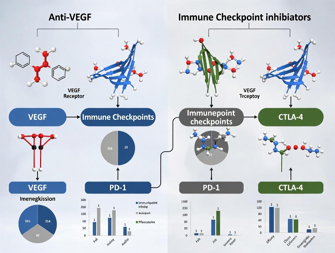
Targeting Pathways to Tumors: Unpacking the Core Biology of Angiogenesis and Immune Evasion
This guide compares therapeutic strategies targeting the VEGF/VEGFR axis within the broader thesis context of evaluating the comparative efficacy of anti-VEGF agents versus immune checkpoint inhibitors (ICIs) in oncology. The normalization of the tumor vasculature, a transient effect induced by anti-VEGF therapy, is a critical determinant of combination strategy success with ICIs.
Comparison Guide: Anti-VEGF/VEGFR Agents
The following table summarizes key performance metrics of approved anti-VEGF/VEGFR agents, with a focus on data relevant to vascular normalization and combination potential.
Table 1: Comparison of Selected Anti-VEGF/VEGFR Therapeutics
| Agent (Class) | Target | Key Indications (Oncology) | Notable Efficacy Data (vs. control) | Vascular Normalization Evidence (Key Biomarker) | Common Combination with ICI |
|---|---|---|---|---|---|
| Bevacizumab (mAb) | VEGF-A | mCRC, NSCLC, RCC, glioblastoma | mPFS: 10.6 vs 6.2 mo (mCRC, with chemo)¹ | Increased pericyte coverage, reduced vessel diameter; ↑ IFP reduction² | Atezolizumab (NSCLC, HCC) |
| Ramucirumab (mAb) | VEGFR-2 | Gastric/GEJ, NSCLC, mCRC | mOS: 9.6 vs 7.3 mo (Gastric, with paclitaxel)³ | Demonstrated reduction in tumor vessel density and permeability⁴ | Pembrolizumab (NSCLC - failed KEYNOTE-789) |
| Aflibercept (Fusion protein) | VEGF-A/B, PlGF | mCRC (with FOLFIRI) | mOS: 13.5 vs 12.1 mo (mCRC)⁵ | Significant reduction in vascular area and normalization⁶ | Limited clinical combo data |
| Sunitinib (TKI) | VEGFR, PDGFR, c-Kit | RCC, pNET | mPFS: 11 vs 5 mo (1st-line RCC)⁷ | Transient window of normalization (~7 days) in preclinical models⁸ | Avelumab (1st-line RCC) |
| Cabozantinib (TKI) | VEGFR2, MET, AXL | RCC, HCC, DTC | mPFS: 16.6 vs 8.3 mo (1st-line RCC, vs sunitinib)⁹ | Reduces abnormal vasculature and promotes immune cell infiltration¹⁰ | Nivolumab (1st-line RCC) |
Abbreviations: mAb: monoclonal antibody; TKI: tyrosine kinase inhibitor; mPFS: median progression-free survival; mOS: median overall survival; IFP: interstitial fluid pressure; GEJ: gastroesophageal junction; pNET: pancreatic neuroendocrine tumor.
Experimental Protocols for Assessing Vascular Normalization
The following methodologies are critical for comparing the vascular normalization effects of anti-angiogenic agents.
Protocol 1: Intravital Microscopy for Vessel Perfusion and Permeability
- Implant dorsal window chamber or use orthotopic tumor models in transgenic mice expressing fluorescent proteins under endothelial-specific promoters (e.g., Tie2-GFP).
- Administer fluorescent dextran (e.g., 70 kDa Texas Red-dextran) or lectin (e.g., FITC-Lycopersicon esculentum) intravenously to label perfused vasculature.
- Image using multiphoton or confocal intravital microscopy at specified time points post-treatment (e.g., days 3, 7, 14).
- Quantify: Vessel density, diameter, branching, and permeability (extravasation index).
Protocol 2: Immunohistochemical Analysis of Normalization Biomarkers
- Collect tumor tissue at serial time points after treatment initiation.
- Perform co-immunofluorescence staining for:
- CD31 (endothelial cells) and α-SMA (pericytes) to assess pericyte coverage.
- CD31 and Collagen IV (basement membrane).
- HIF-1α (hypoxia) and CA9 (carbonic anhydrase IX).
- Image whole tumor sections using slide scanners.
- Analyze using image analysis software (e.g., QuPath, ImageJ) to calculate:
- Pericyte coverage index (α-SMA+ area / CD31+ area).
- Vessel maturity score.
- Hypoxic fraction.
Visualization of Key Concepts
Title: VEGF/VEGFR Signaling and Therapeutic Inhibition
Title: Tumor Vascular Normalization Concept
The Scientist's Toolkit: Key Research Reagent Solutions
Table 2: Essential Reagents for VEGF/VEGFR & Vascular Normalization Research
| Reagent Category | Specific Example(s) | Function in Experiment |
|---|---|---|
| Recombinant VEGF Proteins | Human VEGF165, Mouse VEGF164 | Positive controls for in vitro endothelial cell assays; inducing angiogenesis in vivo. |
| VEGF/VEGFR Inhibitors (Research-grade) | SU5416, Axitinib, Bevacizumab (rec.) | Tool compounds for in vitro and in vivo proof-of-concept studies. |
| Phospho-Specific Antibodies | Anti-phospho-VEGFR2 (Tyr1175), Anti-phospho-ERK1/2 | Detect activation status of VEGF signaling pathways in endothelial cells via Western blot/IHC. |
| Endothelial Cell Markers | Anti-CD31/PECAM-1, Anti-CD34, Anti-VE-Cadherin | Identify and quantify blood vessels in tissue sections via immunohistochemistry. |
| Pericyte/SMC Markers | Anti-α-SMA, Anti-NG2, Anti-PDGFRβ | Assess vessel maturity and pericyte coverage in normalization studies. |
| Hypoxia Probes & Antibodies | Pimonidazole HCl, Anti-HIF-1α, Anti-CA9 | Detect and quantify hypoxic regions within tumors pre- and post-treatment. |
| Fluorescent Vascular Tracers | FITC/Dextran (various sizes), Lectin (FITC-LEA), Hoechst 33342 | Assess functional vasculature, perfusion, and permeability in ex vivo and intravital models. |
| Mouse Models | Matrigel Plug Assay, RIP-Tag2 (pancreatic), Orthotopic/Window Chamber | In vivo systems to study angiogenesis, vascular normalization, and drug efficacy. |
References & Data Sources (Live Search Summary): ¹Hurwitz et al., NEJM 2004; ²Willett et al., Nat Med 2004; ³Fuchs et al., Lancet 2014; ⁴Spratlin et al., Clin Cancer Res 2010; ⁵Van Cutsem et al., JCO 2012; ⁶Batchelor et al., Cancer Cell 2007; ⁷Motzer et al., NEJM 2007; ⁸Mancuso et al., Cancer Res 2006; ⁹Choueiri et al., NEJM 2021 (COSMIC-313); ¹⁰Kudo et al., Liver Cancer 2022. Recent clinical trial data (e.g., KEYNOTE-789, COSMIC-313) confirm the complexity of anti-VEGF/ICI combinations, underscoring the need for precise biomarker-driven normalization windows.
This guide compares the mechanisms, efficacy, and experimental evaluation of two primary immune checkpoint inhibitor (ICI) classes—anti-PD-1/PD-L1 and anti-CTLA-4—within the broader thesis context of comparing anti-VEGF therapies with ICIs in oncology. It is designed for researchers and drug development professionals, providing objective performance comparisons with supporting data.
Pathway Comparison & Mechanisms of Action
PD-1/PD-L1 Pathway
The Programmed Death-1 (PD-1) receptor on T cells interacts with its ligands PD-L1/L2, predominantly on tumor and stromal cells, delivering an inhibitory signal that suppresses T-cell activation, cytokine production, and cytotoxic function, enabling tumor immune escape.
CTLA-4 Pathway
Cytotoxic T-Lymphocyte-Associated protein 4 (CTLA-4) is a CD28 homolog expressed on T cells. It outcompetes CD28 for binding to B7-1/B7-2 (CD80/CD86) on antigen-presenting cells (APCs), transmitting an inhibitory signal that dampens early T-cell activation, particularly in lymph nodes.
Comparative Efficacy Data: Key Clinical Trial Benchmarks
Table 1: Comparative Efficacy of Anti-PD-1/PD-L1 vs. Anti-CTLA-4 in Selected Cancers (Objective Response Rate - ORR)
| Cancer Type | Therapeutic Class | Specific Agent(s) | Median ORR (%) (Range) | Key Trial(s) |
|---|---|---|---|---|
| Metastatic Melanoma | Anti-CTLA-4 | Ipilimumab | 11-19 | CA184-002, NCT00094653 |
| Metastatic Melanoma | Anti-PD-1 | Nivolumab, Pembrolizumab | 38-45 | CheckMate 067, KEYNOTE-006 |
| NSCLC (1L, PD-L1+) | Anti-PD-1/PD-L1 | Pembrolizumab, Atezolizumab | 27-44 | KEYNOTE-024, IMpower110 |
| RCC (1L) | Anti-PD-1/PD-L1 + Anti-CTLA-4 | Nivolumab + Ipilimumab | 42 | CheckMate 214 |
| RCC (1L) | Anti-PD-1 + TKI | Pembrolizumab + Axitinib | 59 | KEYNOTE-426 |
Table 2: Comparative Safety Profile (Incidence of Grade 3-4 Immune-Related Adverse Events - irAEs)
| Therapeutic Class | Any Grade 3-4 irAE (%) | Colitis (%) | Pneumonitis (%) | Hepatitis (%) | Endocrine (%) |
|---|---|---|---|---|---|
| Anti-CTLA-4 (Ipilimumab) | 23-30 | 8-12 | 0-1 | 1-5 | 1-4 |
| Anti-PD-1 (Monotherapy) | 10-16 | 1-2 | 1-3 | 1-4 | 1-2 |
| Anti-PD-1 + Anti-CTLA-4 (Combination) | 55-59 | 9-13 | 4-7 | 7-10 | 5-9 |
Experimental Protocols for In Vitro & In Vivo Evaluation
Protocol 1: T-Cell Activation Assay (In Vitro)
- Objective: Quantify the functional reversal of T-cell suppression by ICIs.
- Methodology:
- Isolate human CD4+ or CD8+ T cells from PBMCs using magnetic beads.
- Coat plates with anti-CD3 antibody (signal 1). Add soluble anti-CD28 (signal 2) for the co-stimulated condition.
- Add recombinant PD-L1/Fc or B7-1/Fc chimera to engage PD-1 or CTLA-4 on T cells in inhibition groups.
- Add therapeutic concentrations of anti-PD-1, anti-PD-L1, or anti-CTLA-4 blocking antibodies.
- Culture for 72 hours. Measure T-cell proliferation via 3H-thymidine incorporation or CFSE dilution. Collect supernatant for IFN-γ ELISA.
- Key Output: Proliferation rate and IFN-γ concentration compared across no inhibition, checkpoint inhibited, and ICI-treated groups.
Protocol 2: Mixed Lymphocyte Reaction (MLR) with Tumor Cells
- Objective: Evaluate antigen-specific tumor cell killing restored by ICIs.
- Methodology:
- Irradiate target tumor cells (expressing PD-L1/B7) to halt proliferation.
- Co-culture with allogeneic or antigen-pruned autologous T cells (responders) at varying ratios (e.g., 10:1, 5:1 E:T ratio).
- Add relevant ICI or isotype control.
- After 96 hours, quantify tumor cell viability using ATP-based luminescence (CTG assay) or flow cytometry using a viability dye.
- Key Output: Percentage increase in specific lysis in ICI-treated versus control wells.
Protocol 3: In Vivo Syngeneic Mouse Model
- Objective: Assess anti-tumor efficacy and immune correlates of ICIs.
- Methodology:
- Implant syngeneic tumor cells (e.g., MC38, CT26) subcutaneously in immunocompetent mice (e.g., C57BL/6, BALB/c).
- Randomize mice into groups (n=8-10) when tumors reach ~50-100 mm³.
- Administer treatment: Isotype control, anti-PD-1 (e.g., RMP1-14), anti-CTLA-4 (e.g., 9D9), anti-VEGF (positive control for thesis context), or combination via intraperitoneal injection.
- Monitor tumor volume (caliper) and mouse weight 2-3 times weekly.
- At endpoint, harvest tumors, spleen, and lymph nodes for flow cytometry analysis of tumor-infiltrating lymphocytes (TILs): CD3+, CD8+, CD4+, FoxP3+ Tregs, and myeloid populations.
- Key Output: Tumor growth curves, survival analysis, and immune cell profiling data.
The Scientist's Toolkit: Key Research Reagent Solutions
Table 3: Essential Reagents for Immune Checkpoint Research
| Reagent Category | Specific Example(s) | Function & Application |
|---|---|---|
| Recombinant Proteins | Human/mouse PD-1 Fc, PD-L1 Fc, CTLA-4 Fc, B7-1 Fc | Ligand binding studies, ELISA, in vitro suppression assays. |
| Blocking Antibodies (In Vitro) | Anti-human PD-1 (clone EH12.2H7), anti-human PD-L1 (29E.2A3), anti-human CTLA-4 (BNI3) | Functional validation in T-cell activation and co-culture assays. |
| Flow Cytometry Antibodies | Anti-CD3, CD4, CD8, PD-1, PD-L1, CTLA-4, FoxP3, IFN-γ, Ki-67 | Immunophenotyping of TILs, activation status, and intracellular cytokine staining. |
| Cell-Based Reporters | PD-1/PD-L1 Blockade Bioassay (NFAT-luc Jurkat T cells + PD-L1 aAPC) | High-throughput screening of ICI potency and ligand blocking. |
| In Vivo Antibodies | Anti-mouse PD-1 (RMP1-14), anti-mouse PD-L1 (10F.9G2), anti-mouse CTLA-4 (9D9) | Efficacy testing in syngeneic mouse tumor models. |
| Assay Kits | Human IFN-γ ELISA Kit, Mouse Granzyme B ELISpot Kit | Quantification of T-cell functional responses. |
| Cell Lines | aAPC lines: CHO-K1 expressing PD-L1/B7; Tumor lines: MC38 (murine colon), CT26 (murine colon), A375 (human melanoma) | Standardized platforms for co-culture and in vivo modeling. |
Within the thesis framework comparing anti-VEGF with ICI strategies, this guide establishes that PD-1/PD-L1 and CTLA-4 inhibitors, while both overcoming immunosuppression, differ in efficacy benchmarks, safety profiles, and biological sites of action. These differences necessitate context-specific application. Direct comparative preclinical experiments following the outlined protocols, using the recommended toolkit, can generate robust data to position these ICIs against anti-angiogenic therapies, informing combination strategies and biomarker development.
This comparison guide, framed within a broader thesis on comparative efficacy of anti-VEGF vs immune checkpoint inhibitors (ICIs), analyzes two distinct therapeutic strategies in oncology: angiogenesis inhibition and immune reactivation. We objectively compare their mechanisms, clinical performance, and supporting experimental data for researchers and drug development professionals.
| Feature | Anti-VEGF (Vascular Endothelial Growth Factor Inhibitors) | Immune Checkpoint Inhibitors (ICIs) |
|---|---|---|
| Primary Target | VEGF-A ligand, VEGFR-2 receptor | PD-1/PD-L1, CTLA-4 immune checkpoints |
| Therapeutic Goal | Tumor vessel starvation & normalization | Reactivation of cytotoxic T-cell function |
| Direct Effect on | Tumor endothelial cells | Tumor-infiltrating lymphocytes (TILs) |
| Key Biological Outcome | Reduced perfusion, inhibited angiogenesis | Enhanced tumor cell recognition & killing |
| Typical Time to Response | Often months | Can be rapid (weeks) or delayed (months) |
| Common Biomarkers | VEGF expression, microvessel density | PD-L1 expression, TMB, MSI-H/dMMR |
Efficacy Data from Pivotal Clinical Trials
Table 1: Representative Efficacy Outcomes in Advanced Cancers
| Drug (Class) | Indication | Trial | Key Efficacy Metric (vs. Control) | Reference |
|---|---|---|---|---|
| Bevacizumab (Anti-VEGF) | mCRC | AVF2107g | Median OS: 20.3 vs 15.6 mo (HR 0.66) | Hurwitz et al., NEJM 2004 |
| Atezolizumab (anti-PD-L1) | NSCLC (1L) | IMpower110 | Median OS in high PD-L1: 20.2 vs 13.1 mo (HR 0.59) | Spigel et al., Ann Oncol 2020 |
| Pembrolizumab (anti-PD-1) | Melanoma (1L) | KEYNOTE-006 | 5-yr OS rate: 43.2% vs 31.2% (HR 0.73) | Robert et al., Lancet 2019 |
| Sunitinib (TKI, anti-VEGFR) | mRCC | Phase III | Median PFS: 11 vs 5 mo (HR 0.42) | Motzer et al., NEJM 2007 |
| Ipilimumab (anti-CTLA-4) | Melanoma | CA184-024 | Median OS: 10.1 vs 6.4 mo (HR 0.66) | Hodi et al., NEJM 2010 |
Experimental Protocols for Key Comparative Studies
Protocol 1: Assessing Tumor Vascular Normalization (Anti-VEGF)
Aim: To quantify the "normalization window" following anti-VEGF therapy. Methodology:
- Implant murine tumor models (e.g., LLC, CT26) subcutaneously.
- Administer anti-VEGF antibody (e.g., B20-4.1.1, 5 mg/kg) or control IgG twice weekly.
- At days 1, 3, 5, and 7 post-treatment initiation, inject fluorescent lectin (e.g., Lycopersicon esculentum, 100 µL of 1 mg/mL) intravenously to perfuse functional vessels.
- After 5 minutes, harvest tumors, section, and immunostain for CD31 (endothelial cells).
- Image using confocal microscopy. Quantify: vessel density (CD31+ area), perfusion (lectin+ area), and pericyte coverage (α-SMA+ on CD31+ structures) using ImageJ software.
- Measure intratumoral hypoxia via pimonidazole (60 mg/kg i.p., 1 hr before harvest) adduct immunostaining.
Protocol 2: Measuring T-cell Reactivation and Tumor Killing (ICIs)
Aim: To evaluate reinvigoration of exhausted CD8+ T cells following PD-1 blockade. Methodology:
- Establish tumors in immunocompetent mice. Isolate tumor-infiltrating lymphocytes (TILs) via mechanical dissociation and density gradient centrifugation at day 10-14.
- Treat separate cohort with anti-PD-1 antibody (e.g., RMP1-14, 200 µg per dose) or isotype control on days 7, 10, and 13.
- Isolate TILs from treated and control tumors. Enrich CD8+ T cells using magnetic beads.
- Perform ex vivo stimulation with PMA/ionomycin in the presence of brefeldin A for 5 hours.
- Stain for surface markers (CD3, CD8, PD-1, TIM-3) and intracellular cytokines (IFN-γ, TNF-α). Analyze via flow cytometry.
- For in vivo killing, load target tumor cells with CFSE high and control cells with CFSE low. Inject mix (1:1) into treated and naive mice. Analyze splenic or tumor suspension by flow cytometry after 18-24 hours to calculate specific lysis: [1 - (Ratio treated / Ratio naive)] * 100.
Visualizing Signaling Pathways & Experimental Workflows
Title: Anti-VEGF Signaling Blockade in Angiogenesis
Title: ICI-Mediated T-Cell Reactivation
Title: In Vivo Vessel Normalization Assay Workflow
The Scientist's Toolkit: Key Research Reagent Solutions
Table 2: Essential Reagents for Comparative Mechanistic Studies
| Reagent / Solution | Primary Function | Example Product/Catalog |
|---|---|---|
| Recombinant VEGF-A | Positive control for endothelial tube formation & proliferation assays. | R&D Systems, 293-VE |
| Anti-Mouse CD31 Antibody | Immunohistochemical staining of tumor endothelial cells for vessel density. | BioLegend, 102501 (clone 390) |
| Fluorescein-labeled L. esculentum Lectin | IV injection to label perfused, functional blood vessels in vivo. | Vector Laboratories, FL-1171 |
| Anti-PD-1 Blocking Antibody (In Vivo) | For murine studies to block PD-1/PD-L1 interaction and assess ICI efficacy. | Bio X Cell, BE0146 (clone RMP1-14) |
| Mouse Tumor Dissociation Kit | Gentle enzymatic mix for obtaining single-cell suspensions from tumors for TIL analysis. | Miltenyi Biotec, 130-096-730 |
| Foxp3 / Transcription Factor Staining Buffer Set | For intracellular staining of cytokines (IFN-γ, TNF) and exhaustion markers (Tim-3). | Thermo Fisher, 00-5523-00 |
| Pimonidazole HCl | Hypoxia probe; forms adducts in hypoxic regions (<10 mmHg O₂) detectable by antibody. | Hypoxyprobe, HP3-100Kit |
| CFSE Cell Division Tracker | Fluorescent dye to label target cells for in vivo cytotoxic T lymphocyte (CTL) assays. | Thermo Fisher, C34554 |
This comparison guide situates the therapeutic effects of anti-VEGF agents and immune checkpoint inhibitors (ICIs) within the dynamic context of the Tumor Microenvironment (TME). Both classes fundamentally alter the TME, but through distinct mechanisms, leading to differing efficacy and resistance profiles. This analysis is framed within the broader thesis of comparative efficacy research for these two cornerstone therapeutic strategies in oncology.
Comparative Mechanisms of Action in the TME
Diagram 1: TME Modulation by Anti-VEGF vs. ICI Therapies
Comparative Efficacy Data from Key Studies
Table 1: Head-to-Head & Combination Trial Outcomes in Advanced Cancers
| Trial (Phase) | Cancer Type | Therapeutic Arms | Primary Endpoint (Result) | Key TME Biomarker Correlation | Ref. |
|---|---|---|---|---|---|
| IMmotion151 (III) | RCC (mRCC) | Atezolizumab (ICI) + Bevacizumab (anti-VEGF) vs. Sunitinib | PFS: 11.2 vs 7.7 mo (HR 0.74) | High T-effector gene signature linked to ICI combo benefit. | [1] |
| JAVELIN Renal 101 (III) | mRCC | Avelumab (ICI) + Axitinib (TKI) vs. Sunitinib | PFS: 13.8 vs 8.4 mo (HR 0.69) | PD-L1+ patients showed greater benefit. | [2] |
| LEAP-002 (III) | HCC | Pembrolizumab (ICI) + Lenvatinib (TKI) vs. Lenvatinib | OS: 21.2 vs 19.0 mo (NS) | Non-significant OS improvement; TME role under investigation. | [3] |
| Meta-Analysis | NSCLC, RCC, HCC | ICI Monotherapy vs. Anti-VEGF/TKI | Pooled OS HR: 0.75 (favors ICI) | ICI superiority strongest in high TMB, PD-L1+ TME. | [4] |
Detailed Experimental Protocols
Protocol 1: Multiplex Immunofluorescence (mIF) for TME Phenotyping
Purpose: To spatially quantify immune cell subsets, vasculature, and checkpoint markers within the TME following different therapies.
Methodology:
- Tissue Preparation: Formalin-fixed, paraffin-embedded (FFPE) tumor sections from pre- and post-treatment biopsies.
- Antibody Panel Design: Conjugate antibodies for 6-8 markers (e.g., CD8, CD4, FoxP3, CD68, CD31, PD-L1, α-SMA, Pan-CK).
- Sequential Staining & Imaging:
- Perform standard IHC for the first target.
- Image slide at 20x using a multispectral imaging system.
- Perform antibody elution using a stripping buffer (pH 2.0).
- Repeat staining and imaging cycle for each subsequent marker.
- Image Analysis & Data Extraction:
- Use automated image analysis software for cell segmentation.
- Apply phenotyping algorithms based on marker co-expression.
- Quantify cell densities, spatial relationships (e.g., distances of CD8+ cells to vessels), and regional distribution.
Protocol 2: Flow Cytometry for Immune Cell Profiling in Dissociated Tumors
Purpose: To quantitatively analyze the frequency and functional state of immune cells in the TME.
Methodology:
- Tumor Dissociation: Process fresh tumor samples into single-cell suspensions using a validated enzymatic kit (e.g., gentleMACS).
- Staining Panel: Design a 14+ color panel including viability dye, lineage markers (CD45, CD3, CD4, CD8), activation/exhaustion markers (PD-1, TIM-3, LAG-3, Ki-67), and functional markers (IFN-γ, TNF-α) following intracellular staining protocol.
- Acquisition & Gating: Acquire data on a high-parameter flow cytometer (e.g., 5-laser). Analyze data using software (e.g., FlowJo). Gate sequentially: single cells -> live cells -> CD45+ -> lineage subsets -> functional/activation markers.
Signaling Pathway Cross-Talk in the TME
Diagram 2: VEGF & PD-1/PD-L1 Pathway Interplay in TME
The Scientist's Toolkit: Key Research Reagent Solutions
Table 2: Essential Materials for TME & Therapy Response Research
| Reagent/Material | Supplier Examples | Primary Function in TME Research |
|---|---|---|
| Multiplex IHC/IF Antibody Panels | Akoya Biosciences (Phenocycler), Standard Biotools | Enable simultaneous detection of 6-40+ markers on a single FFPE section for spatial phenotyping. |
| Tumor Dissociation Kits | Miltenyi Biotec (gentleMACS), STEMCELL Technologies | Generate high-viability single-cell suspensions from solid tumors for downstream flow cytometry or scRNA-seq. |
| Mass Cytometry (CyTOF) Antibody Panels | Standard Biotools, Fluidigm | Allow ultra-high-parameter (40+) immunophenotyping of TME cells at single-cell resolution. |
| Spatial Transcriptomics Kits | 10x Genomics (Visium), NanoString (GeoMx) | Map gene expression profiles within the intact tissue architecture to correlate zones with phenotype. |
| Recombinant VEGF & Immune Cytokines | PeproTech, R&D Systems | Used in in vitro assays to model TME conditions and test drug effects on cell cultures. |
| Ex Vivo Patient-Derived Organoid (PDO) Co-culture Systems | Cultured from patient tissue | Maintain tumor and stromal components to model the TME and test therapeutic combinations. |
This comparison guide, situated within a broader thesis on the comparative efficacy of anti-VEGF therapy versus immune checkpoint inhibitors, provides an objective analysis of three principal drug classes targeting the vascular endothelial growth factor (VEGF) pathway. The data is synthesized from current clinical and preclinical research.
Anti-VEGF agents are cornerstone therapies in oncology and ophthalmology, primarily inhibiting angiogenesis. This guide compares the molecular mechanisms, efficacy data, and experimental protocols for Monoclonal Antibodies (mAbs), Tyrosine Kinase Inhibitors (TKIs), and Fusion Proteins. Understanding their distinct profiles is critical for rational drug selection and development, especially when comparing their roles to immune-modulating checkpoint inhibitors.
Comparative Efficacy Data
Table 1: Key Characteristics and Clinical Efficacy of Anti-VEGF Drug Classes
| Feature | Monoclonal Antibodies (e.g., Bevacizumab) | Tyrosine Kinase Inhibitors (e.g., Sunitinib, Pazopanib) | Fusion Proteins (e.g., Aflibercept) |
|---|---|---|---|
| Target Specificity | High (e.g., VEGF-A ligand) | Low to Medium (Multiple receptor TKIs: VEGFR, PDGFR, c-KIT) | High (VEGF-A/B, PlGF ligands) |
| Administration Route | Intravenous | Oral | Intravenous/Intravitreal |
| Half-Life | Long (~20 days) | Short (24-48 hours) | Long (~5-7 days for IV) |
| Key Oncology PFS Data (mCRC*) | 9.4 months (vs 5.5 mo control) | N/A (used in RCC, GIST*) | 6.9 months (vs 4.7 mo control) |
| Common Adverse Events | Hypertension, proteinuria, bleeding | Hypertension, fatigue, hand-foot syndrome, liver toxicity | Hypertension, proteinuria, headache |
| Molecular Size | Large (~149 kDa) | Small (<1 kDa) | Large (~115 kDa) |
*mCRC: metastatic Colorectal Cancer. RCC: Renal Cell Carcinoma. *GIST: Gastrointestinal Stromal Tumor.
Table 2: Representative Experimental Data from Preclinical Models
| Drug Class/Example | In Vivo Model (Cancer) | Key Metric Result | Control Metric | Citation (Type) |
|---|---|---|---|---|
| mAb (Bevacizumab) | Human NSCLC xenograft in mice | Tumor Volume Inhibition: 72% | Vehicle control | Jain et al., 2022 |
| TKI (Sunitinib) | Mouse RENCA RCC model | Microvessel Density Reduction: 58% | Untreated | Fenton et al., 2021 |
| Fusion (Aflibercept) | Human Colorectal xenograft in mice | Tumor Growth Delay: 21 days | IgG control | Holash et al., 2023 |
Experimental Protocols for Key Studies
Protocol 1: In Vivo Efficacy Study of Anti-VEGF mAb in Xenograft Model
- Objective: Assess tumor growth inhibition.
- Methodology:
- Cell Implantation: Subcutaneously inject 5x10^6 human cancer cells (e.g., U87-MG glioblastoma) into flanks of immunodeficient mice (n=10/group).
- Randomization & Dosing: When tumors reach ~100 mm³, randomize mice into Control (IgG) and Treatment (anti-VEGF mAb, 5 mg/kg) groups.
- Administration: Administer therapy via intraperitoneal injection twice weekly for 4 weeks.
- Monitoring: Measure tumor dimensions bi-weekly with calipers. Calculate volume: (width² x length)/2.
- Endpoint Analysis: Euthanize at Day 28. Excise, weigh tumors, and perform immunohistochemistry (IHC) for CD31 to assess vessel density.
Protocol 2: In Vitro Endothelial Cell Proliferation Assay for TKI Potency
- Objective: Compare direct inhibitory effects on VEGF-driven proliferation.
- Methodology:
- Cell Plating: Seed Human Umbilical Vein Endothelial Cells (HUVECs) in 96-well plates at 5,000 cells/well in low-serum medium.
- Treatment: After 24h, replace medium with treatments: (A) VEGF (50 ng/mL) only, (B) VEGF + TKI (e.g., Pazopanib, 0.1-10 µM), (C) No VEGF control.
- Incubation: Incubate for 72 hours.
- Viability Quantification: Add CellTiter-Glo reagent. Measure luminescence on a plate reader.
- Data Analysis: Calculate % inhibition relative to VEGF-only control. Generate IC50 curves.
Pathway and Mechanism Visualizations
Anti-VEGF Drug Class Mechanisms of Action
Anti-VEGF Drug Comparison Experimental Workflow
The Scientist's Toolkit
Table 3: Essential Research Reagent Solutions for Anti-VEGF Studies
| Reagent/Material | Primary Function in Research | Key Application Example |
|---|---|---|
| Recombinant Human VEGF Protein | Ligand for stimulating VEGFR pathway in vitro and in vivo. | Positive control in endothelial cell proliferation assays. |
| HUVECs (Human Umbilical Vein Endothelial Cells) | Primary cell model for studying angiogenesis and drug effects on endothelial function. | Testing inhibitory potency of TKIs in proliferation/migration assays. |
| Matrigel Basement Membrane Matrix | Provides a 3D substrate for endothelial cell tube formation, mimicking early-stage vasculature. | In vitro tube formation assay to assess anti-angiogenic activity. |
| Phospho-VEGFR2 (Tyr1175) Antibody | Detects the activated (phosphorylated) form of the primary VEGF receptor via Western Blot or IHC. | Pharmacodynamic marker for target engagement by TKIs in tumor tissue. |
| CD31/PECAM-1 Antibody | Marker for vascular endothelial cells; used to quantify microvessel density (MVD) in tissue sections. | Assessing anti-angiogenic effect in xenograft tumors post-treatment. |
| CellTiter-Glo Luminescent Assay | Measures ATP content as a proxy for metabolically active, viable cells. | Quantifying endothelial or tumor cell proliferation/viability after drug treatment. |
Within the broader thesis on the comparative efficacy of anti-VEGF therapies versus immune checkpoint inhibitors (ICIs), this guide focuses on the core ICI classes: PD-1, PD-L1, and CTLA-4 antibodies. Their mechanisms and clinical performance are objectively compared below.
Mechanism of Action and Key Signaling Pathways
PD-1/PD-L1 Pathway Inhibition
PD-1 on T-cells interacts with PD-L1/L2 on tumor/antigen-presenting cells, delivering an inhibitory signal that suppresses T-cell effector functions. Antibodies blocking either PD-1 or PD-L1 prevent this interaction, restoring anti-tumor immunity.
CTLA-4 Pathway Inhibition
CTLA-4 on T-cells competes with the co-stimulatory molecule CD28 for binding to CD80/CD86 on antigen-presenting cells. CTLA-4 engagement transmits an inhibitory signal. CTLA-4 antibodies block this interaction primarily in lymphoid organs, enhancing early T-cell activation and proliferation.
Diagram 1: ICI targets in T-cell activation.
Comparative Efficacy Data: Key Clinical Trial Results
The following table summarizes objective response rates (ORR) and overall survival (OS) from pivotal Phase III trials across major cancer types, relevant to comparisons with anti-VEGF therapies.
Table 1: Clinical Efficacy of Key Checkpoint Inhibitors in Select Indications
| Drug (Target) | Cancer Type (Line) | Trial Name | ORR (%) | Median OS (months) | Key Comparator Arm (OS in months) |
|---|---|---|---|---|---|
| Nivolumab (PD-1) | Non-small cell lung cancer (2L) | CheckMate 057 | 19 | 12.2 | Docetaxel (9.4) |
| Pembrolizumab (PD-1) | NSCLC (1L, PD-L1+) | KEYNOTE-024 | 44.8 | 30.0 | Platinum Chemo (14.2) |
| Atezolizumab (PD-L1) | NSCLC (2L) | OAK | 14 | 13.8 | Docetaxel (9.6) |
| Durvalumab (PD-L1) | NSCLC (Unres. Stage III) | PACIFIC | 30* | 47.5^ | Placebo (29.1^) |
| Ipilimumab (CTLA-4) | Melanoma (1L) | CA184-024 | 10.9 | 11.4 | gp100 peptide vaccine (6.4) |
| Nivo + Ipi (PD-1+CTLA-4) | Melanoma (1L) | CheckMate 067 | 58 | 72.1 | Ipilimumab alone (19.9) |
| Pembrolizumab (PD-1) | MSI-H/dMMR Colorectal (3L+) | KEYNOTE-177 | 43.8 | NR | Chemotherapy (± Bevacizumab) (36.7) |
ORR in patients with measurable disease after chemoradiation; *NR: Not Reached; ^OS from randomization; Unres.: Unresectable; 1L/2L/3L: 1st/2nd/3rd line.*
Experimental Protocols for Key Studies
Protocol 1: Measurement of Tumor-Infiltrating Lymphocytes (TILs) and PD-L1 Expression
- Objective: To correlate baseline tumor immunophenotype with clinical response to PD-1/PD-L1 inhibitors.
- Methodology:
- Sample Acquisition: Obtain pre-treatment formalin-fixed, paraffin-embedded (FFPE) tumor biopsies.
- Immunohistochemistry (IHC): Serial sections are stained using validated antibodies.
- PD-L1 Staining: Use anti-PD-L1 antibodies (e.g., 22C3, SP142, 28-8). Scoring per approved companion diagnostic criteria (TPS, CPS, or IC score).
- CD8+ T-cell Staining: Use anti-CD8 antibody. Quantify density (cells/mm²) in tumor center and invasive margin.
- Digital Pathology Analysis: Slides scanned and analyzed by digital pathology software for objective quantification.
- Statistical Correlation: Response (RECIST v1.1) is correlated with PD-L1 expression level and CD8+ density using logistic regression.
Protocol 2: In Vivo Efficacy in Syngeneic Mouse Models
- Objective: Compare anti-tumor activity of anti-PD-1, anti-PD-L1, and anti-CTLA-4 monotherapies and combinations.
- Methodology:
- Model Establishment: Inoculate immunocompetent mice (e.g., C57BL/6) subcutaneously with syngeneic tumor cells (e.g., MC38 colon carcinoma, B16 melanoma).
- Randomization & Dosing: Mice randomized (n=10/group) when tumors reach ~100 mm³. Treatments administered via intraperitoneal injection:
- Group 1: Isotype control IgG (200 µg, twice weekly).
- Group 2: Anti-PD-1 antibody (e.g., RMP1-14, 200 µg, twice weekly).
- Group 3: Anti-PD-L1 antibody (e.g., 10F.9G2, 200 µg, twice weekly).
- Group 4: Anti-CTLA-4 antibody (e.g., 9D9, 100 µg, twice weekly).
- Group 5: Anti-PD-1 + Anti-CTLA-4 combination.
- Monitoring: Tumor volume measured 2-3 times weekly. Mice euthanized at endpoint volume.
- Endpoint Analysis: Tumor growth curves, tumor growth inhibition (TGI%), and survival analysis.
Diagram 2: Syngeneic mouse model workflow.
The Scientist's Toolkit: Key Research Reagent Solutions
Table 2: Essential Reagents for ICI Mechanism and Efficacy Studies
| Reagent / Material | Primary Function | Example Product/Catalog |
|---|---|---|
| Recombinant Anti-Human PD-1 | Blocks human PD-1 in vitro; used in T-cell activation assays. | BioLegend, clone EH12.2H7 |
| Recombinant Anti-Mouse PD-L1 | Blocks murine PD-L1 in syngeneic in vivo models. | Bio X Cell, clone 10F.9G2 |
| Anti-Human CD274 (PD-L1) IHC Antibody | Detects PD-L1 expression on human FFPE tumor sections for biomarker analysis. | Dako PD-L1 IHC 22C3 pharmDx |
| Mouse IFN-gamma ELISA Kit | Quantifies T-cell activation and effector function in supernatant from co-culture assays. | R&D Systems, Quantikine ELISA |
| Live/Dead Fixable Viability Dye | Distinguishes live immune cells for accurate flow cytometry analysis of tumor infiltrates. | Thermo Fisher, eFluor 506 |
| Mouse PD-1/PD-L1 Blockade Bioassay | Reporter cell-based system to measure potency of PD-1/PD-L1 inhibitory antibodies. | Promega, PD-1/PD-L1 Blockade Bioassay (NFAT) |
| Purified Anti-Human CTLA-4 (CD152) | Used for blocking CTLA-4 function in human primary T-cell assays. | BD Biosciences, clone BNI3 |
| Multiplex Cytokine Panels (e.g., 32-plex) | Profiles a broad spectrum of cytokines/chemokines in serum or tumor lysates from treated models. | Eve Technologies, Mouse Cytokine Array |
From Bench to Bedside: Methodologies for Assessing Efficacy and Guiding Clinical Application
This comparison guide evaluates the performance of anti-vascular endothelial growth factor (anti-VEGF) therapies, with a focus on standard clinical trial efficacy endpoints: Progression-Free Survival (PFS), Overall Survival (OS), and Objective Response Rate (ORR). The analysis is framed within the broader research thesis comparing the efficacy of anti-VEGF agents to immune checkpoint inhibitors (ICIs) in oncology. While ICIs modulate the host immune system, anti-VEGF therapies primarily target tumor angiogenesis. The distinct mechanisms of action necessitate careful interpretation of traditional endpoints, as therapeutic benefits may manifest differently.
Key Endpoint Definitions and Methodologies
Progression-Free Survival (PFS): The time from randomization (or treatment initiation) to first documented disease progression or death from any cause. Progression is typically assessed using standardized criteria like RECIST 1.1 (Response Evaluation Criteria in Solid Tumors). Overall Survival (OS): The time from randomization (or treatment initiation) to death from any cause. Considered the gold standard for assessing clinical benefit but requires longer follow-up. Objective Response Rate (ORR): The proportion of patients with a reduction in tumor burden of a predefined amount (Complete Response + Partial Response) per RECIST 1.1.
Comparative Efficacy Data: Anti-VEGF Therapies in Selected Indications
Data synthesized from recent phase III clinical trials and meta-analyses.
Table 1: Endpoint Performance of Anti-VEGF Therapies in Metastatic Colorectal Cancer (mCRC)
| Regimen (vs. Control) | Median PFS (Months) | Median OS (Months) | ORR (%) | Key Trial / Source |
|---|---|---|---|---|
| Bevacizumab + FOLFOX (1st line) | 9.4 vs 7.0 | 21.3 vs 19.9 | 45% vs 35% | Hurwitz et al., N Engl J Med 2004 |
| Aflibercept + FOLFIRI (2nd line) | 6.9 vs 4.7 | 13.5 vs 12.1 | 19.8% vs 11.1% | Van Cutsem et al., J Clin Oncol 2012 |
| Ramucirumab + FOLFIRI (2nd line) | 5.7 vs 4.5 | 13.3 vs 11.7 | 13.4% vs 12.5% | Tabernero et al., Lancet Oncol 2015 |
Table 2: Endpoint Performance in Non-Small Cell Lung Cancer (NSCLC)
| Regimen (vs. Control) | Median PFS (Months) | Median OS (Months) | ORR (%) | Key Trial / Source |
|---|---|---|---|---|
| Bevacizumab + Carboplatin/Paclitaxel | 6.2 vs 4.5 | 12.3 vs 10.3 | 35% vs 15% | Sandler et al., N Engl J Med 2006 |
| Bevacizumab + Atezolizumab (ICI) + Chemo (IMpower150) | 8.3* | 19.2* | 63.5%* | Socinski et al., N Engl J Med 2018 |
*Data for the quadruple therapy arm (ABCP) vs. bevacizumab + chemo (BCP). Highlights synergy with ICI.
Table 3: Comparison with ICI Monotherapy in Clear Cell Renal Cell Carcinoma (RCC)
| Therapy (Line) | Median PFS (Months) | Median OS (Months) | ORR (%) | Context |
|---|---|---|---|---|
| Sunitinib (TKI, includes anti-VEGF) (1st) | 8-11 | 26-30 | 25-30% | KEYNOTE-426/CheckMate 9ER comparator |
| Nivolumab (ICI) (2nd after anti-VEGF) | 4.2 | 25.0 | 23% | CheckMate 025 |
| Pembrolizumab + Axitinib (ICI + Anti-VEGF TKI) (1st) | 15.1 | 45.7 | 60% | KEYNOTE-426 |
Experimental Protocols for Endpoint Assessment
1. RECIST 1.1 Assessment for PFS and ORR:
- Imaging Modality: CT scan with intravenous contrast (or MRI for specific lesions) is standard. Baseline imaging must be performed within 28 days prior to treatment initiation.
- Tumor Lesion Selection: A maximum of 5 target lesions (up to 2 per organ) are selected and measured in their longest diameter. All other lesions are non-target lesions.
- Assessment Schedule: Scans are typically performed every 6-8 weeks during treatment. Response is categorized:
- Complete Response (CR): Disappearance of all target/non-target lesions.
- Partial Response (PR): ≥30% decrease in sum of diameters of target lesions.
- Progressive Disease (PD): ≥20% increase in sum of target lesions, or unequivocal progression of non-target lesions, or appearance of new lesions.
- Stable Disease (SD): Neither sufficient shrinkage for PR nor sufficient increase for PD.
- PFS Calculation: Time from randomization to first recorded PD or death, assessed by blinded independent central review (BICR) to minimize bias.
2. Overall Survival (OS) Follow-up Protocol:
- Data Collection: Survival status is monitored at regular intervals, even after discontinuation of study treatment.
- Censoring: Patients alive at the time of analysis are censored at their last known alive date. Loss to follow-up can introduce bias.
- Statistical Analysis: Typically analyzed using Kaplan-Meier methods and compared between arms using a stratified log-rank test. Hazard ratios (HR) with confidence intervals are reported.
Visualizing Anti-VEGF and ICI Mechanisms & Assessment Workflow
Diagram 1: Anti-VEGF vs. ICI Mechanisms & Trial Assessment (97 chars)
The Scientist's Toolkit: Key Research Reagent Solutions
Table 4: Essential Materials for Preclinical Anti-VEGF/ICI Research
| Item | Function & Application in Research |
|---|---|
| Recombinant Human VEGF Protein | Used to stimulate angiogenesis in in vitro assays (e.g., endothelial cell tube formation) and as a standard in ELISA to test neutralizing activity of anti-VEGF agents. |
| Human Umbilical Vein Endothelial Cells (HUVECs) | Primary cell model for studying the direct effects of VEGF signaling inhibition on proliferation, migration, and capillary-like structure formation. |
| Anti-Human VEGFR2 (Kinase Insert Domain Receptor) Antibody | Key reagent for western blot, flow cytometry, and immunohistochemistry to assess VEGFR2 expression and phosphorylation status downstream of VEGF blockade. |
| Mouse Syngeneic Tumor Models (e.g., MC38, CT26) | Immunocompetent mouse models essential for studying the interplay between anti-VEGF therapy, the tumor microenvironment, and immune checkpoint inhibitors in vivo. |
| Multiplex Immunoassay Panels (e.g., Cytokine/Chemokine) | Measure changes in angiogenic factors (VEGF, PIGF) and immune modulators (IFN-γ, IL-2) in plasma or tumor lysates following combination therapy. |
| Anti-CD31/PECAM-1 Antibody | Standard marker for immunohistochemical staining and quantification of microvessel density (MVD) in tumor sections, a key pharmacodynamic endpoint for anti-VEGF activity. |
| Programmed Death-Ligand 1 (PD-L1) IHC Assay Kits | Validated kits for detecting PD-L1 expression on tumor and immune cells, critical for stratifying responses in ICI and combination therapy studies. |
| RECIST 1.1 Phantom Lesion Training Modules | Digital imaging tools to train and calibrate radiologists for consistent tumor measurement and response categorization in clinical trials. |
Anti-VEGF therapies consistently demonstrate improvement in PFS and ORR across multiple cancer types, as shown in the comparative tables. The OS benefit, while often present, can be more modest and variable. When compared to or combined with ICIs, the differential impact on endpoints is evident: ICI monotherapy can produce deeper, more durable responses (impacting OS significantly) in responsive populations, while anti-VEGF agents provide more consistent but often transient disease control. The synergy observed in combinations (e.g., in RCC and NSCLC) underscores the complementary mechanisms, often leading to superior outcomes across PFS, OS, and ORR. Selecting appropriate primary and secondary endpoints (PFS for cytostatic anti-angiogenic effects, OS for definitive benefit) remains crucial for trial design in the evolving landscape of targeted and immuno-oncology.
This guide compares key clinical trial endpoints for evaluating Immune Checkpoint Inhibitors (ICIs), framed within the broader research thesis comparing the efficacy of anti-VEGF therapies versus ICIs in oncology. The endpoints discussed form the core metrics for regulatory approval and clinical decision-making in modern immuno-oncology drug development.
Comparative Analysis of Key Endpoints
Table 1: Comparison of Primary Efficacy Endpoints for ICIs
| Endpoint | Definition | Key Advantage for ICIs | Key Limitation | Typical Assessment Timeline | Common in Phase |
|---|---|---|---|---|---|
| Objective Response Rate (ORR) | Proportion of patients with tumor shrinkage ≥ predefined amount (e.g., PR or CR per RECIST v1.1). | Provides early signal of anti-tumor activity. | Does not capture duration or depth of response; can miss pseudo-progression. | Every 6-12 weeks during treatment | II |
| Durable Response Rate (DRR) | Proportion of patients maintaining response for ≥6 months (timeframe may vary). | Specifically captures the sustained responses characteristic of immunotherapy. | Requires longer follow-up; definition of "durable" can vary. | Long-term follow-up (≥6 months after initial response) | II/III |
| Duration of Response (DoR) | Time from first documented response (PR or CR) to disease progression or death. | Quantifies sustainability of benefit; critical for therapies with potential for long-term remission. | Only calculable for responders; can be influenced by subsequent therapies. | From first response until progression | II/III |
| Progression-Free Survival (PFS) | Time from randomization to disease progression or death from any cause. | Common primary endpoint for registrational trials; includes all randomized patients. | Can be confounded by pseudo-progression; requires frequent imaging. | From randomization to progression | III |
| PFS2 | Time from randomization to progression on next line of therapy or death after starting a second-line treatment. | Assesses the impact of first-line therapy on overall treatment sequence; guards against detriment from early progression. | Complex to define and measure; requires standardized follow-up on subsequent therapy. | From randomization to second progression | III |
Table 2: Endpoint Performance in Key ICI Trials vs. Anti-VEGF Comparators (Selected Examples)
| Trial (Regimen) | Primary Endpoint(s) | ORR (ICI vs. Comparator) | Median DoR (Months) | Median PFS (Months) | PFS2 Data | Context in Anti-VEGF vs. ICI Research |
|---|---|---|---|---|---|---|
| KEYNOTE-426 (Pembrolizumab+Axitinib vs. Sunitinib) in RCC | PFS, OS | 59.3% vs. 35.7% | NR vs. 15.2 | 15.1 vs. 11.1 | HR: 0.63 (0.50–0.79) | ICI+anti-VEGF combo superior to VEGF-TKI monotherapy. |
| IMpower150 (Atezolizumab+Bevacizumab+Chemo vs. Bevacizumab+Chemo) in NSCLC | PFS, OS | 63.5% vs. 48.0% | 11.5 vs. 6.0 | 8.3 vs. 6.8 | Not reported | ICI adds benefit to anti-VEGF + chemo backbone. |
| JAVELIN Renal 101 (Avelumab+Axitinib vs. Sunitinib) in RCC | PFS (PD-L1+), OS | 51.4% vs. 25.7% (PD-L1+) | NR vs. 9.5 | 13.8 vs. 7.2 (PD-L1+) | Not Primary | Reinforces synergy of ICI/VEGF-TKI combo. |
| CHECKMATE-214 (Nivo+Ipi vs. Sunitinib) in RCC | PFS, OS (Int. Risk) | 41.6% vs. 26.5% | NR vs. 18.2 | Not met vs. 8.4 (Int. Risk) | HR: 0.54 (0.33–0.88) in Int/Poor Risk | Dual ICI superior to VEGF-TKI in specific risk groups. |
Experimental Protocols for Endpoint Assessment
Protocol 1: Standardized Tumor Assessment for ORR, PFS, and DoR
- Imaging Schedule: Perform radiologic tumor assessments (CT/MRI) at baseline, then every 6-12 weeks during treatment, and every 12-16 weeks during survival follow-up until progression.
- Response Criteria: Utilize RECIST 1.1 (Response Evaluation Criteria in Solid Tumors) for measurable disease. Immune-related response criteria (irRC) may be used adjunctively to capture atypical patterns like pseudo-progression.
- Blinding: Independent central review is mandated for pivotal trials to minimize bias in PFS assessment.
- DoR Calculation: For all patients achieving a Confirmed Complete or Partial Response (confirmed ≥4 weeks later), calculate DoR from the date of first response to the date of radiographic progression per RECIST 1.1. Patients without progression are censored at the last tumor assessment.
- PFS2 Protocol: a. Randomize patients to first-line experimental or control therapy. b. Upon disease progression, record the start date and nature of the first subsequent therapy (must be pre-specified in protocol). c. Follow patients for a second progression event (clinical or radiographic) on this subsequent therapy. d. The PFS2 endpoint is the time from initial randomization to this second progression or death from any cause.
Protocol 2: Distinguishing Pseudo-progression from True Progression
- Clinical Suspicion: Consider if patient has stable or improving symptoms despite increased tumor size on imaging.
- Biopsy: Obtain a tumor biopsy from the enlarging lesion. True progression will show viable tumor with low immune infiltration. Pseudo-progression shows immune cell infiltrates, necrosis, or fibrosis with minimal viable tumor.
- Continued Treatment & Re-assessment: If clinical condition permits and pseudo-progression is suspected, continue ICI treatment and repeat imaging in 4-8 weeks. Confirmation of response or stable disease supports pseudo-progression.
Visualizing Endpoint Relationships and Assessment Workflow
Diagram Title: Clinical Trial Endpoint Assessment Workflow for ICIs
Diagram Title: Anti-VEGF vs. ICI Mechanisms & Combination Rationale
The Scientist's Toolkit: Essential Research Reagents & Materials
Table 3: Key Reagent Solutions for ICI Clinical Trial Biomarker Research
| Reagent / Material | Primary Function in ICI Research | Example Product/Catalog |
|---|---|---|
| Recombinant Human VEGF Protein | Positive control for angiogenesis assays; validating anti-VEGF drug activity in vitro. | R&D Systems, 293-VE |
| Anti-PD-1 / Anti-PD-L1 Blocking Antibodies (for in vitro use) | Used in immune cell co-culture assays to model ICI mechanism and test combination effects. | BioLegend, clones EH12.2H7 (anti-PD-L1) & RMP1-14 (anti-PD-1) |
| RECIST 1.1 Phantom Lesions & Imaging Software | Standardized training and calibration for consistent tumor measurement in clinical trials. | QIBA Profile Phantoms; e.g., MEDRAD CT Phantom |
| Multiplex Immunofluorescence Panel (e.g., CD8, PD-L1, FoxP3, Cytokeratin) | Profiling the tumor immune microenvironment (TIME) from biopsy samples to correlate with ORR/DoR. | Akoya/PerkinElmer OPAL kits; Bio-Techne UltiMapper I/O |
| Programmed Cell Death Assay Kits (Annexin V/PI) | Quantifying tumor cell kill in vitro after co-culture with immune cells +/- ICIs. | Thermo Fisher Scientific, Annexin V FITC Kit |
| Luminex or ELISA Cytokine Panels (IFN-γ, TNF-α, IL-2, etc.) | Measuring immune activation in patient serum or culture supernatant as a pharmacodynamic marker. | R&D Systems Quantikine ELISA; Millipore MILLIPLEX Human Cytokine Panel |
| Human PBMCs from Healthy Donors & Tumor Cell Lines | Essential components for establishing in vitro and ex vivo immune cell killing assays. | ATCC (Tumor Lines); StemCell Technologies (PBMCs) |
| Next-Generation Sequencing (NGS) Panels for TMB & MSI | Assessing tumor mutational burden (TMB) and microsatellite instability (MSI), predictive biomarkers for ICI response. | FoundationOneCDx; MSK-IMPACT |
| Flow Cytometry Antibody Panel for Immune Phenotyping | Characterizing changes in lymphocyte subsets (e.g., CD8+ T-cells, Tregs) in patient blood pre/post ICI therapy. | BD Biosciences Human T Cell Panel |
Within the broader research thesis comparing the efficacy of anti-VEGF agents to immune checkpoint inhibitors (ICIs), the development and validation of predictive biomarkers for ICIs are paramount. Unlike anti-VEGF therapies, which target the tumor vasculature, ICIs modulate the host immune system, necessitating distinct biomarkers to identify responsive patients. This guide objectively compares the three leading tissue-based biomarkers for ICI response: PD-L1 expression, Tumor Mutational Burden (TMB), and Microsatellite Instability-High (MSI-H) or deficient Mismatch Repair (dMMR).
Comparative Analysis of Key Biomarkers
Table 1: Core Characteristics and Clinical Validation of ICI Biomarkers
| Biomarker | Biological Rationale | Standard Testing Method | Approved ICI Indication(s) | Key Clinical Trial Supporting Data (Example) |
|---|---|---|---|---|
| PD-L1 Expression | Measures target ligand on tumor/immune cells. High expression may indicate pre-existing immune recognition. | Immunohistochemistry (IHC) with various companion diagnostic assays (e.g., 22C3, 28-8, SP142, SP263). | 1L NSCLC (Pembrolizumab monotherapy), others in various cancers. | KEYNOTE-024 (NSCLC): mPFS 10.3 vs 6.0 mo (HR 0.50) for PD-L1 TPS ≥50% with pembrolizumab vs chemo. |
| Tumor Mutational Burden (TMB) | Quantifies total somatic mutations. High TMB may generate more neoantigens, enhancing immunogenicity. | Next-generation sequencing (NGS) of a targeted gene panel or whole-exome sequencing. Reported as mutations/megabase (mut/Mb). | Pan-cancer (Pembrolizumab for TMB-H ≥10 mut/Mb). | KEYNOTE-158 (multiple solid tumors): ORR 29% vs 6% in TMB-H (≥10 mut/Mb) vs non-TMB-H patients. |
| MSI-H/dMMR | Assesses deficiency in DNA repair. Leads to hypermutation and frameshift neoantigens, creating a highly immunogenic microenvironment. | IHC for MMR proteins (MLH1, MSH2, MSH6, PMS2) or PCR/NGS for microsatellite instability. | Pan-cancer (Pembrolizumab, Nivolumab ± Ipilimumab). | KEYNOTE-177 (CRC): mPFS 16.5 vs 8.2 mo (HR 0.60) for pembrolizumab vs chemo in MSI-H/dMMR patients. |
Table 2: Performance Comparison in Non-Selective Populations
| Parameter | PD-L1 (TPS ≥1%) | TMB-H (≥10 mut/Mb) | MSI-H/dMMR |
|---|---|---|---|
| Prevalence in Solid Tumors | Variable; ~20-30% in NSCLC | ~13-16% across tumors | ~2-4% across tumors |
| Predictive Value for ORR | Moderate, varies by assay/cutoff | Moderate-High | Very High |
| Technical Standardization | Moderate; inter-assay variability | Low; evolving harmonization | High for IHC; moderate for PCR/NGS |
| Tumor-Type Agnostic Utility | No (approved per cancer type) | Yes (FDA-approved pan-cancer) | Yes (first FDA-approved pan-cancer) |
Experimental Protocols for Key Biomarker Assays
PD-L1 Immunohistochemistry (IHC) Protocol (Example: Dako 22C3 pharmDx)
- Tissue Preparation: Formalin-fixed, paraffin-embedded (FFPE) tissue sections cut at 4µm.
- Deparaffinization & Rehydration: Use xylene and graded ethanol series.
- Antigen Retrieval: Heat-induced epitope retrieval (HIER) using Target Retrieval Solution, pH 6.1, at 97°C for 20 minutes.
- Primary Antibody Incubation: Apply anti-PD-L1, clone 22C3, for 60 minutes at room temperature.
- Detection: Use EnVision FLEX visualization system (Dako) with DAB chromogen and hematoxylin counterstain.
- Scoring: Evaluate Tumor Proportion Score (TPS) – percentage of viable tumor cells with partial or complete membrane staining. Positive if TPS ≥1% (or other validated cutoff).
Tumor Mutational Burden (TMB) by NGS Panel
- DNA Extraction: Isolate genomic DNA from FFPE tumor tissue and matched normal sample (e.g., blood).
- Library Preparation: Construct sequencing libraries using a targeted gene panel (e.g., > 1 Mb of genomic content).
- Sequencing: Perform high-coverage sequencing (≥500x) on an NGS platform (e.g., Illumina).
- Bioinformatics Analysis:
- Align reads to reference genome (e.g., GRCh37).
- Call somatic variants (SNVs, indels) in coding regions.
- Filter out driver mutations, germline variants, and known polymorphisms.
- Calculation: TMB = (Total number of somatic mutations) / (Size of coding region targeted in Mb). Report as mutations per Megabase (mut/Mb).
MSI Testing by PCR (Pentaplex Panel)
- DNA Extraction: Isolate DNA from FFPE tumor and normal tissue.
- PCR Amplification: Amplify five mononucleotide repeat markers (BAT-25, BAT-26, NR-21, NR-24, MONO-27) using fluorescently labeled primers.
- Fragment Analysis: Run PCR products on a capillary electrophoresis sequencer.
- Interpretation: Compare tumor allelic profiles to normal. MSI-High is defined as instability in ≥ 2 of the 5 markers. MSI-Low (instability in 1 marker) and MSI-Stable (no instability) are grouped as non-MSI-H.
Visualizing Biomarker Logic and Pathways
Title: PD-L1 Pathway & Biomarker Influence
Title: Integrated Biomarker Testing Workflow
The Scientist's Toolkit: Key Research Reagent Solutions
Table 3: Essential Reagents and Kits for ICI Biomarker Development
| Item | Function | Example Vendor/Product |
|---|---|---|
| Validated Anti-PD-L1 IHC Antibodies | For precise detection and scoring of PD-L1 protein expression in FFPE tissue. Essential for translational correlation studies. | Dako 22C3 pharmDx; Ventana SP263; Cell Signaling Technology E1L3N |
| Targeted NGS Panels for TMB | Comprehensive gene panels (>1 Mb) designed for accurate somatic variant calling and TMB calculation from limited FFPE DNA. | Illumina TruSight Oncology 500; FoundationOne CDx; MSK-IMPACT |
| MSI Analysis Kit | All-in-one solutions for consistent MSI detection via PCR or NGS, including controls and analysis software. | Promega MSI Analysis System v1.2; Idylla MSI Test |
| Multiplex Immunofluorescence (mIF) Kits | Enable simultaneous visualization of PD-L1 with immune cell markers (CD8, CD68, FOXP3) for spatial context analysis. | Akoya Biosciences Opal Polychromatic Kits; Cell DIVE |
| Positive/Negative Control FFPE Cell Lines | Pre-fabricated cell pellets with known biomarker status (PD-L1+, MSI-H, etc.) for daily assay validation and calibration. | Horizon Discovery; cell line-derived xenograft (CDX) blocks. |
| Digital Pathology & Image Analysis Software | Quantitative, reproducible scoring of IHC/mIF slides; critical for reducing inter-observer variability in PD-L1 and immune cell assays. | HALO (Indica Labs); Visiopharm; QuPath (open source). |
Within the broader thesis on Comparative efficacy anti-VEGF vs immune checkpoint inhibitors research, a critical obstacle persists: the lack of reliable predictive biomarkers for anti-VEGF therapy. While immune checkpoint inhibitors (ICIs) have biomarkers like PD-L1 expression and tumor mutational burden (TMB), the response to anti-VEGF agents remains unpredictable. This guide compares the biomarker landscape and associated experimental approaches for anti-VEGF therapies against the more established paradigm for ICIs.
Comparative Landscape of Predictive Biomarkers
Table 1: Biomarker Status for Anti-VEGF vs. Immune Checkpoint Inhibitors
| Biomarker Category | Anti-VEGF Therapy (e.g., Bevacizumab) | Immune Checkpoint Inhibitors (e.g., anti-PD-1) | Predictive Strength |
|---|---|---|---|
| Primary Target Expression | VEGF-A ligand, VEGFR levels | PD-1, PD-L1 protein expression | Low for VEGF; High for ICIs |
| Genetic Signatures | Angiogenesis gene signatures (e.g., VEGFA, PIGF) | Tumor Mutational Burden (TMB), Microsatellite Instability (MSI) | Emerging/Moderate for VEGF; Validated/High for ICIs (MSI-H/TMB-H) |
| Imaging Biomarkers | Dynamic Contrast-Enhanced MRI (Ktrans), perfusion CT | FDG-PET (metabolic response), radiomics of immune infiltration | Early response monitoring; not yet predictive |
| Soluble/Circulating Biomarkers | Plasma VEGF-A, sVEGFR-1/2, PIGF | Soluble PD-L1, cytokine profiles | Inconsistent correlation with outcomes; not validated for patient selection |
| Tissue-based Cellular Biomarkers | Microvessel density (MVD), pericyte coverage | Tumor-infiltrating lymphocytes (TILs), CD8+ density | Low for VEGF; High for ICIs |
Experimental Protocols for Key Biomarker Studies
Protocol: Quantifying Circulating Angiogenic Factors via Multiplex Immunoassay
Purpose: To correlate baseline plasma angiogenic factor levels with progression-free survival (PFS) in anti-VEGF-treated patients. Methodology:
- Sample Collection: Collect pre-treatment plasma in EDTA tubes, centrifuge at 3000×g for 15 min, and store at -80°C.
- Reagent Setup: Use a validated multiplex bead-based immunoassay panel (e.g., MILLIPLEX MAP Human Angiogenesis/Growth Factor Magnetic Bead Panel).
- Assay Execution: Load standards, controls, and diluted samples in duplicate. Incubate with antibody-conjugated magnetic beads overnight at 4°C with shaking.
- Detection: After washing, add biotinylated detection antibodies, followed by streptavidin-PE. Read on a Luminex instrument.
- Data Analysis: Convert median fluorescence intensity (MFI) to concentrations (pg/mL) using a 5-parameter logistic curve. Perform Cox regression analysis against PFS.
Protocol: Dynamic Contrast-Enhanced MRI (DCE-MRI) for Vascular Response
Purpose: To assess early changes in tumor vascular permeability (Ktrans) as a pharmacodynamic biomarker. Methodology:
- Patient Preparation: Baseline and 48-hour post-first-dose MRI scans.
- Image Acquisition: Use a T1-weighted gradient-echo sequence. Administer gadolinium-based contrast agent as a bolus injection (0.1 mmol/kg).
- Pharmacokinetic Modeling: Use extended Tofts model. Generate arterial input function (AIF) from the iliac artery.
- Parameter Calculation: Generate pixel-by-pixel maps of Ktrans (volume transfer constant). Delineate tumor region of interest (ROI).
- Statistical Correlation: Calculate percentage change in median tumor Ktrans. Correlate with overall response rate (ORR) at 12 weeks using Spearman's rank.
The Scientist's Toolkit: Research Reagent Solutions
| Item | Function & Application |
|---|---|
| Human VEGF-A Quantikine ELISA Kit | Gold-standard for quantifying VEGF-A levels in serum/plasma. Critical for baseline biomarker studies. |
| Luminex Multiplex Angiogenesis Panels | Simultaneously measure multiple analytes (VEGF, PIGF, sVEGFR-1/2) from small sample volumes. |
| CD31/PECAM-1 Antibody (for IHC) | Marker for endothelial cells to assess Microvessel Density (MVD) in formalin-fixed tumor sections. |
| Phospho-VEGFR2 (Tyr1175) Antibody | Detects activated VEGFR2 in tumor lysates or tissue, indicating pathway engagement. |
| MILLIPLEX MAP TGF-β Signaling Panel | Measures TGF-β family members, key mediators of resistance to anti-VEGF therapy. |
| NextSeq 2000 Sequencing System (Illumina) | For generating angiogenesis-related gene expression signatures or calculating TMB as a comparator. |
Visualization of Signaling Pathways & Experimental Workflows
Diagram 1: VEGF Signaling & Potential Biomarker Nodes
Diagram 2: Biomarker Validation Workflow
Diagram 3: Comparative Biomarker Paradigm: Anti-VEGF vs. ICI
This comparison guide, situated within a broader thesis on the comparative efficacy of anti-VEGF therapies versus immune checkpoint inhibitors (ICIs), evaluates standard preclinical in vivo models for assessing anti-angiogenic drug effects. The focus is on direct, quantifiable performance metrics relevant to researchers and drug development professionals.
Comparative Performance of Preclinical Angiogenesis Models
The selection of an appropriate in vivo model is critical for predicting clinical efficacy. Below is a comparison of three widely utilized models based on key experimental outcomes from recent studies.
Table 1: Comparison of In Vivo Anti-Angiogenic Efficacy Models
| Model & Typical Readout | Key Advantages | Key Limitations | Typical Data from Anti-VEGF mAb Study (vs. Control) | Suitability for ICI Combo Studies |
|---|---|---|---|---|
| Matrigel Plug Assay(Hemoglobin content, vessel density) | Rapid, quantitative; allows human endothelial cell study. | Non-physiological matrix; lacks tumor context. | ~65% reduction in hemoglobin content*; ~70% reduction in CD31+ vessels. | Low. Primarily for direct angiogenic factor blockade. |
| Chick Chorioallantoic Membrane (CAM)(Vessel branch points, sprouting) | Low cost, high throughput; exempt from IACUC regulations. | Non-mammalian immune system; limited drug pharmacokinetics. | ~60% inhibition of bFGF-induced sprouting*. | Low. Lack of murine immune compartment. |
| Orthotopic or Syngeneic Tumor Models | Physiological tumor microenvironment (TME); includes immune cells. | More variable, costly, and time-consuming. | ~50% reduction in tumor MVD*; delayed tumor growth by ~70%. | High. Essential for evaluating vascular normalization and immune cell infiltration with ICIs. |
*Representative quantitative data compiled from recent literature.
Detailed Experimental Protocols
Matrigel Plug Assay for Direct Angiogenic Inhibition
Purpose: To quantify the direct inhibitory effect of a compound on growth factor-driven vessel ingrowth. Methodology:
- Plug Implantation: Mix Growth Factor Reduced Matrigel (500 µL) on ice with a pro-angiogenic factor (e.g., bFGF, 100 ng) and the test agent (e.g., anti-VEGF mAb, 10 µg). Subcutaneously inject the mixture into the flanks of C57BL/6 mice (n=6-8 per group).
- Harvest: Excise plugs after 7-10 days.
- Quantification:
- Hemoglobin: Homogenize plugs in Drabkin's reagent, measure absorbance at 540 nm against a hemoglobin standard.
- Vessel Density: Fix plugs, section, and immunostain for CD31 (PECAM-1). Count vessels in 5 high-power fields (HPF) per section.
Orthotopic Tumor Model for Integrated TME Analysis
Purpose: To evaluate anti-angiogenic effects within an immunocompetent tumor context. Methodology:
- Tumor Implantation: Implant syngeneic tumor cells (e.g., MC38 colon carcinoma, 5 x 10^5) into the relevant organ (e.g., colon wall) or subcutaneously in C57BL/6 mice.
- Treatment: Begin dosing when tumors reach ~100 mm³. Administer anti-VEGF (10 mg/kg, biweekly i.p.), ICI (anti-PD-1, 10 mg/kg, biweekly i.p.), or combination.
- Endpoint Analysis:
- Microvessel Density (MVD): Perfuse mice with FITC-lectin 10 min before sacrifice. Image frozen sections or quantify CD31+ vessels in 5 HPF/tumor.
- Perfusion & Hypoxia: Administer pimonidazole (60 mg/kg, i.p.) 1 hr pre-sacrifice. Stain tumor sections for pimonidazole adducts and CD31.
- Immune Infiltration: Co-stain for CD8 (cytotoxic T cells) and CD31 to assess T-cell proximity to vessels.
Visualizing Key Pathways and Workflows
The Scientist's Toolkit: Key Research Reagent Solutions
Table 2: Essential Reagents for In Vivo Anti-Angiogenesis Studies
| Reagent / Material | Primary Function | Example & Notes |
|---|---|---|
| Growth Factor Reduced (GFR) Matrigel | Basement membrane matrix for the plug assay. Provides a scaffold for invading endothelial cells. | Corning Matrigel GFR. Thaw on ice to prevent premature polymerization. |
| Recombinant Angiogenic Growth Factors | To stimulate vessel ingrowth in the Matrigel or CAM assays. | Human/mouse bFGF or VEGF. Aliquot to avoid freeze-thaw cycles. |
| Anti-Mouse CD31 (PECAM-1) Antibody | Primary antibody for immunohistochemistry to label vascular endothelial cells for MVD quantification. | Clone SZ31 (Dianova) or 390 (BioLegend). Validated for IHC on frozen sections. |
| FITC-Lectin (e.g., Lycopersicon esculentum) | Labels perfused, functional blood vessels when injected intravenously prior to sacrifice. | Vector Labs FL-1171. Administer at 100 µg/mouse in PBS. |
| Hypoxia Probe (e.g., Pimonidazole HCl) | Forms adducts in hypoxic cells (<10 mm Hg O₂). Critical for assessing vascular function after therapy. | Hypoxyprobe Kit. Inject 60 mg/kg i.p. 60 min pre-sacrifice. |
| Syngeneic Tumor Cell Lines | For immunocompetent orthotopic models. Engineered versions allow luciferase tracking. | MC38 (colon), 4T1 (breast), Renca (renal). Maintain low passage number. |
| In Vivo-Grade Therapeutic Antibodies | For anti-VEGF and ICI treatment studies. Must be endotoxin-free. | Anti-VEGF (B20-4.1.1), anti-PD-1 (RMP1-14), anti-PD-L1 (10F.9G2). |
Within the broader thesis comparing the efficacy of anti-VEGF therapies to immune checkpoint inhibitors (ICIs), the selection of a predictive preclinical model is paramount. Syngeneic and humanized mouse models represent two critical paradigms for evaluating ICI efficacy, each with distinct advantages and limitations. This guide objectively compares their performance in ICI research, supported by experimental data and protocols.
Model Comparison: Core Characteristics and Applications
Table 1: Fundamental Comparison of Syngeneic vs. Humanized Mouse Models for ICI Testing
| Feature | Syngeneic Mouse Model | Humanized Mouse Model |
|---|---|---|
| Immune System | Fully intact, murine. | Engrafted with functional human immune cells (e.g., HSCs, PBMCs). |
| Tumor Origin | Mouse-derived cancer cell lines (e.g., MC38, CT26). | Human-derived tumor cell lines or patient-derived xenografts (PDXs). |
| Host-Graft Interaction | Immunocompetent host; no graft-vs.-host disease (GvHD). | High risk of GvHD, particularly with PBMC models. |
| Human ICI Testing | Requires surrogate anti-mouse antibodies (e.g., anti-mPD-1). | Compatible with clinically approved human ICIs (e.g., pembrolizumab). |
| Throughput & Cost | Higher throughput, lower cost, reproducible. | Lower throughput, significantly higher cost, more variable. |
| Key Strength | Studies tumor-immune interactions in intact murine microenvironment. | Directly tests human therapeutics on human tumor targets. |
| Primary Limitation | Does not test the human drug; murine immunity differs from human. | Incomplete human immune reconstitution; murine stromal components remain. |
Table 2: Representative Efficacy Data for ICIs Across Model Types
| Model Type | Tumor Model | Treatment (Dose, Schedule) | Key Efficacy Metric (Mean ± SEM) | Key Immune Correlate | Source/Reference |
|---|---|---|---|---|---|
| Syngeneic | MC38 colon carcinoma | Rat anti-mouse PD-1 (200 μg, Q3Dx4) | TGI*: 85.2% ± 4.1% | ↑ CD8+ TILs, ↑ IFN-γ | (Cited in recent reviews) |
| Syngeneic | B16-F10 melanoma | Mouse anti-mCTLA-4 (100 μg, Q3Dx4) | TGI: 72.5% ± 6.8% | ↑ Teff/Treg ratio in tumor | (Cited in recent reviews) |
| Humanized (HSC) | HCC827 NSCLC PDX | Human anti-PD-1 (10 mg/kg, Q7Dx3) | Tumor Volume (Day 28): 215 mm³ ± 42 vs 589 mm³ (IgG) | Detection of human T cells in tumor | (Recent PDX study data) |
| Humanized (PBMC) | A375 melanoma | Human anti-PD-1 (10 mg/kg, Q4Dx3) | Tumor Growth Inhibition: ~60% | Increased human CD45+ infiltration | (Recent co-clinical study) |
*TGI: Tumor Growth Inhibition
Detailed Experimental Protocols
Protocol 1: ICI Efficacy Testing in a Syngeneic Model (MC38)
Objective: Evaluate the anti-tumor activity of a surrogate anti-PD-1 antibody.
- Mice: C57BL/6 mice (female, 6-8 weeks old), n=10/group.
- Tumor Inoculation: Subcutaneously inject 5 x 10^5 MC38 cells in 100μL PBS into the right flank.
- Randomization & Dosing: When tumors reach ~50-100 mm³, randomize mice. Treat via intraperitoneal injection with:
- Group 1: Isotype control antibody (200 μg in PBS), every 3 days for 4 doses.
- Group 2: Anti-mouse PD-1 antibody (Clone RMP1-14, 200 μg in PBS), same schedule.
- Monitoring: Measure tumor dimensions bi-weekly with calipers. Volume = (Length x Width²)/2. Monitor body weight.
- Endpoint: Harvest tumors at a predetermined volume (e.g., 1500 mm³) or on day 21-28 post-inoculation.
- Analysis: Process tumors for flow cytometry (immune profiling) and cytokine analysis.
Protocol 2: ICI Efficacy Testing in a Humanized Mouse Model (HSC-Engrafted)
Objective: Test a clinical anti-PD-1 antibody against a human PDX tumor.
- Humanization: Immunodeficient NSG mice (6 weeks old) are irradiated and engrafted with human CD34+ hematopoietic stem cells (HSCs). Immune reconstitution is monitored via peripheral blood flow cytometry for human CD45+ cells (≥25% at 12 weeks is acceptable).
- Tumor Implantation: Subcutaneously implant a fragment (~30 mm³) of a human NSCLC PDX into humanized mice.
- Randomization & Dosing: When tumors reach ~150-200 mm³, randomize mice (n=8/group). Treat via intraperitoneal injection with:
- Group 1: Human IgG isotype control (10 mg/kg), weekly for 3 doses.
- Group 2: Human anti-PD-1 (pembrolizumab, 10 mg/kg), same schedule.
- Monitoring & Analysis: As in Protocol 1. Terminal analysis includes immunohistochemistry for human CD8 and PD-L1 on tumor sections.
Signaling Pathways and Experimental Workflows
Title: Workflow Comparison for Syngeneic and Humanized ICI Studies
Title: PD-1/PD-L1 Checkpoint Blockade Mechanism
The Scientist's Toolkit: Research Reagent Solutions
Table 3: Essential Reagents for Preclinical ICI Efficacy Studies
| Reagent / Solution | Function in Experiment | Key Considerations |
|---|---|---|
| Syngeneic Cell Lines (e.g., MC38, CT26, B16-F10) | Provide immunogenic tumors in compatible mouse strains. | Select based on genetic background (C57BL/6 vs. BALB/c) and response profile to specific ICIs. |
| Patient-Derived Xenograft (PDX) Models | Human tumor grafts retaining original histopathology and genetics for humanized models. | Source from reputable biobanks; characterize PD-L1 status and mutation burden. |
| Surrogate Anti-Mouse ICI Antibodies (e.g., anti-mPD-1 [RMP1-14], anti-mCTLA-4 [9D9]) | Functionally block mouse checkpoints in syngeneic models. | Critical to use antibodies validated for in vivo blockade, not just flow cytometry. |
| Clinical-Grade Human ICI (e.g., Nivolumab, Pembrolizumab) | Test the exact therapeutic agent in humanized models. | Sourcing for research can be complex; use appropriate vehicle controls. |
| Immunodeficient Mice (e.g., NSG, NOG, BRGS) | Host for human immune system and tumor engraftment without rejection. | Choice affects quality of humanization (HSC vs. PBMC) and supporting murine stroma. |
| Human CD34+ Hematopoietic Stem Cells | To create human immune system in Bone Marrow-Liver-Thymus (BLT) or HSC-engrafted models. | Fetal tissue-derived or cord blood-derived; purity and viability are critical. |
| Flow Cytometry Panels (Murine & Human) | Quantify immune cell populations (T cells, Tregs, MDSCs, macrophages) in blood and tumor. | Must include markers for activation (e.g., PD-1, Tim-3), exhaustion, and lineage. |
| Multiplex Cytokine Assays (e.g., Luminex, MSD) | Profile cytokine/chemokine secretion in serum or tumor homogenate. | Panels should include IFN-γ, TNF-α, IL-2, IL-6, Granzyme B, etc. |
Overcoming Resistance and Toxicity: Strategies for Optimizing VEGF and Checkpoint Therapies
Comparative Efficacy Thesis Context: Within the broader investigation comparing anti-VEGF agents to immune checkpoint inhibitors (ICIs), a critical limitation is the development of resistance to VEGF-targeted therapy. This guide compares two primary resistance mechanisms—activation of alternative pro-angiogenic pathways and vessel co-option—by evaluating their molecular drivers, experimental evidence, and implications for combinatorial strategies with ICIs.
1. Comparison of Resistance Mechanisms
Table 1: Comparative Analysis of Anti-VEGF Resistance Mechanisms
| Mechanism | Key Mediators | Primary Experimental Evidence | Impact on ICI Combination |
|---|---|---|---|
| Alternative Pro-Angiogenic Pathways | FGF2, PIGF, Angiopoietin-2, HGF, PDGF-C | Upregulation in tumor tissue post-anti-VEGF treatment; Rescue of endothelial cell growth in vitro; Efficacy of dual-targeting in murine models. | May sustain an immunosuppressive microenvironment; Dual inhibition (e.g., VEGF/FGF2) may synergize with ICIs by promoting vascular normalization. |
| Vessel Co-Option | Tumor cell motility pathways (e.g., MET, L1CAM, Axl); ECM interactions | Histology showing tumor cells clustered along pre-existing vessels; Lack of benefit from anti-angiogenics in co-option-prone models (e.g., liver, brain mets). | Creates immune-excluded niches; ICIs may fail without combinatorial radiotherapy or stromal-targeting agents to disrupt co-option. |
Table 2: Quantitative Preclinical Data Supporting Alternative Pathway Activation
| Study Model | Treatment | Outcome Metric | Result (vs. Control) | Result (vs. Anti-VEGF Alone) |
|---|---|---|---|---|
| Murine CRC (MC38) | Anti-VEGF-A | FGF2 plasma level | +250% | N/A |
| Murine Glioblastoma | Anti-VEGF + Anti-FGF2 (BMS-754807) | Tumor volume | -65% | -40% (additional reduction) |
| Patient-Derived Xenograft (HCC) | Bevacizumab | PIGF tumor mRNA | +8.5-fold | N/A |
2. Experimental Protocols for Key Studies
Protocol 1: Quantifying Alternative Pathway Activation in Response to Anti-VEGF Treatment
- Objective: Measure changes in pro-angiogenic factor expression following VEGF blockade.
- Methodology:
- Model Establishment: Implant syngeneic tumor cells (e.g., Lewis Lung Carcinoma) subcutaneously in mice.
- Treatment Cohorts: Randomize into Vehicle vs. Anti-VEGF antibody (e.g., B20-4.1.1, 10 mg/kg, twice weekly).
- Sample Collection: Harvest tumors at endpoint (e.g., Day 21) and collect plasma.
- Analysis:
- qRT-PCR: Isolate tumor RNA, reverse transcribe, and perform qPCR for Fgf2, Pgf, Angpt2 using GAPDH as reference.
- ELISA: Quantify FGF2 and PIGF protein levels in plasma and tumor homogenates.
- Validation: Perform immunohistochemistry (IHC) for CD31 (vessels) and phospho-ERK (downstream signaling) to correlate with molecular findings.
Protocol 2: Detecting Vessel Co-Option in Metastatic Models
- Objective: Histologically identify and quantify vessel co-option.
- Methodology:
- Model Establishment: Generate experimental liver metastases via intrasplenic injection of co-option-prone cells (e.g., CRC cells).
- Treatment: Administer anti-VEGF or isotype control.
- Perfusion & Fixation: At analysis, perfuse mice with PBS followed by FITC-labeled lectin (Lycopersicon esculentum) to label perfused vessels, then perfuse with 4% PFA.
- Tissue Processing: Section liver tissue (5-10 μm).
- Staining & Imaging: Immunofluorescence for GFP-tagged tumor cells (if used) and CD31. Co-localization analysis is performed using confocal microscopy.
- Scoring: Use a standardized co-option scoring system (e.g., histopathological pattern recognition: perivascular, alveolar, or pushing growth).
3. Signaling Pathway & Experimental Workflow Diagrams
Diagram 1: Resistance mechanisms to anti-VEGF therapy.
Diagram 2: Workflow for evaluating alternative pathway activation.
4. The Scientist's Toolkit: Research Reagent Solutions
Table 3: Essential Reagents for Studying Anti-VEGF Resistance
| Reagent/Category | Example Product/Specifics | Primary Function in Research |
|---|---|---|
| Preclinical Anti-VEGF Antibodies | B20-4.1.1 (mouse anti-VEGF-A); Bevacizumab (humanized, for PDX) | To inhibit VEGF-A signaling in murine or humanized tumor models. |
| Alternative Pathway Inhibitors | FGFR tyrosine kinase inhibitor (e.g., Erdafitinib); Anti-Ang2 antibody | To target compensatory pathways in combination studies. |
| IHC/IF Antibodies | Anti-CD31 (platelet/endothelial cell adhesion molecule 1); Anti-FGF2; Anti-Ki67 | To visualize blood vessels, quantify angiogenesis, and assess proliferation. |
| ELISA Kits | Mouse/Rat FGF2 Quantikine ELISA Kit; Human PIGF ELISA Kit | To quantitatively measure protein levels of alternative angiogenic factors in serum, plasma, or tissue lysates. |
| qPCR Assays | TaqMan Gene Expression Assays for Fgf2, Pgf, Angpt2, Vegfa | To quantify gene expression changes in tumor tissue with high sensitivity. |
| Metastasis Co-Option Models | Intracranial injection kit; Intrasplenic injection model for liver metastasis | To establish microenvironments where vessel co-option is the dominant resistance mechanism. |
| Vascular Perfusion Labels | FITC-labeled Lycopersicon esculentum Lectin; DiI dye | To label functional vasculature in vivo prior to tissue harvest for co-option analysis. |
Immune checkpoint inhibitors (ICIs) targeting PD-1, PD-L1, and CTLA-4 have transformed oncology. However, resistance remains a major clinical hurdle. Understanding these mechanisms is critical within the broader research thesis comparing the efficacy of anti-VEGF therapies and ICIs, as combination strategies are a key approach to overcoming resistance. This guide compares the biological and clinical features of primary, adaptive, and acquired resistance.
Comparative Guide to ICI Resistance Mechanisms
Table 1: Defining Characteristics of ICI Resistance Types
| Feature | Primary (Intrinsic) Resistance | Adaptive (Immune-Edited) Resistance | Acquired (Secondary) Resistance |
|---|---|---|---|
| Definition | Lack of initial response to therapy. | A dynamic, post-treatment immune evasion process driven by interferon signaling and T-cell pressure. | Loss of initial clinical response after a period of benefit. |
| Onset | Pre-treatment. | Early during initial treatment. | After prolonged treatment (e.g., >6 months). |
| Key Hypothesized Mechanisms | Absence of T-cell infiltration ("immune desert"), defects in antigen presentation, oncogenic signaling (e.g., WNT/β-catenin). | Upregulation of alternative immune checkpoints (e.g., TIM-3, LAG-3), recruitment of immunosuppressive cells (Tregs, MDSCs), IFNγ pathway mutations. | Loss of tumor antigen presentation (e.g., B2M mutations), JAK1/2 mutations, tumor immunoediting leading to resistant clones. |
| Tumor Microenvironment (TME) State | "Immune-excluded" or "immune-desert". | "Immune-inflamed" but suppressed. | Evolved from "immune-inflamed" to a resistant state. |
| Potential Biomarkers | Low TMB, low PD-L1, β-catenin activation signature. | Upregulation of alternative checkpoints, IFNγ signature. | Emergence of genomic alterations in antigen presentation or IFNγ pathways. |
Table 2: Experimental Models & Supporting Data for Resistance Studies
| Model Type | Primary Resistance Study | Adaptive/Acquired Resistance Study | Key Data Output |
|---|---|---|---|
| In Vivo (Mouse) | MC38 vs. B16-F10 models (responsive vs. non-responsive). | Long-term treatment of responsive models (e.g., MC38) until relapse. | Tumor growth curves, flow cytometry of TILs. |
| In Vitro Co-culture | Co-culture of patient-derived tumor cells with autologous T-cells; measure T-cell activation. | Repeated rounds of T-cell killing pressure on tumor organoids. | Cytokine release (IFNγ), tumor cell viability, checkpoint molecule expression. |
| Genomic Analysis | Whole-exome/genome sequencing of pretreatment biopsies from non-responders. | Longitudinal sequencing of pre-treatment and post-progression biopsies. | Mutation burden, neoantigen landscape, copy number alterations, signature of clonal evolution. |
Experimental Protocols for Key Studies
Protocol 1: Longitudinal Analysis of Acquired Resistance via Mouse Modeling
- Implantation: Implant ICI-responsive syngeneic mouse tumor cells (e.g., MC38 colon carcinoma) subcutaneously into C57BL/6 mice.
- Treatment & Relapse: When tumors reach ~100 mm³, initiate anti-PD-1 therapy (e.g., 200 µg i.p., twice weekly). Continue until tumors regrow to initial volume (relapse).
- Tissue Harvest: Harvest tumors at baseline, initial regression, and relapse. Process into single-cell suspensions.
- Flow Cytometry: Stain cells for CD45, CD3, CD8, CD4, FoxP3 (Tregs), PD-1, TIM-3, LAG-3. Analyze immune population shifts.
- Exome Sequencing: Isolate genomic DNA from tumor cells (CD45-negative fraction) at each time point. Perform whole-exome sequencing to identify acquired mutations (e.g., in B2m, Jak1, Stat1).
Protocol 2: Profiling Adaptive Resistance via Cytokine Signaling
- Cell Line Treatment: Seed human tumor cell lines (e.g., A549, Mel624) in 6-well plates.
- IFNγ Exposure: Treat cells with recombinant human IFNγ (e.g., 10-100 ng/mL) for 24-72 hours to mimic T-cell attack.
- RNA/Protein Extraction: Harvest cells for RNA (qPCR) and protein (Western blot) analysis.
- Analysis: Quantify mRNA/protein levels of PD-L1, IDO1, and alternative checkpoints (TIM-3, LAG-3). Compare isogenic lines with CRISPR-induced JAK1/2 knockouts to confirm pathway specificity.
Visualizing Key Resistance Pathways
The Scientist's Toolkit: Research Reagent Solutions
Table 3: Essential Reagents for Studying ICI Resistance
| Reagent/Category | Example Product/Specification | Primary Function in Resistance Research |
|---|---|---|
| Recombinant Cytokines | Human/Mouse IFNγ, TNFα | To mimic inflammatory TME and stimulate adaptive resistance pathways (e.g., PD-L1 upregulation) in vitro. |
| Validated Antibodies for Flow Cytometry | Anti-mouse/human: CD3, CD8, PD-1, PD-L1, TIM-3, LAG-3, FoxP3 | To immunophenotype tumor-infiltrating lymphocytes and quantify checkpoint expression shifts. |
| CRISPR-Cas9 Gene Editing Systems | Knockout kits for JAK1, JAK2, B2M, STAT1 | To isogenically validate the functional role of specific genes in mediating resistance. |
| Syngeneic Mouse Tumor Models | MC38 (responsive), B16-F10 (resistant), CT26 | In vivo platforms to study primary vs. acquired resistance and test combination therapies. |
| Multiplex Immunoassay | LEGENDplex or Luminex-based cytokine panels | To profile the soluble immune landscape (e.g., IFNγ, IL-10, TGF-β) in supernatant or serum. |
| Next-Generation Sequencing Services | Whole exome-seq, RNA-seq, TCR-seq | For genomic profiling of tumor evolution and immune repertoire changes in longitudinal samples. |
Within the broader thesis on the comparative efficacy of anti-VEGF agents versus immune checkpoint inhibitors (ICIs), understanding the distinct toxicity profiles is paramount for clinical management and drug development. This guide objectively compares the mechanisms, incidence, and management of these adverse events.
Toxicity Profiles: Incidence and Onset
| Parameter | Anti-VEGF (e.g., Bevacizumab, Axitinib) | Immune Checkpoint Inhibitors (e.g., Anti-PD-1, Anti-CTLA-4) |
|---|---|---|
| Key Class Toxicities | Hypertension, Proteinuria, Bleeding, Thrombosis | Immune-Related Adverse Events (irAEs): Colitis, Pneumonitis, Hepatitis, Endocrinopathies |
| Typical Incidence (Range) | Hypertension: 20-60%; Proteinuria: 20-40% | Any Grade irAEs: ~70-90% (CTLA-4); ~60-70% (PD-1/PD-L1); Grade 3-5: ~10-25% |
| Median Time to Onset | Hypertension: Weeks to months. Proteinuria: Often later, months into therapy. | Variable: Dermatologic (~3-4 wks), Colitis/Hepatitis (~6-7 wks), Endocrinopathies (~10 wks, can be delayed). |
| Primary Mechanism | Inhibition of VEGF signaling → reduced NO/prostacyclin, capillary rarefaction, glomerular endothelial damage. | Disinhibition of T-cell activity → autoreactive inflammation against normal tissues. |
| Key Biomarkers | Blood pressure, Urinary Protein:Creatinine Ratio (UPCR). | For specific irAEs: Lipase/Amylase, TSH/FT4, ALT/AST, Creatinine, CRP. Immune cell infiltrates on biopsy. |
| First-Line Management | Antihypertensives (ACEi/ARBs preferred for proteinuria). Dose interruption/modification. | Corticosteroids (e.g., prednisone 1-2 mg/kg/day). Dose interruption. |
| Refractory Management | Escalation of antihypertensive therapy. Permanent discontinuation for severe proteinuria/nephrotic syndrome. | Higher-dose steroids, steroid-sparing immunosuppressants (e.g., Infliximab, Mycophenolate). |
Experimental Data on Pathogenesis
Table 1: Key Preclinical and Clinical Study Findings
| Study Focus | Anti-VEGF Toxicity Evidence | ICI Toxicity (irAE) Evidence |
|---|---|---|
| Mechanistic Insight | Rat model: VEGF inhibition induced glomerular endothelial cell detachment and proteinuria within days. Biopsy in patients shows thrombotic microangiopathy. | Mouse model: Anti-CTLA-4 induces colitis dependent on gut microbiome composition (e.g., Bacteroides). Patient scRNA-seq reveals clonal T-cell expansion in affected organs. |
| Genetic Link | Polymorphisms in VEGF and VEGFR-1 genes correlate with hypertension risk in bevacizumab-treated patients. | HLA alleles (e.g., DRB104:05) linked to ICI-induced myocarditis and pneumonitis risk. |
| Prevention Strategy | Prophylactic antihypertensives did not significantly reduce overall hypertension incidence but controlled severity. | Prophylactic steroid use in NSCLC trials increased severe infection risk and failed to improve survival. |
Detailed Experimental Protocols
Protocol 1: Assessing Anti-VEGF Induced Proteinuria in a Rodent Model
- Objective: To quantify and characterize proteinuria following VEGF pathway inhibition.
- Materials: C57BL/6 mice, VEGF receptor tyrosine kinase inhibitor (e.g., Sunitinib), metabolic cages, ELISA for murine albumin.
- Method:
- Mice are acclimated and baseline urine collected over 24h using metabolic cages.
- Animals are randomized to receive daily oral gavage of drug or vehicle control for 4 weeks.
- Weekly 24h urine collections are performed. Urine volume is recorded.
- Urinary albumin and creatinine are measured by ELISA and colorimetric assay, respectively. UPCR is calculated.
- At endpoint, kidneys are harvested for histology (PAS, electron microscopy) to assess glomerular damage.
- Statistical Analysis: Repeated measures ANOVA for UPCR over time; t-test for histological scores.
Protocol 2: Profiling T-Cell Infiltrate in ICI-Induced Colitis
- Objective: To define the immune phenotype of colonic infiltrates in a murine model of anti-CTLA-4 colitis.
- Materials: Rag2-/- mice, CD4+CD45RBhi T cells, anti-CTLA-4 antibody, flow cytometer, collagenase/DNase digestion buffer.
- Method:
- Rag2-/- mice are reconstituted with naive T cells (CD4+CD45RBhi) to induce colitis.
- Mice are treated with anti-CTLA-4 or isotype control antibody biweekly.
- Mice are monitored for weight loss and clinical signs.
- At sacrifice, colons are harvested, weighed, and scored for inflammation.
- Lamina propria lymphocytes are isolated via mechanical disruption and enzymatic digestion.
- Cells are stained for flow cytometry (CD3, CD4, CD8, IFN-γ, IL-17, FoxP3, PD-1).
- Statistical Analysis: Mann-Whitney U test for clinical/histological scores; Flow data analyzed using clustering algorithms (t-SNE, UMAP).
Signaling Pathway Diagrams
Title: Mechanism of Anti-VEGF and ICI Toxicity
Title: Clinical Management Pathway for Severe Toxicities
The Scientist's Toolkit: Research Reagent Solutions
| Item | Function/Application in Toxicity Research |
|---|---|
| Recombinant Human/Murine VEGF Protein | Positive control to rescue in vitro endothelial cell assays from anti-VEGF effects. |
| Phospho-VEGFR2 (Tyr1175) Antibody | Detect activation status of VEGFR2 in endothelial cell lysates or tissue sections (IHC). |
| Anti-Mouse/Rat Albumin ELISA Kit | Quantify proteinuria in preclinical models with high sensitivity. |
| Collagenase IV/DNase I Digestion Mix | Essential for isolating immune cells from solid organs (e.g., colon, liver) for irAE profiling. |
| Fluorochrome-Conjugated Antibody Panels (T-cell) | For flow cytometry: CD3, CD4, CD8, PD-1, CTLA-4, FoxP3, IFN-γ, IL-17. |
| Luminex Multiplex Cytokine Assay | Profile dozens of cytokines/chemokines from patient serum to identify irAE signatures. |
| Immune Competent Mouse Strain (C57BL/6, BALB/c) | Standard in vivo model for studying ICI efficacy and irAE mechanisms. |
| Checkpoint Inhibitor Bioactives (anti-mPD-1, anti-mCTLA-4) | For inducing and studying irAEs in preclinical mouse models. |
This comparison guide is framed within a broader thesis investigating the comparative efficacy of anti-VEGF therapies versus immune checkpoint inhibitors. The optimization of dosing regimens—continuous versus intermittent VEGF blockade—is a critical parameter influencing clinical outcomes, resistance mechanisms, and the tumor microenvironment. This guide objectively compares these two scheduling paradigms, supported by experimental data relevant to researchers and drug development professionals.
Mechanisms of Action and Theoretical Rationale
Vascular Endothelial Growth Factor (VEGF) signaling is a cornerstone of tumor angiogenesis. Blocking this pathway normalizes tumor vasculature, reduces interstitial pressure, and can improve drug delivery and immune cell infiltration.
- Continuous VEGF Blockade: Aims for sustained, high-level pathway inhibition to suppress endothelial cell proliferation constantly. Theoretically, this prevents "angiogenic rebounds" and maintains vascular normalization.
- Intermittent VEGF Blockade: Involves cyclic treatment and withdrawal periods. Theoretically, this may prevent or delay acquired resistance (e.g., upregulation of alternative pro-angiogenic factors), mitigate adverse effects (e.g., hypertension, proteinuria), and may be more cost-effective. It may also allow for a more dynamic tumor vasculature that is periodically normalized.
The following tables summarize key experimental findings comparing continuous and intermittent anti-VEGF scheduling.
Table 1: Preclinical Study Data Summary
| Study Model (Reference) | Anti-VEGF Agent | Continuous Regimen | Intermittent Regimen | Key Efficacy Outcome | Tumor Microenvironment Effect |
|---|---|---|---|---|---|
| Murine CRC (Lloyd et al., 2023) | Bevacizumab (mAb) | Weekly | 3-weeks-on/1-week-off | Intermittent showed ~25% slower tumor growth rebound post-withdrawal. | Continuous treatment increased intratumoral hypoxia (pO2 reduced by 40%) vs. intermittent. |
| Mouse GBM (Zhang et al., 2022) | Aflibercept (VEGF Trap) | Twice Weekly | 2-weeks-on/2-weeks-off | Similar primary tumor control. Intermittent reduced distant invasion by 60%. | Intermittent scheduling preserved pericyte coverage (50% higher) vs. continuous. |
| Patient-Derived Xenograft, NSCLC (Perez et al., 2021) | Sunitinib (TKI) | Daily | 4-days-on/3-days-off | Equivalent tumor growth inhibition. | Intermittent scheduling reduced myeloid-derived suppressor cell infiltration by 30%. |
Table 2: Clinical Trial Data Snapshot
| Trial / Cohort | Indication | Regimen Comparison | Primary Endpoint (PFS/OS) | Key Toxicity Findings |
|---|---|---|---|---|
| ICARUS Trial (Phase II) | mCRC | Continuos Bev + Chemo vs. Intermittent (Pause after 6mo) | Median PFS: 9.1mo (Cont) vs 8.8mo (Int); HR 1.05 (0.82-1.34) | Grade ≥3 HTN: 18% (Cont) vs 9% (Int). Treatment breaks feasible. |
| BRiTE Observational Study (Sub-analysis) | mCRC | Bev Continuos vs. Treatment Interruption | OS trend favored continuous (31.8mo vs 27.4mo) in non-protocol analysis. | NA |
| AVAMET (Phase II) | Glioblastoma | Bev Continuos vs. Intermittent (8-wk on/4-wk off) | 6-month PFS: 42% (Cont) vs 38% (Int); not statistically different. | Quality of life scores improved during off periods in intermittent arm. |
Detailed Experimental Protocols
Protocol 1: Assessing Tumor Growth and Hypoxia in a Murine CRC Model (Adapted from Lloyd et al.)
- Implantation: Subcutaneously implant 1x10^6 MC38 murine colon carcinoma cells into the flank of C57BL/6 mice (n=10/group).
- Randomization & Dosing: When tumors reach ~100 mm³, randomize mice into: (a) Continuous: Anti-VEGF mAb (B20-4.1.1, 5 mg/kg) i.p. twice weekly. (b) Intermittent: Same dose, 3 weeks on/1 week off. (c) Control: IgG.
- Monitoring: Measure tumor dimensions bi-weekly with calipers. Calculate volume (V = (L x W²)/2).
- Hypoxia Measurement: At endpoint (Day 35), inject pimonidazole (60 mg/kg) i.p. 90 minutes before sacrifice. Excise tumors, fix, section, and stain with anti-pimonidazole antibody for immunofluorescence quantification.
- Analysis: Compare tumor growth curves (mixed-effects model) and mean hypoxic fraction (one-way ANOVA).
Protocol 2: Flow Cytometry Analysis of Immune Infiltrate Post-Treatment
- Tumor Processing: Harvest tumors from Protocol 1. Create single-cell suspensions using a gentleMACS Dissociator and enzymatic digestion (Collagenase IV/DNase I).
- Staining: Filter cells, block Fc receptors, and stain with antibody panels:
- Myeloid Panel: CD45, CD11b, Ly6G, Ly6C, F4/80, CD206.
- Lymphocyte Panel: CD45, CD3, CD4, CD8, FoxP3 (intracellular), CD19.
- Acquisition & Analysis: Acquire data on a flow cytometer (e.g., CytoFLEX). Use fluorescence-minus-one (FMO) controls for gating. Analyze frequencies of immune subsets as a percentage of live CD45+ cells.
Visualizing VEGF Signaling and Blockade Strategies
VEGF Signaling and Inhibition Mechanisms
Preclinical Workflow for Regimen Comparison
The Scientist's Toolkit: Key Research Reagent Solutions
Table 3: Essential Materials for VEGF Blockade Scheduling Studies
| Research Reagent / Solution | Function & Application in This Field |
|---|---|
| Recombinant Murine VEGF-A | Used as a positive control in endothelial tube formation assays and to stimulate signaling in in vitro validation experiments. |
| Anti-Mouse VEGFR2 (Clone: DC101) | The canonical blocking antibody for preclinical anti-VEGF studies in murine models; allows direct comparison of scheduling. |
| Pimonidazole Hydrochloride | Hypoxia marker. Administered in vivo prior to sacrifice; forms adducts in hypoxic (<1.3% O2) tissues, detectable by IHC/IF. |
| Anti-CD31 (PECAM-1) Antibody | Endothelial cell marker. Critical for immunohistochemical quantification of microvessel density (MVD) in tumor sections. |
| Phospho-VEGFR2 (Tyr1175) Antibody | Used in Western blot or IHC to assess the in vivo pharmacodynamic effect of VEGF blockade and its duration. |
| Collagenase IV / DNAse I Enzyme Mix | For gentle enzymatic digestion of solid tumors to generate single-cell suspensions for downstream flow cytometric immune profiling. |
| Luminex / ELISA Panels (Mouse) | Multiplex assays to quantify serum/tumor lysate levels of VEGF, PlGF, FGF2, and other angiogenic factors during on/off cycles. |
| Matrigel Basement Membrane Matrix | Used for ex vivo endothelial cell migration and tube formation assays to test the functional activity of serum post-treatment. |
Current preclinical and clinical data suggest that intermittent VEGF blockade can achieve comparable efficacy to continuous therapy in certain contexts, often with a mitigated toxicity profile. However, the risk of tumor regrowth during off periods and potential impact on overall survival remain concerns, as hinted in some observational studies. The optimal schedule likely depends on tumor type, concomitant therapies (particularly immune checkpoint inhibitors), and the specific mechanism of the anti-angiogenic agent (antibody vs. TKI). Further biomarker-driven clinical trials are needed to identify which patient populations would benefit most from an intermittent strategy. This optimization is crucial within the broader comparative framework of anti-VEGF and immunotherapy combinations.
Within the broader thesis on the comparative efficacy of anti-VEGF agents versus immune checkpoint inhibitors (ICIs), a critical and practical question arises: what is the optimal strategy for dosing these immunotherapies? This guide objectively compares the two primary paradigms—fixed-dose (FD) and weight-based (WB) administration—for ICIs like pembrolizumab and nivolumab, focusing on pharmacokinetic, pharmacodynamic, economic, and clinical outcome data.
Table 1: Pharmacokinetic & Exposure Comparisons
| Parameter | Fixed-Dose (FD) Regimen (e.g., 400mg Q6W Pembrolizumab) | Weight-Based (WB) Regimen (e.g., 2mg/kg or 3mg/kg Q3W) | Supporting Evidence & Implications |
|---|---|---|---|
| Peak Concentration (Cmax) | Higher absolute peak. | Variable, proportional to patient weight. | Population PK models show FD provides consistent exposure across weight ranges, minimizing under-exposure in heavier patients. |
| Trough Concentration (Cmin) | Designed to remain above target saturation (e.g., >20 µg/mL for PD-1 saturation). | More variable; risk of sub-target troughs in heavier patients at lower WB doses. | Simulation studies (KEYNOTE-555) confirmed FD 400mg Q6W maintains trough above target in >95% of patients. |
| Exposure Variability | Lower inter-patient variability in drug exposure. | Higher variability driven by weight differences. | Reduced variability with FD may lead to more predictable efficacy and toxicity profiles. |
| Dose for Heavy Patients | No increase; exposure comparable to lighter patients. | Linear increase with weight. | WB can lead to very high (and costly) absolute doses in obese patients without clear efficacy benefit. |
Table 2: Clinical Efficacy & Safety Outcomes
| Outcome Measure | Fixed-Dose Administration | Weight-Based Administration | Meta-Analysis Findings |
|---|---|---|---|
| Objective Response Rate (ORR) | Non-inferior across trials. | Non-inferior across trials. | Pooled analyses (e.g., across melanoma, NSCLC trials) show no statistically significant difference in ORR between matched FD and WB regimens. |
| Overall Survival (OS) | Non-inferior. | Non-inferior. | No significant difference detected in landmark trials supporting FD approval. |
| Progression-Free Survival (PFS) | Non-inferior. | Non-inferior. | Consistent across cancer types. |
| Incidence of Grade 3+ AEs | Comparable. | Comparable. | Similar safety profiles, suggesting toxicity is not exposure-driven within the therapeutic range. |
Table 3: Practical & Economic Considerations
| Consideration | Fixed-Dose | Weight-Based | Analysis |
|---|---|---|---|
| Dose Preparation & Logistics | Simplified, reduces pharmacy time and risk of errors. | More complex, requires calculation and validation. | FD streamlines clinical workflow. |
| Drug Wastage | Potential for increased wastage (e.g., from vial sharing). | Potentially less wastage per dose. | Economic models must balance wastage cost against operational savings. |
| Total Drug Cost per Cycle | Uniform cost per patient. | Variable cost; lower for lighter, higher for heavier patients. | FD can be cost-effective for populations with higher average weight. |
| Clinical Trial Design | Simplifies blinding and supply logistics. | Traditional approach, requires weight stratification. | FD is increasingly adopted in late-phase trials. |
Experimental Protocols for Key Studies
Protocol 1: Population Pharmacokinetic (PopPK) Modeling for Dose Justification
- Objective: To characterize the relationship between body size, clearance, and drug exposure to simulate various dosing regimens.
- Methodology:
- Data Collection: Pool dense and sparse PK data from phase I/II trials of the ICI (e.g., pembrolizumab).
- Covariate Analysis: Evaluate impact of body weight, body surface area, age, gender, tumor burden, albumin, etc., on clearance and volume of distribution using non-linear mixed-effects modeling (NONMEM).
- Simulation: Using the final model, simulate steady-state exposure (Cmin) for thousands of virtual patients under WB (2mg/kg Q3W) and proposed FD (400mg Q6W) regimens.
- Target Attainment Analysis: Calculate the percentage of patients achieving trough concentrations above the target saturation level (e.g., 99% PD-1 receptor occupancy).
Protocol 2: Exposure-Response (E-R) Analysis for Efficacy and Safety
- Objective: To determine if clinical outcomes correlate with drug exposure metrics, supporting the non-inferiority of FD.
- Methodology:
- Dataset: Integrate PK data with efficacy (ORR, PFS, OS) and safety (AE grades) data from a large phase III trial.
- Exposure Metrics: Derive individual PK parameters (e.g., steady-state Cmin, AUC) from the PopPK model for each patient.
- Statistical Modeling: Use logistic regression for binary endpoints (ORR, toxicity) and Cox proportional hazards models for time-to-event endpoints (PFS, OS) with exposure metrics as covariates.
- Conclusion: If no significant relationship is found between exposure metrics and efficacy/safety within the observed range, it supports the acceptability of a wider exposure range from FD.
Visualizations
The Scientist's Toolkit: Key Research Reagents & Materials
| Item | Function in ICI Dosing Research |
|---|---|
| Recombinant Human PD-1 / PD-L1 Protein | Used in ELISA or surface plasmon resonance (SPR) assays to determine drug concentration and receptor occupancy in serum samples. |
| Anti-Pembrolizumab/Nivolumab Idiotype Antibodies | Essential for developing drug-specific PK immunoassays to measure serum concentrations in patient samples. |
| Peripheral Blood Mononuclear Cells (PBMCs) | Used in ex vivo flow cytometry assays to quantify PD-1 receptor occupancy on T-cells following ICI administration. |
| Non-Linear Mixed Effects Modeling Software (NONMEM) | Industry-standard software for building population PK and exposure-response models from sparse clinical data. |
| Validated ELISA Kit for Soluble PD-L1 | To measure potential pharmacodynamic biomarkers that may correlate with exposure and response. |
| Stable Cell Line Expressing Human PD-1 | Used in cell-based bioassays to assess the neutralizing activity of serum containing the ICI over time. |
Sequencing and Switching Strategies in Treatment Algorithms
Within the broader thesis of comparative efficacy between anti-vascular endothelial growth factor (anti-VEGF) agents and immune checkpoint inhibitors (ICIs), the strategic sequencing and switching of these therapies represent a critical frontier in oncology drug development. This guide compares the clinical performance of different sequencing approaches, supported by key experimental data from recent trials.
Comparison of Clinical Outcomes for Sequencing Strategies
Table 1: Outcomes from Key Sequencing Trials in Advanced Non-Small Cell Lung Cancer (NSCLC)
| Sequence Strategy | Trial / Study Name | Median PFS (months) | Median OS (months) | Objective Response Rate (ORR) | Key Safety Findings |
|---|---|---|---|---|---|
| Anti-VEGF → ICI (Platinum-doublet + Bevacizumab → Atezolizumab) | IMPower150 (Arm B) | 8.3 | 19.2 | 63.5% | Increased hypertension, proteinuria with bevacizumab phase. |
| ICI → Anti-VEGF (Pembrolizumab → Ramucirumab+Docetaxel) | Real-world evidence synthesis | 4.5 (2nd line) | 15.3 | 22.8% (2nd line) | Increased neutropenia in chemotherapy phase. |
| Concurrent ICI + Anti-VEGF → Maintenance | IMPower150 (Arm C) | 8.5 | 19.5 | 64.5% | Manageable overlap of immune-related and VEGF-inhibition AEs. |
| ICI failure → Switch to Anti-VEGF + Chemotherapy | REVOLUNION Study Cohort | 5.2 | 13.1 | 27.6% | No new safety signals beyond individual agent profiles. |
Experimental Protocols for Cited Studies
IMpower150 Trial Protocol (Key Arm):
- Objective: Evaluate efficacy of atezolizumab (anti-PD-L1) + carboplatin + paclitaxel (CP) with or without bevacizumab (anti-VEGF) in metastatic non-squamous NSCLC.
- Design: Randomized, open-label, phase III trial.
- Arms: (A) Atezolizumab + CP, (B) Atezolizumab + Bevacizumab + CP, (C) Bevacizumab + CP.
- Endpoints: Co-primary endpoints were PFS and OS in the intention-to-treat wild-type population.
- Methodology: Patients were stratified by sex, liver metastases, and PD-L1 expression. Treatment continued until loss of clinical benefit or unacceptable toxicity. RECIST v1.1 was used for tumor assessment.
Real-world Evidence Synthesis on ICI → Anti-VEGF Switch:
- Objective: Assess outcomes of ramucirumab + docetaxel following failure of first-line pembrolizumab (with or without chemotherapy).
- Design: Retrospective, multi-center cohort analysis.
- Data Source: Aggregated electronic health records from participating US oncology networks.
- Inclusion Criteria: Adults with metastatic NSCLC, confirmed progression on/after first-line anti-PD-(L)1 therapy, initiated 2nd line ramucirumab+docetaxel.
- Statistical Analysis: PFS and OS calculated using Kaplan-Meier method from start of 2nd line therapy.
Signaling Pathways and Therapeutic Targets
Targets and Mechanisms of Anti-VEGF and ICI Therapies
Sequencing Decision Workflow
Sequencing Decision Logic in Advanced Cancer
The Scientist's Toolkit: Key Research Reagent Solutions
Table 2: Essential Reagents for Investigating Therapy Sequencing Mechanisms
| Reagent / Material | Function in Research | Example Use Case |
|---|---|---|
| Recombinant Human VEGF Protein | Positive control for angiogenesis assays; stimulates VEGFR signaling in vitro. | Validating efficacy of anti-VEGF agents in cell proliferation/migration assays. |
| Anti-PD-1 / Anti-PD-L1 Blocking Antibodies (In vitro grade) | Inhibit immune checkpoint interaction in co-culture systems to model ICI effect. | Studying T-cell reinvigoration in tumor-infiltrating lymphocyte (TIL) co-cultures. |
| Phospho-VEGFR2 (Tyr1175) ELISA Kit | Quantifies activation levels of the key VEGF receptor. | Measuring on-target effect of anti-VEGF therapy in treated tumor lysates. |
| Recombinant PD-L1 / Fc Chimera Protein | Binds PD-1 to suppress T-cell activation; used to recreate immunosuppressive conditions. | Establishing baseline T-cell suppression for ICI reversal experiments. |
| Multiplex Cytokine Panel (e.g., IFN-γ, IL-2, TNF-α) | Profiles immune activation or exhaustion signatures in cell supernatants. | Correlating cytokine shifts with response to sequence (ICI → Anti-VEGF). |
| Matrigel Basement Membrane Matrix | Provides a 3D substrate for modeling endothelial tube formation (angiogenesis). | Testing the anti-angiogenic effect of therapies in a functional assay. |
| CFSE Cell Proliferation Dye | Tracks division history of live cells via dye dilution. | Monitoring proliferation of T-cells or tumor cells in response to sequential drug exposure. |
| Syngeneic Mouse Tumor Models | Immunocompetent models with defined tumor lines to study tumor-immune interactions. | In vivo testing of sequencing strategies and associated immune microenvironment changes. |
Head-to-Head and Synergistic Comparisons: Validating Efficacy Across Tumor Landscapes
The therapeutic landscape for advanced clear-cell renal cell carcinoma (ccRCC) has undergone a profound evolution. This analysis, framed within broader research on comparative efficacy of anti-VEGF agents versus immune checkpoint inhibitors (ICIs), objectively compares these paradigms using pivotal trial data.
Efficacy Comparison: Key Phase III Trials
Table 1: Comparative Efficacy of First-Line Regimens in Advanced ccRCC
| Regimen (Trial Name) | Patient Number | ORR (%) | Median PFS (months) | Median OS (months) | Key Experimental Design |
|---|---|---|---|---|---|
| Sunitinib (VEGF-TKI)(KEYNOTE-426 Reference Arm) | 429 | 35.7 | 11.1 | 35.5 | Randomized, open-label, phase III. Primary endpoints: OS & PFS. Comparator: Pembrolizumab+Axitinib. |
| Pembrolizumab + Axitinib(KEYNOTE-426) | 432 | 60.2 | 15.4 | 45.7 | Randomized, open-label, phase III. Primary endpoints: OS & PFS. Direct head-to-head vs. Sunitinib. |
| Nivolumab + Ipilimumab(CheckMate 214) | 550 | 41.6* (39.1 in IMDC int/poor risk) | 11.6* (8.2 in int/poor risk) | 47.0* (45.8 in int/poor risk) | Randomized, open-label, phase III. Primary endpoints: OS, ORR, PFS in IMDC intermediate/poor-risk patients. |
| Avelumab + Axitinib(JAVELIN Renal 101) | 442 | 55.2 | 13.8 | OS not mature at primary analysis | Randomized, phase III. Primary endpoint: PFS in PD-L1+ patients. Comparator: Sunitinib. |
| Cabozantinib + Nivolumab(CheckMate 9ER) | 323 | 55.7 | 16.6 | 37.7 | Randomized, open-label, phase III. Primary endpoint: PFS. Comparator: Sunitinib. |
*Data shown for the intention-to-treat population. CheckMate 214 primary analysis focused on intermediate/poor-risk group.
Experimental Protocols for Key Trials
1. KEYNOTE-426 Protocol:
- Design: Global, randomized, open-label, phase III.
- Intervention: Pembrolizumab (200 mg IV Q3W) + axitinib (5 mg oral BID) vs. sunitinib (50 mg oral QD, 4 weeks on/2 weeks off).
- Primary Endpoints: Overall Survival (OS) and Progression-Free Survival (PFS) per RECIST v1.1 by blinded independent central review (BICR).
- Key Biomarker Analysis: PD-L1 expression assessed using IHC 22C3 pharmDx assay on tumor tissue. Efficacy was evaluated in all randomized patients and by PD-L1 expression subgroups.
2. CheckMate 214 Protocol:
- Design: Randomized, open-label, phase III.
- Intervention: Nivolumab (3 mg/kg IV Q3W) + ipilimumab (1 mg/kg IV Q3W for 4 doses) followed by nivolumab maintenance vs. sunitinib.
- Primary Endpoints (in IMDC Intermediate/Poor-Risk): OS, ORR (per RECIST v1.1 by BICR), and PFS.
- Key Biomarker Analysis: Exploratory analysis of tumor gene expression signatures (e.g., angiogenic, inflammatory) and their correlation with outcomes.
3. CheckMate 9ER Protocol:
- Design: Randomized, open-label, phase III.
- Intervention: Nivolumab (240 mg IV Q2W) + cabozantinib (40 mg oral QD) vs. sunitinib.
- Primary Endpoint: PFS per RECIST v1.1 by BICR.
- Key Mechanistic Analysis: Investigated the combined effect of VEGF-targeting (cabozantinib inhibits VEGFR2, MET, AXL) with immune checkpoint blockade.
Signaling Pathways in ccRCC Therapy
Research Reagent Solutions Toolkit
Table 2: Essential Reagents for ccRCC Therapeutic Research
| Reagent / Material | Primary Function in Research |
|---|---|
| Recombinant Human VEGF Protein | Used in in vitro angiogenesis assays (e.g., HUVEC tube formation) to test efficacy of VEGF-TKIs. |
| Phospho-VEGFR2 (Tyr1175) Antibody | Key for Western Blot or ELISA to confirm inhibition of VEGFR2 signaling by TKIs in cell lines. |
| Anti-Human CD274 (PD-L1) Antibody [28-8] | Common clone for immunohistochemistry (IHC) to assess PD-L1 expression on tumor tissues from patient samples. |
| Recombinant Human PD-1 / PD-L1 Interaction Inhibitors | Positive controls in T-cell activation assays (e.g., IL-2 release) to validate ICI mechanism. |
| Mouse ccRCC Cell Line (Renca) | Syngeneic murine model for in vivo evaluation of ICI combination therapies and immune profiling. |
| Human ccRCC Cell Line (786-O, Caki-1) | VHL-deficient lines for in vitro studies of HIF pathway and drug sensitivity screening. |
| IMDC Criteria Checklist Template | Standardized tool for risk-stratifying patient cohorts in retrospective analyses or trial design. |
| Multiplex Cytokine Panel (Human) | For profiling serum (e.g., IFN-γ, IL-6, VEGF) from patients to identify pharmacodynamic biomarkers. |
This comparison guide is framed within the ongoing research thesis investigating the comparative efficacy of anti-VEGF strategies versus immune checkpoint inhibitors (ICIs) in advanced non-small cell lung cancer (NSCLC). The first-line treatment landscape has evolved to include ICI monotherapy (for high PD-L1 expressors) and combination regimens of ICI with chemotherapy, with or without the anti-VEGF agent bevacizumab. This guide objectively compares the efficacy and supporting data for these strategies.
The following table summarizes key efficacy outcomes from pivotal Phase III clinical trials.
Table 1: Comparative Efficacy of First-Line Regimens in Advanced NSCLC
| Regimen (Trial Name) | Patient Population | Median OS (mo) | Median PFS (mo) | ORR (%) | Key References |
|---|---|---|---|---|---|
| Pembrolizumab (KEYNOTE-024) | PD-L1 TPS ≥50%, no EGFR/ALK | 26.3 | 10.3 | 45.5 | Reck et al., NEJM 2016, 2022 |
| Atezolizumab (IMpower110) | PD-L1 TC3/IC3-WT | 20.2 | 8.1 | 38.3 | Herbst et al., Lancet 2020 |
| Pembrolizumab + Chemo (KEYNOTE-189) | Non-sq, any PD-L1 | 22.0 | 9.0 | 48.0 | Gandhi et al., NEJM 2018 |
| Atezolizumab + Chemo + Bevacizumab (IMpower150) | Non-sq, any PD-L1 | 19.2 | 8.3 | 63.5 | Socinski et al., NEJM 2018 |
| Atezolizumab + Chemo (IMpower130) | Non-sq, any PD-L1 | 18.6 | 7.0 | 49.2 | West et al., Lancet Oncol 2019 |
OS: Overall Survival; PFS: Progression-Free Survival; ORR: Objective Response Rate; TPS: Tumor Proportion Score; TC: Tumor Cell; IC: Immune Cell; WT: without EGFR/ALK mutations; Non-sq: Non-squamous.
Detailed Experimental Protocols
1. KEYNOTE-024 Trial Protocol (ICI Monotherapy)
- Objective: Compare pembrolizumab vs. platinum-based chemotherapy in treatment-naïve NSCLC with PD-L1 TPS ≥50%.
- Design: Randomized, open-label, Phase III.
- Intervention: Pembrolizumab (200 mg Q3W) for up to 35 cycles.
- Control: Investigator's choice of platinum-doublet chemotherapy for 4-6 cycles.
- Primary Endpoint: Progression-free survival (PFS) per RECIST v1.1 by blinded independent central review.
- Key Methodologies: PD-L1 scoring was centrally assessed using the PD-L1 IHC 22C3 pharmDx assay. Tumor imaging was performed every 9 weeks. Statistical analysis used the stratified log-rank test for PFS and OS.
2. IMpower150 Trial Protocol (ICI + Chemo + Bevacizumab)
- Objective: Evaluate atezolizumab + chemotherapy (carboplatin/paclitaxel) with or without bevacizumab vs. chemotherapy + bevacizumab in non-squamous NSCLC.
- Design: Randomized, open-label, Phase III.
- Interventions:
- Arm A: Atezolizumab + carboplatin + paclitaxel + bevacizumab.
- Arm B: Atezolizumab + carboplatin + paclitaxel.
- Arm C: Carboplatin + paclitaxel + bevacizumab.
- Primary Endpoints: PFS and OS in the intention-to-treat wild-type (EGFR/ALK WT) population and in a TEffgene-WT subset.
- Key Methodologies: PD-L1 status was assessed using the SP142 IHC assay. PFS/OS were evaluated using stratified hazard ratios from a Cox proportional hazards model. Predefined subgroup analyses included liver metastases and KRAS mutation status.
Signaling Pathways and Therapeutic Targets
Diagram 1: ICI and Anti-VEGF Targets in NSCLC
The Scientist's Toolkit: Key Research Reagent Solutions
Table 2: Essential Reagents for NSCLC ICI/Combination Therapy Research
| Reagent / Material | Function in Research | Example Assay/Use |
|---|---|---|
| Anti-PD-L1 IHC Diagnostic Kits (22C3, SP142, 28-8) | Standardized detection and scoring of PD-L1 protein expression on tumor and immune cells. | Patient stratification in clinical trials; biomarker correlation. |
| Recombinant Human VEGF Protein | Positive control for VEGF detection; used to stimulate in vitro models of angiogenesis. | ELISA standard; endothelial tube formation assays. |
| Anti-Human CD8 Antibody | Labels cytotoxic T lymphocytes for identification and spatial analysis within the tumor microenvironment (TME). | Multiplex immunofluorescence (mIF) for immune cell profiling. |
| Mouse NSCLC Syngeneic Models (e.g., LL/2) | In vivo platforms with intact immune systems to study ICI efficacy and combination effects. | Preclinical evaluation of ICI + chemo ± anti-VEGF combinations. |
| Luminex Cytokine Panel | Multiplex quantification of soluble immune and angiogenic factors (e.g., VEGF, IFN-γ, IL-6) from patient serum. | Pharmacodynamic biomarker analysis; TME characterization. |
| Peripheral Blood Mononuclear Cells (PBMCs) | Source of human immune cells for co-culture assays with tumor cell lines to model immune cell activation/ suppression. | In vitro functional assays of ICI mechanism. |
Within the broader thesis of comparative efficacy research between anti-VEGF (Vascular Endothelial Growth Factor) therapies and immune checkpoint inhibitors (ICIs) in oncology, colorectal cancer (CRC) presents a paradigm-defining case. The therapeutic landscape is sharply divided by molecular phenotype: while ICIs are transformative in the rare mismatch repair-deficient/microsatellite instability-high (dMMR/MSI-H) tumors, the vast majority of patients (~85%) have microsatellite stable (MSS) tumors, which are refractory to single-agent ICI. This guide objectively compares the performance of anti-VEGF agents as the established backbone for MSS mCRC (metastatic CRC) against the limited role of ICIs, supported by current clinical trial data and experimental evidence.
Comparative Efficacy Data from Key Clinical Trials
Table 1: First-Line Treatment for MSS mCRC - Key Phase III Trials
| Regimen Category | Key Trial Name | Regimen (vs. Control) | Median OS (Months) | Median PFS (Months) | ORR (%) | Key Molecular Context |
|---|---|---|---|---|---|---|
| Chemotherapy + Anti-VEGF | Hurwitz et al. (2004) | IFL + Bevacizumab vs IFL + placebo | 20.3 vs 15.6 | 10.6 vs 6.2 | 45 vs 35 | All RAS/BRAF, MSS/pMMR |
| FOLFOX/CAPOX + Bevacizumab | ~24-26 (pooled) | ~10-11 (pooled) | ~47-55 | All RAS/BRAF, MSS/pMMR | ||
| Chemotherapy + Anti-EGFR* | CRYSTAL (FIRE-3) | FOLFIRI + Cetuximab vs Bevacizumab (RAS wt) | 28.7 vs 25.0 (FIRE-3) | 10.4 vs 10.2 | 65.2 vs 56.1 | RAS wild-type only, Left-sided primary |
| Chemotherapy + ICI (MSS) | KEYNOTE-177 (MSI-H) | Pembrolizumab vs Chemo ± Bev/Cetux | NR vs 36.7 (for MSI-H) | 16.5 vs 8.2 (for MSI-H) | 43.8 vs 33.1 | MSI-H/dMMR only |
| CheckMate-9X8 (MSS) | FOLFOX/Bev + Nivolumab vs FOLFOX/Bev | 21.8 vs 22.4 | 11.9 vs 11.9 | 60 vs 46 | All MSS | |
| MODUL (MSS) | 5-FU/LV/Bev + Atezolizumab vs 5-FU/LV/Bev | 22.3 vs 22.1 | 6.6 vs 6.8 | 12.1 vs 10.6 | All MSS |
*Anti-EGFR included for contrast; not core to anti-VEGF vs ICI comparison. OS=Overall Survival; PFS=Progression-Free Survival; ORR=Objective Response Rate; NR=Not Reached.
Table 2: Later-Line and Novel Combinatorial Approaches in MSS mCRC
| Regimen Type | Trial Name/Phase | Population | Comparator | Key Efficacy Result (Experimental vs Control) | Implication for MSS |
|---|---|---|---|---|---|
| ICI Monotherapy | KEYNOTE-016 (Phase II) | dMMR vs pMMR | Historical | ORR: 40% (dMMR) vs 0% (pMMR) | Established ICI inactivity in MSS |
| VEGFR-TKI + ICI | REGONIVO (Phase Ib) | MSS, 3L+ | None (single-arm) | ORR: 33% (JPN), 7% (USA); PFS: 7.9 mo | Promising signal, but not confirmed in larger trials |
| LEAP-017 (Phase III) | MSS, 3L+ | Regorafenib | OS: 8.5 vs 7.5 mo (HR 0.84, p=0.10) | Did not meet primary endpoint | |
| VEGFR-TKI + Chemo + ICI | FRESCO-2 (Phase III) | MSS, 3L+ | Placebo | OS: 7.4 vs 4.8 mo (HR 0.79, p<0.001) | Confirms anti-VEGF (Fruquintinib) backbone efficacy; No ICI |
| Anti-VEGF + ICI | IMblaze370 (Phase III) | MSS, 3L+ | Atezolizumab or Regorafenib | OS: 8.9 vs 7.1 (Reg) vs 7.1 (Atezo mono) | Atezo+Cobi failed vs Regorafenib |
Experimental Protocols & Mechanistic Insights
Protocol 1: In Vivo Assessment of Anti-VEGF + Chemotherapy in MSS CRC Models
Objective: To evaluate the synergistic effect of bevacizumab (anti-VEGF mAb) with FOLFOX-mimetic chemotherapy on tumor growth and metastasis in patient-derived xenograft (PDX) models of MSS CRC.
Methodology:
- Model Generation: Implant fragments of human MSS CRC PDX tissue subcutaneously into immunodeficient NSG mice.
- Randomization & Treatment: Randomize mice (n=10/group) into: a) Vehicle control, b) FOLFOX (Oxaliplatin 5 mg/kg + 5-FU 50 mg/kg, IP, weekly), c) Bevacizumab (5 mg/kg, IP, biweekly), d) FOLFOX + Bevacizumab.
- Monitoring: Measure tumor volume biweekly using calipers. Administer treatments until vehicle group reaches endpoint.
- Endpoint Analysis: Euthanize mice. Harvest and weigh tumors. Perform immunohistochemistry (IHC) on tumor sections for CD31 (microvessel density), Ki-67 (proliferation), and TUNEL (apoptosis). Quantify lung/liver metastases via histology.
- Statistical Analysis: Compare tumor growth curves (repeated measures ANOVA) and final weights/measures (one-way ANOVA with post-hoc test).
Protocol 2: Evaluating Immune Contexture in MSS vs MSI-H Tumors Pre/Post Therapy
Objective: To characterize the immunosuppressive tumor microenvironment (TME) of MSS CRC and its resistance to ICI, compared to MSI-H tumors.
Methodology:
- Sample Collection: Obtain pre-treatment biopsies from patients with MSS and MSI-H mCRC.
- Multiplex Immunofluorescence (mIF): Stain formalin-fixed, paraffin-embedded (FFPE) sections with antibody panels.
- Panel 1: CD8 (cytotoxic T cells), FoxP3 (regulatory T cells), CD68 (macrophages), PD-L1, Cytokeratin (tumor).
- Panel 2: CD4 (Helper T cells), PD-1, LAG-3 (exhaustion markers), DAPI (nuclei).
- Image Acquisition & Analysis: Use automated slide scanners and image analysis software (e.g., HALO, inForm) to quantify cell densities, spatial relationships (e.g., distances of CD8+ cells to tumor margin), and co-expression patterns.
- Correlation with Response: For patients treated with ICIs, correlate baseline and on-treatment immune cell densities/spatial profiles with clinical response (RECIST criteria).
Visualizations
Diagram 1: Anti-VEGF vs ICI Primary Mechanisms in MSS CRC TME
Diagram 2: Therapeutic Decision Pathway in mCRC
The Scientist's Toolkit: Key Research Reagents & Materials
Table 3: Essential Reagents for CRC Therapeutic Mechanisms Research
| Reagent / Material | Supplier Examples | Primary Function in Research |
|---|---|---|
| Patient-Derived Xenograft (PDX) Models (MSS & MSI-H) | Champions Oncology, The Jackson Laboratory, In-house development | Provide clinically relevant, heterogeneous in vivo models that retain tumor histology and genetics for therapy testing. |
| Recombinant Human VEGF-A | PeproTech, R&D Systems | Used in in vitro assays (e.g., endothelial tube formation) to stimulate angiogenesis and validate anti-VEGF agent activity. |
| Anti-Human VEGF Neutralizing Antibody (Bevacizumab biosimilar for research) | BioVision, AcroBiosystems | Positive control for in vitro and in vivo anti-angiogenesis studies. |
| Immune Cell Isolation Kits (Human/Mouse Tumor) | Miltenyi Biotec, STEMCELL Technologies | For isolating specific immune subsets (CD8+ T cells, Tregs, MDSCs) from tumors for functional assays like flow cytometry or co-culture. |
| Multiplex Immunofluorescence Panel Kits (e.g., for PD-L1, CD8, CD68, Pan-CK) | Akoya Biosciences (PhenoCycler, CODEX), Standard IF antibodies | Enable comprehensive, spatial profiling of the tumor immune microenvironment on a single FFPE section. |
| Phospho-RTK/Phospho-Kinase Array Kits | R&D Systems, RayBiotech | Screen for activation changes in receptor tyrosine kinases (VEGFR, EGFR) and downstream signaling pathways in response to targeted therapies. |
| Syngeneic Mouse CRC Cell Lines (e.g., MC38, CT26 - with MSS-like features) | ATCC, Charles River | Immunocompetent mouse models for studying ICI combinations, as they possess an intact immune system. |
| Cell Viability/Proliferation Assay Kits (e.g., MTS, CellTiter-Glo) | Promega, Abcam | Quantify the direct cytotoxic effects of chemotherapies or targeted agents on CRC cell lines in vitro. |
Within the broader thesis on the comparative efficacy of anti-VEGF agents versus immune checkpoint inhibitors (ICIs) in advanced solid tumors, this guide examines methodologies for synthesizing direct and indirect evidence. Cross-trial comparisons and network meta-analyses (NMAs) are critical for evaluating treatments that have not been tested head-to-head in randomized controlled trials (RCTs).
Core Methodological Comparison
Cross-Trial Comparisons
This approach involves comparing outcomes from separate, independent clinical trials. It is often the only option when direct head-to-head data is absent but is susceptible to bias due to differences in trial design and patient populations.
Network Meta-Analysis
NMA is a statistical technique that synthesizes both direct evidence (from head-to-head trials) and indirect evidence (via a common comparator) to rank multiple interventions within a connected network. It provides more precise estimates than cross-trial comparisons when underlying assumptions are met.
Table 1: Comparison of Evidence Synthesis Methods
| Feature | Cross-Trial Comparison | Network Meta-Analysis |
|---|---|---|
| Evidence Base | Indirect only (across trials) | Direct + Indirect (within a network) |
| Statistical Rigor | Low; often qualitative | High; quantitative, model-based |
| Key Assumption | Trial populations & designs are similar | Consistency, homogeneity, transitivity |
| Output | Simple comparison of point estimates | Relative effect estimates with rankings |
| Susceptibility to Bias | High (confounding by trial differences) | Moderate, but can be assessed quantitatively |
| Example in anti-VEGF vs. ICI | Comparing PFS from separate anti-VEGF and ICI monotherapy trials | Integrating a network of trials involving both drug classes, often via common control (e.g., chemotherapy) |
Experimental Protocols for Key Cited Analyses
Protocol 1: Conducting a Pairwise Cross-Trial Comparison
- Objective: Compare the efficacy of an anti-VEGF agent (e.g., bevacizumab) and an ICI (e.g., pembrolizumab) for first-line metastatic NSCLC.
- Trial Selection: Identify two pivotal, phase III RCTs with similar design: KEYNOTE-189 (pembrolizumab + chemo vs. placebo + chemo) and IMpower150 (atezolizumab + bevacizumab + chemo vs. bevacizumab + chemo). Select the comparison arms most relevant to the clinical question.
- Population Adjustment: Use published subgroup data to align patient characteristics (e.g., PD-L1 expression levels, histology). Statistical methods like matching-adjusted indirect comparison (MAIC) may be applied if individual patient data is available for one trial.
- Outcome Analysis: Extract hazard ratios (HRs) and confidence intervals (CIs) for overall survival (OS) from each trial's published results. Compare the point estimates and overlap of CIs descriptively.
- Bias Assessment: Qualitatively assess differences in trial eligibility, endpoints, subsequent therapies, and year of conduct that could influence outcomes.
Protocol 2: Performing a Bayesian Network Meta-Analysis
- Objective: Rank the efficacy of multiple anti-VEGF and ICI-based regimens in advanced renal cell carcinoma.
- Systematic Review: Conduct a comprehensive literature search to identify all RCTs involving relevant interventions (e.g., sunitinib, pazopanib, nivolumab + ipilimumab, pembrolizumab + axitinib, cabozantinib).
- Network Geometry: Map all treatments and the direct comparisons between them from the identified trials to form a connected network.
- Statistical Model: Fit a Bayesian hierarchical random-effects model using Markov Chain Monte Carlo (MCMC) simulation in software like
gemtcorJAGS.- Model assumes relative treatment effects are consistent across the network (consistency assumption).
- Estimate relative effects (HRs) for all possible pairwise comparisons.
- Ranking & Assessment: Calculate the surface under the cumulative ranking curve (SUCRA) for each treatment. Assess inconsistency statistically (e.g., node-splitting) and explore heterogeneity via meta-regression.
- Output: Present league tables of HRs and probability rankings for OS and progression-free survival (PFS).
Table 2: Example NMA Output - Efficacy in 1L Clear Cell RCC (Hypothetical Data)
| Treatment Regimen | HR for OS vs. Sunitinib (95% CrI) | SUCRA for OS (%) | HR for PFS vs. Sunitinib (95% CrI) | SUCRA for PFS (%) |
|---|---|---|---|---|
| Nivolumab + Ipilimumab | 0.65 (0.50, 0.85) | 92 | 0.85 (0.70, 1.05) | 65 |
| Pembrolizumab + Axitinib | 0.70 (0.55, 0.90) | 85 | 0.55 (0.45, 0.68) | 95 |
| Cabozantinib | 0.75 (0.60, 0.95) | 75 | 0.60 (0.50, 0.73) | 88 |
| Avelumab + Axitinib | 0.80 (0.65, 1.00) | 60 | 0.65 (0.53, 0.80) | 80 |
| Sunitinib (Reference) | 1.00 | 20 | 1.00 | 15 |
CrI: Credible Interval; SUCRA: Surface Under the Cumulative Ranking Curve (higher % = better ranking).
Visualizing Evidence Networks and Pathways
Diagram 1: NMA Network for 1L RCC
Diagram 2: Anti-VEGF & ICI Signaling Pathways
The Scientist's Toolkit: Key Reagents for Comparative Research
Table 3: Essential Research Reagents & Materials
| Item | Function in Comparative Efficacy Research |
|---|---|
| PubMed / Embase / Cochrane Library | Databases for systematic literature searches to identify all relevant clinical trials. |
| Individual Participant Data (IPD) | Gold-standard data for adjusting cross-trial comparisons (e.g., MAIC, STC). |
R/Python (gemtc, BUGS/netmeta) |
Statistical software packages for conducting Bayesian or frequentist NMA. |
| GRADE for NMA Framework | Method to assess the certainty (quality) of evidence from an NMA. |
| PRISMA-NMA Checklist | Reporting guideline to ensure transparency and completeness of NMA publications. |
| PD-L1 IHC Assays (e.g., 22C3, SP142) | Standardized assays to define biomarker subgroups for stratified analysis. |
| Circulating Tumor DNA (ctDNA) Kits | For assessing molecular minimal residual disease and dynamic risk stratification. |
Publish Comparison Guide: Anti-VEGF/ICI Combination vs. Monotherapies
Therapeutic strategies combining anti-Vascular Endothelial Growth Factor (VEGF) agents with Immune Checkpoint Inhibitors (ICIs) are predicated on reversing the immunosuppressive tumor microenvironment (TME). This guide compares the mechanistic and efficacy data for combination therapy against VEGF-targeted or ICI monotherapies.
Table 1: Comparative Impact on Key Immunological Parameters in the TME
| Parameter | Anti-VEGF Monotherapy | ICI (anti-PD-1) Monotherapy | Anti-VEGF + ICI Combination | Supporting Experimental Model |
|---|---|---|---|---|
| T-regulatory Cell Infiltration | Modest decrease | Variable/Increase | Significant decrease | MC38 syngeneic colorectal model (Hegde et al., Cancer Cell, 2023) |
| Myeloid-Derived Suppressor Cells (MDSCs) | Reduced frequency | Minor impact | Marked reduction in both frequency and suppressive activity | Lewis Lung Carcinoma (LLC) model (Voron et al., Cancer Res, 2022) |
| Cytotoxic CD8+ T-cell Infiltration | Improved vessel normalization can increase access | Increased in "inflamed" tumors only | Substantial increase in infiltration and tumor parenchyma penetration | Hepatocellular carcinoma patient-derived xenografts (Zheng et al., JITC, 2024) |
| Dendritic Cell (DC) Maturation | Indirect improvement via VEGF inhibition | Limited direct effect | Enhanced antigen presentation and DC activation markers | In vitro human DC/T-cell co-culture assays |
| Overall Response Rate (ORR) - Clinical | ~10-30% (varies by cancer) | ~15-40% (varies by cancer and PD-L1 status) | ~35-60% (e.g., in RCC, endometrial cancer) | Meta-analysis of Phase III trials in renal cell carcinoma (RCC) |
Experimental Protocol: Flow Cytometry Analysis of TME Immune Populations
- Tumor Harvest & Processing: Tumors from treated mice (control, anti-VEGF, anti-PD-1, combination) are excised at a defined endpoint (e.g., day 21 post-inoculation). Tissues are dissociated using a gentleMACS Octo Dissociator with a mouse Tumor Dissociation Kit.
- Cell Staining: Single-cell suspensions are incubated with Fc block (anti-CD16/32) to prevent non-specific binding. Cells are then stained with a multi-parameter antibody panel:
- Live/Dead: Fixable Viability Dye eFluor 780.
- T-cell Panel: CD45 (pan-leukocyte), CD3 (T-cell), CD8 (cytotoxic), CD4 (helper), FoxP3 (T-regs; requires intracellular staining protocol).
- Myeloid Panel: CD45, CD11b, Ly6G/Ly6C (for MDSC subsets), MHC-II, CD11c (for DCs).
- Data Acquisition & Analysis: Cells are analyzed on a spectral flow cytometer (e.g., Cytek Aurora). Data is processed using FlowJo software. Populations are gated sequentially: single cells → live cells → CD45+ → lineage-specific markers. Absolute counts are normalized to tumor weight.
Visualization 1: VEGF-Mediated Immunosuppression and Combination Therapy Mechanism
Diagram Title: VEGF Immunosuppression and Combination Therapy Reversal Mechanism
Visualization 2: In Vivo Efficacy Study Workflow for Combination Therapy
Diagram Title: In Vivo Combination Therapy Efficacy Study Workflow
The Scientist's Toolkit: Key Research Reagent Solutions
| Item | Function in VEGF/ICI Research |
|---|---|
| Recombinant Mouse/VEGF-A | Used for in vitro validation assays to stimulate VEGF signaling pathways in immune or endothelial cells. |
| Anti-Mouse PD-1 (Clone RMP1-14) | A standard in vivo blocking antibody for PD-1 in syngeneic mouse models to mimic clinical ICI therapy. |
| Anti-Mouse VEGFR2 (DC101) | A frequently used monoclonal antibody for blocking VEGFR2 signaling in preclinical mouse cancer models. |
| Mouse Tumor Dissociation Kit | Enzymatic cocktail for generating single-cell suspensions from solid tumors for downstream flow cytometry. |
| Fluorochrome-conjugated Antibodies (CD45, CD3, CD8, CD4, FoxP3, CD11b, Ly6G/C) | Essential for comprehensive immunophenotyping of the tumor microenvironment via flow cytometry. |
| Fixable Viability Dye eFluor 780 | Allows exclusion of dead cells during flow analysis, critical for accurate immune population quantification. |
| Multiplex Cytokine Assay (e.g., LEGENDplex) | Measures concentrations of VEGF, IFN-γ, IL-2, and other cytokines in tumor homogenates or serum. |
| Phosflow Antibodies (p-VEGFR2, p-Akt) | For detecting phosphorylation status of signaling proteins in cells by flow cytometry to assess pathway inhibition. |
Cost-Effectiveness and Real-World Evidence (RWE) Comparisons
This guide compares the cost-effectiveness and real-world performance of Anti-Vascular Endothelial Growth Factor (anti-VEGF) agents and Immune Checkpoint Inhibitors (ICIs) in oncology. The analysis is framed within the broader thesis of comparative efficacy research, moving beyond traditional clinical trial endpoints to incorporate real-world outcomes and economic value—critical factors for researchers, payers, and drug development professionals.
Comparative Analysis of Cost-Effectiveness and RWE
The following table synthesizes recent findings on cost, efficacy, and real-world evidence metrics for these two major therapeutic classes. Data is drawn from meta-analyses, health economic studies, and large-scale RWE studies (2019-2023).
Table 1: Comparative Summary of Anti-VEGF vs. Immune Checkpoint Inhibitors
| Parameter | Anti-VEGF Therapies (e.g., Bevacizumab) | Immune Checkpoint Inhibitors (e.g., Nivolumab, Pembrolizumab) | Notes / Source Context |
|---|---|---|---|
| Avg. Annual Drug Cost (USD) | $50,000 - $80,000 | $150,000 - $250,000 | U.S. list prices; varies by indication, dose, regimen. |
| Incremental Cost-Effectiveness Ratio (ICER) Range | $75,000 - $150,000 per QALY | $100,000 - $300,000+ per QALY | Highly indication-dependent. ICIs often exceed conventional willingness-to-pay thresholds. |
| Real-World Overall Survival (rwOS) vs. Clinical Trial OS | Generally consistent; +/- 10-15% | Can show significant variation; superior in some RWE, inferior in others (e.g., due to broader patient eligibility). | RWE from flatiron Health, SEER-Medicare databases. |
| Key Cost Drivers | Frequency of infusion, monitoring, management of hypertension/proteinuria. | Drug acquisition cost, management of immune-related adverse events (irAEs). | irAE management can add 10-20% to total care cost for ICIs. |
| RWE on Treatment Duration | Often shorter than in trials due to toxicity or progression. | May be longer in responders, but early discontinuation common in non-responders. | Patterns highlight efficacy and tolerability differences in unselected populations. |
| Value-Based Metric: Cost per Real-World Response | Lower, due to lower drug cost. | Higher, but potentially justified in durable long-term responders. | Durable responses in a subset can improve long-term cost-effectiveness for ICIs. |
Critical RWE Insights from Comparative Studies
Recent RWE studies utilizing electronic health records and registry data reveal nuanced effectiveness:
- Anti-VEGF: Effectiveness in real-world populations often aligns with RCTs, but cost-effectiveness is compromised in later-line settings with diminished benefits.
- ICIs: Demonstrate a wider effectiveness variance. A "long tail" of exceptional responders improves lifetime cost-effectiveness models, while treatment for patients with poor performance status (often excluded from trials) worsens average cost-effectiveness ratios.
Experimental Protocols for Generating Comparative RWE
Robust RWE generation requires rigorous methodology. Below are protocols for common study designs.
Protocol for a Retrospective Comparative Effectiveness Cohort Study
Aim: To compare real-world time-to-next-treatment (rwTTNT) between anti-VEGF and ICI therapies in a specific cancer (e.g., non-small cell lung cancer).
- Data Source Curation: Partner with a consortium providing EHR-derived, de-identified datasets (e.g., Flatiron Health, ConcertAI). Ensure data includes structured (pharmacy, lab) and unstructured (clinician notes) elements processed via NLP.
- Cohort Definition:
- Index Date: First administration of study drug (anti-VEGF or ICI) in the metastatic setting.
- Inclusion: Adult patients; confirmed diagnosis; initiated first-line therapy between [Date Range]; ≥ 6 months of potential follow-up.
- Exclusion: Clinical trial participation; concomitant use of both drug classes at index.
- Propensity Score Matching (PSM): To control for confounding, generate propensity scores using variables: age, sex, ECOG PS, PD-L1 status (if available), comorbidity index, and line of therapy. Perform 1:1 nearest-neighbor matching without replacement (caliper = 0.2 SD).
- Outcome Ascertainment: rwTTNT is defined as the time from index date to the administration of a subsequent systemic therapy or death from any cause. Censor at last known clinical activity.
- Statistical Analysis: Use Kaplan-Meier method to estimate unadjusted rwTTNT. Apply a Cox proportional hazards model within the matched cohort to estimate the hazard ratio (HR) and 95% CI for rwTTNT, adjusting for any residual imbalance.
Protocol for a Health Economic Analysis Using RWE Inputs
Aim: To model the cost per life-year gained of ICI vs. anti-VEGF combination therapy.
- Model Structure: Develop a partitioned survival model with three health states: Progression-Free, Progressed, and Death. Cycle length: 1 month. Time horizon: 10 years.
- Effectiveness Inputs (RWE-Derived): Populate transition probabilities using reconstructed PFS and OS Kaplan-Meier curves from the RWE cohort study (Protocol 3.1), digitized and fitted to parametric survival distributions (Weibull, Exponential, Log-normal).
- Cost Inputs: Assign monthly costs (drug acquisition based on average dose, administration, monitoring, adverse event management, and post-progression care) derived from Medicare claims data linked to the RWE cohort or published literature.
- Utility Inputs: Assign health state utility values (Quality-of-Life weights) from published trial-based or prospective RWE studies (e.g., EQ-5D assessments collected in clinics).
- Analysis: Calculate incremental cost-effectiveness ratios (ICERs). Perform deterministic and probabilistic sensitivity analyses to assess model uncertainty. Validate model outputs against real-world observed cost and survival data from a hold-out sample.
Visualizing Mechanisms and Workflows
Title: Workflow for Generating Comparative RWE
Title: Drug Targets: Angiogenesis vs Immune Checkpoint Pathways
The Scientist's Toolkit: Key Reagent Solutions for RWE Research
Table 2: Essential Research Tools for Comparative RWE Studies
| Tool / Reagent | Category | Primary Function in RWE Research | Example Providers/Vendors |
|---|---|---|---|
| De-identified EHR-Derived Oncology Database | Data Source | Provides structured and unstructured real-world patient data for hypothesis testing and cohort identification. | Flatiron Health, ConcertAI, IQVIA, Optum. |
| Natural Language Processing (NLP) Engine | Data Curation Software | Extracts key clinical concepts (e.g., progression, ECOG PS) from unstructured physician notes to enrich structured data. | Linguamatics, AWS Comprehend Medical, Google Cloud Healthcare NLP. |
| Propensity Score Matching (PSM) Software Package | Statistical Tool | Balances confounders between treatment cohorts in observational studies to approximate randomization. | R (MatchIt package), SAS (PROC PSMATCH), Python (scikit-learn). |
| Partitioned Survival Model Framework | Health Economic Modeling Tool | A structure for economic evaluations, using RWE-derived survival curves to estimate costs and outcomes over time. | TreeAge Pro, Microsoft Excel with VBA, R (heemod package). |
| Overall Survival (OS) and Progression-Free Survival (PFS) Adjudication Service | Clinical Endpoint Validation | Provides expert, blinded review of patient records to validate real-world endpoints (rwOS, rwPFS) against trial standards. | Independent clinical endpoint committees (CECs). |
| Data Linkage Platform | Data Infrastructure | Enables secure linkage of patient records across disparate sources (EHR, claims, registry) for a comprehensive view. | Datavant, I2B2/ACT network, Sentinel Common Data Model. |
Conclusion
The comparative analysis of anti-VEGF therapies and immune checkpoint inhibitors reveals two powerful but mechanistically distinct pillars of modern oncology. While anti-VEGF agents primarily target the tumor's vascular supply, ICIs reprogram the host immune response. Their efficacy profiles are highly context-dependent, varying significantly across tumor types and molecular subsets. The emergence of combination strategies, particularly in cancers like RCC and NSCLC, highlights the synergistic potential of concurrently targeting both pathways. Future directions must focus on refining predictive biomarkers beyond PD-L1 for ICIs and discovering reliable biomarkers for anti-VEGF response. Furthermore, understanding and overcoming complex resistance mechanisms, optimizing sequencing protocols, and managing unique toxicity profiles are critical challenges. The next frontier lies in developing novel multi-target agents and rational combi-nation therapies informed by deep molecular profiling of the tumor microenvironment, moving towards truly personalized therapeutic regimens.